Embed Size (px)
Citation preview
Future Drugs Ltd
10.1586/14750708.3.1.125 © 2006 Future Drugs Ltd ISSN 1475-0708 Therapy (2006) 3(1), 125-138 125
REVIEW
Advances in nonpharmacologic therapies for ventricular arrhythmiasGermanas Marinskis† & Audrius Aidietis†Author for correspondenceVilnius University, Clinic of Heart Diseases, Center of Cardiology and Angiology, Vilnius University Hospital Santariskiu Klinikos, Santariskiu str. 2, LT-08661 Vilnius, LithuaniaTel.: +370 5236 5216Fax: +370 5278 [email protected]
Keywords: catheter ablation, implantable cardioverter–defibrillators, ventricular tachycardia
Ventricular tachycardias differ in their clinical course and prognosis, as this group includes arrhythmias of diverse etiologies. Idiopathic ventricular tachycardias have good prognosis and catheter ablation is an alternative for long-term antiarrhythmic drug treatment. In some patients, sophisticated mapping systems and approaches (pericardial and surgical) have to be used. Ventricular tachycardias related to structural heart disease are more difficult to ablate due to the diffuse character of underlying disease and in most cases with ventricular dysfunction, an option to use an implantable cardioverter–defibrillator should be considered. Recent studies have shown that implantable cardioverter–defibrillators are the only effective option for secondary prevention of sudden cardiac death. Primary prevention of sudden cardiac death is most effective when an implantable cardioverter–defibrillator is implanted; however, this approach is costly and needs elaborating criteria for risk stratification. Optimal treatment of underlying heart disease and correction of risk factors will help to decrease the number of patients suffering from VTs and dying suddenly.
Ventricular tachycardias (VTs) often present as aclinical emergency and are the main mechanismof sudden cardiac death (SCD). A small numberof currently available antiarrhythmic drugs andtheir limited efficacy to treat VT shown by clin-ical trials [1], led to a rise in numerous non-pharmacologic approaches to VT management.The purpose of this review is to highlight thecurrent status of nonpharmacologic therapiesfor VT.
Establishing the diagnosis of VTVT is defined as three or more consecutivepremature beats originating in the ventricles.The width of QRS complexes is more than120 ms due to ventricular origin. VT shouldbe differentiated from other rhythms that maypresent with wide QRS complexes (supra-ventricular arrhythmias with bundle branchblock aberration and pre-excitation). Certaindiagnoses of rhythm origin (ventricular vssupraventricular) are sometimes difficult toestablish, and an intracardiac electrophysio-logic study may be indicated. In everyday prac-tice, especially in acute settings, it is necessaryto rely on the electrocardiographic and clinicalsigns to make the decision and administerappropriate therapy. It is useful to rememberthat VT constitutes approximately 80% ofwide QRS complex tachycardias and evenmore (∼95%) in emergency cases with wideQRS complex tachycardias.
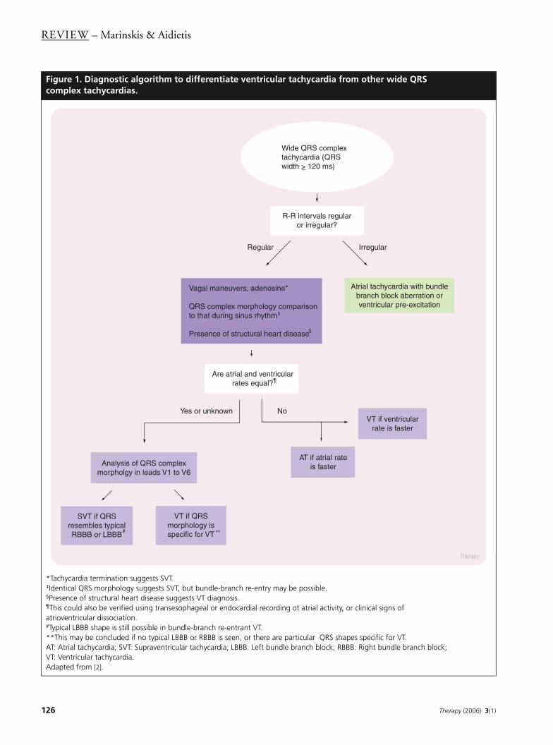
There are many known algorithms for thedifferentiation between VT and supraventric-ular tachycardias with wide QRS complexes.The latest guidelines suggest analyzing the12-lead electrocardiograph (ECG) and clinicalhistory along with the effects of vagal maneuversand adenosine [2]. Very important criterion suchas atrioventricular (AV) dissociation are quiteoften missed on 12-lead ECG; however, its pres-ence or absence can be verified using other easilyapplicable techniques such as transesophagealrecording of atrial activity. A diagnosticapproach to the wide QRS complex tachycardiais presented in Figure 1.
Classification & mechanisms of VTVTs can be classified using many criteria includ-ing etiology, electrophysiologic mechanisms andECG and clinical features – universal classificationincluding all of the criteria would look cumber-some. Thus classifying VTs into idiopathic andrelated to structural heart disease is practical fromclinical and electrophysiologic points of view.
The substrate of idiopathic VTs is limited to asmall area in which a triggered activity or the re-entrant circuit occurs. Ablation of this limitedarea is often effective to cure the patient and theseVTs have a good prognosis even if untreated.
VTs related to structural heart disease moreoften have multiple substrates due to diffusemyocardial changes (Figure 2). The mechanism inthis group is usually re-entry that may use large
REVIEW – Marinskis & Aidietis
126 Therapy (2006) 3(1)
Figure 1. Diagnostic algorithm to differentiate ventricular tachycardia from other wide QRS complex tachycardias.
*Tachycardia termination suggests SVT.‡Identical QRS morphology suggests SVT, but bundle-branch re-entry may be possible.§Presence of structural heart disease suggests VT diagnosis.¶This could also be verified using transesophageal or endocardial recording ot atrial activity, or clinical signs of atrioventricular dissociation.#Typical LBBB shape is still possible in bundle-branch re-entrant VT.**This may be concluded if no typical LBBB or RBBB is seen, or there are particular QRS shapes specific for VT.AT: Atrial tachycardia; SVT: Supraventricular tachycardia; LBBB: Left bundle branch block; RBBB: Right bundle branch block; VT: Ventricular tachycardia.Adapted from [2].
Analysis of QRS complex morpholgy in leads V1 to V6
Atrial tachycardia with bundle branch block aberration or ventricular pre-excitation
Vagal maneuvers, adenosine*
QRS complex morphology comparison to that during sinus rhythm
Presence of structural heart disease
R-R intervals regular or irregular?
IrregularRegular
Therapy
Wide QRS complex tachycardia (QRS width > 120 ms)
Are atrial and ventricular rates equal?
Yes or unknown No
SVT if QRS resembles typical RBBB or LBBB
VT if QRS morphology is specific for VT
VT if ventricular rate is faster
AT if atrial rate is faster
‡
§
# **
¶
www.future-drugs.com 127
Nonpharmacologic therapies for ventricular arrhythmias – REVIEW
area of myocardium or have a 3D path usingdeeper myocardial layers. For these reasons, lin-ear ablation (rather than focal ablation) is neces-sary to block the spread of re-entrant activation.The prognosis is more serious, especially if ven-tricular function is depressed, because VT morequickly causes hemodynamic instability, anddegenerates into a faster form or ventricularfibrillation.
Ablation techniquesNonpharmacologic curative approaches to VTstarted with surgery for VT related to postinfarc-tion scars [3,4]. Encircling ventriculotomy andsubendocardial resection have been accompaniedby cryoablation when necessary. Catheter abla-tion of VT has been used with direct currentshocks applied through endocardial electrodecatheters [5–7]. However, this technique has
limited application due to the necessity ofanesthesia and possible risks of barotrauma andcardiac perforation.
Endocardial catheter ablation of VT began toevolve rapidly when radiofrequency energy wasintroduced into clinical practice. This proce-dure uses standard-sized (tip of 4 mm length)deflectable catheters; however, as largeramounts of energy have to be delivered to cre-ate larger or deeper (transmural) lesions, 8 mmor irrigated-tip (also called cool-tip) cathetersmay be used. These catheters are introducedinto the right or left ventricle under localanesthesia together with several diagnostic cath-eters (usually positioned in the right atrium andHis bundle area). To guide the ablation catheterto the target area, different criteria can be useddepending on the mechanism and substrate ofthe VT, including:
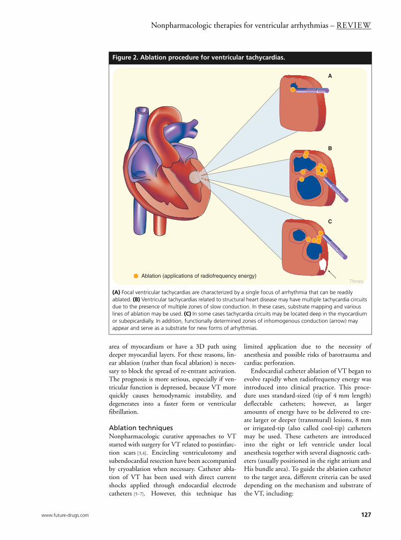
Figure 2. Ablation procedure for ventricular tachycardias.
(A) Focal ventricular tachycardias are characterized by a single focus of arrhythmia that can be readily ablated. (B) Ventricular tachycardias related to structural heart disease may have multiple tachycardia circuits due to the presence of multiple zones of slow conduction. In these cases, substrate mapping and various lines of ablation may be used. (C) In some cases tachycardia circuits may be located deep in the myocardium or subepicardially. In addition, functionally determined zones of inhomogenous conduction (arrow) may appear and serve as a substrate for new forms of arhythmias.
Ablation (applications of radiofrequency energy)
B
C
A
yTherapy
REVIEW – Marinskis & Aidietis
128 Therapy (2006) 3(1)
• Activation mapping which aims to find areaswith the earliest local activation time comparedwith the onset of the QRS complex (-25 ms orearlier is preferable). This technique is appro-priate to find the foci of idiopathic VT, and is,as an adjunct to map substrates in VTs, relatedto structural heart disease
• Pace mapping involves matching the shape ofpaced QRS complexes to that of spontaneousarrhythmia, which may prove useful if thearrhythmia is nonsustained
• Entrainment mapping is a method of pace map-ping applied during ongoing tachycardia. It isuseful during ablation of VTs related to struc-tural heart disease and evaluates both the shapeof QRS complexes and the distance between thepacing spike and the onset of the QRS complex
• Substrate mapping is used to search zones ofabnormal electrical activity (scars, low voltageand delayed electrical activity) during thesinus rhythm [8]. This may be necessarybecause VTs related to structural heart diseasemay cause hemodynamic instability and can-not therefore be mapped directly [9]
Noncontact mapping in patients with poorlytolerable VTs may also be used [10]. TheEnSite™ system uses a multielectrode balloonplaced into the ventricle and reconstructs theelectroanatomic map from the single beat oftachycardia. A number of possible difficultiesmay arise during the ablation procedure andthese are listed in Table 1, together with theirpossible solutions.
Catheter ablation of particular forms of VTIdiopathic VTsIdiopathic VTs occur (by definition) in peoplewithout detectable heart disease or geneticallydetermined molecular abnormalities, and if symp-tomatic, can be treated using catheter ablation.Idiopathic VTs comprise only 10% of all VTs andare not a large clinical problem compared withVTs related to structural heart disease. However,catheter ablation is rewarding as it avoids lifelongmedical therapy and often cures the patients,returning them to a normal life. Details pertinentto catheter ablation of idiopathic VTs are listedin Table 2.
Table 1. Possible difficulties during ablation of ventricular tachycardias.
Type of arrhythmia Possible difficulties Solutions
VTs related to structural heart disease
Complex anatomy of the substrate
3D reconstruction of the electroanatomic map during VT (CARTO™, EnSite™ NavX™ systems)
Hemodynamic instability during VT
Single-beat electroanatomic reconstruction of the heart chamber during VT (EnSite system)
Substrate mapping/ablation during the sinus rhythm
Antiarrhythmic drug administration to slow down the tachycardia
Deep localization of arrhythmia circuits
8 mm or cool-tip catheters to create deeper lesions
Epicardial approach
Multiple substrates/types of VT Substrate mapping/ablation
Noninducibility during ablation Substrate mapping/ablation
Idiopathic VTs Noninducibility or nonsustained character of VT
Intravenous infusion of isoproterenol to induce/facilitate the arrhythmia
Pace-mapping if VT is still not inducible during ablation procedure
VT: Ventricular tachycardia.
www.future-drugs.com 129
Nonpharmacologic therapies for ventricular arrhythmias – REVIEW
Right ventricular outflow tract tachycardiaThis VT is recognized as the most common idio-pathic VT [11]. It can be terminated by vagalmaneuvers, adenosine (this favors the probabilityof triggered cAMP-mediated activity), alsoβ-blockers and verapamil. Catheter ablation ofthe right ventricular outflow tract (RVOT) VThas a high success rate of approximately90% [12]. If there are multiple variants of QRScomplexes in one patient, suspicion concerningthe possible arrhythmogenic right ventricularcardiomyopathy (ARVC) may be raised [13], andother diagnostic tests (genetic analysis, magneticresonance imaging) should be performed. Thefocus of this VT can be found using activationmapping or pace mapping, and intravenousinfusion of catecholamines is often used to pro-voke the tachycardia and to test the effectivenessof ablation. Some foci could not be ablated fromthe right ventricle, and those showing a promi-nent R-wave in lead V1 were thought to belocated subepicardially. It is known now thatthese foci may be ablated in the left ventricle(this group evolved into the entity of the leftventricular outflow tract tachycardia).
Left ventricular outflow tract tachycardiaNonpharmacologic treatment of this form ofVT developed relatively late compared with theRVOT group. Foci of left ventricular outflowtract tachycardia may be located either below oreven above the aortic valve cusps [14]. Therefore,
during ablation of this tachycardia in the aorticsinus of valsalva, caution has to be exercised notto damage the left main coronary artery andoften two catheters have to be introduced viafemoral arteries and positioned in the aortic root– one to cannulate and mark the coronary arteryand another for ablation (Figure 3). There are stillVTs with inferior axis deviation that cannot beablated using the two previous approaches. Astepwise approach to these ‘outflow tract’ VTshas recently been described, including mappingin the right ventricular outflow tract, pulm-onary artery, left ventricular outflow tract, coro-nary sinus branches and via pericardialpuncture [15].
Left fascicular idiopathic tachycardiasAnother common form of idiopathic VT is leftfascicular (verapamil-sensitive) tachycardia. Thecommon (interfascicular) form probably arisesin the Purkinje network of the left posterior fas-cicle and is characterized by right bundlebranch-type QRS morphology with superior axisdeviation. Activation mapping shows the earlyventricular activity at the site of successful abla-tion, and a high-frequency signal, a possiblepotential of Purkinje system, is often observed(or even two potentials, favoring the fact of re-entry rather than triggered activity) [16]. Ablationat these sites is effective in around 90% and theleft posterior hemiblock is often seen after suc-cessful ablation. Other variants of this VT can
Table 2. Forms of idiopathic ventricular tachycardias and details pertinent to catheter ablation.
Form of idiopathic VT Clinical & electrocardiographic features Remarks on catheter ablation
Right ventricular outflow tract tachycardia
Most often stress induced, adenosine and verapamil sensitive
Foci are located in the septal or free wall of the right ventricular outflow tract, sometimes above the pulmonary valve or subepicardially
Left bundle branch block morphology with R-wave transition in leads V3–V4, and inferior axis deviation in frontal plane
Left ventricular outflow tract tachycardia
Adenosine sensitive Foci are located in the left or right coronary cusps of the aortic valve, or below the aortic valve, sometimes subepicardially
Right or left bundle branch block morphology with R-wave transition in leads V2–3, and inferior axis deviation in frontal plane
Left fascicular idiopathic tachycardia Verapamil (not adenosine) sensitive Ablation is guided by activation mapping and search for Purkinje potential in the septal part of the left ventricle
Right bundle branch block with superior axis deviation (common, or posterior fascicular, form)
Block in the corresponding fascicle (posterior fascicle in the typical form) is often seen after successful ablationInferior axis deviation or other frontal QRS plane axis
may be seen in other forms
VT: Ventricular tachycardia.
REVIEW – Marinskis & Aidietis
130 Therapy (2006) 3(1)
arise in the left anterior fascicle. InterfascicularVT (re-entry using both fascicles of the leftbranch) is also possible.
Tachycardias associated with structural heart diseaseVT related to postinfarction scarsThis group is the most common form of VTencountered in clinical practice (80%), due tothe spread of coronary heart disease. It remains tobe discovered how necrosis in the area suppliedby a damaged coronary artery can lead to a tran-sitional area of surviving myocardial cells as wellas areas of slow conduction. Mapping of theseVTs is more complicated and entrainment pac-ing in the critical slow zone is the most reliabletechnique. Many patients tend to have severalforms of postinfarction VT, some of them beingvery fast and poorly tolerated, thus not amenableto catheter ablation. Therefore, substrate map-ping during sinus rhythm may be used, and areasof slow conduction may be connected by abla-tion lines to the mitral valve, or one to another(Figure 2) [17,18]. Substrate mapping during rightventricular pacing can also be performed [19].This can additionally bring out the zones offunctional conduction block (a possible substratefor re-entry) due to the changed direction of ven-tricular activation. Parietal thrombi near theablation target can be dislodged by an ablationcatheter and pose further dangers. The overallsuccess rate of catheter ablation is lower than inidiopathic VTs – approximately 60%. Catheterablation may be used as a single therapy if thereare no risk factors for SCD, or as an adjunct ther-apy to avoid frequent discharges of an implant-able cardioverter–defibrillator (ICD). In theabsence of ICD, long-term follow-up of initiallysuccessful ablation of postinfarction VTs shows along-term mortality of more than 10% [20].Depressed left ventricular function is the mainindependent predictor of poor outcome.
Tachycardias in idiopathic dilated cardiomyopathyIn this group, VTs are often syncopal due to poorleft ventricular function, and the effectiveness ofcatheter ablation is not high due to the diffuse andprogressive character of the disease and multipleforms of VT [21]. Bundle branch re-entry tachy-cardia (see below) is often induced in thesepatients. ICD is the main option to prevent SCDin high-risk patients [22]. Ablation of zones of slowconduction is possible to decrease the frequencyof ICD shocks [23].
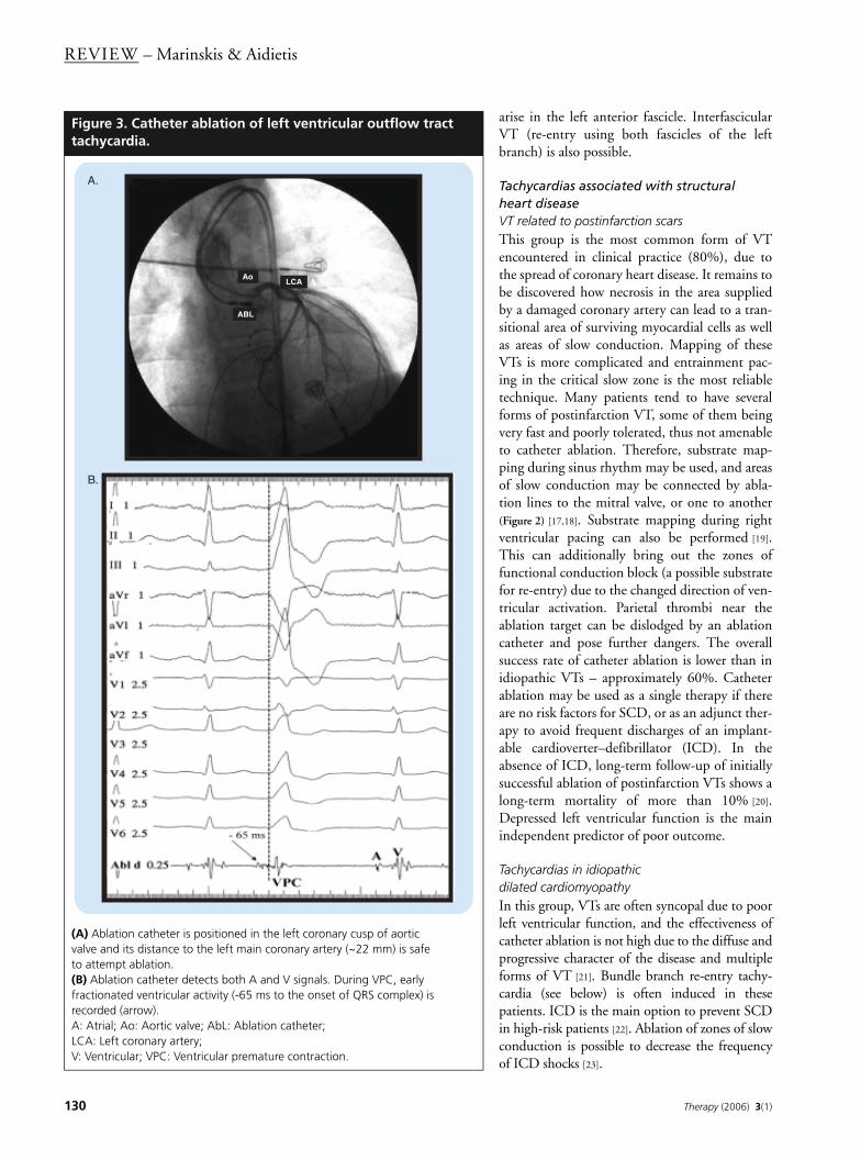
Figure 3. Catheter ablation of left ventricular outflow tract tachycardia.
(A) Ablation catheter is positioned in the left coronary cusp of aortic valve and its distance to the left main coronary artery (~22 mm) is safe to attempt ablation. (B) Ablation catheter detects both A and V signals. During VPC, early fractionated ventricular activity (-65 ms to the onset of QRS complex) is recorded (arrow).A: Atrial; Ao: Aortic valve; AbL: Ablation catheter; LCA: Left coronary artery; V: Ventricular; VPC: Ventricular premature contraction.
AoLCA
ABL
A.
B.
www.future-drugs.com 131
Nonpharmacologic therapies for ventricular arrhythmias – REVIEW
Bundle branch re-entry VTThis form of re-entry is common in patientswith depressed ventricular function and defectsof interventricular conduction. Most commonly,activation spreads down the right bundle branchand ascends via the left bundle branch [21]. Theshape of QRS complexes is indiscernible fromthe left bundle branch block aberration, butcareful analysis of the ECG and clinical signs(i.e., atrioventricular dissociation) leaves fewdoubts concerning the ventricular origin. Thesetachycardias are usually fast and cause hemo-dynamic instability [24]. Ablation of the rightbundle branch abolishes this VT; however, itdoes not eliminate the risk of SCD if other riskfactors are present [25].
VTs in hypertrophic cardiomyopathyPatients with hypertrophic cardiomyopathyhave an increased risk of SCD caused byhypertrophy, disarraying of myocardium anddispersion of electrophysiologic properties.Polymorphic VTs and fibrillation may occur
due to this inhomogeneity, and ICDs areconsidered a measure of secondary and primarySCD prevention [26]. Catheter ablation is notindicated in this group.
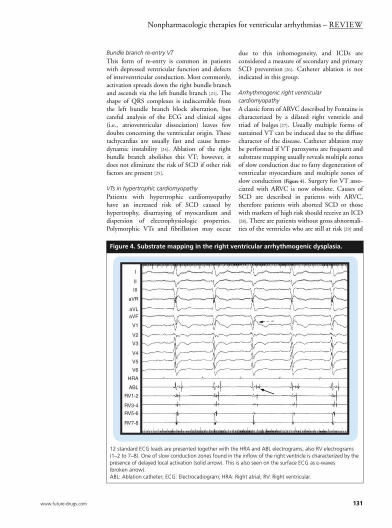
Arrhythmogenic right ventricular cardiomyopathyA classic form of ARVC described by Fontaine ischaracterized by a dilated right ventricle andtriad of bulges [27]. Usually multiple forms ofsustained VT can be induced due to the diffusecharacter of the disease. Catheter ablation maybe performed if VT paroxysms are frequent andsubstrate mapping usually reveals multiple zonesof slow conduction due to fatty degeneration ofventricular myocardium and multiple zones ofslow conduction (Figure 4). Surgery for VT asso-ciated with ARVC is now obsolete. Causes ofSCD are described in patients with ARVC,therefore patients with aborted SCD or thosewith markers of high risk should receive an ICD[28]. There are patients without gross abnormali-ties of the ventricles who are still at risk [29] and
Figure 4. Substrate mapping in the right ventricular arrhythmogenic dysplasia.
12 standard ECG leads are presented together with the HRA and ABL electrograms, also RV electrograms (1–2 to 7–8). One of slow conduction zones found in the inflow of the right ventricle is characterized by the presence of delayed local activation (solid arrow). This is also seen on the surface ECG as ε-waves (broken arrow).ABL: Ablation catheter; ECG: Electrocadiogram; HRA: Right atrial; RV: Right ventricular.
I
II
III
aVR
aVLaVF
V1
V2
V3
V4
V5
V6
HRA
ABL
RV1-2
RV3-4
RV5-6
RV7-8
REVIEW – Marinskis & Aidietis
132 Therapy (2006) 3(1)
the development of molecular genetics shouldgive more diagnostic criteria for the screening ofsuch patients.
VT after surgery for congenital heart diseaseThe most common anomaly that gives rise to aVT substrate after surgical correction is tetra-logy of Fallot [30]. The surgical procedure ofcomplete repair creates scars (anatomic obsta-cles) in the outflow tract and in the ventricularseptum, and VT may rotate around these scars.Catheter ablation is facilitated using various 3Delectroanatomic reconstruction systems andablation line is drawn from the right ventricularinfundibulotomy scar to the pulmonaryvalve [31]. The SCD risk stratification is stillunresolved in this group of patients; however,ICD implantation is necessary if the patient hasbeen resuscitated.
VT related to primary electrical diseasesA number of gene defects responsible for cardiacmyocyte ion channel dysfunction have been dis-covered. This group includes the long QT inter-val syndrome, Brugada syndrome, short QTinterval syndrome, catecholaminergic polymor-phic VT and others [32]. These anomalies create adispersion of myocardial refractoriness and con-duction, leading to a possibility of polymorphicVT and ventricular fibrillation. To date, the onlyoption to prevent SCD in these patients is ICDimplantation. However, some patients can expe-rience multiple VT shocks as a result of frequentVT episodes (‘electrical storm’). It has beennoted that the trigger of polymorphic VT maybe ventricular premature beats originating in onefocus, and their ablation could decrease thenumber of VT episodes. Of note is that electro-grams from these sites often show sharp pre-systolic spikes, probably potentials of thePurkinje system [33,34].
Epicardial approach to ablationIn some cases, the circuit of VT may be locatedcloser to the epicardial surface of the heart andendocardial application of radiofrequency energyis not effective. This is most often observed inpatients with VTs related to Chagas’ disease;however, it may also be applicable to other formsof VT [35,36]. In these cases, the ablation elec-trode is introduced into the pericardial spaceusing a pericardial puncture, and navigated tothe site of interest. Caution has to be exercisednot to damage the coronary arteries that run epi-cardially. The epicardial approach will probably
be used more often since the experience of theepicardial approach to treat other rhythm distur-bances (i.e., the pericardial approach to isolatepulmonary veins to treat atrial fibrillation) iswidening and gained skills and techniques suchas thoracoscopic visualization could be used forablation in more VTs.
Surgical treatment for VTThe principles of mapping various cardiacarrhythmias were first learned during surgicalprocedures, and arrhythmia surgery was thepredecessor of catheter ablation. Surgical isola-tion of the lateral wall of the right ventricle hasbeen used for VT associated with ARVC; how-ever, it has now been replaced by ICD, catheterablation and heart transplantation [37]. Surgicaltreatment of VT related to ischemic heart dis-ease had success rate (abolition of VT) ofapproximately 85%, with a mortality rate of 5to 10%. Today, few centers have experience withmapping-guided VT surgery, and as a stand-alone procedure it has almost been abandonedas less traumatic and almost equally effectivecatheter ablation can be used, or ICD is indi-cated. However, surgical ablation of postinfarc-tion VT can be performed concomitantly withother surgical procedures such as coronaryrevascularization, mitral valve repair, or restora-tion of the left ventricular geometry [38]. Thelatter procedure effectively abolishes VT evenwithout mapping [39]. In the absence of otherindications for cardiac surgery, patients withpostinfarction VT are operated if other treat-ment modalities fail. VTs related to the diffuseinvolvement of myocardium (nonischemiccardiomyopathy) are not usually amenable tosurgical treatment.
Sudden cardiac death & implantable cardioverters–defibrillatorsContemporary ICDs are capable of detectingmultiple forms of VTs and ventricular fibrilla-tion and administering appropriate therapy(Figure 5). These therapeutic measures (tieredtherapy) range from bursts or ramps of antitach-ycardia pacing, R-wave synchronized shocks ofsmall energy, to high energy shocks up to 30 to35 J or 700 to 800 V. Advanced electrogramanalysis and storage features allows therapy to betailored to the patient. Diagnostic algorithmshelp to discriminate VTs from sinus and othersupraventricular tachycardias and decrease thechance of inappropriate shocks. Implantation ofcontemporary ICD devices with endocardial
www.future-drugs.com 133
Nonpharmacologic therapies for ventricular arrhythmias – REVIEW
leads carries lower morbidity and procedure-related mortality, comparable with that whichoccurs with pacemaker implantation.
Guidelines for ICD implantation were firstpublished in 1998, revised in 2002 and arecorrected accordingly to the results of new
trials [40–42]. In patients resuscitated fromSCD due to VT, ICD is the only reliableoption for secondary prevention [43,44]. This isreflected in recent recommendations of SCDmanagement [45]. Trials such as the Multi-center Automatic Defibrillator Implantation
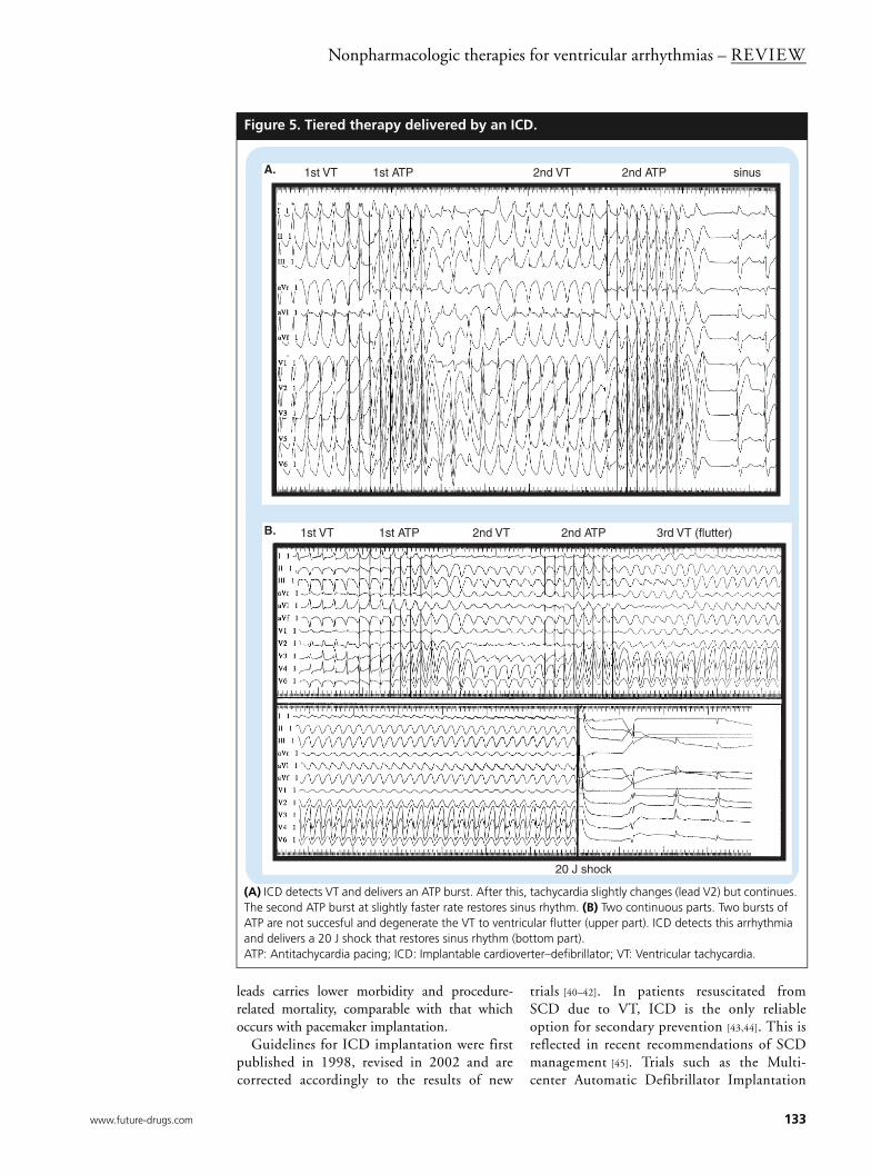
Figure 5. Tiered therapy delivered by an ICD.
(A) ICD detects VT and delivers an ATP burst. After this, tachycardia slightly changes (lead V2) but continues. The second ATP burst at slightly faster rate restores sinus rhythm. (B) Two continuous parts. Two bursts of ATP are not succesful and degenerate the VT to ventricular flutter (upper part). ICD detects this arrhythmia and delivers a 20 J shock that restores sinus rhythm (bottom part).ATP: Antitachycardia pacing; ICD: Implantable cardioverter–defibrillator; VT: Ventricular tachycardia.
1st VT 1st ATP 2nd VT 2nd ATP sinusA.
1st VT 1st ATP 2nd VT 2nd ATP 3rd VT (flutter)
20 J shock
B.
REVIEW – Marinskis & Aidietis
134 Therapy (2006) 3(1)
Trial (MADIT) I, Multicenter UnSustainedTachycardia Trial (MUSTT) and others haveshown that ICDs are the most effective meas-ure for primary SCD prevention in high-riskpatients with VTs [46,47].
Recently, two large trials on primary SCDprevention in patients with depressed left vent-ricular function and without documented VT(MADIT II in patients with coronary heart dis-ease and SCD-HeFT in nonischemic cardio-myopathy) have shown that prophylactic ICDimplantation decreases overall mortality [48].This shows that depressed left ventricular func-tion is an independent predictor of a higher riskof SCD. However, if the expanded indicationssuggested by these trials are followed, thenumber of worldwide ICD implants willincrease more than twice. This raises the debateof economic burden of expanded indications forICD therapy, as indications for ICD implanta-tion for primary prevention vary from country tocountry. ICDs prevent SCD but do not modifythe underlying heart disease (like dilated cardio-myopathy) and in some patients overall lifeexpectancy may not be prolonged significantly.The process of combinating ICDs with bivent-ricular pacing in patients with heart failure is stillcontroversial, and to date only the Comparisonof Medical Therapy, Pacing and Defibrillation inHeart Failure (COMPANION) trial has shownan improvement in prognosis and quality of life(QoL) can be achieved [49,50].
The MADIT II and Dual Chamber and VVIImplantable Defibrillator (DAVID) trials haveshown a bigger incidence of heart failure in ICDpatients with a higher percentage of ventricularpacing. This is due to the fact that the right vent-ricular lead in these patients is in an apical posi-tion, and pacing from this site is less physiologic.Resulting ventricular dyssynchrony and mitralregurgitation worsens heart failure. According tothese data, ventricular pacing should be used aslittle as possible in ICD patients with heart failure.Using the alternate pacing sites, or biventricularpacing, could be considered.
An important problem in view of the expand-ing number of ICD implants is that QoL isdecreased as a result of the adverse psychologiceffects of ICD shocks, regardless of whether or notthey are appropriate. ICD shocks cause discom-fort, anxiety and preoccupation. Recentlyreported results from the Optimal Pharmaco-logical Therapy in Implantable CardioverterDefibrillator patients (OPTIC) trial showed thatamiodarone (especially in combination with a
β-blocker) and to a lesser extent, sotalol or anotherβ-blocker alone, decrease the number of ICDshocks and improve the wellbeing of thepatients [51].
Approach to the patient with diagnosed VTIn patients with idiopathic VTs, no treatmentmay be used. If symptoms are present,antiarrhythmic drugs may be used or catheterablation may be chosen to avoid long-termdrug therapy. Unfortunately, some idiopathicVTs may be related to still poorly identifiablegenetic disorders, such as some forms ofarrrhythmogenic dysplasia.
Patients with VTs related to structural heartdiseases and molecular abnormalities have anincreased risk of SCD. Criteria for risk assess-ment are still yet to be discovered, of which themost important include poor ventricular func-tion, QRS prolongation, and left ventricularhypertrophy [52]. These patients often have mul-tiple forms of VTs, and ablation of one does notreliably decrease the risk of SCD. New areas ofslow conduction leading to dispersion of electro-physiologic parameters (the cause of re-entry)may arise due to disease progression. Transientischemia or catecholamine stress create newzones with different conduction time and refrac-toriness, leading to the possibility of polymor-phic VT and ventricular fibrillation. Otherfactors that lead to inhomogeneity of ventricularmuscle are fibrosis, edema, loss of intercellularcoupling (connexins) and nerve sprouting.
Treating only high-risk groups is not effectivein the prevention of SCD due to the relativelysmall number of people saved. Primary preven-tion in the general population is important formortality decrease in the future, emphasizing theimportance of healthy lifestyle, correcting hyper-tension, diabetes, smoking, obesity and otherrisk factors. Optimal pharmacologic treatmentof underlying heart disease, for example, the useof β-blockers, angiotensin-converting enzymeinhibitors, statins and aldosterone receptorblockers is known to decrease the chance ofarrhythmia [45,53]. Analysis of finished and futuretrials, as well as stratification of risk factors couldprovide the answer as to which patients couldbenefit from particular treatment modalities [54].
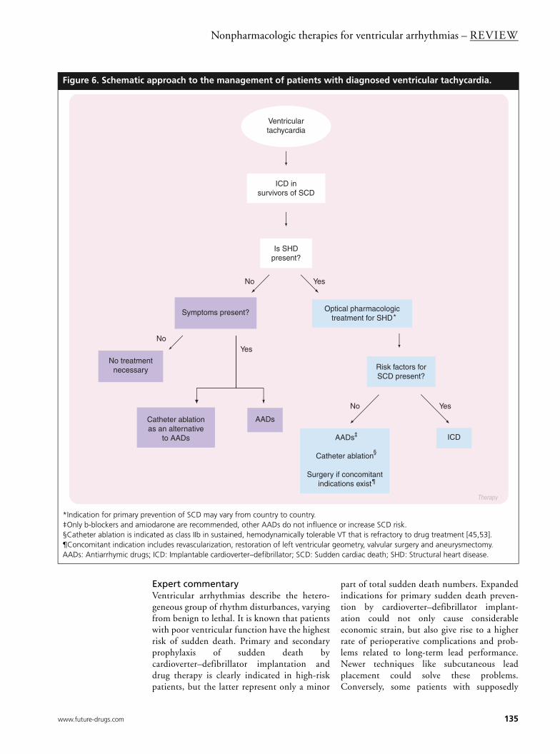
The algorithm of VT management is presentedin Figure 6. Choosing the most appropriate treat-ment should include assessment of numerousclinical factors, and every patient with thisdiagnosis requires an individual approach.
www.future-drugs.com 135
Nonpharmacologic therapies for ventricular arrhythmias – REVIEW
Expert commentaryVentricular arrhythmias describe the hetero-geneous group of rhythm disturbances, varyingfrom benign to lethal. It is known that patientswith poor ventricular function have the highestrisk of sudden death. Primary and secondaryprophylaxis of sudden death bycardioverter–defibrillator implantation anddrug therapy is clearly indicated in high-riskpatients, but the latter represent only a minor
part of total sudden death numbers. Expandedindications for primary sudden death preven-tion by cardioverter–defibrillator implant-ation could not only cause considerableeconomic strain, but also give rise to a higherrate of perioperative complications and prob-lems related to long-term lead performance.Newer techniques like subcutaneous leadplacement could solve these problems.Conversely, some patients with supposedly
Figure 6. Schematic approach to the management of patients with diagnosed ventricular tachycardia.
*Indication for primary prevention of SCD may vary from country to country.‡Only b-blockers and amiodarone are recommended, other AADs do not influence or increase SCD risk.§Catheter ablation is indicated as class IIb in sustained, hemodynamically tolerable VT that is refractory to drug treatment [45,53].¶Concomitant indication includes revascularization, restoration of left ventricular geometry, valvular surgery and aneurysmectomy.AADs: Antiarrhymic drugs; ICD: Implantable cardioverter–defibrillator; SCD: Sudden cardiac death; SHD: Structural heart disease.
Optical pharmacologic treatment for SHD
ICD in survivors of SCD
YesNo
Therapy
Ventricular tachycardia
YesNo
Risk factors for SCD present?
No treatment necessary
Is SHD present?
Symptoms present?
Catheter ablation as an alternative
to AADs
AADs
AADs
Catheter ablation
Surgery if concomitant indications exist
YesNo
ICD
*
‡
§
¶
REVIEW – Marinskis & Aidietis
136 Therapy (2006) 3(1)
benign arrhythmias, such as right ventricularoutflow tract premature beats, die suddenlyafter a reassuring consultation with a cardio-logist offering no therapy. The search for otherfactors to clarify the high-risk patientstherefore remains important.
OutlookUntil newer pharmacologic and gene therapyapproaches are developed for prophylaxis andtreatment of heart diseases, nonpharmacologicapproaches to VT remain the most effective
measure for the prevention of SCD andsymptomatic treatment of VT. Catheter abla-tion remains a very effective method and anexcellent alternative to drug treatment of idio-pathic VTs. In VTs related to structural heartdisease, hybrid approach is needed, includingoptimal medical therapy for underlying dis-ease, antiarrhythmic drugs and catheter abla-tion. ICD devices will evolve and will beimplanted widely for primary prevention inhigh-risk patients, yet more criteria of high riskhave to be defined.
Highlights
• Ventricular tachycardias (VTs) may be subdivided into idiopathic and those related to structural heart disease. The two groups differ both in prognosis and selection of treatment.
• Mapping and ablation techniques have enabled the creation of electroanatomic maps of both ventricles in an attempt to find and ablate arrhythmogenic foci and zones, both endocardially and epicardially. Irrigated-tip and 8-mm tip catheter radiofrequency ablation is able to create lesions of sufficient depth.
• Idiopathic VTs could be ablated in majority of cases (effectiveness >90%) and improve the quality of life of patients. However, idiopathic VTs comprise only 10% of all VTs.
• VTs related to structural heart disease tend to have multiple forms, and ablation is possible to eliminate approximately 60% of arrhythmias. Patients with depressed ventricular function and other markers of high risk are often candidates for ICD implantation. Catheter ablation could be used as adjunct to avoid frequent ICD shocks.
• Surgery for postmyocardial infarction VTs still has a place, when combined with revascularization procedures, mitral valve repair and restoration of the left ventricular geometry.
• ICDs have been shown as the only reliable option for secondary prophylaxis of sudden cardiac death (in resuscitated patients). The superiority of ICDs for primary prophylaxis of SCD has also been shown, but widespread use of ICDs poses significant financial and organisational issues.
• Ventricular pacing in patients with implanted ICDs often worsens the heart failure. Possible solutions could be programming the ICD to lower rates, or use devices capable of biventricular pacing.
• Psychologic problems in patients with implanted ICDs can be decreased using optimal medical therapy to reduce the number of VT episodes.
BibliographyPapers of special note have been highlighted as of interest (•) or of considerable interest (••) to readers.1. Epstein AE, Hallstrom AP, Rogers WJ et al.
Mortality following ventricular arrhythmia suppression by encainide, flecainide, and moricizine after myocardial infarction. The original design concept of the Cardiac Arrhythmia Suppression Trial (CAST). JAMA 270, 2451–2455 (1993).
2. Blomström-Lundqvist C, Scheinman MM, Aliot EM et al. ACC/AHA/ESC guidelines for the management of patients with supraventricular arrhythmias – executive summary: a report of the American College of Cardiology/American Heart Association Task Force on Practice Guidelines, and the European Society of Cardiology Committee for Practice Guidelines (Writing Committee to Develop Guidelines for the Management
of Patients With Supraventricular Arrhythmias). Circulation 108, 1871–1909 (2003).
3. Josephson ME, Horowitz LN, Farshidi A, Spear JF, Kastor JA, Moore EN. Recurrent sustained ventricular tachycardias. 2. Endocardial mapping. Circulation 57, 440–447 (1978).
4. Josephson ME, Karken AH, Horowitz LN. Endocardial excision: a new surgical technique for the treatment of recurrent ventricular tachycardias. Circulation 60, 1430–1439 (1979).
5. Hartzler GO. Electrode catheter ablation of refractory focal ventricular tachycardia. J. Am. Coll. Cardiol. 2, 1107–1113 (1983).
6. Puech P, Gallay P, Grolleau R, Koliopoulos N. Traitement par electrofulguration endocavitaire d’une tachycardie ventriculaire recidivante par dysplasie ventriculaire droite. Arch. Mal. Coeur 77, 826–835 (1984).
7. Fontaine G, Tonet JL, Frank R et al. Traitement d’urgence de la tachycardie ventriculaire chronique apres infarctus du myocarde par la fulguration invicavitaire. Arch. Mal. Coeur 78, 1037–1043 (1985).
8. Arenal A, Glez-Torrecilla E, Oritz M et al. Ablation of electrograms with an isolated, delayed component as treatment of unmappable monomorphic ventricular tachycardias in patients with structural heart disease. J. Am. Coll. Cardiol. 41, 81–92 (2003).
9. Soejima K, Suzuki M, Maisel WH et al. Catheter ablation in patients with multiple and unstable ventricular tachycardias after myocardial infarction. Short ablation lines guided by re-entry circuit isthmuses and sinus rhythm mapping. Circulation 104, 664–669 (2001).
www.future-drugs.com 137
Nonpharmacologic therapies for ventricular arrhythmias – REVIEW
10. Della Bella P, Pappalardo A, Riva S, Tondo C, Fassini G, Trevisi N. Non-contact mapping to guide catheter ablation of untolerated ventricular tachycardia. Eur. Heart J. 23, 742–7552 (2002).
11. Lerman BB, Steik KM, Markowitz SM, Mittal S, Iwai S. Ventricular tachycardia in patients with structurally normal hearts. In: Cardiac Electrophysiology: From Cell to Bedside. Zipes D, Jalife J (Ed). WB Saunders Company, 668–682 (2004).
12. Coggins DL, Lee RJ, Sweeney J et al. Radiofrequency catheter ablation as a cure for idiopathic tachycardia of both left and right ventricular origin. J. Am. Coll. Cardiol. 23, 1333–1341 (1994).
13. O‘Donnell D, Cox D, Bourke J, Mitchell L, Furniss S. Clinical and electrophysiological differences between patients with arrhythmogenic right ventricular dysplasia and right ventricular outflow tract tachycardia. Eur. Heart J. 24, 801–810 (2003).
•• Describes the differences between benign and apparently benign ventricular arrythmias.
14. Ouyang F, Fotuhi P, Ho SY et al. Repetitive monomorphic ventricular tachycardia originating from the aortic sinus cusp. Electrocardiographic characterization for guiding catheter ablation. J. Am. Coll. Cardiol. 39, 500–508 (2002).
15. Tanner H, Hindricks G, Schirdewahn P et al. Outflow tract tachycardia with R/S transition in lead V3: six different anatomic approaches for successful ablation. J. Am. Coll. Cardiol. 45, 418–423 (2005).
• Systematic approach to ablate right/left ventricular outflow tract tachycardias and shows the diversity of this group.
16. Ouyang F, Cappato R, Ernst S et al. Electroanatomic substrate of idiopathic left ventricular tachycardia. Unidirectional block and macroreentry within the Purkinje network. Circulation 105, 462–469 (2002).
17. de Chillou C, Lacroix D, Klug D et al. Isthmus characteristics of reentrant ventricular tachycardia after myocardial infarction. Circulation 105, 726–731 (2002).
18. Kottkamp H, Wetzel U, Schirdewahn P et al. Catheter ablation of ventricular tachycardia in remote myocardial infarction: substrate description guiding placement of individual linear lesions targeting noninducibility. J. Cardiovasc. Electrophysiol. 14, 675–681 (2003).
19. Brunckhorst CB, Stevenson WG, Jackman WM et al. Ventricular mapping during atrial and ventricular pacing. Relationship of multipotential electrograms to ventricular tachycardia reentry circuits after myocardial infarction. Eur. Heart J. 23, 1131–1138 (2002).
20. O’Donnell D, Bourke JP, Anilkumar R, Simeonidou E, Furniss SS. Radiofrequency ablation for postinfarction ventricular tachycardia. Report of a single centre experience of 112 cases. Eur. Heart J. 23, 1699–1705 (2002).
• Long-term follow-up of patients with postinfarction ventricular tachycardia, showing results and limitations of ablation.
21. Hsia HH, Marchlinski FE. Characterization of the electroanatomic substrate for monomorphic ventricular tachycardia in patients with nonischemic cardiomyopathy. Pacing Clin. Electrophysiol. 25, 1114–1127 (2002).
22. Grimm W, Hoffmann J, Muller HH et al. Implantable defibrillator event rates in patients with idiopathic dilated cardiomyopathy, nonsustained ventricular tachycardia on Holter and a left ventricular ejection fraction below 30%. J. Am. Coll. Cardiol. 39, 780–787 (2002).
23. Hsia HH, Callans DJ, Marchlinski F. Characterization of endocardial electrophysiological substrate in patients with nonischemic cardiomyopathy and monomorphic ventricular tachycardia. Circulation 108, 704–710 (2003).
24. Marinskis G, Aidietis A, Ježov V et al. Wide-QRS complex tachycardia presenting with orthostatic syncopes. Sem. Cardiol. 9(4), 50–53 (2003).
25. Mehdirad AA, Keim S, Rist K, Tchou P. Long-term clinical out-come of right bundle branch radiofrequency catheter ablation for treatment of bundle branch reentrant ventricular tachycar-dia. Pacing Clin. Electrophysiol. 18, 213–543 (1995).
26. Olivotto I, Gistri R, Petrone P, Pedemonte E, Vagriu D, Cecchi F. Maximum left ventricular thickness and risk of sudden death in patients with hypertrophic cardiomyopathy. J. Am. Coll. Cardiol. 41, 315–321 (2003).
27. Fontaine G, Fontaliran F, Hebert JL et al. Arrhythmogenic right ventricular dysplasia. Ann. Rev. Med. 50, 17–35 (1999).
28. Corrado D, Leoni L, Link M et al. Implantable cardioverter-defibrillator therapy for prevention of sudden death in patients with arrhythmogenic right ventricular cardiomyopathy/dysplasia. Circulation 108, 3084–3091 (2003).
29. Nasir K, Bomma C, Tandri H et al. Electrocardiographic features of arrhythmogenic right ventricular dysplasia/cardiomyopathy according to disease severity. A need to broaden diagnostic criteria. Circulation 110, 1527–1534 (2004).
30. Khairy P, Landzberg MJ, Gatzoulis MA et al. Value of programmed ventricular stimulation after tetralogy of Fallot repair. A multicenter study. Circulation 109, 1994–2000 (2004).
31. Stevenson WG, Delacretaz E, Friedman PL, Ellison KE. Identification and ablation of macroreentrant ventricular tachycardia with the CARTO electroanatomical mapping system. Pacing Clin. Electrophysiol. 21, 1448–1456 (1998).
32. Wever EFD, Robles de Medina EO. Sudden death in patients without structural heart disease. J. Am. Coll. Cardiol. 43, 1137–1144 (2004).
33. Haissaguerre M, Shoda M, Jais P et al. Mapping and ablation of idiopathic ventricular fibrillation. Circulation 106, 962–697 (2002).
34. Haissaguerre M, Extramiana F, Hocini M et al. Mapping and ablation of ventricular fibrillation associated with long-QT and Brugada syndromes. Circulation 108, 925–928 (2003).
35. Brugada J, Berruezo A, Cuesta A et al. Nonsurgical transthoracic epicardial radiofrequency ablation: an alternative in incessant ventricular tachycardia. J. Am. Coll. Cardiol. 41, 2036–2043 (2003).
36. Soejima K, Stevenson WG, Sapp JL. Endocardial and epicardial radiofrequency ablation of ventricular tachycardia associated with dilated cardiomyopathy. The importance of low-voltage scars. J. Am. Coll. Cardiol. 43, 1834–1842 (2004).
37. Cox JL. Cardiac surgery for arrhythmias. J. Cardiovasc. Electrophysiol. 15, 250–262 (2004).
38. Page PL. Surgery for cardiac arrhythmias. In: Cardiac Electrophysiology: From Cell to Bedside. Zipes D, Jalife J (Eds). WB Saunders Company, 1104–1115 (2004).
39. Sosa E, Scanavacca M, d'Avila A, Fukushima J, Jatene A. Long-term results of visually guided left ventricular reconstruction as single therapy to treat ventricular tachycardia associated with postinfarction anteroseptal aneurysm. J. Cardiovasc. Electrophysiol. 9, 1133–1143 (1998).
REVIEW – Marinskis & Aidietis
138 Therapy (2006) 3(1)
40. Gregoratos G, Cheitlin MD, Conill A et al. ACC/AHA guidelines for implantation of cardiac pacemakers and antiarrhythmia devices: a report of the American College of Cardiology/American Heart Association Task Force on Practice Guidelines (Committee on Pacemaker Implantation). Circulation 97, 1325–1335 (1998).
41. Gregoratos G, Abrams J, Epstein AE et al. ACC/AHA/NASPE 2002 guideline update for implantation of cardiac pacemakers and antiarrhythmia devices: a report of the American College of Cardiology/American Heart Association Task Force on Practice Guidelines (ACC/AHA/NASPE Committee to update the 1998 pacemaker guidelines). Circulation 106, 2145–2161 (2002).
42. Epstein AE. An update on implantable cardioverter-defibrillator guidelines. Curr. Opin. Cardiol. 19, 23–25 (2004).
43. Kuck KH, MD; Cappato R, Siebels J, Ruppel R, for the Cardiac Arrest Study Hamburg (CASH) investigators. Randomized comparison of antiarrhythmic drug therapy with implantable defibrillators in patients resuscitated from cardiac arrest. The CASH. Circulation 102, 748–754 (2000).
44. A comparison of antiarrhythmic-drug therapy with implantable defibrillators in patients resuscitated from near-fatal ventricular arrhythmias. The Antiarrhythmics Versus Implantable Defibrillators (AVID) investigators. N. Engl. J. Med. 337, 1576–1583 (1997).
45. Priori SG, Aliot E, Blomstrom-Lundqvist C et al. Task force on sudden cardiac death of the European Society of Cardiology. Eur. Heart J. 22, 1374–1450 (2001).
46. Moss AJ, Hall WJ, Cannom DS et al. Improved survival with an implanted defibrillator in patients with coronary disease at high risk for ventricular arrhythmia. Multicenter Automatic Defibrillator Implantation Trial Investigators. N. Engl. J. Med. 335, 1933–1940 (1996).
47. Buxton AE, Lee KL, Fisher JD, Josephson ME, Prystowsky EN, Hafley G. A randomized study of the prevention of sudden death in patients with coronary artery disease. Multicenter UnSustained Tachycardia Trial (MUSTT) investigators. N. Engl. J. Med. 341, 1882–1890 (1999).
48. Moss AJ, Zareba W, Hall WJ et al. Prophylactic implantation of a defibrillator in patients with myocardial infarction and reduced ejection fraction. N. Engl. J. Med. 346, 877–883 (2002).
•• Shows the effectiveness of implantation of cardioverter–defibrillator for primary prophylaxis of sudden cardiac death.
49. Young JB, Abraham WT, Smith AL et al., for the Multicenter Insync ICD RAndomized CLinical Evaluation (MIRACLE ICD) trial investigators. Combined cardiac resynchronization and implantable cardioversion defibrillation in advanced chronic heart failure. The MIRACLE ICD trial. JAMA 289, 2685–2694 (2003).
50. Bristow MR, Saxon LA, Boehmer J et al. Cardiac-resynchronization therapy with or without an implantable defibrillator in advanced chronic heart failure. N. Engl. J. Med. 350, 2140–2150 (2004).
51. Connolly SJ, Hohnloser S, Dorian P et al. Optimal Pharmacological Therapy in Implantable Cardioverter Defibrillator Patients (OPTIC) trial. Presented at: The American College of Cardiology Annual Scientific Session 6–9 March, FL, USA (2005).
52. Zimetbaum PJ, Buxton AE, Batsford W et al. Electrocardiographic predictors of arrhythmic death and total mortality in the Multicenter UnSustained Tachycardia Trial (MUSTT). Circulation 110, 766–769 (2004).
53. Priori SG, Aliot E, Blomstrom-Lundqvist C et al. Update of the guidelines on sudden cardiac death of the European Society of Cardiology. Eur. Heart J. 24, 13–15 (2003).
54. Josephson M, Wellens HJJ. Implantable defibrillators and sudden cardiac death. Circulation 109, 2685–2691 (2004).
AffiliationsGermanas MarinskisVilnius University, Clinic of Heart Diseases, Center of Cardiology and Angiology, Vilnius UniversityHospital Santariskiu Klinikos,Santariskiu str. 2, LT-08661 Vilnius, LithuaniaTel.: +370 5236 5216Fax: +370 5278 [email protected]
Audrius AidietisVilnius University, Clinic of Heart Diseases, Center of Cardiology and Angiology, Vilnius UniversityHospital Santariskiu Klinikos,Santariskiu str. 2, LT-08661 Vilnius, LithuaniaTel.: +370 5236 5216Fax: +370 5236 [email protected]