Embed Size (px)
Citation preview
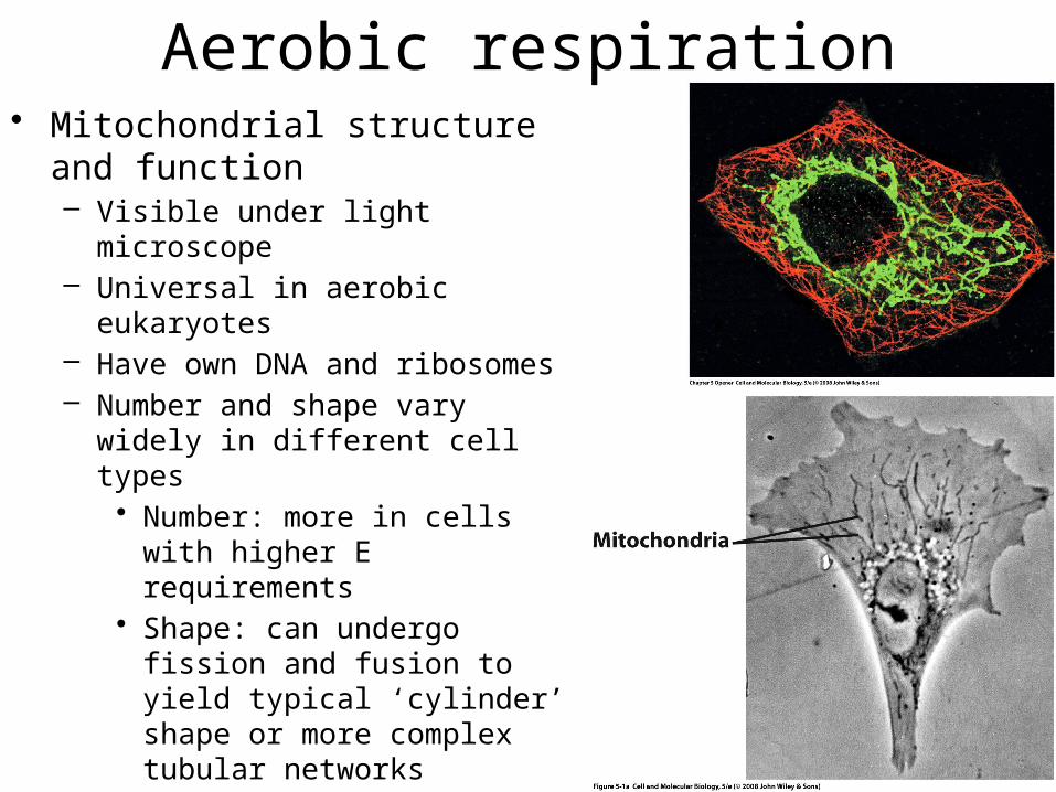
Aerobic respiration• Mitochondrial structure and function
– Visible under light microscope– Universal in aerobic eukaryotes– Have own DNA and ribosomes– Number and shape vary widely in
different cell types• Number: more in cells with higher E
requirements• Shape: can undergo fission and
fusion to yield typical ‘cylinder’ shape or more complex tubular networks
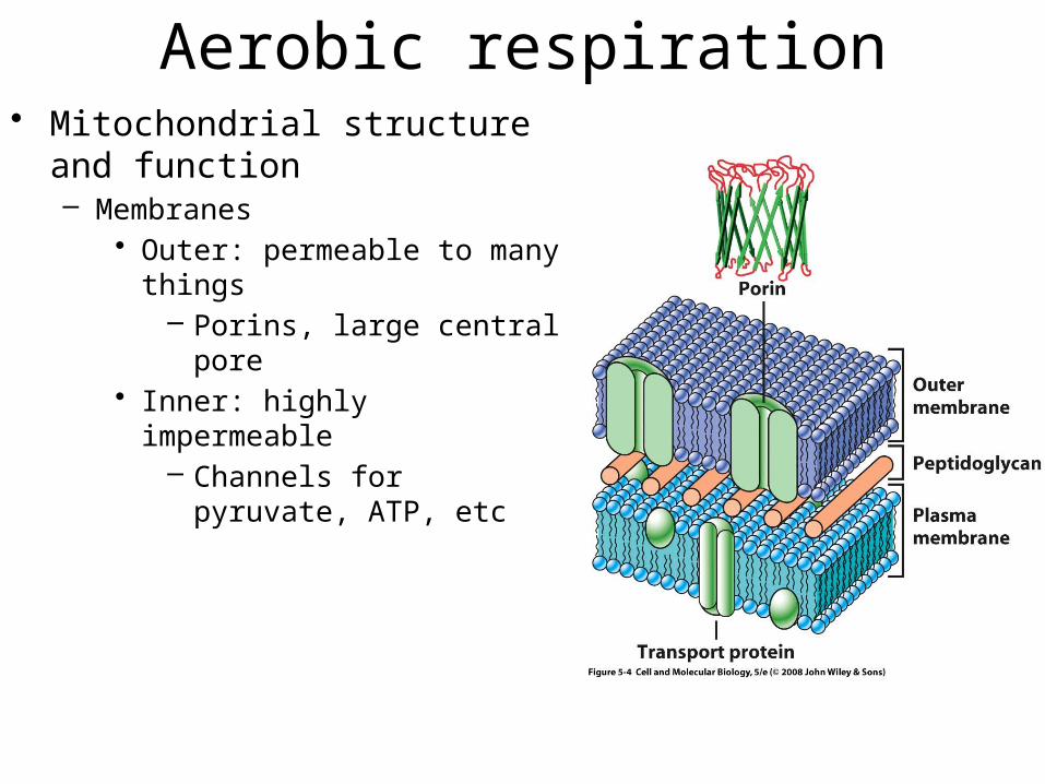
Aerobic respiration• Mitochondrial structure and function
– Membranes• Outer: permeable to many things
– Porins, large central pore• Inner: highly impermeable
– Channels for pyruvate, ATP, etc
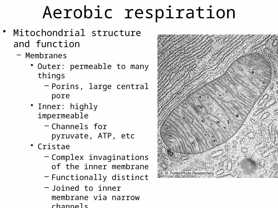
Aerobic respiration• Mitochondrial structure and function
– Membranes• Outer: permeable to many things
– Porins, large central pore• Inner: highly impermeable
– Channels for pyruvate, ATP, etc• Cristae
– Complex invaginations of the inner membrane
– Functionally distinct– Joined to inner membrane via
narrow channels
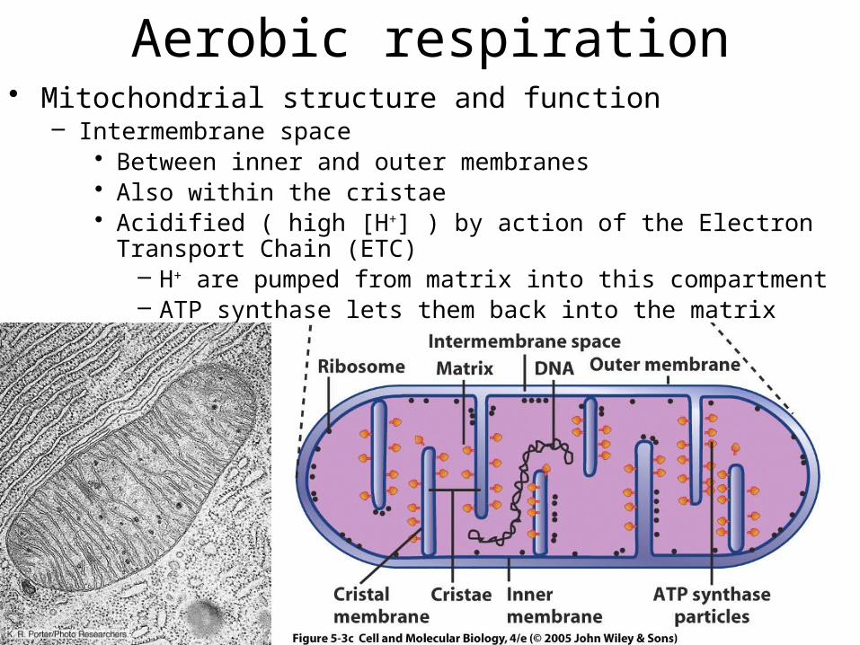
Aerobic respiration• Mitochondrial structure and function
– Intermembrane space• Between inner and outer membranes• Also within the cristae• Acidified ( high [H+] ) by action of the Electron Transport Chain (ETC)
– H+ are pumped from matrix into this compartment– ATP synthase lets them back into the matrix
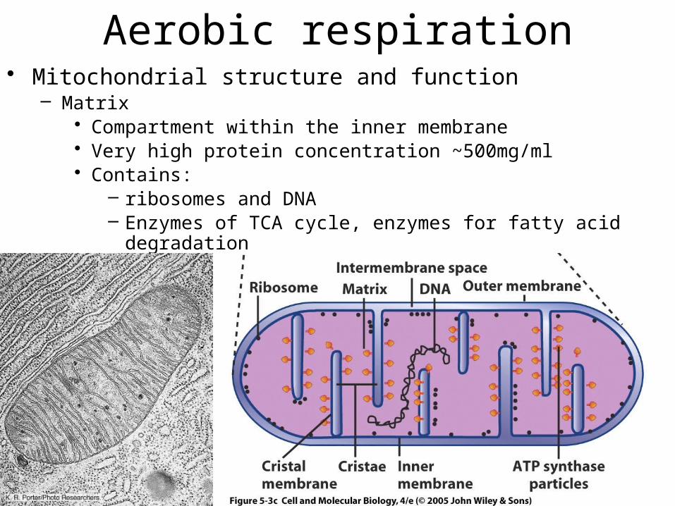
Aerobic respiration• Mitochondrial structure and function
– Matrix• Compartment within the inner membrane• Very high protein concentration ~500mg/ml• Contains:
– ribosomes and DNA– Enzymes of TCA cycle, enzymes for fatty acid degradation
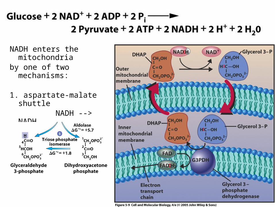
NADH enters the mitochondriaby one of two mechanisms:
1. aspartate-malate shuttle NADH --> NADH2. glycerol phosphate shuttle
NADH --> FADH2
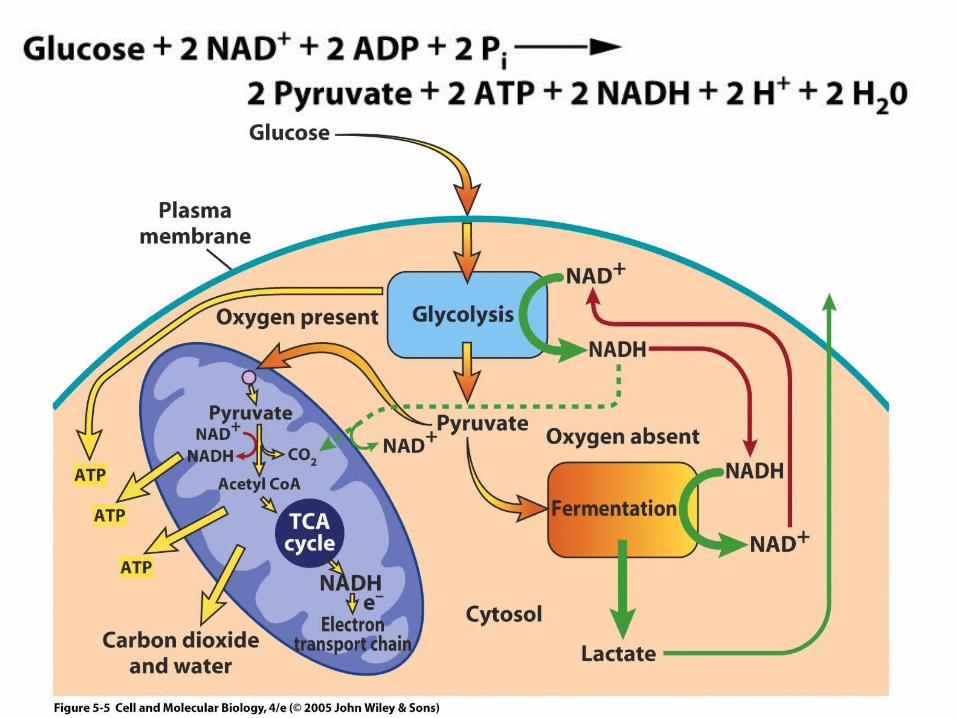
• Pyruvate to TCA
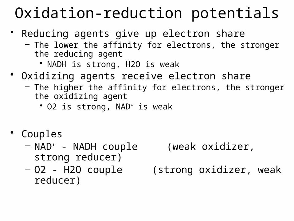
Oxidation-reduction potentials• Reducing agents give up electron share
– The lower the affinity for electrons, the stronger the reducing agent• NADH is strong, H2O is weak
• Oxidizing agents receive electron share– The higher the affinity for electrons, the stronger the oxidizing agent
• O2 is strong, NAD+ is weak
• Couples– NAD+ - NADH couple (weak oxidizer, strong
reducer)– O2 - H2O couple (strong oxidizer, weak reducer)
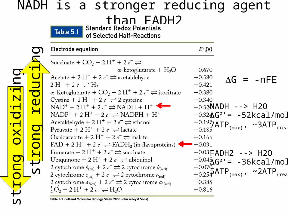
NADH is a stronger reducing agent than FADH2strong oxidizing
strong reducing
NADH --> H2OG0’= -52kcal/mol7ATP(max), ~3ATP(real)
FADH2 --> H2OG0’= -36kcal/mol5ATP(max), ~2ATP(real)
DG = -nFE

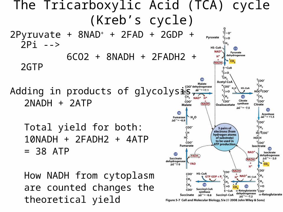
2Pyruvate + 8NAD+ + 2FAD + 2GDP + 2Pi --> 6CO2 + 8NADH + 2FADH2 + 2GTP
Adding in products of glycolysis,2NADH + 2ATP
Total yield for both:10NADH + 2FADH2 + 4ATP= 38 ATP
How NADH from cytoplasmare counted changes thetheoretical yield
The Tricarboxylic Acid (TCA) cycle (Kreb’s cycle)
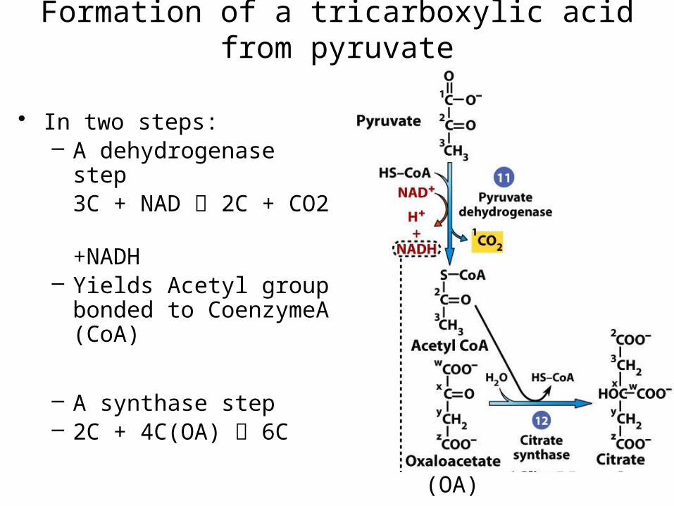
• In two steps:– A dehydrogenase step
3C + NAD 2C + CO2
+NADH– Yields Acetyl group bonded
to CoenzymeA (CoA)
– A synthase step– 2C + 4C(OA) 6C
Formation of a tricarboxylic acid from pyruvate
(OA)
The Tricarboxylic Acid (TCA) cycle (Kreb’s cycle)
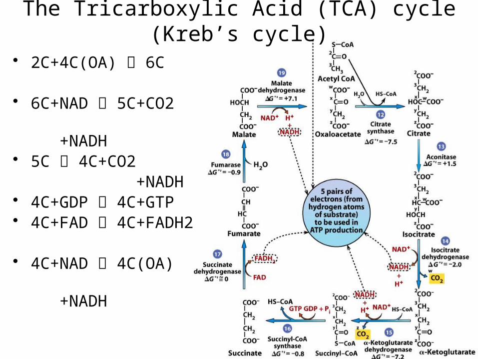
• 2C+4C(OA) 6C
• 6C+NAD 5C+CO2 +NADH
• 5C 4C+CO2 +NADH
• 4C+GDP 4C+GTP• 4C+FAD 4C+FADH2
• 4C+NAD 4C(OA) +NADH
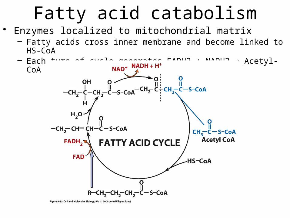
Fatty acid catabolism• Enzymes localized to mitochondrial matrix
– Fatty acids cross inner membrane and become linked to HS-CoA– Each turn of cycle generates FADH2 + NADH2 + Acetyl-CoA
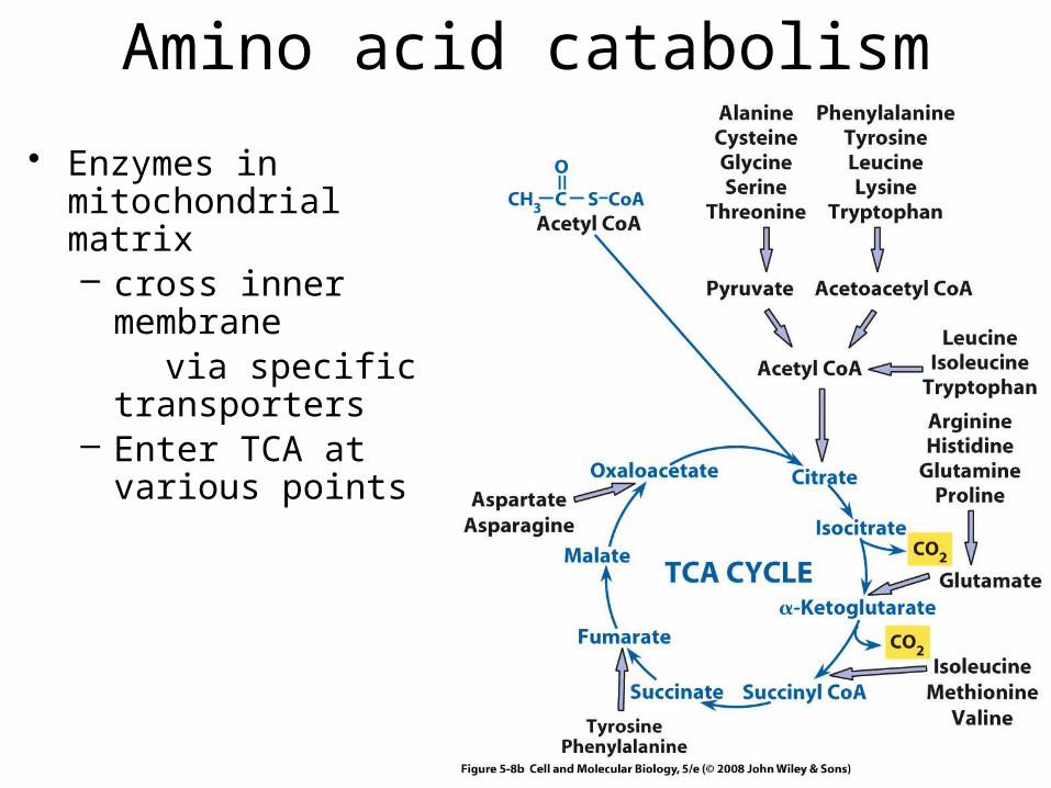
Amino acid catabolism
• Enzymes in mitochondrial matrix– cross inner membrane via specific transporters– Enter TCA at various
points
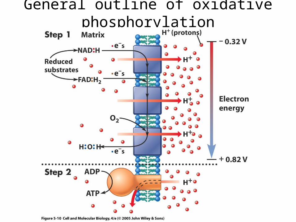
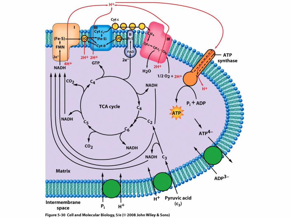
General outline of oxidative phosphorylation
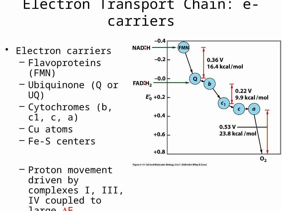
Electron Transport Chain: e- carriers
• Electron carriers– Flavoproteins (FMN)– Ubiquinone (Q or UQ)– Cytochromes (b, c1, c, a)– Cu atoms– Fe-S centers
– Proton movement driven by complexes I, III, IV coupled to large DE
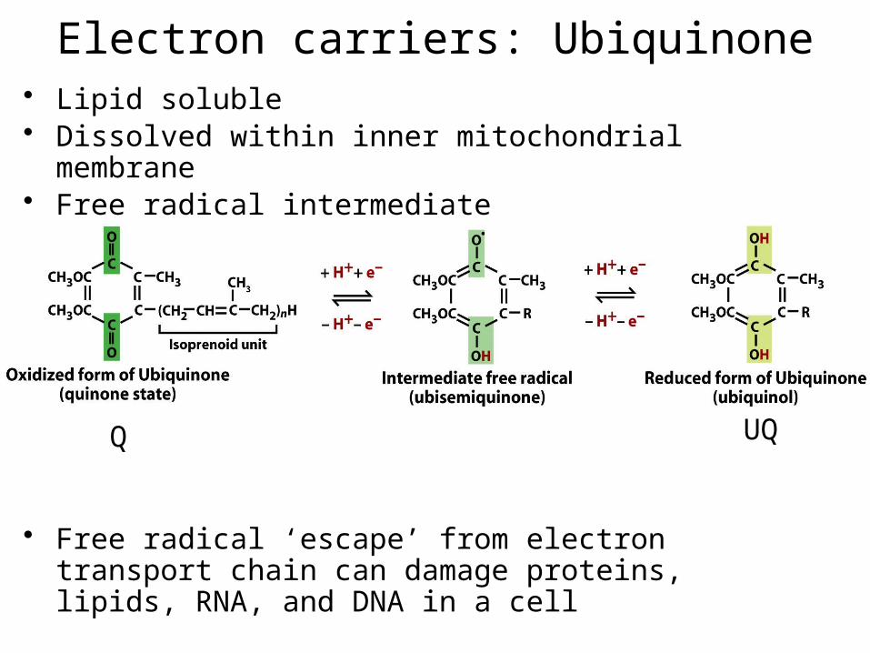
Electron carriers: Ubiquinone• Lipid soluble• Dissolved within inner mitochondrial membrane• Free radical intermediate
• Free radical ‘escape’ from electron transport chain can damage proteins, lipids, RNA, and DNA in a cell
Q UQ
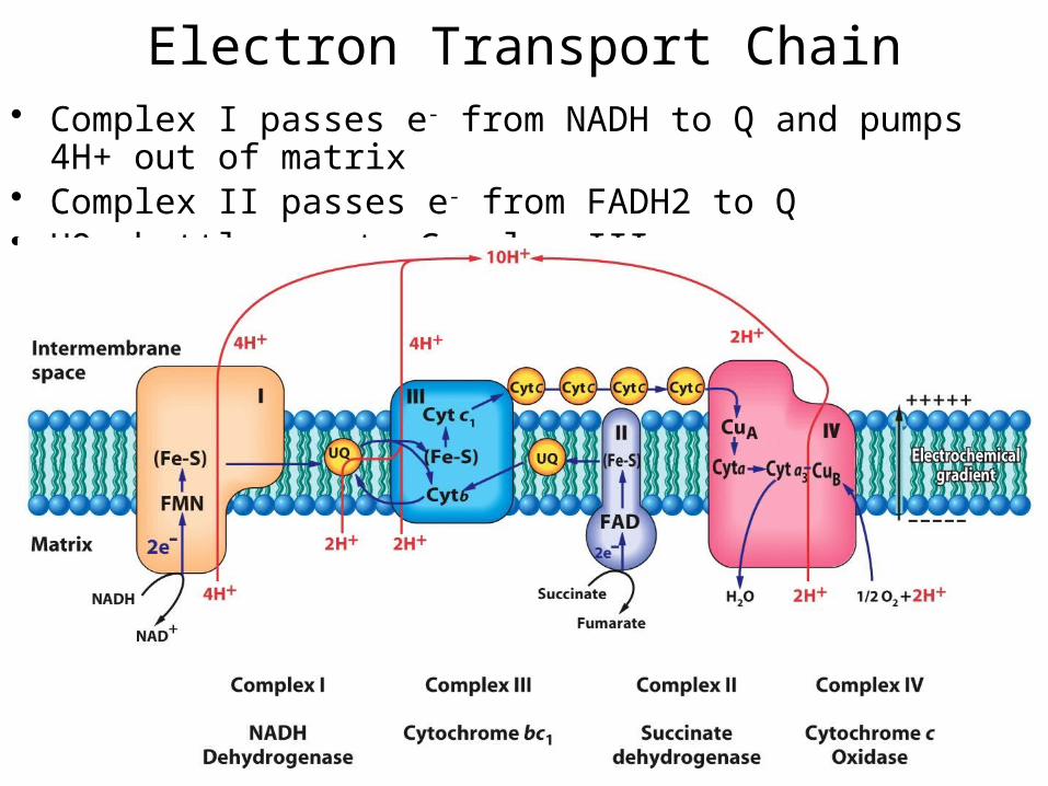
Electron Transport Chain• Complex I passes e- from NADH to Q and pumps 4H+ out of matrix• Complex II passes e- from FADH2 to Q• UQ shuttles e- to Complex III
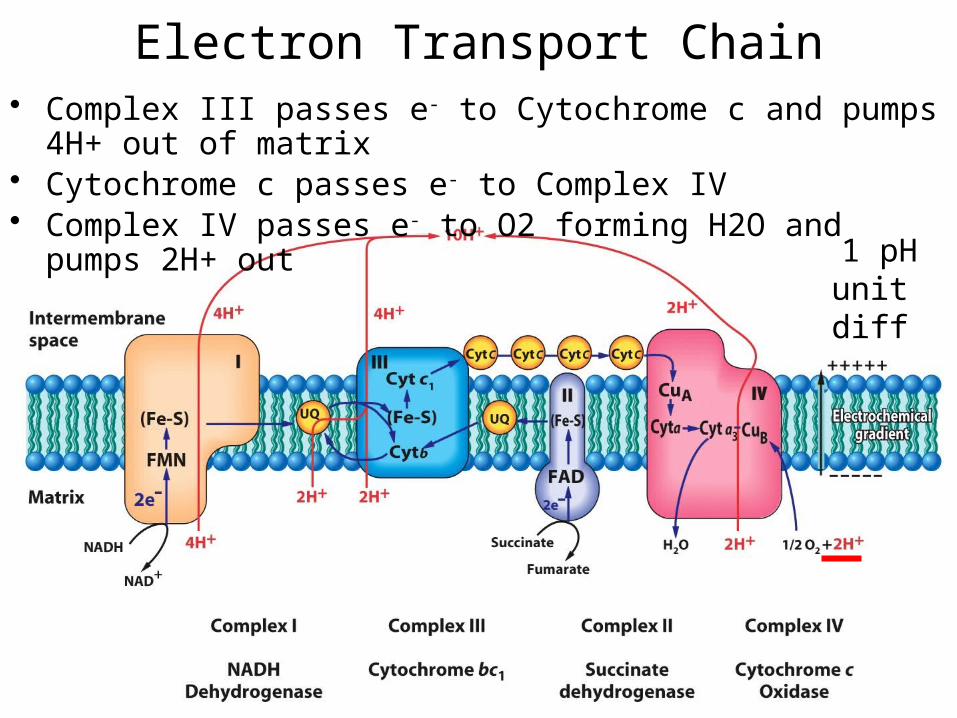
Electron Transport Chain• Complex III passes e- to Cytochrome c and pumps 4H+ out of matrix• Cytochrome c passes e- to Complex IV• Complex IV passes e- to O2 forming H2O and pumps 2H+ out
1 pH unit diff
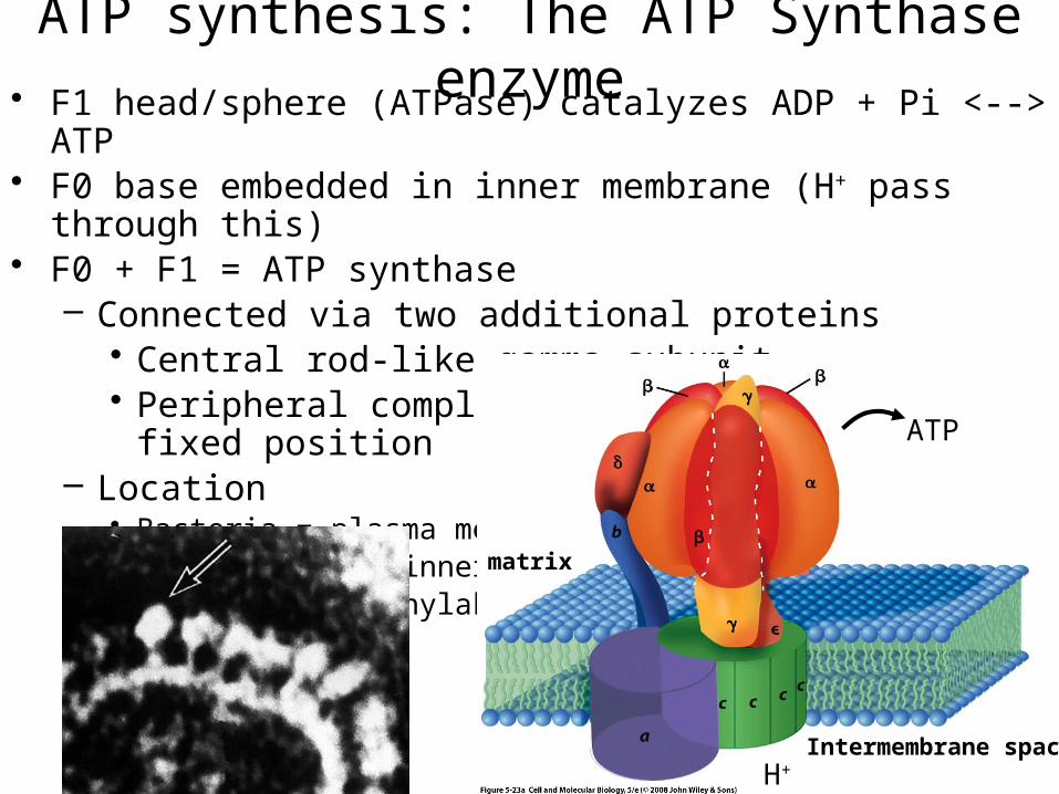
ATP synthesis: The ATP Synthase enzyme• F1 head/sphere (ATPase) catalyzes ADP + Pi <--> ATP• F0 base embedded in inner membrane (H+ pass through this)• F0 + F1 = ATP synthase
– Connected via two additional proteins• Central rod-like gamma subunit• Peripheral complex (abd) holds F1 in a fixed position
– Location• Bacteria = plasma mem• Mitochondria = inner mem• Chloroplast = thylakoid
Intermembrane space
matrix
H+
ATP
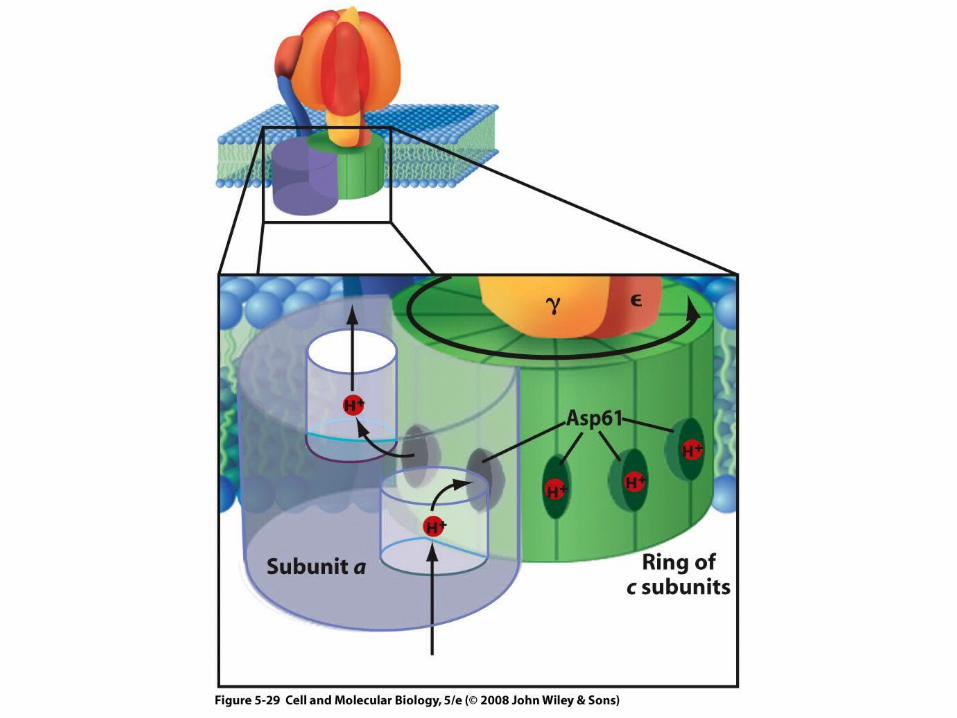
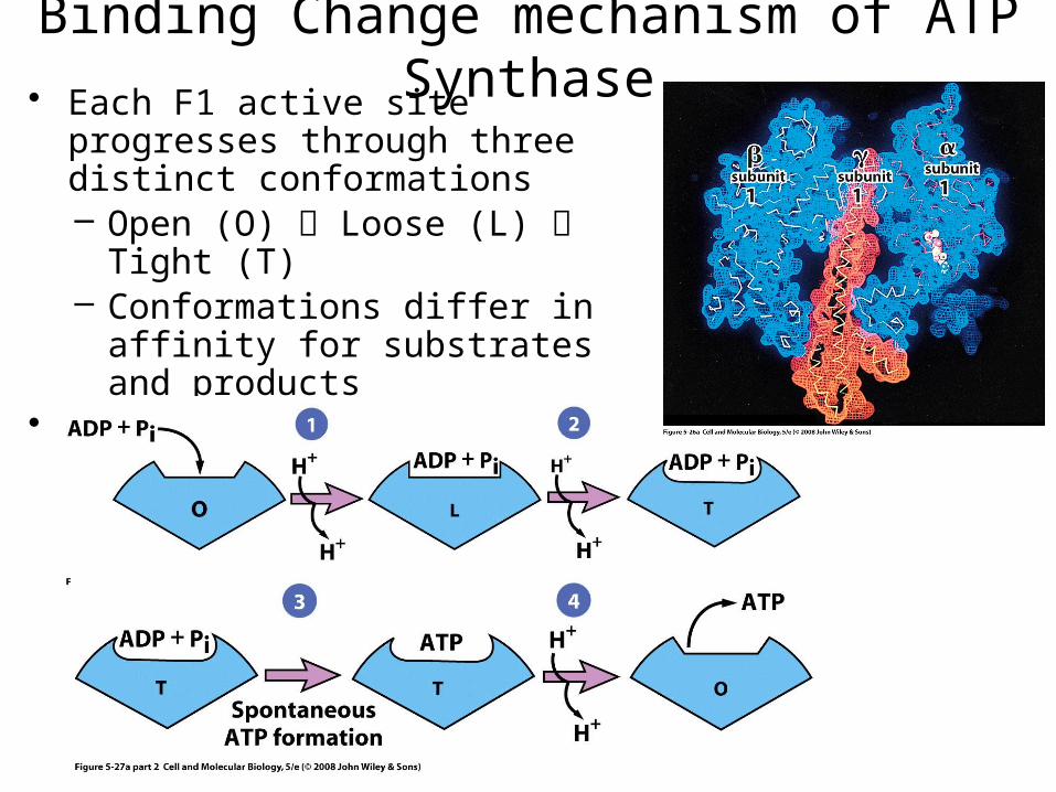
Binding Change mechanism of ATP Synthase• Each F1 active site progresses through
three distinct conformations– Open (O) Loose (L) Tight (T)– Conformations differ in affinity for
substrates and products• Central gamma () subunit rotates
causing conformation changes
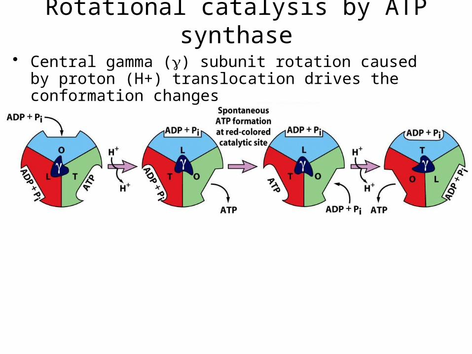
Rotational catalysis by ATP synthase
1 pH unit diff
• Central gamma () subunit rotation caused by proton (H+) translocation drives the conformation changes
Rotational catalysis by ATP synthase
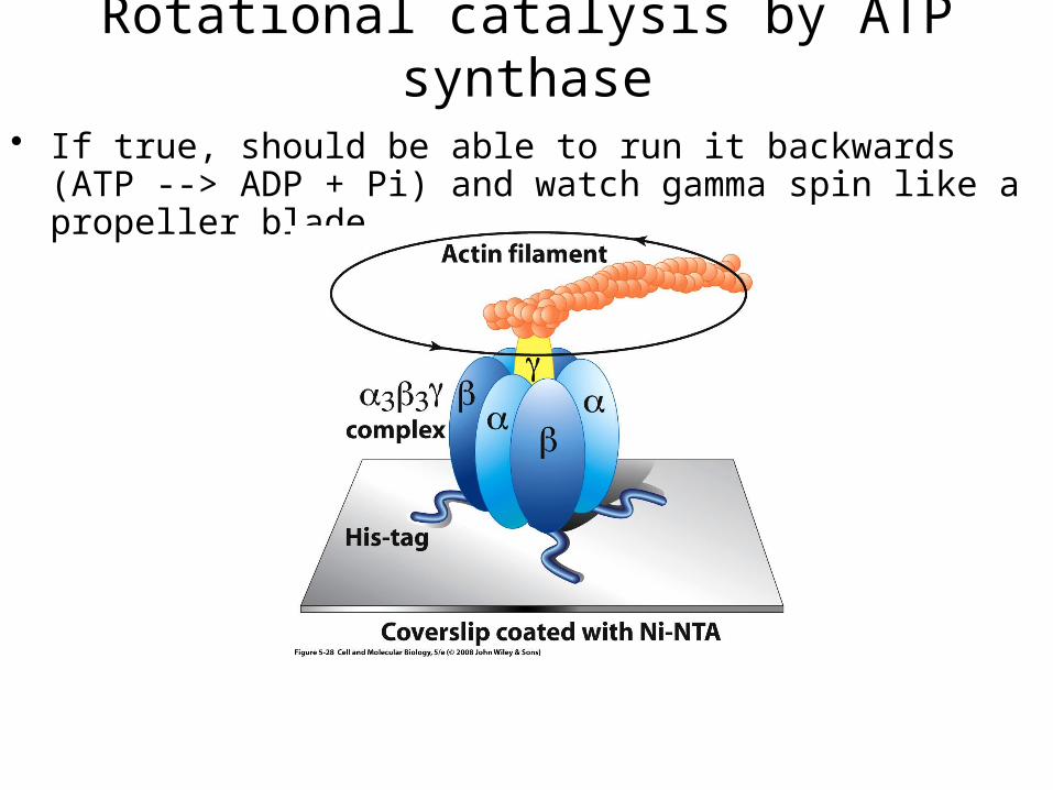
• If true, should be able to run it backwards (ATP --> ADP + Pi) and watch gamma spin like a propeller blade
Rotational catalysis by ATP synthase
Other fxns of electrochemical gradient
• E also used for:– Import of ADP + Pi (+H+) and export of ATP– Import of pyruvate (+H+)
• Uncoupling sugar oxidation from ATP synthesis– Uncoupling proteins (UCP1-5)
• UCP1/thermogenin, shuttles H+ back to matrix (endothermy)– Brown adipose tissue
» Present in newborns (lost with age) and hibernating animals» Generates heat
– 2,4-dinitrophenol (DNP)• Ionophore that can dissolve in inner membrane and shuttle H+ across
– 1930’s stanford diet pill trials: overdose causes a fatal fever