Embed Size (px)
Citation preview
HAEMOLYTIC ANEMIA IN TWO 500-YEAR-OLD SKULLS FROM THE CARDAMOMSMOUNTAINS, CAMBODIA: PALEORADIOLOGY STUDY
A.F. Yim*, C. Trompoukis**, C. Romagnoli *, F. Demeter***, R.K. Chhem*
AbstractTwo 500 year-old skulls, with possible signs of thalassemia, were selected from a burial jar
site located in the Cardamoms mountains in Southwest Cambodia for a multidisciplinary investi-gation. This study includes physical and radiological examinations. The prevalence of thalassemia ishigh in the extant population of the Southeast-Asian mainland. We report the radiological findings,which include both plain films and computed tomography (CT) and discuss the role of radiology inthe diagnosis of thalassemia in ancient bones.
IntroductionLegend has it that the Cardamoms forest may have served as an important place for people
to hide, particularly for the royal family after the invasion of Loevek city by the Siamese in 1593 CE.No evidence to support this theory has yet been found, however the discovery of burial jars sites byJean Ellul, French ethnologist in the 1960s may help determine the significance of this forest1.Bioarchaeological analysis of the skeletons obtained therein may give insight into how people mayhave survived in an area where malaria is endemic. Therefore the purpose of this paper is to describe
223
*Department of Diagnostic Radiology and Nuclear Medicine, Schulich School of Medicine, University of WesternOntario.** Department of History of Medicine, School of Medicine, University of Crete, Greece.*** Collège de France.1 Martin 1997.
04_Chamrithy:Udaya7 12/28/2007 11:15 AM Page 223
the radiological findings in two 500-year-old skulls that were suggestive of a chronic hemolyticanemia, most likely thalassemia. The pathogenesis of thalassemia in the ancient population living inthe malaria-infested Cardamom Mountains in Cambodia will be discussed.
Materials and Methods
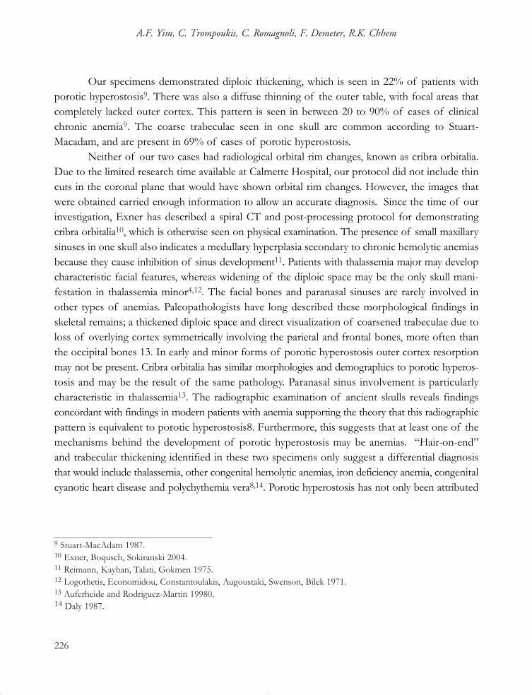
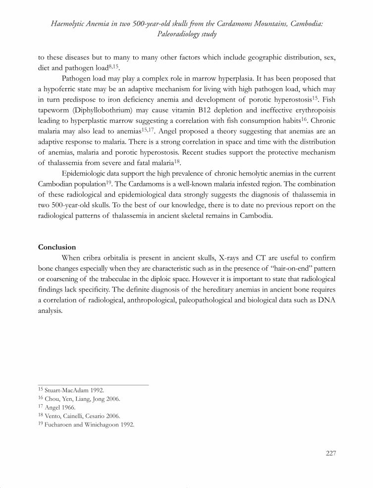
1. Skeletons and archaeological context In March 2000, under the Cardamom Conservation Program working with the department
of Forestry and Wildlife and the Agriculture Forestry and Fisheries ministry, 15 post-Angkorianburial jar sites were re-discovered within the Cardamoms forest of Southwest Cambodia. InFebruary 2003, a multidisciplinary team headed by the senior author (RC) was sent to the malaria-infested Cardamoms forest to investigate the burial jar site near Phum Roleak Kang Cheung village.The expedition was supported by funds from the National Geographic Channel, Australia, forwhich a documentary film was produced (Becker Entertainment). Forty jars were found. Two ofthe most intact of these jars were brought to Phnom-Penh for further analysis.
One jar contained four individuals, another three; with a total of one child and six adults.Of the adults, five were female aged 18-25 years old.
2. Radiological analysis X-ray analysis was performed in the Radiology Department of Calmette Hospital in
Phnom-Penh. Radiographs of skulls were obtained using standard X-ray machine.CT protocols: Two skulls with cribra orbitalia underwent a radiological investigation using
both conventional X-rays and CT scan. CT (Toshiba, Auklet, Japan) was performed using thefollowing technical parameters: 120kV, 200mAs, 5mm thickness, 10mm skip and 1 second scantime per rotation. Multiplanar reconstruction was performed to convert the acquired axial planeinto coronal plane. The data was saved on a CD and selected images were printed on hardcopyfilm for interpretation.
3. Biomolecular studies were not conducted because they are not available in Cambodia, andexporting the skeletal materials was not allowed by the Cambodian authorities.
ResultsAnalysis of the ceramics from the burial jars site revealed three different origins. One is
224
A.F. Yim, C. Trompoukis, C. Romagnoli, F. Demeter, R.K. Chhem
04_Chamrithy:Udaya7 12/28/2007 11:15 AM Page 224
typical for Siamese, another middle Ming period Chinese and the third resembles known Siamesewares but is of uncertain origin. The ceramic techniques date to a period from the 14th to 15th
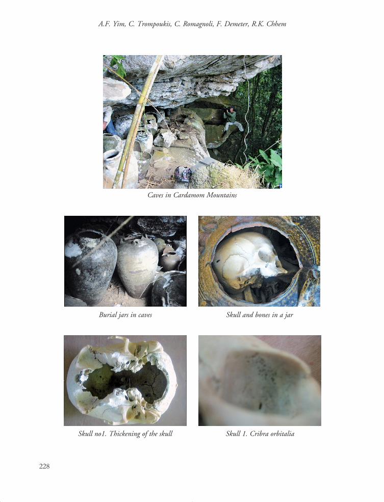
centuries. Carbon14 dating of the human remains within supports this placing them in the 1430-1480 CE range. Despite the fact that these remains are 100 years older than would be expected ofthe fleeing royal family, given the multiple origins of these ceramics it is likely that the people wereat least important enough to have had access to external trade. On gross inspection two of the sixadults showed cribiform changes on the roof of the orbit known as cribra orbitalia.
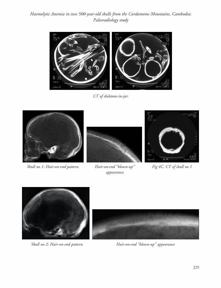
Radiographic findings in both skulls include a diffuse calvarial thickening. This was associatedwith a “hair-on-end” pattern in one skull and a trabecular coarsening of the diploic space on theother. The “hair-on-end” pattern was identified in the parietal bones. The frontal sinuses of theskull displaying this pattern were small. They were normal in size in the latter skull.
CT confirmed the thickening of the calvarium but did not clearly demonstrate the “hair-on-end” pattern or trabecular coarsening. In one skull the maxillary sinuses were small, with thickeningof the sinus wall.
DiscussionThe radiological findings described in these specimens have been recognized by paleo-
pathologists in previous skeletal remains under the term porotic hyperostosis, and are well knownto radiologists as suggesting a chronic hemolytic anemia in extant populations2,3,4,5,6,7.
In our case, the “hair-on-end” pattern involves the parietal bone, which is the most commonlocation for porotic hyperostosis5, In modern patients these changes may result from bone marrowhyperplasia in the diploic space. In severe and chronic anemia the outer table is perforated byproliferating marrow, which undermines and uplifts the periosteum developing bony spicules thatgive rise to the “hair-on-end” pattern8. Of the anemias, changes are most pronounced in thalassemiamajor4.
225
Haemolytic Anemia in two 500-year-old skulls from the Cardamoms Mountains, Cambodia:Paleoradiology study
2 Agarwal, Dhar, Shah, Bhardwaj 1970.3 Aksoy, Camli, Erdem 1966.4 Mosely 1974.5 Ponec and Resnick 1984.6 Sebes and Diggs 1979.7 Steinbock 1976.8 Hart 1981.
04_Chamrithy:Udaya7 12/28/2007 11:15 AM Page 225
Our specimens demonstrated diploic thickening, which is seen in 22% of patients withporotic hyperostosis9. There was also a diffuse thinning of the outer table, with focal areas thatcompletely lacked outer cortex. This pattern is seen in between 20 to 90% of cases of clinicalchronic anemia9. The coarse trabeculae seen in one skull are common according to Stuart-Macadam, and are present in 69% of cases of porotic hyperostosis.
Neither of our two cases had radiological orbital rim changes, known as cribra orbitalia.Due to the limited research time available at Calmette Hospital, our protocol did not include thincuts in the coronal plane that would have shown orbital rim changes. However, the images thatwere obtained carried enough information to allow an accurate diagnosis. Since the time of ourinvestigation, Exner has described a spiral CT and post-processing protocol for demonstratingcribra orbitalia10, which is otherwise seen on physical examination. The presence of small maxillarysinuses in one skull also indicates a medullary hyperplasia secondary to chronic hemolytic anemiasbecause they cause inhibition of sinus development11. Patients with thalassemia major may developcharacteristic facial features, whereas widening of the diploic space may be the only skull mani-festation in thalassemia minor4,12. The facial bones and paranasal sinuses are rarely involved inother types of anemias. Paleopathologists have long described these morphological findings inskeletal remains; a thickened diploic space and direct visualization of coarsened trabeculae due toloss of overlying cortex symmetrically involving the parietal and frontal bones, more often thanthe occipital bones 13. In early and minor forms of porotic hyperostosis outer cortex resorptionmay not be present. Cribra orbitalia has similar morphologies and demographics to porotic hyperos-tosis and may be the result of the same pathology. Paranasal sinus involvement is particularlycharacteristic in thalassemia13. The radiographic examination of ancient skulls reveals findingsconcordant with findings in modern patients with anemia supporting the theory that this radiographicpattern is equivalent to porotic hyperostosis8. Furthermore, this suggests that at least one of themechanisms behind the development of porotic hyperostosis may be anemias. “Hair-on-end”and trabecular thickening identified in these two specimens only suggest a differential diagnosisthat would include thalassemia, other congenital hemolytic anemias, iron deficiency anemia, congenitalcyanotic heart disease and polychythemia vera8,14. Porotic hyperostosis has not only been attributed
226
A.F. Yim, C. Trompoukis, C. Romagnoli, F. Demeter, R.K. Chhem
9 Stuart-MacAdam 1987.10 Exner, Boqusch, Sokiranski 2004.11 Reimann, Kayhan, Talati, Gokmen 1975.12 Logothetis, Economidou, Constantoulakis, Augoustaki, Swenson, Bilek 1971.13 Auferheide and Rodriguez-Martin 19980.14 Daly 1987.
04_Chamrithy:Udaya7 12/28/2007 11:15 AM Page 226
to these diseases but to many to many other factors which include geographic distribution, sex,diet and pathogen load8,15.
Pathogen load may play a complex role in marrow hyperplasia. It has been proposed thata hypoferric state may be an adaptive mechanism for living with high pathogen load, which mayin turn predispose to iron deficiency anemia and development of porotic hyperostosis15. Fishtapeworm (Diphyllobothrium) may cause vitamin B12 depletion and ineffective erythropoisisleading to hyperplastic marrow suggesting a correlation with fish consumption habits16. Chronicmalaria may also lead to anemias15,17. Angel proposed a theory suggesting that anemias are anadaptive response to malaria. There is a strong correlation in space and time with the distributionof anemias, malaria and porotic hyperostosis. Recent studies support the protective mechanismof thalassemia from severe and fatal malaria18.
Epidemiologic data support the high prevalence of chronic hemolytic anemias in the currentCambodian population19. The Cardamoms is a well-known malaria infested region. The combinationof these radiological and epidemiological data strongly suggests the diagnosis of thalassemia intwo 500-year-old skulls. To the best of our knowledge, there is to date no previous report on theradiological patterns of thalassemia in ancient skeletal remains in Cambodia.
ConclusionWhen cribra orbitalia is present in ancient skulls, X-rays and CT are useful to confirm
bone changes especially when they are characteristic such as in the presence of “hair-on-end” patternor coarsening of the trabeculae in the diploic space. However it is important to state that radiologicalfindings lack specificity. The definite diagnosis of the hereditary anemias in ancient bone requiresa correlation of radiological, anthropological, paleopathological and biological data such as DNAanalysis.
227
Haemolytic Anemia in two 500-year-old skulls from the Cardamoms Mountains, Cambodia:Paleoradiology study
15 Stuart-MacAdam 1992.16 Chou, Yen, Liang, Jong 2006.17 Angel 1966.18 Vento, Cainelli, Cesario 2006.19 Fucharoen and Winichagoon 1992.
04_Chamrithy:Udaya7 12/28/2007 11:15 AM Page 227
228
A.F. Yim, C. Trompoukis, C. Romagnoli, F. Demeter, R.K. Chhem
Caves in Cardamom Mountains
Burial jars in caves Skull and bones in a jar
Skull no1. Thickening of the skull Skull 1. Cribra orbitalia
04_Chamrithy:Udaya7 12/28/2007 11:16 AM Page 228
229
Haemolytic Anemia in two 500-year-old skulls from the Cardamoms Mountains, Cambodia:Paleoradiology study
Skull no 1: Hair-on-end pattern. Hair-on-end “blown-up”appearance.
Skull no 2: Hair-on-end pattern Hair-on-end “blown-up” appearance
Fig 4C. CT of skull no 1
CT of skeletons-in-jar.
04_Chamrithy:Udaya7 12/28/2007 11:16 AM Page 229
References Cited
Agarwal, K.N., Dhar, N., Shah, M.M., Bhardwaj, O.P., “Roentgenologic changes in iron deficiencyanemia”, American Journal of Roentgenology, 1970, 110(3): 635-637.
Angel, J.L., “ Porotic hyperostosis, anemias, malarias and marshes in the prehistoric EasternMediterranean”, Science, 1966 August, 153(737): 760-763.
Aksoy, M., Camli, N., Erdem, S., “Roentgenographic bone changes in chronic iron deficiency anemia”,Blood 1966, 27(5): 677-686.
Auferheide, A.C. and Rodriguez-Martin, C., “Hematological Disorders”, in: The CambridgeEncyclopedia of Human Paleopathology. 1st ed. Cambridge, United Kingdom:Cambridge University Press, 1998: 345-350.
Chou, H.F., Yen, C.M., Liang, W.C., Jong, Y.J., “Diphyllobothriasis Latum: the first child casereport in Taiwan”, The Kaohsiung Journal of Medical Sciences, 2006 July, 22 (7): 346-350.
Daly, D., “Hair-on-end”, Pattern in the skull. Seminars in Roentgenology 1987 July, ;22(3): 144-145.
Exner, S., Boqusch, G., Sokiranski, R., “Cribra orbitalia visualized in computed tomography”, Annalsof Anatomy, 2004 April, 186(2): 169-172.
Fucharoen, S. and Winichagoon, P. “Thalassemia in Southeast Asia: problems and strategy forprevention and control”, The Southeast Asian Journal of Tropical Medicine and PublicHealth, 1992 December, 23(4): 647-655.
Hart, G.D., “Anemia in ancient times”, Blood Cells, 1981, 7(3): 485-493.
Logothetis, J., Economidou, J., Constantoulakis, M., Augoustaki, O., Swenson, R.B., Bilek, M.,“Cephalofacial deformities in thalassemia major (Cooley’s Anemia)“, AmericanJournal of Diseases of Children, 1971 April, 121(4): 300-306.
Martin, M.A. Les Khmer Daeum: Khmer de l’Origine. Société Montagnarde et Exploitation de la Forêt,
230
A.F. Yim, C. Trompoukis, C. Romagnoli, F. Demeter, R.K. Chhem
04_Chamrithy:Udaya7 12/28/2007 11:16 AM Page 230
de l’Écologie à l’Histoire, Presses de l’École Française d’Extrême Orient, 1997.Mosely, J.E., “Skeletal changes in the Anemias”, Sem Roentgenol, 1974 July, 9(3):169-184.
Ponec, D.J., and Resnick, D., “On the etiology and pathogenesis of porotic hyperostosis of theskull”, Investigative Radiology, 1984 Jul-Aug, 19(4): 313-317.
Reimann, F., Kayhan, V., Talati, U., Gokmen, “X-ray and clinical study of the nose, sinuses andmaxilla in patients with severe iron deficiency diseases”, Laryngologie RhinologieOtologie, 1975 November, 54(11): 880-890.
Sebes, J.I., Diggs, L.W., “Radiographic changes of the skull in sickle cell anemia”, American Journal ofRoentgenology, 1979 March, 132(3): 373-377.
Steinbock, R.T., “Hematologic Disorders- the Anemias”, in: Paleopathological diagnosis and inter-pretation: Bone diseases in ancient human populations, 1st ed. Springfield, Illinois:Charles C Thomas, 1976: 213-248.
Stuart-MacAdam, P., “A radiographic study of porotic hyperostosis”, American Journal of PhysicalAnthropology, 1987 December, 74(4): 511-520.
Stuart-MacAdam, P., “Porotic hyperostosis a new perspective”, American Journal of PhysicalAnthropology 1992 January, 87(1): 39-47.
Vento, S., Cainelli, F., Cesario, F., “Infections and thalassaemia”, The Lancet Infectious Diseases,2006 April, 6(4): 226-233.
231
Haemolytic Anemia in two 500-year-old skulls from the Cardamoms Mountains, Cambodia:Paleoradiology study
04_Chamrithy:Udaya7 12/28/2007 11:16 AM Page 231

232
A.F. Yim, C. Trompoukis, C. Romagnoli, F. Demeter, R.K. Chhem
04_Chamrithy:Udaya7 12/28/2007 11:16 AM Page 232