Embed Size (px)
Citation preview

Loyola University ChicagoLoyola eCommons
Master's Theses Theses and Dissertations
1967
Age and Generation Cycle of Oral EpitheliumNabil John BarakatLoyola University Chicago
This Thesis is brought to you for free and open access by the Theses and Dissertations at Loyola eCommons. It has been accepted for inclusion inMaster's Theses by an authorized administrator of Loyola eCommons. For more information, please contact [email protected].
This work is licensed under a Creative Commons Attribution-Noncommercial-No Derivative Works 3.0 License.Copyright © 1967 Nabil John Barakat
Recommended CitationBarakat, Nabil John, "Age and Generation Cycle of Oral Epithelium" (1967). Master's Theses. Paper 2076.http://ecommons.luc.edu/luc_theses/2076

AGE AND GENBRA'1'ION CYCLE
OF ORAL EPITHELIUM
by
NuU J. Barakat.
A The.i. Sw.1tted to t.he Facult.y o~ the Graduate School
of Loyola Unl.erait.y in Partial Pulfl1lmant o~
the Requirement.. for the Degre. of
Ma.ter of Sclence
June 1961
- I UBRARY , l,UYOI:A UN1VERSlTY MEDICP.L a::r,,1"JLj1

LIFE
Nab11 John Barakat. was born on March 25 t 1940,
in <:a1ro, EtYpt.. He graduated :from HOly Fam11y·.
Je.uit coll.,. 1n 1958. From 1958 to 1959 be attended
t.he Faoulty of Scitance., Calro Un!verslt.y.
ae recelved hi. Bachelor of Dental surqery deqr ..
from the School of D6ftt1atry, Cairo Unlveralty, in 1963.
Upon graduatlon, he staned a rotat.ln9 int.ernship at.
ca1ro·a Un.lveralty heap1Ul.
In January 1,965, he b8CJan a two-year Graduate
pzoqram a1t Loyola unlvers1t.y le.dinq t.o a Mast.r of sclenee e
4eqr.. 1n Oral Biology. Clinical t.rUni.n9 waa clone in t.he
Oral Surgery t>ep&.rt.lIeJlt. u.n4er Dr. Niebolas ChOUkas.
During' his coura. of at.u41es, he inat.ructed Phyalolo;y
and PharmacolOCJY' at. th. achool.

ACKNOWLBDGBMENTS
The author would like t.o expre.s hi. appreciat.ion
t.o Dr. Patrick Toto for hi. encouragement, many augge.tlons,
and idea., during t.he pa8t year.
TO Dr. Harry Sierher and Dr. John O' Malley, lIf!;/
Sincere thank. for their helpful crlticiam.
I am lnd.beed to ~ faculty advisor, Dr. Nicholas
Cheukaa, for hi. guidance and the philosophy of the deaire
to learn.
Appreciat.ion 18 offered to Dr. Allen Goldberq,
Veterans Administration Hospital, Htnes, Illinois, for
hia aid in obtatniftf the photom1croc;rapha.
It baa b41en a, great opportunity and privileqe to
work under such celebrated and gifted teachers.
Finally I to Arqonne Nat.ional Laboratory I Lemont.,
Illinoi8, and Dr. samuel Lesher, for~. apectmana they
made available tor atudy, I am deeply thankful.

I.
II.
TABLE OF CONTBN'l'S
INTRODUCTION • • • • • .. .. • .. • • • • • .. •
RBVIBW OF LXTERA'l'URE. • . .. ,. . . • • • > ••
A. HISTOLOGY OF THE ORAL MUCOSA . .. . .. • •
1
3 , B. CBLL RBNBWAL SYSTEMS • .. • .. • • • .. II.. 6
C.
1. Renewal of Cell Popula~1on • .. .. .... 6
2. Cause. of Cell R8n ... l .. • • • . . ,. :I.. Method. of Det.8rm1na't1on. of t.he
Ga.nt.1oft Cycle of cells · . . .. . . ..
.. . . . . .
10
11
18
26
III. MATERIALS AND MB'l'HODS • • .. • • *' • • .. • ... 21
V.
FINDINGS ..
DISCUSSION
,"i · .. .. .. . . . .. .. .. . . .. .. . ..
· .. • • • .. . . .. . .. it • • . . .. 35
VI. StJMM.AR'!l AND CONCLUSIONS ,.....:..... •• :41
BIBLIOGRAPHY .. ,. .. .. ..... ,. •• .. • • ... 43
APPENDIX • • .. .. .. · .. .. .. .. A. Photomicroqraph. • .. • .. .. B. 'l'able • • .. • • .. • .. .. .. c. Gzoaph • • • • • • • • • •
.. " .. .. .
.. .. • • • .. j, • • .. .. • • .. ..
.. . • •
• •
.. •
4' 58
59

L
CHAPTER I
INTRODUCTION
1
In the adult mammal, larqe number of aells are
produaed and lost everyday. All aell populations increase
in number durinq the period of qrollth of the orqanism. In
some cell populations, the rate of proliferation is <Jreater
than that needed to sustain qrowt:h, this means that some
cells are lost and replaced by newly formed aells. Such
cell populations are aalled nCell renewal systems"
(Leblond 1956). However, upon aompletion of the qrowinq
period for a given renewal system a steady state is estab
lished, Cell ~renewal> balance. cell los ••
The f~r.t author to rais. the question whether
physiologic regeneration of epithelia actually slows with
aqe was Ortiz Picon (1933), who studied the epidermis of
abdomen and baak of mice from birth to 33 months of ag8.
At 6 months the rate of division was twice as hiqh as at one
month and remained hiqher than at one month for the entire
period studied. In 1949, Bullouqh found the mitotic::
frequency in mouse epidermis to be considerably hiqher in
middle-aqed. than in younq animals even before they have
reachad full size. Marwah (1956) found the mitotiC frequency
in human qinq1va to be 50% h1qher 1n the older aqe qroups.

2
The.e tinding. contradict the common concept ot
aging, according to Which bodily proc •••••• low down and
the r8gen.rative oapacity ot the ti.sue. dimini.he. with
age. Th ••• observation. would point -to the po •• ibility
that the mechanisms ~ which normal cellular prolit.ration
i. controlled are reduced in the older indiVidual. The
incidenc. ot mali9ftaney i. known to incr .... with age.
Marwah state. that one might .uspect the tull emancipation
trom control ot the tull tledged malignant. c.ll i. prepared
tor ~ the partial 8mancU.pation t rom control Which oacur.
with age.
Th. pre.ent study will attempt to •• timate the
g.n.ration cycle ot the oral .pithelium in 600 day old miae.
In partiaular this .tudy employs the "direct graphic method"
to .tudy the renewal ot cell population.

CHAPTER II
REVIEW OF THE LITERATURE
A. HISTOLOGY OF THE ORAL MUCOSA
Kutuzov and Sieher (1953) described in detail
the dorsum of the mouse tonque.
Macr08copically, the dorsum of the tonque can be
divided into 3 reqion., eaoh oharacterized by a specific
type of papillae.
1. Anterior portion of 8imple conical papillae.
2. Intermolar em1nence characterized by the qiant
papillae.
3. Posterior portion beset with the filiform
papillae.
The stratified squamous epithelium of theae three areas is
keratinized.
Sonntag (1924) pointed out that thou9'h animals
compri8in9' the Order of Rodentia are very diversified the
lin9'U&l characters do not differ very much specifically.
In 1959, Medak qava a detailed aacount of the
oral epithelium of the mouse. ae described it as having a
lamina propria consistinq of connective tissue cells and
fibers, a basement membrane with collagenous and reticular
fibers and a stratified squamous epithelium.
3

4 The basal layer of the epithelium haa a sinq1e row
of columnar cella. They are perpendicular to the baaement
membrane. The cytopla.mic nucleic acid content. are hiqher
in the.e cells than the cella fOrming the rest of the epithe
lium. The.e nucleic acid contents decrease peripheralward.
The strat.um apinosum bas larqe polygonal cella with
atar-ahaped outline. and intercellular connections. Kerato
hyaline granule. are found in the cell. of the stratum
spinoaum in later staqe. of oellular differentiation. In
1967, CUtright stated that the keratohyaline granules, whlch
are eventually shed with maturing cells, do not conta1n any
DNA component.
Madak (1956) described the "ratum qranulosum as
havlng no residual cytopla.m and the unstained Clranule. are
of uniform 81ze only adjacent to the compact keratin areas.
There ls evidence of phosphamidaa. actlvity 1n the kerato
hyaline granules. CUtright (1967) noted clear apaces in the
nuclei of oells approaohing the keratin layer and attrlbuted
lt to the normal loss of their nuclear DNA in the oour.. of
kerat.lnization.
Leblond (1956) described the morpholoq1cal change
in the ba.al cell layer. A. th... cella move outwards they
abu<,Je from columnar to polyhedral and finally to flattened,
granule containing c.lls, whlch then transform abruptly into
a homoqenoua qla.ay layer.

5 This, in turn, ohan~.8 into soft k.ratin.
In 1967, CUtright atated that the normal oral
.pith.lium haa the property to aohieve oytoplaam1c diff.ren
tiation while und.rgoinq nucl.ar deg.n.ration coupl.d with •
mechanism by which the DNA rele.aed from k.ratinizing c.lls
i. reutiliz.d by newly proliferating cell ••
Normal hlstol9.9X Of aging oral soft tissue,
Wentz .1 Al (1952) founcS an absenc. of aqe ohanqes
in the thickne.. of epithelium and length and confiquratlon
of the epith.lial ridges of the gingiva. Rllnqaberg (1960)
found no major alterationa in the keratin lay.r and the
ov.rall .pith.lium with incre.sing a~e.
MArwah (1956) found 80me ag. Chang •• in human
qinglva aa followl
(1) a deore... in incidence of a granular layer (2) a decrease
in inoidence of parakeratosis and (3) a decrea •• in the
.. aociation of a granular layer with paraken'tosis, (4) an
incre .. e in cell density by a third. a. attributed that in
cr .... in cell d.nsit.y t.o be due in pan. t.o lower wat..r
content of the t1.sue and in pan. to the smaller sl.e of cell ••
Burzinak1 at Al (1965) repon.ed:.::.t.hat there was no
aignificant difference in the palatal t.issues with ag1nq
between sax.. of',: the same age groups I and 1n palatal tissu ••
of l-year-old and 4-year-old animals in non-collagenous to
collagenoua ratiOS.

B. CELL RBNBWAL SYSTBMS
I. Renewal of Cell Population
6
In 1956, Leblond r.viewed in detail the proce ••
of the renewal of oell population. He attributed the lack
of in~reat of classical histologi.t. in the subject to
their belief that new cell. were proc!uae4 by "amito.1s" a.
well as by "m:Lt081." and therefore the true rate of pro
liferation could not be estimated. However, in 1955, Ria
poin'ted out that amitosis consist. of a _re nuclear
fragmentation, a proce.. which venerally precludes further
proliferation. Thus mito.i. 1. the only means of cell
production, and the number of new cells formed in this
tissue.
Howard (1953) divided the "9eneration cycle" into
the followinq phas •• , S phase (DNA syntheSiS time) durinq
Which sufficient DNA is synthe.ized to form a new complement
of chromosomes, G2 ph .. e which ls an interval between S
Phase and beqinninv of mitoais, M pha •• in which mitOSis
occur. and a Gl phase (rest.ing- phas.) betwe.n termination
of mitosis and c:onmencement of the S ph .. e. The perlod
~ween the two mitosis ls called an .. int.rphaa .... - In 1963,
Patt. pointed out that. the duration of Gl (restlnq pha.e)
vari.s wid.ly, ln typical mammalian cell syetems, and is
malnly responsible for the very larqe difference. ln cell
cycle time amonq varioua tis.ues.

7 ~he time required for one generation cycle to occur is
referred to as the "qeneration Ume" which should be clearly
distingui.hed trom the "turnover time" which i. defined as
the time taken tor the replacement at a number of cells equal
to that in the whole population (Leblond 1956).
In 1960, Messier considered the stratified squamous
epithelium ot the tonque as an example ot renewing cell popu
lation with a "restricted proliferation site." New cell.
are always for..ed in the stratum germ1nativum of the epithe
lium, since only cell. in this layer are labeled (that i.,
synthesizing DNA prior to mitosis) one hour after isotope
injections. ae further stated that cell "miqration" i.
common in the tongue epithelium a. shown by the migration
of a labeled baaal cell to the upper strata. CUtright (1967)
concluded that the greater the speed ot epithelial cell
turnover, the more cells miqrate.
CO'Wdry (1942) reported that the dauqhter cells of
a cell dividing in the reprodUCing layer may either become
member. of the surtace layers in whiCh no mitosis occur
(Poat mitotic cell) or remain in the qerminative layer and
there undezvo further mitosis (veqatativ. intemitotics and
differentiating' interm1t~ic.) •
. In 1965, Leblond pointed out that two dauqbt.er cells
Of a mitosis coma out independently and at random, thus the
division of a bual cell may qive two basal, or one basal

8 and one spinous, or two spinous cells. He considered the
coming out ot the basal layer as the cri't.ical step I after
Which the evolu't.ion of the cell is irreversible up to
keratiniza'tion and sheddinq. He concluded, theretore, that
on the average halt ot the cell. ari8ing from mito.is diVide
again and half transtorm into spinous cell., thus, there is
no change in the number ot cells of the epithelium and the
steady state ot the cell population i8 maintained.
In 1961, CUtright demonstrated that the maintenance
ot epithelial homeosta.is i. helped by the reutilization of
the DNA, released fram keratinizing cells, by the newly
proliterating cells_ He poin~ed outt.hat this recycling
could be important when rapid regeneration is needed, since
it make. the epithelium le.. dependent on blood-born DNA
precursor. and provides a local pool of e •• ential DNA
metabolite ••
Henry (1952) used the "colchicine method" to
•• timate the rate ot regeneration ot the rabbit's oral
epithelium and tound it to be "S.5" days. Since then,
many workers have qathered "data" on the "turnovert.1me"
ot t.he oral epithelium. They estimated it to be between 3
days (Gott..sbe1'ge 1963) and 5.S days (Cutriqht 1961) tor
the various portions ot the oral mucosa. These values are
.ummarized in Table I.

Toto (1962) conducted an investigation of the
qeneration cyele of the epithelium of the tongue in mice
in which he concluded that the duplication staqe lasts for
ten hours. In 1964, Dhawan found that the synthesis time
9
1s eiqht hours for the palate and doraa1 surfaae of the
tonque, and 81qht and one-half hours for the ventral surfaae
of the tongue, in the 60 day old miae.
Leblond (1956) pointed out that variations in the
•• timation of the duration of mitosis were probably due to
observational difficu1ti.s, since prophase and telophase are
hard to delimit in livinq ae1ls. Henry (1952) concluded
that the mitosis required 64 minutes 1n the oral epithelium
of rabbit. In cue of mice's oral mUCOsa 1t was found to
require 40 minutes by Medak (1959). This findinq is in
agreement with tho.e of Toto (1962) and Dhawan (1964).
In 1908, Minot studied the epidermis of rabbit
embryos at varying age ranges, he found different mitotia
indiaes in various tissu.s, thus establishint evidence of
.differential rate. of q«."Owth. He further, described the
mitotia index (MI) as the number of cells to be found at
any given moment in the active proc... of division out of
a total of 1,000 cells. Mitotic index is used to measure
the renewal time. Messier (1960) pointed out that mitotic
index is not altogether satisfaatory to assess the rate of

10
cell proliferation in a tissue beaause the counts of mitosis
in sections are proportional not only to the number of cell.
entering mitosls, but a180 to the time taken by the.e cells
to complete vislble mitosi., also, mitotlc figure. are
difficult to reooqnlze in small and elongated cells.
II. Cause. of Cell Renewal
In 1963, Bertalantfy assumed that cell renewal is
a protective mechanism. ae stated that many epithelia are
almoat continuouslYf:·exposed to .tress of varlous kinds, e.g.,
mechanical (oral epithellum) I enzymatic (duodenal epithelium),
bacterial agents (respiratory epithelium), to site a few,
therefore, were there no cell renewal, aells of varlous
epit.helia might .oon become abraded by mechanical contact or
dige.ted by proteolytic enzyme.. Vulpe (1954) found that
irritation due to urine causes mitosis ln the transitional
epithelium of the bladder. Le.her (1964) estimated that the
turnover time Of duodenal epithelium of the germ-free mice
i. longer than that of conventional mice. ae postulated
that the germ-free envlronment reduce. attrition of cells
on the villi and the re.ulting decreased need for renewal 1.
recoqnized and responded to by decreased cell production.
In 1956, Leblond demonstrated that most renewal systems,
though responslve to extrinsic force., are capable of re
newing in their ab.ence, e.g. , cells were renewed even in

isolated intestinal loops not exposed to the mechanical
.tres. that may be exerted by inqe.ted food. Accordinqly,
the ability to proliferate is an inherent property of the
cells of the renewal system.
11
Leblond (1965) pointed out 'that the addition of
new cells by mitosis tends to increase population pre.sure
in the basal layer .s shown, often, by the racket shape of
the cells leav1ng this layer, as if squeezed out by pressure.
Bierman (1955) sugyested that cell. may be el~
1nated by hormonal factors as s.en 1n caee of lymphocytes
by the adrenal cortical hormone (hydrocortisone).
Harvey (1963) stated that the production rate in
hematopoietic tissues is death-controlled. That is, ad
Justed by variations 1n the mortality of newly formed cells.
Berta1anffy (1963) concluded that the component cells of
tis.ue. with cell renewal are replaced at constant rate.
governed by the steady state, whether they are stres •• d at
any particular time or not.
III. Methods of Determination of the aeneration cycle of
Cella
There are various methods for determination of the
rate of regeneration of cella preferred by different workers
for different reasons. some of the methods used will be
described.
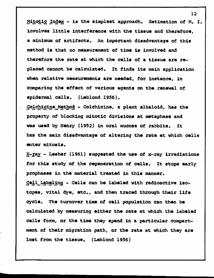
12
,MitO!ig, ln~~ - is the simplest approach. Est.imation of M. I.
involves little interterence wit.h the tissue and therefore,
a minimum of artifacts. An important disadvantaqe of this
method is t.hat no measurement. of time 1s involved and
therefore t.he rat.e at Whlch t.he cells of a tissue are re
placed cannot be calculat.ed. It. finds its main applicat.ion
when relatlve measurements are needed, for 1nst.ance, in
comparlng t.he effect of various aqenta on t.he renewal of
epldarmal cells. (Leblond 1956).
£o!c.hlg,l!!a_M.!t~ - Colchicina, a plant alkalOid, has tha
property of blOCking mitotic divisions at metaphase and
was used by Henry (1952) in oral mucosa of rabbits. It
has the main disadvantaqa of altaring the rate at. whlch cells
enter mitosls.
~-.la.x - Lesher (1961) sWigested the use Of x-ray irradiations
for this study of the reweneration of cells. It stops early
prophases 1n the material treated in this manner •
.£e!l_L,!b,!l!n,i - Cell. can be labeled with radioactive i.o
topes, Vital dye, etc., and then traced through their life
cycle. The turnover time of cell population can then be
calculated by measuring either t.he rate at Which the labeled
cell. form, or the time they spend 1n a particular compart
ment of t.heir migration path, or t.he rate at which they are
lost from the ti •• ue. (Leblond 1956)

13
Volkman (1950) aa8.ssed the renewal time of the
epidermia by meaaurinq the rate at. which India ink injected
into the deep layers was c:arried to the surface.
Leblond (1965) atate. that the advent of radio
autoqraphy qave hi.tology a new dimenaion, time. When cella
double their DNA prior to mltosl., they take up varlous DNA
precursors. If labeled precursors are injected lnto animals,
they are incorporated ln divldinq cella, which are then
t.awed anet may be traaed in radioautographs.
Phosphate-p32 , Adanine-c14, and c14 formate have
been used ln thls manner (Lajtha 1954). However, the firat
one of the.e preaursora 1s taken up int.o a varlety of
substances besldea DNA, the second and th1rd lnto both RNA
and DNA and only "Thymidine" ia taken into DNA alone
(Relchard 1951). Amano (1959) ob •• rved that I after
thymidine-H3, radioautoqraphi<: reactiona were present only
over nuclei, and that pretreatment of the sections with the
enzyme Deoxyribonuoleaa. prevented the oacurrenoe of
reactions. It may, therefore, be atated that the only
raetioa=lve substance present. in •• ctiona after labeled
thymidine inject10n 1a DNA, and that any cell tound to be
labeled ls about to d1v1de by mitoaia. Thymidlne haa the
advant.aqe ot be1nq aval1able wlt.h a trltium (H3) label,
Whlch produce. Beta-ray wlt.h such a low energy that they are

recorded in t.he phot.ographic emulslon very Cl0S8 to their
site of emission, the re.ult is a f1ne radiographlc reso
lution (Taylor 1951).
Me.sier (1960) used thymidine Hl for hi. studi ••
of the cell proliferation and mJ,gration ln male rat. and
mice. The frequencY of labeled cella ls proportional to
14
the duration of tbe duplicatlon lItaq_. A close relationship
exi.ts between the labeled thymidine uptake and mitosis ln
the tissues. He further .tates that the radioactlve index
(perc:entag'e labeled aells) i. a rOug'h lndex of the rate of
cell dlvislon, slnce it is lntluenaed by the rate of incor
poration of the label into the inter,mediate substance leading'
t.o DNA synthesl. and by the duration of the perlod of DNA
synthesls. Neverthele.s, the radioactive 1rldax has two
advantaqes. (1) labeled cella are more readl1y ldentlfled
than mitotic flgures in many ti.sue. and are many times as
numerous, (2) the cells t.aqqe4 by t.hymidlne-H] could be
traced lon, after mito.ls had been accomplished.
There is a 41fference of opinion., among' sclentlst.,
a. to the extent of injury to chromosomes caused by
thymidine-H] • Schoenhe14er (1960) I reviewed the method of
autoradioqraphy, as.ume4 that the chemical behavlor of the
labeled substance 1s ldentical with that of lts stable
countexpart. It 1. llke a "tracer."
\

15
Leblond (1965) stated that the autoradiography 8hows movement
under physiological conditions, on the other hand Quaatler
(1959) pointed out that this method of inv •• tiqation is not
physiological, because of the qrowth factor in young' animal.,
the stres. of lnjec~lon and the injury to the chromosomes
caused by triated t.hymidine. In 1962, Toto concluded t.hat
there is no m1.n1mal threshold for triated thymicU.ne at which
injury t.o the chrOmosomes does not oc:c:ur.
Lesher (1961) u.ed both the graphic method and
the staqe durat.ion index one to determine t.he qeneration
t:Lme. He concluded that the qraphic method 1a superior to
t.he at.aqe durat.ion indek one because the lat.t.er require.
preci.. knowledge of the 81z. of the proliferative pool and
of the duration of a particular staqe of the qeneration
cycle which are difficult to obtain. Its only advantage is
t.hat it can be applied wit.hOut observing' the complete
qeDerat.ion ayele. Waher (1961), Toto (1962) and Dhawan
(1964) calculat.ed the 9eneration cycle of cells by a4dinq
the DNA synthesis per104 (s), a reet. period (G2), the dura
tion of mitosi. eM) I and t.he interphase CG1 ).
Lesher (1961) reported t.hat. the qeneration time
computed by the staqe durat.ion index formula,
Number of Cells in Mito.1a = Duration of Mitosi. Number of Calla in Inte~ha.e = Duration of Interphase
i8 unreliable proportioned to the deqre. to which the ••

estimate. of the mitotic duration are unreliable.
In 1967, CUtriqht used a pure hiatoloqic and a
mathematical technique to determine the turnover time of
16
the Stratified squamous epithelium. The histoloqic method
expressed the renewal time as thet:..1me required for the
last initially labeled cell to pas. from the stratum qermi
nativum to t.he kerat.in layer whioh was used as an end point
because it normally contains no living cells and t.hus no
labeled nuclei. On the other hand, the mathematical method
used the racU.oac:tlve index (percent.age labeled cells) of the
basal layer and calculation i. al.o baaed on the t.1me needed
for DNA aynt.hesis, 7.5 hours. The following formula was
used.
100 x 7.5 Radloac:t.i ve Index 24 hour.
He pointed out. t.hat t.hi. approach is based on the assumpt.ion
t.hat. all newly formed cell. derive f~ the basal layer and
that., therefore, the percentage of proliferat.ing' cella
within the baaal layer raflecrt the t1.tmover .tat.e. He
considered the mathematical method to be superior t.o the
histolOjJic one.

17
TABLE I
TURNOVER TIME OF ORAL EPITHELIUM
Author Source P'ind-and Method of lnqs In
Year Used Material Reqion Days
Henry Colchicine Rabbit Oral Mucosa 8.5 (1952)
Toto Autora41o- Mice Tongue Epithelium 4.1 (1962)
KoWrq Autoradio- Mice Hard Palate 4.4 (1962) grapby
;BJlu:talanffy Colcbicine Rat Buccal Mucosa 4.3 (1963) Tongue Epithelium 4.9
Gottesberqe Autoradio- Mice Ginqival Epithelium 3.5 (1964) qraphy
Ohawan Auto radio- Mice Palatal and Dorsal 4"""--(1964) qraphy Tongue Epithelium
Ventral Tonque 4.1 ....-Epithelium
cutright Autoradio- Rat Post Dorsal Tongue 3.2 ../ (1967) qraphy Epithelium
Anterior Tonque 3.5 Epithelium
Ventral Tonque 5.1 Epithelium
Hard palate-Ginqiva, 4.3 attac:hed

18
c. FACTORS AFFECTING MITOTIC ACTIVI'r'f
Wherever mitotic activity i. pre.ent in adult
mammalian ti •• u.. its rate is commonly variable from hour to
hour and from day to day. It is always evid.nt, however,
that there .xist strict limits to this variation and that
these limits are characteri.tic for each particular tis.ue.
Amonq the factors known to influence the daily variation in
mitotiC activity are.
Hormonal. Many workers in the past have d.termined
.. that endocrinological influenc.. are important to the rate of
mitotic activity. Earthly (1951) showed that "thyroxin." in
.mall do •• s d.pr ••••• mitotic activity and that Ute.to.t.rone"
in mal. enhanc.s the rate of cell division. In 1950, Bullouqh
suqq •• ted a po •• ibl. mechani.m for thyroxine's action. Energy
r.quired for mitosis is supplied by qlycogen. Now, thyroxine
has the well known ability to deplete tissues of qlycoqen,
and con.equently deerease mitotic activity. Leblond (1955)
stated that "Growth hormone" accelerates mitosis. Bullouqh
(1961) showed that mitotic activity is siqnificantly increased
in "adrenalectomized" or "hypophysectomized" animals.
Bullouqh (1946) pre.ented evidence that ·the "e.trone" level
in the female mouse affected the renewal of epidermal cell ••
However, in 1961, he stated that while the mitotic rate of
a tissue may be influenced by hormone., it is not controlled

by them. The findings for some of the investiqators are
summarized in Table II.
19
Physioal Stimulation I Carleton (1934) found that
continuous exposure of animals to light caused an alteration
in the rhythmical periodicity of mitosis. In 1955, Peters
suqqested that irritation causes cell proliferation, since
it i8 noticed that at the edqes of a wound the mitotic
activity may rise to more than ten times the normal rate.
Bullough (1960) found that even qentle massaqa causes a
marked increase in the epidermal mitotic rate both invitro
and invivo.
Temperature, BulloUg'h (1949) concluded that
mitotic activity is high during lowered temperatures and
low durinq higher temperature. Blumenfeld (1939), in a
study of mitosis 1n the epidermiS of the albino rat, spec
ulatedthat the early morning increase was related to a
high body temperat.ure at that time • In 1950 I storey found
a rise of mitotio activity at a temperature of 25° - 30°C.
in rats epidermi8. However, Leblond (1956) is of the opinion
that in an integrated animal there is probably no effect
because of the internal temperature regulation mechanism.
Blood syqar Levels, Diller (1946) pointed out
that starvation lowers mitotic activity in the rat intestine.
In 1949, Bullough observed the existence of a direct relation
ship between mitotiC activity and blood sugar levels.
,

20 He found that epidermal glycogen content increases durinq
sleep when the mitotic rate increases and considers the
glucose or glycogen to be the critical substance effecting
the mitotic aotivity 1n the epiderm1s of the adult mouse and
of man. In 1954, he reported evidenoe that a moderate in
orea.e 1n insulin conoentration may lead to a raised epidermal
mitotio rate. O'Connor (1954) reported that the mitotic
activity of a tissue 1s dependent on the rate of carbohy
drate metabolism.
Gluoose OXidation. Orban (1942) USing a repeated
application of 30% hydrogen peroxide whioh in turn produced
monomeriC oxygen, found the basal oell layer of human
gingival epithelium to form from four to six layers of oella
with a remarkable increa.e in mitosis. Xn (1961) Garqiulo
after .ingle exposure. of 30% hydrogen peroxide for a
thirty-day period, found an apparent prolongation of the
mitotic period in the attached interdental gingival epithe
lium of humans. This seemect to be more specifio in prophase.
This apparently gives evidence that the mitotic process is
retarded by the colchicine-like effect of hydro;en peroxide.
Medawar (1947) found that anoerobic conditions prevent both
epidermal mitotic activity and epidermal migration. By
re-introducing oxygen it is possible to obtain a normal rate
of division in the same cells. In 1951, Bullough concluded
that mitotic activity could be increased by stimulating the

21
golucose OXidation. Th. number of mitoses can be increased
by stimulating ~. rat. of .nergy production from qlucoa.
oxidation. Thi. has it. .ffect immediately prior to the
prophase which appears to be a critical ttma in the
histophysiology of mito.is. However, GliCkman (1950)
discouraged the assumption that the artificial introduction
of oxyg.n would appreciably hasten the normal cellular
proce •• e. in the cour.e of gingival healing.
Str.... Bullough (1952) demon.trat.d that .tre ••
ia an influencinq factor in mitotic activity and that the
qlucocorticoid hormone. play a role in the antimitotic
mechanisma. In 1954, Maoapanpan found an increa.e in
activity of fibroblasts in the periodontal ligament space
When abnormal force was exerted on the teeth.
Radiation a Chase (1961) found that the mito.i.
inhibition .ffects of ionizing radiation on the normal
oral epith.lium of the buccal mucosa in human caused a
reduction in the number of cella. In 1963, Patt stated
that different transitions in the proliferative cycle are
blocked for different lengths of time by a given dose of
ionizing radiation.
Chemical.. H.nry (1952) reported. that "colchicin."
arr.sts all m1to.~ •• special1y in the metaphase stag.. In
1958, Orr demonstrat.d an increase in the mitotic activity
of the epidermis and hair follicles by paintinq the skin

22 with "croton oil" or ItMethylocholanthrene."
Mitotic RhythmiCitYI Many investigators have
shown the existence of a diurnal mitotic: dycle in both
animals and man. As yet, however, there is no agreement as
to the timing of peak mitotic activity.
Bullough (1947) reported that there are hiqher
mitotic activity with sleep and lower mitotic activity with
those of wakefulness. Experimentally, the proliferation
could be either increased by inducing sleep with an anes
thetic or decreased by having an animal run in a revolvinq
box to keep him awake. In 1961, he attributed this diurnal
mitotic cycle to the mitotic inhibition caused by adrenaline
Any mouse when awake is evidently under suffiCient stress to
cause an increased adrenal in output and consequently a
reduced epidermal mitotic rate, conversely a sleeping mouse
with less circulating adrena11n shows a raised epidermal
mitotic rate. on the other hand, Muhlemann (1954) found a
night low and day hiqh in retromolar epithelium and perio
dontal membrane of five month old black male rats. Henry
(1952) found a maximal activity of 7.2 and a minimal activ
ity of 3.8 mitOSis per 1.000 cells in the oral mucosa of
a 3~ month old rabbit at 1.00 P.M. and 10.00 P.M.,
respectively. A summary of these values is found in
Table III.

Other Factor.. In 1960, Marwah found that
I'chronic inflammation" increase. mit.otie activity in 50%
of the ea ••• ob.erved in both age group ••
Halberg (1954) showed an inhibition of mitotic
activity in the connective tissue of the int.rdental
papillae of rats which were treat.d with large dos.. of
.. corti.on ....
23
Thuring.r (1924, 1928) found reqional "differene.s. II
H. counted on. mitosi. for 2,414 cells on the saalp, for
378,325 ael1s on the leg, and 268,275 on the .ar.
storey (1949) described "seasonal variations"
in the mitotic activity of epidermis. He recorded the
turnover time of .pith.lium during summer (9.8 days), in
the autumn (16.3 days) in the wint.r (18.8 days).
Marwah (1956) reported that the pr.sence of a
"granular layer" and the presence ot "parakeratosis"
influ.nae mitotiC activity. The former markedly r.duced
the mitotiC rate, the latter increased it slightly.

24
TABLE II
HORMONES AND ITS EFFECT ON MITOTIC ACTIVITY
Hormone.
Author Year Stimulate Inhibit
Bullouqh 1946 Estrone
Earthly 1951 Testosterone Thyroxin
Bulloug'h 1952 Testosterone Glucocorticoid
Bulloug'h 1954 Insulin
Halberg- 1954 Cortisone
Leblond 1955 Growth Hormone
Bullough 1961 ACI rana 1 in Nor-ACIrenalin

25
TABLE III
MITOTIC RHYTHMICITY
Time of Source Mitotic Cyclea
Author and of MaXimal Minimal Year. Material Q~an Activity Activity
Carleton Miee Hair Follicle 8,00 P.M. 12,00 A.M '(1934)
Blumenfeld Rat Skin 8100 A.M. Early (1939) Evening'
Bullouqh Mice Skin 6,00 A.M. 10,00 A.M • (1947) 2.00 P.M. 8,00 1l.M •
Henry Rabbits Oral Buccal laOO P.M. 10.00 P.M • (1952) Mucosa
Halberq Rats Retromolar Niqht Morning (1954) Epithelium
. and Perio-dontal Membrane
Mul1 emann Rats Retromolar Day Night Pad

26
D. AGING AND MITOTIC ACTIVITY
In 1933, ortiz Plcon lnvestigated the rate of
division in the .pld.~s of abdomen and back of mice from
birth to 33 months of ag.. He found that at six months the
rate of divlsion was twlce as hlgh as at one month and
remalned higher than at one month for the entlre perlod.
Bullouqh (1949) reported that mitotlc frequenoy ln mouse
epidermis ls oonslderably hlgher ln mlddle-aged than ln
young anlmals even before they have reached. full slze. On
the other hand, K11junen (1956) found that the mltotic index
in the epldermis of young rats ls hlgher 'than that of older
ones.
In 1961, Lesher investlgated the effeat of age on
the generatlon tlme of the mouse duodenal eplthelial cell.
He found that the fraction of the crypt populatlon ln the
S-8tage ls practlcally independent of age.
The first mitotlc lndex of human oral mucous
membrane was publlshed by Marwah (1956). He reported that
the frequency of mitosis ln eplthelium of human glnglva ls
5~ hlgher 1n older age groups (SO to 78 years) than ln
younger age groups (25 to 55 years). A study on human
attached glnglval epithellum by Garglulo (1961) showed age
ohanges ln the same dlreetion. In 1956, Marwah stated that
thls flndlng would seem to contradlct the oommon conoept of

aging, according to which bodily processes slow down and
the regenerative capacity Of the tiS8ue diminishes with
age. He concluded that the concept of aging as a general
slowing down of physiologic regenerative process cannot
be maintained for epithelia.
27
In 1956, Falzone indicated that age changes which
are not demonstrable in resting tissue can be discerned
under condition of 8tress. Carter (1956) te8ted the
reaction8 of gingiva to a brushing stimulus at different
age8. He found that brushing led to a significant increase
in the thickness of cornified cella and the height of the
epithelial rete pegs. He, a180, noted that within the
range studied (early adult to middle age) aging per se
was without significant influence on the thickness of the
cornified layer or of the epithelium. On the other hand,
Klingsberg (1960) investigated the adaptive potentiality
of the oral epithelium, to an acute stress, with aging of
the individual. He nOted a significantly greater increase ~
in the adaptive capaCity on the part of the younger animal
a8 compared to the older one.
In 1961, Lesher reported that the generation time
and heterogenicity of the cell population is increased.

28
He concluded th~t whatever mechanism is responsible in old
age, the reproduction of epithelial cells in the intestinal
crypt ot mice is changed.
A review of the literature reveals no studies
reported on the effect of age on the generation eycle of
oral epitheliwn.

Materials
CHAPTER III
MATERIALS AND METHODS
Twenty-four 600 day old mice, a strain from C57,
Black and Albino, weighing an average of 37 grams, and
being fed a diet of Wayne Lab Blox for mouse or rat were
injected intraperitoneally with thymidine-H3 (specific
gravity 1.9 auriesper milli-mole) at a dose rate of one
miarocurie per gram of animal weight.
The time of injection was recorded for each
mouse to assure preaise timing at sacrifice. The mice
were sacrificed at intervals of a quarter of an hour, 1,
9, 10, 12, 14, 24, 30, 44, 54, and 100 hours.
29
The tongues were dissected out, cut mid-sagittally,
fixed in formalin, dehydrated with ascending series of
alcohol, and embedded in paraffin. Seetions of 3 micron
thickness were made.
Methods
Autoradioqrama were prepared by a modified
Fitzgerald's (1959) technique using Kodax NTB3 emulsion,
the exposure time of 30 days at 40 e was used. The slide.
were developed in KOdak 0-19 developer for 5 minute. at
190 e. The slides were rinsed in distilled water and fixed
in aci4 fixer with hardener (Kodak). The sli4.s are then

washed in runninq water, cleaned and the tissue sections
stained, throuqh the overlyinq emulsion layer, with
(1) Hematoxylin and eosin or (2) Nuclear-faat red and
indiqo carmine.
The times at which the first labeled prophase.
and telophases were .een were recorded. OWinq to the
difticulty ot identifyinq prophase. and telophases in
beavily labeled cells, only metaphases and anaphas.s were
scored. One hundred mitotic tiqures tor each int.rval of
time were observed tor each ot the specimens under pres.nt
investiqations and the percentaqe of labeled mitotic
tiqures was determined.
The staqes ot mitosis are identified accordinq
to DeRobertis (1954) standards, as tollows:
30
1. prophase is identified by an increased visibility
ot chromatin threads, and an increased basophilia
of nuclei.
2. Metaphas. is recognized by the absence ot a
nuclear membrane and the chromosomes arranqinq
themselves: along' the equatorial plate.
3. Anaphase i8 characterized by tbe separation ot
the dauqhter chromosomes in the undivided cell
body.
4. Telophase is identified by the division of the
cell body into two daughter cells.

A curve plottinq the percentage of labeled
mitotic fiqures as a function of time wasprapared. The
two coordinates representinq 50% labeling on the ascending
and descending limbs of the curve were located and the
time interval recorded a. the DNA synthesis time.
Radioactive index: Under high dry magnification
"400 basal cells" c:ontiquously were counted in the one
hour interval sections. The number of labeled nUClei
observed in each of the 400 cell. was then recorded. The
count of labeled cell. was divided by 4 to give the per
centage of labeled cells.
The followinq formula was used to estimate the
turnover time I
100 Radioactive Index
DNA syn'th.sis time 24 hours (1 day)
Where t.he radioactive index i. the percentaqe
of labeled call. at 1 hour period. The DNA synthesiS
time is taken from the curve.
31

CHAPTER IV
FINDINGS
The stratified squamous epithelium on the dorsal
aurfaee of the tongue 1n 600 day old mice, at any given
aaorifice period showed always some oells in preparation
for mitOSis.
At the f1fteen minute interval, following in
ject10n of the T_H3, some labeled cella are observed in
the basal oell layer of the epithelium. Theae labelad
cells are randomly distributed.
Within the first hour of injection, the time
that triated thymidine 1s available, it i8 bound in the
DNA of the oells 1n the S-phase fram whioh it oannot
escape during the period under consideration.
At one hour interval, the basal cell layer of
the dorsal surface of thetonque showed a radioactive
index (percentage of labeled cells) of 10 which represents
the percentage of the oell population in the S stage at
injec:tion.
Only cells in the stratum germ1nativum are
labeled one hour after isotope injection. Also nuclei
of fibroblasts and salivary glands oontained the isotopes.
(Figure 3)
32

33
In the course of 8 hours the number of labeled
mitotic figures rose rapidly from zero to 100 percent
(ascending limb), where it remained for about 4 hours
(plateau), then fell rapidly to 50% at 14 hours (descending
11mb). Graph 1
No labeled mitotiC figures were found either
at 24, 30, 44, 54, or at 100 hours following the injection
of the triated thymidine. (Table IV)
The first labeled prophase. are observed at 15
minute. and the first telophase. at one hour following
the injection with triated thymidine. (Figure 1, 2)
The time interval between the coordinate. extra
polated on the curve a. repre.enting fifty percent labeling
is 12 hours (Graph I).
Between the 8 and 14 hour interval labeled cells
are observed in the stratum germinativum, often paired and
showing a reduction of grains over the nuclei, appearing
approximately half as many grains, as the originally
labeled cella. The paired cells were, uaually, arranged
as tollowwa 2 baaal, one baaal and ofta spinous or two
spinous. (Figure 4)
Many cells had migrated tZ"Om the stratum
germinativum to the upper layer of the .eratum spinosum
at the 24 hour period. (Fiqure 5) ~FGBOO~~\ .( 0 LOYOLA ·11
UNIVERSiTY 1/

Labeled cells are obaerved near the stratum
corneum at the 44-54 hour period. Clear apace. are noted
ln the nuclei Of oells approachinq the keratln layer.
Individual trltlum expo.ed .ilver grains are observed
out.alde the nuclel of the migratlftg' oella undel'901nq
ke rat 1nlzat. ion • (Flqure 6, 7 )
One hundred hours following the inJectlon of
TH3, a marked deerea.e ln number of heavily labeled Cells
le observed as compared t.o the 44-54 hour periods. Many
cel18 ln t.he stratum qerminatlvum contaln. nuclel d1s
playinq 2-4 tritlum exposed .11ver qralna. (Flqure 8)
34

CHAPTBR V
DISCUSSION
~e stratified squamoua epithelium llninq the
dorsal surface of the tongue is constantly renewed by
multiplication of cells in the stratum qerm1nativum. This
findinq ia supported by Leblond (1965) who stated that
cell proliferation occurs within the basal layer of the
epithelium, since only cella in this layer of the stratum
qarm1nativum are labeled one hour af,ter iSotope injeat:.ion.
Med.ak (1959) a180 pointed out that in the oral epithelium
of mice, the baaal cella contributed 6~98% in the
renewal of the cell population.
At any qiven tim., there are alway. some cella
in the S phase. It i. durlnq this stag-e that the injected
triat.ed thymidine ent.ers the DNA molecule in place of the
naturally occurinq thymine. < The perc.nug'e of labeled
35
cells (radioactive index) found one hour followinq in
jection represent. all .taga. 1n DNA synthesis aa determined
by the availability of the t.riate4 t.hymidine during this
time.. some labeled cells represented by the aacend1nq
11mb are in lat.e stag. of DNA synthesis period, those
represented by the 4e.eendlnq 11mb are 1n an early phase
of synthesis and thoae shown on the plateau are in some
interval in bet.ween.

...
Th. number of c.lla in the S stag. at the time
of inj.ction 18 10% of the cell. in the verminative lay.r.
It indicates the rate of homeo.tatic epithelial call re
placement. Thi. ob.ervation i. very clo.. to the radio
active index, at tha one hour perioct, estimated by Toto
(1962, 1966) and Cutright (1967) for the stratum vermi
nativum of the dorsal epithelium of the tongue 1n the 300
and 400 days old mice anet 100 day old rat.. Therefore,
the fraction of the cell population 1n the S stage at the
time of injection is practJ.cally independent of &qe. It.
36
is concluded t.hat there ia no increa.. in mitotic activity
oocuinq wit.h aqing'. Furthermore, lAsher (1967) inv •• tiqated
the generation cycle of the mou.e duodenal .pithelium. ae
found out that the fraction of the crypt population in the
s atage at the time of injection 1. .mallet in the older
animal. than in the younqer on.s. The •• finding. dl.aqr.e
wit.h previOus reports of an increased mitotic actiVity
with age (Ortiz Picon) (1933), Bullough (1949), Marwah
(1956) and Gatgiulo (1961).
~The radioactive index increas.. .s the ttm. of
.acrifice inor.a.... Thi. i. due to the division of the
l.~led cell population a. indioated by the paired labeled
cell showinq approximat.ely balf as many qrains per nucleus
•• t.b. originally labeled cell.

37
Mitotic division, migration and maturation of
the epithelial cells occur completely at random to assure
the maintenance of a uniform eplthellum and a steady state
ot the cell population ls, consequently, aChieved.
Bertalantfy (1962) st.ated t.hat ln spite of mit.otic formation
of cell., in the adult, organs with cell renewal inorease
neither in siza nor weight. This ia because identioal
number of cel1111 ara oontinuously be1n9 extruded from the
o%9ana.
The fint labeled propha... are obaerved at
fifteen minut.. folloWing the injection of tr1&te4
thymidine. The intelVal of time between tthe end Of DNA
synthesis and prophase, which i. known as G 2' DlUS"t. be 1 •••
t.han 15 minut.s but. more than zero 1. e. I approximately
12 minutes. The first. labeled telopha.e. are •• an at one
how: I the period Of approximately 48 minute. is est.imat..d
1::.0 be required for mito.is. The •• findin'l. are close to
those caloula1::.ed by Toto (1962, 1966) and Dhawan (1964)
for the dorsal surface of ~e t.ongue epithelium of adult
anet yoUft9 m1ce. They 8IIIt1mat.ed a G2 IMtriod of twenty
minutes and forty minute. required by the cell division.
The DNA eyntheai. time tor the doraal surface
of the tongue is 12 hoU%'8 .s determined by extrapOlation
on the curve.

Toto (1962) found it to be 10 hours in the adult mice and
Dhawan (1964) found it to be 8 hours in the 60 days old
mice. It is concluded therefore that the DNA synthesis
time increases with age. This observation is supported
by Lesher (1961, 1967) who found out that the S phase is
longer in the older animals than in the younger ones.
Cutright (1967) u.ed the followinq formula
100 Radioactive Index (at 1 hour period)
to calculate 'turnover time.
)( DNA Synthesis Time 24 hours (1 day)
In the pre.ent studies, thfr
39
calculations based on such a method show that the qen.ra
tion cycle for the dorsal surface of the tonque epithelium,
in the 600 days old mice, to be 5 days (120 hours). No
autoradiographic studie. have been published which could
be used for comparison. Toto (1962, 1965), Dhawan (1964)
and Cutriqht (1967) used the autoradi09'raphic technic to
estimate the turnover time of the dorsal surface of the
tongue epithelium in 300 days, 400 days, and 600 days old
mice and 100 days old rats. They found it to be 4.1 days,
3.7 days, 4 days and 3.2 days, respectively. It is
concluded that the physiologic regeneration of epithelia
slowed with aqe. This findinq agrees wi'th the common
concept of aginq, accordinq to which bodily processes slow
down and the regenerative capacity of the tissues diminishes

/with age.
Lesher (1961), Toto (1962, 1965) and Dhawan
(1964) calculated the generation cyole of oells by adding
;' the DNA synthesis (s), a rest period (G2), the duration
39
of mitosis (M), and the interphase (Gl ). Using this method,
the Gl phase is estimated to be 107 hours. The preparation
of the cells for DNA synthesis occurs in Gl , it is suggested
that this preparation period is prolonged.
Epithelial cells in the process of maturation
und.~o karyolysis, karyorhexis and the formation of
keratohyalin. granules. The loss of their nuolear DNA in
the course of keratinization is indicated by the scattering
of labeled particles of DNA around degenerating nuclei as
soon as karyolysis begins.
The labeled DNA particles, scattered in the inter-
cellular spaces, may be taken up by the "basal eells lt in
the S phase 1.e., the DNA extruded from maturing epithelial
oells may be reutilized. Cutright (1967) stated that the
reutilization of intra-epithelially released DNA is independent
of the Circulation, since, it occurs in tissue explants and
does not include the oells of the submueosa •.. tturthermore,
the labeled DNA is inoorporated first by those epithelial
cells which lie near DNA-releasing oells.

He concluded that squamous epithelium, with its unique
property of nuclear degeneration during physio1oqic
maturation, thus conserves the fragmented nuclear material
for the local use of newly formed cells. This recycling
may be important when rapid regeneration is needed, sinoe
it makes the epithelium less dependent on blood-borne
DNA precursors and provides a loeal pool of essential
DNA metabolites.
40

CHAPTER VI
SUMMARY AND CONCLUSIONS
Twenty-tour 600 day old mic., a strain ot CS7 '
were injected with triated thymidine intra peritoneally.
They were sacrificed at intervals ot trom lS
minutes throuqh 100 hours.
The tonques were dissected out and cut mid-
aag-itally.
'"" . Autoradioqrams were prepared and stained with
(1) Hematoxylin and Eosin or (2) Nuclear - fast red and
indigo carmine.
The number of labeled cells in the stratum ge~
inativum 1 hour after injection was determined.
One hundred mitotiC tigures tor each interval
of time were counted tor each ot the specimens under
investigations and the percentage of labeled mitotic
figures was determined.
A curve plotting the percentage ot labeled
mitotic tiqures as a tunction of time was prepared.
41
The DNA synthesis time is 12 hours, G2, the
interval between end DNA synthesis and prophase, 12 minutes1
mitotiC time 48 minutes, interphase, 107 hours. The esti
mated generation cycle ot the dorsal surface ot the tongue
epithelium is 5 days (120 hours).

The stratified squamous epithelium lininq the
dorsal surface of the tonque is constantly renewed by
multiplication of cells in the stratum qerminativum.
At any qiven time, 10% of the epithelial cells,
of the dorsal surface of the tonque in 600 day old mice,
are in preparation for mitosis.
Mitotic division, miqration and maturation of
the epithelial cells occur completely at random to assure
a uniform epithelium and maintain a steady state of the
cell population.
There is no substantial difference in the
duration of G2 and Mitosis (M) in the old animals as
eompared to the younqer ones.
The synthesis time, interphase (Gl ) and the
qeneration time of the oral epithelium are increased with
aqe.
Epithelial cells in the proces. of maturation
loose its nuclear DNA into the surroundinq intercellular
spece ••
The DNA extruded from maturinq epithelial cell.
may be reutilized by the basal cells in the S phaae.
The physioloqle reqeneration of oral epithelia
slowa with aqe.
42

BIBLIOGRAPHY
Amano, M., Me.sier, B., and Leblond, C. P. "specificity ot Labeled Thymidine as a DNA Precursor in RadioautO<,1raphy, If J. Histoehem. Cytoehem., 7.153, 1959
Berta1anffy, F. D. llMitotic Rates and Renewal Ti.sues of the Diqestive Tract Epithelia in the Rat," Acta Anat., 40,130, 1960
Berta1anfty, F. D., and Lau, C. IICe11 Renewal, II International Review ot Cytology, 13, 357, 1962
Bertalanffy, F. D. "Cell Renewal aa the Ba.i. of Diaqno.tic Exfo1iat.ive Cytology,lI Am. J. Obat. and Gynec:., 85, 383, 1963
Bierman, H. R., Kelly, K. H., and Cordea, F. L. Ann. N. Y. Acad. sc., 59. 850, 1955
Blumenfeld, C. M. "Periodic Activity in Epidermis of .. Albinos Rat.," SCience, 90, 446, 1939
Bullough, W. S. "Mitot.ic ACt.ivity 1n t.he Adult Mous., tI
Philoa. Tran. B. 231. 452, 1946
Bullough, w. S. "Mitotic Activity in the Adult Male Mouse. The diurnal Cycle. and Their Relation to Wakinq and Sleepinq," 35. 212, 1941
Bul10ugh, W. S. "The Relations Between Epidermal Mitotic Activity and the Blood SUg'ar Level Of the Mouse Musculus," J. Exp. Biol., 26. 261, 1949
Bul10uqh, w. s., and Eiaa, E. A. J. Exp. Biol. 27: 275, 1950
Bul10Ug'h, W. S. Tension,"
"Bpidermal Mitotic Activity and OXyqen Nature 167. 488, 1951
Bul10U9h, W. S. "The Enel9Y Relat.ions of Mitotic Activity, .. Biol. Rev., Cam. Philos. Soc. 27. 133, 1952
Bu110uqh, W. S./ and Laurenee, B. B. ItTh. Control of Epidermal Mitotic Activity in the Mouse,·t ~. Roy. Soc. B., 151. 517, 1960

Bullouqh, W. S., and Laurenae, E. B. tiThe Study of Mammalian Bpidermal Mitosls ln vltro," £Xe. Cell Res. 24. 289, 1961
44
Bullough, W. S. "The control of Mitotic Activity ln Adult Mammallan Tlssue.," Bl01. aev., 37. 307, 1962
Burzynski, J. N., and Rodgers, B. J. "Ob.ervation of Chemlcal Changes in the Palate of GUinea Pigs with Relation. to Aging," J. Dent. Re •• , 44. 1410, 1965
Carleton, A. "Rhythmical Periodicity in Animal Cell.," J. Anat., 68. 251, 1934
Cowdry, E. V. roing of Individual Cells in Prob1e_ of A~' B olS?91aal and Midiaal ~P!cts, Edited by E. v. Co ry, Baltimore, wiIiiam8 Wilkins Company, 1942
Cronkite, B. P., Bond, V. P" Pliedman, T. M., and Rubine, J. R. "The Us. ot Triated Thymidine in the Study ot DNA Synthesis and Cell TUrnover in Hemopoeltic Tis.ue," Lab. Invest., 8. 1959
eut.riqht, D. B. and Bauer, H. "Call Renewal in the Oral Mucosa of the Rat I Turnover Time," Oral Surg., Oral Mad. and Oral Path., 23. 249, 196'
Cutriqht, D. E., and Bauer, H. "Cell Renewal in the Oral Mucosa of the bt. II DNA Conservat.ion and Reuti1izat.ion During the Renewal Cye1e in vivo and in Tissue Explants, II Oral surg., Oral Med. and Oral path., 23: 260, 1967
Dhawan, S. S. "Renewal ot Cell Population in the Palate and Tongue Epithelium ot Mice" Thesis tor Master of Scienee, Loyola University, 1964
Diller, I. C. , and BlanCh, B. M. Growth, 10. 331, 1946
Earthy, H., Grad, B., and Leblond, C. P. tiThe Antagonistid Relat.ion.hlp Between Testo.terone and Thyroxine in Maint.ain1nq ·the.Epidermis of t.he Male Rat, It Bn~grinol~, 49. 677, 1951
pitzqerald, P. J. Simmel, B. B., Wainstein, iI., and Martini C. II Radioautoqraph, Theory I Technic and Applications," Lab. Invest.., 2. 181, 19S9.

Gargiulo, W. A., Wentz, M. F., and Orban, B. "Mitotic Activity of Human Oral Epithelium Expoaed to 30% Hydr09'en Peroxide t t. Oral Surg., Oral Med., and Oral Path, 14, 474, 1961
Glickman, I., Tur.sk:y, S., and Manhold, J. "OXyeJen Consumption of Healing Gingiva," J. D. Res., 29. 429, 1950
45
Gotteaberge, A. M. ZUlU, and Koburg-, E. "Autoradi09raphical Investigations of Cell Formation in the Re.piratory Tract, Suatachian Tube, Middle Bar, and axternal Auditory Canal, It Acta QtolaZ'Y!!i., 56. 353, 1963
Halberg, F., Zander, H. A. , Honqhem, M. W. , and Mublem.ann, H. R. "Daily Variations in Tiasue Mitosi., Blood Eosinophils I and Rectal Temperature of Rats," Am. J. Physiol. 1771 361, 1954
Henry, J. L., Meyer, J., Weinmann, J. P.,and schour, I "Pattern of Mitotic: ActiVity in oral Epithelium of Rabbits, It AMA Arch. Path. I 54. 281, 1952
Boward, A., and Pele, S. R. "Synth.sis of DNA in Normal and Irradiated Cella and its Relation to Chromosome Breakaq.," Beridity Suepl., 6, 261, 1953
Kiljun.-n, A. "Mitotic Activity in Normal and Mali9ftant Ett1dermal Tissue of the Rat," Acta Path., at Microbiol., seandinav., 1121 121, 1956
Klinq8berg, J. t Conoellare, L. , an4 Butcher, O. E. "Effects of Aqe and Exteriorization on the Hamster Cheek poueh,ft Aging' • • • • • • • • • • • • • • 65. 383, 1960
Koburg, E. "Autoradidiraphic Stadie. onehe Rate of Cell Reqeneration in the Epithelium of the Upper Respiratory and Digestive Tracts," Arch Ohren-Nas.en-u Kehlkopfh, 180. 616, 1962
Kutuzov I H. and Sieher, H. nComparative Anatomy of the Mucosa of the Tonque and the Palate of the Laboratory Mouse," Anat., Rec., 4. 4Q9, 1953
Layt:.ha, L. G., Oliver, R. , and Elli., F. Brit. J. Cancer, 8. 36, 1954
Laytha, L. G., Nature, 173. 587, 1954

46
Leblond, C. P. Walker, B. E. "Renewal of Cell Population," Physiol, Rev., 36, 255, 1956
Leblond, C. p. "The Time Dimension in Histology, II Am. J. Anat., 116: I, 1965
Lesher, S., Fry, R. 3., and Kahn, H. I. "Aqe and the Generation Time of the Mouse Duodenal Epithelial Cell," SKp, Cell Res., 24: 334, 1961
Lesher, S. I Walburq, E. H., and Sacher I A. G. "Generation Cycle in Duodenal Crypt Cells of Germ Free and Conventional Mice," Nature, 202. 884, 1964
Lesher, S. Argonne National Laboratory, Lemont, Illinois Personal Communication (1967)
Macapanpan, L. e., Weinmann, J. P., and Brodie, A. G. "Early Tissue Chanqes Followlnq Tooth Movement in Rats,ft Anqle, Ortho, 24. 79, 1954
Marwah, A. 5., Weinmann, J. p., and Meyer, J. "Effect. of ChroniC Inflammation on the Epithelial Turnover of Human Ginqiva," Arch. of Path., 691 147, 1960
Medak, H. "Histo Differentiation of Oral Epithelium in Adult Mouse," Thesi. for Ph.D., University of Illinois, 1959
Madawar, P. B. "The Behaviour of Mammalian Skin Epithelium Under Strictly Anarobic Condition," Quadrant 3. Microsco2, Sc1., 88: 27, 1947
Messier, B., and Leblond, C. P. "Cell Proliferation and Miqration aa Rev1aled by Autoradloqraphy After Injection of Thymidine - H . Into Male Rats and Mice, It Am. J. of Anatomy, 106, 247, 1960
Meyer, J. Marwah, A. S., and Weinmann, J, P."M1totic Rate of Ginqi val Epithelium in Two Aqe Groups," 3. Invest. Dermat., 27, 237, 1956
Minot, C. S. Aqe, Growth and Death, 1908
Muhlemann, H, R., Zander, H. A., and Halbe%'g', F. "MitotiC Aetivity in the Periodontal Tis.ues of the Rat Molar," J, Dent. Res., 33: 459, 1954

47
O'Connor, R. J, J. Embrxol, and Exp. Moph., 2. 26, 1954
orban, B, "Action of Oxygan on Chemically Inflamed Tissue," J. Am. Dent, A., 29. 2018, 1942
Orr, J. W. The Mechanism of Chemioal Carcinogenesis Brit. Med. Bull., 14. 99, 1955
Ortiz, PiCon, J. M. "Ober zellteilunqs frequenz and Zellt.eilung in dar Epidermis der HaUS. Ztsch. F. Zellforsch. v. Mikr. Anat., 19: 489, 1933
Pat·t., H. M" and Quast.ler, H. "Radiat.ion Effeats in Cell Renewal and Related Systems," Phxsiol, Rev., 43. 357, 1963
Petera, A. Inaugural Dis.ertat.ion, Bonn, (Quoted by Nioholas, J. C., Vertebrat.es. Analysi8 of Development., Philadelphia Saunders, 1955)
Quastler, H" and Sherman, F. G. "Cell population Kinetios in t.he Intestinal Epithelium of the Mouse," Exp. Cell !!l., 17. 420, 1959
Reiahard, P., and Estborn, B. "Utilization of Deoxyriboseides in the Synthesis of Polynucleotide.," J. Bio, ahem., lS8. S39, 1951
Ri8, H. Cell Division, AnalysiS of Development, Philadelphia, Saunders, 1955
schoenheider, W. A. "DNA Metabolill11l of the Basal Cell Layer of the Epithelium of the Tongue in the Mowle" Thesis tor Master of SCience, Loyola University, 1960
Sonntag', C, "The COmparat.ive Anat~ of the Tongue of the Mammalian, .. Pro.e. zool. soC. London, 21 725, 1924
Storey, W. F. "Renewal T1m.e of EpideJ:'mis in the White Rat" (Mast.er of SCience Thesis) Montrealj MaGill Univ., 1949
Storey, W. F., and Leblond, C. P. "Measurement. of the Rate of Proliferation of Epidel"IR1a of Associated St.rueture.··
Taylor, J. H., . Woods, P. S., and Hugh •• ; W. L. tiThe Organization and Duplioation of ChrdmolOmas as Revealed by Autoradioqraphic Studies Using Tritium Labeled Thymidine," Proc, Nat. Ac:ad, sci., 43. 122, 1957

Thurinqer, J. M. Epithelium,"
Thurinqar, J. M. Epidermis,"
IfReqenerat.ion of stratified. squamous Anat. Rec., 28, 31-44, 1924
"studias on Cell Division in t.he Human Anat. Ree., 30. 1-13, 1928
48
Tot.o, P" and. Ojha, G. llGeneration Cycle of Oral Epit.helium in Mice," J, D. Re •• , 41, 388, 1962
Tot.o, P., and Dhawan, S. A. "Generat.1on Cycle of Oral Epithelium 1n 400-day-old mice," J. 0, Rea., 45. 949 1966
Von Volkem&nn, R. Anat. Nachr., 1: 86, 1950
VUlpe, M. "Renewal of Epit.helium of the Urinary Bladder" (Master of science The.1s), Montreal, MeGill University, 1954
Wentz, F. M' I Maier, A. W., and Orban, B. "Aqe Chanq •• and sax Differences in the Chemically Normal Gingiva, It
J. Periodont., 23. 13, 1952

49
figure 1 At 15 minutes in'terval. (A ) the firs 't prophase is observed. (H & E X 750)

Figure 2 At 1 hour interval. is observed. (H & E X 750)
50
(A) 'the firs 't t :elophase

Figure 3 At 1 hour interval. All labeled cells are in the stratum germinativum. (H & E X 300)
51
«

Figure 4 At 12 hours in'terval. All mitotic figures are labeled.
52
Daughter cells can be seen: (A) 2 basal cells (B) 2 spinous cells (C) 1 basal and 1
spinous cell (H & E X 750)

Figure 5 At 24 hours interval. (A), (B) & (C) represent labeled cells migrating from the basal layer ,to the stratum spinosum. (H & E X 750)
53
. ,

Figure 6 At 44 hours interval. (A), (B) & (C) represent migrating labeled cells in the upper layers of the stratum spinosum. (H & E X 750)
54
«

55
Figure 7 At 54 hours interval. (A) & (C) represent labeled cells in the process of degeneration and showing scattering of labeled DNA particles outside 'their nuclei. (B) represents an unlabeled mitotio figure (early Telophase). (H & E X 750)
rt

Figure 8 At 100 hours interval. Scattered labeled DNA particles can be seen in the stratum germinativum. (A) represents a labeled (2 grains) mitotiC figqre (Metaphase). (H & E X 300)
56
•

58
TABLE IV
EPITHELIUM - DORSAL SURFACE OF THE TONGUE IN 600 DAY OLD MICE
THE NUMBER OF MITOTIC FIGURES COUNTED IN EACH INTERVAL OF TIME IS ONE HUNDRED.
Hours (Following the Percentaqe Injection of Mean Labeled Labeled Triated ThYmidinel Mitotic Figures Mitotic F1iures
~ 0 0 1 40 40 8 100 100
10 100 100 12 100 100 14 50 50 24 0 0 30 0 0 44 0 0 50 0 0
100 0 0

~ 0
1 ~ 0
~ ~ ~\ 0
~
~
z 0 , - ~ -~ 0 ~ -/
~ 0
~ 0
-o o

APPR.OVAL SHEET
The thesis submitted by Nabil J. Barakat has
been read and approved by the director of theth.sia.
FUrthermore, the final copies have been examined by the
director and the signature which appears below verifi.. the
fact that any neces.ary chanqes have been incorporated,
and that the t.he.is is now 9iven final approval with
reference to content and form.
The thesi. is therefore accepted in partial
fulfillment of the requirement. for the Deqre. of Master
of Science.
~d.i1/@,Z Date Siqnature of Advisor