Embed Size (px)
Citation preview
Aging
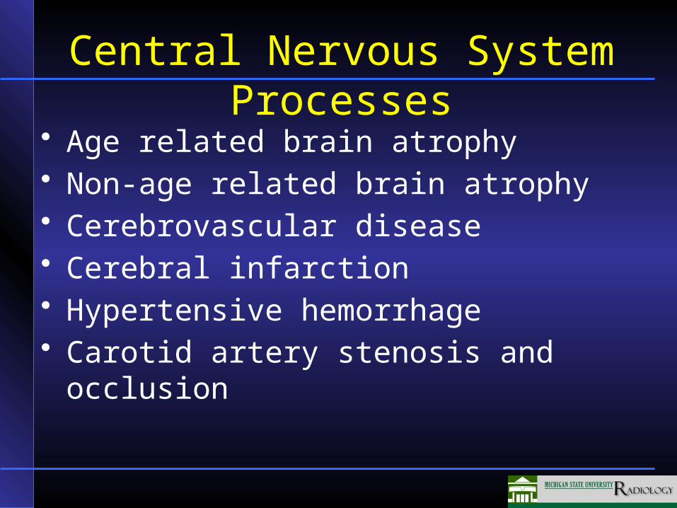
Central Nervous System Processes
• Age related brain atrophy• Non-age related brain atrophy• Cerebrovascular disease• Cerebral infarction• Hypertensive hemorrhage• Carotid artery stenosis and occlusion
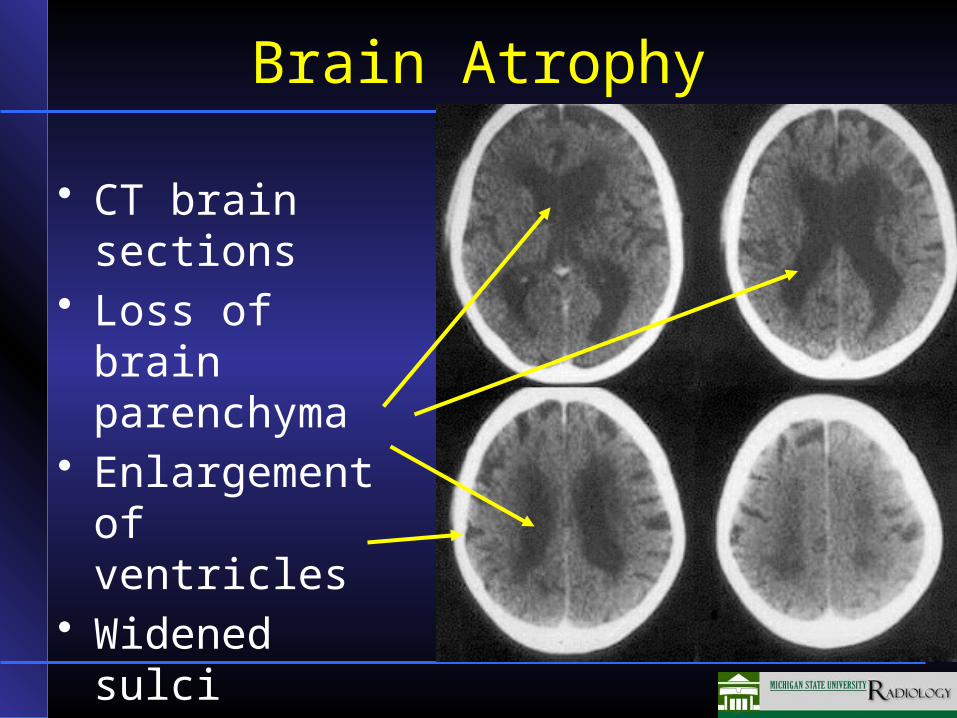
Brain Atrophy
• CT brain sections
• Loss of brain parenchyma
• Enlargement of ventricles
• Widened sulci
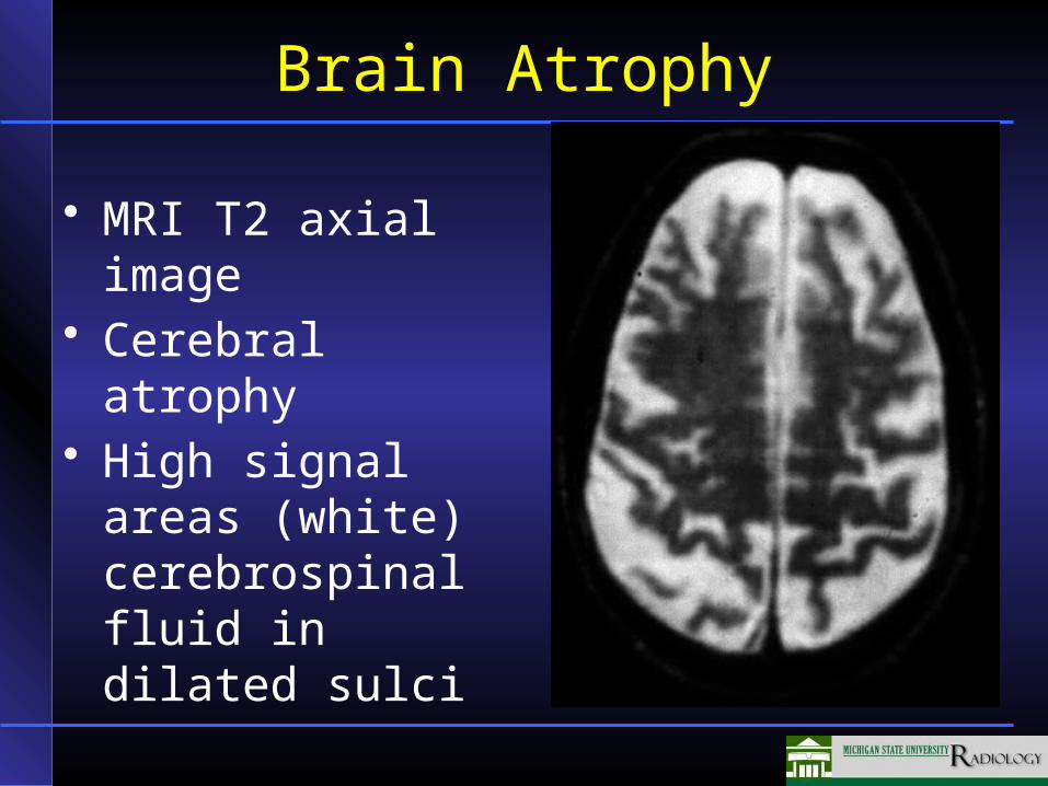
Brain Atrophy
• MRI T2 axial image
• Cerebral atrophy• High signal areas
(white) cerebrospinal fluid in dilated sulci
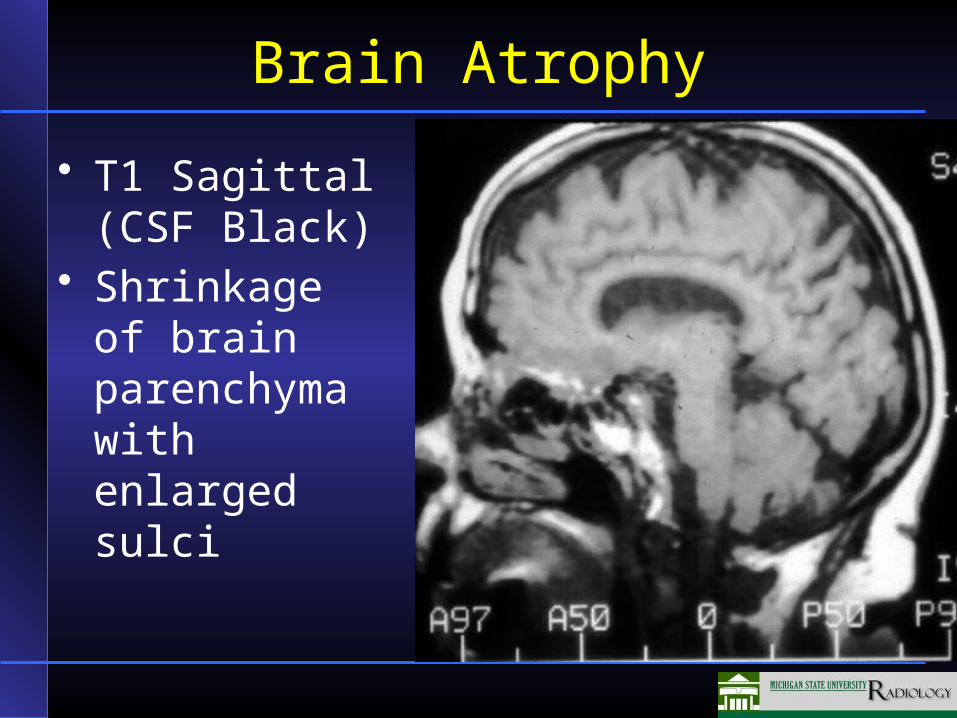
Brain Atrophy
• T1 Sagittal (CSF Black)
• Shrinkage of brain parenchyma with enlarged sulci
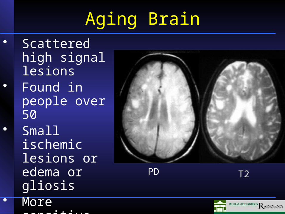
Aging Brain• Scattered high
signal lesions• Found in
people over 50• Small ischemic
lesions or edema or gliosis
• More sensitive than CT PD T2
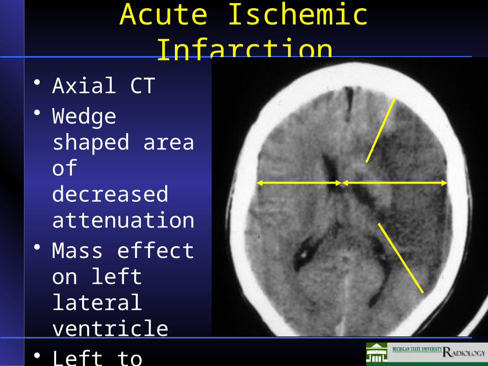
Acute Ischemic Infarction
• Axial CT• Wedge shaped
area of decreased attenuation
• Mass effect on left lateral ventricle
• Left to right shift
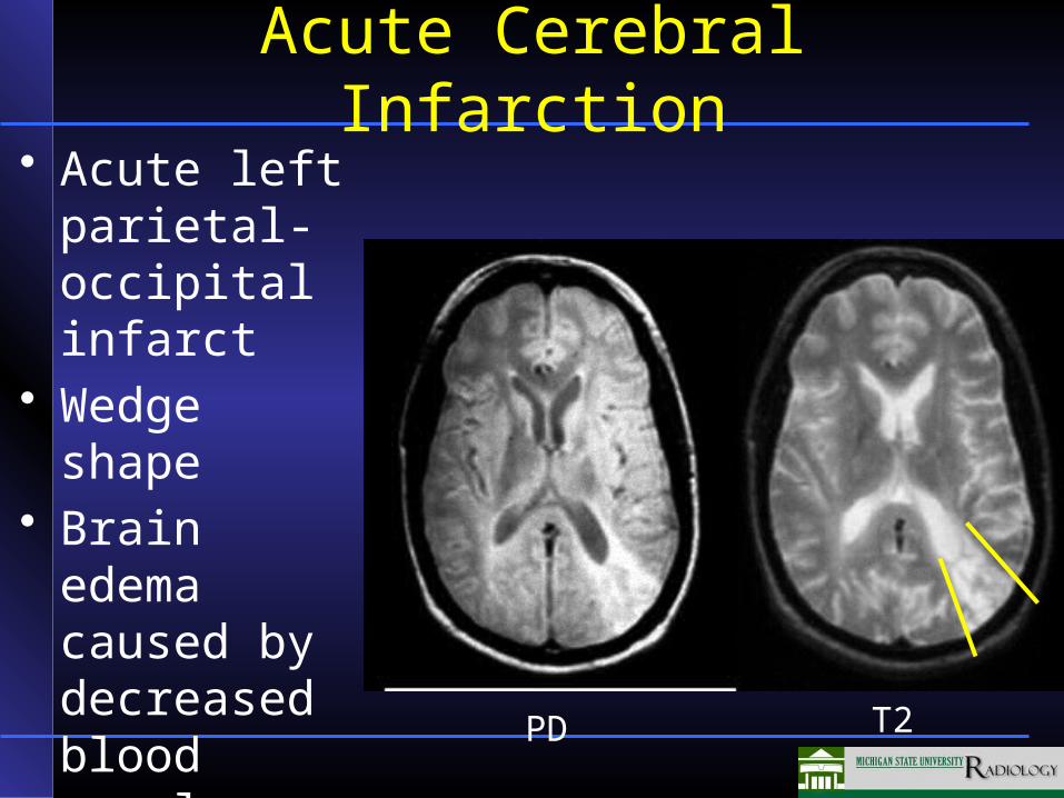
Acute Cerebral Infarction• Acute left
parietal-occipital infarct
• Wedge shape• Brain edema
caused by decreased blood supply
PD T2
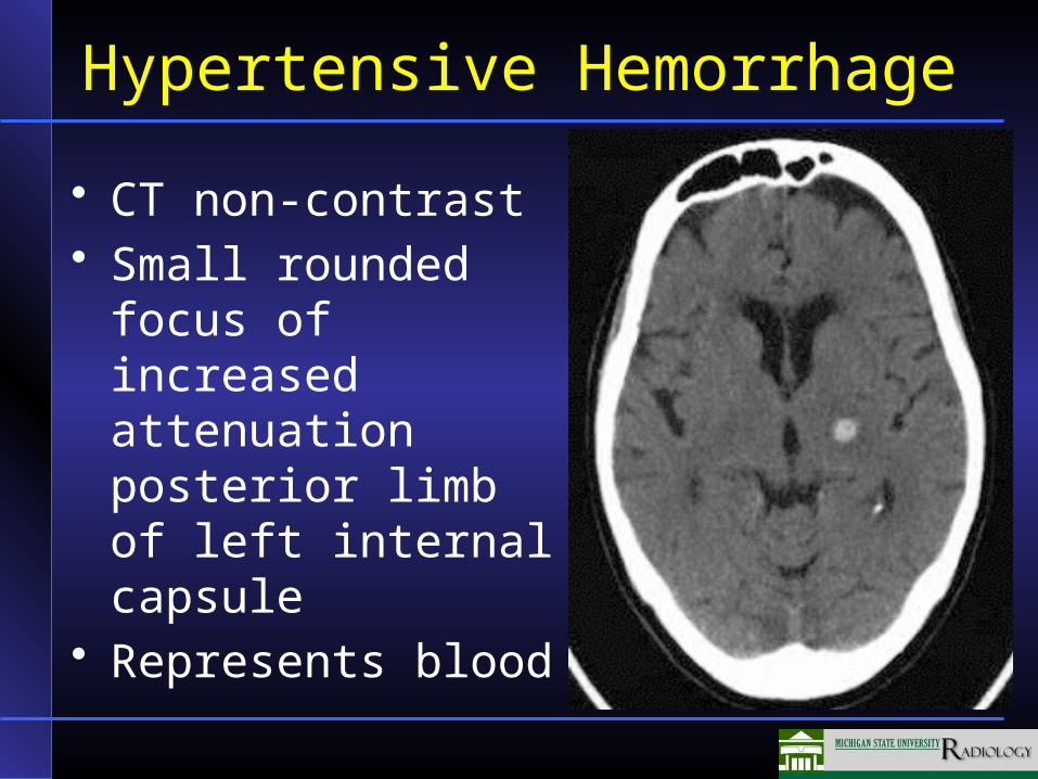
Hypertensive Hemorrhage
• CT non-contrast• Small rounded focus
of increased attenuation posterior limb of left internal capsule
• Represents blood
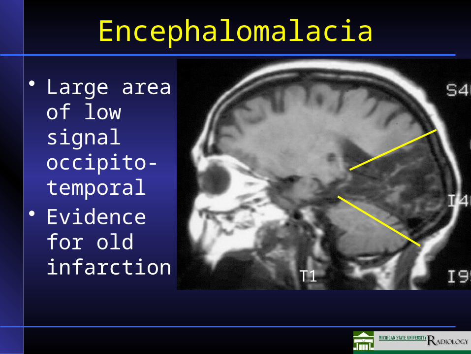
Encephalomalacia
• Large area of low signal occipito-temporal
• Evidence for old infarction
T1
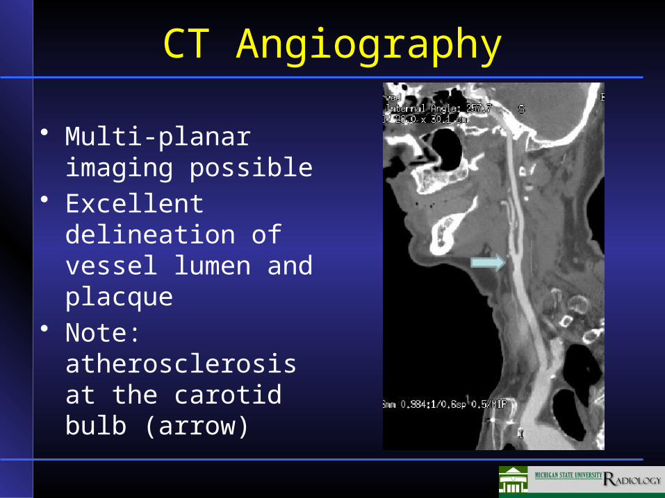
CT Angiography
• Multi-planar imaging possible
• Excellent delineation of vessel lumen and placque
• Note: atherosclerosis at the carotid bulb (arrow)
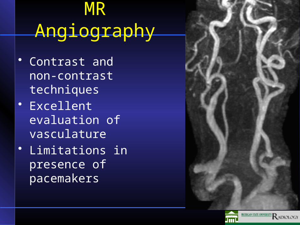
MR Angiography
• Contrast and non-contrast techniques
• Excellent evaluation of vasculature
• Limitations in presence of pacemakers
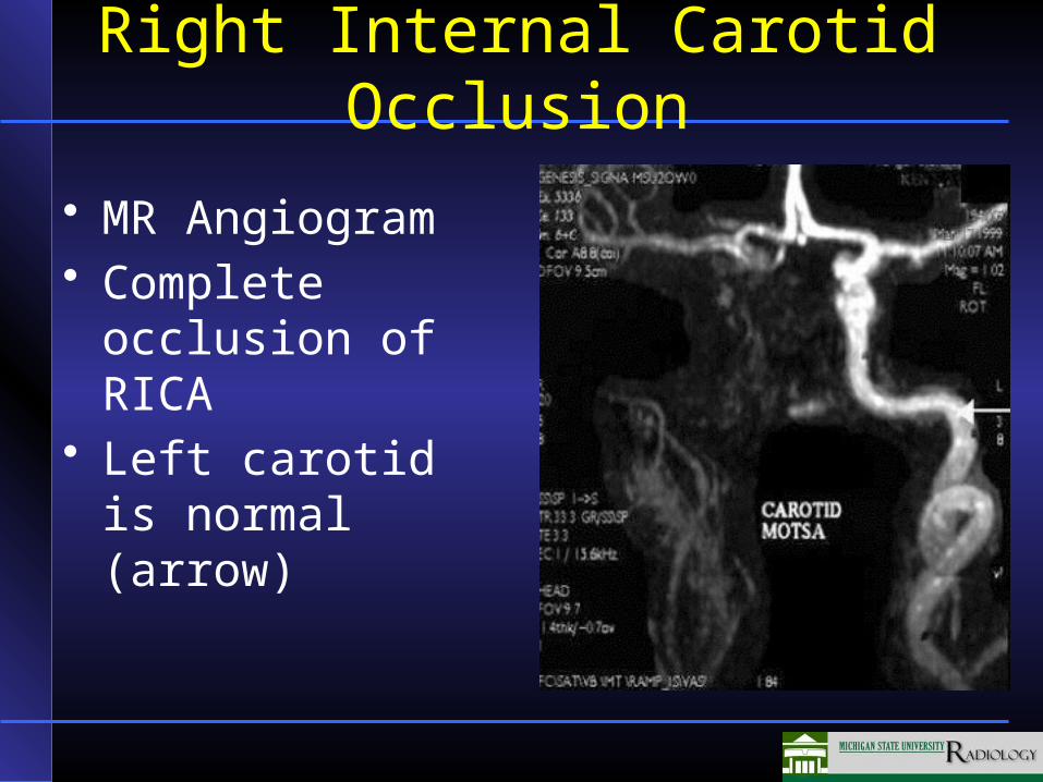
Right Internal Carotid Occlusion
• MR Angiogram• Complete
occlusion of RICA
• Left carotid is normal (arrow)
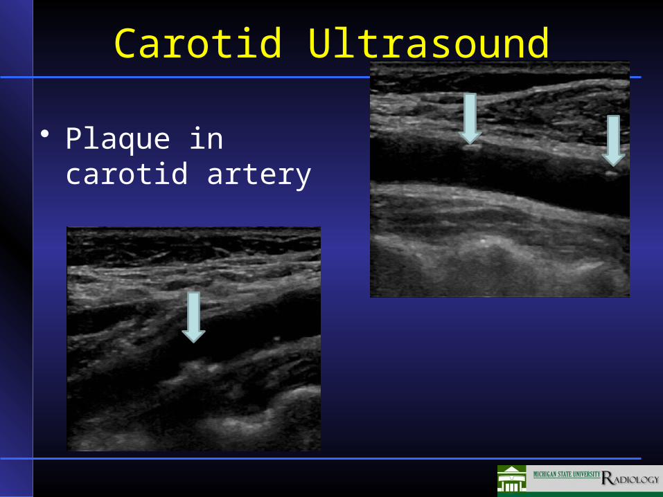
Carotid Ultrasound
• Plaque in carotid artery
Doppler Ultrasound
• Dynamic imaging study using frequency shift of returning sound
• Can detect blood flow (including very small vessels)
• Can observe abnormal blood flow such as–Left-to-right shunt in heart disorders–Turbulent flow in aneurysms and dilated
post-stenotic vessels• Can evaluate and estimate the degree of
stenosis
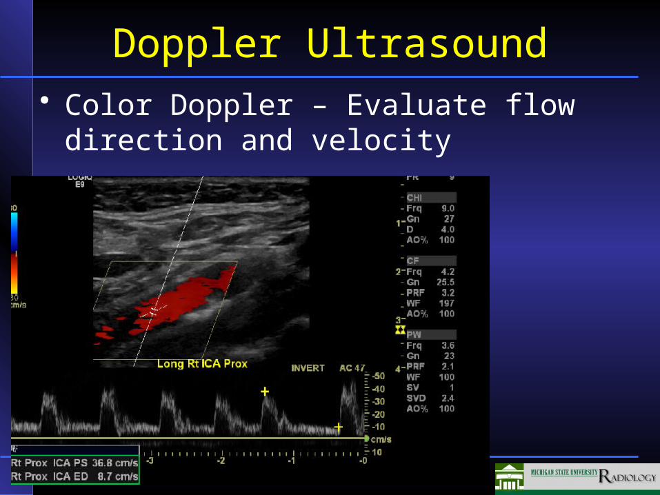
Doppler Ultrasound• Color Doppler – Evaluate flow direction
and velocity
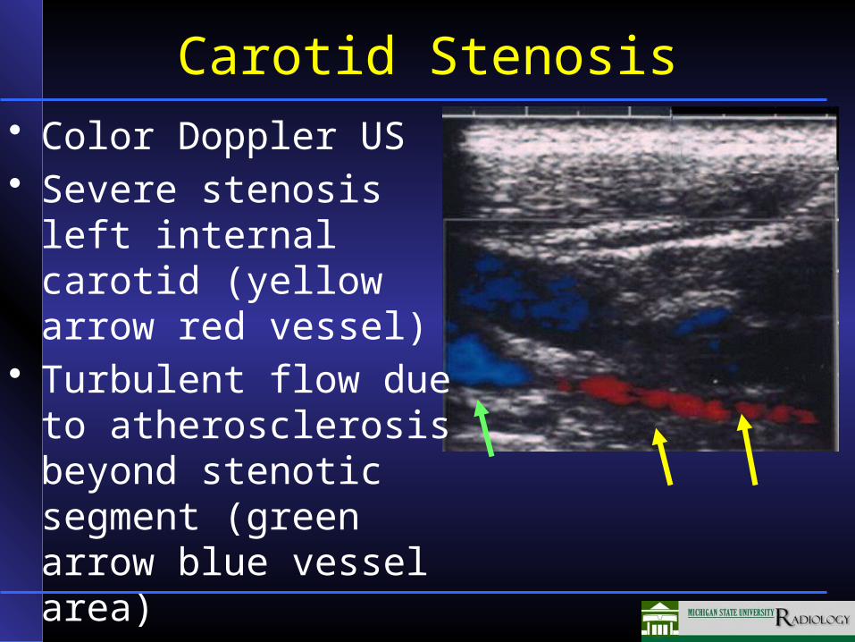
Carotid Stenosis• Color Doppler US• Severe stenosis left
internal carotid (yellow arrow red vessel)
• Turbulent flow due to atherosclerosis beyond stenotic segment (green arrow blue vessel area)
• Jugular vein (blue vessel more superficial)
Aging of the Skeletal System
• Osteoporosis• Osteoarthritis• Other arthritis
– Rheumatoid arthritis– Gout
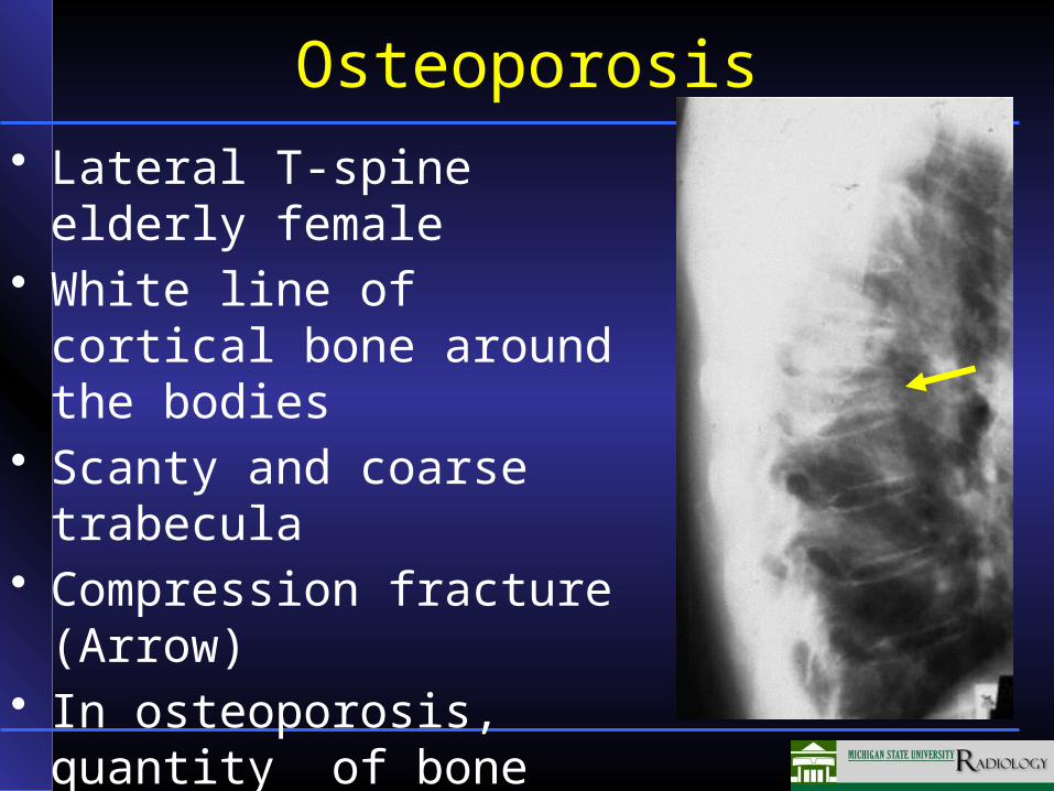
Osteoporosis• Lateral T-spine elderly
female• White line of cortical bone
around the bodies• Scanty and coarse trabecula• Compression fracture
(Arrow)• In osteoporosis, quantity of
bone decreased, composition normal
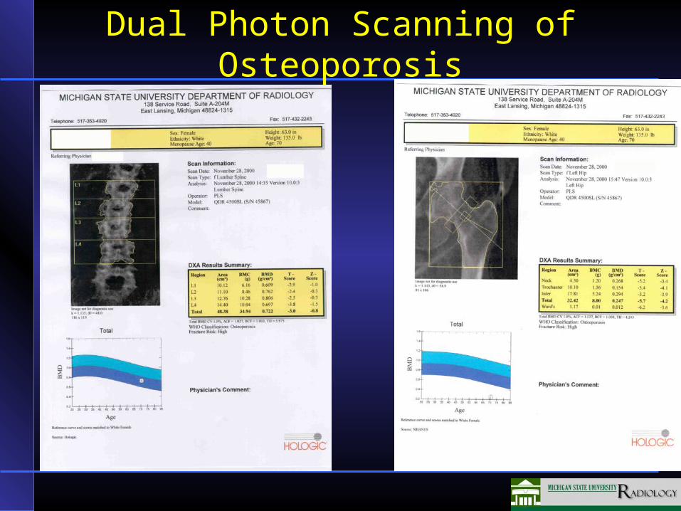
Dual Photon Scanning of Osteoporosis
Osteoporosis
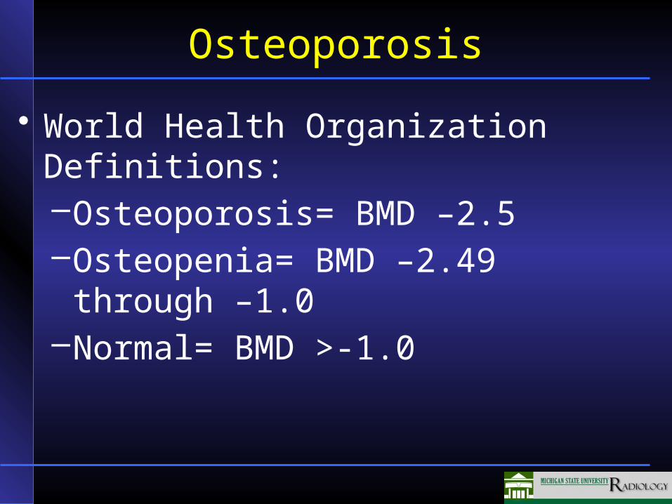
• World Health Organization Definitions:–Osteoporosis= BMD –2.5–Osteopenia= BMD –2.49 through –1.0–Normal= BMD >-1.0
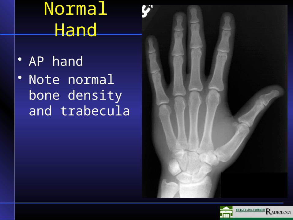
Normal Hand
• AP hand• Note normal bone
density and trabecula
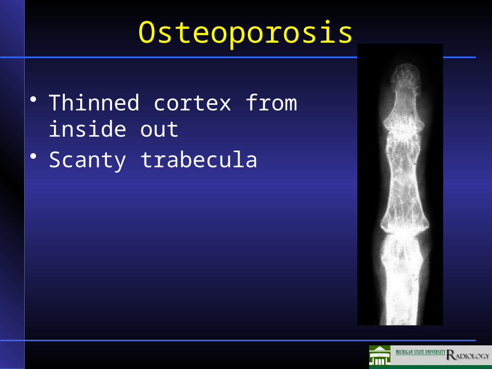
Osteoporosis
• Thinned cortex from inside out
• Scanty trabecula
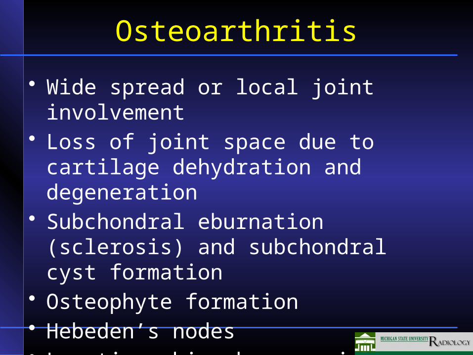
Osteoarthritis
• Wide spread or local joint involvement• Loss of joint space due to cartilage
dehydration and degeneration• Subchondral eburnation (sclerosis) and
subchondral cyst formation• Osteophyte formation• Hebeden’s nodes• Location: hip, knee, spine, hand, post-
trauma
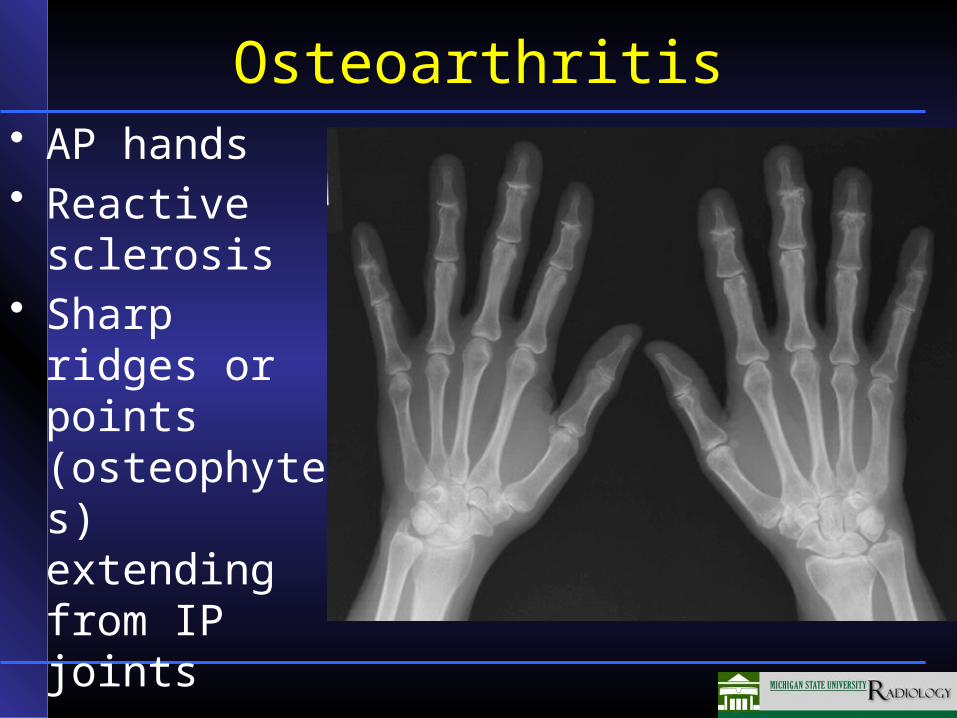
Osteoarthritis• AP hands• Reactive
sclerosis• Sharp ridges
or points (osteophytes) extending from IP joints
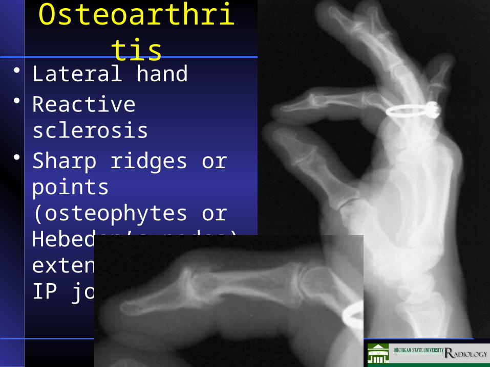
Osteoarthritis• Lateral hand• Reactive sclerosis• Sharp ridges or points
(osteophytes or Hebeden’s nodes) extending from IP joints
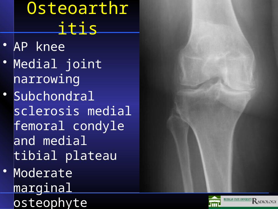
Osteoarthritis
• AP knee• Medial joint narrowing• Subchondral sclerosis
medial femoral condyle and medial tibial plateau
• Moderate marginal osteophyte medial tibial plateau
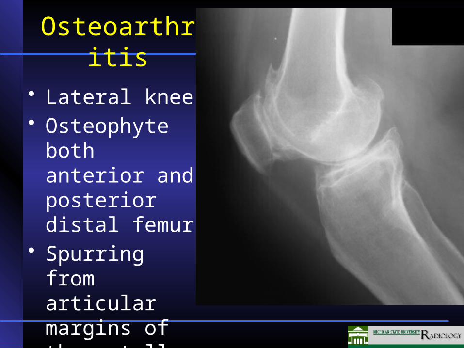
Osteoarthritis
• Lateral knee• Osteophyte
both anterior and posterior distal femur
• Spurring from articular margins of the patella
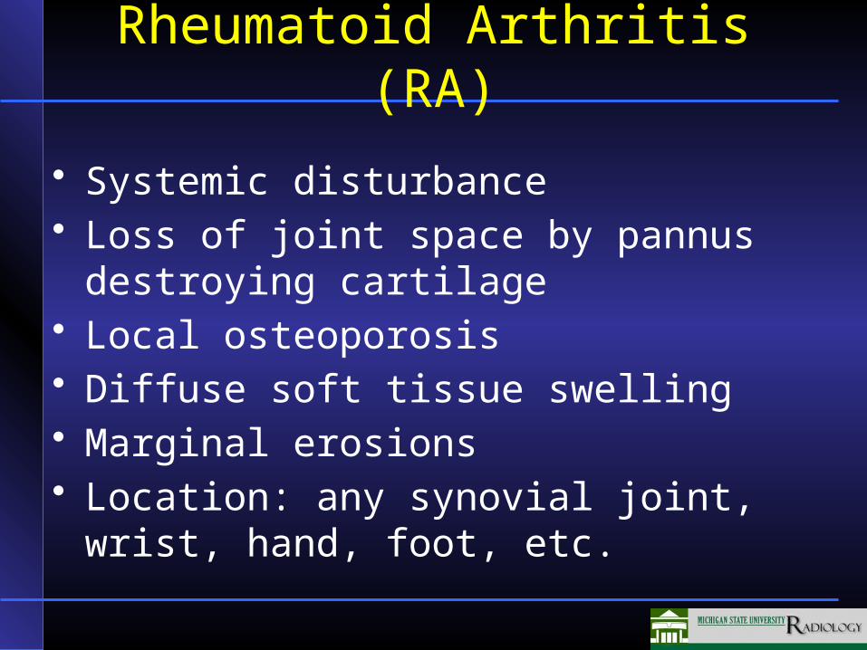
Rheumatoid Arthritis (RA)
• Systemic disturbance• Loss of joint space by pannus destroying
cartilage• Local osteoporosis• Diffuse soft tissue swelling• Marginal erosions• Location: any synovial joint, wrist, hand,
foot, etc.
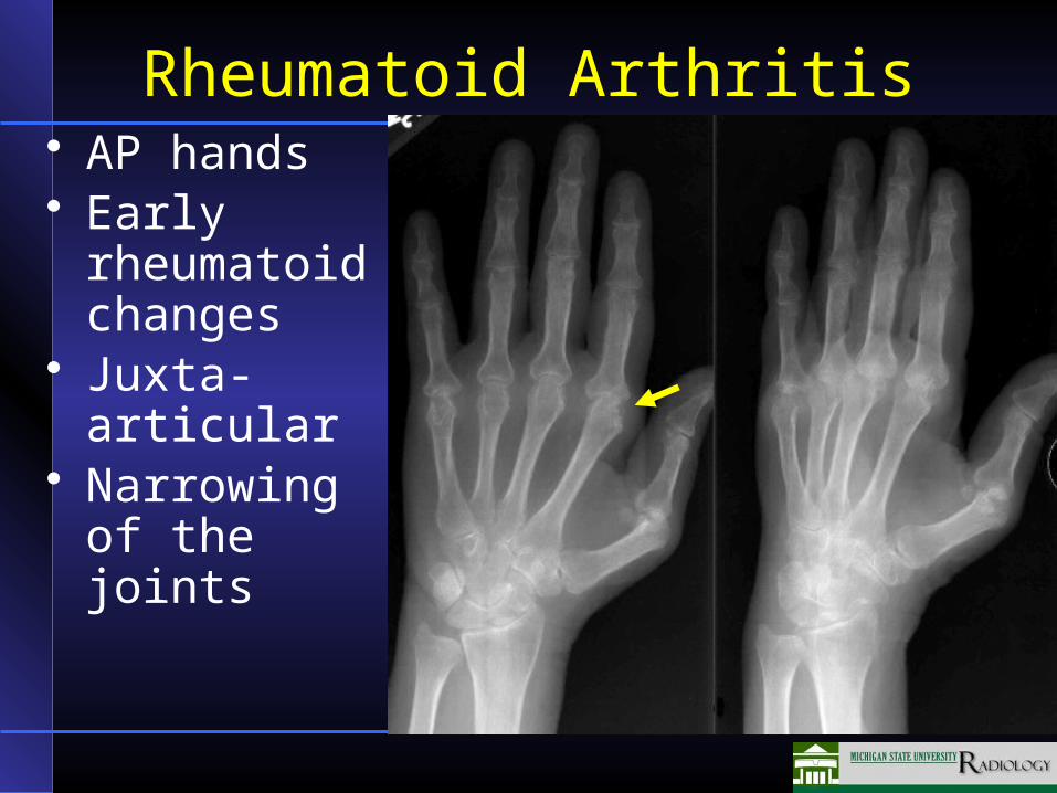
Rheumatoid Arthritis• AP hands• Early
rheumatoid changes
• Juxta-articular
• Narrowing of the joints

Rheumatoid Arthritis• Osteoporosis
most prominent at the metacarpal-carpal joints and IP joints
• Marginal erosions several joints (arrow)
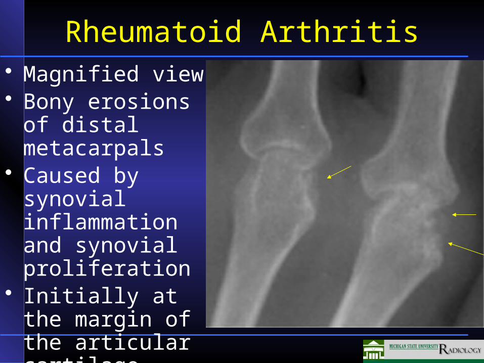
Rheumatoid Arthritis• Magnified view• Bony erosions of
distal metacarpals• Caused by
synovial inflammation and synovial proliferation
• Initially at the margin of the articular cartilage
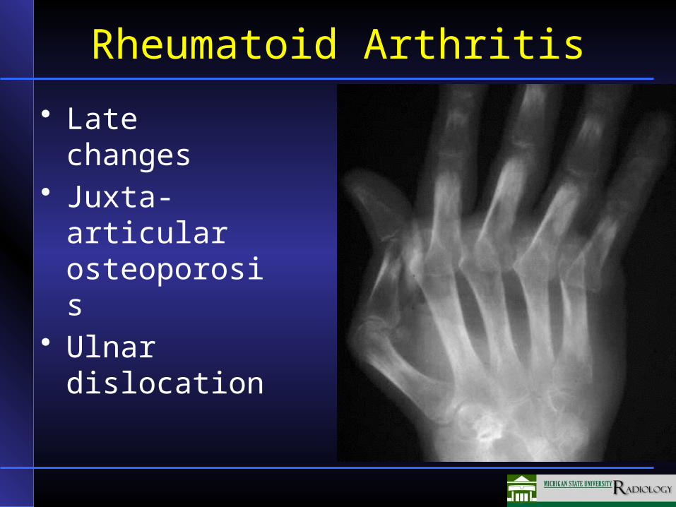
Rheumatoid Arthritis
• Late changes• Juxta-articular
osteoporosis• Ulnar
dislocation
Gout
• Metabolic disorder• Bone destruction secondary to urate
deposits• Loss of joint space• Lumpy soft tissue swelling • Primary location: first metatarsal-
phalangeal joint
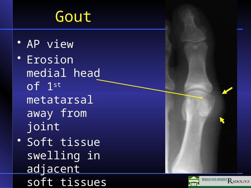
Gout
• AP view• Erosion medial
head of 1st metatarsal away from joint
• Soft tissue swelling in adjacent soft tissues
Cardiovascular System
• Coronary artery disease (Ischemic heart disease)–Cardiac Catheterization–Radionuclide study–PET Scanning–MRI
• Congestive heart failure• Future Modalities
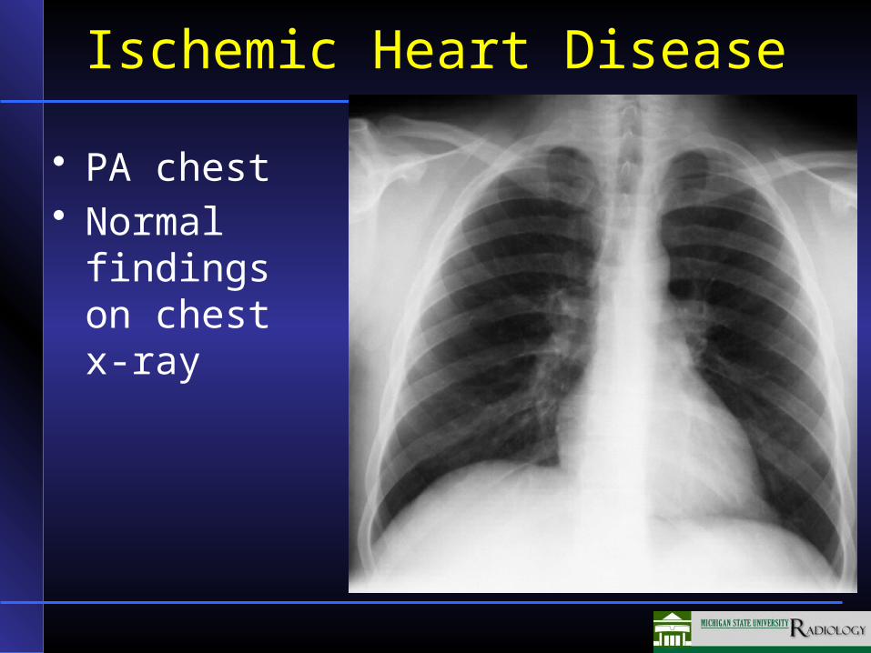
Ischemic Heart Disease
• PA chest• Normal
findings on chest x-ray
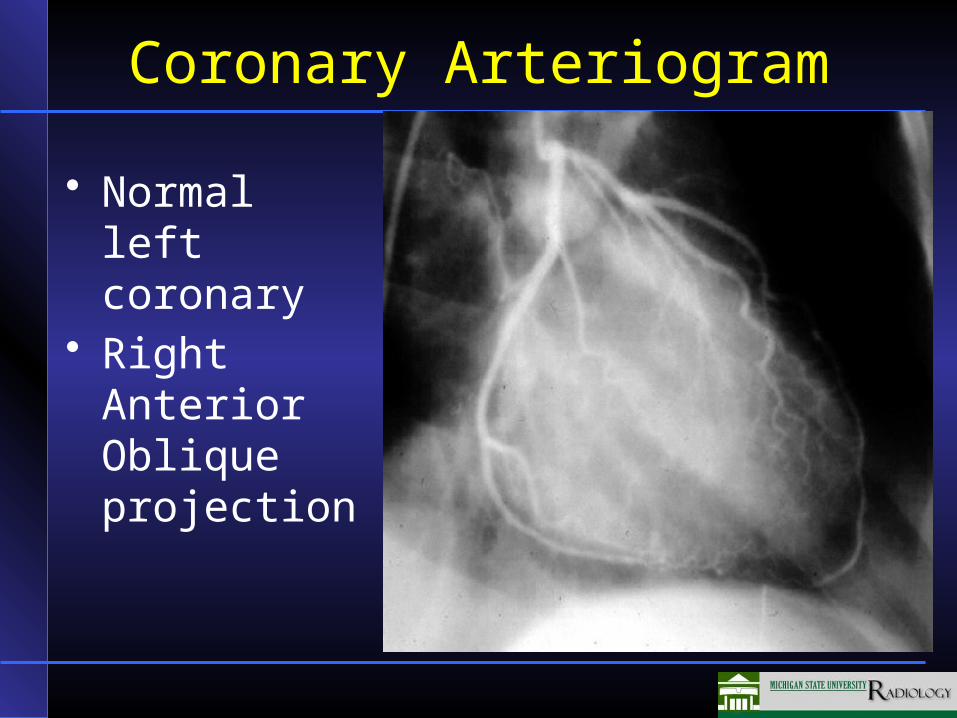
Coronary Arteriogram
• Normal left coronary
• Right Anterior Oblique projection
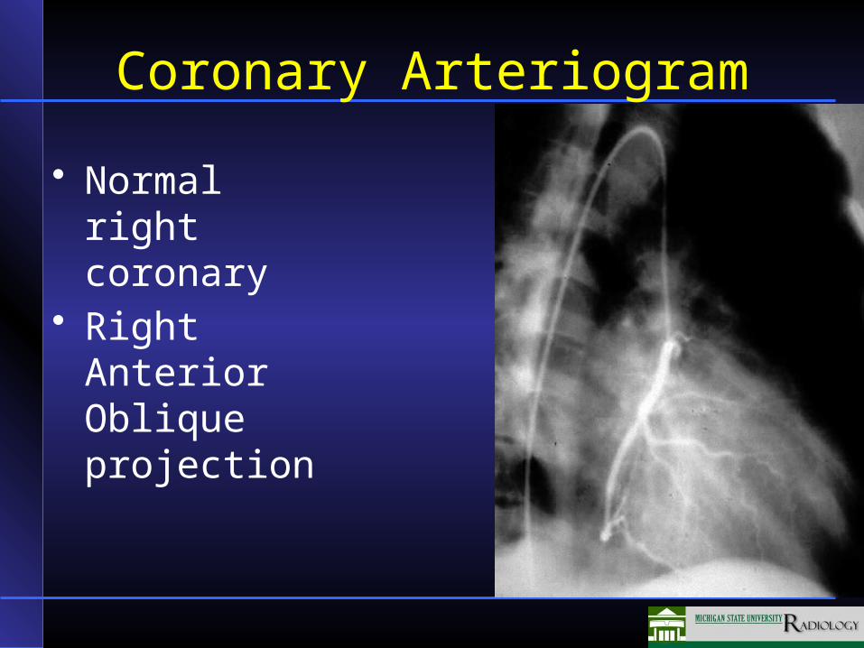
Coronary Arteriogram
• Normal right coronary
• Right Anterior Oblique projection
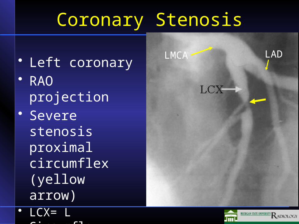
Coronary Stenosis
• Left coronary • RAO projection • Severe stenosis
proximal circumflex (yellow arrow)
• LCX= L Circumflex, LMCA = left main coronary, LAD = left anterior descending
LMCA LAD
Coronary CTA
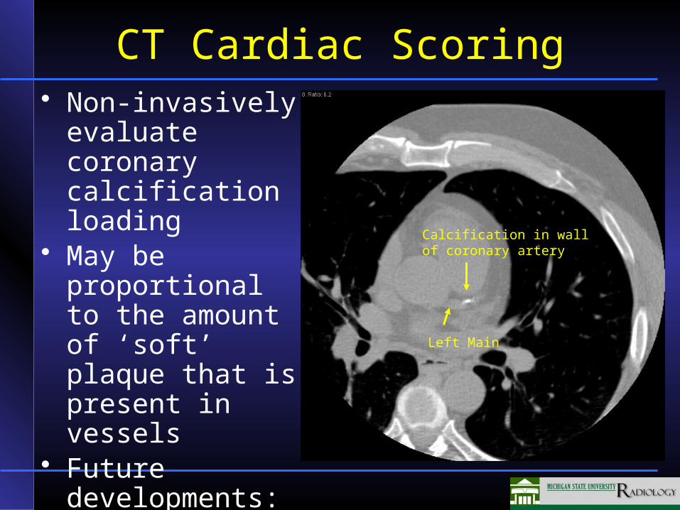
CT Cardiac Scoring• Non-invasively
evaluate coronary calcification loading
• May be proportional to the amount of ‘soft’ plaque that is present in vessels
• Future developments: CT coronary angiograms
Calcification in wall of coronary artery
Left Main
Thallium and Sestamibi Cardiac Scanning
• Radionuclide agents for scanning to identify cardiac perfusion
• Thallium functions as a Potassium analogue and demonstrates perfusion at the time of injection and shortly thereafter
• Sestamibi provides a snapshot of cardiac perfusion at the time of injection that persists
• Compare Stress and Rest images
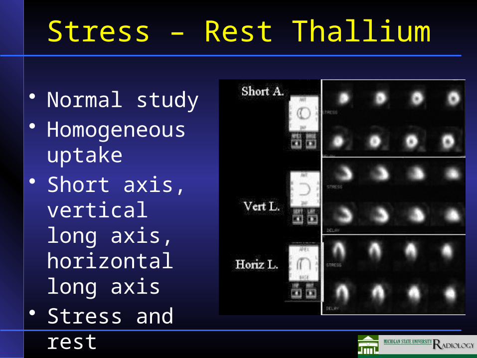
Stress – Rest Thallium
• Normal study• Homogeneous
uptake• Short axis,
vertical long axis, horizontal long axis
• Stress and rest
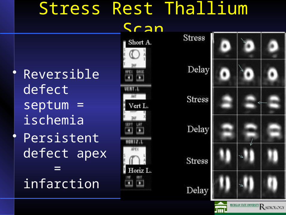
Stress Rest Thallium Scan
• Reversible defect septum = ischemia
• Persistent defect apex = infarction
Cardiac PET
• PET requirement for cyclotron access to radioactive N-13
• PET less widely available than SPECT
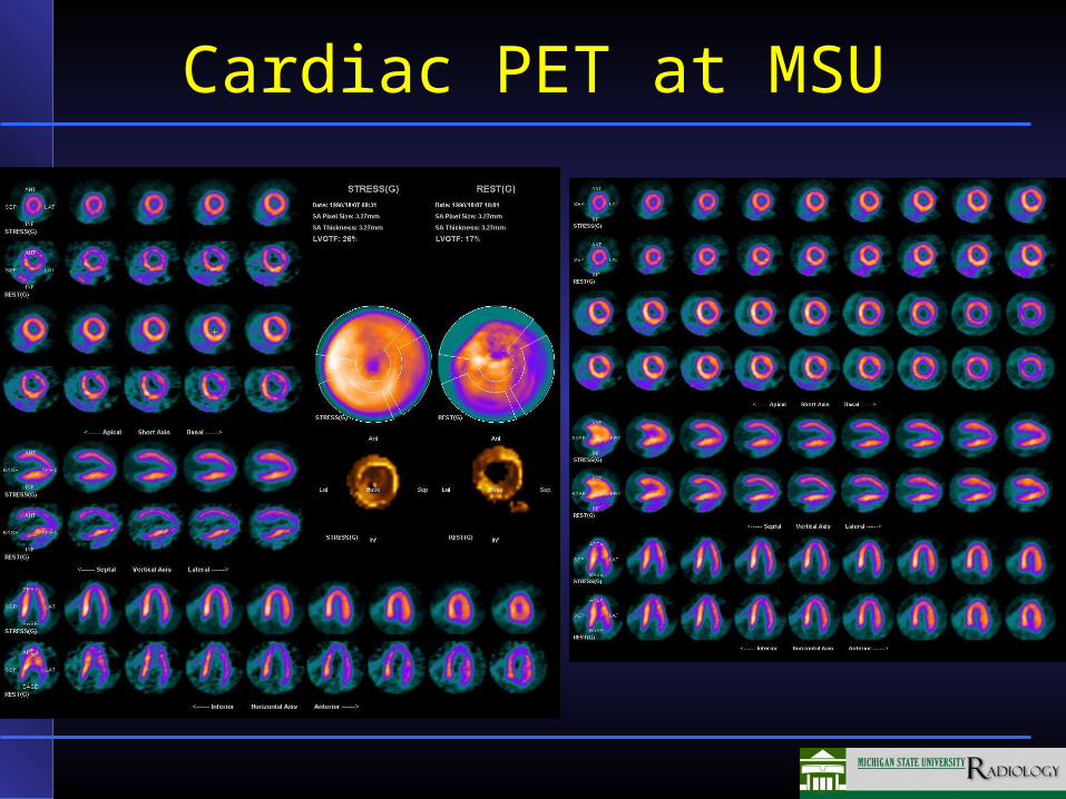
Cardiac PET at MSU
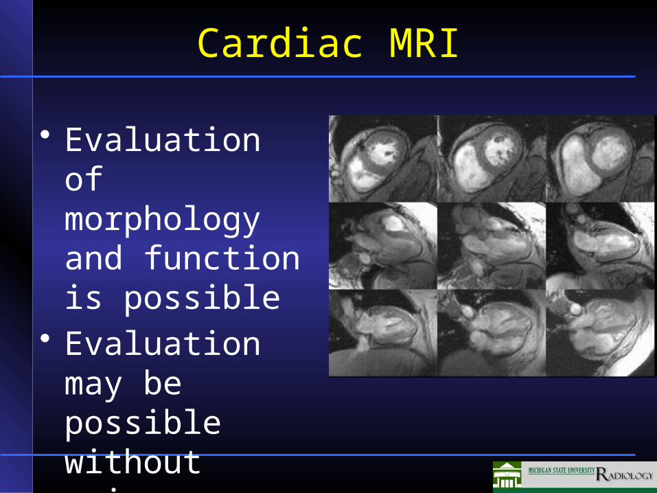
Cardiac MRI
• Evaluation of morphology and function is possible
• Evaluation may be possible without using contrast
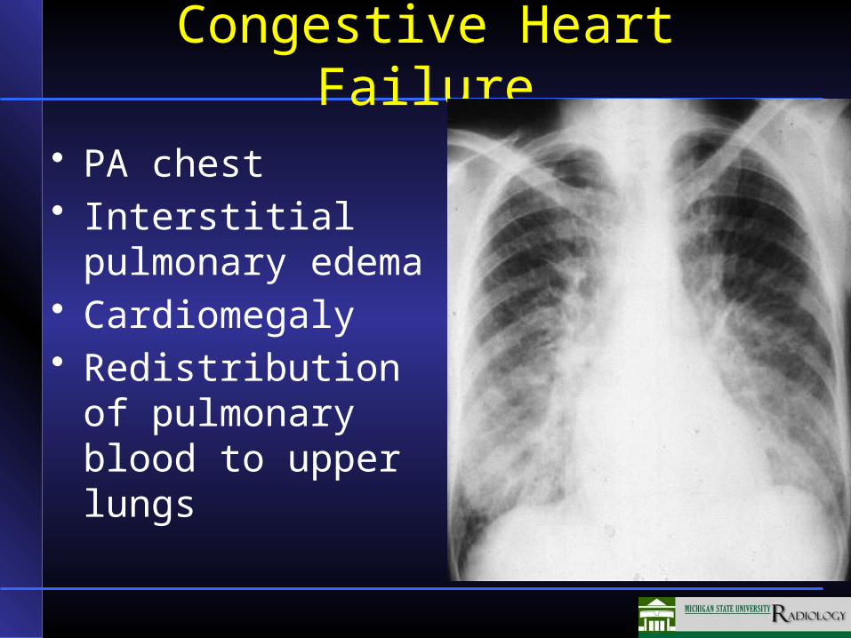
Congestive Heart Failure
• PA chest• Interstitial
pulmonary edema• Cardiomegaly• Redistribution of
pulmonary blood to upper lungs
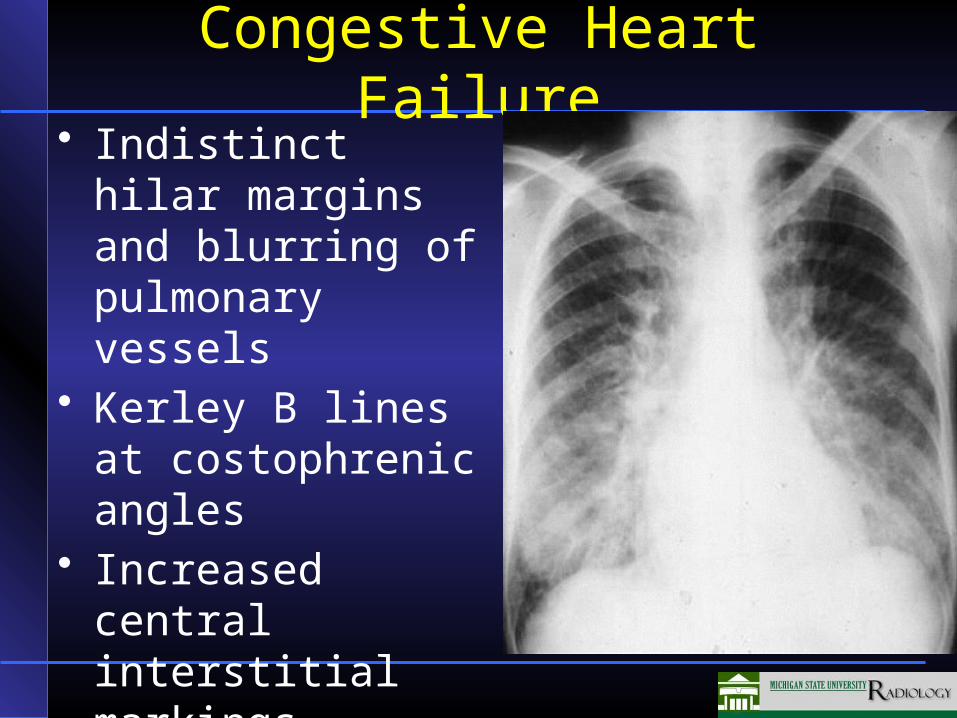
Congestive Heart Failure• Indistinct hilar
margins and blurring of pulmonary vessels
• Kerley B lines at costophrenic angles
• Increased central interstitial markings
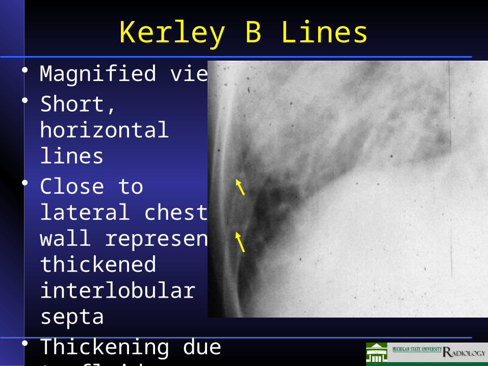
Kerley B Lines• Magnified view• Short, horizontal
lines • Close to lateral
chest wall represent thickened interlobular septa
• Thickening due to fluid
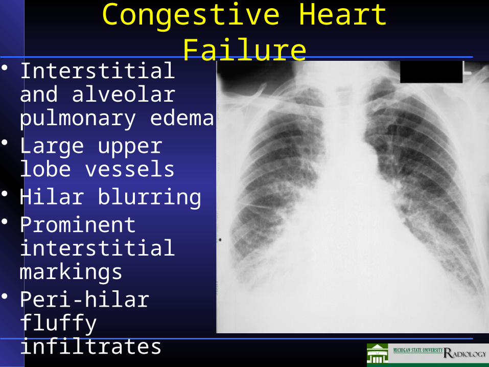
Congestive Heart Failure• Interstitial and
alveolar pulmonary edema
• Large upper lobe vessels
• Hilar blurring• Prominent interstitial
markings• Peri-hilar fluffy
infiltrates


![Evaluation of Brain Atrophy Estimation Algorithms using ...1. Introduction Brain atrophy is a common feature of many neuro-degenerative diseases such as Multiple Sclerosis (MS)[1]](https://img.pdfslide.net/doc/110x75/60366758719d65527f1d649e/evaluation-of-brain-atrophy-estimation-algorithms-using-1-introduction-brain.jpg)