Embed Size (px)
Citation preview
NeuroImage: Clinical 3 (2013) xxx–xxx
YNICL-00167; No. of pages: 9; 4C:
Contents lists available at ScienceDirect
NeuroImage: Clinical
j ourna l homepage: www.e lsev ie r .com/ locate /yn ic l
Thalamic atrophy in antero-medial and dorsal nuclei correlates withsix-month outcome after severe brain injury☆
Evan S. Lutkenhoff a, David L. McArthur b, Xue Hua c, Paul M. Thompson c,d,Paul M. Vespa b,e, Martin M. Monti a,b,⁎a Department of Psychology, University of California Los Angeles, Los Angeles, CA 90095, USAb Brain Injury Research Center (BIRC), Department of Neurosurgery, David Geffen School of Medicine at UCLA, Los Angeles, CA 90095, USAc Laboratory of Neuro Imaging (LONI), Department of Neurology, David Geffen School of Medicine at UCLA, Los Angeles, CA 90095, USAd Department of Psychiatry, University of California Los Angeles, Los Angeles, CA 90095, USAe Department of Neurology, David Geffen School of Medicine at University of California Los Angeles, Los Angeles, CA 90095, USA
☆ This is an open-access article distributed under the tAttribution-NonCommercial-No Derivative Works License,use, distribution, and reproduction in any medium, provideare credited.⁎ Corresponding author at: University of California
Psychology, Los Angeles, Ca 90095, USA. Tel.: +1 310 825E-mail address: [email protected] (M.M. Monti).
2213-1582/$ – see front matter © 2013 The Authors. Pubhttp://dx.doi.org/10.1016/j.nicl.2013.09.010
Please cite this article as: Lutkenhoff, E.S., etsevere brain injury, NeuroImage: Clinical (20
a b s t r a c t
a r t i c l e i n f oArticle history:Received 28 June 2013Received in revised form 26 September 2013Accepted 27 September 2013Available online 5 October 2013
Keywords:Traumatic brain injuryThalamusTensor brain morphometryMagnetic resonance imaging
The primary and secondary damage to neural tissue inflicted by traumatic brain injury is a leading cause of deathand disability. The secondary processes, in particular, are of great clinical interest because of their potentialsusceptibility to intervention. We address the dynamics of tissue degeneration in cortico-subcortical circuitsafter severe brain injury by assessing volume change in individual thalamic nuclei over the first six-monthspost-injury in a sample of 25 moderate to severe traumatic brain injury patients. Using tensor-basedmorphom-etry, we observed significant localized thalamic atrophy over the six-month period in antero-dorsal limbic nucleias well as in medio-dorsal association nuclei. Importantly, the degree of atrophy in these nuclei was predictive,even after controlling for full-brain volume change, of behavioral outcome at six-months post-injury. Further-more, employing a data-driven decision tree model, we found that physiological measures, namely the extentof atrophy in the anterior thalamic nucleus, were themost predictive variables of whether patients had regainedconsciousness by six-months, followed by behavioral measures. Overall, these findings suggest that the second-ary non-mechanical degenerative processes triggered by severe brain injury are still ongoing after the first weekpost-trauma and target specifically antero-medial and dorsal thalamic nuclei. This result therefore offers a poten-tial window of intervention, and a specific target region, in agreement with the view that specific cortico-thalamo-cortical circuits are crucial to the maintenance of large-scale network neural activity and thereby therestoration of cognitive function after severe brain injury.
© 2013 The Authors. Published by Elsevier Inc. All rights reserved.
1. Introduction
Traumatic brain injury (TBI) is a leading cause of death and severedisability in the United States (Coronado et al., 2011; MacKenzie,2000), and has long been projected to be a leading cause of death anddisability in the world (Murray and Lopez, 1997). The principal mecha-nisms of TBI typically include focal brain damage due to contact injurytypes resulting in contusion, laceration, intracranial hemorrhage, anddiffuse brain damage due to acceleration/deceleration injury types
erms of the Creative Commonswhich permits non-commerciald the original author and source
, Los Angeles, Department of8546; fax: +1 310 206 5895.
lished by Elsevier Inc. All rights reser
al., Thalamic atrophy in ante13), http://dx.doi.org/10.101
resulting in diffuse axonal injury (DAI) or brain swelling (Werner andEngelhard, 2007). However, the damage inflicted by TBI, as well as thepotential outcome, crucially depends not only on the local effects of theprimary insult, but also on the secondary, delayed, non-mechanical pro-cesses consequent to axonal damage and Wallerian degeneration (Hallet al., 2005). These secondary processes, which in humans can takefrom several hours to days (Christman et al., 1994), involve necroticand apoptotic death cascades in brain regions distal to the primary siteof the trauma, assumed to result from membrane failure and disruptionof ionic homeostasis inducing rapid degradation of the neuronal cyto-skeleton and its cytoplasmic constituents (Povlishock and Katz, 2005).This aspect of neuronal damage is particularly relevant in the clinical set-ting because, unlike the site of primary damage, which is typically onlysusceptible to preventive measures, secondary processes might be sus-ceptible to therapeutic intervention (Werner and Engelhard, 2007). Un-derstanding themechanisms of secondary insult, aswell as the regions ofthe brain most affected by it, might therefore offer novel strategies fortherapeutic interventions in TBI survivors.
ved.
ro-medial and dorsal nuclei correlates with six-month outcome after6/j.nicl.2013.09.010

Table 1Patient demographics, behavioral scores, acute-to-chronic percent brain volume change(PBVC) and days post-injury at which acute and chronic MRI scans (MRIA and MRIC, re-spectively) were performed.
Pat Age Sex GCS GOSe PBVC (%) MRIA MRIC
1 31 M 9 3 2.18 2 2012 33 M 12 4 −3.02 3 2383 20 M 4 4 2.34 12 1954 25 M 14 8 −0.66 1 2005 45 M 14 8 −2.3 2 1766 34 M 7 4 −1.36 4 2617 62 F 7 3 −2.12 5 1948 60 M 3 3 −4.06 2 2049 29 F 14 8 −0.74 12 13810 64 M 3 2 −7.2 23 19911 45 M 6 3 −1.2 4 43012 25 M 14 7 0.15 32 20913 60 M 14 3 −4.05 3 19414 23 M 3 8 −1.62 1 16215 17 M 3 4 −4.63 2 18116 47 M 12 6 2.06 28 18217 41 M 3 2 −1.73 1 20218 18 M 12 4 1.32 10 17619 34 M 8 3 −4.77 3 18620 25 F 8 5 −2.65 1 18421 54 M 3 5 −1.23 24 20722 40 M 3 5 0.95 2 18223 16 M 3 3 −7.93 2 19424 16 M 3 5 2.76 2 19625 27 F 3 2 −5.7 2 246
2 E.S. Lutkenhoff et al. / NeuroImage: Clinical 3 (2013) xxx–xxx
One region of the brain particularly vulnerable to secondary mecha-nisms is the bilateral thalamus, an area that due to its central location isrelatively more protected from direct impact in TBI (Fearing et al.,2008). Indeed, the thalamus has been shown in animal models to be asite of retrograde neuronal apoptosis after cortical damage (Martinet al., 2001; Natale et al., 2002) within the first few days post-trauma(Hall et al., 2005). The important role of the thalamus in TBI has alsobeen shown by reports demonstrating associations between subcorticallesion volume and 1 to 4 years post-injury performance across severalneuropsychological tests (Babikian et al., 2005). Furthermore, at thehigh end of severity, thalamic circuits are considered to play a centralrole in permanent vegetative states (Monti, 2012). Post-mortem exam-inations have shown that 80 to 100% of patients in permanent vegeta-tive state exhibit widespread thalamic damage (Adams et al., 2000).Indeed, damage to the thalamo-cortical axis has been reported to besufficient to induce a vegetative state (VS) even in the presence of intactcortico-cortical connectivity (Boly et al., 2009). Conversely, direct orindirect stimulation of the thalamus has been associatedwith functionalimprovements in both moderate (Kang et al., 2012) and severe (Schiffet al., 2007; Yamamoto et al., 2013) TBI survivors.
In what follows, we employ tensor-based morphometry (TBM;Thompson et al., 2000) to assess the dynamics of structural changewithin individual thalamic nuclei occurring between the acute andchronic phases of moderate to severe TBI. Tensor-based morphometry,a technique related to deformation-based morphometry (Ashburneret al., 1998), employs deformations obtained from nonlinear registra-tion of two brain images (e.g., an individual anatomical image to afollow-up image, or to an anatomical template) to infer 3D patterns ofstatistical differences in brain volume or shape between two images(Ashburner and Friston, 2000; Ashburner et al., 1998; Chung et al.,2001, 2003; Wang et al., 2003). In the past 15 years, this techniquehas been successfully applied to measure structural neuroanatomicalchanges over time, including mapping of growth patterns in the devel-oping brain (Chung et al., 2001; Thompson et al., 2000), degenerativerates in Alzheimer's disease and other forms of dementia (Fox andFreeborough, 1997; Fox et al., 1996, 1999, 2000, 2001; Freeboroughand Fox, 1997; Freeborough et al., 1996; O'Brien et al., 2001;Studholme et al., 2001), as well as tumor growth (Lemieux et al.,1998) and multiple sclerosis lesions (Ge et al., 2000; Rey et al., 2002).As detailed below, we will show that the degree of structural change(namely, atrophy) within specific dorsal and anterior sections of thethalamus predicts 6-month change in behavioral outcome measures.Our approach differs from virtually all previous studies in two keyaspects. First, our longitudinal design allows us to look at the dynamicsof thalamic structural change in the first 6months post-TBI. Second, wefocus on individual nuclei within the thalamus, as opposed to consider-ing it as a unitary structure, a degree of resolution that is very importantconsidering that specific regions of the thalamus may play a key role insevere brain injury (Schiff, 2010).
2. Materials and methods
2.1. Patient population
A convenience sample of 25 acute TBI patients (21 male, 4 female;mean age 35.6 years, SD = 15.25) was enrolled in the study (seeTable 1 for individual patient demographic, behavioral scores andacute-to-chronic percent brain volume change). Patientswere recruitedduring a time-span of 30 months as part of the UCLA Brain InjuryResearch Center (BIRC) activity. The main inclusion criterion was anadmission Glasgow Coma Scale (GCS; Teasdale and Jennett, 1974)score of ≤8, or an admission GCS of 9–14 with computerized tomogra-phy (CT) brain scans demonstrating intracranial bleeding. The main ex-clusion criteria were GCS N14 with a non-significant head CT, history ofneurologic disease or TBI, brain death, and unsuitability to enter theMRIenvironment (e.g., due to any nonMRI-safe implant). Informed written
Please cite this article as: Lutkenhoff, E.S., et al., Thalamic atrophy in antesevere brain injury, NeuroImage: Clinical (2013), http://dx.doi.org/10.101
assent was obtained from the patient's legal representative. The studywas approved by theUCLA institutional review board. Informed consentwas obtained from surrogates as per state regulations.
2.2. Experimental design
Each patient underwent two structural MRI scans, one shortly afterthe ictal event (henceforth, “acute”), and one at an approximate6 month interval (henceforth, “chronic” or “follow-up,” interchange-ably). The specific day on which the acute scan occurred dependedupon the decision of medical personnel blinded to the aims of thisstudy, and was mainly driven by the patient being stable enough to un-dergo the session, and general patient safety concerns. (The day of thechronic scan was mainly driven by contingent factors such as patienttransportation to the hospital, patient availability, and scheduling.) Inaddition, patients also underwent two behavioral assessments. An ini-tial GCS assessment was conducted acutely in the emergency room(post-resuscitation), and a Glasgow Outcome Scale extended (GOSeWilson, 1998) assessment was conducted at follow-up. Acute data ac-quisition occurred between 1 and 32 days post-injury, with 72% of thepatients being scanned within 5 days and the remaining 28% beingscanned between 10 and 32 days. The median acute scan occurred onday 3. Follow-up scanning occurred between 138 and 430 days post-injury, with 72% of the patients being scanned before 202 days, andthe remaining between 204 and 430days. The median chronic data ac-quisition occurred at the 195-daymark. The temporal distance betweenacute and chronic scans ranged between 126 and 426days, with 76% ofthe patients being re-scanned within 200days from the acute visit, andonly the remaining 6 being scanned at a longer interval (between 201and 426days). The median inter-scan interval was 183days.
2.3. MRI data acquisition
T1-weightedMP-RAGE images (TR=2250ms, TE=2.99ms, FA=9°,FOV=256×240×160mm, resolution=1mm3 isovoxel)were acquiredon a 3T Siemens Tim Trio scanner at the Ronald ReaganMedical Centerat UCLA.
ro-medial and dorsal nuclei correlates with six-month outcome after6/j.nicl.2013.09.010
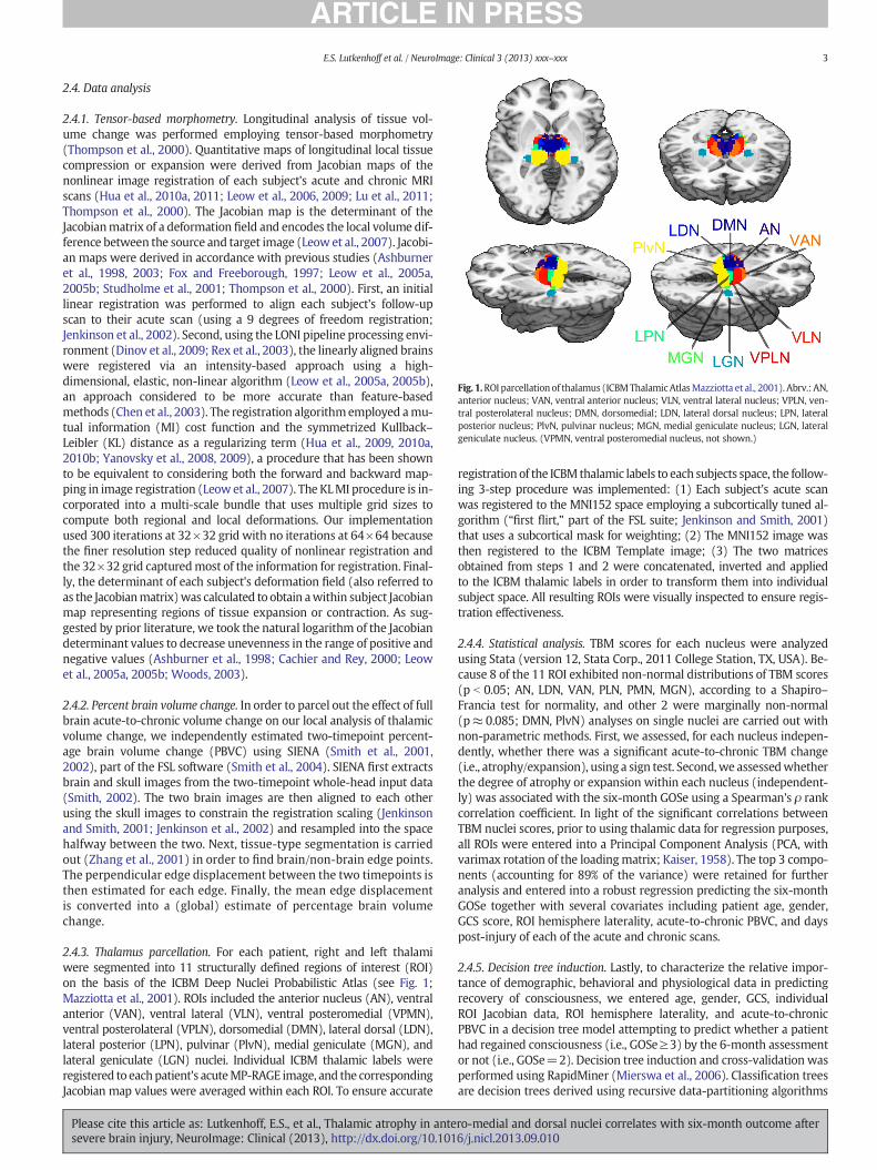
Fig. 1.ROI parcellation of thalamus (ICBMThalamicAtlasMazziotta et al., 2001). Abrv.: AN,anterior nucleus; VAN, ventral anterior nucleus; VLN, ventral lateral nucleus; VPLN, ven-tral posterolateral nucleus; DMN, dorsomedial; LDN, lateral dorsal nucleus; LPN, lateralposterior nucleus; PlvN, pulvinar nucleus; MGN, medial geniculate nucleus; LGN, lateralgeniculate nucleus. (VPMN, ventral posteromedial nucleus, not shown.)
3E.S. Lutkenhoff et al. / NeuroImage: Clinical 3 (2013) xxx–xxx
2.4. Data analysis
2.4.1. Tensor-based morphometry. Longitudinal analysis of tissue vol-ume change was performed employing tensor-based morphometry(Thompson et al., 2000). Quantitative maps of longitudinal local tissuecompression or expansion were derived from Jacobian maps of thenonlinear image registration of each subject's acute and chronic MRIscans (Hua et al., 2010a, 2011; Leow et al., 2006, 2009; Lu et al., 2011;Thompson et al., 2000). The Jacobian map is the determinant of theJacobianmatrix of a deformation field and encodes the local volume dif-ference between the source and target image (Leow et al., 2007). Jacobi-an maps were derived in accordance with previous studies (Ashburneret al., 1998, 2003; Fox and Freeborough, 1997; Leow et al., 2005a,2005b; Studholme et al., 2001; Thompson et al., 2000). First, an initiallinear registration was performed to align each subject's follow-upscan to their acute scan (using a 9 degrees of freedom registration;Jenkinson et al., 2002). Second, using the LONI pipeline processing envi-ronment (Dinov et al., 2009; Rex et al., 2003), the linearly aligned brainswere registered via an intensity-based approach using a high-dimensional, elastic, non-linear algorithm (Leow et al., 2005a, 2005b),an approach considered to be more accurate than feature-basedmethods (Chen et al., 2003). The registration algorithmemployed amu-tual information (MI) cost function and the symmetrized Kullback–Leibler (KL) distance as a regularizing term (Hua et al., 2009, 2010a,2010b; Yanovsky et al., 2008, 2009), a procedure that has been shownto be equivalent to considering both the forward and backward map-ping in image registration (Leow et al., 2007). The KLMI procedure is in-corporated into a multi-scale bundle that uses multiple grid sizes tocompute both regional and local deformations. Our implementationused 300 iterations at 32×32 grid with no iterations at 64×64 becausethe finer resolution step reduced quality of nonlinear registration andthe 32×32 grid capturedmost of the information for registration. Final-ly, the determinant of each subject's deformation field (also referred toas the Jacobianmatrix)was calculated to obtain awithin subject Jacobianmap representing regions of tissue expansion or contraction. As sug-gested by prior literature, we took the natural logarithm of the Jacobiandeterminant values to decrease unevenness in the range of positive andnegative values (Ashburner et al., 1998; Cachier and Rey, 2000; Leowet al., 2005a, 2005b; Woods, 2003).
2.4.2. Percent brain volume change. In order to parcel out the effect of fullbrain acute-to-chronic volume change on our local analysis of thalamicvolume change, we independently estimated two-timepoint percent-age brain volume change (PBVC) using SIENA (Smith et al., 2001,2002), part of the FSL software (Smith et al., 2004). SIENA first extractsbrain and skull images from the two-timepoint whole-head input data(Smith, 2002). The two brain images are then aligned to each otherusing the skull images to constrain the registration scaling (Jenkinsonand Smith, 2001; Jenkinson et al., 2002) and resampled into the spacehalfway between the two. Next, tissue-type segmentation is carriedout (Zhang et al., 2001) in order to find brain/non-brain edge points.The perpendicular edge displacement between the two timepoints isthen estimated for each edge. Finally, the mean edge displacementis converted into a (global) estimate of percentage brain volumechange.
2.4.3. Thalamus parcellation. For each patient, right and left thalamiwere segmented into 11 structurally defined regions of interest (ROI)on the basis of the ICBM Deep Nuclei Probabilistic Atlas (see Fig. 1;Mazziotta et al., 2001). ROIs included the anterior nucleus (AN), ventralanterior (VAN), ventral lateral (VLN), ventral posteromedial (VPMN),ventral posterolateral (VPLN), dorsomedial (DMN), lateral dorsal (LDN),lateral posterior (LPN), pulvinar (PlvN), medial geniculate (MGN), andlateral geniculate (LGN) nuclei. Individual ICBM thalamic labels wereregistered to eachpatient's acuteMP-RAGE image, and the correspondingJacobian map values were averaged within each ROI. To ensure accurate
Please cite this article as: Lutkenhoff, E.S., et al., Thalamic atrophy in antesevere brain injury, NeuroImage: Clinical (2013), http://dx.doi.org/10.101
registration of the ICBM thalamic labels to each subjects space, the follow-ing 3-step procedure was implemented: (1) Each subject's acute scanwas registered to the MNI152 space employing a subcortically tuned al-gorithm (“first flirt,” part of the FSL suite; Jenkinson and Smith, 2001)that uses a subcortical mask for weighting; (2) The MNI152 image wasthen registered to the ICBM Template image; (3) The two matricesobtained from steps 1 and 2 were concatenated, inverted and appliedto the ICBM thalamic labels in order to transform them into individualsubject space. All resulting ROIs were visually inspected to ensure regis-tration effectiveness.
2.4.4. Statistical analysis. TBM scores for each nucleus were analyzedusing Stata (version 12, Stata Corp., 2011 College Station, TX, USA). Be-cause 8 of the 11 ROI exhibited non-normal distributions of TBM scores(p b 0.05; AN, LDN, VAN, PLN, PMN, MGN), according to a Shapiro–Francia test for normality, and other 2 were marginally non-normal(p≈ 0.085; DMN, PlvN) analyses on single nuclei are carried out withnon-parametric methods. First, we assessed, for each nucleus indepen-dently, whether there was a significant acute-to-chronic TBM change(i.e., atrophy/expansion), using a sign test. Second,we assessedwhetherthe degree of atrophy or expansion within each nucleus (independent-ly) was associated with the six-month GOSe using a Spearman's ρ rankcorrelation coefficient. In light of the significant correlations betweenTBM nuclei scores, prior to using thalamic data for regression purposes,all ROIs were entered into a Principal Component Analysis (PCA, withvarimax rotation of the loadingmatrix; Kaiser, 1958). The top 3 compo-nents (accounting for 89% of the variance) were retained for furtheranalysis and entered into a robust regression predicting the six-monthGOSe together with several covariates including patient age, gender,GCS score, ROI hemisphere laterality, acute-to-chronic PBVC, and dayspost-injury of each of the acute and chronic scans.
2.4.5. Decision tree induction. Lastly, to characterize the relative impor-tance of demographic, behavioral and physiological data in predictingrecovery of consciousness, we entered age, gender, GCS, individualROI Jacobian data, ROI hemisphere laterality, and acute-to-chronicPBVC in a decision tree model attempting to predict whether a patienthad regained consciousness (i.e., GOSe≥3) by the 6-month assessmentor not (i.e., GOSe=2). Decision tree induction and cross-validation wasperformed using RapidMiner (Mierswa et al., 2006). Classification treesare decision trees derived using recursive data-partitioning algorithms
ro-medial and dorsal nuclei correlates with six-month outcome after6/j.nicl.2013.09.010

Table 2Description of each nucleus' average (M), standard deviation (SD),minimumandmaximumacute-to-chronic change. p-Value refers to a sign test assessing whether the average six-month change is significantly different from zero. ‘**’ indicates p≤0.05 Bonferroni corrected.
Nucleus M SD Min Max p-Value
AN −15.26 19.69 −58.94 20.34 b0.001**LDN −13.86 15.73 −62.45 10.33 b0.001**DMN −6.35 11.14 −34.48 11.72 0.015VAN −4.27 10.97 −34.85 18.18 0.015VLN 0.77 9.16 −25.88 24.95 n.s.VPMN 2.97 6.41 −9.28 26.34 0.033VPLN 3.76 7.90 −13.01 31.34 b0.001**LPN −3.64 10.01 −37.68 21.56 0.033LGN 2.00 11.18 −24.93 33.68 n.s.MGN 5.31 8.91 −14.24 38.57 0.003**PlvN −2.24 8.54 −28.74 19.32 n.s.
4 E.S. Lutkenhoff et al. / NeuroImage: Clinical 3 (2013) xxx–xxx
that classify eachobservation into oneof the class labels for the outcome(in our design: VS or better; cf. Shmueli et al., 2010). Binary classifica-tion trees are induced “top–down” by starting with all the data andpartitioning it into two subsets (a left and a right daughter node). Inturn, each sub-partition (or daughter node) is further split into leftand right daughters. The process is repeated recursively until the treecannot be partitioned further due to lack of data or some stopping crite-rion is reached (Ishwaran and Rao, 2009). The type of partitioning con-sidered here is univariate in the sense that splits are performed alongone unique dimension (i.e., variable). The dimension along which tosplit the data, at each iteration, is chosen on the basis of so-called “impu-rity” algorithms,which is to say, algorithms thatmeasure how homoge-neous the induced partitions are with respect to the outcome variable.The more homogeneous the observations within each branch of thetree, with respect to the outcome variable, the purer it is, hence the bet-ter the partitioning. Here, the variable along which to partition the dataat each iteration was thus induced on the basis of an information gaincriterion (Quinlan, 1987), an algorithm that uses the concept of entropyto quantify impurity (Rokach andMaimon, 2005). In this setting, entro-py can be thought of as the degree of uncertainty concerningwhether agiven patient falls in the six-month GOSe group. The un-partitioneddataset has the highest level of entropy, since all patients, whetherthey are VS or better, fall in the same group. The perfectly partitioneddataset, in which all patients with a GOSe of 2 are in one branch of thetree and all other patients are in a different branch, would have theleast entropy. At each split, then, information gain is the change (possi-bly the reduction) in entropy afforded by partitioning the observationsinto two sub-groups determined by one of the attributes (e.g., GCSscores, or degree of atrophy in an ROI). To avoid over-fitting and toimprove generality of the result, we employed pruning and pre-pruning in order to exclude splits with low or non-significant infor-mation gain (Fürnkranz, 1997). Assessment of the accuracy of the in-duced tree was performed using an exhaustive leave-one-out k-foldcross-validation, as implemented in Rapid Miner. In this approach,the decision tree is built on the basis of n− 1 observations, and itsaccuracy is assessed by testing the induced tree on the nth observation(the one that had been left out). This procedure is repeated by holdingout once, in turn, each observation.
3. Results
3.1. Behavioral assessments
Patients' admission GCS score ranged between 3 and 14, with 64% ofpatients falling in the severe range (i.e.,≤8). Themedian GCS scorewas7 while the mode was 3. At follow-up, GOSe scores ranged between 2and 8 with a median of 4 and a mode of 3. All patients with a severeacute GCS exhibited 6-month GOSe scores indicating lower moderatedisability or worse (i.e., GOSe≤ 5; cf. Table 1). Of these patients, halfwere in a vegetative state or lower severe disability state (i.e., GOSeequal to 2 or 3, respectively) at 6-months post-injury. The remainingpatientswere in a state of upper severe disability or lowermoderate dis-ability (i.e., GOSe equal to 4 or 5, respectively). One patientwith a severeacute GCS progressed, by 6-months post-injury, to upper good recovery(i.e., GOSe=8). Of the patientswith an acute GCSof 9 ormore, 4were ina severe disability state (evenly split between upper and lower severedisability; i.e., GOSe=3 or 4, respectively), 4 progressed to make goodrecovery (evenly split between upper and lower good recovery;i.e., GOSe=7 or 8, respectively), and 1 patient progressed to a state ofupper moderate disability (i.e., GOSe=6).
3.2. Neuroimaging results
Acute to chronic volume change data for each nucleus are report-ed in Table 2 and Fig. 2. Collapsing across all patients, several nucleipresented an acute-to-chronic volume change significantly different
Please cite this article as: Lutkenhoff, E.S., et al., Thalamic atrophy in antesevere brain injury, NeuroImage: Clinical (2013), http://dx.doi.org/10.101
from zero. Nonetheless, only 4 nuclei were statistically significantafter Bonferroni correction for multiple comparisons; namely, theAN and LDN ROIs exhibited severe atrophy over time, while theVPLN and MGN exhibited small, but significant, tissue expansion.
As shown in Fig. 3, however, only for a few ROIs the degree of atro-phy was significantly correlated with the six-month outcome.
When corrected formultiple comparisons, only one nucleus exhibiteda significant association with the six-month GOSe, the AN, which corre-lated positively with the outcomemeasure (ρ=0.46, pb0.001). Consid-ering that the TBM values in this nucleus aremostly negative (cf., Table 2and Fig. 3), this association is best interpreted as implying that the lessthe atrophy in this nucleus the better the outcome. The DMN and LDNalso exhibited the same pattern of associationwith the outcomemeasure(ρ = 0.31, p ≈ 0.03, for both ROIs), although neither met family-wiseBonferroni criterion. One ROI exhibited the reverse association (althoughit did not reach Bonferroni criterion). Namely, the MGN exhibited asignificant negative correlation with the six-month GOSe score(ρ=−0.34, p≈0.02). Considering that values in this ROI are mostlypositive (cf., Table 2 and Fig. 3), this association is best interpreted asimplying that the less the tissue expansion in this region the better theoutcome. Finally, the VLPN also exhibited this same pattern of associa-tion, but was only marginally significant at the individual test level(ρ=−0.27, p≈0.06), and thus also did not survive family-wise statisti-cal correction.
When all ROIs were entered into a PCA, the data reduction proce-dure returned 3 components cumulatively accounting for 89% of thetotal variance. The first component captured the postero-lateral andventro-lateral aspect of the thalamus, including the LPN, VPLN, VPMN,PlvN and MGN ROIs. The second component grouped anterior and dor-sal aspects of the thalamus, encompassing the AN, DMN, LDN, and VANnuclei. Finally, the last component captured the LGN ROI, as well as theVLN (although this last one contributed almost equally to the first com-ponent). The three components were entered in a robust regression topredict the outcome score (GOSe) at 6months post-injury. In addition,we entered in the regression several covariates: the initial GCS score,age, gender, ROI hemisphere laterality, as well as the acute-to-chronicPBVC to parcel out overall brain atrophy occurring between the twotime-points. We also included as covariates the days post-injury atwhich each scanwas performed in order to account for the known asso-ciation between time and atrophy, and for the across patient variance inthe day of examination. The results of the regression indicated that themodel was significant (F(10,39)=7.64, pb0.001) and exhibited a gooddata fit (R2 = 0.44), as computed by the appropriate iteratively re-weighted least squares procedure (see Street et al., 1988). In particular,the second component (namely, the anterior and medial-dorsal ROIs)was significantly related to the six-month outcome measure (β̂=0.27,p=0.045) entailing that the less the atrophy in these regions, the higherthe chances of a better six-month GOSe. Neither of the remaining tha-lamic components appeared to be predictive of the outcome measure.The acute-to-chronic PBVC also exhibited a positive relationship with
ro-medial and dorsal nuclei correlates with six-month outcome after6/j.nicl.2013.09.010

Fig. 2. Average TBM values for each thalamic nucleus across all patients. Cold colors markregions of tissue atrophy,warm colorsmark regions of tissue expansion. Brighter colors in-dicate regions presenting greater volume change. (This visualization was obtained byassigning to each of the ROIs shown in Fig. 1 the average TBM value across patients; andnot by transforming individual patient data into a common template.)
5E.S. Lutkenhoff et al. / NeuroImage: Clinical 3 (2013) xxx–xxx
the follow-up outcome measure (β̂=0.17, p=0.014). Considering thatPBVC ismostly negative (cf. Table 1), this relationship is best interpretedas indicating that the less the acute-to-chronic atrophy, the better thechances of a higher 6-month GOSe. Two other covariates appeared tobe significantly related to the six-month GOSe score: post-resuscitationGCS (β̂=0.09, p=0.029) and age (β̂=−0.03, p=0.013). As expected,the former measure correlated positively with the six-month outcome,entailing that a less severe GCS score was indicative of a better outcome,whereas the latter correlated negatively, indicating that the older the
Fig. 3. Scatterplots depicting the distribution of ROI tensor-based morphometry values as a funcient ‘ρ’ is reported, for each variable; ‘***’ indicates p b 0.001; ‘*’ indicates 0.05≤ p≤ 0.01; ‘†’ i
Please cite this article as: Lutkenhoff, E.S., et al., Thalamic atrophy in antesevere brain injury, NeuroImage: Clinical (2013), http://dx.doi.org/10.101
patient the worse the GOSe measure. Finally, we note that althoughonly marginally, the days post-injury of the acute scan also correlatedpositively with the follow-up GOSe (β̂=0.04, p=0.058). This positivemarginal association demonstrates that it was important to include thetemporal variables in the analysis. Nonetheless, it is difficult to interpretan association according to which the later a patient underwent theacuteMRI scan, the better the behavioral outcomemeasure. Thismargin-al association might thus be spurious, or reflect some other factor whichmight have had itself an association with the choice of day of scanning(which, as stated in the Materials and methods section, was decided in-dependently by medical personnel blind to the aims of this study). Atpresent we cannot distinguish between the two possibilities.
The binary decision tree induced over the data is depicted in Fig. 4.Overall, the decision tree achieved 84% accuracy in differentiatingpatients who remained in a VS at six months (i.e., GOSe of 2) versus pa-tients who regained consciousness. As shown in the figure, two attri-butes including physiological and behavioral measures formed thebasis of the decision tree: volume change in the anterior nucleus ROIsand the post-resuscitation GCS score. The TBM measurement in theAN was the most important attribute, itself separating patients intotwo groups (approximately 61% and 39% for the left and right branches,respectively) according to whether they exhibited extreme atrophy inthis ROI (with threshold value of−16.74%). Patientswho did not exhib-it extreme atrophy in the AN all regained consciousness, exhibiting ascore of 3 or more in the 6-month GOSe. Of the patients who exhibitedextreme atrophy in the AN, those with a post-resuscitation GCS of atleast 5 also regained consciousness. Of the patients who exhibitedextreme atrophy in the AN and an initial GCS of 4 or less, 80% failed toregain consciousness, while 20% did. In other words, all patients whofailed to regain consciousness presented extreme atrophy in the ANand an initial GCS of 4 or less. There was, however, a small fraction ofpatients with this same neurophysiological and behavioral profile whodid regain consciousness. Further research and larger samples will haveto address which factor(s) might have contributed to this small uncer-tainty. Taken together, this multivariate and data driven methodologyis in good agreement with the hypothesis-driven analysis described
ction of the six-month GOS (with confidence ellipsis). Spearman's rank correlation coeffi-ndicates p=0.06; ‘n.s.’ indicates not significant.
ro-medial and dorsal nuclei correlates with six-month outcome after6/j.nicl.2013.09.010

Fig. 4. Binary decision tree classifying patients who did not regain consciousness(i.e., 6-month GOSe of 2; “−”) versus patients who did regain consciousness (6-monthGOSe≥ 3;“+”). (AN, anterior nucleus; GCS, Glasgow Coma Scale).
6 E.S. Lutkenhoff et al. / NeuroImage: Clinical 3 (2013) xxx–xxx
above, and provides a (preliminary) flow-chart integrating and rankingthe importance (in our data) of behavioral and quantitative physiologicalmetrics of brain change. This highlights the relative predictive power ofthe collected variables and suggests the power of pooling together mul-tiple sources of information.
4. Discussion
In this report, we have examined the relation between acute-to-chronic structural change within the subregions of the thalamus andbehavioral change in a group of moderate to severe TBI patients. Asreported in previous research, thalamus is significantly affected by TBIon a global basis (Adams et al., 2000; Babikian et al., 2005; Fearinget al., 2008). Our nucleus-wise analysis, however, allowed us to isolatedifferent patterns of structural change. Specifically, we find that theantero-dorso-medial aspect of the thalamus is the target of localized tis-sue atrophy, consistent with the finding that these same regions appearto be particularly atrophied in chronic vegetative state and minimallyconscious state patients (Fernández-Espejo et al., 2010), and with a re-cent proposal discussing the crucial role of the thalamus in the recoveryof awareness and cognitive function after severe brain injury (Schiff,2010). Our data therefore support the view that, perhaps because oftheir connectivity geometry, some aspects of the thalamus are crucialto sustaining functional integration through long-range cortico-corticalpathways as well as cortico-striatopallidal-thalamocortical loop connec-tions (Schiff, 2008). Indeed, there is a remarkable overlap betweenthe regions known to be directly interconnected with the antero-dorso-medial aspect in the thalamus and medial and lateral prefron-tal cortices which, when metabolically dysfunctional, are consideredto be the hallmark of chronic vegetative states after severe brain in-jury (Laureys, 2005). Consistent with this view, the thalamo-frontalconnectivity has been reported to be restored upon recovery of apatient's behavioral responsiveness after protracted unconscious-ness (Laureys et al., 2000), and some pharmacologic interventionshave been shown to induce behavioral ameliorations concurrentlywith a restoration of frontal metabolism (Brefel-Courbon et al.,2007). Similarly, the successful increase in behavioral responsive-ness of some patients with impairments of consciousness afterdeep brain stimulation to the (central) thalamus (Schiff et al., 2007)has been interpreted as following a functional restoration of thalamo-cortical circuits (Schiff, 2008, 2010).
The anterior nucleus,which is encased between the armsof the rostralsegments of the internalmedullary lamina is reciprocally connectedwiththe limbic cortex including the anterior cingulate gyrus, retrosplenial areaand pre- and para-subiculum (Kaitz and Robertson, 1981; Robertson andKaitz, 1981). This region is believed to play an important role in learningandmemory (Gabriel et al., 1983) since lesions in this area are known toimpair mnestic functions (inducing diencephalic amnesia; Van der Werf
Please cite this article as: Lutkenhoff, E.S., et al., Thalamic atrophy in antesevere brain injury, NeuroImage: Clinical (2013), http://dx.doi.org/10.101
et al., 2003), possibly through a disruption of neural plasticity processesin distal limbic brain regions (Dumont et al., 2012). In particular, damageto this region induces severe anterograde and temporally graded retro-grade amnesia, along with impaired subjective memory, resembling pa-tients with hippocampal system injury (Hampstead and Koffler, 2009).Furthermore, lesions in this region have also been associated with im-paired regulation of affective responses to environmental conditions,through its connection with medial prefrontal limbic regions (Dupireet al., 2013). From a connectivity as well as a structural point of view,the lateral dorsal nucleus, which is also encased within the two rostralarms of the internal medullary lamina, is very similar to the anterior nu-clei (vanGroen et al., 2002), although the projections from the entorhinalcortex, presubiculum and parasubiculum to the lateral dorsal nucleusmight be denser (Saunders et al., 2005). Sharing its connection geometrywith the anterior nucleus, the lateral dorsal nucleus is also considered tobe part of the thalamo-hippocampal system, and therefore important forrecollection (Cipolotti et al., 2008). Overall, these two nuclei appear to bethemajor thalamic recipients of projections from limbic cortex (Kaitz andRobertson, 1981). The dorsomedial nucleus, which is particularly large inhumans (Nieuwenhuys et al., 2008), represents the main subcorticalstructure that projects to the prefrontal cortex (PFC), and is reciprocallyconnected with lateral orbitofrontal, medial frontal/cingulate, and lateralprefrontal cortex (Klein et al., 2010). From a functional point of view, thisnucleus is believed to play a key role in regulating the cognitive functionsof prefrontal cortex (Rotaru et al., 2005), and to therefore participate, per-haps mostly through its connection to dorsolateral prefrontal cortex, inhigher cognitive functions (Watanabe and Funahashi, 2004) includingvarious types of memory and the construction of prospective infor-mation (Watanabe and Funahashi, 2012) as well as executive func-tioning (Van der Werf et al., 2003). Furthermore, the DMN has alsobeen involved in aspects of consciousness because of its role (togeth-er with the intra-laminar group) in the generation of absence sei-zure-like episodes in rodents (Banerjee and Snead, 1994; Katoet al., 2008). The ventral anterior nucleus is reciprocally connectedwith the premotor sections of frontal cortex, including the frontal eyefields (Brodmann area 8) and the supplementary motor area (SMA),as well as prefrontal cortex, and particularly the anterior cingulategyrus. In addition, this nucleus also receives afferent connections fromthe substantia nigra pars compacta and the globus pallidus parsmedialis, completing a cortico-subcortical circuit crucial formotor plan-ning (Nieuwenhuys et al., 2008).
Overall, we find that different classes of thalamic nuclei(Nieuwenhuys et al., 2008) are affected by severe traumatic brain injury,and that the degree of atrophy in a specific subset of thalamic nuclei iscorrelated with a patient's behavioral outcome at six months post-injury. On the one hand, association nuclei (i.e., DMN) and limbic nuclei(i.e., AN, LDN)were the site of tissue atrophy, and exhibited increased at-rophy in patientswith poor six-month behavioral outcome. On the otherhand, we find that two sensory nuclei (i.e., MGN and VPLN) underwentlittle, but systematic, tissue expansion over the first six months post-injury. Their association with the behavioral outcome, however, wasnot very strong (neither survived Bonferroni criterion, and the VPLN cor-related only marginally with the GOSe when individually tested; p =0.06— see the Results section). Consistentwith this pattern, the principalcomponent in which both nuclei were included was not significantly re-lated to the outcome measure either. Nonetheless, thalamic expansionfollowing brain injury has been reported previously in conjunctionwith acute and secondary delayed processes triggered by brain injury(Osteen et al., 2001; Pierce et al., 1998). In particular, the medial genicu-late nucleus and the ventro-posterior aspect (among others) of the ro-dent thalamus were found to be enlarged due to calcium accumulationreflecting secondary cell death (Osteen et al., 2001). While previousstudies have also shown that pathologies such as necrotic neurons, mac-rophages, reactive astrocytes and cellular debris, can selectively affect in-dividual nuclei (Bramlett et al., 1997), at present we can only speculateabout the importance of the specific cytoarchitectonics of each nucleus
ro-medial and dorsal nuclei correlates with six-month outcome after6/j.nicl.2013.09.010

7E.S. Lutkenhoff et al. / NeuroImage: Clinical 3 (2013) xxx–xxx
in determining the dynamic cascade of neural events triggered by severebrain injury.
As discussed above, the nuclei exhibiting severe atrophy receiveafferent connections and project efferent fibersmainly tomedial limbic,andmedial and lateral prefrontal cortices, regions at the heart of severalcognitive functions spanning memory, executive functions, and aware-ness— capacities central to a normal brain and the disruption of whichis consistent with very low performance on a GOSe assessment (al-though the mediating factor of motor impairments, which might be as-sociated with VAN atrophy, cannot be discounted here; cf. Bekinschteinet al., 2008).
Finally, it is worth closing with a note of caution concerning somelimitations of the present study. First, whilewe have adopted thewidelyemployed “nuclear” view of the thalamus, this approach is notuncontroversial see (Sherman and Guillery, 2001) for discussion).Specifically, it does not account for the fact that each individual nucleusor nuclear subdivision can host a variety of intermingled relay cell typeswhich might be differently susceptible to brain injury. Similarly, ourapproach is also blind to the fact that thalamic nuclei are likely to differin both extrinsic connectivity (e.g., different laminar organization ofthalamocortical projections; cf., Herkenham, 1980) as well as intrinsicorganization (e.g., distribution of different excitatory and inhibitorymetabotropic receptors), a set of differences which might bear someexplanatory power relevant to the uneven role of different nuclei in cog-nitive recovery after TBI. Second, it is not possible for us to differentiateamong different modes of firing of thalamic cells (i.e., in tonic versusburst mode), a characteristic of thalamic neurons that is likely to playa key role in its function. Finally, we stress that our findings are boundby the specific methodological choices. On the one hand, we arebound by the structural atlas we chose which, although based onan extremely large number of individual datasets, does not allowus to evaluate some specific subregions of the thalamus, such as theanterior and posterior intralaminar nuclei, which have been pro-posed to be central to recovery after TBI (by visual inspection thesenuclei appear to be included within the AN and DMN ROIs; Schiff,2008). Furthermore, even though the use of an atlas based on alarge population is likely to properly label ROIs, on average, thisapproach is necessarily introducing greater variance than if we haddefined ROIs on the basis of more detailed individual neuroanatom-ical images (which were not available in this setting). On the otherhand, we are also bound, with respect to the precision of our mea-surement, by the use of the GOSe scale, which although very fittingin this setting, does not have the fine-grained resolution affordedby other scales often employed in severe TBI (e.g., Coma RecoveryScale — Revised Giacino et al., 2004).
In sum, our findings address secondary processes following severebrain injury, and in particular structural changes across different thalam-ic nuclei. According to our data, substantial secondary non-mechanicaldamage triggered by severe trauma occurs well after the first weekpost-injury (although we cannot tell at which point within our two-timepoint sampling), suggesting a temporal window within whichintervention might be possible. Furthermore, our results suggest aclear role for specific subregions of the thalamus within its anteriorand medio-dorsal aspects in TBI outcome, consistent with the viewthat these specific sections of the thalamus are crucial to awarenessand to maintaining neuronal firing patterns across long-rangecortico-cortical as well as cortico-subcortical loops crucial to globalnetwork activity and recovery post severe brain injury (Schiff, 2008,2010).
References
Adams, J.H., Graham, D.I., Jennett, B., 2000. The neuropathology of the vegetative stateafter an acute brain insult. Brain 123 (Pt 7), 1327–1338.
Ashburner, J., Friston, K.J., 2000. Voxel-based morphometry—the methods. Neuroimage11 (6 Pt 1), 805–821.
Please cite this article as: Lutkenhoff, E.S., et al., Thalamic atrophy in antesevere brain injury, NeuroImage: Clinical (2013), http://dx.doi.org/10.101
Ashburner, J., Hutton, C., Frackowiak, R., Johnsrude, I., Price, C., Friston, K., 1998. Identify-ing global anatomical differences: deformation-based morphometry. Hum. BrainMapp. 6 (5–6), 348–357.
Ashburner, J., Csernansky, J.G., Davatzikos, C., Fox, N.C., Frisoni, G.B., Thompson, P.M.,2003. Computer-assisted imaging to assess brain structure in healthy and diseasedbrains. Lancet Neurol. 2 (2), 79–88.
Babikian, T., Freier, M.C., Tong, K.A., Nickerson, J.P., Wall, C.J., Holshouser, B.A., Burley, T.,Riggs, M.L., Ashwal, S., 2005. Susceptibility weighted imaging: neuropsychologic out-come and pediatric head injury. Pediatr. Neurol. 33 (3), 184–194.
Banerjee, P.K., Snead III, O.C., 1994. Thalamic mediodorsal and intralaminar nuclear lesionsdisrupt the generation of experimentally induced generalized absence-like seizures inrats. Epilepsy Res. 17 (3), 193–205.
Bekinschtein, T.A., Coleman, M.R., Niklison, J., Pickard, J.D., Manes, F.F., 2008. Can electro-myography objectively detect voluntary movement in disorders of consciousness?J. Neurol. Neurosurg. Psychiatry 79 (7), 826–828.
Boly, M., Tshibanda, L., Vanhaudenhuyse, A., Noirhomme, Q., Schnakers, C., Ledoux, D.,Boveroux, P., Garweg, C., Lambermont, B., Phillips, C., Luxen, A., Moonen, G., Bassetti,C., Maquet, P., Laureys, S., 2009. Functional connectivity in the default network duringresting state is preserved in a vegetative but not in a brain dead patient. Hum. BrainMapp. 30 (8), 2393–2400.
Bramlett, H.M., Dietrich, W.D., Green, E.J., Busto, R., 1997. Chronic histopathological conse-quences of fluid-percussion brain injury in rats: effects of post-traumatic hypothermia.Acta Neuropathol. 93 (2), 190–199.
Brefel-Courbon, C., Payoux, P., Ory, F., Sommet, A., Slaoui, T., Raboyeau, G., Lemesle, B.,Puel, M., Montastruc, J.L., Demonet, J.F., Cardebat, D., 2007. Clinical and imagingevidence of zolpidem effect in hypoxic encephalopathy. Ann. Neurol. 62 (1),102–105.
Cachier, P., Rey, D., 2000. Symmetrization of the non-rigid registration problem usinginversion-invariant energies: application to multiple sclerosis. Medical Image Com-puting and Computer-Assisted Intervention–MICCAI 2000. Springer, pp. 472–481.
Chen, H.M., Chung, A.C., Yu, S.C., Norbash, A., Wells, W., 2003. Multi-modal image registra-tion by minimizing Kullback–Leibler distance between expected and observed jointclass histograms. Computer Vision and Pattern Recognition, 2003. Proceedings.2003 IEEE Computer Society Conference. IEEE, vol. 2, p. II, 570–576.
Christman, C.W., Grady, M.S., Walker, S.A., Holloway, K.L., Povlishock, J.T., 1994. Ultra-structural studies of diffuse axonal injury in humans. J. Neurotrauma 11 (2), 173–186.
Chung, M.K., Worsley, K.J., Paus, T., Cherif, C., Collins, D.L., Giedd, J.N., Rapoport, J.L., Evans,A.C., 2001. A unified statistical approach to deformation-based morphometry.Neuroimage 14 (3), 595–606.
Chung,M.K.,Worsley, K.J., Robbins, S., Paus, T., Taylor, J., Giedd, J.N., Rapoport, J.L., Evans, A.C.,2003. Deformation-based surface morphometry applied to gray matter deformation.Neuroimage 18 (2), 198–213.
Cipolotti, L., Husain, M., Crinion, J., Bird, C.M., Khan, S.S., Losseff, N., Howard, R.S., Leff, A.P.,2008. The role of the thalamus in amnesia: a tractography, high-resolution MRI andneuropsychological study. Neuropsychologia 46 (11), 2745–2758.
Coronado, V.G., Xu, L., Basavaraju, S.V., McGuire, L.C., Wald, M.M., Faul, M.D., Guzman, B.R.,Hemphill, J.D., 2011. C.f.D.C., (CDC), P. Surveillance for traumatic brain injury-relateddeaths—United States, 1997–2007. MMWR Surveill. Summ. 60 (5), 1–32.
Dinov, I.D., Van Horn, J.D., Lozev, K.M., Magsipoc, R., Petrosyan, P., Liu, Z., Mackenzie-Graham, A., Eggert, P., Parker, D.S., Toga, A.W., 2009. Efficient, distributed and interac-tive neuroimaging data analysis using the LONI pipeline. Front. Neuroinform. 3, 22.
Dumont, J.R., Amin, E., Poirier, G.L., Albasser, M.M., Aggleton, J.P., 2012. Anterior thalamicnuclei lesions in rats disruptmarkers of neural plasticity in distal limbic brain regions.Neuroscience 224, 81–101.
Dupire, A., Kant, P., Mons, N., Marchand, A.R., Coutureau, E., Dalrymple-Alford, J., Wolff, M.,2013. A role for anterior thalamic nuclei in affective cognition: interaction with envi-ronmental conditions. Hippocampus 23 (5), 392–404.
Fearing, M.A., Bigler, E.D., Wilde, E.A., Johnson, J.L., Hunter, J.V., Li, Xiaoqi, Hanten, G.,Levin, H.S., 2008. Morphometric MRI findings in the thalamus and brainstem inchildren after moderate to severe traumatic brain injury. J. Child Neurol. 23 (7),729–737.
Fernández-Espejo, D., Junque, C., Bernabeu, M., Roig-Rovira, T., Vendrell, P., Mercader, J.,2010. Reductions of thalamic volume and regional shape changes in the vegetativeand the minimally conscious states. J. Neurotrauma 27 (7), 1187–1193.
Fox, N.C., Freeborough, P.A., 1997. Brain atrophy progression measured from registeredserial MRI: validation and application to Alzheimer's disease. J. Magn. Reson. Imaging7 (6), 1069–1075.
Fox, N.C.,Warrington, E.K., Freeborough, P.A., Hartikainen, P., Kennedy, A.M., Stevens, J.M.,Rossor, M.N., 1996. Presymptomatic hippocampal atrophy in Alzheimer's disease. Alongitudinal MRI study. Brain 119 (Pt 6), 2001–2007.
Fox, N.C., Scahill, R.I., Crum, W.R., Rossor, M.N., 1999. Correlation between rates of brainatrophy and cognitive decline in AD. Neurology 52 (8), 1687–1689.
Fox, N.C., Cousens, S., Scahill, R., Harvey, R.J., Rossor, M.N., 2000. Using serial registeredbrain magnetic resonance imaging to measure disease progression in Alzheimer dis-ease: power calculations and estimates of sample size to detect treatment effects.Arch. Neurol. 57 (3), 339–344.
Fox, N.C., Crum,W.R., Scahill, R.I., Stevens, J.M., Janssen, J.C., Rossor, M.N., 2001. Imaging ofonset and progression of Alzheimer's disease with voxel-compression mapping ofserial magnetic resonance images. Lancet 358 (9277), 201–205.
Freeborough, P.A., Fox, N.C., 1997. The boundary shift integral: an accurate and robustmeasure of cerebral volume changes from registered repeat MRI. IEEE Trans. Med.Imaging 16 (5), 623–629.
Freeborough, P.A., Woods, R.P., Fox, N.C., 1996. Accurate registration of serial 3DMR brainimages and its application to visualizing change in neurodegenerative disorders.J. Comput. Assist. Tomogr. 20 (6), 1012–1022.
Fürnkranz, J., 1997. Pruning algorithms for rule learning. Mach. Learn. 27 (2), 139–171.
ro-medial and dorsal nuclei correlates with six-month outcome after6/j.nicl.2013.09.010

8 E.S. Lutkenhoff et al. / NeuroImage: Clinical 3 (2013) xxx–xxx
Gabriel,M., Lambert, R.W., Foster, K., Orona, E., Sparenborg, S.,Maiorca, R.R., 1983. Anteriorthalamic lesions and neuronal activity in the cingulate and retrosplenial cortices dur-ing discriminative avoidance behavior in rabbits. Behav. Neurosci. 97 (5), 675–696.
Ge, Y., Grossman, R.I., Udupa, J.K., Wei, L., Mannon, L.J., Polansky, M., Kolson, D.L., 2000.Brain atrophy in relapsing–remitting multiple sclerosis and secondary progressivemultiple sclerosis: longitudinal quantitative analysis. Radiology 214 (3), 665–670.
Giacino, J.T., Kalmar, K.,Whyte, J., 2004. The JFK ComaRecovery Scale-Revised:measurementcharacteristics and diagnostic utility. Arch. Phys. Med. Rehabil. 85 (12), 2020–2029.
Hall, E.D., Sullivan, P.G., Gibson, T.R., Pavel, K.M., Thompson, B.M., Scheff, S.W., 2005.Spatial and temporal characteristics of neurodegeneration after controlled corticalimpact in mice: more than a focal brain injury. J. Neurotrauma 22 (2), 252–265.
Hampstead, B.M., Koffler, S.P., 2009. Thalamic contributions to anterograde, retrograde,and implicit memory: a case study. Clin. Neuropsychol. 23 (7), 1232–1249.
Herkenham, M., 1980. Laminar organization of thalamic projections to the rat neocortex.Science 207 (4430), 532–535.
Hua, X., Leow, A.D., Levitt, J.G., Caplan, R., Thompson, P.M., Toga, A.W., 2009. Detectingbrain growth patterns in normal children using tensor-based morphometry. Hum.Brain Mapp. 30 (1), 209–219.
Hua, X., Hibar, D.P., Lee, S., Toga, A.W., Jack Jr., C.R., Weiner, M.W., Thompson, P.M.,A.D.N.I., 2010a. Sex and age differences in atrophic rates: an ADNI study withn= 1368 MRI scans. Neurobiol. Aging 31 (8), 1463–1480.
Hua, X., Lee, S., Hibar, D.P., Yanovsky, I., Leow, A.D., Toga, A.W., Jack Jr., C.R., Bernstein, M.A.,Reiman, E.M., Harvey, D.J., Kornak, J., Schuff, N., Alexander, G.E., Weiner, M.W., Thomp-son, P.M., A.D.N.I., 2010b. Mapping Alzheimer's disease progression in 1309MRI scans:power estimates for different inter-scan intervals. Neuroimage 51 (1), 63–75.
Hua, X., Gutman, B., Boyle, C.P., Rajagopalan, P., Leow, A.D., Yanovsky, I., Kumar, A.R., Toga,A.W., Jack Jr., C.R., Schuff, N., Alexander, G.E., Chen, K., Reiman, E.M., Weiner, M.W.,Thompson, P.M., A.D.N.I., 2011. Accuratemeasurement of brain changes in longitudinalMRI scans using tensor-based morphometry. Neuroimage 57 (1), 5–14.
Ishwaran, H., Rao, J.S., 2009. Decision tree: introduction. In: Kattan, M.W., Cowen, M.E.(Eds.), Encyclopedia of Medical Decision Making. SAGE Publications, Thousand Oaks,CA, pp. 323–328.
Jenkinson, M., Smith, M., 2001. A global optimisationmethod for robust affine registrationof brain images. Med. Image Anal. 5, 143–156.
Jenkinson, M., Bannister, P.R., Brady, J.M., Smith, S.M., 2002. Improved optimisation for therobust and accurate linear registration and motion correction of brain images.Neuroimage 17, 825–841.
Kaiser, H.F., 1958. The varimax criterion for analytic rotation in factor analysis.Psychometrika 23, 187–200.
Kaitz, S.S., Robertson, R.T., 1981. Thalamic connectionswith limbic cortex. II. Corticothalamicprojections. J. Comp. Neurol. 195 (3), 527–545.
Kang, E.K., Kim, D.Y., Paik, N.J., 2012. Transcranial direct current stimulation of the leftprefrontal cortex improves attention in patients with traumatic brain injury: apilot study. J. Rehabil. Med. 44 (4), 346–350.
Kato, K., Urino, T., Hori, T., Tsuda, H., Yoshida, K., Hashizume, K., Tanaka, T., 2008. Experi-mental petit mal-like seizure induced bymicroinjection of kainic acid into the unilat-eralmediodorsal nucleus of the thalamus. Neurol. Med. Chir. (Tokyo) 48 (7), 285–290(discussion 290–1).
Klein, J.C., Rushworth, M.F.S., Behrens, T.E.J., Mackay, C.E., de Crespigny, A.J., D'Arceuil, H.,Johansen-Berg, H., 2010. Topography of connections between human prefrontal cortexand mediodorsal thalamus studied with diffusion tractography. Neuroimage 51 (2),555–564.
Laureys, S., 2005. The neural correlate of (un)awareness: lessons from the vegetativestate. Trends Cogn. Sci. 9 (12), 556–559.
Laureys, S., Faymonville, M.E., Luxen, A., Lamy, M., Franck, G., Maquet, P., 2000. Restora-tion of thalamocortical connectivity after recovery from persistent vegetative state.Lancet 355 (9217), 1790–1791.
Lemieux, L., Wieshmann, U.C., Moran, N.F., Fish, D.R., Shorvon, S.D., 1998. The detectionand significance of subtle changes in mixed-signal brain lesions by serial MRI scanmatching and spatial normalization. Med. Image Anal. 2 (3), 227–242.
Leow, A., Huang, S.C., Geng, A., Becker, J., Davis, S., Toga, A., Thompson, P., 2005a. Inverseconsistent mapping in 3D deformable image registration: its construction and statis-tical properties. Inf. Process. Med. Imaging 19, 493–503.
Leow, A., Yu, C.L., Lee, S.J., Huang, S.C., Protas, H., Nicolson, R., Hayashi, K.M., Toga,A.W., Thompson, P.M., 2005b. Brain structural mapping using a novel hybrid im-plicit/explicit framework based on the level-set method. Neuroimage 24 (3),910–927.
Leow, A.D., Klunder, A.D., Jack Jr., C.R., Toga, A.W., Dale, A.M., Bernstein, M.A., Britson, P.J.,Gunter, J.L., Ward, C.P., Whitwell, J.L., Borowski, B.J., Fleisher, A.S., Fox, N.C., Harvey, D.,Kornak, J., Schuff, N., Studholme, C., Alexander, G.E., Weiner, M.W., Thompson, P.M.,Study, A.P.P., 2006. Longitudinal stability of MRI for mapping brain change usingtensor-based morphometry. Neuroimage 31 (2), 627–640.
Leow, A.D., Yanovsky, I., Chiang, M.C., Lee, A.D., Klunder, A.D., Lu, A., Becker, J.T., Davis,S.W., Toga, A.W., Thompson, P.M., 2007. Statistical properties of Jacobian maps andthe realization of unbiased large-deformation nonlinear image registration. IEEETrans. Med. Imaging 26 (6), 822–832.
Leow, A.D., Yanovsky, I., Parikshak, N., Hua, X., Lee, S., Toga, A.W., Jack Jr., C.R., Bernstein,M.A., Britson, P.J., Gunter, J.L., Ward, C.P., Borowski, B., Shaw, L.M., Trojanowski, J.Q.,Fleisher, A.S., Harvey, D., Kornak, J., Schuff, N., Alexander, G.E.,Weiner,M.W., Thompson,P.M., A.D.N.I., 2009. Alzheimer's disease neuroimaging initiative: a one-year follow upstudy using tensor-based morphometry correlating degenerative rates, biomarkersand cognition. Neuroimage 45 (3), 645–655.
Lu, P.H., Thompson, P.M., Leow, A., Lee, G.J., Lee, A., Yanovsky, I., Parikshak, N., Khoo, T.,Wu, S., Geschwind, D., Bartzokis, G., 2011. Apolipoprotein e genotype is associatedwith temporal and hippocampal atrophy rates in healthy elderly adults: a tensor-based morphometry study. J. Alzheimers Dis. 23 (3), 433–442.
Please cite this article as: Lutkenhoff, E.S., et al., Thalamic atrophy in antesevere brain injury, NeuroImage: Clinical (2013), http://dx.doi.org/10.101
MacKenzie, E.J., 2000. Epidemiology of injuries: current trends and future challenges.Epidemiol. Rev. 22 (1), 112–119.
Martin, L.J., Kaiser, A., Yu, J.W., Natale, J.E., Al-Abdulla, N.A., 2001. Injury-induced apoptosisof neurons in adult brain is mediated by p53-dependent and p53-independent path-ways and requires bax. J. Comp. Neurol. 433 (3), 299–311.
Mazziotta, J., Toga, A., Evans, A., Fox, P., Lancaster, J., Zilles, K., Woods, R., Paus, T., Simpson,G., Pike, B., Holmes, C., Collins, L., Thompson, P., MacDonald, D., Iacoboni, M.,Schormann, T., Amunts, K., Palomero-Gallagher, N., Geyer, S., Parsons, L., Narr, K.,Kabani, N., Le Goualher, G., Boomsma, D., Cannon, T., Kawashima, R., Mazoyer, B.,2001. A probabilistic atlas and reference system for the human brain: InternationalConsortium for Brain Mapping (ICBM). Philos. Trans. R. Soc. Lond. B Biol. Sci. 356(1412), 1293–1322.
Mierswa, I., Wurst, M., Klinkenberg, R., Scholz, M., Euler, T., 2006. YALE: rapid prototypingfor complex data mining tasks. In: Ungar, L., Craven, M., Gunopulos, D., Eliassi-Rad, T.(Eds.), KDD '06: Proceedings of the 12th ACM SIGKDD International Conference onKnowledge Discovery and Data Mining. ACM, New York, NY, USA, pp. 935–940.
Monti, M.M., 2012. Cognition in the vegetative state. Annu. Rev. Clin. Psychol. 8,431–454.
Murray, C.J., Lopez, A.D., 1997. Global mortality, disability, and the contribution of riskfactors: global burden of disease study. Lancet 349 (9063), 1436–1442.
Natale, J.E., Cheng, Y., Martin, L.J., 2002. Thalamic neuron apoptosis emerges rapidly aftercortical damage in immature mice. Neuroscience 112 (3), 665–676.
Nieuwenhuys, R., Voogd, J., van Huijzen, C., 2008. The Human Central Nervous System,4th ed. Springer.
O'Brien, J.T., Paling, S., Barber, R.,Williams, E.D., Ballard, C., McKeith, I.G., Gholkar, A., Crum,W.R., Rossor, M.N., Fox, N.C., 2001. Progressive brain atrophy on serial MRI in demen-tia with Lewy bodies, AD, and vascular dementia. Neurology 56 (10), 1386–1388.
Osteen, C.L., Moore, A.H., Prins, M.L., Hovda, D.A., 2001. Age-dependency of 45calciumaccu-mulation following lateral fluid percussion: acute and delayed patterns. J. Neurotrauma18 (2), 141–162.
Pierce, J.E., Smith, D.H., Trojanowski, J.Q., McIntosh, T.K., 1998. Enduring cognitive, neuro-behavioral and histopathological changes persist for up to one year following severeexperimental brain injury in rats. Neuroscience 87 (2), 359–369.
Povlishock, J.T., Katz, D.I., 2005. Update of neuropathology and neurological recovery aftertraumatic brain injury. J. Head Trauma Rehabil. 20 (1), 76–94.
Quinlan, J.R., 1987. Simplifying decision trees. Int. J. Man Mach. Stud. 27 (3), 221–234.Rex, D.E., Ma, J.Q., Toga, A.W., 2003. The LONI pipeline processing environment.
Neuroimage 19 (3), 1033–1048.Rey, D., Subsol, G., Delingette, H., Ayache, N., 2002. Automatic detection and segmentation
of evolving processes in 3D medical images: application to multiple sclerosis. Med.Image Anal. 6 (2), 163–179.
Robertson, R.T., Kaitz, S.S., 1981. Thalamic connectionswith limbic cortex. I. Thalamocorticalprojections. J. Comp. Neurol. 195 (3), 501–525.
Rokach, L., Maimon, O., 2005. Top–down induction of decision trees classifiers—a survey.IEEE Trans. Syst. Man Cybern. Part C Appl. Rev. 35 (4), 476–487.
Rotaru, D.C., Barrionuevo, G., Sesack, S.R., 2005. Mediodorsal thalamic afferents to layer iiiof the rat prefrontal cortex: synaptic relationships to subclasses of interneurons.J. Comp. Neurol. 490 (3), 220–238.
Saunders, R.C., Mishkin, M., Aggleton, J.P., 2005. Projections from the entorhinal cortex,perirhinal cortex, presubiculum, and parasubiculum to the medial thalamus in ma-caque monkeys: identifying different pathways using disconnection techniques.Exp. Brain Res. 167 (1), 1–16.
Schiff, N.D., 2008. Central thalamic contributions to arousal regulation and neurologicaldisorders of consciousness. Ann. N. Y. Acad. Sci. 1129, 105–118.
Schiff, N.D., 2010. Recovery of consciousness after brain injury: a mesocircuit hypothesis.Trends Neurosci. 33 (1), 1–9.
Schiff, N.D., Giacino, J.T., Kalmar, K., Victor, J.D., Baker, K., Gerber, M., Fritz, B., Eisenberg, B.,Biondi, T., O'Connor, J., Kobylarz, E.J., Farris, S., Machado, A., McCagg, C., Plum, F., Fins,J.J., Rezai, A.R., 2007. Behavioural improvements with thalamic stimulation aftersevere traumatic brain injury. Nature 448 (7153), 600–603.
Sherman, S.M., Guillery, R.W., 2001. Exploring the Thalamus. Academic Press.Shmueli, G., Patel, N., Bruce, P., 2010. Data mining for business intelligence: concepts,
techniques, and applications in Microsoft Office Excel with XLMiner, 2nd ed. Wiley,Hoboken, NJ.
Smith, S.M., 2002. Fast robust automated brain extraction. Hum. BrainMapp. 17, 143–155.Smith, S., De Stefano, N., Jenkinson, M., Matthews, P., 2001. Normalized accurate measure-
ment of longitudinal brain change. J. Comput. Assist. Tomogr. 25, 466–475.Smith, S.M., Zhang, Y., Jenkinson, M., Chen, J., Matthews, P.M., Federico, A., De Stefano, N.,
2002. Accurate, robust and automated longitudinal and cross-sectional brain changeanalysis. Neuroimage 17, 479–489.
Smith, S.M., Jenkinson, M., Woolrich, M.W., Beckmann, C.F., Behrens, T.E., Johansen-Berg,H., Bannister, P.R., De Luca, M., Drobnjak, I., Flitney, D.E., Niazy, R.K., Saunders, J.,Vickers, J., Zhang, Y., De Stefano, N., Brady, J.M., Matthews, P.M., 2004. Advances infunctional and structural MR image analysis and implementation as FSL. Neuroimage23, S208–S219.
Street, J.O., Carroll, R.J., Ruppert, D., 1988. A note on computing robust regression esti-mates via iteratively reweighted least squares. Am. Stat. 42 (2), 152–154.
Studholme, C., Cardenas, V., Schuff, N., Rosen, H., Miller, B., Weiner,M., 2001. Detecting spa-tially consistent structural differences in Alzheimer's and fronto temporal dementiausing deformation morphometry. Medical Image Computing and Computer-AssistedIntervention–MICCAI 2001. Springer, pp. 41–48.
Teasdale, G., Jennett, B., 1974. Assessment of coma and impaired consciousness: a practicalscale. Lancet 304 (7872), 81–84.
Thompson, P.M., Giedd, J.N., Woods, R.P., MacDonald, D., Evans, A.C., Toga, A.W., 2000.Growth patterns in the developing brain detected by using continuum mechanicaltensor maps. Nature 404 (6774), 190–193.
ro-medial and dorsal nuclei correlates with six-month outcome after6/j.nicl.2013.09.010

9E.S. Lutkenhoff et al. / NeuroImage: Clinical 3 (2013) xxx–xxx
Van der Werf, Y.D., Jolles, J., Witter, M.P., Uylings, H.B.M., 2003. Contributions of thalamicnuclei to declarative memory functioning. Cortex 39 (4–5), 1047–1062.
van Groen, T., Kadish, I., Wyss, J.M., 2002. The role of the laterodorsal nucleus of thethalamus in spatial learning and memory in the rat. Behav. Brain Res. 136 (2),329–337.
Wang, L., Swank, J.S., Glick, I.E., Gado, M.H., Miller, M.I., Morris, J.C., Csernansky,J.G., 2003. Changes in hippocampal volume and shape across time distinguishdementia of the Alzheimer type from healthy aging. Neuroimage 20 (2),667–682.
Watanabe, Y., Funahashi, S., 2004. Neuronal activity throughout the primate mediodorsalnucleus of the thalamus during oculomotor delayed-responses. I. Cue-, delay-, andresponse-period activity. J. Neurophysiol. 92 (3), 1738–1755.
Watanabe, Y., Funahashi, S., 2012. Thalamic mediodorsal nucleus and working memory.Neurosci. Biobehav. Rev. 36 (1), 134–142.
Werner, C., Engelhard, K., 2007. Pathophysiology of traumatic brain injury. Br. J. Anaesth.99 (1), 4–9.
Please cite this article as: Lutkenhoff, E.S., et al., Thalamic atrophy in antesevere brain injury, NeuroImage: Clinical (2013), http://dx.doi.org/10.101
Wilson, S.L., 1998. Magnetic-resonance imaging and prediction of recovery from post-traumatic vegetative state. Lancet 352 (9126), 485.
Woods, R.P., 2003. Characterizing volume and surface deformations in an atlas framework:theory, applications, and implementation. Neuroimage 18 (3), 769–788.
Yamamoto, T., Katayama, Y., Obuchi, T., Kobayashi, K., Oshima, H., Fukaya, C., 2013. Deepbrain stimulation and spinal cord stimulation for vegetative state and minimally con-scious state. World Neurosurg. 80 (3–4), S30.e1–S30.e9.
Yanovsky, I., Thompson, P.M., Osher, S., Leow, A.D., 2008. Asymmetric and symmetricunbiased image registration: statistical assessment of performance. IEEE ComputerSociety Conference on. IEEE, pp. 1–8.
Yanovsky, I., Leow, A.D., Lee, S., Osher, S.J., Thompson, P.M., 2009. Comparing registrationmethods for mapping brain change using tensor-based morphometry. Med. ImageAnal. 13 (5), 679–700.
Zhang, Y., Brady,M., Smith, S.M., 2001. Segmentation of brainMR images through a hiddenMarkov random fieldmodel and the expectationmaximization algorithm. IEEE Trans.Med. Imaging 20, 45–57.
ro-medial and dorsal nuclei correlates with six-month outcome after6/j.nicl.2013.09.010