Embed Size (px)
Citation preview

Dr. Nikki LePage 1
Formalin fumes! 1
2014 AHL outreach presentations
2
Zoonoses update 2014 3
Ruminants VTEC diarrhea Campylobacter abortion Brain removal Field PM images Theileria buffeli anemia
4 5 5 6 7
Horses ORC Death Registry PHF abortion
8 9
Swine Swine OAHN
10
Avian/fur/exotic Yellow fungus disease in
bearded dragons
11
Companion animals Leptospiral nephritis
12
In this issue:
AHL Newsletter Volume 19, Number 1, page 1 March, 2015 ISSN 1481-7179
Canada Post Publications number - 40064673
Welcome Dr. Véronique LePage, aquatic animal pathologist
Dr. Véronique (Nikki) LePage has joined us as our AHL
contract fish pathologist, working mostly from her home office.
Nikki is a Guelph graduate (BSc 2006, DVM 2011, MSc 2012)
and is familiar to many in the aquaculture industry.
The fish pathology lab has been transferred from Dr. John
Lumsden to the AHL, and our primary focus will be on Ontario
farmed fish - trout, tilapia, char - but Nikki has broader interests,
and we expect this scope to grow. Fish cases are processed
through accessioning in our LIMS through to the AHL Molecu-
lar Biology lab for gross pathology, bacteriology, and virology. Nikki will add histopathol-
ogy and case integration.
As noted in the December 2014 AHL Newsletter, we have validated identification tests
available for aquatic animal bacteria and viral hemorrhagic septicemia virus.
Welcome Nikki! AHL
Unstained cytology and hematology slides should never be transported with, or stored near, tissue samples contained
in formalin. Formalin fumes can penetrate almost any packaging, even biopsy samples enclosed in plastic jars with
screw-top lids that are sealed in plastic bags. Exposure of unstained slide preparations to formalin fumes results in
altered staining characteristics and poor cellular preservation due to the partial fixation of cellular material by
the formalin fumes. Subsequent staining of the slides reveals the characteristic blue-green tinged erythrocytes and
lysed nucleated cells, making slide interpretation impossible.
Air-dried, well-stained blood smear. Cytology smear exposed to formalin
fumes before staining.
Beware formalin fumes! Kristiina Ruotsalo

AHL Newsletter, Volume 19, Number 1 March, 2015 2
AHL Newsletter March, 2015 - Volume 19, Number 1
Editor: Grant Maxie, DVM, PhD, Diplomate ACVP
Editorial Assistants: Helen Oliver, April Nejedly
The AHL Newsletter is published quarterly (March, June, Septem-
ber, December) by the Animal Health Laboratory, Laboratory Ser-
vices Division, University of Guelph.
Its mission is to inform AHL clients and partners about AHL
current activities, and laboratory-based animal disease events
and disease trends. All material is copyright 2015. Ideas and
opinions expressed herein do not necessarily reflect the opin-
ions of the University or the Editor.
Articles may be reprinted with the permission of the editor and with
appropriate credit given to the AHL Newsletter. Mailing address & contact information: (please return all undeliverable Canadian addresses to:)
Animal Health Laboratory
Laboratory Services Division, University of Guelph
Box 3612, Guelph, Ontario, Canada N1H 6R8
Phone: (519) 824-4120 ext. 54538; fax: (519) 821-8072
Email: [email protected]
ISSN 1481-7179
Canada Post Publications number - 40064673
Contributors to this issue
- from the Animal Health Laboratory: Melanie Barham, DVM
Pat Bell-Rogers, MSc
Marina Brash, DVM, DVSc, Diplomate ACVP
Andrew Brooks, DVM, PhD, Diplomate ACVP
Hugh Cai, DVM, MSc, DVSc
Josepha DeLay, DVM, DVSc, Diplomate ACVP
Murray Hazlett, DVM, DVSc, Diplomate ACVP
Megan MacAlpine, BSc, AHT
Beverly McEwen, DVM, PhD, Diplomate ACVP
Davor Ojkic, DVM PhD
Kristiina Ruotsalo, DVM, DVSc, Diplomate ACVP
Jan Shapiro, DVM, DipPath, DipEqSurg
Durda Slavic, DVM, PhD
Maria Spinato, DVM, DVSc, Diplomate ACVP
Margaret Stalker, DVM, PhD, Diplomate ACVP
Andrew Vince, DVM, DVSc, Diplomate ACVP
Other contributors:
Jessika Bronsoiler, DVM, MSc, Brantford, ON
Geert Jongert, DVM, Seaforth, ON
Our continued thanks to all of the non-author AHL clerical, tech-
nical, and professional staff who contribute to the generation of
results reported in the AHL Newsletter.
Selected AHL outreach presentations, 2014 Barham M, Vince A, et al. 6 OAHN podcasts: small ruminant,
equine, swine, rabies. Fall 2014. http://barhamm.podbean.com/.
Brash M. Diagnostic Pathology Submissions to the AHL. Parrot
Conference and Expo Canada. Cambridge, ON. April 12, 2014.
Brash M. 2014 Canadian Food Inspection Agency Veterinary Pro-
fessional Update Course – Poultry Health Update. Ontario Veteri-
nary College, Guelph, ON. May 27, 2014.
Brash M, Martin E, Stalker M, Hoyland S, Coventry J, San-
drock C, Ojkic D. Clinical & Pathological Features of IBV 4/91
Infection in Ontario’s Commercial Poultry. WAPV Scientific
Seminar. Banff, AB. Oct 7, 2014.
Brooks A. Hepatic alveolar echinococcosis in a dog in Ontario.
Ann CAHLN-RCTLSA Meeting, Ottawa. July 2014.
Brooks A. Cache valley virus causing lamb malformations in On-
tario. Annual CAVP-ACPV Meeting, Ottawa, ON, June 1, 2014.
Brooks A. Lyme borreliosis in a Labrador retriever. Annual CAVP
-ACPV Meeting, Ottawa, ON, June 1, 2014.
Brooks A, Ruotsalo K. Bovine anaplasmosis in eastern Ontario.
Annual CAVP-ACPV Meeting, Ottawa ON, June 1, 2014.
Fairles J, Ojkic D, Hazlett M, Maxie G. PEDV – A case study of
one lab's response to an emerging disease. CAHLN-RCTLSA
annual mtg, OLF, Nepean ON. June 3, 2014; and CAVP-ACPV
annual meeting, Ottawa ON. June1, 2014.
Fournier D, Venne D, Lejeune M, Brash M. Oh my aching head. A
broiler chicken’s tale of woe. WAPV Scientific Seminar. Banff,
AB. Oct 7, 2014.
Hazlett MJ. Food Animal Diagnostic Pathology – Diseases of
Swine. 4th year OVC students. Dec 11, 2014.
Martin E, Brash M, Reid A. White striping in breast muscles
a.k.a. 'wooden breast'. OAPP meeting, Guelph ON. Nov27, 2014.
Maxie G, Alves D, Pasma T, McNab B. DSP – Disease Surveil-
lance Program – the Ontario plan. CAHLN-RCTLSA annual mtg,
OLF, Nepean, ON. June 2, 2014.
Maxie G, Cai H, Ojkic D, Slavic D, DeLay J, Barham M. Veter-
inary laboratory knowledge translation and transfer. AAVLD
annual meeting. Kansas City, MO. Oct 18, 2014.
Maxie G, Fairles J, Ojkic D. PEDV and PDCoV: Canadian per-
spective. Laboratory Directors Committee, AAVLD annual meet-
ing. Kansas City, MO. Oct 18, 2014.
McEwen BJ. Veterinary Forensic Pathology: I. State of veterinary
pathology & Lessons learned from a precedent setting case. II
Expectations of investigators & what to expect being an expert
witness. III Asphyxia (non-drowning) & Méli-Mélo (aging of
lesions, postmortem interval. Programme Annuel de Formation
Continue, Les Laboratoires de Pathologie Vétérinaires, MAPAQ,
St Hyacinthe, Quebec. October 26-27, 2014.
McEwen BJ. Veterinary Forensic Pathology. CAVP-ACPV Annu-
al Meeting, Ottawa, ON. June 1, 2014.
McEwen BJ, DeLay J. Jugular Vein Lesions in Racehorses. Inter-
national Veterinary Forensic Sciences Association Annual Meet-
ing, Orlando, Florida. May 25, 2014.
Ojkic D, Hazlett M, Fairles J, Marom A, Slavic D, Maxie G,
Alexandersen S, Pasick J, Alsop J, Burlatschenko S. Porcine
Epidemic Diarrhea Update. Mike Wilson Research Day. Arbore-
tum, Guelph, ON. June 4, 2014.
Shapiro J. Malignant catarrhal fever in swine in eastern Ontario.
CAVP-ACPV Annual Meeting, Ottawa, ON. June 1, 2014.
Sunohara-Neilson J, Nagy E, Brash M, Tapscott B, Turner PV.
Development of diagnostic tests for Leporid herpesvirus 4 infec-
tion of Ontario Rabbits. 2014 Ontario Food Safety Research Fo-
rum, Guelph, ON. May 8, 2014.
Varga C. Brash M, Barham M. Ontario Poultry Health Update.
2014 Poultry Producer Updates, OMAFRA & PIC. Brodhagen,
ON. Dec 10, 2014.
Zechel J, Cai, H. Honey bee testing at the Animal Health Labora-
tory. Ontario Beekeeper's Association Annual General Meeting,
Markham, ON. Oral presentation. Nov 20-21, 2014.
AHL Newsletter, Volume 19, Number 1 March, 2015 3
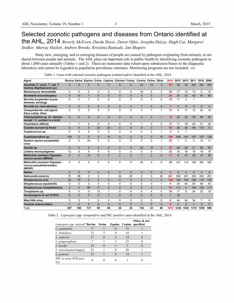
Selected zoonotic pathogens and diseases from Ontario identified at the AHL, 2014 Beverly McEwen, Durda Slavic, Davor Ojkic, Josepha DeLay, Hugh Cai, Margaret
Stalker, Murray Hazlett, Andrew Brooks, Kristiina Ruotsalo, Jan Shapiro
Many new, emerging, and re-emerging diseases of people are caused by pathogens originating from animals, or are
shared between people and animals. The AHL plays an important role in public health by identifying zoonotic pathogens in
about 1,000 cases annually (Tables 1 and 2). These are numerator data reliant upon submission biases to the diagnostic
laboratory and cannot be regarded as population prevalence estimates. Monitoring programs are not included. AHL
Table 1. Cases with selected zoonotic pathogens isolated and/or identified at the AHL, 2014
Leptospira spp. serovar* Bovine Swine Equine Canine Other & not
specified
L. autumnalis 17 1 11 33 1
L. bratislava 23 3 9 10 1
L .canicola 27 0 4 14 0
L. grippotyphosa 7 1 3 25 0
L. hardjo 24 0 1 3 0
L. icterohaemorrhagiae 25 1 8 20 1
L. pomona 33 1 6 14 1
IHC or urine PCR posi-tive
0 0 0 1 0
Table 2. Leptospira spp. seropositive and IHC positive cases identified at the AHL, 2014
Agent Bovine Swine Equine Ovine Caprine Chicken Turkey Canine Feline Other 2014 2013 2012 2011 2010 2009 Ascarids (T. canis, T. cati, T. leonina, Baylisascaris sp.)
0 0 0 0 0 0 0 23 14 3 40 36 35 ND ND ND
Blastomyces dermatitidis 0 0 0 0 0 0 0 19 0 3 22 17 10 10 5 10 Bordetella bronchiseptica 0 10 1 0 0 0 0 5 5 7 28 24 33 43 54 60 Borrelia burgdorferi (Lyme disease), serology
0 0 7 0 0 0 0 5 0 0 12 11 3 1
Brucella sp. (non-abortus) 0 0 0 0 0 0 0 1 0 0 1 0 0 0 0 0 Campylobacter coli/ jejuni/ fetus subsp. fetus
3 0 0 9 0 0 0 3 1 1 17 6 17 12 24 14
Chlamydophila sp. (C. abortus except 1 C. psittaci in a bird)
0 0 0 5 9 0 0 0 0 1 15 25 33 39 58 29
Clostridium difficile 1 7 2 0 0 0 0 1 0 0 11 11 19 40 31 24 Coxiella burnetii (Q fever) 6 0 0 25 23 0 0 0 0 1 55 28 36 99 115 9 Cryptococcus sp. 0 0 0 0 0 0 0 0 2 1 3 2 1 Cryptosporidium sp. 155 3 0 5 13 0 0 0 0 10 186 206 141 147 157 128 Eastern equine encephalitis virus
0 0 24 0 0 0 0 0 0 1 25 1 0 5 12 11
Giardia sp. 8 0 0 2 0 0 0 30 10 0 50 48 26 31 60 55 Listeria monocytogenes 12 0 0 4 6 0 0 0 0 1 23 15 18 18 19 18 Methicillin-resistant Staphylo-coccus aureus (MRSA)
0 2 10 0 0 2 0 1 2 0 17 8 24 49 74 36
Methicillin-resistant Staphylo-coccus pseudintermedius (MRSP)
0 0 0 0 0 0 0 45 0 0 45 141 114 192 ND ND
Rabies 0 0 0 0 0 0 0 0 0 0 0 0 0 0 3 8 Salmonella enterica 70 68 3 3 1 34 20 2 0 20 221 308 281 256 256 281 Streptococcus suis 23 76 0 3 2 0 0 0 1 0 105 126 144 106 110 120 Streptococcus equisimilis 0 0 3 0 0 0 0 0 0 1 4 34 45 59 48 43 Streptococcus zooepidemicus 2 0 86 0 0 0 0 0 4 1 93 112 4 149 152 117 Toxoplasma sp. 0 0 0 13 1 0 0 0 4 0 18 11 8 24 22 19 Verotoxigenic E.coli (VTEC) 6 0 0 0 1 0 0 0 0 0 7 18 West Nile virus 0 0 1 0 0 0 0 0 0 5 6 44 36 34 7 6 Yersinia enterocolitica 1 0 0 0 0 0 0 1 0 4 6 4 2 1 2 0 Total 287 166 137 69 56 36 20 136 43 60 1010 1236 1043 1315 1209 988

AHL Newsletter, Volume 19, Number 1 March, 2015 4
RUMINANTS
AHL Lab Reports
Verotoxigenic E.coli associated with diarrhea and mortality in young calves Andrew Brooks, Durda Slavic, Margaret Stalker, Murray Hazlett, Maria Spinato, Beverly McEwen
VTEC are a heterogeneous group of E.coli that express
one or more Shiga toxins (stx). VTEC have important public
health significance because some strains cause hemorrhagic
colitis and hemolytic uremia syndrome in people. VTEC are
carried subclinically in the intestine of healthy cattle, which
are an important reservoir for human infection, and sporadi-
cally cause diarrhea and dysentery in young calves.
Verotoxigenic E.coli (VTEC) was determined to be
the cause of diarrhea or death in 9 calves submitted to the
AHL for postmortem or histological examination from Janu-
ary 2013 to December 2014. The major clinical signs were
diarrhea and sudden death in calves 4-30 days of age (Table
1). The most common gross lesion was enterocolitis that was
often mild; in some cases fibrinonecrotic or fibrinohemor-
rhagic intestinal exudates were present. The common histo-
logic lesion was attachment of bacilli to the apical border of
small intestinal or colonic enterocytes (Fig. 1). E. coli iso-
lates from the intestine (1 isolate was from feces) were geno-
typed at the AHL with a PCR assay that detects the intimin
(eaeA), hemolysin (hlyA), and Shiga toxin 1 and 2 (stx1/2)
virulence genes. The majority of isolates from these cases
were positive for eaeA, hlyA, and stx1. Co-infections with
other pathogens associated with calf diarrhea were common. AHL
Case Breed Age
(days) Major clinical
problems Major postmortem lesions E. coli virulence factor genotype Co-infections
eaeA hylA stx1 stx2
1 Holstein 4 Bloody feces Found dead
Fibrinonecrotic enteritis + + + -
Cryptosporidium
2* not given 10 Diarrhea Erosive enterocolitis + + + - Cryptosporidium
3 Holstein 10 Diarrhea,
pneumonia Enterocolitis, mycotic ru-
menitis +/- 1 +/- +/- - Enterotoxigenic E. coli
Cryptosporidium Coronavirus
4 Mixed-
beef 9 Diarrhea, sud-
den death Enterocolitis Septicemia
+ + + - Rotavirus
5 Charolais 14 Diarrhea Enterocolitis Mycotic rumenitis/reticulitis
+ + + - Rotavirus
6 Holstein 30 Found dead Enteritis, rumenitis, meningi-
tis, pyelonephritis + + + - -
7 Jersey 15 Diarrhea Enterocolitis, abomasitis,
rumenitis + + + - Cryptosporidium
Rotavirus
8 Holstein 3 Found dead Fibrino-hemorrhagic enteri-
tis, abomasitis - + + + -
9 Holstein 10 Acute death Enterocolitis, abomasitis +/- + + - Coronavirus Salmonella Muenster Cryptosporidium
Table 1. Case summaries of diarrhea and mortality in calves associated with VTEC infection
* Field postmortem; 1. +/- denotes suspicious PCR result.
Figure 1. VTEC attached to surface enterocytes of the
small intestine (from case 4).

AHL Newsletter, Volume 19, Number 1 March, 2015 5
An outbreak of Campylobacter jejuni abortions in a small dairy herd Maria Spinato, Andrew Vince, Durda Slavic, Geert Jongert
The producer of a closed, well-managed herd of 85 Hol-
stein cattle reported 9 abortions during a 2-week period. Cat-
tle are vaccinated with a modified live, 5-way vaccine
(IBRV, BVDV types 1 and 2, BRSV, PI3V) at freshening.
Most of the aborted fetuses were between 200-260 days ges-
tation. Three fetuses and placentas were submitted to the
AHL for postmortem examination and ancillary testing. Pla-
centas contained variably-sized, relatively uniform tan coty-
ledons; slight marginal cupping and congestion were noted
in several cotyledons in the placenta of fetus 3. All 3 fetuses
were moderately autolysed. The only remarkable observation
in fetal tissues was the finding of an edematous sheet of fi-
brin overlying the lung of fetus 3. Fibrinous pleuritis is
most often observed in abortions caused by bacterial in-
fections; therefore, Campylobacter culture and Leptospira
MAT were performed, in addition to routine aerobic bacteri-
al culture. Campylobacter jejuni subsp. jejuni was isolated
in one or more samples of lung, abomasal fluid and pla-
centa cultured from all 3 fetuses. Leptospira MATs and
PCR tests for BVDV, BoHV-1 (IBRV), and Coxiella bur-
netii were negative. Neospora caninum ELISAs performed
on maternal sera of fetuses 1 and 2 were also negative. His-
tologic lesions included multifocal neutrophilic necrotizing
placentitis, colonization of chorionic stroma and occasional
trophoblastic epithelial cells by abundant Gram negative
bacteria, and hypercellular alveolar septa in lung sections
due to circulating neutrophils. The allantois of fetus 1 also
had a few arterioles characterized by hyaline walls and fibrin
thrombi.
Upon further investigation, it was discovered that a cow
had aborted on this farm approximately 2.5-3 weeks prior to
the outbreak. This cow was housed in a hospital pen that is
scraped out twice weekly using the same tractor and bucket
used for making the total mixed ration (TMR). Although the
bucket was rinsed with water after scraping the hospital pen,
it was not thoroughly cleaned or disinfected. It is suspected
that this abortion was the index case (not submitted for test-
ing), and that the placenta contaminated the TMR which was
the source of infection for the other aborting cows via inges-
tion. Subsequent to this outbreak, a premature live calf and a
first trimester abortion at 60 days gestation were reported on
this farm; however, no additional testing was performed and
their relation to the outbreak could not be confirmed.
Campylobacter jejuni is a normal commensal organism
found in the gastrointestinal tract of ruminants and other
food-producing animals. Although sporadic abortions have
been reported in cattle, abortion outbreaks due to C. jejuni
are extremely rare. Conversely, outbreaks of C. jejuni abor-
tion in small ruminants are a more common occurrence. The
emergence of tetracycline-resistant strains in Canada and the
USA is of significant concern to the small ruminant indus-
tries, as tetracycline is used both therapeutically and prophy-
lactically for infectious causes of abortion (1). Minimum
inhibitory concentration (MIC) analysis of the C. jejuni iso-
late in this case confirmed that it was susceptible to tetracy-
cline.
This case highlights the importance of expanding on-
farm biosecurity protocols to include appropriate dispos-
al and disinfection procedures for aborted fetuses and
placentas. Many abortifacient bacterial species in ruminants
also pose a significant zoonotic risk for producers, their fam-
ilies and veterinarians. Campylobacter jejuni is recognized
world-wide as a significant cause of gastroenteritis in hu-
mans, and contamination of food and water supplies by bo-
vine feces is a known risk factor. AHL
Reference
Slavic D, et al. Tetracycline susceptibility of Campylobacter iso-
lates causing ovine abortions in Ontario. AHL Newsletter, June
2012:16.
An alternative method for brain removal Josepha DeLay, Andrew Brooks
See: AHL LabNote 33, February 26, 2015.
Posted at: http://www.guelphlabservices.com/AHL/LabNotes.aspx
An alternative approach and, for
large animals especially, often easi-
er method to expose brain is by
using a lateral approach, cutting
through a coronal plane. Figure 6. Brain removal in rostral and caudal
sections. These may be sagittally sectioned,
with half of each section fixed in formalin for
histopathology, and the remaining halves
stored fresh or frozen for microbiologic tests. Figure 1. Landmarks for brain removal
by lateral approach.

AHL Newsletter, Volume 19, Number 1 March, 2015 6
Field and clinic postmortems. I: Simplified protocol and image list Josepha DeLay
Digital images captured during postmortem (PM) examinations provide a permanent record of lesions. The images are a
very useful communication tool when consulting with pathologists and other specialists. PM images provide valuable sup-
plemental information for pathologists evaluating tissue samples submitted to a diagnostic laboratory for histologic exami-
nation. Images may be emailed to AHL pathologists at [email protected]
The image list below provides both a step-wise guide to the postmortem procedure and a suggested set of images that
are applicable to all species of companion and food-producing animals. Establishing and following a standard routine for
PM procedures is important. This allows the practitioner to spend more time identifying and interpreting lesions, rather than
concentrating on the logistics of the exam. Developing a PM routine is similar to having a routine protocol for physical ex-
amination in a live patient.
Remove ear tag or create ID label, and include with all photos.
Image 1. External views: full body, head, thorax / abdomen, perineum
- for unexpected deaths, take image in situ, in location and position where body was discovered.
- include views that depict body condition, hydration (eyes), evidence of predation or trauma, etc.
Open abdominal and thoracic cavities.
Image 2. Opened thorax (with organs in situ).
Image 3. Heart in situ, with pericardial sac opened (check for fluid, exudate, etc.).
Remove pluck.
Image 4. Pluck, with focus on lungs (dorsoventral view, with right and left lung visible).
Image 5. Cross-section of right and left lung.
Image 6. Cross-section of heart through both ventricles.
Image 7. Larynx (including thyroid glands) and trachea: opened and mucosal surface exposed.
Image 8. Opened abdomen (with organs in situ).
In ruminants, remove omentum. In all species, fan out intestines and locate cecum and ileum.
Image 9. Opened abdomen with intestines fanned out.
Image 10. Open cecum, ileum, and jejunum to expose mucosal surface.
Image 11. Open colon to expose mucosal surface.
Image 12. Open duodenum to expose mucosal surface.
Image 13. Liver – capsular surface. For ruminants, include opened caudal vena cava.
Image 14. Liver – cross section.
Image 15. Abomasum / stomach – serosal surface.
Image 16. Abomasum / stomach – mucosal surface.
Image 17. Ruminants: rumen – serosal surface.
Image 18. Ruminants: rumen – mucosal surface and content.
Image 19. Solid organs:
- kidneys: sagittal sections, with cut surfaces exposed
- spleen: cross section
- adrenal glands
Unexpected death / neurologic cases: Remove brain. Also remove spinal cord if required, based on clinical signs.
Image 20. Brain.
The next installment in this series will focus on ancillary test selection and sample collection during PM exams. AHL
References and additional reading
1.Janzen ED. Field Postmortems. Presentation to the annual meeting of the Ont Assoc Bovine Practitioners. Guelph, ON. Nov 20, 2014.
2.Griffin D. Field necropsy of cattle and diagnostic sample submission. Vet Clin North Am Food Anim Pract 2012;28:391-405.

AHL Newsletter, Volume 19, Number 1 March, 2015 7
Theileriosis in a dairy cow from eastern Ontario Kris Ruotsalo, Jan Shapiro
A 3-year-old Holstein cow from eastern Ontario had a 2-
month clinical history of pyrexia, diarrhea, lameness, and
weight loss. Significant hematological changes included
marked, mildly responsive anemia; hematocrit 0.15 L/L
(reference interval 0.21-0.30 L/L), hemoglobin 48 g/L
(reference interval 84-120 g/L), and mild lymphopenia. Sig-
nificant biochemistry changes included hypoproteinemia
(total protein 50 g/L) and hyperbilirubinemia (total bilirubin
20 µmol/L).
Peripheral blood smear examination revealed numer-
ous intraerythrocytic organisms (Fig. 1). These organisms
were pleomorphic, exhibiting round, rod, comma, and signet
ring forms. A tentative diagnosis of Theileria spp. infection
was made. No commercially available test to confirm theiler-
iosis is available, and the species of Theileria cannot be mor-
phologically distinguished on the basis of a blood smear.
Confirmatory testing by 18S RNA amplification and gene
sequence analysis was performed at the Animal Health Diag-
nostic Laboratory, Cornell University, Ithaca, NY. Analysis
revealed that the Theileria organisms within this blood sam-
ple were part of the Theileria buffeli complex.
The affected cow continued to deteriorate clinically, and
euthanasia was elected. Repeated CBC analysis just prior to
euthanasia revealed the ongoing presence of Theileria organ-
isms within erythrocytes, although the anemia had marginal-
ly improved (hematocrit 0.18 L/L and hemoglobin 60 g/L).
The cow was submitted to AHL-Kemptville for postmortem
examination. Significant gross postmortem lesions consisted
of marked edema and diffuse enlargement of the pelvic, he-
patic, and peri-ruminal lymph nodes, mildly watery pale
blood, and mild icterus of internal fat. Lameness was at-
tributed to localized fibrinopurulent myositis of left thigh
muscles, and periarthritis and arthritis of the left femorotibial
joint. Histology of the enlarged lymph nodes showed cortical
lymphoid hyperplasia, with germinal centers. In one node,
medullary cords were populated predominantly by small
lymphocytes, with scattered foci of extramedullary hemato-
poiesis. Lymphocytic schizonts were not seen in the lymph
nodes, and are reported as uncommon in cows infected with
T. buffeli. Bone marrow taken at postmortem was autolysed,
and only adipose tissue with mild intercellular hemorrhage
and scattered foci of hematopoiesis were identified.
This is the first documented identification of this par-
asite in cattle in Canada. Neither the affected cow nor her
clinically unaffected herd mates had ever travelled outside of
Ontario. Theileria are tick transmitted, protozoal erythropar-
asites of ruminants. The tick species associated with T. buf-
feli transmission has not been identified. The role of wildlife
such as white-tailed deer as a possible parasite reservoir is
also unclear. Although this cow had been on pasture, it is not
known how she became infected.
Theileria buffeli has been reported previously in individ-
ual cows in the United States (Kansas 1950, Texas 1975,
Missouri 2000, Michigan 2002 and 2014). T. buffeli has been
considered non-pathogenic or less pathogenic than other
Theileria spp. such as T. parva and T. annulata which are the
agents of the rapidly fatal East Coast fever in Africa, and
tropical theileriosis in the Mediterranean and Asia, respec-
tively. Unlike the intralymphocytic schizonts which are con-
sidered the major pathogenic stage for T. parva and which
also play a role in T. annulata infections, the intraerythrocyt-
ic piroplasms are the major pathogenic state for T. buffeli. T.
buffeli has been identified both in asymptomatic cattle, as
well as those with clinical evidence of anemia. The patho-
genesis of anemia has not been clearly established and may
be multifaceted, involving both immune-mediated mecha-
nisms as well as erythrocyte fragmentation and damage by
proteases and oxygen radicals. Lymphoid hyperplasia and
lymphoma have been previously identified with bovine theil-
eriosis. It is known that intra-lymphocytic theilerial parasites
can transform cells, and lead to the clonal proliferation of
lymphocytes, although it is unclear if this occurs with T. buf-
feli infections.
Currently, Theileria can only be detected by examina-
tion of peripheral blood smears. Therefore evaluation of a
well-prepared blood smear is strongly recommended for all
anemic ruminants.
Theileriosis is on the federal and provincial lists of im-
mediately notifiable diseases, and therefore this case was
reported to OMAFRA and the CFIA. AHL References
Cossio-Bayagur R, et al. Theileria buffeli infection of a Michigan
cow confirmed by small subunit ribosomal RNA gene analysis.
Vet Parasitol 2002;105:105-110.
OMAFRA Industry Update: New diagnosis of tick-borne disease in
Ontario. January 29, 2015
Stockham SL, et al. Theileriosis in a Missouri beef herd caused by
Theileria buffeli: case report, herd investigation, ultrastructure,
phylogenetic analysis, and experimental transmission. Vet Pathol
2000;37:1-11.
Figure 1. Intraeryrthrocytic piroplasms of Theileria buffeli
(arrows).

AHL Newsletter, Volume 19, Number 1 March, 2015 8
HORSES
Ontario Racing Commission Death Registry: 2014 postmortem summary
The Ontario Racing Commission (ORC) has a long-
established record and takes pride in its proactive approach to
advancing the welfare of the racehorse and the safety of the par-
ticipant. In 2003, Ontario became one of the first North American
racing jurisdictions to require mandatory reporting of racehorse
deaths, in order to monitor, research, and improve our knowledge
of why these tragic events occur. The ORC Death Registry contin-
ues to provide excellent data regarding the causes of morbidity
and mortality in racehorses in this province.
Summaries of postmortem submissions to the AHL under this
program and diagnoses by body system are provided in the fol-
lowing tables. AHL
Table 1. Breed distribution of ORC Death Regis-
try submissions to the AHL, 2003-2014
Breed /
Year
Standard-
bred
Thorough-
bred
Quarter
Horse
Total
2003 67 (54%) 58 (46%) 0 125
2004 82 (58%) 60 (42%) 0 142
2005 59 (54%) 51 (46%) 0 110
2006 58 (54%) 47 (44%) 2 (2%) 107
2007 66 (54%) 53(43%) 3(3%) 122
2008 27 (53%) 24(47%) 0 51
2009 28 (62%) 16 (36%) 1 (2%) 45
2010 22 (69%) 8 (25%) 2 (6%) 32
2011 24 (52%) 18 (39%) 4 (9%) 46
2012 20 (59%) 14 (41%) 0 34
2013 19 (40%) 27 (56%) 2 (4%) 48
2014 21 (41%) 23 (45%) 7 (14%) 51
Table 2. Postmortem diagnoses of ORC Death Registry submissions by body system, 2003-2014.
Diagnosis by body system: 2003 2004 2005 2006 2007 2008 2009 2010 2011 2012 2013 2014
Fracture / limbs 53
(42%)
69
(49%)
48
(44%)
42
(39%)
54
(44%)
16
(31%)
4
(9%)
9
(28%)
6
(13%)
2
(6%)
23
(48%) 23
(45%)
Fracture / other 10 4 7 13 10 5 0 3 6 2 2 7
Non-fracture musculoskeletal 8 6 6 8 6 5 2 3 1 0 3 4
Gastrointestinal 15 19 17 16 18 5 4 7 5 6 4 6
Respiratory
(including EIPH)
21 17 9 11 16 9 21 6 9 7 4 5
Cardiovascular 5 6 5 5 2 4 6 2 4 1 7 3
CNS 6 11 7 4 1 1 2 0 5 4 3 0
Integumentary 0 0 1 2 2 1 1 0 0 0 0 0
Renal 0 2 0 0 2 0 1 0 0 0 0 0
Hematopoietic 2 1 1 0 0 0 0 0 0 0 0 0
Other / whole body condi-
tions (e.g., septicemia)
1 7 5 2 9 0 4 0 6 6 2 1
Cause of death undetermined 4
(3.2%)
0
(0%)
4
(3.6%)
4
(3.7%)
2
(1.6%)
5
(9.8%)
0
(0%)
2
(6%)
4
(9%)
6
(18%)
0
(0%) 2
(4%)
Total 125 142 110 107 122 51 45 32 46 34 48 51

AHL Newsletter, Volume 19, Number 1 March, 2015 9
Table 3. Musculoskeletal injuries in ORC Death Registry sub-
missions by breed and anatomic site, 2014.
Lesion TB SB QH Total
P1 fracture - LF 0 2 0 2
P1 fracture - RF 1 1 0 2
P1 fracture - LH 2 1 0 3
P1 fracture - RH 0 2 0 2
Carpal fracture - R 2 0 1 3
Proximal sesamoid fracture – LF
medial and lateral sesamoids
2 1 0 3
Fetlock failure - LF 1 0 0 1
Fetlock failure - RF 2 0 0 2
Humerus fracture - L 0 2 0 2
Metatarsal III fracture - L 0 2 0 2
Tibia fracture - L 1 0 0 1
Pelvis fracture 1 0 0 1
Rib fracture 0 1 0 1
Vertebral fracture 0 1 1 2
Skull fracture 3 0 0 3
Sacroiliac subluxation 0 0 1 1
Flexor tendon laceration – RF 0 1 0 1
Carpus DJD - bilateral 1 0 0 1
Multiple soft tissue lacerations 0 0 1 1
Total by breed 16 14 4 34
Gastrointestinal: Hemorrhagic enterocolitis associat-
ed with Clostridium perfringens (1) Colitis associated with Clostridium
difficile (1) Typhlitis / typhlocolitis / colitis,
etiology undetermined (3) Large colon torsion (1)
Respiratory: Exercise-induced pulmonary hem-
orrhage (EIPH) (2) Pulmonary congestion and edema,
suspect heart failure (1) Laryngotracheal evulsion second-
ary to trauma (1) Tracheal malformation (1)
Cardiovascular: Hemoabdomen, with concurrent
EIPH (2) Hemoabdomen and retroperitoneal
hematoma (1)
Other / whole body
conditions: Hypothyroidism (1)
Table 4. Non-musculoskeletal diagnoses in ORC
Death Registry submissions, 2014.
ORC Death registry, annual summary, 2014 - continued
In December and January of 2014-2015, we received 2
aborted foals, the first at 5 months gestation, the second at 7
months gestation – one from eastern Ontario, one from
southwestern Ontario. In the first foal, there was 100 mL
blood-tinged free fluid in the peritoneal cavity and diffuse
firmness of both lungs. The second foal had generalized ic-
terus; multifocal hemorrhages in the skin, skeletal muscle,
and small intestinal mucosa; subtle enlargement and pallor
of the liver; and large quantities of fluid in the small intes-
tine and colon. Tissues from both animals were negative by
PCR for equine herpesvirus-1 (EHV-1). No significant path-
ogens were isolated on bacterial culture of lung, stomach
content, or placenta (as available).
Despite divergent gross lesions, histologic lesions were
similar in both cases, and included hepatitis, myocarditis,
thymic necrosis/thymitis, and most distinctively erosive en-
teritis (an unusual lesion in an aborted foal). The first foal
also had a distinctive vasculitis in placenta, lung, and thy-
mus. More uncommon differential diagnoses for equine
abortion were discussed, including leptospirosis, equine viral
arteritis (EVA), and Potomac horse fever (PHF). The first
fetus was negative for EVA (PCR on lung) and leptospirosis
(immunohistochemistry on various tissues). PCR tests of
spleen and liver for PHF was positive in both foals. This
result strongly implicated PHF as causal in both abortions.
Neorickettsia risticii is the causal organism underlying
Potomac horse fever, a disease characterized by acute enter-
ocolitis in horses first identified in 1979. It is principally
associated with disease in spring, summer, and fall, and is
most common in farms bordering rivers. This is a very in-
frequently identified cause of equine abortion in Ontario.
Abortion typically occurs months after infection and
clinical disease in the mare, and is associated with fetal in-
fection resulting in enterocolitis, hepatitis, myocarditis, pla-
centitis, and variable hyperplasia or depletion of lymphoid
organs. The gross and histologic appearance may initially
resemble EHV-1 (though without classic microscopic her-
pesviral inclusions), and PHF should be considered a dif-
ferential diagnosis for abortion in such cases if EHV-1
PCR/IHC testing is negative, regardless of season. PCR
testing of tissues (usually lung, liver, spleen, commonly col-
lected for EHV-1 PCR in such cases) is available at the
AHL. AHL
Potomac horse fever (Neorickettsia risticii) infection in 2 aborted Ontario foals Andrew Vince, Andrew Brooks, Hugh Cai

AHL Newsletter, Volume 19, Number 1 March, 2015 10
The Swine OAHN (Ontario Animal Health Network) Melanie Barham
SWINE
The Ontario Animal Health Network is up and running in the swine sector!
What is OAHN? OAHN is a new way for Ontario commodity
groups to tackle important disease issues in their sector, col-
laborate with other industries, and access valuable resources.
Each sector will have an “Expert Network” comprising an
AHL, OVC, and OMAFRA species specialist and up to 3 pri-
vate practitioners. The Expert Network will meet regularly to
discuss pertinent diseases and issues affecting the sector. La-
boratory data will be discussed, together with the results of a
quarterly veterinary survey. The network’s focus is on identi-
fying trends and actionable items for the industry, and will
work together with producer groups. Networks will also par-
ticipate in cross-species information sharing, and the OAHN
plan will be able to link in with other provinces and a national
program as these initiatives are developed.
Networks currently in operation:
Fish, poultry, small ruminants, swine.
Under development:
Bees, bovine, companion animals, equine, fur-bearing/alternative, wildlife.
Swine OAHN
OAHN is patterned on CSHIN (Canadian Swine Health Information Network), and will serve as the Ontario node if
CSHIN is funded and continues.
The second swine network teleconference was held January 29th to discuss Oct/Nov/Dec 2014 clinical impression sur-
vey information. The AHL and Gallant Custom Laboratories provided lab data for the quarter.
A veterinary report and a producer report were published and distributed. The producer report focus was post-weaning
colibacillosis.
OAHN data is being shared with CSHIN’s national program.
Ontario Pork and OSHAB are close advisors for this network and we look forward to a continued close relationship
with producer groups. The full reports can be obtained by emailing [email protected] .
Ongoing surveillance testing for PED has been funded through OAHN.
Check out our OAHN podcast about neonatal piglet diarrhea!
OAHN Podcasts - Our podcasts are available in iTunes, so you can access them via any Apple device and subscribe so you
never miss an episode. As always, they are also available on our podbean site: barhamm.podbean.com .
Social media - Our Facebook and Twitter (@OntAnHealthNet) feeds offer up to date disease notifications, as well as news
stories and information for you to repost to your clinic website or Facebook/Twitter feeds if you so choose.
Website - We are designing a new website for the OAHN program, launching this spring.
Farm press - The OAHN program was also featured in an Ontario Farmer article.
Questions? Comments? Would you like a copy of the reports? Do you want to be included on surveys or mailing lists?
Contact Dr. Melanie Barham at (519) 824-4120 x53364 or [email protected].
Website: OAHN Website
Podcasts: OAHN Podcasts
1Improved animal health management

AHL Newsletter, Volume 19, Number 1 March, 2015 11
AVIAN/FUR/EXOTIC SPECIES Yellow fungus disease in bearded dragons
Marina Brash, Durda Slavic, Hugh Cai, Pat Bell-Rogers, Megan MacAlpine
In the last quarter of 2014, the AHL received numerous
submissions of primarily juvenile bearded dragons with his-
tories of lethargy and death often following the development
of skin lesions. Gross skin lesions ranged from yellow to
grey/brown ulcerated to thickened, roughened and scaly
patches involving the face, head, neck, abdomen, legs and
tail (Fig. 1) with splitting of the skin reported in some chron-
ic cases.
Histologically, variable numbers of fungal hyphae were
on the surface and within a variably thickened hyperkeratotic
layer of epidermis, and the underlying epithelium was erod-
ed/ulcerated or hyperplastic. Often the dermis was expanded,
hypercellular, fibrotic, and infiltrated by nodules or sheets of
macrophages and multinucleated giant cells, often containing
fungal hyphae. Occasionally the nodules had central cores of
eosinophilic necrotic debris. Granulomas were also within
the underlying skeletal muscle, connective tissue between
muscle bundles, and adjacent to resorbing bone. Organs in-
cluding lung and liver were less frequently affected (Fig. 2).
Fresh skin samples were submitted for fungal culture,
and wet mount microscopic examination, which revealed a
large number of hyphae confirming histological observa-
tions. Fungal cultures yielded Chrysosporium anamorph of
Nannizziopsis vriesii (CANV) which was confirmed by the
mycology laboratory at the Public Health Agency of Canada
and also by 18S rRNA gene sequencing at the AHL.
In the past, it was widely accepted that CANV and a vari-
ety of Chrysosporium spp. cause yellow fungus disease in
reptiles. Advanced molecular characterization of over 40
isolates from reptiles showed that they had been phylogenet-
ically misclassified. Isolates from reptile dermatitis cases
belong to 3 different fungal genera, namely Nannizziopsis,
Paranannizziopsis, and Ophidiomyces. These isolates may
have species predilections, with O. ophiodiicola causing
disease in snakes only, whereas N. guarroi primarily affects
bearded dragons.
There are slight morphological differences among the
isolates belonging to these genera. Since, at present, molecu-
lar characterization is time consuming, expensive, and likely
does not affect the clinical outcome, AHL reports fungal
isolates from reptile dermatitis cases as CANV complex
which currently encompasses all 3 fungal genera noted
above. AHL
Figure 2 a. Surface keratin contains large numbers of fungal hyphae (*). The
underlying epidermis is hyperplastic (200X, H & E). b. Multiple nodules and
clusters of macrophages and giant cells (M), often containing fungal hyphae
(see inset (400X, H & E) are within the expanded, fibrotic dermis and infiltrat-
ing into the underlying skeletal muscle (S) (100X, H & E). c, d. Granulomas
occasionally containing fungal hyphae (*) within liver (c) and lung (d). Exu-
date (E) is accumulating in faveolar lumens (100X, H & E).
Figure 1 a. Orange bearded dragon with grey/brown
irregularly thickened and crusted skin of the caudal
back, R leg, and proximal tail. The R leg and tail are
swollen, and the tail is shortened with a dark crust on
the tip. b. Ventrum of head of a bearded dragon; gener-
alized yellow discoloration of the skin and yellow
crusts of exudate at the neck. c. Skin of the ventral
abdomen is slightly thickened and yellow with scat-
tered small brown foci. (Photo courtesy of Dr. Rick
Axelson, The Links Road Animal & Bird Clinic).

AHL Newsletter, Volume 19, Number 1 March, 2015 12
AHL Newsletters and LabNotes are available on the Web at - http://ahl.uoguelph.ca
Leptospiral nephritis in a Shih Tzu dog Margaret Stalker, Jessika Bronsoiler
A 4-year-old spayed female Shih Tzu living in Brant-
ford, Ontario, was presented with a history of partial anorex-
ia of one-month duration. Clinical workup had been previ-
ously refused, and the dog had been treated symptomatically
with famotidine. On presentation, the dog was dehydrated
and severely azotemic. The dog was hospitalized, treated
with intravenous fluids, antibiotics, and antiemetics without
significant improvement and the owner elected euthanasia.
Sections of kidney collected at postmortem were submitted
for histologic examination at the AHL.
Histology revealed extensive renal interstitial inflam-
matory infiltrates (Fig. 1), with accompanying mild intersti-
tial fibrosis and tubular atrophy. The inflammatory infiltrate
was of mixed cell types, with nodular aggregates as well as
more diffuse infiltrates of lymphocytes and plasma cells,
scattered macrophages, and infiltrates of neutrophils within
the cortical interstitium. There was edema and patchy irregu-
lar foci of fibrin exudation and neutrophilic infiltrates within
the medullary interstitium. The unusually severe and active
inflammation of this lymphoplasmacytic and suppurative
tubulointerstitial nephritis prompted us to perform immuno-
histochemistry, looking for Leptospira interrogans. Wide-
spread positive staining was present (Fig. 2), including
extensive intense linear and granular staining (the latter rep-
resenting degraded bacteria) within tubular lumens, tubular
epithelial cells, and also within macrophages in the interstiti-
um. Staining with Warthin-Starry silver stains also highlight-
ed intact organisms morphologically compatible with lepto-
spires in renal tubular lumens (Fig. 3).
Naturally occurring infection with Leptospira spp.
is relatively common in various wildlife populations in
southern Ontario, principally raccoons and striped
skunks. Infection in these species is apparently self-limiting
and asymptomatic, with the host recovering from the initial
stages of the disease while potentially remaining as a reser-
voir capable of continued urinary shedding of infectious bac-
teria. Epidemiologic studies of canine leptospirosis demon-
strate increased risk of infection in middle-aged, intact male
hounds, herding, working or mixed-breed dogs, presumably
associated with increased outdoor activity of these breeds.
However, the presence of large urban populations of reser-
voir wildlife species such as raccoons increases the chance
of exposure of urban dogs with access to the outdoors as
well.
The severe lymphoplasmacytic and neutrophilic tubu-
lointerstitial nephritis seen in this case is a compatible lesion
of subacute infection in dogs, although acute infections may
cause significant hepatorenal dysfunction (with depression,
anorexia, vomiting, diarrhea, icterus, dehydration, severe
azotemia) with only minimal, subtle histologic lesions in the
kidney and liver. Antemortem diagnosis has relied on detec-
tion of high serum antibody titers using the microscopic ag-
glutination test (MAT), although peracute/acute infections
may have low titers. PCR testing of blood or urine, and
immunohistochemistry on renal biopsies may be useful
for detection of early infections and confirmation of in-
fection in some instances. AHL
Figure 1. Lymphoplasmacytic, histiocytic and suppurative
tubulointerstitial nephritis in a dog (H&E).
Figure 2. Positive immunostaining for Leptospira interrogans in
tubular lumens, tubular epithelial cells and interstitial macrophages.
Figure 3. Aggregates of typical curving spirochete bacteria
in tubular lumens (GMS silver stain).
COMPANION ANIMALS


![Ahl Al Dar - HR Newsletter [Q2]](https://img.pdfslide.net/doc/110x75/555ddc4dd8b42a192c8b495d/ahl-al-dar-hr-newsletter-q2.jpg)