Embed Size (px)
Citation preview
AIDS MALIGNANCY
CLINICAL TRIALS CONSORTIUM
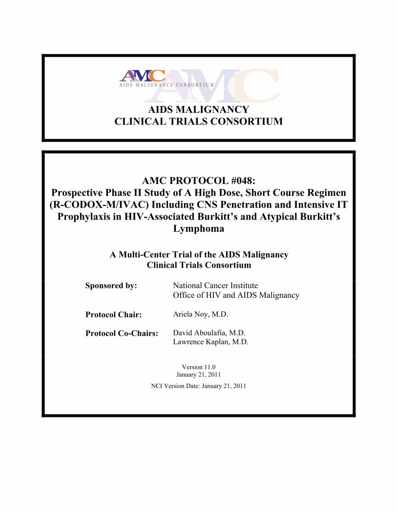
AMC PROTOCOL #048:
Prospective Phase II Study of A High Dose, Short Course Regimen (R-CODOX-M/IVAC) Including CNS Penetration and Intensive IT
Prophylaxis in HIV-Associated Burkitt’s and Atypical Burkitt’s Lymphoma
A Multi-Center Trial of the AIDS Malignancy
Clinical Trials Consortium
Sponsored by: National Cancer Institute
Office of HIV and AIDS Malignancy
Protocol Chair: Ariela Noy, M.D.
Protocol Co-Chairs:
David Aboulafia, M.D. Lawrence Kaplan, M.D.
Version 11.0
January 21, 2011
NCI Version Date: January 21, 2011
AMC PROTOCOL SIGNATURE PAGE
I, , Principal Investigator at site , agree to conduct and follow this protocol: AMC Protocol #048 - Prospective phase II study of a high dose, short course regimen (R-CODOX-M/IVAC) including CNS penetration and intensive IT prophylaxis in HIV-associated Burkitt’s and atypical Burkitt’s lymphoma (Version 11.0, 01/21/2011), as written according to AMC, NCI and FDA guidelines. I understand that no deviations from the above protocol may be made without written permission from the Protocol Chair(s). _________________________________ _____________________ Signature Date (mm/dd/yyyy)
AMC-048 (Version 11.0) 01/21/2011 ii NCI Version Date 01/21/2011
TABLE OF CONTENTS
AMC PROTOCOL SIGNATURE PAGE ......................................................................................ii
PROTOCOL ROSTER ................................................................................................................... vi
vii
1
2
2
2
5
9
9
9
12
12
................................................................................................ 12
13
13
14
14
15
16
16
16
17
17
18
19
19
20
21
STUDY SCHEMA ..........................................................................................................................
1.0 ................................................................................................... PROTOCOL SUMMARY
2.0 ......................................................................... OBJECTIVES AND SCIENTIFIC AIMS
2.1 ................................................................................................... Primary Objective
2.2 .............................................................................................. Secondary Objectives
3.0 ................................................................................3 BACKGROUND AND RATIONALE
3.1 ................................................................................................... 3 Primary Objective
3.2 .................................................. Secondary Objectives to be Pursued in this Study
4.0 ................................................... OVERVIEW OF STUDY DESIGN/INTERVENTION
4.1 ..................................................................................................................... Design
4.2 ............................................................................................................. Intervention
5.0 .................................................................... THERAPEUTIC/DIAGNOSTIC AGENTS
5.1 .......................................................................................................... Doxorubicin
5.2 Cyclophosphamide
5.3 ............................................................................................................. Vincristine
5.4 ........................................................................................... Rituximab (Rituxan )®
5.5 ................................................................................................. Ifosfamide (Ifex )®
5.6 ............................................................................................................. Cytarabine
5.7 ................................................................................................. Mesna (Mesnex )®
5.8 ............................................................................................................ Leucovorin
5.9 ......................................................................................................... Methotrexate
5.10 ............................................................................................................... Etoposide
5.11 .......................................................................... G-CSF (Filgrastim, Neupogen )®
5.12 ......................................................... Pegfilgrastim (Neulasta ) (Pegylated-CSF)®
5.13 ........................................................................... Liposomal cytarabine (Depocyt)
6.0 ................................................................... CRITERIA FOR SUBJECT ELIGIBILITY
6.1 ..................................................................................... Subject Inclusion Criteria
6.2 .................................................................................... Subject Exclusion Criteria
7.0 .................................................................................................... RECRUITMENT PLAN
AMC-048 (Version 11.0) 01/21/2011 iii NCI Version Date 01/21/2011
7.1 .......................................................................................... Enrollment Procedures 21
22
22
22
22
23
25
25
25
25
26
26
26
27
30
30
31
32
32
33
33
34
34
35
36
37
38
38
40
41
41
41
8.0 ................................................................................ PRETREATMENT EVALUATION
8.1 .................................................................................... Complete Medical History
8.2 ........................................................................... Complete Physical Examination
8.3 ................................................................................................... Laboratory Tests
8.4 ................................................................................................ Staging Evaluation
9.0 ............................................ EVALUATIONS DURING AND AFTER TREATMENT
9.1 ............................................................................... Evaluation During Treatment
9.2 .................................................................................... Post-Treatment Evaluation
9.3 ........................................................... Disease Progression/Off Study Evaluation
10.0 ........................................................................ TREATMENT/INTERVENTION PLAN
10.1 ........................................................................... Treatment Plan by Disease Risk
10.2 ................................................................................... Regimen A: R-CODOX-M
10.3 ...................... Guidelines for Leucovorin Rescue After High Dose Methotrexate
10.4 ......................................................................... Re-treatment on Regimen A or B
10.5 ................................................................................................. Regimen B: IVAC
10.6 ........................................................................................ Prophylactic Antibiotics
10.7 .............................. Concurrent Highly Active Antiretroviral Therapy (HAART)
10.8 ......................................................................................... Tumor Lysis Syndrome
11.0 .................................................. ADVERSE EVENTS AND DOSE MODIFICATIONS
11.1 ............................................................................................... Dose Modifications
11.2 .................................................................................................. Treatment Delays
11.3 ...............................................................................................................................
Classification of AEs by Severity and Relationship to Study Drug Administration
11.4 ....................................................................................... Expedited AE Reporting
11.5 ........................................................................................... Routine AE Reporting
11.6 ................................................................................. Secondary AML/MDS/ALL
12.0 ........... CRITERIA FOR THERAPEUTIC RESPONSE/OUTCOME ASSESSMENT
12.1 .......................................................................................... Response to Treatment
13.0 ............................................................... CRITERIA FOR REMOVAL FROM STUDY
14.0 .............................................................................. STATISTICAL CONSIDERATIONS
14.1 ........................................................................................................... Sample Size
14.2 ................................................................................... Stopping Rule for Toxicity
AMC-048 (Version 11.0) 01/21/2011 iv NCI Version Date 01/21/2011
AMC-048 (Version 11.0) 01/21/2011 v NCI Version Date 01/21/2011
14.3 ................................................................................................ Statistical Analysis 41
42
43
44
44
44
44
44
45
48
52
53
54
56
58
62
64
65
14.4 .......................................................................... Data Safety and Monitoring Plan
15.0 ................................................................................................... DATA MANAGEMENT
16.0 ....................................................................... PROTECTION OF HUMAN SUBJECTS
16.1 .................................................................................................. Informed Consent
16.2 ......................................................................................... Research Authorization
16.3 ......................................................................................... Subject Confidentiality
16.4 ......................................................................................... Women and Minorities
17.0 ................................................................................................................... REFERENCES
APPENDIX I: SCHEDULE OF EVALUATIONS .....................................................................
APPENDIX II: PERFORMANCE STATUS SCALE................................................................
APPENDIX III: ANN ARBOR STAGING CRITERIA ............................................................
APPENDIX IV: DIAGNOSTIC BIOPSIES.................................................................................
APPENDIX V: ACSR SPECIMEN PREPARATION & SHIPPING INSTRUCTIONS .......
APPENDIX VI: ACSR INFORMED CONSENT.......................................................................
APPENDIX VII: AMC DATA SAFETY MONITORING PLAN ............................................
APPENDIX VIII: METHODS FOR EBV STUDIES.................................................................
APPENDIX IX: CSF PROCESSING FOR FLOW CYTOMETRY ........................................
PROTOCOL ROSTER
AMC Protocol #048 Prospective Phase II Study of A High Dose, Short Course Regimen (R-CODOX-M/IVAC) Including CNS Penetration and Intensive IT Prophylaxis in HIV-Associated Burkitt’s and
Atypical Burkitt’s Lymphoma
Protocol Chair: Statistician: Ariela Noy, M.D. Jeannette Y. Lee, Ph.D. Memorial Sloan-Kettering Cancer Center AMC Statistical Center Lymphoma and Clinical Immunology University of Arkansas for Medical Sciences 1275 York Avenue 4301 W. Markham, #781 New York, NY 10021 Little Rock, Arkansas 72205-7199 Phone: 212-639-7423 Phone: 501-526-6712 Fax: 646-422-2284 Fax: 501-526-6729 Email: [email protected] Email: [email protected] Protocol Co-Chairs: Data Management/Operations: David Aboulafia, M.D. AMC Operations Center University of Washington The EMMES Corporation Virginia Mason Medical Center 401 N. Washington St., Suite 700 1100 Ninth Avenue Rockville, MD 20850 Seattle, WA 98101 Phone: 301-251-1161 Phone: 206-223-6193 Fax: 240-238-2842 Fax: 206-223-6914 Email: [email protected] Email: [email protected]
Laboratory Chair: Lawrence Kaplan, M.D. University of California, San Francisco Richard F. Ambinder UCSF Lymphoma Program Johns Hopkins School of Medicine 400 Parnassus Ave, Room A-502 1650 Orleans San Francisco, CA 94143 Baltimore, MD 21231 Phone: 415-353-2661 Phone: 410-955-8839 Fax: 415-353-2467 Fax: 410-955-0960 Email: [email protected] Email: [email protected]
AMC-048 (Version 11.0) 01/21/2011 vi NCI Version Date 01/21/2011
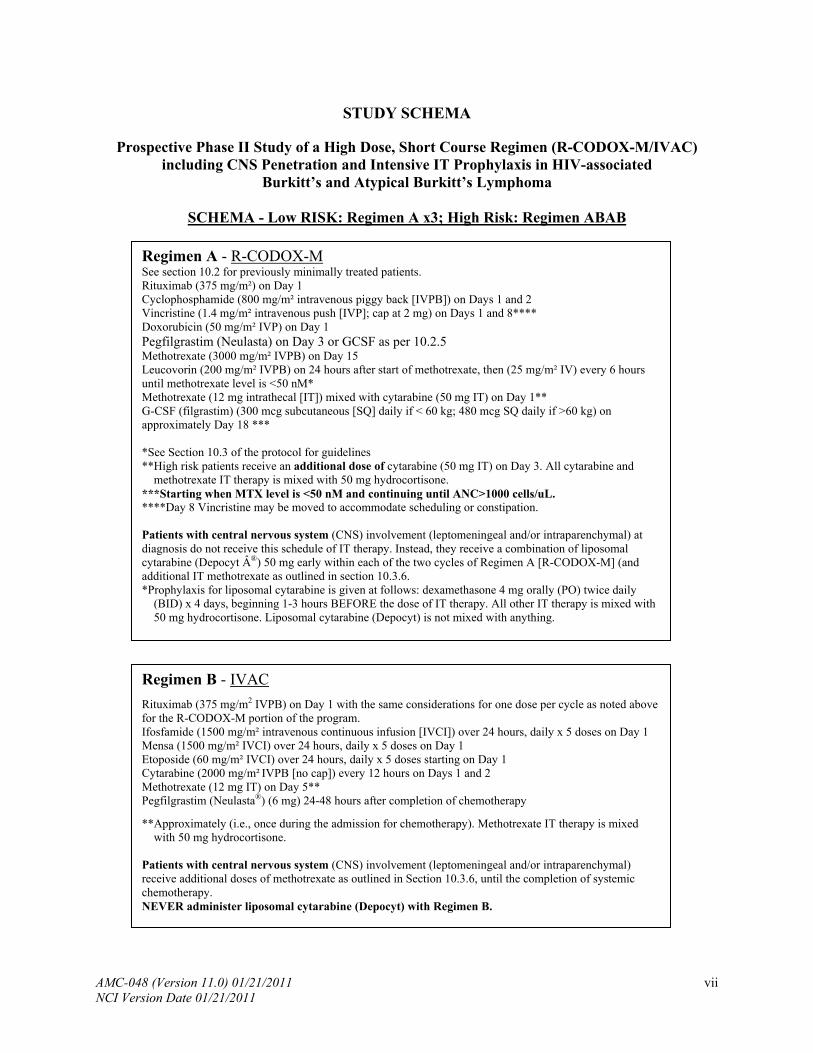
STUDY SCHEMA
Prospective Phase II Study of a High Dose, Short Course Regimen (R-CODOX-M/IVAC) including CNS Penetration and Intensive IT Prophylaxis in HIV-associated
Burkitt’s and Atypical Burkitt’s Lymphoma
SCHEMA - Low RISK: Regimen A x3; High Risk: Regimen ABAB
Regimen A - R-CODOX-M See section 10.2 for previously minimally treated patients. Rituximab (375 mg/m²) on Day 1 Cyclophosphamide (800 mg/m² intravenous piggy back [IVPB]) on Days 1 and 2 Vincristine (1.4 mg/m² intravenous push [IVP]; cap at 2 mg) on Days 1 and 8**** Doxorubicin (50 mg/m² IVP) on Day 1 Pegfilgrastim (Neulasta) on Day 3 or GCSF as per 10.2.5 Methotrexate (3000 mg/m² IVPB) on Day 15 Leucovorin (200 mg/m² IVPB) on 24 hours after start of methotrexate, then (25 mg/m² IV) every 6 hours until methotrexate level is <50 nM* Methotrexate (12 mg intrathecal [IT]) mixed with cytarabine (50 mg IT) on Day 1** G-CSF (filgrastim) (300 mcg subcutaneous [SQ] daily if < 60 kg; 480 mcg SQ daily if >60 kg) on approximately Day 18 *** *See Section 10.3 of the protocol for guidelines **High risk patients receive an additional dose of cytarabine (50 mg IT) on Day 3. All cytarabine and
methotrexate IT therapy is mixed with 50 mg hydrocortisone. ***Starting when MTX level is <50 nM and continuing until ANC>1000 cells/uL. ****Day 8 Vincristine may be moved to accommodate scheduling or constipation. Patients with central nervous system (CNS) involvement (leptomeningeal and/or intraparenchymal) at diagnosis do not receive this schedule of IT therapy. Instead, they receive a combination of liposomal cytarabine (Depocyt ®) 50 mg early within each of the two cycles of Regimen A [R-CODOX-M] (and additional IT methotrexate as outlined in section 10.3.6. *Prophylaxis for liposomal cytarabine is given at follows: dexamethasone 4 mg orally (PO) twice daily
(BID) x 4 days, beginning 1-3 hours BEFORE the dose of IT therapy. All other IT therapy is mixed with 50 mg hydrocortisone. Liposomal cytarabine (Depocyt) is not mixed with anything.
Regimen B - IVAC
Rituximab (375 mg/m2 IVPB) on Day 1 with the same considerations for one dose per cycle as noted above for the R-CODOX-M portion of the program. Ifosfamide (1500 mg/m² intravenous continuous infusion [IVCI]) over 24 hours, daily x 5 doses on Day 1 Mensa (1500 mg/m² IVCI) over 24 hours, daily x 5 doses on Day 1 Etoposide (60 mg/m² IVCI) over 24 hours, daily x 5 doses starting on Day 1 Cytarabine (2000 mg/m² IVPB [no cap]) every 12 hours on Days 1 and 2 Methotrexate (12 mg IT) on Day 5** Pegfilgrastim (Neulasta®) (6 mg) 24-48 hours after completion of chemotherapy
**Approximately (i.e., once during the admission for chemotherapy). Methotrexate IT therapy is mixed with 50 mg hydrocortisone.
Patients with central nervous system (CNS) involvement (leptomeningeal and/or intraparenchymal) receive additional doses of methotrexate as outlined in Section 10.3.6, until the completion of systemic chemotherapy. NEVER administer liposomal cytarabine (Depocyt) with Regimen B.
AMC-048 (Version 11.0) 01/21/2011 vii NCI Version Date 01/21/2011
AMC-048 (Version 11.0) 01/21/2011 viii NCI Version Date 01/21/2011
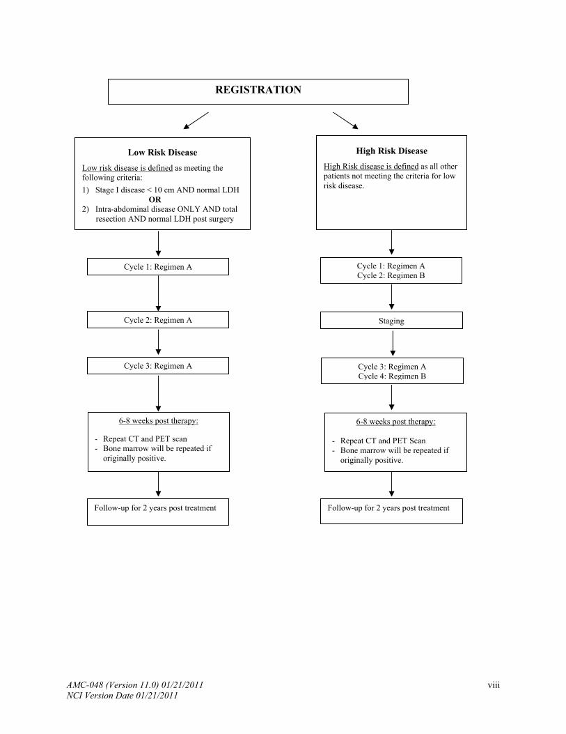
REGISTRATION
High Risk Disease
High Risk disease is defined as all other patients not meeting the criteria for low risk disease.
Low Risk Disease
Low risk disease is defined as meeting the following criteria:
1) Stage I disease < 10 cm AND normal LDH OR 2) Intra-abdominal disease ONLY AND total
resection AND normal LDH post surgery
Cycle 1: Regimen A Cycle 2: Regimen B
Cycle 1: Regimen A
Cycle 2: Regimen A Staging
Cycle 3: Regimen A Cycle 3: Regimen A Cycle 4: Regimen B
6-8 weeks post therapy:
- Repeat CT and PET scan - Bone marrow will be repeated if
originally positive.
6-8 weeks post therapy:
- Repeat CT and PET Scan - Bone marrow will be repeated if
originally positive.
Follow-up for 2 years post treatment Follow-up for 2 years post treatment
1.0 PROTOCOL SUMMARY
This will be a prospective phase II study of a maximum of 34 patients with human immunodeficiency virus (HIV)-associated Burkitt’s or atypical Burkitt’s Non-Hodgkin’s Lymphoma (NHL). Patients will be treated with a modification of the R-CODOX-M/IVAC regimen to assess efficacy. Most of these modifications were recently reported in a phase II study of 14 patients without HIV[1] to reduce toxicity and potentially improve efficacy. The study will be conducted by the AIDS Malignancy Clinical Trials Consortium (AMC). Accrual is anticipated over a 36-month period.
AMC-048 (Version 11.0) 01/21/2011 1 NCI Version Date 01/21/2011
2.0 OBJECTIVES AND SCIENTIFIC AIMS
2.1 Primary Objective
Primary Objectives: To determine the efficacy and safety of rituximab plus modified CODOX-M/IVAC regimen in patients with HIV-associated Burkitt’s (BL) or atypical Burkitt’s. The primary study endpoint is overall survival (OS) at one year, and secondary endpoints include complete response (CR) rate, failure-free survival (FFS), event-free survival, and toxicity.
2.2 Secondary Objectives
2.2.1 Evaluate downstream effectors of apoptosis as mechanisms of chemotherapy resistance and prognosis and perform exploratory analysis of their relationship to treatment effect.
2.2.2 Evaluate multi-drug resistance gene expression as a mechanism of chemotherapy
resistance and prognosis and perform exploratory analysis of their relationship to treatment effect.
2.2.3 Confirm the use of flow cytometry in the identification of occult leptomeningeal
disease and determine whether abnormal flow cytometry is predictive of CNS cytology is negative for malignant cells.
2.2.4 Determine the biologic and prognostic significance of Epstein-Barr Virus (EBV) +
Burkitt’s Lymphoma (BL) in the highly active antiretroviral therapy (HAART) era and perform exploratory analysis of their relationship to treatment effect.
2.2.5 Evaluate genotyping in BL and determine whether it is similar to that described in
HIV negative cases. Moreover, determine whether cases are uniform in their genetic profile or whether some cases are more like DLBCL.
2.2.6 Determine if EBV detection in CSF at diagnosis is predictive of leptomeningeal
disease.
2.2.7 Explore modifications of CODOX-M/IVAC with regard to possible reduced toxicity.
AMC-048 (Version 11.0) 01/21/2011 2 NCI Version Date 01/21/2011
3.0 BACKGROUND AND RATIONALE
3.1 Primary Objective
3.1.1 Burkitt’s Lymphoma in Immunocompetent Patients: Clinical Characteristics, Prognosis, and Treatment
Burkitt’s lymphoma (BL) is an aggressive non-Hodgkin’s lymphoma (NHL) characterized by rapid proliferation and a nearly 100% growth phase fraction. Originally identified as “undifferentiated lymphoma,” it has since been classified as diffuse small non-cleaved cell lymphoma in the Working Formulation,[2] and as Burkitt’s lymphoma in the Revised European-American Lymphoma[3] and World Heath Organization (WHO) classification systems.[4] In the general population, BL is more common in children or adolescents, but accounts for 1-2% of NHL in adults. Most patients present with advanced disease involving multiple extra nodal sites, most commonly in the abdomen. Although historically BL was one of the first cancers to respond to chemotherapy [5], early relapses occurred, often in the central nervous system (CNS), with rapid disease progression[6]. Cure rates of 50-60% were achieved in early stage patients treated with chemotherapy regimens containing high dose cyclophosphamide, anti-metabolites, and prophylactic intrathecal (IT) chemotherapy. However, only 20% of patients with bone marrow (BM) or CNS involvement achieved durable responses.[7] [8, 9] In 1996, Magrath and colleagues reported a 92% 2-year event-free survival (EFS) rate in HIV-negative adult and pediatric patients following intensive chemotherapy with cyclophosphamide, doxorubicin, high-dose methotrexate/ifosfamide, etoposide, high dose cytarabine (CODOX-M/ IVAC). Results were particularly impressive in patients with BM and/ or CNS involvement, with an 80% two-year disease-free survival.[10] A recent international multicenter phase II study confirmed that CODOX-M/ IVAC led to high cure rates particularly in International Prognostic Index stratified high risk patients with a reported 2-year EFS of 65% and 2-year overall cure rate of 72.8%, although results were inferior to the previous Magrath report [10, 11] Similar success has been reported with other regimens that incorporate. high dose cytarabine and methotrexate and intensive IT prophylaxis.[12, 13]
3.1.2 HIV-Associated Burkitt’s Lymphoma: Clinical Characteristics, Prognosis, and
Treatment
BL comprises 25-40% of NHL in HIV infected individuals. [14-16] Clinically, HIV-associated BL closely resembles BL in the general population. A recent review of 75 adults with BL (46 HIV+) found no difference between HIV+ and HIV-negative patients in terms of disease stage, marrow (33-35%), CNS (17-19%) involvement [17]
and histology [18]. The optimal treatment of adult patients with HIV-associated BL is not clear. In the era prior to highly active anti-retroviral therapy (HAART), combination chemotherapy was less successful for patients with HIV-associated NHL than for HIV-negative patients with similar NHL histopathology. Early deaths were often due to opportunistic infection [19]. Improved immune function in the HAART era has led to a reevaluation of full-dose chemotherapy for HIV-associated lymphoma. It has previously been proposed that HIV+ patients with BL and preserved immune
AMC-048 (Version 11.0) 01/21/2011 3 NCI Version Date 01/21/2011
function may represent a subset of acquired immune deficiency syndrome (AIDS)-related lymphoma patients who would benefit from more aggressive chemotherapeutic approaches with acceptable toxicities [7]. However, because of the perceived risk of increased hematologic and infectious complications, many patients with HIV-associated BL continue to be treated with cyclophosphamide, doxorubicin, vincristine, and prednisone (CHOP) and other moderate dose chemotherapy regimens despite the fact that such treatment is known to be inferior to intensive chemotherapy in HIV-negative BL patients. Indeed, a recent single institution retrospective analysis also demonstrated that the prognosis of HIV-associated lymphoma has improved dramatically in the HAART era, but the subset of patients with BL treated with CHOP-like regimens continues to do poorly.[20] At Memorial Sloan-Kettering Cancer Center (MSKCC) BL patients are routinely treated with chemotherapy regimens irrespective of HIV status. A retrospective review of 14 HIV+ adult patients with BL treated between 1988-2000[21] described eight patients who received an intensive regimen (CODOX-M/ IVAC) in the HAART era, and six with non-intensive regimens primarily in the pre-HAART era. Treatment outcomes and toxicities were compared with 24 concomitantly treated HIV-negative BL patients. Among the 14 HIV+ patients, CR rates were 63% after CODOX-M/ IVAC treatment versus 67% after other chemotherapy. Two-year event free survival (EFS) was 59% versus 63% after CODOX-M/ IVAC or other regimens, respectively. One HIV+ patient treated with CODOX-M/ IVAC died of PCP during chemotherapy related neutropenia. Similar outcomes were seen despite the fact that 88% of CODOX-M/ IVAC-treated HIV+ patients had stage IV disease as compared with 33% (2/6) of HIV+ patients treated with other chemotherapy. HIV status did not adversely affect long term EFS independent of treatment regimen (p=0.88). When EFS was examined by chemotherapy independent of HIV status, CODOX-M/ IVAC was associated with improved EFS (p= 0.05) in all patients particularly those with high risk BL. HIV+ patients treated with CODOX-M/ IVAC tolerated chemotherapy well with similar rates of myelosuppression and infectious complications as HIV-negative patients. The small, retrospective nature of this study precludes definitive statements, but suggests that intensive chemotherapy with CODOX-M/ IVAC is feasible and well-tolerated in HIV+ adults with BL. In the post HAART era, intensive chemotherapy such as CODOX-M/ IVAC may be appropriate in all adult patients with BL, especially those with poor prognostic factors, regardless of HIV status. The current proposal intends to build upon these preliminary findings by prospectively studying patients with high grade or highly aggressive HIV-associated NHL. We will employ a modification of the CODOX-M/ IVAC regimen, although it is recognized that a comparison study of the various Burkitt regimens has not been performed. We will follow a set of modifications recently reported in a phase II study of 14 patients [1] to reduce toxicity and potentially improve efficacy; the latter, in part by reducing toxicity related treatment delays and discontinuation of therapy. Despite reductions in methotrexate and vincristine, 12 of 14 patients achieved CR. There were no treatment-related deaths, no grade 3/4 neuropathies, and only 1 case of grade 3/4 mucositis was reported. Disease-free survival was 64% at 29 months.
AMC-048 (Version 11.0) 01/21/2011 4 NCI Version Date 01/21/2011
3.2 Secondary Objectives to be Pursued in this Study
3.2.1 Background and Rationale for Correlative Studies
Evaluate downstream effectors of apoptosis as mechanisms of chemotherapy resistance and prognosis. Rationale: Alterations of downstream effectors of apoptosis may explain why some cancers are resistant to chemotherapy. Caspase-mediated apoptosis has been demonstrated in the Ramos cell line[22] In a study of 16 adult and 16 pediatric patients (1 patient being HIV+) treated with the French regimen LMB, c-flip expression conferred a negative prognosis with 24% 2-year overall survival compared with 93% in the absence of this marker (P=0.04) [23] c-Flip inhibits caspase-8-mediated apoptosis[24] [25, 26]. Confirming this correlation in HIV-associated BL would provide a rationale to explore other treatment approaches in this subset of patients. Given a correlation with NF-Kappa B activity, [23] inhibitors of this pathway, such as bortezimib, may be justified. In addition, p53 mutations may play an important role (Kishor Bhattia, personal communication) and this will be evaluated to the extent possible using immunohistochemistry in paraffin sections under the auspices of Central Pathology review. Evaluate multidrug resistance (MDR) gene expression as a mechanism of chemotherapy resistance and prognosis. Rationale: MDR has been implicated in HIV- diffuse large B-cell lymphomas (DLBCL) chemotherapy resistance [27], but has not been evaluated in HIV+BL. This will be evaluated to the extent possible using immunohistochemistry in paraffin sections under the auspices of Central Pathology Review.
Confirm the use of flow cytometry in the identification of occult leptomeningeal
disease. Rationale: Hegde et al. [28] reported a surprising high rate of leptomeningeal disease detected by multi-color flow cytometry in the absence of positive cytology. The patients had diffuse large B cell lymphoma (DLBCL) in 91% vs. BL 9%. HIV infection was documented in 54% of the patients. In the newly diagnosed cohort, 11/51 (22%) of patients had a positive CSF by flow, yet only one was detected by routine cytology. The CSF fluid in these samples contained only a median of 2 WBC/µL and only 7% of the cells were characterized as malignant. Patients with high risk, but negative cytology received 12 prophylactic intrathecal (IT) therapies in addition to chemotherapy without blood-brain penetration, while the 10 with flow positive occult disease and the one with a cytologic positive received an intensive treatment beginning with therapy twice weekly and concluding with maintenance. In the 10 patients with follow-up at the NIH, seven received IT via lumbar puncture, three via Ommaya reservoir.
Despite this very aggressive strategy, five patients (45%) relapsed in the CNS and died. In contrast, in patients at increased risk of CNS involvement, but with negative flow cytometry, the relapse risk was only 8% (3/40). Additionally, all three patients with relapsed systemic disease and positive CSF flow cytometry died.
This study demonstrates that flow cytometry is more sensitive than cytology in detecting occult leptomeningeal disease at diagnosis of systemic NHL with high
AMC-048 (Version 11.0) 01/21/2011 5 NCI Version Date 01/21/2011
risk features. However, this related primarily to DLBCL and whether this can be generalized to BL is unknown. One cannot predict if patients with occult CSF + BL would have a higher rate of relapse given the intensive CSF prophylaxis. We propose a simplified flow cytometric evaluation of staining for (Kappa) and (Gamma) light chains in CD19-positive cells to be performed at each individual sites. Flow cytometry should be performed even if cells are not seen in the hemocytometer as detailed in the Appendix. This study will help examine this technique in a multi-institutional setting and determine the natural history of CSF occult positivity in BL. If this is found to be a negative prognostic factor, future trials can address whether IT therapy can be more efficacious in those with flow positive CSF if given via Ommaya, or if agents with longer half lives such as rituximab or liposomal cytarabine are used. Determine the biologic prognostic significance of EBV + BL in the HAART era. Rationale: EBV-driven lymphomas may develop in a unique pathway that may be more resistant to chemotherapy. However, NF-Kappa B activation in these tumors may create a new therapeutic window. The prevalence of EBV+ in the era of HAART is unknown. Tumors will be stained for EBV and the prognostic significance of EBV will be evaluated in multivariate analyses of prognostic factors. In addition, recently it has been recognized that DNA in cell-free blood (plasma or serum) may serve as tumor markers. Many studies have shown that tumor DNA is released into the blood. In nasopharyngeal carcinoma, a tumor that is always EBV-associated, viral DNA can almost always be detected in serum or plasma. Pretreatment EBV copy number has emerged as the single most important pretreatment prognostic factor (more important than classical prognostic factors such as stage)[29, 30]. Similar findings have been reported in nasal lymphoma. [31] The viral DNA detected is not virion (encapsidated DNA), but rather it is free DNA. Free DNA and virion DNA can be distinguished by a number of techniques described below in preliminary results, including sensitivity to DNAse and fragment size. In these non-immunocompromised patients, free DNA, not virion DNA, is detected. Our goal is to determine if in the fraction of AIDS-BL that are EBV-associated, the viral copy number will identify very high risk patients. To evaluate the role of c-flip expression on overall survival, the Cox proportional hazards model will be used. The role of EBV viral load on overall survival will also be evaluated using the Cox proportional hazards model.
Recent articles demonstrate that molecular profiling may better classify Burkitt’s
lymphoma from other lymphoma subtypes[32-34] Morphology alone was sometimes misleading, although the myc oncogene and expression of associated genes continue to play a central role. Comparing genotyping to histopathology for HIV lymphoma could provide new information regarding HIV-BL. When available, frozen tissue will be evaluated for genotyping.
AMC-048 (Version 11.0) 01/21/2011 6 NCI Version Date 01/21/2011
EBV in CSF fluid: EBV in DLBCL tumor DNA or in CSF has been shown to be predictive of CNS lymphoma when a brain lesion is present.[35] It is not known if EBV in the tumor or CSF is predictive of leptomeningeal disease.
3.2.2 Background and Rationale for Treatment Modifications
A number of modifications to the original McGrath regimen will be made in this study and the rationale is presented here:
Rationale for addition of rituximab
Modification A: Rituximab: Examine safety and added efficacy of rituximab when added to this regimen. Rationale: Rituximab has been shown to improve chemotherapy efficacy without appreciable added toxicity in a number of studies in the HIV-negative setting in both DLBCL[36] and follicular lymphoma.[37] Concerns were raised, however, in AMC-010 that excessive sepsis and toxic deaths may occur in the subpopulation of patients with a CD4 <50.[38]
Nonetheless, in patients with CD4 <100, rituximab may have improved the CR rate and OS. In addition, preliminary data from the NIH suggest that rituximab improved the outcome in patients with DLBCL treated with etoposide, vincristine, doxorubicin with cyclophosphamide (EPOCH) when CD4<100, as long as antibiotic prophylaxis and careful monitoring was performed.[28] Similarly, recent data suggest a possible improved efficacy of intensive BL regimens after the addition of rituximab without added toxicity in an HIV-negative cohort.[39] Investigators at MD Anderson Cancer Center added rituximab to the HyperCVAD/AraC/MTX regimen. A CR was achieved in 24 of 28 (86%) evaluable HIV-negative patients including 17 with BL. Equal efficacy was noted in patients with BL, its variants or B-ALL. The 3 year OS, EFS and DFS rates were 89%, 80%, and 88%, respectively. Although numbers were small, Thomas et al. reported only 1 induction death in patients over age 60 with all remaining patients in continuous CR. This was in striking contrast to their previous 19% treatment-related mortality in patients over age 60. Most significantly, rituximab improved the relapse rate compared to their prior study of hyperCVAD alone: (7% vs 34% overall, P=0.004; 0% vs 50% for elderly, P=0.02; 11% vs 20% for age < 60, P=0.11) In summary, the outcomes were markedly superior for all subgroups when rituximab was combined with chemotherapy. Clearly, other factors played a role in improving the treatment-related mortality, but in the absence of a randomized trial, these are the best data to date showing that rituximab improved the efficacy of chemotherapy in BL and its variants. In conclusion, the addition of rituximab in the current proposed study has been chosen with the hopes of improving CR and OS. Given the anticipated universal neutropenia with the chemotherapy alone, it is unlikely that toxicity will be augmented. Prophylactic antibiotics will be given with the intention to reduce toxicity as well. Modification B: Evaluate ifosfamide and etoposide infusion rather than bolus administration. Rationale: Infusional therapy has been championed as a way of combating high proliferative lymphomas. [40] [41] [42] Given that the IVAC portion
AMC-048 (Version 11.0) 01/21/2011 7 NCI Version Date 01/21/2011
of the regimen is given as an inpatient and that there is no reason to expect that infusional therapy will be inferior to bolus therapy, the modification will be made to give the same dose of ifosfamide 1500 mg/m2 daily x 5 days and etoposide 60 mg/m2 daily x 5 days as an infusion rather than a bolus. Mesna will be given as a concomitant infusion. Modification C: Reduce toxicity by lowering the dose of methotrexate while maintaining CNS penetration. Rationale: The published dose of methotrexate (methotrexate 1200 mg/m2 IV, Day 1, followed by methotrexate 240 mg/m2 every hour [q.h.] x 23 hours, total 5520mg/m2) is very high and can be difficult to clear. It typically takes several days and in some instances more than a week. This dose is also commonly associated with significant mucositis, which contributes to neutropenic sepsis. The dose of methotrexate in primary CNS lymphomas (3000 mg/m2) will be substituted, as it is designed for the same purpose and has been piloted in a small prospective study of HIV-negative patients.[1] More rapid clearance of methotrexate should result in less neutropenia and mucositis, and consequent shortening of the treatment cycle. This, in turn, may improve efficacy. Modification D: Reduce neurotoxicity by decreasing vincristine dose. Rationale: Patients with HIV are at risk for neuropathy both related to HIV and to HAART. Reducing the dose of vincristine to a maximum of 2 mg may improve tolerability and reduce missed doses. [1] Finally, patients with DLBCL occasionally have tumors with proliferative indices in the 90-100% range. The morphology is intermediate between BL (small, non-cleaved cell) and large-cell lymphoma. Similar to BL, the translocation t(8;14)(q24;q32) and variants involving the c-myc rearrangement are often present. No direct comparison trial has addressed whether this subgroup of aggressive lymphoma in fact does more poorly with CHOP-like regimens, but most investigators consider this to be similar to BL due to the high proliferative rate. In the WHO classification, this disease is considered a variant of BL, called atypical Burkitt’s. In light of this classification and the clinical characteristics, BL and atypical Burkitt’s are often treated with the same regimens, whether on or off clinical trials. Consequently, atypical Burkitt’s will be included in the current study.
Modification E: Reduce toxicity by moving the day 10 methotrexate in CODOX-M to day 15. In the original schema methotrexate is given on day 10 of the CODOX-M cycle coinciding with the nadir from the first few days of chemotherapy. Patients almost invariably neutropenic and sometimes have mucositis from the doxorubicin. It is not uncommon for patients to also have neutropenic fever. This neutropenia and mucositis is then compounded by the high dose methotrexate. This often results in prolonged hospitalizations for bacteremia and mucositis requiring TPN. If the methotrexate is moved by 5 days the first nadir will have passed and the toxicity of the treatment may be improved. It is likely the overall length of the cycle will be retained or possibly shortened if there is better blood count recovery, less mucositis and shorter hospitalization.
AMC-048 (Version 11.0) 01/21/2011 8 NCI Version Date 01/21/2011
AMC-048 (Version 11.0) 01/21/2011 9 NCI Version Date 01/21/2011
4.0 OVERVIEW OF STUDY DESIGN/INTERVENTION
4.1 Design
This phase II study will be conducted as a multi-institutional study through the AIDS Malignancy Consortium (AMC). A maximum of 34 patients with HIV-associated Burkitt’s and atypical Burkitt’s NHL will be enrolled. The CR rate, EFS and OS will be assessed. A modification of CODOX-M/ IVAC will be used. This regimen is one of a few standard regimens used in BL and atypical Burkitt’s. It is not known if any of the currently available regimens is superior to another. The modifications have been studied in a cohort of 14 patients and are believed to improve tolerability and toxicity. Within the scope of this limited study, efficacy did not appear to be compromised. Rituximab will also be added to the regimen with the goal to improve treatment efficacy, as has been seen in many lymphoma subtypes. Other secondary objectives will be studied as previously described.
4.2 Intervention
Patients with low risk disease will receive three cycles of R-CODOX-M.
Low risk disease:
1. Stage I, with a single focus of disease < 10 cm AND normal LDH
OR
2. Intra-abdominal disease ONLY AND total resection AND normal LDH post surgery.
High risk disease:
All other patients will receive R-CODOX-M/ IVAC in an A/B/A/B sequence. Note: In patients presenting with anasarca, pleural effusion, or ascites, methotrexate can pool causing difficulties with clearance. Treatment can be given in a reverse sequence: B/A/B/A. Cycles should remain 21-28 days in length. If the patient has CNS disease, treatment of the leptomeninges with liposomal cytarabine and methotrexate must be in the appropriate cycle. Specifically, do NOT administer liposomal cytarabine (Depocyt) in any of the B cycles.
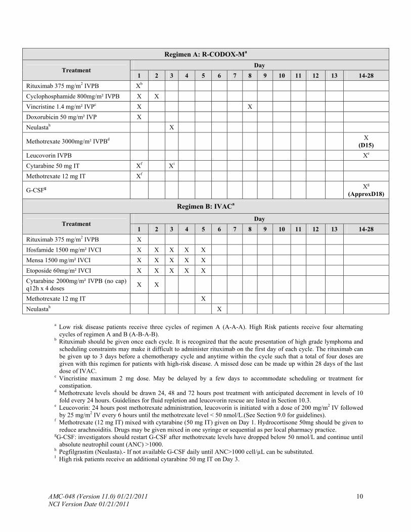
Regimen A: R-CODOX-Ma
Day Treatment
1 2 3 4 5 6 7 8 9 10 11 12 13 14-28
Rituximab 375 mg/m2 IVPB Xb
Cyclophosphamide 800mg/m² IVPB X X
Vincristine 1.4 mg/m² IVPc X X
Doxorubicin 50 mg/m² IVP X
Neulastah X
Methotrexate 3000mg/m² IVPBd X (D15)
Leucovorin IVPB Xe
Cytarabine 50 mg IT Xf Xi
Methotrexate 12 mg IT Xf
G-CSFg Xg
(ApproxD18)
Regimen B: IVACa
Day Treatment
1 2 3 4 5 6 7 8 9 10 11 12 13 14-28
Rituximab 375 mg/m2 IVPB X
Ifosfamide 1500 mg/m² IVCI X X X X X
Mensa 1500 mg/m² IVCI X X X X X
Etoposide 60mg/m² IVCI X X X X X
Cytarabine 2000mg/m² IVPB (no cap) q12h x 4 doses
X X
Methotrexate 12 mg IT X
Neulastah X
a Low risk disease patients receive three cycles of regimen A (A-A-A). High Risk patients receive four alternating
cycles of regimen A and B (A-B-A-B). b Rituximab should be given once each cycle. It is recognized that the acute presentation of high grade lymphoma and
scheduling constraints may make it difficult to administer rituximab on the first day of each cycle. The rituximab can be given up to 3 days before a chemotherapy cycle and anytime within the cycle such that a total of four doses are given with this regimen for patients with high-risk disease. A missed dose can be made up within 28 days of the last dose of IVAC.
c Vincristine maximum 2 mg dose. May be delayed by a few days to accommodate scheduling or treatment for constipation.
d Methotrexate levels should be drawn 24, 48 and 72 hours post treatment with anticipated decrement in levels of 10 fold every 24 hours. Guidelines for fluid repletion and leucovorin rescue are listed in Section 10.3.
e Leucovorin: 24 hours post methotrexate administration, leucovorin is initiated with a dose of 200 mg/m2 IV followed by 25 mg/m2 IV every 6 hours until the methotrexate level < 50 nmol/L.(See Section 9.0 for guidelines).
f Methotrexate (12 mg IT) mixed with cytarabine (50 mg IT) given on Day 1. Hydrocortisone 50mg should be given to reduce arachnoiditis. Drugs may be given mixed in one syringe or sequential as per local pharmacy practice.
gG-CSF: investigators should restart G-CSF after methotrexate levels have dropped below 50 nmol/L and continue until absolute neutrophil count (ANC) >1000.
h Pegfilgrastim (Neulasta).- If not available G-CSF daily until ANC>1000 cell/µL can be substituted. I High risk patients receive an additional cytarabine 50 mg IT on Day 3.
AMC-048 (Version 11.0) 01/21/2011 10 NCI Version Date 01/21/2011
AMC-048 (Version 11.0) 01/21/2011 11 NCI Version Date 01/21/2011
Methorexate and cytarabine intrathecal therapy is mixed with 50 mg hydrocortisone. Patients with CNS involvement (leptomeningeal and/or intraparenchymal) at diagnosis by cytology criteria do not receive this schedule of IT therapy. Instead, they receive a combination of liposomal cytarabine (Depocyt) 50 mg and methotrexate as outlined in Section 10.3.6. (Note: liposomal cytarabine (Depocyt) is not mixed with anything.) Prophylaxis for liposomal cytarabine is given at follows: dexamethasone 4 mg po BID x 4 days beginning 1-3 hours BEFORE the dose of IT therapy. Regimen A or B will be repeated every 21-28 days depending upon recovery from the previous cycle. Treatment may be administered as soon as the following criteria have been met: (1) post-nadir neutrophil count > 1000/µL, (2) platelets > 75,000/µL (if at least 75,000/µL at Baseline), (patients with platelets between 50,000 and 75,000 at baseline that is not lymphoma related should have return to baseline platelet count) (3) satisfactorily recovered from any infectious complication, and (4) satisfactorily recovered from other grade 3-4 non-hematologic toxicity.
5.0 THERAPEUTIC/DIAGNOSTIC AGENTS
5.1 Doxorubicin
5.1.1 Mechanism of action: Doxorubicin is an anthracycline antibiotic derived as a fermentation product of Streptomyces peucetius (caesius). The drug is tightly bound to DNA, preventing DNA-directed DNA and RNA synthesis. The drug may also act via a free radical mechanism. It appears to be active in all phases of the cell cycle.
5.1.2 Formulation: The drug is supplied reconstituted in 10, 50 and 200 mg vials.
5.1.3 Storage: Reconstituted solutions are stable at room temperature for 24 hours and
under refrigeration for 48 hours.
5.1.4 Administration: The drug is administered via a freely-flowing IV line over 15 minutes. Care must be taken to avoid extravasation.
5.1.5 Toxicity: Includes nausea, vomiting, itching, hives or red rash at the injection site.
Urine can be pink or red in color for as long as 48 hours after the treatment. Alopecia, stomatitis, and reversible myelosuppression can occur.
Extravasation may occur if leakage around the intravenous site occurs. Cardiomyopathy has been reported with this compound, usually in patients
who have received total doses in excess of 500 mg/m2.
5.1.6 Supplier: The drug is commercially available for purchase.
5.2 Cyclophosphamide
5.2.1 Mechanism of action: Cyclophosphamide, a nitrogen mustard derivative, is converted to polyfunctional alkylating metabolites by hepatic microsomal enzymes. It interferes with DNA replication and RNA transcription, and possesses potent immunosuppressive activity.
5.2.2 Formulation: The drug is supplied as a lyophilized powder in 100 mg, 200 mg,
500mg, l gram, and 2 gram vials.
5.2.3 Preparation/Storage: It is reconstituted to result in a concentration of
20 mg/mL. It is stable for 24 hours at room temperature or for six days refrigerated (2-8ºC).
5.2.4 Administration: cyclophosphamide is given IVPB over 30-60 minutes as the dose in
this protocol is <1200 mg/m2.
5.2.5 Toxicity: Includes nausea, vomiting, anorexia, edema, cardiomyopathy, skin rash,
alopecia, reversible myelosuppression, hemolytic anemia, possible sterility, hemorrhagic cystitis, and syndrome of inappropriate antidiuretic hormone production.
AMC-048 (Version 11.0) 01/21/2011 12 NCI Version Date 01/21/2011
5.2.6 Supplier: The drug is commercially available for purchase.
5.3 Vincristine
5.3.1 Mechanism of action: Vincristine is a member of the vinca alkaloid class of natural product anti-tumor agents. It exerts its antineoplastic effects by binding to tubulin, resulting in inhibition of microtubule assembly. This, in turn, blocks formation of the mitotic spindle resulting in the accumulation of cells in mitosis.
5.3.2 Formulation: The drug is supplied reconstituted to a concentration of
1 mg/mL.
5.3.3 Preparation/Storage: 1 mg/mL solution, 2 mL vial, stored under refrigeration.
5.3.4 Administration: The drug is administered via a freely-flowing IV line over 1-2
minutes, with care taken to avoid extravasation.
5.3.5 Toxicity: Includes peripheral neuropathy, constipation, alopecia, metallic taste in the
mouth, mild nausea, paraesthesia and paresis. Extravasation may result in soft tissue necrosis.
5.3.6 Supplier: The drug is commercially available for purchase.
5.4 Rituximab (Rituxan®)
5.4.1 Mechanism of action: Rituximab binds to the CD20 antigen expressed on B-cells and causes cell death by complement mediated lysis and ADCC.
5.4.2 Formulation: Rituximab is supplied as 100 mg and 500 mg sterile, preservative-free,
single-use vials.
5.4.3 Preparation: The appropriate dose is withdrawn and diluted to a final concentration of
1-4 mg/mL in either 0.9% sodium chloride or 5% dextrose solution. The solution is then stable at 2-8oC for 24 hours and at room temperature for an additional 12 hours.
5.4.4 Storage: Vials can be stored at 2-8oC. They should be protected from sunlight.
5.4.5 Administration: The first infusion should be administered at an initial rate of
50 mg/hour. If hypersensitivity or infusion-related events do not occur, the rate may be increased by 50 mg/hour every 30 minutes up to a maximum of 400 mg/hour. Subsequent infusions may be started at 100 mg/hour and the rate increased by 100 mg/hour at every 30 minutes to a maximum of 400 mg/hour, as tolerated. Patients will be pre-medicated as per institutional standards. For severe reactions, the
AMC-048 (Version 11.0) 01/21/2011 13 NCI Version Date 01/21/2011
infusion will be stopped and can be resumed at 50% of the prior rate once the reactions are treated and symptoms resolved or per institutional standards.
For subsequent infusions: Administration of rituximab can be given over one hour if commensurate with each individual institution’s guidelines.
5.4.6 Toxicity: Common: fever, chills, fatigue, headache; less common: nausea, vomiting,
rhinitis, pruritus, hypotension; rare: neutropenia, thrombocytopenia, asthenia, arrhythmia, tumor lysis syndrome, shock, angioedema, acute respiratory distress, arthritis, vasculitis, lupus-like syndrome, pleuritis, bronchiolitis obliterans, uveitis, optic neuritis, and skin reactions such as toxic epidermal necrolysis and pemphigus.
5.4.7 Supplier: The drug is commercially available for purchase.
5.5 Ifosfamide (Ifex®)
5.5.1 Mechanism of action: Ifosfamide is activated in the liver by microsomal enzymes and the subsequent ifosfamide mustard causes direct alkylation of DNA.
5.5.2 Formulation: Ifosfamide is supplied in single dose vials for constitution and
administration by IV infusion. Each contains 1 gram or 3 grams of sterile ifosfamide.
5.5.3 Preparation: Injections are prepared by adding sterile water to the vial. The 1 gram
dose is mixed with 20 mL sterile water, and the 3-gram dose with 60 mL sterile water for a final concentration of 50 mg/mL.
5.5.4 Storage: The dry powder may be stored at room temperature.
5.5.5 Administration: IVCI over 24 hours.
5.5.6 Toxicity: Alopecia, nausea and vomiting, hematuria, gross hematuria, CNS toxicity,
infection, renal dysfunction, allergic reactions and at high doses, cardiotoxicity.
5.5.7 Supplier: The drug is commercially available for purchase.
5.6 Cytarabine
5.6.1 Mechanism of action: Antimetabolite antineoplastic.
5.6.2 Formulation: 2000 mg vial with 20 mL sterile water for injection. Yields
concentration of 100 mg/mL. Other formulations available.
AMC-048 (Version 11.0) 01/21/2011 14 NCI Version Date 01/21/2011
5.6.3 Preparation: Dilute with final concentration up to 100 mg/mL. When given IT, dilute in preservative free saline.
5.6.4 Storage: Store vials at room temperature.
5.6.5 Administration: IVPB over 1 hour, every 12 hours; IT; or via Ommaya.
5.6.6 Toxicity: Leukopenia, thrombocytopenia, anemia, nausea and vomiting commonly
occurs with high doses (rarely with low doses), and is more severe after rapid administration; diarrhea, anorexia, oral and anal ulceration, gastrointestinal (GI) ulceration (high-dose therapy), pancreatitis, elevations in bilirubin, aspartate aminotransferase (AST), alanine aminotransferase (ALT), and Alk Phos, noncardiogenic pulmonary edema, resembles Acute Respiratory Distress Syndrome (ARDS), somnolence, cerebral and cerebellar dysfunction (high-dose therapy, usually reversible), maculopapular rash, erythema, blistering and peeling of skin, especially, hands and feet, alopecia, fever, conjunctivitis (high dose).
5.6.7 Supplier: The drug is commercially available for purchase.
5.7 Mesna (Mesnex®)
5.7.1 Mechanism of action: Mesna was developed as a prophylactic agent to inhibit hemorrhagic cystitis induced by ifosfamide and is analogous to the cysteine-cystine system. Mesna is rapidly metabolized to mesna disulfide and acts as a free radical scavenger.
5.7.2 Formulation: Mesna is a sterile preservative free aqueous solution of clear, colorless
appearance in clear glass vials for IV administration. Mesna injection contains 100 mg/mL Mesna, 0.25mg/Ll acetate disodium, and sodium hydroxide to maintain pH 6.5-8.5.
5.7.3 Preparation: For IV administration the drug is diluted in sterile solution to the
desired concentration.
5.7.4 Storage: Diluted solutions are chemically and physically stable for 24 hours at room
temperature. It is recommended that solutions be refrigerated and used within 6 hours.
5.7.5 Administration: 24 hour IVCI.
5.7.6 Toxicity: Nausea, vomiting, diarrhea.
5.7.7 Supplier: The drug is commercially available for purchase.
AMC-048 (Version 11.0) 01/21/2011 15 NCI Version Date 01/21/2011
5.8 Leucovorin
5.8.1 Mechanism of action: Leucovorin (folinic acid) is the formyl derivative and active form of folic acid. Leucovorin is used to diminish the toxicity and counteract the effect of high doses of folic acid antagonists.
5.8.2 Formulation: PO or IV.
5.8.3 Preparation: 100 mg vial powder: add 10 mL sterile water for injection to yield final
concentration of 10 mg/mL. Equivalent substitutions acceptable.
5.8.4 Storage: Store solution for injection vials in refrigerator. Store powder for injection
vials at room temperature. Reconstituted vials are stable for 7 days refrigerated at room temperature. Infusions prepared in dextrose 5% in water (D5W) are stable for 7 days.
5.8.5 Administration: IVPB or PO.
5.8.6 Toxicity: Nausea, vomiting, diarrhea.
5.8.7 Supplier: The drug is commercially available for purchase.
5.9 Methotrexate
5.9.1 Mechanism of action: methotrexate is an antimetabolite antineoplastic.
5.9.2 Formulation: The drug is supplied in 50 mg, 250 mg and 1 gram vials; 25 mg/mL
solution or 20 mg lyophilized powder preservative-free. Use preservative-free vials for IT preparations.
5.9.3 Preparation/Storage: Room temperature.
5.9.4 Administration: IVPB over 2-4 hours (in the current protocol) or IT.
5.9.5 Toxicity: Includes nausea, vomiting, anorexia, reversible myelosuppression,
mucositis and renal failure.
5.9.6 Supplier: The drug is commercially available for purchase.
5.10 Etoposide
5.10.1 Mechanism of action: Mitotic inhibitor belonging to the epipodophyllotoxin group.
AMC-048 (Version 11.0) 01/21/2011 16 NCI Version Date 01/21/2011
5.10.2 Formulation: 100 mg, 500 mg, 1000 mg vials; 20mg/mL.
5.10.3 Administration: IVCI over 24 hours (in the current protocol).
5.10.4 Toxicity: Includes hematologic, nausea and vomiting, hypotension, elevated liver
enzymes, dermatologic and neurologic.
5.10.5 Storage: Room temperature. Stability is concentration-dependent.
5.10.6 Supplier: The drug is commercially available for purchase.
5.11 G-CSF (Filgrastim, Neupogen® )
5.11.1 Mechanism of action: Neupogen is a human protein, which is involved in the promotion of the growth and maturation of granulocytic progenitors and the stimulation of functional activity.
5.11.2 Formulation: Available as a recombinant DNA product supplied as 1 or 1.6 mL vials
containing clear, colorless, sterile protein solution.
5.11.3 Administration: SQ.
5.11.4 Storage: It can be stored at 2-6 °C and is stable for at least 30 months.
5.11.5 Toxicity: Bone pain, exacerbation of preexisting autoimmune disorders, transient and
reversible changes in alkaline phosphatase (ALP), uric acid and LDH.
5.11.6 Supplier: The drug is commercially available for purchase.
5.12 Pegfilgrastim (Neulasta®) (Pegylated-CSF)
5.12.1 Mechanism of action: Neulasta is a human protein involved in the promotion of the growth and maturation of granulocytic progenitors and the stimulation of functional activity. It is a covalent conjugate of recombinant methionyl human G-CSF (filgrastim) and monomethoxypolyethylene glycol.
5.12.2 Formulation: Available as a pegylated recombinant DNA product supplied as 0.6-mL
pre-filled syringe containing clear, colorless, sterile protein solution.
5.12.3 Storage: It can be stored at 2-8 °C and is stable for at least 30 months.
AMC-048 (Version 11.0) 01/21/2011 17 NCI Version Date 01/21/2011
5.12.4 Toxicity: Bone pain, exacerbation of preexisting autoimmune disorders, transient and reversible changes in ALP, uric acid and LDH, nausea, fatigue, alopecia, diarrhea, vomiting, constipation, fever, anorexia, skeletal pain, headache, taste perversion, dyspepsia, myalgia, insomnia, abdominal pain, arthralgia, generalized weakness, peripheral edema, dizziness, granulocytopenia, stomatitis, mucositis, and neutropenic fever.
5.12.5 Supplier: The drug is commercially available for purchase.
5.13 Liposomal cytarabine (Depocyt)
5.13.1 Mechanism of action: DepoCyt is a sustained release formulation of the chemotherapeutic agent, cytarabine, used for the treatment of patients with lymphomatous meningitis. DepoCyt gradually releases cytarabine into the cerebral spinal fluid resulting in a significantly extended half-life, prolonged exposure to the therapy, and a more uniform distribution. Cytarabine is an antimetabolite neoplastic.
5.13.2 Formulation: sterile injectable suspension.
5.13.3 Administration: intathecal or intraOmmaya reservoir.
5.13.4 Storage: 2-8 °C and is stable for at least 30 months.
5.13.5 Toxicity: chemical arachnoiditis to include but not limited to headache, fever,
confusion, somnolence, nausea and vomiting. Systemic absorption is not expected, but cannot be excluded. Monitoring for myelosuppression is advised.
5.13.6 Supplier: The drug is commercially available for purchase.
AMC-048 (Version 11.0) 01/21/2011 18 NCI Version Date 01/21/2011
6.0 CRITERIA FOR SUBJECT ELIGIBILITY Patients with documented HIV infection and newly-diagnosed untreated Burkitt’s or atypical Burkitt’s lymphoma will be eligible.
6.1 Subject Inclusion Criteria
6.1.1 Histologic diagnosis of BL or atypical Burkitt’s as per WHO criteria. As of 2009, the new WHO criteria replacing atyipical Burkitt’s is “B cell lymphoma unclassifiable, with features intermediate between DLBCL and Burkitt lymphoma.”
6.1.2 Patients may be entered in either of the following two categories:
Untreated: Patients should be untreated for the diagnosis of BL or atypical Burkitt’s with the exception of up to 7 days of consecutive steroids alone or in combination with a non-CHOP regimen necessary for patient stabilization (e.g., cycolphosphamide and steroids for normalization of disease-related hyperbilirubinemia). OR
After 1 cycle of CHOP or fractionated CHOP (e.g. CODOX) with or without rituximab. This allows patients with newly diagnosed HIV infection or minimal therapy at diagnosis to be entered onto the study. (See section 10.2.)
6.1.3 Patients must have normal baseline cardiac function based upon echocardiogram or
multigated acquisition (MUGA) blood pool scan with an ejection fraction ≥ 50%.
6.1.4 Patients must have a serum creatinine of ≤ 1.5 mg/dL. If creatinine >1.5 mg/dL,
creatinine clearance must be ≤ 60 mL/minute.
6.1.5 Patients must have ANC ≥ 1000/µL and Platelets ≥ 50,000/µL unless disease-related.
Patients with bone marrow involvement due to BL will be eligible irrespective of blood count.
6.1.6 Patients must have a direct bilirubin level of ≤ 2.0 mg/dL. If, however, the elevated
bilirubin is felt to be secondary to antiretroviral therapy, patients will be allowed to enroll on protocol if the total bilirubin is ≤ 3.5 mg/dL provided that the direct bilirubin is normal.
6.1.7 AST (SGOT) and ALT (SGPT) ≤ 3 x the upper limit of normal.
6.1.8 All patients of childbearing age must be using an acceptable form of birth control.
6.1.9 Women who are pre-menopausal must have a negative pregnancy test.
AMC-048 (Version 11.0) 01/21/2011 19 NCI Version Date 01/21/2011
6.1.10 Patients must be HIV positive documented by enzyme-linked immunosorbent assay [ELISA] and Western Blot, or measurable HIV viral load.
6.1.11 If patients have a history of malignancy other than curatively treated cutaneous basal
cell or squamous cell carcinoma, carcinoma in situ of the cervix, or cutaneous Kaposi’s sarcoma (KS), they must be disease-free for > 5 years at the time of enrollment.
6.1.12 Patients or their guardians must be capable of providing informed consent.
6.1.13 Karnofsky performance status > 30% (given the aggressiveness of this disease and
the often severely debilitated nature of the patients at initial presentation).
6.1.14 All stages of disease.
6.1.15 Measurable or non-measurable tumor parameter(s). Non-measurable tumor
parameters will be defined as not having bidimensional measurements (i.e., gastric or marrow involvement), but can be followed for response by other diagnostic tests such as gallium, PET imaging and/or bone marrow biopsy.
6.1.16 Age ≥ 18 years.
6.1.17 Patients already receiving erythropoietin or G-CSF for treatment of HIV-related
cytopenia are eligible, although the growth factors must be discontinued at last 24 hours prior to chemotherapy.
6.2 Subject Exclusion Criteria
6.2.1 Any lymphoma subtype other than BL or atypical Burkitt’s.
6.2.2 Previous therapy other than seven consecutive days of steroids.
6.2.3 Known pregnancy or breast-feeding.
6.2.4 Medical illness unrelated to NHL, which in the opinion of the attending physician
and Principal Investigator (PI) will preclude administration of chemotherapy safely. This includes patients with uncontrolled infection (including opportunistic), chronic renal insufficiency, myocardial infarction (MI) within the past 6 months, unstable angina, cardiac arrhythmias other than chronic atrial fibrillation.
6.2.5 History of any malignancy for which the disease-free interval is <5 years, excluding
curatively treated cutaneous basal cell or squamous cell carcinoma and carcinoma in situ of the cervix or cutaneous KS. Patients with visceral KS are excluded.
AMC-048 (Version 11.0) 01/21/2011 20 NCI Version Date 01/21/2011
7.0 RECRUITMENT PLAN
Patients seen in the inpatient or outpatient setting who meet eligibility criteria will be recruited to this study. Participation is voluntary. The patient will be made aware of his or her diagnosis, current nature of this treatment program. All patients will be required to sign a statement of informed consent that conforms to Food and Drug Administration (FDA) and Institutional Review Board (IRB) guidelines.
7.1 Enrollment Procedures
This study will be available for enrollment at all AMC sites. Each site must have this protocol approved by their IRB and be registered with the AMC Operations Center before they may enroll patients. After it has been determined that the patient is eligible and an informed consent has been signed by the patient, the patient must be registered on-line via the AMC AdvantageEDCSM Internet Data Entry System. Enrollment and data collection will occur via the AMC Internet Data Entry System. The participating site will ensure the patient meets all eligibility criteria prior to completing the protocol-specific eligibility checklist. Patients will be enrolled on-line via the AMC Internet Data Entry System no more than one week prior to the initiation of treatment (enrollment one day prior to or on the day of treatment is strongly encouraged). Once the eligibility checklist is submitted and eligibility is confirmed, a system generated confirmation email will be sent to the enroller upon successful completion of the patient enrollment. If the on-line system is inaccessible, the site should notify the AMC Operations Center (via e-mail at [email protected] or phone at 301-251-1161) for further instructions.
AMC-048 (Version 11.0) 01/21/2011 21 NCI Version Date 01/21/2011
8.0 PRETREATMENT EVALUATION
See Appendix I, Schedule of Evaluations. PRETREATMENT: The following will be obtained no more than 14 days prior to study enrollment:
8.1 Complete Medical History
8.1.1 Duration of AIDS diagnosis, history of prior opportunistic illness.
8.1.2 Date of initial diagnosis of lymphoma. A copy of the pathology report must be
available in the medical record.
8.1.3 Presence or absence of “B”-symptoms (unexplained fevers, night sweats, involuntary
weight loss greater than 10% normal body weight).
8.1.4 History of other symptoms related to NHL.
8.1.5 History of drug allergies.
8.1.6 Medication list to include all antiviral, antibiotics and opportunistic prophylaxis.
List duration of current antiviral (HIV) therapy.
8.2 Complete Physical Examination
Includes Karnofsky performance score (see Appendix II), vital signs, weight, height, body surface area, neurologic examination, careful measurement of all palpable peripheral lymph nodes and measurement of other sites of disease present on physical examination.
8.3 Laboratory Tests
8.3.1 Hematology: complete blood count (CBC), platelet count and differential.
8.3.2 Blood chemistries: sodium, potassium, chloride, CO2, creatinine, calcium,
phosphorus, uric acid, total bilirubin, AST, ALT, alkaline phosphatase, total protein, albumin, LDH.
8.3.3 T cell subsets (CD4, CD8), circulating B-cells (CD20 and/or CD19).
8.3.4 HIV viral load.
8.3.5 EBV viral load (see Appendix VIII).
AMC-048 (Version 11.0) 01/21/2011 22 NCI Version Date 01/21/2011
8.3.6 Quantitative immunoglobulins (IgG, IgM, IgA).
8.3.7 Donation of biopsy material, blood, marrow or CSF to AIDS and Cancer Specimen
Resource (ACSR). (Optional patient participation). (Please see Appendices VI and VII for ACSR instructions and consent form.)
8.3.8 Urinalysis.
8.3.9 Electrocardiogram (EKG).
8.3.10 Serum pregnancy test for women of child-bearing age within 48 hours of starting
therapy (excluding only post-menarche patients, those who are 2 years post the last menses or post hysterectomy).
8.3.11 Hepatitis B surface antigen, surface antibody and core antibody; Hepatitis C
antibody.
8.3.12 Material sent for Central Pathology Review (see Appendix IV, Diagnostic Biopsies).
This includes diagnostic material to confirm diagnosis and unstained slides for correlative studies. Frozen tissue if available should be sent for genotyping analysis to Central Pathology (see Appendix IV).
8.4 Staging Evaluation
The following studies will be done for baseline evaluation of the extent of disease. The Ann Arbor staging classification will be used (Appendix III). Staging evaluations should be performed within 28 days of study entry.
8.4.1 Chest X-ray.
8.4.2 CT or magnetic resonance imaging (MRI) scan of the chest, abdomen and pelvis.
PET scans are recommended, but not required.
8.4.3 Bone marrow (bilateral or single core with total aggregate 2.0 cm preferred).
8.4.4 Lumbar puncture with routine studies and cytology in addition to EBV PCR and flow
cytometry as follows:
*In accordance with Secondary Objective 3: (confirm the use of flow cytometry in the identification of occult leptomeningeal disease), 3 cc of CSF should be sent to flow cytometry for CD19, CD10 and CD45 by routine methods on the first CSF sample to be sent. If there are sufficient cells, kappa and lambda should also be run.
AMC-048 (Version 11.0) 01/21/2011 23 NCI Version Date 01/21/2011
Thereafter, CSF should be sent for cytology and routine studies (cell count and protein) only with each lumbar puncture. In accordance with the secondary objective correlating EBV PCR in the CSF, one cc of CSF fluid should be sent as specified Appendix V.
8.4.5 Central Pathology Review: All specimens will be reviewed by a panel of pathologists
and must be submitted within 30 days of study enrollment (see Appendix IV). Investigators will be notified of patients deemed ineligible for the study by the Central Pathology Review and will be removed from study and replaced by eligible patients.
8.4.6 When available diagnostic frozen tissue should be sent to Central Pathology for
genotyping studies (see Appendix IV).
AMC-048 (Version 11.0) 01/21/2011 24 NCI Version Date 01/21/2011
AMC-048 (Version 11.0) 01/21/2011 25 NCI Version Date 01/21/2011
9.0 EVALUATIONS DURING AND AFTER TREATMENT
9.1 Evaluation During Treatment
9.1.1 Within 48 hours of treatment and prior to methotrexate courses as indicated in Appendix I: Physical exam, CBC, serum chemistries and assessment of toxicity prior to each chemotherapy.
9.1.2 Interim restaging (including all previously positive imaging studies and bone marrow
examinations):
In patients with low risk disease, interim restaging will not be performed.
In patients with high risk disease, interim restaging will be performed after 2 cycles of chemotherapy, i.e., after cycle 1 (CODOX-M) and cycle 2 (IVAC) and prior cycle 3 (R-CODOX-M) and will include CT scan and bone marrow if previously positive. PET scans are recommended, but not required if they are not covered by third party payers.
9.2 Post-Treatment Evaluation
9.2.1 6-8 weeks after completion of all therapy: CT scans and PET imaging will be repeated. Bone Marrow will be repeated if originally positive. HIV viral load and HIV, B and T-cell subsets (CD4, CD8, CD19, CD20) will be tested.
9.2.2 Patients should have repeat imaging with CT scans every 4 months for 2 years post
treatment. Follow up after this time point will be as clinically indicated and not part of the current study.
9.2.3 HIV viral load and CD4 should be monitored every 4 months concurrent with the
patient’s re-staging for 2 years.
9.3 Disease Progression/Off Study Evaluation
Patients who develop disease progression during the treatment period and who do not begin other anti-cancer therapy will continue to be followed for routine safety and efficacy for a total of 2 years post completion of treatment to allow for analysis of overall survival.
A case report form must be completed that includes notification of new lymphoma directed therapy.
10.0 TREATMENT/INTERVENTION PLAN
10.1 Treatment Plan by Disease Risk
Patients with low risk disease will receive 3 cycles of R-CODOX-M.
10.1.1 Low risk disease is defined as meeting the following criteria:
1. Stage I, with a single focus of disease < 10 cm AND normal LDH OR
2. Intra-abdominal disease ONLY AND total resection AND normal LDH post surgery.
10.1.2 High risk disease: All other patients will receive R-CODOX-M/ IVAC in an A/B/A/B
sequence for a total of four cycles.
High dose methotrexate and IVAC will be given as an inpatient procedure. All other chemotherapy may be given as an inpatient procedure, or as an outpatient procedure, at the discretion of the Investigator. However, depending on the burden of disease, patients may be at risk of tumor lysis during the first cycle of chemotherapy. This should be taken into account. All patients should receive some form of preventative treatment for uric acid nephropathy during the first cycle of chemotherapy, either in the form of allopurinol or rasburicase, at the discretion of the Investigator.
10.1.3 Rituximab 375 mg/m2 IVPB will be given once each cycle. It is recognized that the acute presentation of high-grade lymphoma and scheduling constraints may make it difficult to administer rituximab on the first day of each cycle. Rituximab can be given up to 3 days before a chemotherapy cycle and anytime within the cycle such that a total of 3 doses are given with regimen A (CODOX-M x 3) and a total of 4 doses are given with regimen B (R-CODOX-M/ IVAC). A missed dose can be made up within 28 days of the last dose of IVAC.
10.1.4 Hepatitis B
Hepatitis B virus reactivation, sometimes resulting in varying severity of liver failure, has been reported in some patients taking rituximab. Most of these patients were taking rituximab in combination with chemotherapy. It is thought that rituximab contributed to activating the hepatitis B virus. Patients found to have an active hepatitis B infection, either before or after initiation of treatment (hepatitis B surface antigen +), should have rituximab held. Any patient receiving rituximab with active hepatitis B infection must receive dual antiviral therapy. Consultation with a hepatitis expert is strongly encouraged.
10.2 Regimen A: R-CODOX-M
NOTE: Patients entering the study after a previous cycle of CHOP or CHOP-like treatment should start protocol therapy with the methotrexate 3000 mg/m² IVPB portion (10.2.6) after neutrophils recover >1000 cells/mL. Patients entering after a non-anthracycline-containing regimen for stabilization as outlined in Section 6.1.2 should begin with the first cycle of CODOX similar to untreated patients.
AMC-048 (Version 11.0) 01/21/2011 26 NCI Version Date 01/21/2011
10.2.1 Rituximab 375 mg/m2 IVPB on Day 1 (considerations for moving the timing of the dose are noted in Section 10.1.3).
10.2.2 Cyclophosphamide 800 mg/m² IVPB will be given on Days 1 and 2.
10.2.3 Vincristine 1.4 mg/m² IVP (cap at 2 mg) will be given on Days 1 and 8. (May be
delayed by a few days to accommodate scheduling or treatment for constipation.)
10.2.4 Doxorubicin 50 mg/m² IVP will be given on Day 1.
10.2.5 Pegfilgrastim (Neulasta) 6 mg subcutaneously will be given on day 3. If not available
G-CSF daily until ANC>1000 cell/µL can be substituted.
10.2.6 Methotrexate 3000 mg/m² IVPB mixed in 1 L normal saline (NS) will be given on
Day 15 over a 2-4 hour infusion, even in the setting of neutropenia and thrombocytopenia, unless an interim event has occurred that precludes safe administration in the judgment of the treating physician. Methotrexate may be delayed until the acute event resolves. Serum creatinine should be ≤ 2 mg/dL within 24 hours of each dose of methotrexate. Particular questions should be addressed to the Protocol Chair. See section 13.0 for criteria for study withdrawal.
IV hydration for methotrexate should be given as follows:
Hydration:
Pre-dose:
o Infuse 1 liter D5W + 100 mEq sodium bicarbonate over 4 hours. o Urine output should be > 150 mL/hour and urine pH >7.5 prior to the start of
the high-dose methotrexate. Notify MD/Nurse Practitioner (NP) if these criteria are not met for adjustments.
o Sodium bicarbonate 50 mEq in 25 mL D5W given IV x 1, 15 minutes prior to methotrexate infusion.
Post-Dose:
o Infuse D5W + 50mEq sodium bicarbonate/L at a rate of 150 mL/hour x 72 hours.
o Sodium bicarbonate tablets 1300 mg PO x 1, or 50 mEq in 25 mL D5W IV x 1 for each urinalysis with pH < 7.5.
o Send stat urinalysis prior to treatment and every 6 hours until methotrexate level <50 nmol/L and notify Investigator/NP if urine pH is < 7.5.
10.3 Guidelines for Leucovorin Rescue After High Dose Methotrexate
10.3.1 For the R-CODOX-M lymphoma regimen leucovorin should be given as follows:
200 mg/m2 IVPB 24 hours after the start of methotrexate.
AMC-048 (Version 11.0) 01/21/2011 27 NCI Version Date 01/21/2011
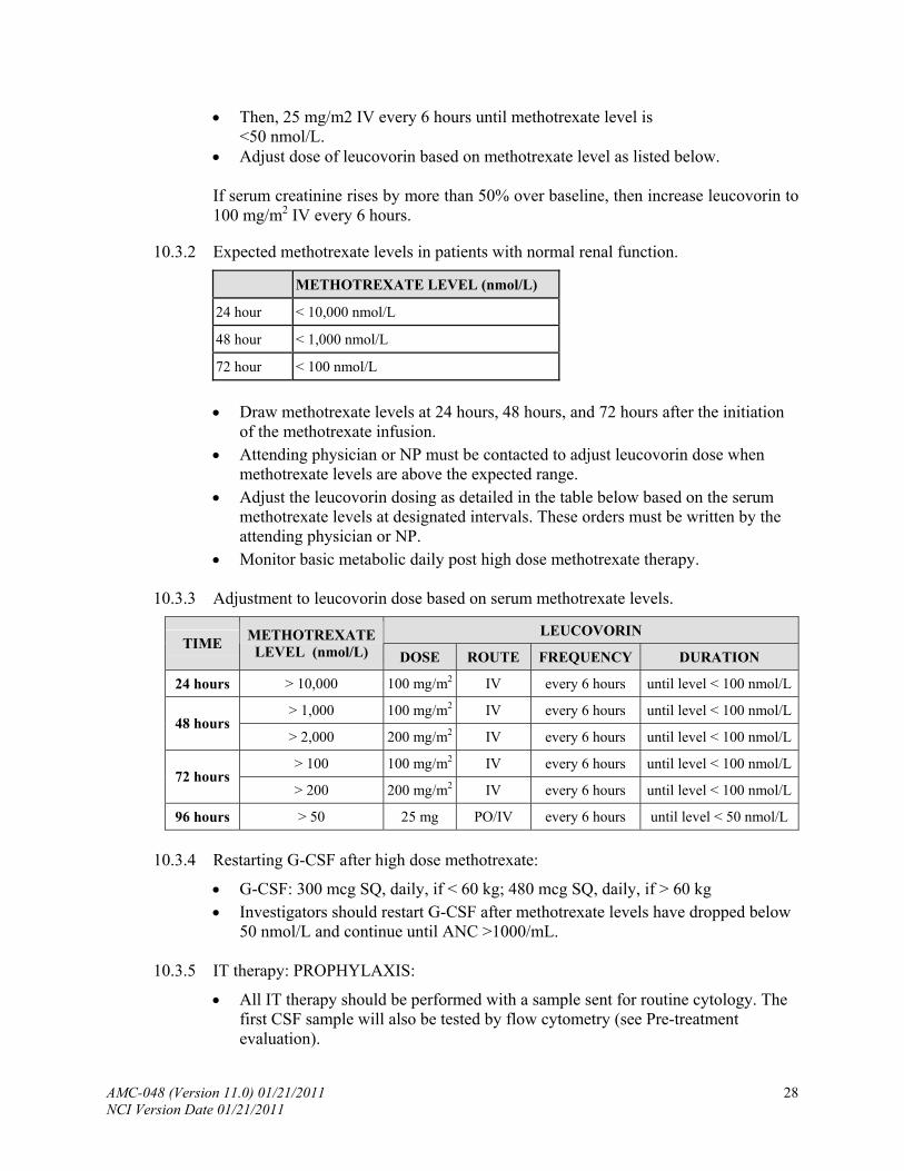
Then, 25 mg/m2 IV every 6 hours until methotrexate level is <50 nmol/L.
Adjust dose of leucovorin based on methotrexate level as listed below.
If serum creatinine rises by more than 50% over baseline, then increase leucovorin to 100 mg/m2 IV every 6 hours.
10.3.2 Expected methotrexate levels in patients with normal renal function.
METHOTREXATE LEVEL (nmol/L)
24 hour < 10,000 nmol/L
48 hour < 1,000 nmol/L
72 hour < 100 nmol/L
Draw methotrexate levels at 24 hours, 48 hours, and 72 hours after the initiation
of the methotrexate infusion. Attending physician or NP must be contacted to adjust leucovorin dose when
methotrexate levels are above the expected range. Adjust the leucovorin dosing as detailed in the table below based on the serum
methotrexate levels at designated intervals. These orders must be written by the attending physician or NP.
Monitor basic metabolic daily post high dose methotrexate therapy.
10.3.3 Adjustment to leucovorin dose based on serum methotrexate levels.
LEUCOVORIN TIME
METHOTREXATE LEVEL (nmol/L) DOSE ROUTE FREQUENCY DURATION
> 10,000 100 mg/m2 IV every 6 hours until level < 100 nmol/L24 hours
100 mg/m2 IV every 6 hours until level < 100 nmol/L> 1,000 48 hours
> 2,000 200 mg/m2 IV every 6 hours until level < 100 nmol/L
100 mg/m2 IV every 6 hours until level < 100 nmol/L> 100 72 hours
> 200 200 mg/m2 IV every 6 hours until level < 100 nmol/L
96 hours > 50 25 mg PO/IV every 6 hours until level < 50 nmol/L
10.3.4 Restarting G-CSF after high dose methotrexate:
G-CSF: 300 mcg SQ, daily, if < 60 kg; 480 mcg SQ, daily, if > 60 kg Investigators should restart G-CSF after methotrexate levels have dropped below
50 nmol/L and continue until ANC >1000/mL.
10.3.5 IT therapy: PROPHYLAXIS:
All IT therapy should be performed with a sample sent for routine cytology. The first CSF sample will also be tested by flow cytometry (see Pre-treatment evaluation).
AMC-048 (Version 11.0) 01/21/2011 28 NCI Version Date 01/21/2011
Methotrexate 12 mg IT mixed with cytarabine 50 mg IT given Day 1. High risk patients receive an additional dose of cytarabine 50 mg IT on Day 3. All methotrexate and cytrabine IT therapy is mixed with 50 mg hydrocortisone.
10.3.6 IT Therapy: TREATMENT of CNS DISEASE
Patients with CNS involvement (leptomeningeal and/or intraparenchymal) at diagnosis do not receive this schedule of IT therapy. Instead, they receive a combination of liposomal cytarabine (Depocyt), methotrexate and cytarabine as follows: Cycle 1: Regimen A: CODOX-M: CNS disease will likely be diagnosed with either imaging or with first lumbar puncture (LP). In the former case, the Day 1 cytarabine/methotrexate combination should be replaced by liposomal cytarabine (Depocyt) 50 mg once. In the latter case, i.e. CNS diagnosis made by the first LP, the Day 3 cyatrabine should be replaced with liposomal cytarabine (Depocyt) 50 mg.
If the CNS disease is diagnosed with the Day 3 LP, the subject should receive an
additional dose of liposomal cytarabine (Depocyt) 50 mg on Day 5. High dose methotrexate IV proceeds on Day 15 in each of the above 3 scenarios. Prophylaxis for liposomal cytarabine is given as follows: dexamethasone 4 mg
PO BID x 4 days beginning 1-3 hours BEFORE the dose of IT therapy. When feasible, patients with CNS disease should have an Ommaya reservoir
placed for optimal therapy administration as this may improve efficacy, tolerability and safety.
In the unlikely event that a subject with low risk disease (see Section 10.1) has a positive CSF cytology, he/she will be reassigned to the high risk group. Specifically, this patient will receive 4 cycles as AB/AB (CODOX-M/IVAC/CODOX-M/IVAC).
Cycle 2: Regimen B: IVAC: DO NOT administer liposomal cytarabine (Depocyt) with this cycle. Administer methotrexate 12 mg IT/IO (intrathecal or intra-Ommaya) on Day 5 of this cycle. Add two additional doses of methtrexate 12 mg on Day 7 and Day 9. All methotrexate IT/IO therapy is given with hydrocortisone 50 mg IT/IO. RATIONALE: In small trials of leptomeningeal lymphoma, liposomal cytarabine (Depocyt) 50 mg appears to have increased efficacy when compared to cytarabine with respect to improvement in neurological symptoms. However, SEVERE neurotoxicity, including DEATH, has been reported in 16% of patients who received liposomal cytarabine following moderate dose methotrexate IV and high dose cytrabine.
[43]
[44]
Cycle 3: Regimen A: CODOX-M: Administer one dose of liposomal cytarabine (Depocyt) 50 mg on Day 1. Do not administer any additional IT/IO therapy. Proceed with pegfilgrastim on Day 3 and methotrexate IV on Day 15. Cycle 4: Regimen B: IVAC: DO NOT administer liposomal cytarabine (Depocyt) with this cycle. Administer methotrexate 12 mg IT/IO (intrathecal or intra-Ommaya)
AMC-048 (Version 11.0) 01/21/2011 29 NCI Version Date 01/21/2011
on Day 5 of this cycle. Methotrexate IT/IO therapy is given with hydrocortisone 50 mg IT/IO.
10.4 Re-treatment on Regimen A or B
Begins when the ANC reaches 1000/µL and platelets reach 75,000/µL (patients with platelets between 50,000 and 75,000 at baseline that is not lymphoma related should have return to baseline platelet count). Ideally, up to one additional week is allowed for a patient to recover from fatigue, recent hospitalization or other Grade 3-4, non-hematologic toxicity. Typically cycles last for 21-28 days. See section 13.0 for criteria for study withdrawal.
10.5 Regimen B: IVAC
10.5.1 Rituximab 375 mg/m2 IVPB on Day 1 (considerations for moving the timing of the dose are noted in Section 10.1.3).
10.5.2 Ifosfamide 1500 mg/m² IVCI over 24 hours daily x 5 doses, starting on Day 1.
10.5.3 Mensa 1500 mg/m² IVCI over 24 hours daily x 5 doses starting on Day 1.
10.5.4 Etoposide 60 mg/m² IVCI over 24 hours daily x 5 doses starting on Day 1.
10.5.5 Cytarabine 2000 mg/m² IVPB over 1 hour (no cap) every 12 hours x 4 doses starting
on Day 1.
10.5.6 Methotrexate 12 mg IT given on, or about Day 5 (once during the admission for
chemotherapy). All IT therapy is mixed with 50 mg hydrocortisone.
Patients with CNS involvement (leptomeningeal and/or intraparenchymal) at diagnosis do not receive this schedule of IT therapy. Instead, they should follow the schedule for treatment of CNS disease outline in Section 10.3.6.
10.5.7 Pegfilgrastim (Neulasta) 6mg subcutaneously should be given 24–48 hours after
completion of chemotherapy.
10.5.8 Pegfilgrastim (Neulasta) - If not available, G-CSF daily until ANC > 1000/µL can be
substituted.
10.5.9 Returning to Regimen A: Regimen A begins when the ANC reaches 1000/µL and
platelets reach 75,000/µL. Up to one additional week is allowed for patient to recover from fatigue, recent hospitalization or other toxicity. (2) platelets > 75,000/µL (if at least 75,000/µL at Baseline), (patients with platelets between 50,000 and 75,000 at baseline that is not lymphoma related should have return to baseline platelet count).
AMC-048 (Version 11.0) 01/21/2011 30 NCI Version Date 01/21/2011
10.6 Prophylactic Antibiotics
All patients should be on prophylaxis for pneumocystis (PCP) as follows: pentamidine 300 mg aerosolized monthly. (Clinicians may substitute another standard form of PCP prophylaxis, but should be aware that trimpethoprim/sulfamehtoxazole will likely contribute to neutropenia). Famciclovir 250 mg PO BID. (Clinicians may substitute another standard form of herpes virus prophylaxis, but should be aware that acyclovir or valcyclovir will likely contribute to neutropenia).
Mycobacterium avium complex (MAC) prophylaxis should be given to all patients with CD4<50: azithromycin 1200 mg weekly. It should be considered that patients with baseline low CD4 counts may drop their CD4 count below 50 during treatment, but a reevaluation of the CD4 count during treatment and prophylaxis for MAC is at the discretion of the treating physician.
Quinolone prophylaxis: Antibiotic prophylaxis will be used on account of the anticipated depth and duration of neutropenia, the use of rituximab, and the inherent immunocompromised state of HIV-infected patients. Quinolone prophylaxis should be given as follows:
R-CODOX-M cycles: Begin a quinolone (choice at the Investigator’s discretion) Day 8 and continue for 8 days unless the patient is still neutropenic, in which case, prophylaxis should be continued until the ANC>1000 cells/mL. If the ANC recovers before Day 16, antibiotics maybe be discontinued to minimize antibiotic exposure, unless antibiotics are required clinically to treat an interim infection. Prophylactic antibiotics are also suggested after high dose methotrexate on Day 15 if the patient becomes neutropenic.
IVAC cycles: Begin a quinolone (choice at the Investigator’s discretion) on Day 8 and continue for 8 days unless the patient is still neutropenic, in which case, prophylaxis should be continued until the ANC>1000 cells/mL. If the ANC recovers before Day 16, antibiotics maybe be discontinued to minimize antibiotic exposure, unless antibiotics are required clinically to treat an interim infection.
Patients who are allergic or intolerant of quinolones are advised to use an alternative antibiotic. The prophylactic antibiotic should be recorded in the concomitant medication form.
In the event of febrile neutropenia or other infection, prophylactic quinolone antibiotics may be discontinued and therapeutic antibiotics instituted as appropriate. It should be noted that nephrotoxic antibiotics, including aminoglycosides, should be avoided if at all possible during and after high-dose methotrexate, for at least 7 days.
AFTER high-dose methotrexate and after IVAC: CBCs should be monitored at the discretion of the treating physician whether the patient is inpatient or outpatient. Appropriate support should be given in the form of transfusions and erythropoietic hormones at the discretion of the investigator. CBCs can be discontinued when it is clear that the patient is no longer going to require transfusion support for a given cycle.
AMC-048 (Version 11.0) 01/21/2011 31 NCI Version Date 01/21/2011
10.7 Concurrent Highly Active Antiretroviral Therapy (HAART)
The treating physician(s) will choose an antiretroviral strategy at the time of registration onto the study and this will be noted on the patient case report form (CRF).
The use of HAART will be at the discretion of the individual Investigator. Physicians may continue or discontinue therapy at the initiation of chemotherapy. In addition, HAART may be discontinued as needed for toxicity. If using HAART, a multiagent program of at least three drugs is required. At no time should zidovudine (AZT) be used concurrently, given its myelosuppression. Concurrent HAART therapy should be listed in detail on the patient’s CRF.
Concurrent vs. discontinued HAART therapy is a controversial area of investigation. To wit, fearing drug interactions, the NCI investigators discontinued HAART therapy quite successfully during EPOCH for HIV-associated NHL (largely DLBCL) with spectacular results.[41] HIV suppression after chemotherapy is highly desirable and all patients completing treatment should receive HAART with the intent of maximal HIV suppression and immune reconstitution.
Investigators should also be aware of drug interactions which can affect the levels of chemotherapy drugs. The implications of these interactions are not known at this time. However, this is particularly true of many of the protease inhibitors as they inhibit the CYP 3A cytochrome pathway. Investigators may want to switch to HAART agents that are not CYP 3A inhibitors or withhold HAART during therapy.
10.8 Tumor Lysis Syndrome
Investigators should be aware that tumor lysis and tumor lysis syndrome during the first treatment are anticipated complications of high-grade lymphomas, particularly in the setting of high tumor burden or high LDH. Investigators should follow the standard of care and, if applicable, institutional guidelines for the prevention and/or treatment of tumor lysis. This should include, but is not limited to aggressive IV fluid hydration, monitoring of electrolytes, prophylaxis against hyperuricemia, and management of hyperphosphatemia, hyperkelmia, hypocalcemia and hyperphosphatemia.
AMC-048 (Version 11.0) 01/21/2011 32 NCI Version Date 01/21/2011
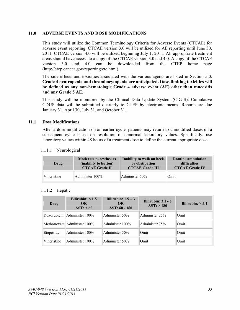
11.0 ADVERSE EVENTS AND DOSE MODIFICATIONS
This study will utilize the Common Terminology Criteria for Adverse Events (CTCAE) for adverse event reporting. CTCAE version 3.0 will be utilized for AE reporting until June 30, 2011. CTCAE version 4.0 will be utilized beginning July 1, 2011. All appropriate treatment areas should have access to a copy of the CTCAE version 3.0 and 4.0. A copy of the CTCAE version 3.0 and 4.0 can be downloaded from the CTEP home page (http://ctep.cancer.gov/reporting/ctc.html).
The side effects and toxicities associated with the various agents are listed in Section 5.0. Grade 4 neutropenia and thrombocytopenia are anticipated. Dose-limiting toxicities will be defined as any non-hematologic Grade 4 adverse event (AE) other than mucositis and any Grade 5 AE.
This study will be monitored by the Clinical Data Update System (CDUS). Cumulative CDUS data will be submitted quarterly to CTEP by electronic means. Reports are due January 31, April 30, July 31, and October 31.
11.1 Dose Modifications
After a dose modification on an earlier cycle, patients may return to unmodified doses on a subsequent cycle based on resolution of abnormal laboratory values. Specifically, use laboratory values within 48 hours of a treatment dose to define the current appropriate dose.
11.1.1 Neurological
Drug Moderate paresthesias
(inability to button) CTCAE Grade II
Inability to walk on heels or obstipation
CTCAE Grade III
Routine ambulation difficulties
CTCAE Grade IV
Vincristine Administer 100% Administer 50% Omit
11.1.2 Hepatic
Drug Bilirubin: < 1.5
OR AST: < 60
Bilirubin: 1.5 – 3 OR
AST: 60 - 180
Bilirubin: 3.1 - 5 AST: > 180
Bilirubin: > 5.1
Doxorubicin Administer 100% Administer 50% Administer 25% Omit
Methotrexate Administer 100% Administer 100% Administer 75% Omit
Etoposide Administer 100% Administer 50% Omit Omit
Vincristine Administer 100% Administer 50% Omit Omit
AMC-048 (Version 11.0) 01/21/2011 33 NCI Version Date 01/21/2011
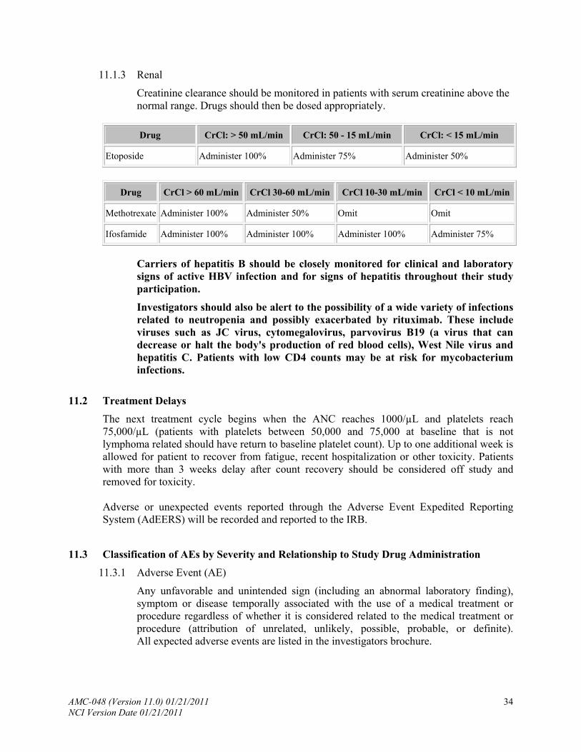
11.1.3 Renal
Creatinine clearance should be monitored in patients with serum creatinine above the normal range. Drugs should then be dosed appropriately.
Drug CrCl: > 50 mL/min CrCl: 50 - 15 mL/min CrCl: < 15 mL/min
Etoposide Administer 100% Administer 75% Administer 50%
Drug CrCl > 60 mL/min CrCl 30-60 mL/min CrCl 10-30 mL/min CrCl < 10 mL/min
Methotrexate Administer 100% Administer 50% Omit Omit
Ifosfamide Administer 100% Administer 100% Administer 100% Administer 75%
Carriers of hepatitis B should be closely monitored for clinical and laboratory signs of active HBV infection and for signs of hepatitis throughout their study participation.
Investigators should also be alert to the possibility of a wide variety of infections related to neutropenia and possibly exacerbated by rituximab. These include viruses such as JC virus, cytomegalovirus, parvovirus B19 (a virus that can decrease or halt the body's production of red blood cells), West Nile virus and hepatitis C. Patients with low CD4 counts may be at risk for mycobacterium infections.
11.2 Treatment Delays
The next treatment cycle begins when the ANC reaches 1000/µL and platelets reach 75,000/µL (patients with platelets between 50,000 and 75,000 at baseline that is not lymphoma related should have return to baseline platelet count). Up to one additional week is allowed for patient to recover from fatigue, recent hospitalization or other toxicity. Patients with more than 3 weeks delay after count recovery should be considered off study and removed for toxicity. Adverse or unexpected events reported through the Adverse Event Expedited Reporting System (AdEERS) will be recorded and reported to the IRB.
11.3 Classification of AEs by Severity and Relationship to Study Drug Administration
11.3.1 Adverse Event (AE)
Any unfavorable and unintended sign (including an abnormal laboratory finding), symptom or disease temporally associated with the use of a medical treatment or procedure regardless of whether it is considered related to the medical treatment or procedure (attribution of unrelated, unlikely, possible, probable, or definite). All expected adverse events are listed in the investigators brochure.
AMC-048 (Version 11.0) 01/21/2011 34 NCI Version Date 01/21/2011
11.3.2 Life-Threatening AE
Any adverse event that places the patient or subject, in view of the investigator, at immediate risk of death from the reaction as it occurred, i.e., it does not include a reaction that, had it occurred in a more severe form, might have caused death.
11.3.3 Serious Adverse Event (SAE)
Any adverse drug experience occurring at any dose that results in any of the following outcomes: death, a life-threatening adverse drug experience, inpatient hospitalization, or prolongation of existing hospitalization, a persistent or significant disability/incapacity, or a congenital anomaly/birth defect. Important medical events that may not result in death, be life threatening, or require hospitalization may be considered a serious adverse drug experience when, based upon medical judgment, they may jeopardize the patient or subject and may require medical or surgical intervention to prevent one of the outcomes listed in this definition.
Please note for hospitalization – For this study, hospitalization is defined as an inpatient hospital stay equal to or greater than 24 hours. A hospital visit where a patient is admitted for observation or minor treatment (e.g., hydration) and released in less than 24 hours would not meet the requirements for hospitalization.
11.3.4 Toxicity
Toxicity is a term NOT clearly defined by regulatory organizations. Toxicity has been described as an adverse event that has an attribution of possibly, probably or definitely related to investigational treatment. It is recommended that the term toxicity NOT be utilized for adverse event reporting purposes.
11.3.5 Unexpected AE
Unexpected adverse events for commercial agents are those not listed in available sources including the package insert, the Investigator’s Brochure, or the protocol.
11.3.6 Attribution
The determination of whether an adverse event is related to a medical treatment or procedure.
Attribution categories:
Definite - The AE is clearly related to the investigational agent(s). Probable - The AE is likely related to the investigational agent(s). Possible - The AE may be related to the investigational agent(s). Unlikely - The AE is doubtfully related to the investigational agent(s). Unrelated - The AE is clearly NOT related to the investigational agent(s).
11.4 Expedited AE Reporting
11.4.1 Expedited AE reporting for this study must use AdEERS, accessed via the CTEP home page (http://ctep.cancer.gov). The reporting procedures to be followed are
AMC-048 (Version 11.0) 01/21/2011 35 NCI Version Date 01/21/2011
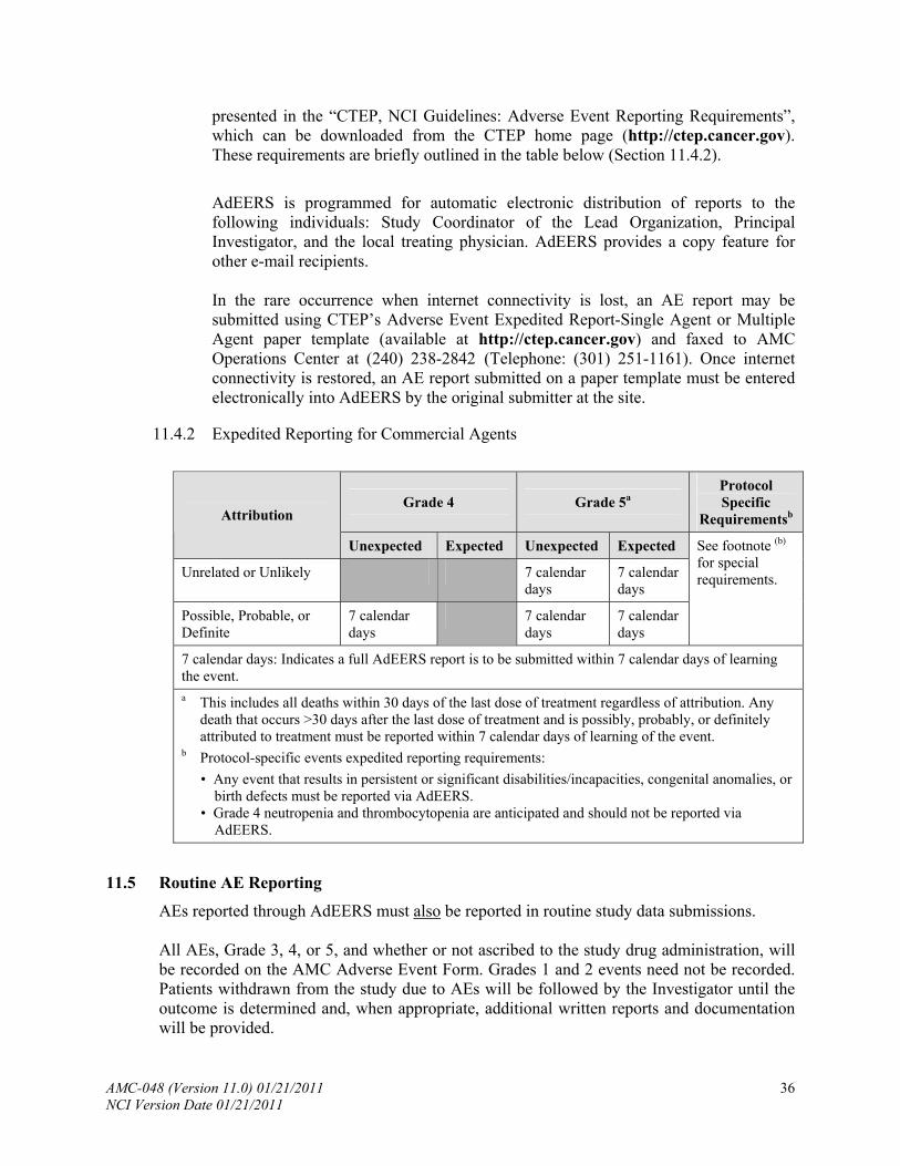
presented in the “CTEP, NCI Guidelines: Adverse Event Reporting Requirements”, which can be downloaded from the CTEP home page (http://ctep.cancer.gov). These requirements are briefly outlined in the table below (Section 11.4.2).
AdEERS is programmed for automatic electronic distribution of reports to the following individuals: Study Coordinator of the Lead Organization, Principal Investigator, and the local treating physician. AdEERS provides a copy feature for other e-mail recipients. In the rare occurrence when internet connectivity is lost, an AE report may be submitted using CTEP’s Adverse Event Expedited Report-Single Agent or Multiple Agent paper template (available at http://ctep.cancer.gov) and faxed to AMC Operations Center at (240) 238-2842 (Telephone: (301) 251-1161). Once internet connectivity is restored, an AE report submitted on a paper template must be entered electronically into AdEERS by the original submitter at the site.
11.4.2 Expedited Reporting for Commercial Agents
Grade 4 Grade 5a Protocol Specific
Requirementsb Attribution
Unexpected Expected Unexpected Expected
Unrelated or Unlikely 7 calendar days
7 calendar days
7 calendar days
7 calendar days
See footnote (b) for special requirements.
Possible, Probable, or Definite
7 calendar days
7 calendar days: Indicates a full AdEERS report is to be submitted within 7 calendar days of learning the event. a This includes all deaths within 30 days of the last dose of treatment regardless of attribution. Any
death that occurs >30 days after the last dose of treatment and is possibly, probably, or definitely attributed to treatment must be reported within 7 calendar days of learning of the event.
b Protocol-specific events expedited reporting requirements:
• Any event that results in persistent or significant disabilities/incapacities, congenital anomalies, or birth defects must be reported via AdEERS.
• Grade 4 neutropenia and thrombocytopenia are anticipated and should not be reported via AdEERS.
11.5 Routine AE Reporting
AEs reported through AdEERS must also be reported in routine study data submissions. All AEs, Grade 3, 4, or 5, and whether or not ascribed to the study drug administration, will be recorded on the AMC Adverse Event Form. Grades 1 and 2 events need not be recorded. Patients withdrawn from the study due to AEs will be followed by the Investigator until the outcome is determined and, when appropriate, additional written reports and documentation will be provided.
AMC-048 (Version 11.0) 01/21/2011 36 NCI Version Date 01/21/2011
AMC-048 (Version 11.0) 01/21/2011 37 NCI Version Date 01/21/2011
11.6 Secondary AML/MDS/ALL
All cases of acute myeloid leukemia (AML), myelodysplastic syndrome (MDS), and acute lymphocytic leukemia (ALL) that occur in patients on or following treatment on NCI-sponsored chemotherapy protocols must be reported to the AMC Operations Center using the NCI/CTEP Secondary AML/MDS Report Form. This form can be downloaded from the CTEP web site (http://ctep.cancer.gov). Refer to the “CTEP, NCI Guidelines: Adverse Event Reporting Requirements” (available at http://ctep.cancer.gov) for additional information about secondary AML/MDS reporting. The following must be submitted within 30 days of an AML/MDS/ALL diagnosis occurring after treatment for cancer on an NCI-sponsored trial:
A completed NCI/CTEP Secondary AML/MDS Report Form (do not use AdEERS); A copy of the pathology report confirming the AML/MDS/ALL; and A copy of the cytogenetics report (if available).
The AMC Operations Center will forward copies to the Investigational Drug Branch (IDB) of the NCI Cancer Therapy Evaluation Program (CTEP).
12.0 CRITERIA FOR THERAPEUTIC RESPONSE/OUTCOME ASSESSMENT
12.1 Response to Treatment
Response to treatment will be assessed after the post-treatment evaluation outlined in Section 9.0. The following response criteria will be employed:
12.1.1 Complete Response (CR)
1. Complete disappearance of all detectable clinical and radiographic evidence of disease and resolution of all disease-related symptoms. Any abnormal biochemical values (e.g. LDH) clearly attributable to lymphoma must have normalized.
2. All involved lymph nodes and nodal masses must measure < 1.5 cm in their short axes if they measured >1.5 cm prior to therapy. Involved nodes measuring 1.1 -1.5 cm prior to treatment must either measure < 1 cm or have a > 75% reduction in the sum of the products of the diameters (SPD).
3. If the bone marrow was involved with lymphoma, the repeat biopsy must be free of disease.
4. If the spleen was considered to be enlarged prior to treatment, it must have regressed in size and must not be palpable on exam. Similarly, other organs considered to be involved prior to therapy must have diminished in size. Macroscopic tumor nodules previously detectable on imaging studies in any organ must have resolved.
5. If the CSF was positive by cytology at diagnosis, it should be negative by cytology. If the CSF was positive by flow cytometry only at diagnosis, it should be monitored by flow cytometry until it becomes negative.
6. If a PET scan was obtained at diagnosis and/or restaging, the results should be recorded, yet they will not be considered in the criteria for CR.
12.1.2 Partial Response (PR)
1. > 50% decrease in SPD of the 6 largest dominant nodes or nodal masses.
2. No increase in size of other nodes, liver, or spleen.
3. Splenic and hepatic nodules must regress by at least 50% in the SPD.
4. Bone marrow and organs other than the spleen and liver cannot be considered for evaluation for PR because involvement in these sites is considered evaluable and not measurable.
5. No new sites of disease.
A PR is converted to a CR if biopsy confirms no evidence of disease.
AMC-048 (Version 11.0) 01/21/2011 38 NCI Version Date 01/21/2011
12.1.3 Stable Disease (SD)
Response is less than that which constitutes a PR and disease does not meet criteria for progressive disease.
12.1.4 Progressive Disease (PD)
1. > 50% increase in SPD of any previously identified abnormal node or nodule.
2. Appearance of any new lesion at the end of therapy.
AMC-048 (Version 11.0) 01/21/2011 39 NCI Version Date 01/21/2011
13.0 CRITERIA FOR REMOVAL FROM STUDY
The following patients will be removed from study:
1. Patients who have progression of disease while on treatment. These patients may be offered alternate treatments.
2. Patients who develop unacceptable toxicity precluding drug administration including uncontrolled opportunistic infections.*
3. Treatment delays greater than 4 weeks.
4. Patients who request that they be removed from study. This will not compromise the care they receive at the treating institution.
5. Patients who are non-compliant with treatment or follow-up. *For patients with ifosfamide encephalopathy, prophylactic therapy with methylene blue should be considered for the second cycle containing ifosfamide. It is highly recommend that investigators discuss toxicities with the protocol team in advance of protocol removal. NOTE: Patients removed from study remain in follow up for 2 years post completion of treatment to allow for analysis of overall survival. Case report forms include a notification of new lymphoma directed therapy whether it be protocol or not.
AMC-048 (Version 11.0) 01/21/2011 40 NCI Version Date 01/21/2011
14.0 STATISTICAL CONSIDERATIONS
14.1 Sample Size
The primary endpoint is one-year overall survival. A sample size of 31 patients is needed to test the null hypothesis that the one-year survival rate is 0.65[20] will be tested against the alternative that it is 0.85 at the one-sided 0.10 significance level with power of 0.90 using a nonparametric survival model. To allow for a 10% dropout rate, 34 patients will be enrolled.[45]
It is estimated that 10 patients will be enrolled nationally per year. This allows for one patient per core AMC site and 2 patients total for affiliated or enrolling sites. Accrual will require 3 years and all patients will be followed for a minimum of one year. The total study duration is expected to be 4 years.
14.2 Stopping Rule for Toxicity
Grade 4 neutropenia and thrombocytopenia are anticipated.
A stopping rule for treatment-related, including infection-related death, was derived by determining the number of infection-related deaths, which would occur 10% of the time if the true infection-related mortality rate was 4%. There was a 2% incidence of infection-related deaths in patients treated with CHOP alone in a recently completed AMC Study (AMC-010). This stopping rule is designed to detect a two-fold or greater increased risk of treatment related death in this study. The stopping rule has a power of 0.90.
The trial will be stopped if the number of treatment-related deaths (X) is greater than or equal to the following:
X > When N =
2 2-13
3 14-28
4 29-34
14.3 Statistical Analysis
Kaplan-Meier curves will be generated to describe OS, EFS and failure-free survival. The median survival time, EFS time and failure-free survival times will be estimated using the method of Brookmeyer and Crowley [45] The one-year OS rate will be estimated as the cumulative proportion surviving at one year and by its 95% confidence interval, estimated using the standard errors derived using Greenwood’s formula. The CR rate will be estimated using the binomial proportion and its exact 95% confidence interval.
Toxicity data will be presented by severity. The incidence of toxicities will be estimated using the binomial proportion and its exact 95% confidence interval. The incidence of infection-related deaths will be estimated using the binomial proportion and its exact 95% confidence interval. The results of the safety evaluation will be tabulated. The log-rank test will be used to evaluate c-flip expression, p53 expression, and MDR expression separately on OS, failure-free survival and EFS. If two or more of these factors are found to be associated with any of the time to event endpoints, the proportional hazards
AMC-048 (Version 11.0) 01/21/2011 41 NCI Version Date 01/21/2011
model will be used to assess the two factors together on the time to event endpoint. To evaluate the utility of flow cytometry in detecting leptomeningeal disease, McNemar’s chi-square test will be used. The degree of disconcordance between the flow cytometry and CNS cytology results will be estimated using the binomial proportion and its 95% confidence interval.
The log-rank test will be used to evaluate the association of EBV and the time to event endpoints. The proportional hazards model will be used to evaluate the role of EBV load measurements and the time to event endpoints. If the EBV load measurements are not normally distributed, a normalizing transformation may be used.
14.4 Data Safety and Monitoring Plan
This protocol will follow the AMC’s policy for data monitoring (See Appendix VII).
AMC-048 (Version 11.0) 01/21/2011 42 NCI Version Date 01/21/2011
15.0 DATA MANAGEMENT
CRFs will be provided for each subject via the AMC AdvantageEDCSM Internet data entry system upon enrollment. Subjects will not be identified by name on any study document. Data will be recorded on the CRFs using the unique subject identification number assigned at registration.
All signs, symptoms, HIV-related and AIDS-defining events, AEs, laboratory results and deaths must be recorded on the CRFs. Sample CRFs will be available on the AMC Operations Center website. Instructions concerning the recording of study data on CRFs will be provided by the Operations Center.
AMC-048 (Version 11.0) 01/21/2011 43 NCI Version Date 01/21/2011
AMC-048 (Version 11.0) 01/21/2011 44 NCI Version Date 01/21/2011
16.0 PROTECTION OF HUMAN SUBJECTS
16.1 Informed Consent
The principles of informed consent described in the Food and Drug Administration (FDA) regulations (21 CFR part 50) must be followed. IRB approval of the protocol and the informed consent form must be given in writing. Participating sites must forward a copy of the letter of approval from the IRB, which specifically approves the protocol and informed consent, to the AMC Operations Center before patient enrollment.
The IRB must also approve any significant changes to the protocol and documentation of this approval must be sent to the AMC Operations Center. Records of all study review and approval documents must be kept on file by the investigator and are subject to FDA inspection during or after completion of the study. Adverse events (AEs) must be reported to the IRB.
The IRB should receive notification of completion of the study. The Investigator will maintain an accurate and complete record of all submissions made to the IRB, including a list of all reports and documents submitted.
16.2 Research Authorization
Each institution should insert the appropriate research authorization sections into the informed consent document.
16.3 Subject Confidentiality
To maintain confidentiality, all laboratory specimens, evaluation forms, reports and other records will be identified by a coded number only. Clinical information will not be released without written permission of the subject, except as necessary for monitoring by the FDA, the AMC or the NCI.
16.4 Women and Minorities
This is a study being conducted by the NCI-sponsored AIDS Malignancy Consortium (AMC). Each participating site within the AMC, and the AMC as a whole, are required to assure that the participation of women and minority subjects reflects the percentage representation of these populations in their geographic region and, for the AMC, the United States as a whole. As such, it is expected that the representation of subjects on this trial will reflect the constitution of the respective populations.
17.0 REFERENCES
1. Lacasce, A., O. Howard, S. Lib, et al., Modified magrath regimens for adults with Burkitt and Burkitt-like lymphomas: preserved efficacy with decreased toxicity. Leuk Lymphoma, 2004. 45(4): p. 761-7.
2. The Non-Hodgkin’s Lymphoma Pathologic Project. National Cancer Institute sponsored study of classification of non-Hodgkin’s lymphomas. Summary and description of a working formulation for clinical usage. Cancer, 1982. 49: p. 2112.
3. Harris, N.L., E.S. Jaffe, H. Stein, et al., A revised European-American classification of lymphoid neoplasms: a proposal from the International Lymphoma Study Group. Blood, 1994. 84(5): p. 1361-92.
4. Harris, N.L., E.S. Jaffe, J. Diebold, et al., World Health Organization classification of neoplastic diseases of the hematopoietic and lymphoid tissues: report of the Clinical Advisory Committee meeting-Airlie House, Virginia, November 1997. J Clin Oncol, 1999. 17(12): p. 3835-49.
5. Burkitt, D., Long-term remissions following one and two-dose chemotherapy for African lymphoma. Cancer, 1967. 20(5): p. 756-9.
6. Ostronoff, M., C. Soussain, E. Zambon, et al., Burkitt's lymphoma in adults: a retrospective study of 46 cases. Nouv Rev Fr Hematol, 1992. 34(5): p. 389-97.
7. Straus, D.J., Treatment of Burkitt's lymphoma in HIV-positive patients. Biomed Pharmacother, 1996. 50(9): p. 447-50.
8. Kaplan, L.D., D.J. Straus, M.A. Testa, et al., Low-dose compared with standard-dose m-BACOD chemotherapy for non-Hodgkin's lymphoma associated with human immunodeficiency virus infection. National Institute of Allergy and Infectious Diseases AIDS Clinical Trials Group. N Engl J Med, 1997. 336: p. 1641-8.
9. Levine, A.M., J. Sullivan-Halley, M.C. Pike, et al., Human immunodeficiency virus-related lymphoma. Prognostic factors predictive of survival. Cancer, 1991. 68(11): p. 2466-72.
10. Magrath, I., M. Adde, A. Shad, et al., Adults and children with small non-cleaved-cell lymphoma have a similar excellent outcome when treated with the same chemotherapy regimen. J Clin Oncol, 1996. 14(3): p. 925-34.
11. Mead, G.M., M.R. Sydes, J. Walewski, et al., An international evaluation of CODOX-M and CODOX-M alternating with IVAC in adult Burkitt's lymphoma: results of United Kingdom Lymphoma Group LY06 study. Ann Oncol, 2002. 13(8): p. 1264-74.
12. Thomas, D.A., J. Cortes, S. O'Brien, et al., Hyper-CVAD program in Burkitt's-type adult acute lymphoblastic leukemia. J Clin Oncol, 1999. 17(8): p. 2461-70.
13. Patte, C., J. Michon, D. Frappaz, et al., Therapy of Burkitt and other B-cell acute lymphoblastic leukaemia and lymphoma: experience with the LMB protocols of the SFOP (French Paediatric Oncology Society) in children and adults. Baillieres Clin Haematol, 1994. 7(2): p. 339-48.
14. Carbone, A., A. Gloghini, G. Gaidano, et al., AIDS-related Burkitt's lymphoma. Morphologic and immunophenotypic study of biopsy specimens. Am J Clin Pathol, 1995. 103(5): p. 561-7.
15. Levine, A.M., Acquired immunodeficiency syndrome-related lymphoma. Blood, 1992. 80: p. 8.
16. Beral, V., T. Peterman, R. Berkelman, and H. Jaffe, AIDS-associated non-Hodgkin lymphoma. Lancet, 1991. 337(8745): p. 805-9.
AMC-048 (Version 11.0) 01/21/2011 45 NCI Version Date 01/21/2011
17. Spina, M., U. Tirelli, V. Zagonel, et al., Burkitt's lymphoma in adults with and without human immunodeficiency virus infection: a single-institution clinicopathologic study of 75 patients. Cancer, 1998. 82(4): p. 766-74.
18. Davi, F., H.J. Delecluse, P. Guiet, et al., Burkitt-like lymphomas in AIDS patients: characterization within a series of 103 human immunodeficiency virus-associated non-Hodgkin's lymphomas. Burkitt's Lymphoma Study Group. J Clin Oncol, 1998. 16(12): p. 3788-95.
19. Gill, P.S., A.M. Levine, M. Krailo, et al., AIDS-related malignant lymphoma: results of prospective treatment trials. J Clin Oncol, 1987. 5(9): p. 1322-8.
20. Lim, S.T., R. Karim, B.N. Nathwani, et al., AIDS-related Burkitt's lymphoma versus diffuse large-cell lymphoma in the pre-highly active antiretroviral therapy (HAART) and HAART eras: significant differences in survival with standard chemotherapy. J Clin Oncol, 2005. 23(19): p. 4430-8.
21. Wang, E.S., D.J. Straus, J. Teruya-Feldstein, et al., Intensive chemotherapy with cyclophosphamide, doxorubicin, high-dose methotrexate/ifosfamide, etoposide, and high-dose cytarabine (CODOX-M/IVAC) for human immunodeficiency virus-associated Burkitt lymphoma. Cancer, 2003. 98(6): p. 1196-205.
22. Kiyokawa, N., T. Mori, T. Taguchi, et al., Activation of the caspase cascade during Stx1-induced apoptosis in Burkitt's lymphoma cells. J Cell Biochem, 2001. 81(1): p. 128-42.
23. Valnet-Rabier, M.B., B. Challier, S. Thiebault, et al., c-Flip protein expression in Burkitt's lymphomas is associated with a poor clinical outcome. Br J Haematol, 2005. 128(6): p. 767-73.
24. Irmler, M., M. Thome, M. Hahne, et al., Inhibition of death receptor signals by cellular FLIP. Nature, 1997. 388(6638): p. 190-5.
25. Thome, M., P. Schneider, K. Hofmann, et al., Viral FLICE-inhibitory proteins (FLIPs) prevent apoptosis induced by death receptors. Nature, 1997. 386(6624): p. 517-21.
26. Scaffidi, C., I. Schmitz, P.H. Krammer, and M.E. Peter, The role of c-FLIP in modulation of CD95-induced apoptosis. J Biol Chem, 1999. 274(3): p. 1541-8.
27. Levine, A.M., A. Tulpule, B. Espina, et al., Liposome-encapsulated doxorubicin in combination with standard agents (cyclophosphamide, vincristine, prednisone) in patients with newly diagnosed AIDS-related non-Hodgkin's lymphoma: results of therapy and correlates of response. J Clin Oncol, 2004. 22(13): p. 2662-70.
28. Hegde, U., A. Filie, R.F. Little, et al., High incidence of occult leptomeningeal disease detected by flow cytometry in newly diagnosed aggressive B-cell lymphomas at risk for central nervous system involvement: the role of flow cytometry versus cytology. Blood, 2005. 105(2): p. 496-502.
29. Lo, Y.M., A.T. Chan, L.Y. Chan, et al., Molecular prognostication of nasopharyngeal carcinoma by quantitative analysis of circulating Epstein-Barr virus DNA. Cancer Res, 2000. 60(24): p. 6878-81.
30. Lin, J.C., W.Y. Wang, K.Y. Chen, et al., Quantification of plasma Epstein-Barr virus DNA in patients with advanced nasopharyngeal carcinoma. N Engl J Med, 2004. 350(24): p. 2461-70.
31. Lei, K.I., L.Y. Chan, W.Y. Chan, et al., Diagnostic and prognostic implications of circulating cell-free Epstein-Barr virus DNA in natural killer/T-cell lymphoma. Clin Cancer Res, 2002. 8(1): p. 29-34.
AMC-048 (Version 11.0) 01/21/2011 46 NCI Version Date 01/21/2011
AMC-048 (Version 11.0) 01/21/2011 47 NCI Version Date 01/21/2011
32. Dave, S.S., K. Fu, G.W. Wright, et al., Molecular diagnosis of Burkitt's lymphoma. N Engl J Med, 2006. 354(23): p. 2431-42.
33. Harris, N.L. and S.J. Horning, Burkitt's lymphoma--the message from microarrays. N Engl J Med, 2006. 354(23): p. 2495-8.
34. Hummel, M., S. Bentink, H. Berger, et al., A biologic definition of Burkitt's lymphoma from transcriptional and genomic profiling. N Engl J Med, 2006. 354(23): p. 2419-30.
35. Cingolani, A., R. Gastaldi, L. Fassone, et al., Epstein-Barr virus infection is predictive of CNS involvement in systemic AIDS-related non-Hodgkin's lymphomas. J Clin Oncol, 2000. 18(19): p. 3325-30.
36. Coiffier, B., E. Lepage, J. Briere, et al., CHOP chemotherapy plus rituximab compared with CHOP alone in elderly patients with diffuse large-B-cell lymphoma. N Engl J Med, 2002. 346(4): p. 235-42.
37. Marcus, R., K. Imrie, A. Belch, et al., CVP chemotherapy plus rituximab compared with CVP as first-line treatment for advanced follicular lymphoma. Blood, 2005. 105(4): p. 1417-23.
38. Kaplan, L.D., J.Y. Lee, R.F. Ambinder, et al., Rituximab does not improve clinical outcome in a randomized phase III trial of CHOP with or without rituximab in patients with HIV-associated non-Hodgkin's lymphoma: AIDS-malignancies consortium trial 010. Blood, 2005. 106(5) p. 1538-43.
39. Thomas, D.A., S. Faderl, S. O'Brien, et al., Chemoimmunotherapy with hyper-CVAD plus rituximab for the treatment of adult Burkitt and Burkitt-type lymphoma or acute lymphoblastic leukemia. Cancer, 2006. 106(7): p. 1569-80.
40. Tirelli, U., M. Spina, U. Jaeger, et al., Infusional CDE with rituximab for the treatment of human immunodeficiency virus-associated non-Hodgkin's lymphoma: preliminary results of a phase I/II study. Recent Results Cancer Res, 2002. 159: p. 149-53.
41. Little, R.F., S. Pittaluga, N. Grant, et al., Highly effective treatment of acquired immunodeficiency syndrome-related lymphoma with dose-adjusted EPOCH: impact of antiretroviral therapy suspension and tumor biology. Blood, 2003. 101(12): p. 4653-9.
42. Spina, M., U. Jaeger, J.A. Sparano, et al., Rituximab plus infusional cyclophosphamide, doxorubicin, and etoposide in HIV-associated non-Hodgkin lymphoma: pooled results from 3 phase 2 trials. Blood, 2005. 105(5): p. 1891-7.
43. Glantz, M.J., S. LaFollette, K.A. Jaeckle, et al., Randomized trial of a slow-release versus a standard formulation of cytarabine for the intrathecal treatment of lymphomatous meningitis. J Clin Oncol, 1999. 17(10): p. 3110-6.
44. Jabbour, E., S. O'Brien, H. Kantarjian, et al., Neurologic complications associated with intrathecal liposomal cytarabine given prophylactically in combination with high-dose methotrexate and cytarabine to patients with acute lymphocytic leukemia. Blood, 2007. 109(8): p. 3214-8.
45. Brookmeyer, R.C., J., A confidence interval for the median survival time. Biometrics, 1982. 38: p. 29-41.
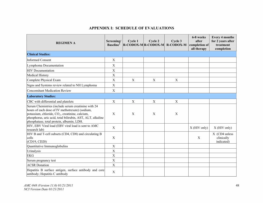
APPENDIX I: SCHEDULE OF EVALUATIONS
REGIMEN A Cycle 2
R-CODOX-MCycle 3
R-CODOX-M
6-8 weeks after
completion of all therapy
Every 4 months for 2 years after
treatment completion
Screening/ Baseline1
Cycle 1 R-CODOX-M
Clinical Studies:
Informed Consent X
Lymphoma Documentation X
HIV Documentation X
Medical History X
Complete Physical Exam X X X X
Signs and Systems review related to NH Lymphoma X
Concomitant Medication Review X
Laboratory Studies:
CBC with differential and platelets X X X X
Serum Chemistries (include serum creatinine with 24 hours of each dose of IV methotrexate) (sodium, potassium, chloride, CO2, creatinine, calcium, phosphorus, uric acid, total bilirubin, AST, ALT, alkaline phosphatase, total protein, albumin, LDH.
X X X X
HIV, EBV Viral load (EBV viral load is sent to AMC research lab)
X X (HIV only) X (HIV only)
HIV B and T-cell subsets (CD4, CD8) and circulating B cells (CD19, CD20)
X X X (CD4 unless
clinically indicated)
Quantitative Immunoglobulins X
Urinalysis X EKG X
Serum pregnancy test X
ACSR Donation X
Hepatitis B surface antigen, surface antibody and core antibody; Hepatitis C antibody
X
AMC-048 (Version 11.0) 01/21/2011 48 NCI Version Date 01/21/2011
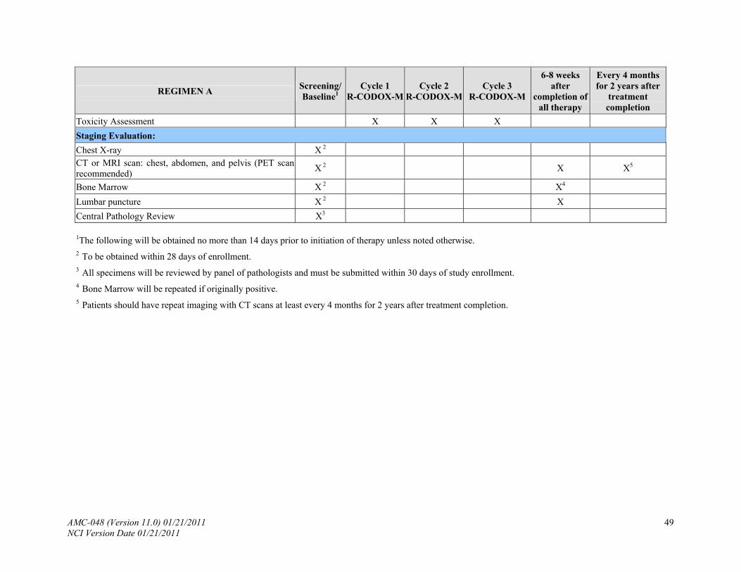
AMC-048 (Version 11.0) 01/21/2011 49 NCI Version Date 01/21/2011
REGIMEN A Screening/ Baseline1
Cycle 1 R-CODOX-M
Cycle 2 R-CODOX-M
Cycle 3 R-CODOX-M
6-8 weeks after
completion of all therapy
Every 4 months for 2 years after
treatment completion
Toxicity Assessment X X X
Staging Evaluation:
Chest X-ray X 2 CT or MRI scan: chest, abdomen, and pelvis (PET scan recommended)
X 2 X X5
Bone Marrow X 2 X4
Lumbar puncture X 2 X
Central Pathology Review X3
1The following will be obtained no more than 14 days prior to initiation of therapy unless noted otherwise. 2 To be obtained within 28 days of enrollment. 3 All specimens will be reviewed by panel of pathologists and must be submitted within 30 days of study enrollment. 4 Bone Marrow will be repeated if originally positive. 5 Patients should have repeat imaging with CT scans at least every 4 months for 2 years after treatment completion.
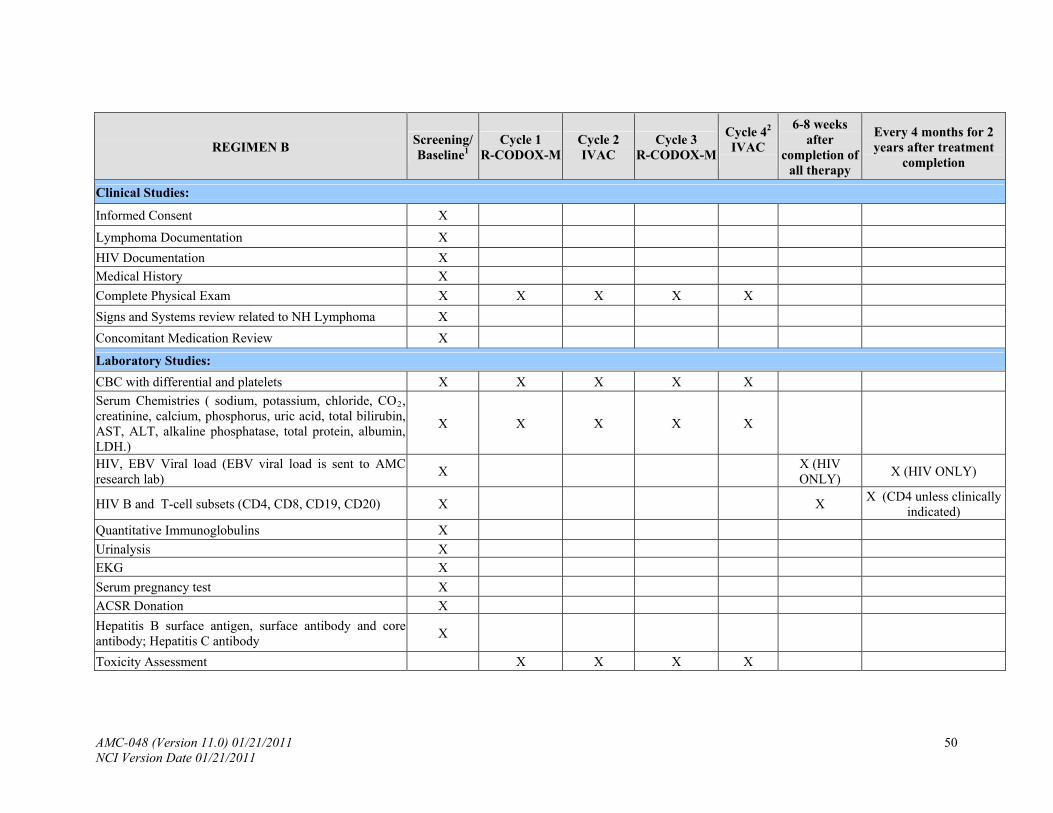
REGIMEN B Screening/ Baseline1
Cycle 1 Cycle 3 R-CODOX-M
Cycle 42
IVAC
6-8 weeks after
completion of all therapy
Every 4 months for 2 years after treatment
completion
Cycle 2 R-CODOX-M IVAC
Clinical Studies:
Informed Consent X
Lymphoma Documentation X
HIV Documentation X Medical History X
Complete Physical Exam X X X X X
Signs and Systems review related to NH Lymphoma X
Concomitant Medication Review X
Laboratory Studies:
CBC with differential and platelets X X X X X
Serum Chemistries ( sodium, potassium, chloride, CO2, creatinine, calcium, phosphorus, uric acid, total bilirubin, AST, ALT, alkaline phosphatase, total protein, albumin, LDH.)
X X X X X
HIV, EBV Viral load (EBV viral load is sent to AMC research lab)
X X (HIV ONLY)
X (HIV ONLY)
X (CD4 unless clinically indicated)
HIV B and T-cell subsets (CD4, CD8, CD19, CD20) X X
Quantitative Immunoglobulins X
Urinalysis X EKG X
Serum pregnancy test X
ACSR Donation X
Hepatitis B surface antigen, surface antibody and core antibody; Hepatitis C antibody
X
Toxicity Assessment X X X X
AMC-048 (Version 11.0) 01/21/2011 50 NCI Version Date 01/21/2011
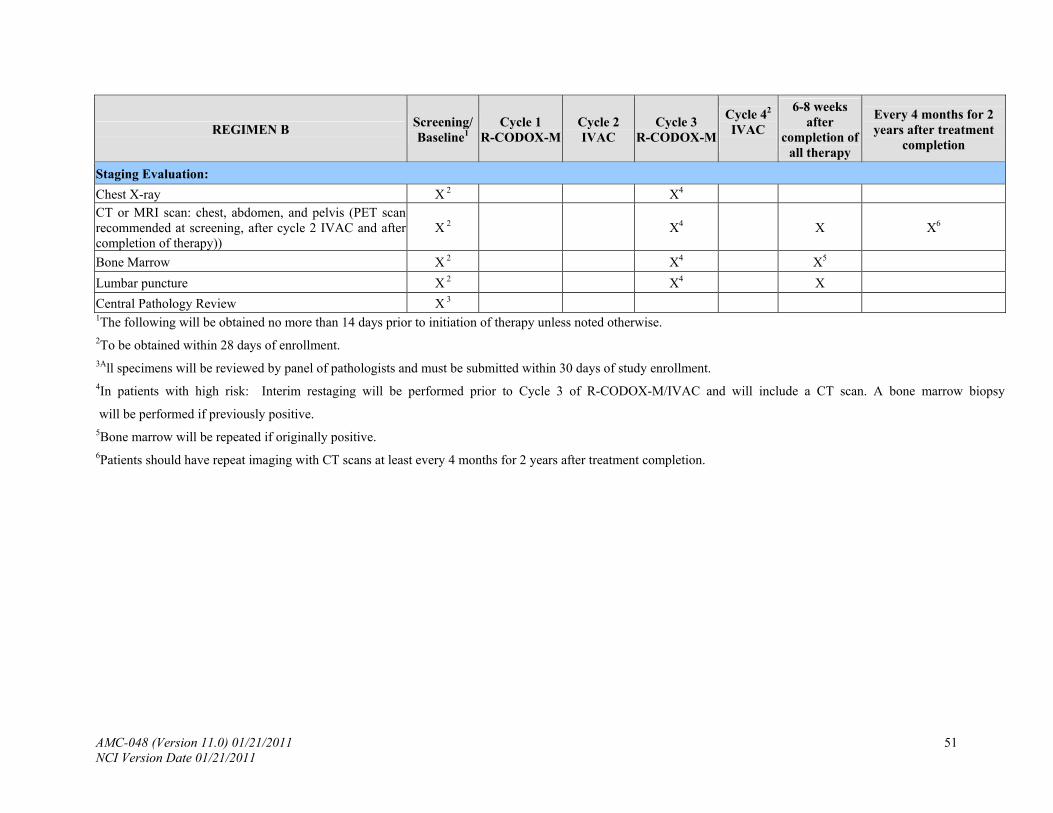
AMC-048 (Version 11.0) 01/21/2011 51 NCI Version Date 01/21/2011
REGIMEN B Screening/ Baseline1
Cycle 1 R-CODOX-M
Cycle 2 IVAC
Cycle 3 R-CODOX-M
Cycle 42
IVAC
6-8 weeks after
completion of all therapy
Every 4 months for 2 years after treatment
completion
Staging Evaluation:
Chest X-ray X 2 X4 CT or MRI scan: chest, abdomen, and pelvis (PET scan recommended at screening, after cycle 2 IVAC and after completion of therapy))
X 2 X4 X X6
Bone Marrow X 2 X X4 5
Lumbar puncture X 2 X4 X
Central Pathology Review X 3 1The following will be obtained no more than 14 days prior to initiation of therapy unless noted otherwise. 2To be obtained within 28 days of enrollment. 3All specimens will be reviewed by panel of pathologists and must be submitted within 30 days of study enrollment. 4In patients with high risk: Interim restaging will be performed prior to Cycle 3 of R-CODOX-M/IVAC and will include a CT scan. A bone marrow biopsys
will be performed if previously positive. 5Bone marrow will be repeated if originally positive. 6Patients should have repeat imaging with CT scans at least every 4 months for 2 years after treatment completion.
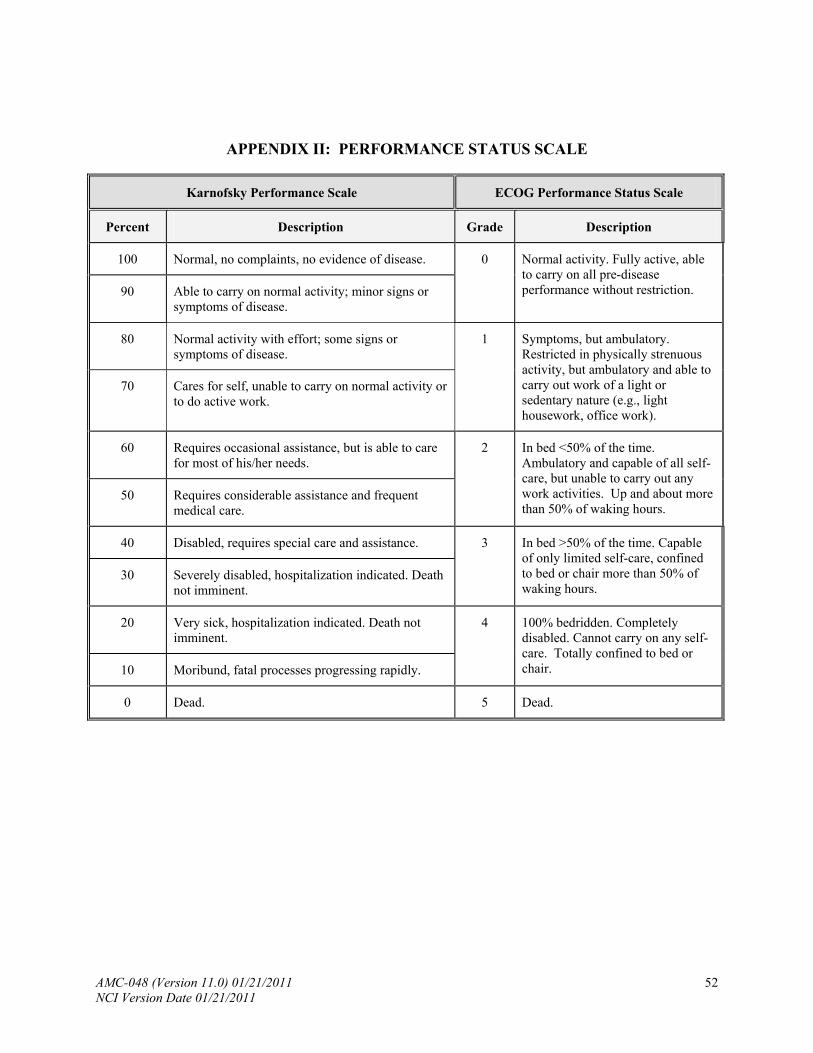
AMC-048 (Version 11.0) 01/21/2011 52 NCI Version Date 01/21/2011
APPENDIX II: PERFORMANCE STATUS SCALE
Karnofsky Performance Scale ECOG Performance Status Scale
Percent Description Grade Description
100 Normal, no complaints, no evidence of disease.
90 Able to carry on normal activity; minor signs or symptoms of disease.
0 Normal activity. Fully active, able to carry on all pre-disease performance without restriction.
80 Normal activity with effort; some signs or symptoms of disease.
70 Cares for self, unable to carry on normal activity or to do active work.
1 Symptoms, but ambulatory. Restricted in physically strenuous activity, but ambulatory and able to carry out work of a light or sedentary nature (e.g., light housework, office work).
60 Requires occasional assistance, but is able to care for most of his/her needs.
50 Requires considerable assistance and frequent medical care.
2 In bed <50% of the time. Ambulatory and capable of all self-care, but unable to carry out any work activities. Up and about more than 50% of waking hours.
40 Disabled, requires special care and assistance.
30 Severely disabled, hospitalization indicated. Death not imminent.
3 In bed >50% of the time. Capable of only limited self-care, confined to bed or chair more than 50% of waking hours.
20 Very sick, hospitalization indicated. Death not imminent.
10 Moribund, fatal processes progressing rapidly.
4 100% bedridden. Completely disabled. Cannot carry on any self-care. Totally confined to bed or chair.
0 Dead. 5 Dead.
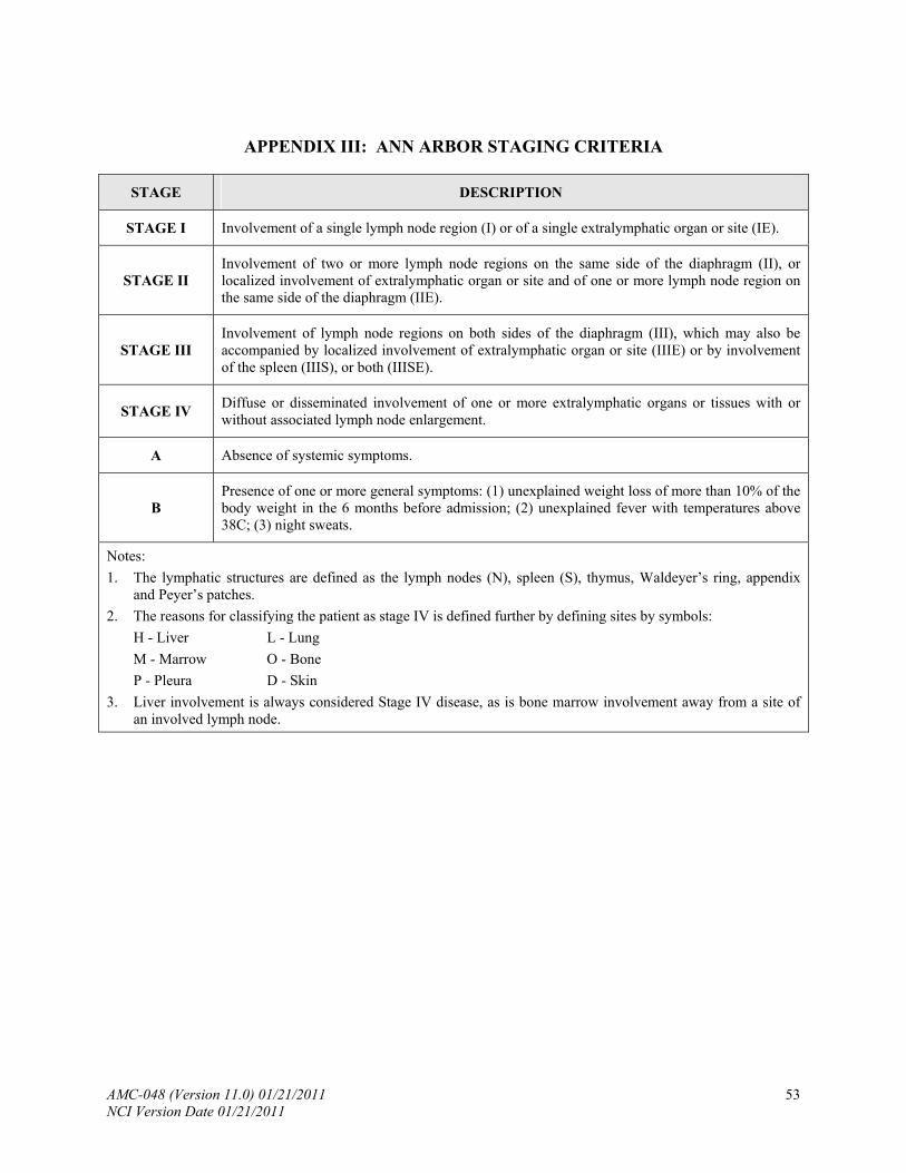
AMC-048 (Version 11.0) 01/21/2011 53 NCI Version Date 01/21/2011
APPENDIX III: ANN ARBOR STAGING CRITERIA
STAGE DESCRIPTION
STAGE I Involvement of a single lymph node region (I) or of a single extralymphatic organ or site (IE).
STAGE II Involvement of two or more lymph node regions on the same side of the diaphragm (II), or localized involvement of extralymphatic organ or site and of one or more lymph node region on the same side of the diaphragm (IIE).
STAGE III Involvement of lymph node regions on both sides of the diaphragm (III), which may also be accompanied by localized involvement of extralymphatic organ or site (IIIE) or by involvement of the spleen (IIIS), or both (IIISE).
STAGE IV Diffuse or disseminated involvement of one or more extralymphatic organs or tissues with or without associated lymph node enlargement.
A Absence of systemic symptoms.
B Presence of one or more general symptoms: (1) unexplained weight loss of more than 10% of the body weight in the 6 months before admission; (2) unexplained fever with temperatures above 38C; (3) night sweats.
Notes:
1. The lymphatic structures are defined as the lymph nodes (N), spleen (S), thymus, Waldeyer’s ring, appendix and Peyer’s patches.
2. The reasons for classifying the patient as stage IV is defined further by defining sites by symbols:
H - Liver L - Lung
M - Marrow O - Bone
P - Pleura D - Skin
3. Liver involvement is always considered Stage IV disease, as is bone marrow involvement away from a site of an involved lymph node.
AMC-048 (Version 11.0) 01/21/2011 54 NCI Version Date 01/21/2011
APPENDIX IV: DIAGNOSTIC BIOPSIES
A. GENERAL
1. Diagnostic biopsy specimens required for this trial are to be shipped to the ASCR George Washington site for batching and dispersal to central laboratories.
2. Tumor blocks in paraffin are the preferred specimens, but 20 unstained slides are acceptable.
B. SPECIMEN PREPARATION
Pathology reports should accompany slides or blocks and identifiers linking these particular specimens to the paperwork must be present. In addition paperwork identifying the specimen as follows is required.
Label formalin specimen with the following information: Protocol #: AMC-048 9 digit Patient # Patient initials Date and time of collection Specimen type- “Tissue Block” or “Tissue (Unstained Slide)” Specimen purpose: Diagnostic Tumor Biopsy
C. PACKAGING and FedEx FORMS
1. Place the labeled paraffin block or slides into a specimen container in bubble wrap.
2. Affix the FED-EX airbill on blank side of the shipper making sure that it is marked “FED-EX PRIORITY OVERNIGHT”.
3. Mark “OTHER” in the airbill under “Packaging”.
4. Under airbill section “special Handling” indicate “YES-SHIPPERS DECLARATION NOT REQUIRED”.
5. Enter FED-EX account #:
6. Place “From/To” information onto areas provided on the shipper. Specimens are accepted MONDAY through THURSDAY only. All specimens should be shipped by overnight express to:
Dr. Sylvia Silver George Washington University Medical Center Pathology Bank, Room 507 2300 I Street, NW Washington, DC 20037 Phone: (202) 994-1444 Fax: (202) 994-5056
7. Make certain that shipper is already either pre-labeled with ‘UN#3373’ stamp, or make a paper label with ‘UN#3373” and affix it to the shipper.
AMC-048 (Version 11.0) 01/21/2011 55 NCI Version Date 01/21/2011
8. Make certain that the net volume of the specimen being shipped is written in the space provided on the shipper or make a separate label with the volume in ml (write in the number of ml of formalin the sample is in) and affix to the shipper.
9. Affix airbill to shipper so that the ‘UN’ and ‘VOLUME’ labels are visible.
10. RETAIN THE TOP COPY OF THE AIRWAY BILL FOR YOUR RECORDS.
11. Place the box in the FedEx pickup area at your site or call to request a package pickup. D. RECORD OF SPECIMENS
This study will track specimens via GlobalTraceSM, a component of the AMC AdvantageEDCSM system. The GlobalTraceSM shipment manifest must accompany all specimens.
AMC-048 (Version 11.0) 01/21/2011 56 NCI Version Date 01/21/2011
APPENDIX V: ACSR SPECIMEN PREPARATION & SHIPPING INSTRUCTIONS
A. GENERAL
To ship these specimens, use a diagnostic shipper approved for a volume of at least 30 cc. The use of the SAF-T-PAK STP 210 diagnostic cardboard shipper is recommended. These shippers may be ordered at the SAF-T-PAK website www.saftpak.com. The following instructions below are for use with the recommended STP-210 shipper. If using another federally approved diagnostic shipper, please follow instructions provided for that specific shipper.
NOTE: Specimens MUST BE SHIPPED Monday through Wednesday as an OVERNIGHT PRIORITY shipment. Specimens are NOT ACCEPTED ON FRIDAYS OR SATURDAYS in the Ambinder Lab.
B. SPECIMEN PREPARATION
BLOOD SPECIMENS
1. Draw three 10 cc (ml) yellow top (ACD) tubes from study patient. With a black, water resistant, sharpie pen, label each specimen with the following information: Protocol #: AMC-048 9 digit Patient Study ID # Patient initials Date and time of collection Specimen type- "Whole Blood"
2. Seal the tops of the three 10 cc heparin green tops with parafilm.
C. PACKAGING and FedEx FORMS
1. Place the three sealed tubes into bubble wrap (provided in STP-210 kit).
2. Tape around the bubble wrap so that the roll stays together and the tubes cannot fall out or break.
3. Place absorbent material sheet around the bubble wrapped tubes and slip into a biohazard poly-bag and “self-seal”.
4. Place poly-bag containing tubes into the white TYVEK bag and seal.
5. Place the TYVEK bag into the STP-210 diagnostic cardboard shipper. Seal the cardboard shipper with clear packing/shipping tape.
6. Affix the FED-EX airbill on blank side of the shipper making sure that it is marked “FED-EX PRIORITY OVERNIGHT”.
7. Mark “OTHER” in the airbill under “Packaging”. Please use FedEx #: .
8. Under airbill section “Special Handling” indicate “YES-SHIPPERS DECLARATION NOT REQUIRED”.
9. Place “From/To” information onto areas provided on the shipper.
AMC-048 (Version 11.0) 01/21/2011 57 NCI Version Date 01/21/2011
Shipping Address:
ACSR Blood Receiving Lab Johns Hopkins Oncology 1650 Orleans Street, CRB-384 Baltimore, MD 21231-1000 TEL: (410) 955-8721 FAX: (443) 287-3217
10. Make certain that shipper is already either pre-labeled with ‘UN#3373’ stamp, or make a paper label with ‘UN#3373” and affix it to the shipper.
11. Make certain that the net volume of the specimen being shipped is written in the space provided on the shipper or make a separate label with the volume in ml (so three 10 cc tubes is 30 ml) and affix to the shipper.
12. Affix airbill to shipper so that the ‘UN’ and ‘VOLUME’ labels are visible.
13. RETAIN THE TOP COPY OF THE AIRWAY BILL FOR YOUR RECORDS.
14. Place the box in the FedEx pickup area at your site or call to request a package pickup.
Please Note: The shippers will be mailed back to each AMC site.
CSF SPECIMENS
1. Aliquot 1 cc CSF into individual Nalgene cryovials (2 ml tubes) with 0.5 ml per cryovial. With a black, water resistant, sharpie pen, label each specimen with the following information: Protocol #: AMC-048 9 digit Patient Study ID # Patient initials Date and time of collection Specimen type- "CSF"
2. Freeze CSF specimens at -20º or less. After specimens are frozen, they can be shipped.
D. PACKAGING and FedEx FORMS for SHIPPING
1. Approximately 2 kg (4.4 lbs) of dry ice pellets or chunks are needed for packaging samples.
2. Specimens must be shipped on dry ice in a styrofoam box, and then shipped in an outer cardboard box (required by Fed ex).
3. Place Hazard Label (9mm diamond) on outer cardboard box, along with FedEx Air Bill. (Please use FedEx #: to ship specimens.)
4. Ship to the address listed above (Ambinder lab) as for blood specimens.
E. RECORD OF SPECIMENS
This study will track specimens via GlobalTraceSM, a component of the AMC AdvantageEDCSM system. The GlobalTraceSM shipment manifest must accompany all specimen shipments.
AMC-048 (Version 11.0) 01/21/2011 58 NCI Version Date 01/21/2011
APPENDIX VI: ACSR INFORMED CONSENT
INFORMED CONSENT FORM RESEARCH STUDY
AIDS AND CANCER SPECIMEN RESOURCE (ACSR) A. INTRODUCTION
You are being asked to donate tissue for research. Before you decide to be a part of this research study, you need to understand the risks and benefits so that you can make an informed decision. This is known as informed consent. This consent form provides information about the research study, which has been explained, to you. Once you understand the study and the tests it requires, you will be asked to sign this form if you want to take part in the study. Your decision to take part in the study is voluntary. This means that you are free to choose if you will take part in the study.
B. PURPOSE
The National Cancer Institute has set up a Biorepository for tissues and biological fluids from HIV-positive and HIV-negative individuals in order to have specimens available for scientists studying malignancies associated with HIV disease. Individuals who have had biopsies to determine a malignancy are being asked for permission to store some of the tissue in the ACSR. Only tissue in excess of that required for decision making will be given to the ACSR. If it turns out that your physician needs more of your tissue for additional studies, the ACSR will release all of your tissue back to your doctor. No additional tissues will be taken from your body for the Bank. In addition, you are requested to donate some of your blood to the ACSR so that scientists will also be able to look for any deviation in these body fluids that may explain the malignancy.
C. PROCEDURES
You are being asked for consent to place some of the biopsy material in the ACSR. If you agree to allow the ACSR to have some of your tissue, we would also like to:
1. Confidentially obtain some clinical information from your medical records that could be useful to research investigators. The report of the information retrieved from your medical record that is given to research investigators will not have your name, or include any information which could personally identify you.
2. Obtain some blood for the ACSR. Up to twenty (20) milliliters of blood will be obtained at your next visit to your physician.
If during the course of treatment by your physician, it is necessary to perform any of the following procedures for diagnostic reasons, you will be asked, at that time, to consent to having a portion of that specimen sent to the ACSR. These requests will not require you to make any additional visits to your doctor or have any additional specimens taken just for the
AMC-048 (Version 11.0) 01/21/2011 59 NCI Version Date 01/21/2011
ACSR. The ACSR will only receive part of your specimen, and only what is in excess. No additional materials will be removed for the purposes of the Bank alone. Samples of interest would include, (but are not limited to): Spinal fluid. Airway washes. Fluid around lungs and intestines. Additional biopsy material. You will not be asked to fill out any forms for any of these specimens.
D. POSSIBLE RISKS
There is a possibility of a bruise and slight pain at the time the blood samples are taken. There is also the possibility of fainting and infection at the site of the blood draw.
E. POSSIBLE BENEFITS
It may be that there will be no direct benefit to you by consenting to allow the ACSR to have portions of your biopsies and biological fluids. However, there may be possible benefits to medical knowledge and HIV-infected individuals in the future.
F. COSTS
There will not be any additional costs to you for consenting to participate in the AIDS and Cancer Specimen Resource.
G. PAYMENT FOR INJURY OR HARM
As the lists of risks shows, taking part in this research study may result in injury or harm to you. If you require immediate medical care, you should go to an emergency room. Otherwise, the doctor in charge of the study will take care of you or help you get the care you need. You will be sent a bill for whatever medical care you receive. All or part of your bill may be paid by the sponsor of the research study (according to its agreement with the AIDS Associated Malignancies Clinical Trials Consortium), or by your health insurance. (Institution) will not pay for the care. Likewise, (Institution) will not pay you for pain, worry, lost income, or non-medical care costs that might occur from taking part in this research study.
H. PRIVACY
Your hospital medical records will be confidentially reviewed to obtain clinical information that could be useful to research investigators. However, the report of this information will not have your name or social security number anywhere on the report, so you will not be easily identified. The results of this research study will be given to the sponsor, the National Cancer Institute (NCI), AIDS Malignancy Consortium, and may be asked for by the Department of Health and Human Services. In addition, the Institutional Review Board may see your records. Except for these people, records from this study will be kept private unless you
AMC-048 (Version 11.0) 01/21/2011 60 NCI Version Date 01/21/2011
authorize their release or release is required by law (i.e. court subpoena). Any publications of this study will not use your name, identify you personally, or include any information that could personally identify you.
I. QUESTIONS
If you have any questions about this research study, you should contact Dr. (_____________) at (Phone Number) (day) or (Phone Number) (night), or the person in charge of the study, (_____________), the study coordinator, at (Phone Number). If you have any questions about your rights as a research subject, you should call (IRB Representative), in (Institution) Office of Human Research at (______________). (IRB Representative) is your representative and is not employed by the individuals conducting the study.
J. SIGNATURES
Statement of professional obtaining consent I have fully explained this research study to the patient or guardian of patient ______________________________. In my judgment and the patient’s or guardian’s, there was sufficient access to information, including risks and benefits to make an informed decision. Date: ___________ Physician’s Signature: ______________________________ Physician’s Name: (Print)
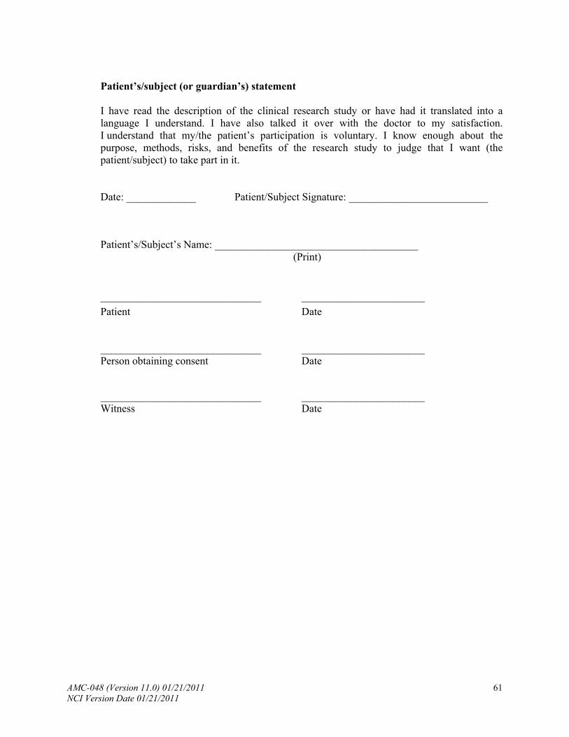
AMC-048 (Version 11.0) 01/21/2011 61 NCI Version Date 01/21/2011
Patient’s/subject (or guardian’s) statement I have read the description of the clinical research study or have had it translated into a language I understand. I have also talked it over with the doctor to my satisfaction. I understand that my/the patient’s participation is voluntary. I know enough about the purpose, methods, risks, and benefits of the research study to judge that I want (the patient/subject) to take part in it. Date: _____________ Patient/Subject Signature: __________________________
Patient’s/Subject’s Name: ______________________________________ (Print)
______________________________ _______________________
Patient Date ______________________________ _______________________ Person obtaining consent Date
______________________________ _______________________ Witness Date
APPENDIX VII: AMC DATA SAFETY MONITORING PLAN
Monitoring the Progress of Trials and the Safety of Participants
All AMC protocols follow the Cancer Therapy Evaluation Program (CTEP) guidelines for reporting of adverse events. All adverse events that meet the expedited reporting requirements of the National Cancer Institute (NCI) are reported to the Investigational Drug Branch (IDB) of the NCI via the Adverse Event Expedited Reporting System (AdEERS) web application. All expedited adverse event reports are also required to be submitted to the local Institutional Review Board (IRB) of the reporting institution. If NCI holds the IND or no IND is required for a study, the AMC sites report serious adverse events directly to the AMC Operations and Data Management Center (ODMC) via AdEERS. In some instances, the AMC sites may report serious adverse events directly to the commercial sponsor holding the IND who will then in turn report to the AMC ODMC. However, most AMC protocol require that sites report all serious adverse events via AdEERS with the AMC ODMC forwarding a copy of the report to the sponsor. Unless an AMC protocol specifies an alternate plan for the review and submission of serious adverse events, all serious adverse events received by the AMC ODMC will be reviewed by the AMC Medical Monitor at the AMC ODMC prior to submission to NCI and the sponsor. For protocols for which the IDB does not have an assigned drug monitor to review serious adverse event reports, in the event of disagreement between the reporting physician and the AMC Medical Monitor regarding the attribution of the event to the investigational agent(s) (i.e., determination of whether the relationship is unrelated, unlikely, possible, probable, or definite), the AMC Medical Monitor will provide the final determination of the relationship.
The AMC ODMC provides a listing of serious adverse events to the Protocol Chair and Co-chair(s) for review on a regular basis. The AMC ODMC compiles these events in a tabular format and posts them on the password-protected section of the AMC web site. The AMC web site is accessible to all AMC investigators, co-investigators, and their staff. Email notification that this information is available on the web site will be sent to all site PIs. It is the responsibility of each site to provide this information to their respective IRBs, if required by their IRB. For blinded studies, the serious adverse events are reviewed and tabulated without treatment assignment.
Accrual summaries for each AMC trial are updated nightly on the password-protected section of the AMC web site. The progress of each AMC trial is reviewed regularly by the Protocol Chair and also by the appropriate disease-oriented Working Group during scheduled conference calls. For phase I dose escalation trials, dose escalation (or dose de-escalation) is based on the rules in the protocol and the Protocol Chair and Group Statistician determine whether these criteria have been met. For phase II trials, stopping the trial for toxicity or efficacy, or suspending enrollment pending observation of responses in a multi-stage phase II trial, is based on meeting criteria stated in the protocol, and the Protocol Chair and Group Statistician determine whether these criteria have been met.
For phase III trials, the AMC has formed an independent Data Safety and Monitoring Committee (DSMC). Voting members of the DSMC are physicians, statisticians, and a patient advocate. All voting members are from outside the AMC. Non-voting members are the NCI scientific project officers and an NCI statistician. The AMC Data Safety and Monitoring Committee reviews AMC phase III studies in accordance with the National Cancer Institute’s Policy for Data Safety and Monitoring. Confidential reports of all phase III trials are prepared by the AMC Group Statistician with support from the AMC ODMC. A written report containing the current status of each trial monitored, and when appropriate, any toxicity and outcome data, are sent to DSMC members by the AMC ODMC allowing sufficient time for DSMC members to review the report prior to the meeting.
AMC-048 (Version 11.0) 01/21/2011 62 NCI Version Date 01/21/2011
This report addresses specific toxicity concerns as well as concerns about the conduct of the trial. The report may contain recommendations for consideration by the DSMC concerning whether to close the trial, report the results, or continue accrual or follow-up.
The results of each DSMC meeting are summarized in a formal report sent by the DSMC Chair to the Group Chair and AMC ODMC within 1 week of the meeting. The DSMC report contains recommendations on whether to close each study reviewed, whether to report the results, and whether to continue accrual or follow-up. A primary recommendation (e.g., continue with no change; recommended or required modification; stop) must be included in the document. The Group Chair is then responsible for notifying the Protocol Chair and relevant Disease-oriented Working Group Chair before the recommendations of the DSMC are carried out. In the unlikely event that the Protocol Chair does not concur with the DSMC, then the NCI Division Director or designee must be informed of the reason for the disagreement. The Study Chair, relevant Disease-oriented Working Group Chair, Group Chair, DSMC Chair and NCI Division Director or designee will be responsible for reaching a mutually acceptable decision about the study. CTEP approval of a formal amendment will be required prior to any implementation of a change to the study.
Following a DSMC meeting, a summary of the serious adverse events reported to the DSMC is posted to the AMC web site. It is each site’s responsibility for conveying this information to its IRB.
Plans for Assuring Compliance with Requirements Regarding the Reporting of Adverse Events (AE)
For trials monitored by the NCI’s Clinical Data Update System (CDUS), adverse event information is transmitted electronically to NCI on a quarterly basis. For trials monitored by NCI’s Clinical Trials Monitoring Service (CTMS), adverse event information is transmitted electronically to NCI every two weeks.
Plans for Assuring that any Action Resulting in a Temporary or Permanent Suspension of an NCI-Funded Clinical Trial is Reported to the NCI Grant Program Director Responsible for the Grant
In the event that termination of the trial or major modification to the protocol is under consideration, the Protocol Chair will convene the AMC Data Coordinator and Disease-oriented Working Group Chair by conference call to discuss the options. For phase I and II trials, the Protocol Chair also has the option of asking the AMC DSMC to review the study. The AMC ODMC will inform the CTEP Protocol Information Office (PIO) when studies are temporarily or permanently closed. The Cancer Treatment and Evaluation Program (CTEP) of the National Cancer Institute (NCI) must approve all protocol amendments prior to distributing to the AMC sites.
Plans for Assuring Data Accuracy and Protocol Compliance
All study data for AMC clinical trials are entered directly by AMC site staff into AdvantageEDCSM (a web-based data entry and enrollment system). During data entry, the system performs validation checks on many fields and performs consistency checks between select fields. Range checks are placed on each field to eliminate entry of out-of-range values. Edit check programs are run on the database on a set schedule to identify and resolve inconsistencies between forms or data collected at different points in time. AMC ODMC staff routinely interacts with site staff to resolve any data problems.
AMC-048 (Version 11.0) 01/21/2011 63 NCI Version Date 01/21/2011
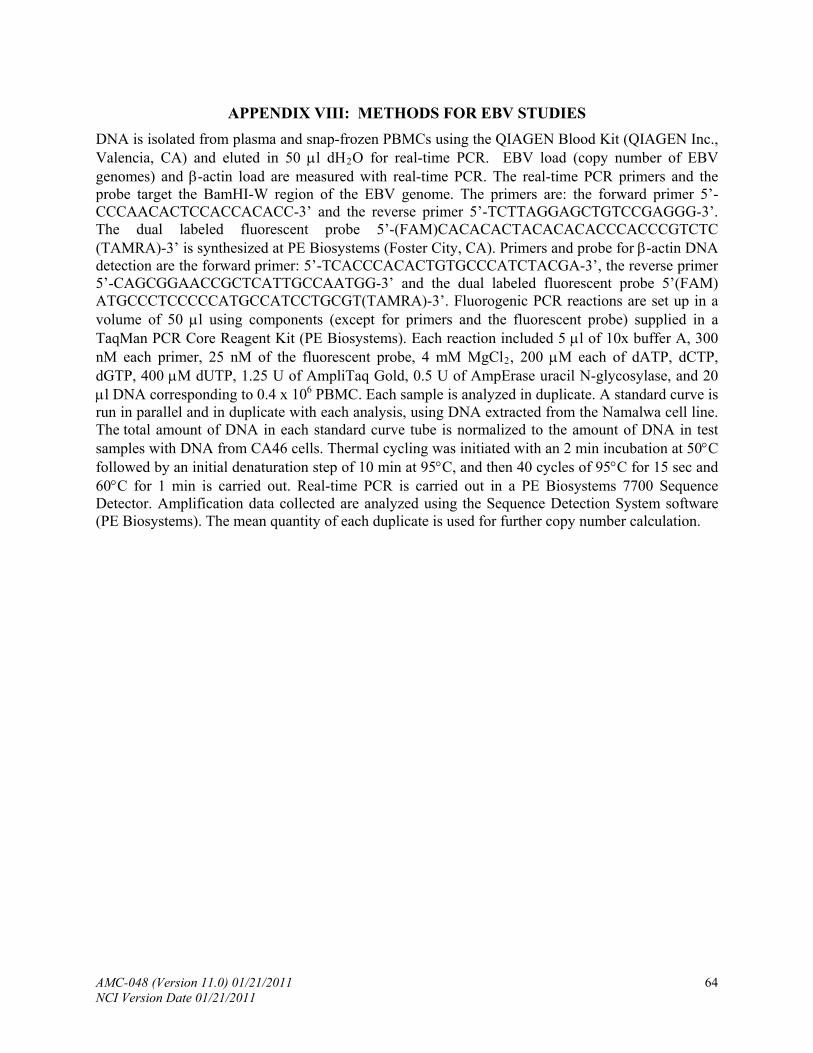
APPENDIX VIII: METHODS FOR EBV STUDIES
DNA is isolated from plasma and snap-frozen PBMCs using the QIAGEN Blood Kit (QIAGEN Inc., Valencia, CA) and eluted in 50 l dH2O for real-time PCR. EBV load (copy number of EBV genomes) and -actin load are measured with real-time PCR. The real-time PCR primers and the probe target the BamHI-W region of the EBV genome. The primers are: the forward primer 5’-CCCAACACTCCACCACACC-3’ and the reverse primer 5’-TCTTAGGAGCTGTCCGAGGG-3’. The dual labeled fluorescent probe 5’-(FAM)CACACACTACACACACCCACCCGTCTC (TAMRA)-3’ is synthesized at PE Biosystems (Foster City, CA). Primers and probe for -actin DNA detection are the forward primer: 5’-TCACCCACACTGTGCCCATCTACGA-3’, the reverse primer 5’-CAGCGGAACCGCTCATTGCCAATGG-3’ and the dual labeled fluorescent probe 5’(FAM) ATGCCCTCCCCCATGCCATCCTGCGT(TAMRA)-3’. Fluorogenic PCR reactions are set up in a volume of 50 l using components (except for primers and the fluorescent probe) supplied in a TaqMan PCR Core Reagent Kit (PE Biosystems). Each reaction included 5 l of 10x buffer A, 300 nM each primer, 25 nM of the fluorescent probe, 4 mM MgCl2, 200 M each of dATP, dCTP, dGTP, 400 M dUTP, 1.25 U of AmpliTaq Gold, 0.5 U of AmpErase uracil N-glycosylase, and 20 l DNA corresponding to 0.4 x 10⁶ PBMC. Each sample is analyzed in duplicate. A standard curve is run in parallel and in duplicate with each analysis, using DNA extracted from the Namalwa cell line. The total amount of DNA in each standard curve tube is normalized to the amount of DNA in test samples with DNA from CA46 cells. Thermal cycling was initiated with an 2 min incubation at 50C followed by an initial denaturation step of 10 min at 95C, and then 40 cycles of 95C for 15 sec and 60C for 1 min is carried out. Real-time PCR is carried out in a PE Biosystems 7700 Sequence Detector. Amplification data collected are analyzed using the Sequence Detection System software (PE Biosystems). The mean quantity of each duplicate is used for further copy number calculation.
AMC-048 (Version 11.0) 01/21/2011 64 NCI Version Date 01/21/2011
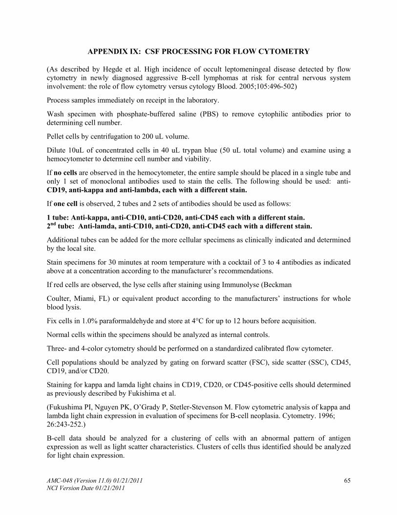
AMC-048 (Version 11.0) 01/21/2011 65 NCI Version Date 01/21/2011
APPENDIX IX: CSF PROCESSING FOR FLOW CYTOMETRY
(As described by Hegde et al. High incidence of occult leptomeningeal disease detected by flow cytometry in newly diagnosed aggressive B-cell lymphomas at risk for central nervous system involvement: the role of flow cytometry versus cytology Blood. 2005;105:496-502)
Process samples immediately on receipt in the laboratory.
Wash specimen with phosphate-buffered saline (PBS) to remove cytophilic antibodies prior to determining cell number.
Pellet cells by centrifugation to 200 uL volume.
Dilute 10uL of concentrated cells in 40 uL trypan blue (50 uL total volume) and examine using a hemocytometer to determine cell number and viability.
If no cells are observed in the hemocytometer, the entire sample should be placed in a single tube and only 1 set of monoclonal antibodies used to stain the cells. The following should be used: anti-CD19, anti-kappa and anti-lambda, each with a different stain.
If one cell is observed, 2 tubes and 2 sets of antibodies should be used as follows:
1 tube: Anti-kappa, anti-CD10, anti-CD20, anti-CD45 each with a different stain. 2nd tube: Anti-lamda, anti-CD10, anti-CD20, anti-CD45 each with a different stain.
Additional tubes can be added for the more cellular specimens as clinically indicated and determined by the local site.
Stain specimens for 30 minutes at room temperature with a cocktail of 3 to 4 antibodies as indicated above at a concentration according to the manufacturer’s recommendations.
If red cells are observed, the lyse cells after staining using Immunolyse (Beckman
Coulter, Miami, FL) or equivalent product according to the manufacturers’ instructions for whole blood lysis.
Fix cells in 1.0% paraformaldehyde and store at 4°C for up to 12 hours before acquisition.
Normal cells within the specimens should be analyzed as internal controls.
Three- and 4-color cytometry should be performed on a standardized calibrated flow cytometer.
Cell populations should be analyzed by gating on forward scatter (FSC), side scatter (SSC), CD45, CD19, and/or CD20.
Staining for kappa and lamda light chains in CD19, CD20, or CD45-positive cells should determined as previously described by Fukishima et al.
(Fukushima PI, Nguyen PK, O’Grady P, Stetler-Stevenson M. Flow cytometric analysis of kappa and lambda light chain expression in evaluation of specimens for B-cell neoplasia. Cytometry. 1996; 26:243-252.)
B-cell data should be analyzed for a clustering of cells with an abnormal pattern of antigen expression as well as light scatter characteristics. Clusters of cells thus identified should be analyzed for light chain expression.