Embed Size (px)
Citation preview
Braz J Otorhinolaryngol. 2017;83(3):299---312
www.bjorl.org
Brazilian Journal of
OTORHINOLARYNGOLOGY
ORIGINAL ARTICLE
Airway reconstruction: review of an approach to theadvanced-stage laryngotracheal stenosis�
Mohamad Ahmad Bitara,b,c,d,∗, Randa Al Barazia, Rana Barakeha
a American University of Beirut, Faculty of Medicine and Medical Center, Department of Otolaryngology and Head & Neck Surgery,Beirut, Lebanonb American University of Beirut, Faculty of Medicine and Medical Center, Department of Pediatrics and Adolescent Medicine,Beirut, Lebanonc University of Sydney, Sydney Medical School, The Children’s Hospital at Westmead, Department of ENT Surgery, Sydney, Australiad Al Jalila Children’s Specialty Hospital, Department of Otolaryngology Head & Neck Surgery, Dubai, UAE
Received 9 December 2015; accepted 31 March 2016Available online 27 April 2016
KEYWORDSLaryngotrachealstenosis;Subglottic stenosis;Laryngotrachealreconstruction;Cricotrachealresection;Staging;Mapping
AbstractIntroduction: The management of laryngotracheal stenosis is complex and is influenced bymultiple factors that can affect the ultimate outcome. Advanced lesions represent a specialchallenge to the treating surgeon to find the best remedying technique.Objective: To review the efficacy of our surgical reconstructive approach in managing advanced-stage laryngotracheal stenosis treated at a tertiary medical center.Methods: A retrospective review of all patients that underwent open laryngotrachealrepair/reconstruction by the senior author between 2002 and 2014. Patients withmild/moderate stenosis (e.g. stage 1 or 2), or those who had an open reconstructive pro-cedure prior to referral, were excluded. Patients who had only endoscopic treatment (e.g.laser, balloon dilatation) and were not subjected to an open reconstructive procedure at ourinstitution, were not included in this study. Variables studied included patient demographics,clinical presentation, etiology of the laryngotracheal pathology, the location of stenosis, thestage of stenosis, the type of corrective or reconstructive procedure performed with the typeof graft used (where applicable), the type and duration of stent used, the post-reconstructioncomplications, and the duration of follow-up. Outcome measures included decannulation rate,total number of reconstructive surgeries needed to achieve decannulation, and the number ofpost-operative endoscopies needed to reach a safe patent airway.
Results: Twenty five patients were included, aged 0.5 months to 45 years (mean 13.5 years,median 15 years) with 16 males and 9 females. Seventeen patients (68%) were younger than 18years. Most patients presented with stridor, failure of decannulation, or respiratory distress.Majority had acquired etiology for their stenosis with only 24% having a congenital pathology.�
Please cite this article as: Bitar MA, Al Barazi R, Barakeh R. Airway reconstruction: review of an approach to the advanced-stagelaryngotracheal stenosis. Braz J Otorhinolaryngol. 2017;83:299---312.∗ Corresponding author. Current address: Pediatric Otolaryngology Program, Al-Jalila Children’s Specialty Hospital, Dubai, UAE.E-mail: [email protected] (M.A. Bitar).
Peer Review under the responsibility of Associacão Brasileira de Otorrinolaringologia e Cirurgia Cérvico-Facial.
http://dx.doi.org/10.1016/j.bjorl.2016.03.0121808-8694/© 2016 Associacao Brasileira de Otorrinolaringologia e Cirurgia Cervico-Facial. Published by Elsevier Editora Ltda. This is an openaccess article under the CC BY license (http://creativecommons.org/licenses/by/4.0/).
300 Bitar MA et al.
Thirty-two reconstructive procedures were performed resulting in decannulating 24 patients(96%), with 15/17 (88%) pediatric patients and 5/8 (62.5%) adult patients requiring only a sin-gle reconstructive procedure. Cartilage grafts were mostly used in children (84% vs. 38%) andstents were mostly silicone made, followed by endotracheal tubes. The number of endoscopiesrequired ranged from 1 to 7 (mean 3). More co-morbidities existed in young children, resultingin failure to decannulate one patient. Adult patients had more complex pathologies requiringmultiple procedures to achieve decannulation, with grafting less efficacious than in youngerpatients. The pediatric patients had double the incidence of granulation tissue compared toadults. The decannulated patients remained asymptomatic at a mean follow-up of 50.5 months.Conclusion: The review of our approach to open airway repair/reconstruction showed its effi-cacy in advanced-stage laryngotracheal stenosis. Good knowledge of a variety of reconstructivetechniques is important to achieve good results in a variety of age groups.© 2016 Associacao Brasileira de Otorrinolaringologia e Cirurgia Cervico-Facial. Publishedby Elsevier Editora Ltda. This is an open access article under the CC BY license (http://creativecommons.org/licenses/by/4.0/).
PALAVRAS-CHAVEEstenoselaringotraqueal;Estenose subglótica;Reconstrucãolaringotraqueal;Resseccãocricotraqueal;Estadiamento;Mapeamento
Reconstrucão de via aérea: revisão de uma abordagem à estenose laringotraquealem estágio avancado
ResumoIntroducão: A conduta da estenose laringotraqueal é complexo e é influenciado por váriosfatores que podem afetar o resultado final. Lesões em estágio avancado representam um desafioespecial para o cirurgião encontrar a melhor técnica de tratamento.Objetivo: Avaliar a eficácia de nossa abordagem de reconstrucão cirúrgica no tratamento deestenose laringotraqueal em estágio avancado em um centro médico terciário.Método: Revisão retrospectiva de todos os pacientes que foram submetidos a tratamentocirúrgico/reconstrucão laringotraqueal aberta pelo autor principal, entre 2002 e 2014. Ospacientes com estenose leve (por exemplo, estágio 1 ou 2), ou aqueles submetidos a proced-imento de reconstrucão aberta antes da indicacão, foram excluídos. Pacientes que tinhamsido submetidos somente a tratamento endoscópico (por exemplo, laser, dilatacão por balão)e não haviam sido submetidos a procedimento de reconstrucão aberta em nossa instituicão,não foram incluídos neste estudo. As variáveis estudadas incluíram dados demográficos dospacientes, apresentacão clínica, etiologia da doenca laringotraqueal, local da estenose, estágioda estenose, o tipo de procedimento corretivo ou reconstrutor realizado com o tipo de enxertoutilizado (onde aplicável), tipo e duracão do stent utilizado, complicacões pós-reconstrucão,e duracão do seguimento. Os resultados incluíram taxas de decanulacão, número total decirurgias reconstrutoras necessárias para possibilitar a decanulacão, e o número de endoscopiaspós-operatórias necessárias para obter uma via aérea patente e segura.Resultados: Vinte e cinco pacientes foram incluídos, com idade de 0,5 meses a 45 anos (médiade 13,5 anos, mediana de 15 anos) com 16 homens e 9 mulheres. Dezessete pacientes (68%)eram menores de 18 anos. A maioria dos pacientes apresentava estridor, falha de decanulacãoou desconforto respiratório. A maioria das estenoses era adquirida, enquanto apenas 24%presentavam apresentavam causa congênita. Trinta e dois procedimentos reconstrutores foramrealizados, resultando em decanulacão de 24 pacientes (96%), com 15/17 (88%) pacientespediátricos e 5/8 pacientes (62,5%) adultos que necessitaram de apenas um único procedimentoreconstrutor. Enxertos de cartilagem foram utilizados principalmente em criancas (84% vs. 38%)e a maioria dos stents era feita principalmente de silicone, seguidos por tubo endotraqueal.O número de endoscopias necessárias variou de 1 a 7 (média de 3). Mais comorbidades foramobservadas em criancas pequenas, resultando em falha de decanulacão em um paciente.Pacientes adultos apresentavam doencas mais complexas que requereram vários procedimentospara decanulacão, com enxertos menos eficazes do que em pacientes mais jovens. Os pacientespediátricos apresentaram o dobro da incidência de tecido de granulacão em comparacão aosadultos. Os pacientes decanulados permaneceram assintomáticos em um seguimento médiode 50,5 meses.Conclusão: A revisão da nossa abordagem para tratamento cirúrgico/reconstrucão aberta dasvias aéreas demonstraram eficácia na estenose laringotraqueal em estágio avancado. O con-hecimento de uma variedade de técnicas de reconstrucão é importante para conseguir bonsresultados em vários grupos etários.© 2016 Associacao Brasileira de Otorrinolaringologia e Cirurgia Cervico-Facial. Publicadopor Elsevier Editora Ltda. Este e um artigo Open Access sob uma licenca CC BY (http://creativecommons.org/licenses/by/4.0/).
nosi
ruut
etguIit
Ceiupfosoat
tiigrct
ba
w
e
Airway reconstruction in advanced-stage laryngotracheal ste
Introduction
A significant increase in the incidence of laryngotrachealstenosis (LTS) occurred after the advent of neonatal intu-bation in the 1960s as described first by McDonald andStocks.1 However, over the past few decades, the inci-dence has decreased given the effort put in the educationof the nursing and medical staff involved in endotrachealtube care and the development of new tube material.2
Laryngotracheal stenosis can be congenital or acquired andcan affect the supraglottis, glottis, subglottis, the tra-chea, or a combination of these levels at the same time,although the most common location in children is thesubglottis.2,3
On the other hand, LTS in the adult population has adifferent spectrum of pathologies. The main cause of air-way stenosis in adults has been reported by Pena et al., tobe endotracheal intubation followed by laryngeal trauma,hamartoma and amyloidosis.4 As such, the trachea is themost common site to be affected (2ry to trauma from thetube’s cuff) followed by the larynx.
The management of LTS can be challenging with multi-ple factors involved that can affect the ultimate prognosis.Treatment should be personalized as per the patient’s char-acteristics. The most commonly used approach so far islaryngotracheal airway reconstruction (LTR). Other methodsinclude laser ablation, and endoscopic balloon dilatation.The latter is usually used in patients with mild stenosis(stage 1 or 2), in early immature lesions or soon after anairway reconstruction procedure to prevent restenosis. Bal-loon dilatation has recently become popular and sometimesoverused. We believe LTR is still the treatment modality ofchoice for mature and advanced LTS.
Our approach to LTS has been to adapt to each patient’stype of pathology by relying on mapping the lesion preop-eratively to choose the most appropriate corrective surgicaltechnique for that particular patient. It also relies on usinga staging system specific to each type of pathology toensure proper documentation, transmission of information,and reporting of data. In this study we revisit the surgicaltreatment of LTS, by reviewing our experience and assessingthe efficacy of our approach in managing advanced-stagelaryngotracheal stenosis treated at a tertiary medicalcenter.
Methods
We performed a retrospective review of all patients whowere managed by the senior author (MAB) for LTS between2002 and 2014. The institutional review board approvedthe study (Ethical committee approval number OTO.MB.11).Patients with mild stenosis (e.g. stage 1 or 2), or thosewho had an open reconstructive procedure prior to referral,were excluded. Patients, who had only endoscopic treat-ment (e.g. laser, balloon dilatation) and were not subjectedto an open reconstructive procedure at our institution,were not included in this study. Variables studied included
patient demographics, clinical presentation, etiology of thelaryngotracheal pathology, the location of the stenosis, thestage of the stenosis using various grading systems appro-priate to the topography of lesion, the type of corrective ors 301
econstructive procedure performed with the type of graftsed (where applicable), the type and duration of stentsed, the post-reconstruction complications, and the dura-ion of follow-up.
Our adopted approach includes mapping the variousncountered airway pathologies preoperatively. On presen-ation, all patients had a flexible fiberoptic nasopharyn-olaryngoscopy performed to evaluate the patency of thepper airways and assess the mobility of the vocal cords.f the patient had already a tracheostomy in place, a flex-ble fiberoptic tracheoscopy was performed through theracheostomy tube to assess the lower airways.
If not already available from the referring physician, aT scan of the neck/chest was then ordered to study thextent of the lesion prior to further evaluation in the operat-ng theater. Direct laryngoscopy and bronchoscopy was thenndertaken for a final and direct mapping of the lesion. Ifossible distal endoscopy through the stenotic area was per-ormed to mark the distal part of the stenosis. The locationf the tracheostomy tube (if present) with respect to thetenotic segment was also assessed. The final topographyf the lesion was delineated by combining the results ofll the above and recorded in the chart; including location,hickness, and length.
To better document the findings and properly transmithe information to other physicians, we classified the stud-ed stenoses using lesion-appropriate staging systems. Thesencluded; the Cotton---Myer staging system5 for isolated Sub-lottic Stenosis (SGS), the Cohen’s classification6 for Ante-ior Glottic webs/stenosis (AGS), the Bogdasarian---Olsonlassification7 for Posterior Glottic webs/stenosis (PGS) andhe McCaffrey staging system8 for LTS.
The Cotton---Myer staging system5 describes the stenosisased on the percent relative reduction in cross-sectionalrea of the subglottis and it consists of four grades:
Grade I --- less than 50% obstruction;Grade II --- 51---70% obstruction;Grade III --- 71---99% obstruction;Grade IV --- no detectable lumen or complete obstruction.
Cohen proposed the classification for anterior glotticeb/stenosis6:
Type I --- involvement of 35% or less of the glottis with littleor no subglottic involvement;Type II --- involvement of 35---50% of the glottis with minimalsubglottic involvement;Type III --- involvement of 50---75% of the glottis extendingto the lower border of the cricoids;Type IV --- a thick web covering 75---90% of the glottis andextending to the lower border of the cricoids.
On the other hand, Bogdasarian and Olson classified thextent of posterior glottic web/stenosis into four types7:
Type I --- vocal process adhesion;Type II --- posterior commissure stenosis with scarring in theinter-arytenoid plane and internal surface of the posteriorcricoid lamina;
3
fmtsi
htATvo
ttra
oa
‘pf
gfibullts
Lrtwp
as
sp
ttsbaa
asoo
R
W1STpfraa(
a
four had a cervical tracheal pathology while one had athoracic location. One had severe, two moderate and twomild narrowing. Three of the patients had a lesion affecting
Table 1 Etiology of the airway pathology.
Etiology Number of patients
Acquired 19
Prolonged intubation 13Polytrauma 7Neurological disorder 3Respiratory failurea 1Suicidal attempt 1Post-operative complicationb 1
Non-closure of tracheostomy site 3Tracheal tearc 1Chemical injury 2Congenitald 6Total 25
a Patient had congenital heart disease.b Patient had subglottic stenosis following intubation for rhino-
02
Type III --- posterior commissure stenosis with unilateralcricoarytenoid joint ankylosis;Type IV --- posterior commissure stenosis with bilateralcricoarytenoid joint ankylosis.
The McCaffrey staging system8 was developed and usedor LTS in the adult patient. Though it was not validated toeasure the outcome in the pediatric age group, we opted
o use it just for documentation because of lack of a similarystem in children. The McCaffrey staging system is dividednto four stages describing the site of stenosis:
Stage I --- lesions confined to the subglottis or trachea thatare less than 1 cm long;Stage II --- subglottic lesions longer than 1 cm within thecricoid ring and not extending to the glottis or trachea;Stage III --- subglottic lesions extending into the upper tra-chea but not involving the glottis;Stage IV --- lesions involving the glottis with fixation or paral-ysis of one or both vocal cords.
The classification of an isolated tracheal pathology wasard as there is no specifically adopted staging system forhat location. We have adopted the classification used bynand et al.9 to stratify the managed tracheal pathologies.he lesion is classified depending on its location (cervicals. thoracic), length (1---3 cm vs. > 3 cm) and severity ofbstruction (mild, moderate or severe).
The outcome was measured by the decannulation rate,he total number of reconstructive procedures requiredo achieve decannulation, and the number of post-econstructive endoscopies needed to reach a safe patentirway.
The type of surgery performed on each patient was tail-red according to the preoperative mapping of the lesion,nd the stability of the laryngotracheal framework.
Supraglottic stenosis’ correction (what we like to call‘supraglottic reconstruction’’) was the most challengingrocedure which always involves stenting and requires closeollow-up.
Laryngotracheal reconstruction was used to expand thelottic, subglottic or laryngotracheal stenotic segment. Theramework should be stable enough to accommodate annserted graft. The expansion may be anterior, posterior oroth depending on the topography of the stenosis at a partic-lar site. The tracheal segment of a combined stenosis (i.e.aryngotracheal) can be shortened by excising it if needed toimit the number of used grafts or if it is circumferential ashe graft will only expand the anterior part of the trachealegment.
Cricotracheal resection (CTR) was used in advanced stageTS, in cases where the framework was unstable due toeplacement of the cartilage with fibrosis, in adults wherehe ossification of the rib cartilage and the airway frame-ork was present, and in revision LTR’s where grafts werereviously tried.
Tracheal resection and anastomosis was used to removen isolated segment of the trachea where circumferential
tenosis exists.The reconstructive procedure was sometimes a singletage where the patient did not need a tracheostomyresent postoperatively. This was feasible in cases where
Bitar MA et al.
he stenotic lesion was excised or expanded and the resul-ant reconstruction was stable enough to require short or notenting in the postoperative period. At other times, a dou-le stage was needed, where decannulation was performedfter ensuring that the reconstructed area healed properlynd the airway was safe.
As the studied population included both pediatric anddult patients, we further analyzed the results as twoeparate series to compare patients’ characteristics, pre-perative findings, the type of surgeries needed and theirutcome.
esults
e reviewed 25 patients aged 0.5 months to 45 years (mean3.5 years, median 15 years) with 16 males and 9 females.eventeen patients (68%) were younger than 18 years.he clinical presentation was variable among the studiedatients; 36% presented with stridor, 28% were referredor failure of decannulation, 20% presented with history ofespiratory distress, 8% were seen for failure of intubation,nd 8% complained of inability to swim. Most patients had ancquired cause with only 24% having a congenital pathologyTable 1).
After preoperative mapping, we could classify the lesionss:
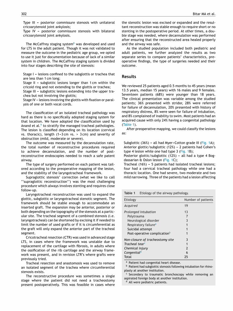
Subglottic (36%) --- all had Myer---Cotton grade III (Fig. 1A);Anterior glottic/subglottic (12%) --- 2 patients had Cohen’stype 4 lesion while one had type 3 (Fig. 1B);Posterior glottic/subglottic (12%) --- all had a type 4 Bog-dassarian & Oslon lesion (Fig. 1C);Tracheal (16%) --- 5 patients had isolated tracheal lesions;
plasty at another institution.c Secondary to traumatic bronchoscopy while removing an
aspirated foreign body at another institution.d All were pediatric patients.
Airway reconstruction in advanced-stage laryngotracheal stenosis 303
Figure 1 Mapping of various pathologies. (A) Grade 3 isolated subglottic stenosis; (B) type 4 glottic web; (C) type 4 posteriorglottic stenosis; (D) cervical, moderate, 1---3 cm isolated tracheal stenosis.
aotasprttasT
fPatp
>3 cm of the tracheal length while the other 2 had a lesioninvolving 1---3 cm of the trachea (Fig. 1D);Laryngotracheal (20%) --- 4 patients with LTS had McCaffreystage 3, while one had stage 4;Supraglottic (4%).
Thirty-two open reconstructive procedures were per-formed on 25 patients (Tables 2 and 3). Sixteen patientshad already had a procedure attempted prior to an opensurgical reconstruction whether it was a tracheotomy, bal-loon dilatation, or laser treatment. All the intraoperativefindings corresponded to the topography resultant from thepreoperative mapping.
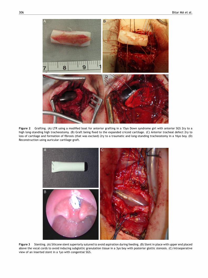
Cartilage grafts were used to expand the airway whenneeded; these were mainly cartilage rib grafts (for cricoidexpansion), conchal graft (for tracheal expansion) and thy-roid alar graft (in infants) (Fig. 2). Stenting was needed tosupport the reconstructed area in 84% of the performed pro-cedures. The stents were different in types and included
silicone stents (part of Montgomery T-tube), endotrachealtubes, Aboulker stents, Montgomery T-tubes, and keels(Fig. 3). The duration of stenting varied from one to 40 dayswith a mean of 12.5 and a median of 14.5 days.me(
The outcome of the various corrective procedures wasssessed based on the decannulation rate, and the numberf needed corrective procedures to achieve decannula-ion. The number of needed endoscopy was also calculatednd was not found to correlate with the degree of steno-is or type of surgery performed. Twenty four out of 25atients were eventually decannulated (96%). Most patientsequired only one reconstructive procedure (80%) to achievehat. The number of endoscopies required to follow-up onhe reconstructive procedures ranged from 1 to 7 with
mean of 2.8 and a median of 3. A general compari-on between pediatric and adult patients is summarized inable 4.
The most common complication was granulation tissueormation, which affected mainly patients with stents (75%).ostoperative complications are summarized in Table 5,long with the interventional steps taken to remedyhem and their effect on the decannulation rate of theatients.
The decannulated patients remained asymptomatic at aean follow-up of 50.5 months. They had good exercise tol-
rance and were able to carry on their normal daily activitieswhere applicable). No objective testing (e.g. pulmonary
304
Bitar M
A et
al.
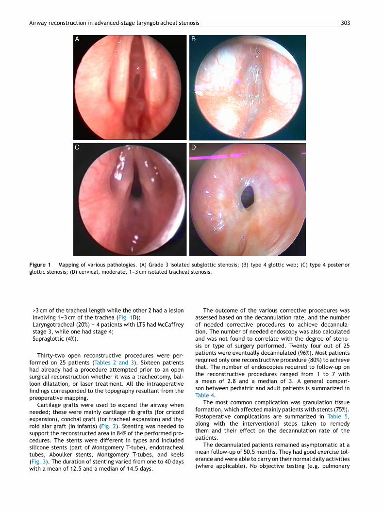
Table 2 Reviewed pediatric patients with advanced laryngeal and or tracheal stenosis.N Age Co-morbidities Lesion Stage Tracheostomy timing Procedures Stenting
(type/duration/g.t.)Number ofendoscopiesneeded after eachsurgery
Outcome
1 12d Cardiac anomalies SGS Cotton Myer III None LTR + AG (SS) ET Tube-5 days-No Three Decannulated2 3m Sturge-Weber
syndromeSGS Cotton Myer III During the 1st
procedureEndoscopic CO2 laserablation
Decannulated
Subglottichemangioma
LTR + AG (SS) ET tube-5 days-No Two
3 8m Congenital TOF SGS Cotton Myer III Prior to the procedure LTR + APG (DS) Siliconea 5 days---Yes Seven DecannulatedEsophageal atresiaDuodenal atresia
4 1y None AGS Cohen IV Prior to the 1stprocedure
Anterior cricoidsplit + AG (DS)
Keel-19days-Yes-Mitomycin
Four Decannulated
LTR + AG (DS) Silicone-12days-Yes-Mitomycin
Five
5 2y Seizures SGS Cotton Myer III Prior to 1st theprocedure
LTR + APG (DS) Silicone-7 days-No Four DecannulatedLTR + AG (SS) ET tube-3 days-No Two
6 3y Bilateral severehearing loss
PGS Bogdassarian Olson IV During the procedure LTR + APG (DS) Silicone-21 days-Yes Four Decannulated
7 5y None Tracheal Anand (tracheal,moderate, >3 cm)
After the procedureb Primary repair throughThoracotomy (DS)
ET tube-11 days-No Three Decannulated
8 6y Cerebral palsy LTS Mc-Caffrey III Prior to the procedure LTR + AG (DS) None One Not decannulated9 9y None SGS Cotton Myer III During the 1st
procedureEndoscopic dilatation DecannulatedLTR + APG (DS) Silicone-7 days-Yes Two
10 9y None LTS Mc-Caffrey III Prior to the 1stprocedure
Endoscopic dilatation DecannulatedLTR + AG (DS) Abulkheir-5 days-No FiveEndoscopic dilatation
11 12y None Tracheal Anand (cervical,severe, >3 cm)
Prior to the procedure R + A (SS) ET Tube-8 days-No Two Decannulated
12 13y None PGS Bogdassarian Olson IV None LTR + PG (SS) ET tube-7 days-No One Decannulated13 15y None SGS Cotton Myer III Prior to the procedure LTR + APG (DS) Silicone-21 days-Yes Two Decannulated14 15y Down syndrome SGS Cotton Myer III Prior to the procedure LTR + AG (DS) None-Yes Five Decannulated15 15y Mild mental
retardation posttrauma (car accident)
PGS Bogdassarian Olson IV During the procedure LTR + APG (DS) Silicone-25 days-Yes Three Decannulated
16 16y None Tracheal Anand (cervical, mild,1---3 cm)
None Tracheoplasty + AG (SS) None One Decannulated
17 17y None AGS Cohen III During the 2ndprocedure
Endoscopic excision ofweb
Decannulated
LTR + AG (DS) Keel-27days-Yes-Mitomycin
Three
Endoscopic excision ofweb
SGS, subglottic stenosis; LTR, laryngotracheal reconstruction; AG, anterior graft; APG, anterior and posterior grafts; CTR, cricotracheal resection; R + A, resection and anastomosis; PG,posterior graft; ET, endotracheal tube; SS, single stage; DS, double stage; g.t., granulation tissue.
a Silicone stent is made of one of the flanges of a Montgomery T-tube, it is always plugged caudally to avoid aspiration with the upper tip placed just above the level of the vocal cords.b Tracheotomy was performed after ET tube removal to help toileting and avoid prolonged intubation.
Airway
reconstruction in
advanced-stage laryngotracheal
stenosis
305
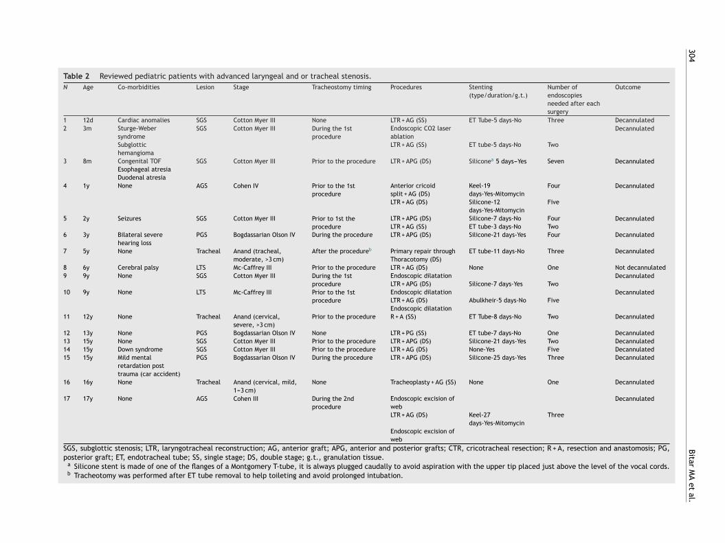
Table 3 Reviewed adult patients with advanced laryngeal and or tracheal stenosis.
N Age Co-morbidities Lesion Stage Tracheostomytiming
Procedures Stenting(type/duration/g.t.)
Number ofendoscopiesneeded aftereach surgery
Outcome
1 18y None LTS Mc-Caffrey III During the 3rdprocedure
R + A (SS) ET Tube 1 day-No One DecannulatedEndoscopicDilatationLTR + APG (DS) Montgomery
T-tube 21 days-NoFive
Tracheoplasty + AG(DS)
Siliconea 40days-Yes
One
2 18y Vocal cordsparalysis
LTS Mc-Caffrey IV Prior to the 1stprocedure
CTR (DS) None Three DecannulatedRight Posteriorcordotomy
3 18y None Supra-glottic NA Prior to the 1stprocedure
Supraglotticreconstruction(DS)
Silicone 21days-No
One Decannulated
Release ofadhesionsRelease ofadhesions
4 22y None Tracheal Anand (cervical,mild, 1---3 cm)
None Tracheoplasty + AG(SS)
ET tube 1 day-No One Decannulated
5 23y None LTS Mc-Caffrey III Prior to the 1stprocedure
R + A (DS) MontgomeryT-tube 7 days-No
Two Decannulated
LTR + APG (DS) Silicone-17days-No
Three
6 25y None SGS Cotton Myer III Prior to theprocedure
LTR + APG (SS) ET Tube 4 days-No Three Decannulated
7 29y GERD AGS Cohen IV Prior to theprocedure
CTR + AG (DS) None-Yes Three Decannulated
8 45y None SGS Cotton Myer III During the 3rdprocedure
EndoscopicDilatation
Decannulated
LTR + APG (SS) ET tube 5 days-No FourLTR + APG (DS) Silicone 19
days-NoTwo
EndoscopicdilatationCTR (DS) None-Yes-
MitomycinOne
SGS, subglottic stenosis; LTR, laryngotracheal reconstruction; AG, anterior graft; APG, anterior and posterior grafts; CTR, cricotracheal resection; R + A, resection and anastomosis; PG,posterior graft; ET, endotracheal tube; SS, single stage; DS, double stage; g.t., granulation tissue.
a Silicone stent is made of one of the flanges of a Montgomery T-tube, it is always plugged caudally to avoid aspiration with the upper tip placed just above the level of the vocal cords.
306 Bitar MA et al.
Figure 2 Grafting. (A) LTR using a modified boat for anterior grafting in a 15yo Down syndrome girl with anterior SGS 2ry to ahigh long-standing high tracheostomy. (B) Graft being fixed to the expanded cricoid cartilage. (C) Anterior tracheal defect 2ry toloss of cartilage and formation of fibrosis (that was excised) 2ry to a traumatic and long-standing tracheostomy in a 16yo boy. (D)Reconstruction using auricular cartilage graft.
Fav
igure 3 Stenting. (A) Silicone stent superiorly sutured to avoid aspbove the vocal cords to avoid inducing subglottic granulation tissueiew of an inserted stent in a 1yo with congential SGS.
iration during feeding. (B) Stent in place with upper end placed in a 3yo boy with posterior glottic stenosis. (C) Intraoperative

Airway reconstruction in advanced-stage laryngotracheal stenosis 307
Figure 4 (A) and (B) Suggested algorithm to follow when managing advanced airway stenotic lesions. The key is to map the lesionfirst, stage it properly and then tailor the surgical procedure accordingly.
P
Wytto
p
function test) was performed on these patients as they hadno clinical indication for it.
The voice was evaluated postoperatively in our patientsby the speech pathologist. The assessment evaluated theneed for speech therapy or other measures in case the voicewas not adequate and or not acceptable to the patient andor corresponding parents. All patients with SGS or posteriorglottic/subglottic stenosis had a normal voice, even thosepatients who needed more than one procedure.
We have devised an algorithm to manage advanced laryn-geal and or tracheal stenosis (Fig. 4A and B), focusing onaccurately mapping the lesion and staging it before decidingon choosing a particular surgical procedure.
stmr
ediatric patients
e operated on 17 pediatric patients, aged 12 days to 17ears, mean 8.2 years, median 9 years (Table 2). Eight ofhem (47%) had associated co-morbidities that could poten-ially affect the postoperative course and eventually theutcome (except the hearing loss).
Subglottic stenosis was the most common encounteredathology followed by PGS and AGS. Therefore, the glottic/
ubglottic location of the stenosis was landmark of encoun-ered advanced pediatric airway stenosis (71%), as such,ost pediatric patients underwent LTR with cartilageib graft (16/19 procedures). Most of these LTR’s were
308 Bitar MA et al.
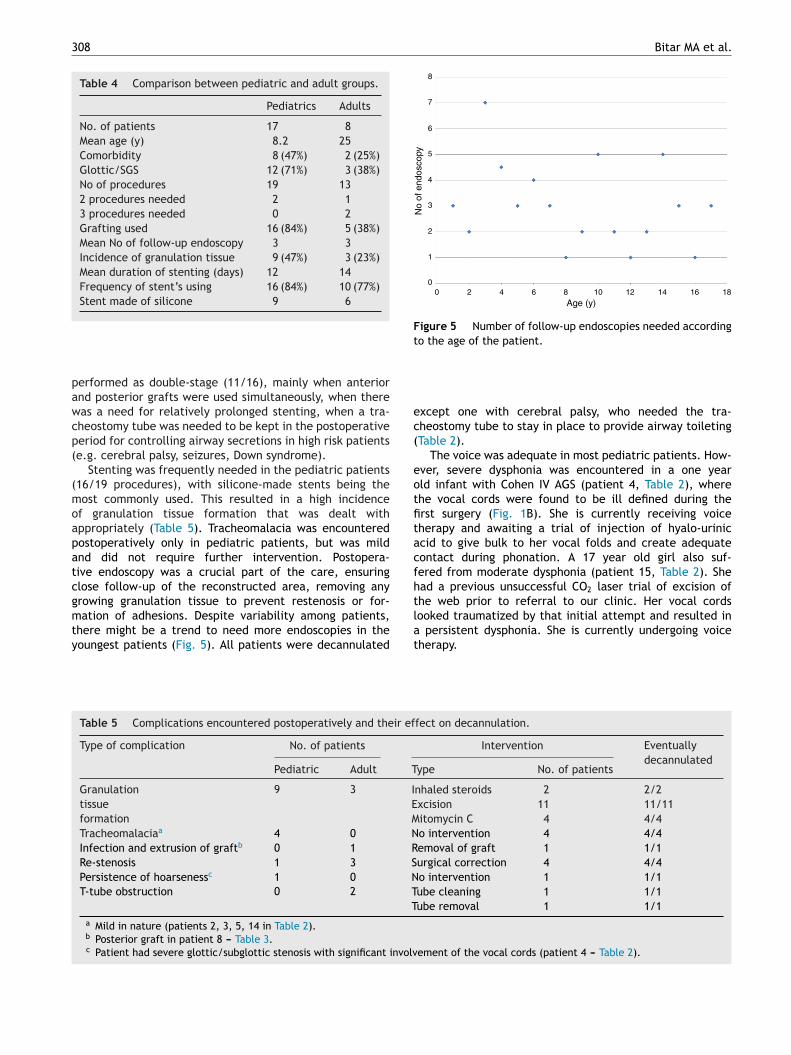
Table 4 Comparison between pediatric and adult groups.
Pediatrics Adults
No. of patients 17 8Mean age (y) 8.2 25Comorbidity 8 (47%) 2 (25%)Glottic/SGS 12 (71%) 3 (38%)No of procedures 19 132 procedures needed 2 13 procedures needed 0 2Grafting used 16 (84%) 5 (38%)Mean No of follow-up endoscopy 3 3Incidence of granulation tissue 9 (47%) 3 (23%)Mean duration of stenting (days) 12 14Frequency of stent’s using 16 (84%) 10 (77%)Stent made of silicone 9 6
pawcp(
(moapatcgmty
8
7
6
5
4
3
No
of e
ndos
copy
2
00 2 4 6 8
Age (y)10 12 14 16 18
1
Figure 5 Number of follow-up endoscopies needed accordingto the age of the patient.
ec(
eotfitacfhthe web prior to referral to our clinic. Her vocal cords
erformed as double-stage (11/16), mainly when anteriornd posterior grafts were used simultaneously, when thereas a need for relatively prolonged stenting, when a tra-heostomy tube was needed to be kept in the postoperativeeriod for controlling airway secretions in high risk patientse.g. cerebral palsy, seizures, Down syndrome).
Stenting was frequently needed in the pediatric patients16/19 procedures), with silicone-made stents being theost commonly used. This resulted in a high incidence
f granulation tissue formation that was dealt withppropriately (Table 5). Tracheomalacia was encounteredostoperatively only in pediatric patients, but was mildnd did not require further intervention. Postopera-ive endoscopy was a crucial part of the care, ensuringlose follow-up of the reconstructed area, removing anyrowing granulation tissue to prevent restenosis or for-
ation of adhesions. Despite variability among patients,here might be a trend to need more endoscopies in theoungest patients (Fig. 5). All patients were decannulated
lat
Table 5 Complications encountered postoperatively and their ef
Type of complication No. of patients
Pediatric Adult T
Granulationtissueformation
9 3 IEM
Tracheomalaciaa 4 0 NInfection and extrusion of graftb 0 1 RRe-stenosis 1 3 SPersistence of hoarsenessc 1 0 NT-tube obstruction 0 2 T
Ta Mild in nature (patients 2, 3, 5, 14 in Table 2).b Posterior graft in patient 8 --- Table 3.c Patient had severe glottic/subglottic stenosis with significant involv
xcept one with cerebral palsy, who needed the tra-heostomy tube to stay in place to provide airway toiletingTable 2).
The voice was adequate in most pediatric patients. How-ver, severe dysphonia was encountered in a one yearld infant with Cohen IV AGS (patient 4, Table 2), wherehe vocal cords were found to be ill defined during therst surgery (Fig. 1B). She is currently receiving voiceherapy and awaiting a trial of injection of hyalo-uriniccid to give bulk to her vocal folds and create adequateontact during phonation. A 17 year old girl also suf-ered from moderate dysphonia (patient 15, Table 2). Shead a previous unsuccessful CO2 laser trial of excision of
ooked traumatized by that initial attempt and resulted in persistent dysphonia. She is currently undergoing voiceherapy.
fect on decannulation.
Intervention Eventuallydecannulated
ype No. of patients
nhaled steroids 2 2/2xcision 11 11/11itomycin C 4 4/4o intervention 4 4/4emoval of graft 1 1/1urgical correction 4 4/4o intervention 1 1/1ube cleaning 1 1/1ube removal 1 1/1
ement of the vocal cords (patient 4 --- Table 2).

nosi
ia
rpmspoof
m55Gadawespwodoa
ttssitsdsCsbtai
dtdWctepc
cua
Airway reconstruction in advanced-stage laryngotracheal ste
Adult patients
Eight adult patients were reviewed, aged 18---45 years, mean25 years, median 22.5 years (Table 3). Only two patients hadco-morbidities that did not affect the outcome, except forthe quality of voice. In contrast to the pediatric patients,adult patients had more lesions affecting multiple levels,including supraglottic and tracheal. Multiple procedureswere needed in 3 patients, including one patient that failedLTR twice and required a salvage CTR. The latter becamethe procedure of choice for adults with advanced LTS afterencountering difficulties (e.g. infection, re-stenosis, delayin healing) using expansion procedures using rib cartilagegrafts. Stents were used as frequently as in the pediatricpatients for a comparable duration of time too, withsurprisingly less granulation tissue formation. However,adult patients had other complications detailed in Table 5.
The postoperative voice of the adult patients was goodin general. One patient (patient 7, Table 3) had gastro-esophageal reflux causing intermittent mild dysphonia; itwas treated by PPI with good improvement. Another patient(patient 2, Table 3) had moderate dysphonia secondary topre-existing bilateral vocal cord paralysis (2ry to his initialneck trauma). He is receiving voice therapy to improve hisphonation.
Discussion
Congenital airway stenosis includes laryngeal atresia, laryn-geal web, posterior glottic stenosis, subglottic stenosisand tracheal stenosis (complete tracheal rings). Most ofthese pathologies are believed to result from failure ofrecanalization of the airway during embryological devel-opment. Congenital subglottic stenosis is defined as asubglottic diameter of less than 4.5 mm in a newborn or lessthan 4 mm in a premature infant, in the absence of acquiredcauses of stenosis.10 It is the most common cause of con-genital airway narrowing and the third most common causeof congenital stridor after laryngomalacia and vocal cordparalysis. It can be due to a cartilaginous malformation, afibrous narrowing or a glandular hyperplasia. It tends to bemilder than an acquired stenosis, having a better prognosisand allowing in some cases a wait-and-see policy.3
Acquired LTS is more common and results from pro-longed endotracheal intubation in 90% of the cases. It isestimated that 1---5% of intubated children may eventuallydevelop LTS.11 Other factors may include external trauma,inflammatory conditions or tumors. In children, the mostsusceptible area is the subglottis, as it is the narrowest partof the larynx, has a delicate mucosa and submucosa, andis formed of a complete cartilaginous ring.3 The posteriorglottic/subglottic area can be another site of pathology as itmay be subjected to direct pressure trauma from the endo-tracheal tube. Other sites of trauma include the trachea,due to balloon or tracheostomy tube injury, and the glottissecondary to intubation or external trauma.
In adults, LTS is usually acquired and is the result of intu-
bation’s trauma in more than 50% of the cases. Autoimmunedisease and idiopathic etiology can account for 18% of thecases, each. The site of stenosis differs according to theetiological factor. The trachea, for example, is commonlyt
cs
s 309
nvolved in autoimmune and iatrogenic causes, while lessffected in idiopathic etiology.12,13
Treatment may include balloon dilatation, which hasecently gained popularity and has been tried even inatients with advanced stenosis or as a primary treatmentodality.14,15 It was used in some of our patients but was not
uccessful, resulting in the need of an open reconstructiverocedure. Nonetheless, the use of balloon dilatation post-peratively might be beneficial to treat an early re-stenosisr stricture and prevent its progression into a more severeorm.
In a systematic review of dilatation as a primary treat-ent modality for LTS, Chueng and Chadha (2013) reported a
0% success rate with balloon dilatation, which increased to0---78% upon coupling it with adjuvant therapy.16 Recently,ünaydın et al. (2014) compared balloon dilatation to LTRs a primary treatment modality and noticed that balloonilatation needs more repetitive interventions than LTR with
higher re-stenosis rate (63.2% vs. 31.3%).17 More concernsere raised in another recent comparative study by Maresht al. (2014) who stated that there is a poor definition of theafety profile for balloon dilatation.18 They believe that therocedure carries risks of worsening the stenosis, affects air-ay tissue integrity, and in particular increases the chancef needing urgent airway intervention. Balloon dilatation hasefinitely its role but does not replace the effective rolef LTR in providing a long lasting safe airway, especially indvanced stenosis.
We have shown in our study that LTR is an impor-ant tool in the airway surgeon’s hand to repair moderateo severe laryngotracheal stenosis. Laryngotracheal airwayurgery includes a variety of techniques depending on theite and extent of the airway pathology. The aim is toncrease the airway lumen diameter and allow the patiento be decannulated as early as possible. Attempts to relieveuch an obstruction started way back in 1956, when Rethiescribed posterior splitting or cricoidotomy with long-termtenting.2,19 Anterior cricoid split was then performed byotton and Seid in 1980, to enable extubation of infantsuffering from SGS. These procedures were later modifiedy introducing costal cartilage grafts with or without sten-ing the expanded area.20,21 Since then, various correctivend reconstructive techniques have been described includ-ng cricotracheal resection.
Because laryngotracheal airway pathologies can affectifferent areas of the larynx and trachea, it would be impor-ant to use appropriate mapping of the lesion prior toeciding on the best reconstructive/corrective procedure.e have used a combination of assessment techniques to
orrectly map the location and extent of the lesions andhis combined method proved to be valid and beneficial,specially that intraoperative finding corresponded to ourreoperative topographic delineation of the lesion in allases.
The various available staging systems are quite useful toorrectly document the present stenosis. One should avoidsing a single staging system to describe any type of stenosis,s this may lead to inaccurate description of the lesion and
o inappropriate reporting of results.The Cotton---Myer staging system5 is one of the mostommonly used grading systems to classify an airwaytenosis. Though it was devised to stage isolated SGS, it has
3
blasanobgpucabanTr
avsupwlsuttsavts(
rpswtcahisTfldha
ttwIctrg
caorobo
tc
ttanwcs
ccc
gfm
dbap
ntfaec
au(nt
wbsrorlPdssT
10
een used in several reports to stage other stenotic areas,ike tracheal and laryngotracheal, something we do notgree on or advise. A symptomatic isolated SGS often needsurgical intervention. It can be caused by a narrowingnterior shelf, bilateral lateral shelves or a circumferentialarrowing. The mode of expansion will depend on the typef narrowing. An anterior shelf can be adequately correctedy an anterior cricoid split and a modified boat cartilageraft to maintain the expansion. It is often a single stagerocedure that needs a short-term or no stenting, which issually done using an endotracheal tube. A subglottis withircumferential stenosis or bilateral shelves are managed byn anterior and posterior cricoid split which are supportedy a boat shaped posterior and modified boat shapednterior grafts. The reconstructed area almost alwayseeds stenting to stabilize the area while healing occurs.he duration of stenting will depend on the stability of theeconstructed area at the end of the procedure.
Stents are often a source of granulation tissue formationnd care should be taken to monitor such a reaction to pre-ent restenosis or formation of obstructive adhesions. In oureries, not every patient of the 21 who had a stent, got gran-lation tissue, and granulation tissue even occurred in someatients who had no stent (Tables 2 and 3). The ET tubeas used in 10 patients and was not associated with granu-
ation tissue formation, in contrast to the silicone stent thathowed a reaction in 8 out of 12 patients in whom it wassed. The age range was similar between both groups, buthe mean duration of stenting was different (5 days for ETube vs. 21 days for the silicone stent), reflecting the neces-ity to limit the stenting period. Looking specifically at thege of the patients, pediatric patients seemed to be moreulnerable to form granulation tissue than adult patients andhus should be more closely monitored with frequent endo-copies until resolution of the granulation tissue formationTable 4).
Performing a single-stage or a double-stage operationelies on the ability to avoid a tracheostomy at the end of therocedure while achieving a safe airway. It also relies on theeverity of the present pathology, and the stability of the air-ay. Including grafts during reconstruction would decrease
he required stenting duration. Cartilage grafts are mostommonly harvested from the ribs but alternatives includeuricular, thyroid alar and septal cartilage.2,22 Rib grafts arearvested with an intact perichondrium on one side to facil-tate mucosalization. Their success in reconstructing theubglottic area exceeds that in correcting tracheal stenosis.hey are also noticed to better integrate with the airwayramework in the pediatrics than in adults. Adult’s carti-age has foci of ossification which makes its carving moreifficult, suturing it into the airway framework harder, andealing slower, with a possibility of acquiring an infectionnd extruding.
When the subglottic area is totally occluded (grade 4),he area cannot be expanded and is rather resected, hencehe CTR. In addition, severe grade 3 stenosis, especiallyhen framework fibrosis exists, is best treated with CTR.23
t is a more challenging procedure, but with a higher suc-
ess rate.24 Though only few cases of CTR were reviewed inhe current study, we have found this procedure particularlyewarding in the adult patients, where using of cartilage ribrafts is avoided.t&il
Bitar MA et al.
When the stenosis involves both the larynx and the tra-hea, the management may include tracheal resection andnastomosis and or airway expansion using cartilage graftr CTR. In these cases, the reconstruction method is tailo-ed specifically to the present pathology, according to thebtained preoperative topography of the lesion. These maye tough cases and decannulation may not happen followingne reconstructive procedure (Table 3).
Looking at those cases that failed an initial reconstruc-ive procedure despite adequate preoperative mapping, weould realize the following:
Two patients (patients 1 and 5, Table 3) were subjectedo initial resection and anastomosis of the involved upperrachea, which resulted in aggravation of the existing SGSt the site of anastomosis (crico-tracheal junction). Thisecessitated additional reconstruction of the subglottic areaith an anterior and posterior graft. These 2 proceduresould have been avoided by performing a CTR from thetart.
Patients 5 (Table 2) and 1 (Table 3) had an additional pro-edure (LTR with anterior graft) to correct a suprastomalollapse, which is often associated with a long standing tra-heostomy tube.
Patient 8 (Table 3) taught us to avoid using rib cartilagerafts in subsequent repair of an adult airway stenosis. A CTRrom the start would have spared the patient two additionalajor procedures.Patient 4 (Table 2) was a challenging case and two proce-
ures could not be avoided. The pathology was of what coulde classified as partial laryngeal atresia. These are delicatend tough cases that are expected to require more than onerocedure to reach a safe airway.
Isolated tracheal pathology are hard to stage as there iso single commonly used grading system that can assess allracheal pathologies. When expansion is needed, we haveound the auricular cartilage graft of great use both in pedi-tric and adults patients due to its appropriate contour andlasticity that conforms with the normal shape of the tra-heal rings.25
Glottic stenosis is less common but can usually be man-ged successfully with a single surgical procedure. Again,sing appropriate classification for each type of stenosisanterior vs. posterior glottis) will ensure proper dissemi-ation of information about the existing pathology amongreating surgeons and in published reports.
Our overall decannulation or extubation rate was 96%hich is comparable to that present in the literature, foroth pediatric and adult patients.2,4,9,19,23,24,26---36 Operation-pecific decannulation rate is a more common method ofeporting success rate; however, it is a simple way that mayverlook the type, location and extent of the lesion. Theates may get over-rated by including lesions that are ofow stages (Rizzi et al.,2 Agrawal et al.,19 White et al.33).erforming a single stage or a double stage operation willepend on several factors already discussed above, andhould not be a criteria for reporting success rate as done byome authors (Saunders et al.,34 Gustafson et al.,35 Rhee &oohill36). We prefer reporting the success rate according to
he type/site of pathology (like Rutter & Cotton27 and WyattHartley28) with emphasis on differentiating between treat-ng mild (stage 1 or 2) vs. moderate to severe (stage 3 or 4)esions.
nosi
1
1
1
1
1
1
1
1
1
2
2
2
2
2
2
2
2
2
2
3
Airway reconstruction in advanced-stage laryngotracheal ste
Though both pediatric and adult patients had favorableoutcome, it is worth mentioning that pediatric patients(especially the infants and young children) need more metic-ulous techniques during airway reconstruction due to thesmaller dimensions of the airway and the tendency to formgranulation tissue when a stent is used. Postoperative care inthe intensive care unit adds another aspect to the challengesencountered in the pediatric patients regarding the needfor sedation and tracheostomy/endotracheal tube care,and other medical treatment especially if co-morbiditiesexist.
To be transparent, it is very important to specify howmany reconstructive/corrective procedures were neededto achieve decannulation. Requiring multiple proceduresmay reflect either the complexity of the case (e.g. multi-levels stenosis, co-morbidities) or the inefficacy of the usedtechnique for the particular lesion. Details will be able topinpoint the reason behind a particular failure.
Conclusion
The review of our approach to open airway repair/reconstruction showed its efficacy in advanced-stagelaryngotracheal stenosis. Good knowledge of a variety ofreconstructive techniques is crucial to achieve good resultsin a variety of age groups.
Conflicts of interest
The authors declare no conflicts of interest.
References
1. McDonald IH, Stocks JG. Prolonged nasotracheal intubation. Areview of its development in a pediatric hospital. Brit J Anaesth.1965;37:161---73.
2. Rizzi MD, Thorne MC, Zur KB, Jacobs IN. Laryngotrachealreconstruction with posterior costal cartilage grafts: out-comes at a single institution. Otolaryngol Head Neck Surg.2009;140:348---53.
3. Lesperance MM, Zalzal GH. Laryngotracheal stenosis in children.Eur Arch Otorhinolaryngol. 1998;255:12---7.
4. Pena J, Cicero R, Marín J, Ramírez M, Cruz S, NavarroF. Laryngotracheal reconstruction in subglottic stenosis: anancient problem still present. Otolaryngol Head Neck Surg.2001;125:397---400.
5. Myer CM 3rd, O’Connor DM, Cotton RT. Proposed grading systemfor subglottic stenosis based on endotracheal tube sizes. AnnOtol Rhinol Laryngol. 1994;103:319---23.
6. Cohen SR. Congenital glottic webs in children: a retrospec-tive review of 51 patients. Ann Otol Rhinol Laryngol. 1985;121:2---16.
7. Bogdasarian RS, Olson NR. Posterior glottic laryngeal stenosis.Otolaryngol Head Neck Surg. 1980;88:765---72.
8. McCaffrey TV. Classification of laryngotracheal stenosis. Laryn-goscope. 1992;102:1335---40.
9. Anand VK, Alemar G, Warren ET. Surgical considerations in tra-cheal stenosis. Laryngoscope. 1992;102:237---43.
10. Zalzal GH, Cotton RT. Glottic and subglottic stenosis. In: Cum-mings CW, Fredrickson JM, Krause CJ, Schuller DE, editors.Otolaryngology-head and neck surgery. 2nd edn St. Louis:Mosby-Year Book; 1993. p. 1981---2000.
3
3
s 311
1. Cotton RT. Prevention and management of laryngeal stenosis ininfants and children. J Pediatr Surg. 1985;20:845---51.
2. Lorenz R. Adult laryngotracheal stenosis: etiology and sur-gical management. Curr Opin Otolaryngol Head Neck Surg.2003;11:467---72.
3. Gelbard A, Francis D, Sandulache V, Simmons J, Donovan D,Ongkasuwan J. Causes and consequences of adult laryngotra-cheal stenosis. Laryngoscope. 2015;125:1137---43.
4. Guarisco JL, Yang CJ. Balloon dilation in the management ofsevere airway stenosis in children and adolescents. J PediatrSurg. 2013;48:1676---81.
5. Hautefort C, Teissier N, Viala P, Van Den Abbeele T. Balloondilation laryngoplasty for subglottic stenosis in children. ArchOtolaryngol Head Neck Surg. 2012;138:235---40.
6. Chueng K, Chadha N. Primary dilatation as a treatment for pedi-atric laryngotracheal stenosis: a systematic review. Int J PediatrOtorhinolaryngol. 2013;77:623---8.
7. Günaydın RO, Süslü N, Bajin MD, Kuscu O, Yılmaz T, Ünal ÖF,et al. Endolaryngeal dilatation versus laryngotracheal recon-struction in the primary management of subglottic stenosis. IntJ Pediatr Otorhinolaryngol. 2014;78:1332---6.
8. Maresh A, Preciado DA, O’Connell AP, Zalzal GH. A compara-tive analysis of open surgery vs endoscopic balloon dilation forpediatric subglottic stenosis. JAMA Otolaryngol Head Neck Surg.2014;140:901---5.
9. Agrawal N, Black M, Morrison G. Ten-year review of laryngotra-cheal reconstruction for paediatric airway stenosis. Int J PediatrOtorhinolaryngol. 2007;71:699---703.
0. Fearon B, Cotton R. Surgical correction of subglottic stenosisof the larynx. Preliminary report of an experimental surgicaltechnique. Ann Otol Rhinol Laryngol. 1972;81:508---13.
1. Evans T. Laryngo-tracheoplasty. J Laryngol Otol. 1974;88:589---97.
2. Fayoux P, Devisme L, Merrot O, Chevalier D. Thyroid alarcartilage graft in laryngoplasty anatomical study in prema-ture and newborn babies. Int J Pediatr Otorhinolaryngol.2002;66:259---63.
3. Hartley BEJ, Rutter MJ, Cotton RT. Cricotracheal resection as aprimary procedure for laryngotracheal stenosis in children. IntJ Pediatr Otorhinolaryngol. 2000;54:133---6.
4. Monnier P, Lang F, Savary M. Partial cricotracheal resectionfor pediatric subglottic stenosis: a single institution’s expe-rience in 60 cases. Eur Arch Otorhinolaryngol. 2003;260:295---7.
5. Lusk RP, Kang DR, Muntz HR. Auricular cartilage grafts inlaryngotracheal reconstruction. Ann Otol Rhinol Laryngol.1993;102:247---54.
6. Rea F, Callegaro D, Loy M, Zuin A, Narne S, Gobbi T, et al. Benigntracheal and laryngotracheal stenosis: surgical treatment andresults. Eur J Cardio-thoracic Surg. 2002;22:352---6.
7. Rutter MJ, Cotton RT. The use of posterior cricoid grafting inmanaging isolated posterior glottic stenosis in children. ArchOtolaryngol Head Neck Surg. 2004;130:737---9.
8. Wyatt ME, Hartley BE. Laryngotracheal reconstruction in con-genital laryngeal webs and atresias. Otolaryngol Head NeckSurg. 2005;132:232---8.
9. Rutter MJ, Hartley BEJ, Cotton RT. Cricotracheal resectionin children. Arch Otolaryngol Head Neck Surg. 2001;127:289---92.
0. Younis RT, Lazar RH, Astor F. Posterior cartilage graft in single-stage laryngotracheal reconstruction. Otolaryngol Head NeckSurg. 2003;129:168---75.
1. White DR, Cotton RT, Bean JA, Rutter MJ. Pediatric cricotra-cheal resection: surgical outcomes and risk factor analysis. Arch
Otolaryngol Head Neck Surg. 2005;131:896---9.2. Johnson RF, Rutter M, Cotton R, Vijayasekeran S, White D. Crico-tracheal resection in children 2 years of age and younger. AnnOtol Rhinol Laryngol. 2008;117:110---2.

3
3
3
3
12
3. White DR, Bravo M, Vijayasekaran S, Rutter MJ, Cotton RT, ElluruRG. Laryngotracheoplasty as an alternative to tracheotomy ininfants younger than 6 months. Arch Otolaryngol Head Neck
Surg. 2009;135:445---7.4. Saunders MW, Thirlwall A, Jacob A, Albert DM. Single-or-two-stage laryngotracheal reconstruction; comparison of outcomes.Int J Pediatr Otorhinolaryngol. 1999;50:51---4.
3
Bitar MA et al.
5. Gustafson LM, Hartley BE, Liu JH, Link DT, Chadwell J, KoebbeC, et al. Single-stage laryngotracheal reconstruction in chil-dren: a review of 200 cases. Otolaryngol Head Neck Surg.
2000;123:430---4.6. Rhee JS, Toohill RJ. Single-stage adult laryngotrachealreconstruction without stenting. Laryngoscope. 2001;111:765---8.