Embed Size (px)
Citation preview
Doctorate Program in Molecular Oncology and Endocrinology
Doctorate School in Molecular Medicine
XX cycle - 2004–2007 Coordinator: Prof. Giancarlo Vecchio
“The function of PED/PEA-15 in brain: identification of new protein-protein
interactions and their biological relevance”
Alessia Paola Maria Barbagallo
University of Naples Federico II
Dipartimento di Biologia e Patologia Cellulare e Molecolare “L. Califano”
Administrative Location
Dipartimento di Biologia e Patologia Cellulare e Molecolare “L. Califano” Università degli Studi di Napoli Federico II
Partner Institutions Italian Institutions Università di Napoli “Federico II”, Naples, Italy Istituto di Endocrinologia ed Oncologia Sperimentale “G. Salvatore”, CNR, Naples, Italy Seconda Università di Napoli, Naples, Italy Università del Sannio, Benevento, Italy Università di Genova, Genoa, Italy Università di Padova, Padua, Italy Foreign Institutions Johns Hopkins School of Medicine, Baltimore, MD, USA Johns Hopkins Krieger School of Arts and Sciences, Baltimore, MD, USA National Institutes of Health, Bethesda, MD, USA Ohio State University, Columbus, OH, USA Université Paris Sud XI, Paris, France Universidad Autonoma de Madrid, Spain Centro de Investigaciones Oncologicas (CNIO), Spain Universidade Federal de Sao Paulo, Brazil Albert Einstein College of Medicine of Yeshiwa University, USA Supporting Institutions Università di Napoli “Federico II”, Naples, Italy Ministero dell’Istruzione, dell’Università e della Ricerca Istituto Superiore di Oncologia (ISO) Terry Fox Foundation, Canada Istituto di Endocrinologia ed Oncologia Sperimentale “G. Salvatore”, CNR, Naples, Italy Centro Regionale di Competenza in Genomica (GEAR)
Faculty
Italian Faculty Giancarlo Vecchio, MD, Co-ordinator Salvatore Maria Aloj, MD Francesco Beguinot, MD Maria Teresa Berlingieri, PhD Angelo Raffaele Bianco, MD Bernadette Biondi, MD Francesca Carlomagno, MD Gabriella Castoria, MD Angela Celetti, MD Annamaria Cirafici, PhD Mario Chiariello, MD Vincenzo Ciminale, MD Annamaria Colao, MD Alma Contegiacomo, MD Sabino De Placido, MD Monica Fedele, PhD Pietro Formisano, MD Alfredo Fusco, MD Massimo Imbriaco, MD Paolo Laccetti, MD Antonio Leonardi, MD Barbara Majello, PhD Rosa Marina Melillo, MD Claudia Miele, PhD Francesco Oriente, MD Roberto Pacelli, MD Giuseppe Palumbo, PhD Silvio Parodi, MD Giuseppe Portella, MD Giorgio Punzo, MD Antonio Rosato, MD Massimo Santoro, MD Giampaolo Tortora, MD Donatella Tramontano, PhD Giancarlo Troncone, MD Bianca Maria Veneziani, MD Giuseppe Viglietto, MD Roberta Visconti, MD
Foreign Faculty National Institutes of Health (USA) Michael M. Gottesman, MD Silvio Gutkind, PhD Stephen Marx, MD Ira Pastan, MD Phil Gorden, MD Johns Hopkins School of Medicine (USA) Vincenzo Casolaro, MD Pierre Coulombe, PhD James G. Herman MD Robert Schleimer, PhD Johns Hopkins Krieger School of Arts and Sciences (USA) Eaton E. Lattman, MD Ohio State University, Columbus (USA) Carlo M. Croce, MD Albert Einstein College of Medicine of Yeshiwa University (USA) Luciano D’Adamio, MD Nancy Carrasco Université Paris Sud XI (France) Martin Schlumberger, MD Universidad Autonoma de Madrid (Spain) Juan Bernal, MD, PhD Pilar Santisteban Centro de Investigaciones Oncologicas (Spain) Mariano Barbacid, MD Universidade Federal de Sao Paulo (Brazil) Janete Maria Cerutti Rui Maciel
“The function of PED/PEA-15 in brain: identification of new protein-protein
interactions and their biological relevance”
TABLE OF CONTENTS
ABSTRACT.....................................................................................................................6 BACKGROUND.............................................................................................................7 1. Classification of neurodegenerative diseases........................................................7 2. Protein aggregation and neurodegeneration..........................................................8 3. Alzheimer’s Disease.................................................................................................9 4. Parkinson’s Disease................................................................................................12 5. Interaction between genes and environment in neurodegenerative diseases.....................................................................................................................14 6. Association between neurodegenerative disorders, insulin-resistance and diabetes mellitus......................................................................................................15 7. Insulin action...........................................................................................................16 8. Insulin-resistance and impaired glucose tolerance.............................................17 9. The protein PED/PEA-15......................................................................................19 AIM OF THE STUDY.................................................................................................23 MATERIALS AND METHODS................................................................................24 RESULTS AND DISCUSSION.............................................................................. ..27 Isolation and Identification of APP and ADAM10 as novel PED/PEA-15 interacting proteins.........................................................................27 APP and ADAM10 interact with PED/PEA-15 in vitro as well as in mammalian cells and mouse brain...................................................................29 Effect of PED/PEA-15 on APP-ADAM10 interaction......................................32 Effect of PED/PEA-15 on APP proteolytic processing.....................................33 The effect of PED/PEA-15 on APP processing is mediated by cPKCs..........37 CONCLUSIONS..........................................................................................................39 REFERENCES.............................................................................................................40 ACKNOWLEDGEMENTS.......................................................................................48

LIST OF PUBLICATIONS
This dissertation is based upon the following publications:
1. Trencia A, Fiory F, Maitan MA, Vito P, Barbagallo APM, Perfetti A, Miele C, Ungaro P, Oriente F, Cilenti L, Zervos AS, Formisano P, Beguinot F. Omi/HtrA2 Promotes Cell Death by Binding and Degrading the Anti-apoptotic Protein ped/pea-15. J Biol Chem 2004; 279: 46566–46572.
2. Perfetti A, Oriente F, Iovino S, Alberobello AT, Barbagallo APM, Esposito
I, Fiory F, Teperino R, Ungaro P, Miele C, Formisano P, Beguinot F. Phorbol Esters Induce Intracellular Accumulation of the Anti-apoptotic Protein PED/PEA-15 by Preventing Ubiquitinylation and Proteasomal Degradation. J Biol Chem 2007; 282:8648-8657.
6
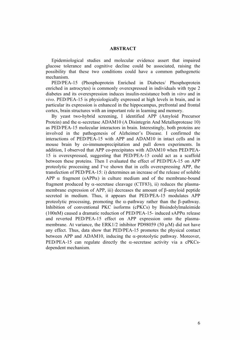
ABSTRACT
Epidemiological studies and molecular evidence assert that impaired glucose tolerance and cognitive decline could be associated, raising the possibility that these two conditions could have a common pathogenetic mechanism.
PED/PEA-15 (Phosphoprotein Enriched in Diabetes/ Phosphoprotein enriched in astrocytes) is commonly overexpressed in individuals with type 2 diabetes and its overexpression induces insulin-resistance both in vitro and in vivo. PED/PEA-15 is physiologically expressed at high levels in brain, and in particular its expression is enhanced in the hippocampus, prefrontal and frontal cortex, brain structures with an important role in learning and memory.
By yeast two-hybrid screening, I identified APP (Amyloid Precursor Protein) and the α-secretase ADAM10 (A Disintegrin And Metalloprotease 10) as PED/PEA-15 molecular interactors in brain. Interestingly, both proteins are involved in the pathogenesis of Alzheimer’s Disease. I confirmed the interactions of PED/PEA-15 with APP and ADAM10 in intact cells and in mouse brain by co-immunoprecipitation and pull down experiments. In addition, I observed that APP co-precipitates with ADAM10 when PED/PEA-15 is overexpressed, suggesting that PED/PEA-15 could act as a scaffold between these proteins. Then I evaluated the effect of PED/PEA-15 on APP proteolytic processing and I‘ve shown that in cells overexpressing APP, the transfection of PED/PEA-15: i) determines an increase of the release of soluble APP α fragment (sAPPα) in culture medium and of the membrane-bound fragment produced by α-secretase cleavage (CTF83), ii) reduces the plasma-membrane expression of APP, iii) decreases the amount of β-amyloid peptide secreted in medium. Thus, it appears that PED/PEA-15 modulates APP proteolytic processing, promoting the α-pathway rather than the β-pathway. Inhibition of conventional PKC isoforms (cPKCs) by Bisindolylmaleimide (100nM) caused a dramatic reduction of PED/PEA-15- induced sAPPα release and reverted PED/PEA-15 effect on APP expression onto the plasma-membrane. At variance, the ERK1/2 inhibitor PD98059 (50 µM) did not have any effect. Thus, data show that PED/PEA-15 promotes the physical contact between APP and ADAM10, inducing the α-proteolytic pathway. Moreover, PED/PEA-15 can regulate directly the α-secretase activity via a cPKCs-dependent mechanism.
7
BACKGROUND
The increase in life span in Western societies is accompanied with an increased incidence of age-related diseases including neurodegeneration and type 2 diabetes, that interfere with the quality of life and become a global issue. Currently, the prevalence of dementia within the population over the age of 65 is about 10-15%. Moreover, it has been estimated that almost one half of the population over 85 will suffer dementia. According to the Global Burden of Disease Study (GBD), a collaborative study of the World Health Organization (WHO), the World Bank and the Harvard School of Public Health, dementia and other neurodegenerative diseases will be, in 2020, the eighth cause of disease burden for developed regions (Murray and Lopez 1996, Menken et al 2000), while neurodegenerative diseases will become the world’s second leading cause of death by the middle of the century, overtaking cancer (Menken et al 2000). Diabetes is calculated to affect 5.9% of the world’s adult population (20-79 age group). Overall, the prevalence of diabetes is expected to increase worldwide from 246 million to 380 million people between 2007 and 2025, representing 7.1% of the adult population (Diabetes Atlas 2006). It is known that approximately 20% of neurodegenerative disorders are associated with insulin-resistance and type 2 diabetes (Ristow 2004).
Neurodegenerative diseases represent a varied assortment of central nervous system (CNS) disorders, inherited or sporadic, characterized by the gradual and progressive loss of specific subsets of neurons in specific functional anatomic systems. Although the causes may differ, patients with neurodegenerative disorders are likely to show localized to generalized atrophy of brain, leading to two principal phenotypic effects, not mutually exclusive: conditions causing problems with movements, such as ataxia, and conditions affecting memory. This compromising in mental and/or physical function dramatically reduces the quality of life for the patients, that, in the later stages of dementia, will lose the ability to care for oneself and will become dependent on other people, increasing the burden on the family and caregivers. §1. Classification of neurodegenerative diseases
The number of neurodegenerative diseases is currently estimated to be a few hundred, and, among these, many appear to overlap with one another clinically and pathologically, rendering their practical classification quite challenging. The issue is further complicated by the fact that, in diseases such as multi-system atrophy in which several areas of the brain are affected, different combinations of lesions can give rise to different clinical pictures (Burn and Jaros 2001). Furthermore, the same neurodegenerative process, especially at the beginning, can affect different areas of the brain, making a given disease appear very different from a symptomatic standpoint. Despite these difficulties, the most popular categorization of neurodegenerative disorders is still based on the predominant clinical feature or the topography of the predominant lesion,
8
or often on a combination of both. Indeed, clinical and pathological manifestations are determined by the location and seriousness of the disorder. Accordingly, neurodegenerative disorders of the CNS may, for example, be first grouped into diseases of the cerebral cortex, the basal ganglia, the brainstem and cerebellum, or the spinal cord. Then, within each group, a given disease may be further classified based on its main clinical features (Przedborski et al. 2003).
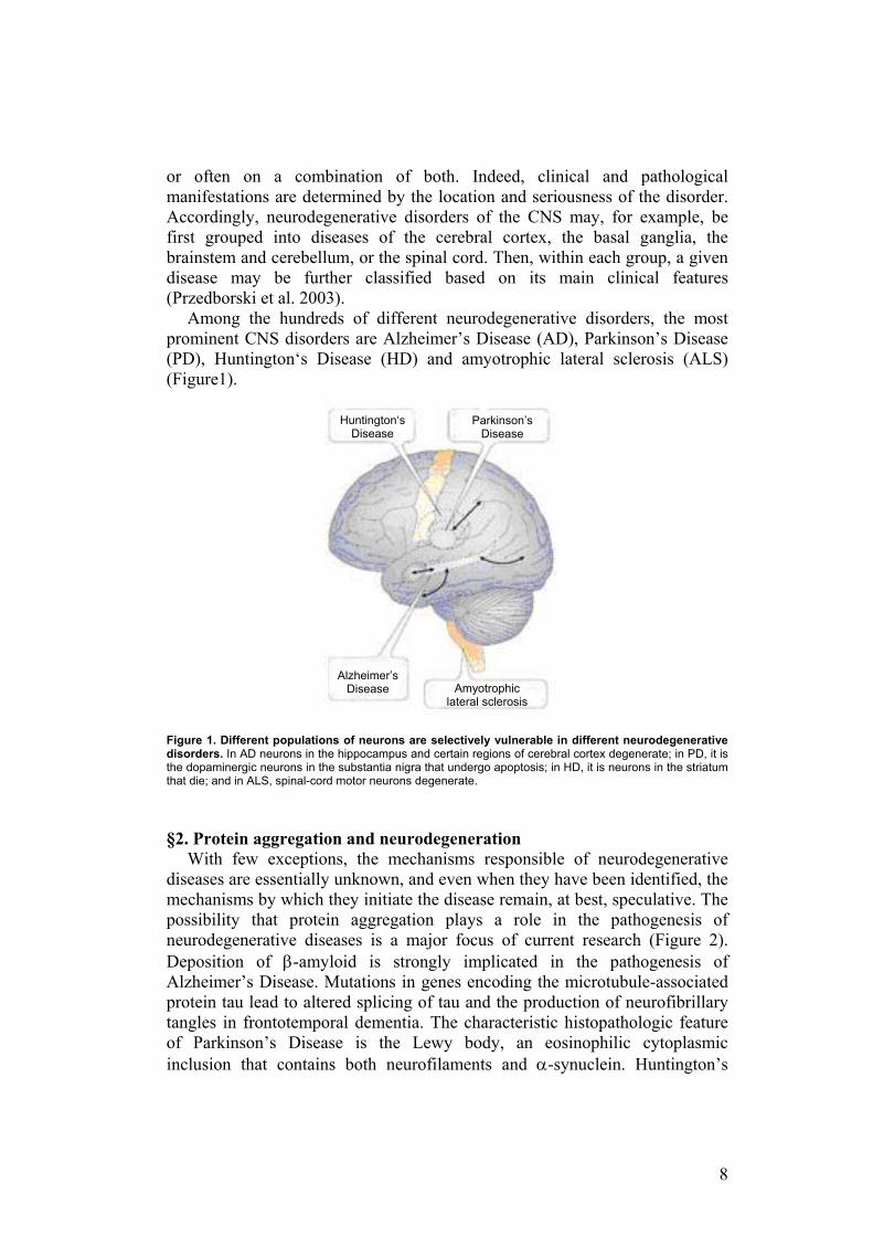
Among the hundreds of different neurodegenerative disorders, the most prominent CNS disorders are Alzheimer’s Disease (AD), Parkinson’s Disease (PD), Huntington‘s Disease (HD) and amyotrophic lateral sclerosis (ALS) (Figure1). Figure 1. Different populations of neurons are selectively vulnerable in different neurodegenerative disorders. In AD neurons in the hippocampus and certain regions of cerebral cortex degenerate; in PD, it is the dopaminergic neurons in the substantia nigra that undergo apoptosis; in HD, it is neurons in the striatum that die; and in ALS, spinal-cord motor neurons degenerate. §2. Protein aggregation and neurodegeneration
With few exceptions, the mechanisms responsible of neurodegenerative diseases are essentially unknown, and even when they have been identified, the mechanisms by which they initiate the disease remain, at best, speculative. The possibility that protein aggregation plays a role in the pathogenesis of neurodegenerative diseases is a major focus of current research (Figure 2). Deposition of β-amyloid is strongly implicated in the pathogenesis of Alzheimer’s Disease. Mutations in genes encoding the microtubule-associated protein tau lead to altered splicing of tau and the production of neurofibrillary tangles in frontotemporal dementia. The characteristic histopathologic feature of Parkinson’s Disease is the Lewy body, an eosinophilic cytoplasmic inclusion that contains both neurofilaments and α-synuclein. Huntington’s
Huntington‘s Disease
Parkinson’s Disease
Amyotrophic lateral sclerosis
Alzheimer’s Disease
9
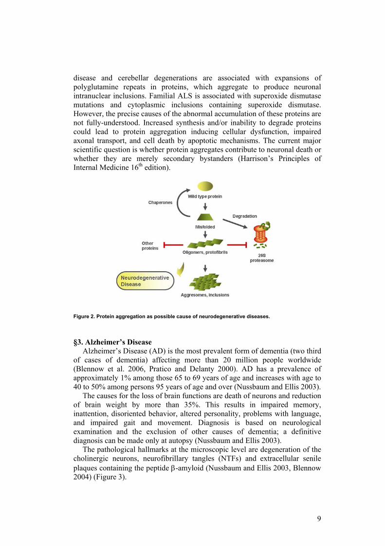
disease and cerebellar degenerations are associated with expansions of polyglutamine repeats in proteins, which aggregate to produce neuronal intranuclear inclusions. Familial ALS is associated with superoxide dismutase mutations and cytoplasmic inclusions containing superoxide dismutase. However, the precise causes of the abnormal accumulation of these proteins are not fully-understood. Increased synthesis and/or inability to degrade proteins could lead to protein aggregation inducing cellular dysfunction, impaired axonal transport, and cell death by apoptotic mechanisms. The current major scientific question is whether protein aggregates contribute to neuronal death or whether they are merely secondary bystanders (Harrison’s Principles of Internal Medicine 16th edition).
Figure 2. Protein aggregation as possible cause of neurodegenerative diseases. §3. Alzheimer’s Disease
Alzheimer’s Disease (AD) is the most prevalent form of dementia (two third of cases of dementia) affecting more than 20 million people worldwide (Blennow et al. 2006, Pratico and Delanty 2000). AD has a prevalence of approximately 1% among those 65 to 69 years of age and increases with age to 40 to 50% among persons 95 years of age and over (Nussbaum and Ellis 2003).
The causes for the loss of brain functions are death of neurons and reduction of brain weight by more than 35%. This results in impaired memory, inattention, disoriented behavior, altered personality, problems with language, and impaired gait and movement. Diagnosis is based on neurological examination and the exclusion of other causes of dementia; a definitive diagnosis can be made only at autopsy (Nussbaum and Ellis 2003).
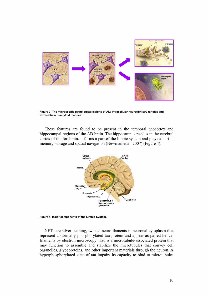
The pathological hallmarks at the microscopic level are degeneration of the cholinergic neurons, neurofibrillary tangles (NTFs) and extracellular senile plaques containing the peptide β-amyloid (Nussbaum and Ellis 2003, Blennow 2004) (Figure 3).
10
Figure 3. The microscopic pathological lesions of AD: intracellular neurofibrillary tangles and extracellular β-amyloid plaques.
These features are found to be present in the temporal neocortex and hippocampal regions of the AD brain. The hippocampus resides in the cerebral cortex of the forebrain. It forms a part of the limbic system and plays a part in memory storage and spatial navigation (Newman et al. 2007) (Figure 4).
Figure 4. Major components of the Limbic System.
NFTs are silver-staining, twisted neurofilaments in neuronal cytoplasm that represent abnormally phosphorylated tau protein and appear as paired helical filaments by electron microscopy. Tau is a microtubule-associated protein that may function to assemble and stabilize the microtubules that convey cell organelles, glycoproteins, and other important materials through the neuron. A hyperphosphorylated state of tau impairs its capacity to bind to microtubules
11
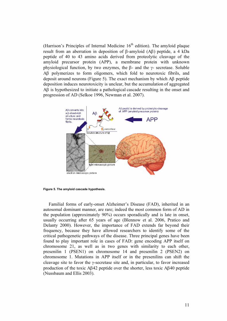
(Harrison’s Principles of Internal Medicine 16th edition). The amyloid plaque result from an aberration in deposition of β-amyloid (Aβ) peptide, a 4 kDa peptide of 40 to 43 amino acids derived from proteolytic cleavage of the amyloid precursor protein (APP), a membrane protein with unknown physiological function, by two enzymes, the β- and the γ- secretase. Soluble Aβ polymerizes to form oligomers, which fold to neurotoxic fibrils, and deposit around neurons (Figure 5). The exact mechanism by which Aβ peptide deposition induces neurotoxicity is unclear, but the accumulation of aggregated Aβ is hypothesized to initiate a pathological cascade resulting in the onset and progression of AD (Selkoe 1996, Newman et al. 2007).
Figure 5. The amyloid cascade hypothesis.
Familial forms of early-onset Alzheimer’s Disease (FAD), inherited in an autosomal dominant manner, are rare; indeed the most common form of AD in the population (approximately 90%) occurs sporadically and is late in onset, usually occurring after 65 years of age (Blennow et al. 2006, Pratico and Delanty 2000). However, the importance of FAD extends far beyond their frequency, because they have allowed researchers to identify some of the critical pathogenetic pathways of the disease. Three principal genes have been found to play important role in cases of FAD: gene encoding APP itself on chromosome 21, as well as in two genes with similarity to each other, presenilin 1 (PSEN1) on chromosome 14 and presenilin 2 (PSEN2) on chromosome 1. Mutations in APP itself or in the presenilins can shift the cleavage site to favor the γ-secretase site and, in particular, to favor increased production of the toxic Aβ42 peptide over the shorter, less toxic Aβ40 peptide (Nussbaum and Ellis 2003).
12
A discovery of great importance has implicated the Apoε gene on chromosome 19 in the pathogenesis of late-onset familial and sporadic forms of AD. Apoε is involved in cholesterol transport (Harrison’s Principles of Internal Medicine 16th edition) and has three alleles, designated 2, 3, and 4. The Apoε4 allele has a strong association with AD in the general population, including sporadic and late-onset familial cases. Approximately 40 to 65% of AD patients, compared to 24 to 30% of the nondemented Caucasian population, has at least one ε4 allele. Many AD patients have no ε4 allele, however, and individuals with ε4 may never develop AD. Nevertheless, it is clear that the Apoε4 allele, especially in the homozygous 4/4 state, is an important risk factor for AD (Harrison’s Principles of Internal Medicine 16th edition). The molecular mechanisms by which the various Apoε alleles alter the age at onset and, therefore, the lifetime risk of Alzheimer’s Disease are unknown. A number of associations of the disease with variants of genes other than Apoε have also been reported but remain to be confirmed and are the subject of ongoing research (Myers and Goate 2001). §4. Parkinson’s Disease
Parkinson’s Disease is the second most common neurodegenerative disorder, after Alzheimer’s Disease. It has a prevalence of approximately 0.5 to 1% among persons 65 to 69 years of age, rising to 1 to 3% among persons 80 years of age and older (Nussbaum and Ellis 2003). It is characterized clinically by parkinsonism (resting tremor, bradykinesia, rigidity, and postural instability) and pathologically by the progressive loss of dopaminergic neurons in the substantia nigra and nigrostriatal pathway of the midbrain (Harrison’s Principles of Internal Medicine 16th edition). Microscopically there is degeneration of the dopaminergic cells with the presence of ubiquitinated protein deposits in the cytoplasm of the remaining neurons (Lewy bodies, LBs) and thread-like proteinaceous inclusions within neurites (Lewy neurites) (Harrison’s Principles of Internal Medicine 16th edition, Nussbaum and Ellis 2003) LBs have a high concentration of α-synuclein and are the pathologic hallmark of the disorder.
Although >90% of cases of idiopathic PD appear to be sporadic, increasing evidence indicates that genetic factors play an important role in many forms of PD. Familial clusters of autosomal dominant and recessive forms of PD comprise ~5% of cases (Table 1). These are characterized by an earlier age of onset (typically before age 50 years) and a longer course than the more typical “sporadic” PD. Four genes have been clearly linked to familial forms of PD (Table 1), and a number of other candidate genes or genetic loci have been identified as possibly causative of PD.
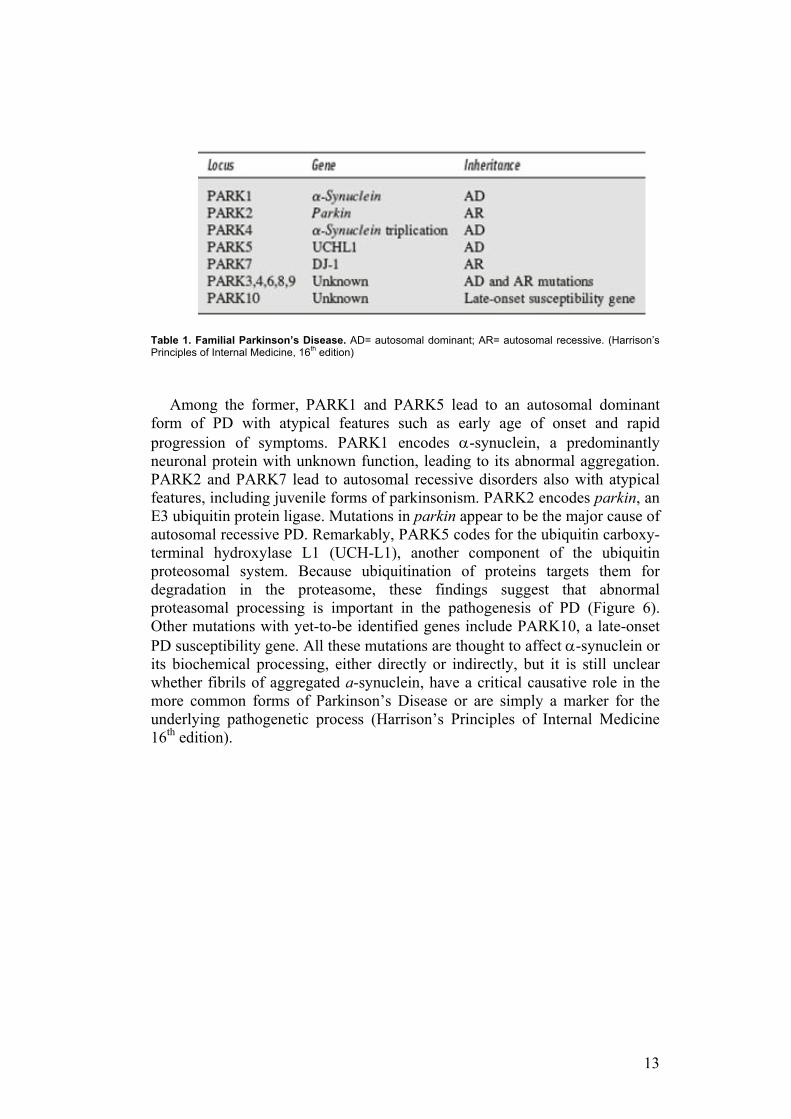
13
Table 1. Familial Parkinson’s Disease. AD= autosomal dominant; AR= autosomal recessive. (Harrison’s Principles of Internal Medicine, 16th edition)
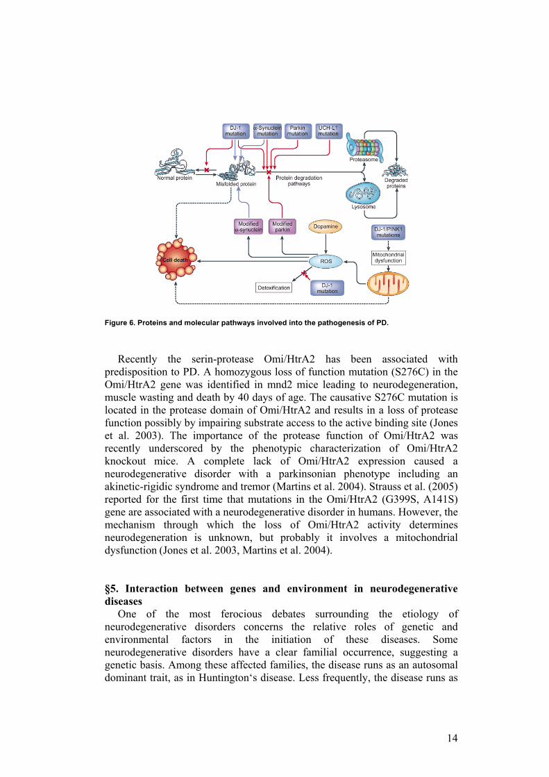
Among the former, PARK1 and PARK5 lead to an autosomal dominant form of PD with atypical features such as early age of onset and rapid progression of symptoms. PARK1 encodes α-synuclein, a predominantly neuronal protein with unknown function, leading to its abnormal aggregation. PARK2 and PARK7 lead to autosomal recessive disorders also with atypical features, including juvenile forms of parkinsonism. PARK2 encodes parkin, an E3 ubiquitin protein ligase. Mutations in parkin appear to be the major cause of autosomal recessive PD. Remarkably, PARK5 codes for the ubiquitin carboxy-terminal hydroxylase L1 (UCH-L1), another component of the ubiquitin proteosomal system. Because ubiquitination of proteins targets them for degradation in the proteasome, these findings suggest that abnormal proteasomal processing is important in the pathogenesis of PD (Figure 6). Other mutations with yet-to-be identified genes include PARK10, a late-onset PD susceptibility gene. All these mutations are thought to affect α-synuclein or its biochemical processing, either directly or indirectly, but it is still unclear whether fibrils of aggregated a-synuclein, have a critical causative role in the more common forms of Parkinson’s Disease or are simply a marker for the underlying pathogenetic process (Harrison’s Principles of Internal Medicine 16th edition).
14
Figure 6. Proteins and molecular pathways involved into the pathogenesis of PD. Recently the serin-protease Omi/HtrA2 has been associated with
predisposition to PD. A homozygous loss of function mutation (S276C) in the Omi/HtrA2 gene was identified in mnd2 mice leading to neurodegeneration, muscle wasting and death by 40 days of age. The causative S276C mutation is located in the protease domain of Omi/HtrA2 and results in a loss of protease function possibly by impairing substrate access to the active binding site (Jones et al. 2003). The importance of the protease function of Omi/HtrA2 was recently underscored by the phenotypic characterization of Omi/HtrA2 knockout mice. A complete lack of Omi/HtrA2 expression caused a neurodegenerative disorder with a parkinsonian phenotype including an akinetic-rigidic syndrome and tremor (Martins et al. 2004). Strauss et al. (2005) reported for the first time that mutations in the Omi/HtrA2 (G399S, A141S) gene are associated with a neurodegenerative disorder in humans. However, the mechanism through which the loss of Omi/HtrA2 activity determines neurodegeneration is unknown, but probably it involves a mitochondrial dysfunction (Jones et al. 2003, Martins et al. 2004). §5. Interaction between genes and environment in neurodegenerative diseases
One of the most ferocious debates surrounding the etiology of neurodegenerative disorders concerns the relative roles of genetic and environmental factors in the initiation of these diseases. Some neurodegenerative disorders have a clear familial occurrence, suggesting a genetic basis. Among these affected families, the disease runs as an autosomal dominant trait, as in Huntington‘s disease. Less frequently, the disease runs as
15
an autosomal recessive trait, an X-linked trait, or even a maternally inherited trait. However, only a small minority of cases are of purely genetic origin. Most neurodegenerative disorders occur sporadically and they belong to the very long list of diseases that could arise through interactions among genetic and environmental factors. Parkinson’s Disease, for example, is considered a multifactorial disease resulting from the effect of environmental factors and genetic susceptibility. In addition to increasing age and male gender, risk factors for PD include head injury, exposure to pesticides, consumption of well water, and rural living (Harrison’s Principles of Internal Medicine 16th edition). No specific environmental risk factor was found to be consistently associated with Alzheimer’s Disease. Studies have reported associations of AD with depression, traumatic head injury, and cardiovascular-related disorders; however, it remains unclear whether these are true risk factors or simply co-morbidities that increase severity of cognitive disorders (Przedborski S et al. 2003).
Recently, increasing evidence indicate that there is an association between impaired glucose tolerance, a common feature of several pathological conditions such as insulin-resistance, diabetes and metabolic syndrome, and neurodegenerative disorders. Impairment of insulin signaling in the brain has been linked to cognitive dysfunctions, but the extent and nature of the association have not been elucidated to date. §6. Association between neurodegenerative disorders, insulin-resistance and diabetes mellitus
Many investigators have reported that approximately 20% of neurodegenerative disorders are associated with insulin-resistance and type 2 diabetes. Compared to the overall incidence of diabetes mellitus in the general population of 4–8% (Zimmet et al. 2001), individuals suffering from neurodegenerative disorders exhibit a significantly higher prevalence of diabetes mellitus. For example, impaired glucose tolerance is frequently observed in Parkinson’s Disease and affects up to 80% of patients (Ristow 2004).
Several large, population-based studies in Europe and the United States suggest that impaired glucose tolerance and insulin resistant conditions, including diabetes and hyperinsulinemia, increase the risk for developing cognitive dysfunction. In the Kuopio study, investigators reported that older adults (mean age = 72.9 years) with persistent impaired glucose tolerance performed worse than did healthy older adults (mean age = 73.3 years) on the Mini-Mental State Examination and on long-term verbal memory from the Buschke Selective Reminding Test (Vanhanen et al. 1998, Watson and Craft 2004). In the Honolulu–Asia Aging Study, type 2 diabetes was associated with an increased risk for incident dementia, incident Alzheimer’s Disease, and incident vascular dementia for a cohort of Japanese–American men who were followed for 3 years (Peila et al. 2002). In the Rotterdam and the Mayo studies,
16
type 2 diabetes increased the risk for Alzheimer’s Disease, independent of vascular dementia (Leibson et al. 1997, Ott et al. 1999, Watson and Craft 2006). Conversely, Alzheimer’s Disease seems to predispose for insulin resistance (Razay and Wilcock 1994), insulin hypersecretion (Razay and Wilcock 1994), and type 2 diabetes mellitus (Janson et al. 2004).
Peripheral and CNS insulin abnormalities have been reported in Alzheimer’s Disease patients. These patients have an increased risk for hyperinsulinemia and hyperglycemia, relative to healthy controls (Watson and Craft 2006). Patients with Alzheimer’s Disease, when compared with healthy controls, have lower cerebrospinal fluid (CSF) insulin levels, higher plasma insulin levels, and reduced insulin-mediated glucose disposal, a pattern consistent with insulin resistance (Craft et al 1999). Moreover, AD brains show reduced insulin receptor density and tyrosine kinase activity markers (Frolich et al. 1998). Collectively, these findings suggest that Alzheimer’s Disease may be associated with reduced insulin sensitivity and impaired insulin signaling (Messier 2003, Watson and Craft 2006).
Thus, epidemiological studies and molecular evidence assert that insulin-resistance and type 2 diabetes seem to be independent risk factors for Alzheimer’s Disease and other neurodegenerative diseases (European Journal of Pharmacology 2004, whole issue), suggesting that insulin-resistance can be involved in the development of cognitive disorders.
§7. Insulin action Insulin is the most potent anabolic hormone known. It is involved in the
control of tissue growth and development and in the regulation of glycidic, protein and lipid metabolism. Secreted by pancreatic beta cells in response to increase of plasmatic glucose and amino acids levels after feeding, insulin promotes the synthesis and storage of carbohydrates, lipids and proteins, while inhibiting their degradation and release into the circulation. Insulin stimulates the uptake of glucose, amino acids and fatty acids into cells, and increases the expression or activity of enzymes that catalyse glycogen, lipid and protein synthesis, while inhibiting the activity or expression of those that catalyse degradation. Insulin acts on three principal insulin-dependent target tissues, increasing glucose uptake in muscle and fat, and inhibiting hepatic glucose production (glycogenolysis and gluconeogenesis), thus serving as the primary regulator of blood glucose concentration (Saltiel and Kahn 2001).
Until recently, the brain was described as an insulin insensitive organ; however, a growing body of evidence demonstrates that insulin participates in a number of normal and pathophysiological functions in the CNS (Watson and Craft 2004, Schwartz and Porte 2005, Watson and Craft 2006, Strachan 2003, European Journal of Pharmacology 2004, whole issue). Pancreatic insulin is transported across the blood-brain barrier (BBB) by a saturable, insulin receptor-mediated transport process (Baura et al. 1993, Banks et al. 1997a, Banks et al. 1997b). Acutely raising peripheral insulin levels also elevates
17
insulin levels in cerebrospinal fluid (Watson et al. 2003, Watson and Craft 2006). Neuronal insulin synthesis has been demonstrated in animals (Rulifson et al. 2002, Devaskar et al. 1994, Watson and Craft 2006), and rats appear to express insulin genes in the hippocampus (Singh et al. 1997). However, insulin gene expression and synthesis is yet to be demonstrated in brain of mature adult humans. Insulin and insulin receptors are abundant but selectively distributed in the brain. Rodent studies have shown that insulin binding is highest in the olfactory bulb, cerebral cortex, hippocampus, hypothalamus, amygdala, and septum (Watson and Craft 2006). Insulin receptors are also expressed in the substantia nigra, basal ganglia, and frontal cortex (Watson and Craft 2006). In light of these distributions, insulin may contribute to selective brain functions such as the control of food intake, body weight and reproduction (Bruning et al. 2000, Schulingkamp et al. 2000, Watson and Craft 2004). Insulin may influence cerebral glucose metabolism in specific brain regions. In the CNS, insulin-sensitive GLUT4 and GLUT8 transporters are selectively distributed. Rats express GLUT4 transporters in cerebellum, sensorimotor cortex, hippocampus, pituitary, and hypothalamus (Apelt et al. 1999), and substantial co-localization has been reported for GLUT4 transporters, insulin-containing neurons, and insulin receptors (Apelt et al. 1999). Likewise, GLUT8 transporters are present in hippocampus and hypothalamus (Reagan et al. 2001). Therefore, overlapping distributions of insulin, insulin receptors, and insulin-sensitive GLUT isoforms constitute a platform for insulin-stimulated glucose uptake in selective brain regions, such as the hippocampus (Apelt et al. 1999). Insulin administration studies corroborate the notion that insulin contributes to normal memory functioning (Watson and Craft 2004). In rats, intracerebroventricular insulin administration enhances memory acutely on a passive-avoidance task, whereas control infusates (saline and heat-inactivated insulin) do not enhance memory (Park et al. 2000). In healthy older adults and adults with Alzheimer’s Disease, increasing plasma insulin levels (via intravenous insulin infusion) while maintaining euglycemia, facilitates recall of verbal declarative memory (i.e., story recall and word list recall) and enhances selective attention (Watson and Craft 2006). Moreover insulin seems to regulate the neurotransmitter release and the expression of neurotransmitter receptors, and so the synaptic plasticity. (Watson and Craft 2006). Thus, it is likely that insulin modulates learning and memory through diverse mechanisms including effects related to insulin signaling, cerebral glucose metabolism, neurotransmitter modulation (Watson and Craft 2004, Watson and Craft 2006). §8. Insulin-resistance and impaired glucose tolerance
Once beta cells are stimulated by glucose, insulin is released in blood circulation and is distributed to all tissues. At this level, insulin acts on a specific receptor at high affinity, essential for insulin signaling transduction.
18
Alterations in insulin function are known as insulin-resistance (IR). Insulin resistance is defined as the condition in which normal amounts of circulating insulin are inadequate to produce a physiological insulin response from target cells. IR in fat cells results in hydrolysis of stored triglycerides, which elevates free fatty acids in the blood plasma; IR in muscle reduces glucose uptake, whereas IR in liver reduces glucose storage, with both effects serving to elevate blood glucose (Pickup and Williams 2005). Moreover, since brain could be considered an insulin-dependent tissue, it is possible to hypothesize the existence of cerebral insulin-resistance. High plasma levels of insulin and glucose due to insulin resistance often lead to metabolic syndrome and type 2 diabetes. This is often seen when pancreatic β-cells are unable to produce adequate insulin to maintain normal blood sugar levels (euglycemia). The inability of the β-cells to produce more insulin in a condition of hyperglycemia is what characterizes the transition from insulin resistance to type 2 diabetes.
Impaired Glucose Tolerance (IGT) is the name given to define blood glucose levels that are higher than normal, but below the level of a person with diabetes. In people with IGT, the rise in blood glucose that occurs after consuming 75g glucose is greater than normal, although not as great as in people with type 2 diabetes. Fasting blood glucose levels are normal or moderately raised. IGT is associated with insulin resistance and increased risk of cardiovascular pathology and it carries a high risk of progressing to type 2 diabetes, leading to it being referred to as ‘pre-diabetes’.
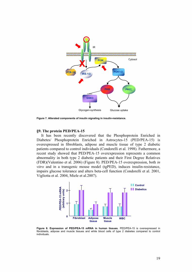
Alterations of components of insulin signal transduction are known mechanisms of IR (Figure 7). Synthesis of anomalous unfunctioning insulin is found in rare cases of IR. Genetic alterations of insulin receptor are the cause of severe insulin-resistance syndromes, such as leprecaunism, in which mutations of insulin receptor gene alter its expression or function. Then, several polymorphisms of Insulin Receptor Substrate 1 (IRS-1) and Phosphatidyl Inositol-3-Kinase (PI3K), proteins involved in insulin signaling, are linked to type 2 diabetes. Moreover, proteins involved in the regulation of insulin signaling could be associated to IR. For example, Phospho-Tyrosin-Phosphatase-1B (PTP-1B) is a phosphotyrosine phosphatase that desphosphorylates insulin receptor and insulin receptor substrate, and it is responsible for the switching off of insulin signaling transduction. Its activity is increased in patients affected be type 2 diabetes (Saltiel and Kahn 2001).
19
Figure 7. Alterated components of insulin signaling in insulin-resistance. §9. The protein PED/PEA-15
It has been recently discovered that the Phosphoprotein Enriched in Diabetes/ Phosphoprotein Enriched in Astrocytes-15 (PED/PEA-15) is overexpressed in fibroblasts, adipose and muscle tissue of type 2 diabetic patients compared to control individuals (Condorelli et al. 1998). Futhermore, a recent study showed that PED/PEA-15 overexpression represents a common abnormality in both type 2 diabetic patients and their First Degree Relatives (FDR)(Valentino et al. 2006) (Figure 8). PED/PEA-15 overexpression, both in vitro and in a transgenic mouse model (tgPED), induces insulin-resistance, impairs glucose tolerance and alters beta-cell function (Condorelli et al. 2001, Vigliotta et al. 2004, Miele et al.2007).
Figure 8. Expression of PED/PEA-15 mRNA in human tissues. PED/PEA-15 is overexpressed in fibroblasts, adipose and muscle tissues and white blood cells of type 2 diabetes compared to control individuals.
Cytosol
IR
Glycogen-synthesis
GSK3
Glucose uptake
X
PI3K
PTP-1B IRS-1/2 PDK1/2
PKB PKCζ
ControlDiabetics
Fibroblast Adipose tissue
Muscle tissue
PED/PEA-15
mR
NA
(a
rbitr
ary
units
)
1
2
0 WBC
20
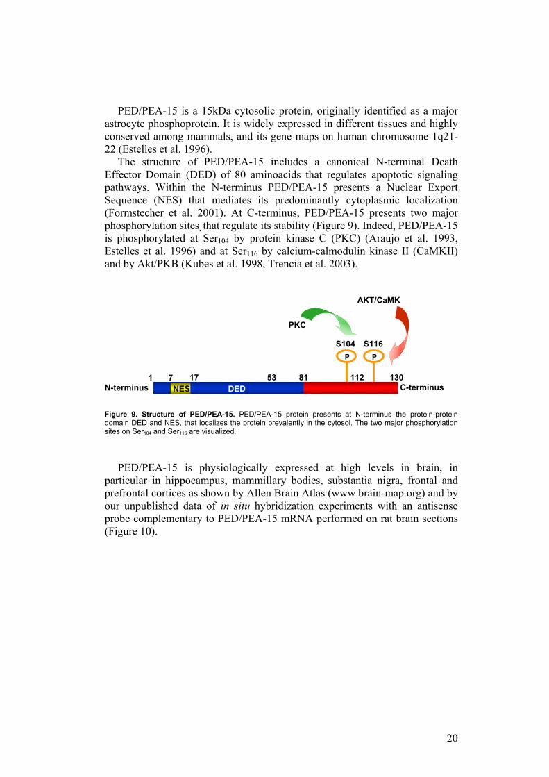
PED/PEA-15 is a 15kDa cytosolic protein, originally identified as a major astrocyte phosphoprotein. It is widely expressed in different tissues and highly conserved among mammals, and its gene maps on human chromosome 1q21-22 (Estelles et al. 1996).
The structure of PED/PEA-15 includes a canonical N-terminal Death Effector Domain (DED) of 80 aminoacids that regulates apoptotic signaling pathways. Within the N-terminus PED/PEA-15 presents a Nuclear Export Sequence (NES) that mediates its predominantly cytoplasmic localization (Formstecher et al. 2001). At C-terminus, PED/PEA-15 presents two major phosphorylation sites, that regulate its stability (Figure 9). Indeed, PED/PEA-15 is phosphorylated at Ser104 by protein kinase C (PKC) (Araujo et al. 1993, Estelles et al. 1996) and at Ser116 by calcium-calmodulin kinase II (CaMKII) and by Akt/PKB (Kubes et al. 1998, Trencia et al. 2003).
Figure 9. Structure of PED/PEA-15. PED/PEA-15 protein presents at N-terminus the protein-protein domain DED and NES, that localizes the protein prevalently in the cytosol. The two major phosphorylation sites on Ser104 and Ser116 are visualized.
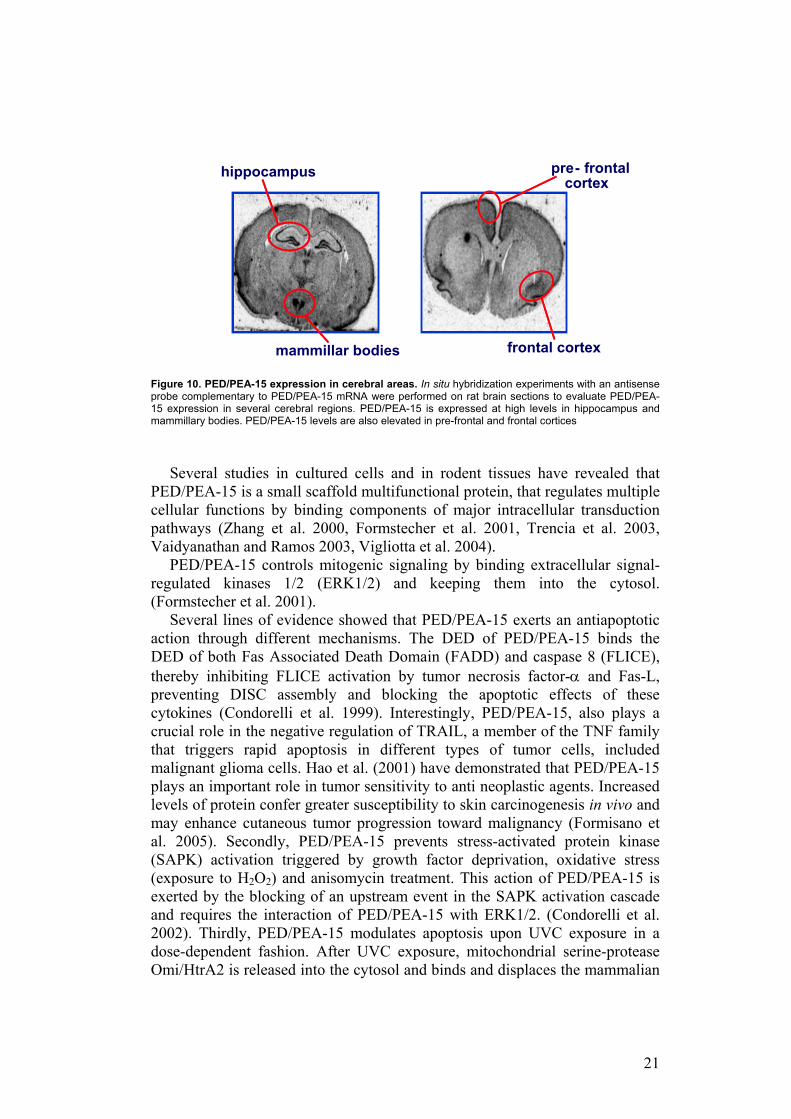
PED/PEA-15 is physiologically expressed at high levels in brain, in particular in hippocampus, mammillary bodies, substantia nigra, frontal and prefrontal cortices as shown by Allen Brain Atlas (www.brain-map.org) and by our unpublished data of in situ hybridization experiments with an antisense probe complementary to PED/PEA-15 mRNA performed on rat brain sections (Figure 10).
P P
81 130 C-terminus 7
S104 S116
53 112NES DED
17
PKC
AKT/CaMK
N-terminus 1
21
Figure 10. PED/PEA-15 expression in cerebral areas. In situ hybridization experiments with an antisense probe complementary to PED/PEA-15 mRNA were performed on rat brain sections to evaluate PED/PEA-15 expression in several cerebral regions. PED/PEA-15 is expressed at high levels in hippocampus and mammillary bodies. PED/PEA-15 levels are also elevated in pre-frontal and frontal cortices
Several studies in cultured cells and in rodent tissues have revealed that PED/PEA-15 is a small scaffold multifunctional protein, that regulates multiple cellular functions by binding components of major intracellular transduction pathways (Zhang et al. 2000, Formstecher et al. 2001, Trencia et al. 2003, Vaidyanathan and Ramos 2003, Vigliotta et al. 2004).
PED/PEA-15 controls mitogenic signaling by binding extracellular signal-regulated kinases 1/2 (ERK1/2) and keeping them into the cytosol. (Formstecher et al. 2001).
Several lines of evidence showed that PED/PEA-15 exerts an antiapoptotic action through different mechanisms. The DED of PED/PEA-15 binds the DED of both Fas Associated Death Domain (FADD) and caspase 8 (FLICE), thereby inhibiting FLICE activation by tumor necrosis factor-α and Fas-L, preventing DISC assembly and blocking the apoptotic effects of these cytokines (Condorelli et al. 1999). Interestingly, PED/PEA-15, also plays a crucial role in the negative regulation of TRAIL, a member of the TNF family that triggers rapid apoptosis in different types of tumor cells, included malignant glioma cells. Hao et al. (2001) have demonstrated that PED/PEA-15 plays an important role in tumor sensitivity to anti neoplastic agents. Increased levels of protein confer greater susceptibility to skin carcinogenesis in vivo and may enhance cutaneous tumor progression toward malignancy (Formisano et al. 2005). Secondly, PED/PEA-15 prevents stress-activated protein kinase (SAPK) activation triggered by growth factor deprivation, oxidative stress (exposure to H2O2) and anisomycin treatment. This action of PED/PEA-15 is exerted by the blocking of an upstream event in the SAPK activation cascade and requires the interaction of PED/PEA-15 with ERK1/2. (Condorelli et al. 2002). Thirdly, PED/PEA-15 modulates apoptosis upon UVC exposure in a dose-dependent fashion. After UVC exposure, mitochondrial serine-protease Omi/HtrA2 is released into the cytosol and binds and displaces the mammalian
hippocampus
mammillar bodies frontal cortex
pre - frontal cortex
22
caspase inhibitor XIAP, releasing its suppressive effect on caspases activity. Interestingly, the mitochondrial release of Omi/HtrA2 is accompanied by a significant decrease in cellular PED/PEA-15 levels. Indeed, Omi/HtrA2 is able to interact with and to cleave PED/PEA-15 in the cytosol. Thus, at least in part, apoptosis following Omi/HtrA2 mitochondrial release is mediated by increased PED/PEA-15 degradation. By contrast, PED/PEA-15 overexpression blocks Omi/HtrA2-XIAP interaction thereby inhibiting the activation of the apoptotic pathway. Thus, the relative concentrations of Omi/HtrA2 and PED/PEA-15 play an important role in committing the cells to apoptosis. (Trencia et al. 2004).
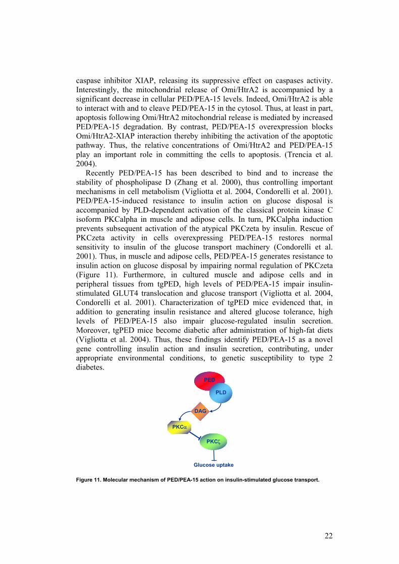
Recently PED/PEA-15 has been described to bind and to increase the stability of phospholipase D (Zhang et al. 2000), thus controlling important mechanisms in cell metabolism (Vigliotta et al. 2004, Condorelli et al. 2001). PED/PEA-15-induced resistance to insulin action on glucose disposal is accompanied by PLD-dependent activation of the classical protein kinase C isoform PKCalpha in muscle and adipose cells. In turn, PKCalpha induction prevents subsequent activation of the atypical PKCzeta by insulin. Rescue of PKCzeta activity in cells overexpressing PED/PEA-15 restores normal sensitivity to insulin of the glucose transport machinery (Condorelli et al. 2001). Thus, in muscle and adipose cells, PED/PEA-15 generates resistance to insulin action on glucose disposal by impairing normal regulation of PKCzeta (Figure 11). Furthermore, in cultured muscle and adipose cells and in peripheral tissues from tgPED, high levels of PED/PEA-15 impair insulin-stimulated GLUT4 translocation and glucose transport (Vigliotta et al. 2004, Condorelli et al. 2001). Characterization of tgPED mice evidenced that, in addition to generating insulin resistance and altered glucose tolerance, high levels of PED/PEA-15 also impair glucose-regulated insulin secretion. Moreover, tgPED mice become diabetic after administration of high-fat diets (Vigliotta et al. 2004). Thus, these findings identify PED/PEA-15 as a novel gene controlling insulin action and insulin secretion, contributing, under appropriate environmental conditions, to genetic susceptibility to type 2 diabetes.
Figure 11. Molecular mechanism of PED/PEA-15 action on insulin-stimulated glucose transport.
Glucose uptake
PED
PLD
DAG
PKCα
PKCζ
23
AIM OF THE STUDY
In the last years increasing evidences indicate that there is an association between impaired glucose tolerance and cognitive decline. Indeed, alterations of genes involved in insulin signaling have also been found in patients with Alzheimer Disease (AD) and other neurodegenerative disorders (Watson and Craft 2006). Furthermore, epidemiologic studies indicate that patients with neurodegenerative disorders present an increased prevalence of insulin-resistance or type 2 diabetes (T2D) compared to control individuals (Watson and Craft 2004, Watson and Craft 2006). This link suggests the possibility of a common pathogenetic mechanism for these conditions.
PED/PEA-15 gene is overexpressed in tissue from type 2 diabetics (Condorelli et al. 1998, Valentino et al. 2006). PED/PEA-15 is involved in the regulation of insulin sensitivity and its overexpression causes insulin-resistance in cultured cells and in transgenic mice (tgPED) (Vigliotta et al. 2004, Miele et al.2007).
PED/PEA-15 is physiologically expressed at high levels in brain, and in particular in hippocampus and in frontal and pre-frontal cortex, cerebral areas of limbic system. Nevertheless, its function in brain is still unclear.
The aim of this work is to clarify PED/PEA-15 role in cerebral function and its possible involvement into pathogenesis of neurodegenerative diseases.
24
MATERIALS AND METHODS Yeast two Hybrid screening
The yeast strain AH109, the mouse brain cDNA library in the pGADT7 vector, and the Matchmaker Gal4 two-hybrid system 3 were from Clontech (CA). Generation of pGBKT7 yeast expression plasmids encoding the ped/pea-15 full length cDNA was obtained by amplifying ped/pea-15, respectively, with the following set of primers: ped/pea-15 5’ NdeI (5’-GGGAATTCCATATGGTTGAGTACGGG-3’) and 3’ped/pea-15 BamHI (5’-CGCGGATCCTCAGGCCTTCTTCGCTGG GGGACC-3’). Plasmid DNA transformations were performed using a high efficiency lithium acetate procedure (Gietz et al. 1992). Cotransformants were propagated on Trp-, Leu- plates, and potential interacting clones selected in Trp-, Leu-, His-, Ade- media. After 4 days of incubation at 30 °C, positive clones were further tested for β galactosidase activity by liquid culture assays using the substrate o-nitrophenyl-β-D-galactopyranoside as described in Experiments in Molecular Genetics (1972). Clones of interest were analyzed by DNA sequencing and BLAST analysis. Cell culture and transfection The HEK293 human kidney embryonic cells stably expressing ped/pea-15 cDNA have been previously described (Condorelli et al. 2002); the N2a rat neuroblastoma cells stably expressing app cDNA were kindly provided by Prof. L. D’Adamio (Albert Einstein College of Medicine of Yeshiwa University, USA). The HEK293 and N2a cells were grown in Dulbecco’s modified Eagle’s medium supplemented with 10% fetal bovine serum, 100 IU/ml penicillin, 100 IU/ml streptomycin, and 2% L-glutamine in a 5% humidified CO2 incubator. Transient transfection with RcCMV vector encoding app cDNA (kindly provided by Prof. N. Zambrano, DBBM of University of Naples “Federico II”, Italy) and pcDNA3 vector encoding ped/pea-15 cDNA (Condorelli et al. 1998) were performed using the Lipofectamine method according to the manufacturer’s instructions (Invitrogen, CA). Cells were also incubated for 24 hours with bisindolylmaleimide 100nM or PD98059 50µM. Western Blot analysis
HEK293 were lysed in lysis buffer (50mM HEPES pH 7.6, 150mM NaCl, 10mM EDTA, 10mM Na4P2O7, 2mM Na3VO4, 100mM NaF, 10% glycerol, 1mM PMSF, 100 IU/ml aprotinin, 20µM leupetin, 1% Triton X-100) and N2a cells were lysed in 50mM Tris pH 7.4, 150mM NaCl, 2mM EDTA, 1% Triton X-100, 0.1% SDS, 1mM PMSF, for 2 h at 4 °C and lysates were centrifuged at 14,000 x g for 15 minutes to remove cellular debris. Mouse brains were homogenized in a Polytron in T-PERTM extraction reagent (Pierce, IL) and

25
following manufacture’s instruction. Culture medium (100µl) was collected from cells 48 hours after transfection and was centrifuged for 10 min at 16,000 x g to remove cellular debris. Tissues homogenates, cell lysates and media were separated by sodium dodecyl sulfate-polyacrylamide gel electrophoresis
(SDS-PAGE) and analyzed by western blot as previously described (Laemmli 1970). Nitrocellulose membranes were probed with antibodies to PED/PEA-15 (Condorelli et al. 1998), APP (clone 6E10; Sigma-Aldrich, MO), APP-C-term (Sigma-Aldrich, MO), sAPPα (IBL, Japan) ADAM10 (Stressgen, Canada). Immunoreactive bands were detected by enhanced chemiluminescence according to the manufacturer’s instructions (GE Healthcare, NJ).
Co-immunoprecipitation Lysates from HEK293 and Na2 transfected and untrasfected cells and mouse brains were incubated with pre-immune serum, PED/PEA-15 antibody or ADAM10 antibody (Santa Cruz Biotechnology, CA) at 4 °C for 16 hours. After incubation the mixture was incubated with protein A sepharose resin (Sigma-Aldrich, MO) pre-equilibrated with HNT buffer (50mM HEPES pH 7.5, 150mM NaCl, 0,1 % Triton X-100) for 2 hours at 4 °C with vibrant shaking. The bound antibody-antigen complexes were washed three times with HNT and were then eluted in SDS sample buffer (Laemmli 1970). Total elute was separated by SDS-PAGE followed by immunoblotting with appropriate antibodies. Pull-down experiment PED/PEA-15-glutatione S-transferase (GST) fusion protein was generated as described by Trencia et al. 2003. Cell lysates (500 µg of protein) were incubated in the presence of Sepharose-bound GST-PED/PEA-15 (5 µg) for 2 h at 4 °C. The beads were washed four times with HNT buffer and then resuspended in SDS sample buffer followed by boiling for 5 min and centrifugation at 25,000 x g for 3 min. Supernatants were separated by SDS-PAGE followed by immunoblotting with appropriate antibodies. Cell surface Biotinylation
To label cell surface molecules, cells were placed on ice and washed twice
with PBS/Ca/Mg buffer (8mM Na2HPO4, 1,5mM KH2PO4, 2,7mM KCl, 137mM NaCl, 0,1mM CaCl2, 1mM MgCl2, pH 7.4) and were then incubated in PBS/Ca/Mg buffer supplemented with 0.5 mg/ml EZ-LinkTM Sulfo-NHS-LC-Biotin (Pierce, IL) for 30 min at 4 °C. Cells were then washed three times with PBS/Ca/Mg buffer plus 15mM glycine and were lysed in lysis buffer. Biotinylated cell surface proteins were immunoprecipitated with APP antibody, and immunoprecipitates were eluted by boiling in SDS sample buffer, electrophoresed on polyacrylamide gels, transferred to nitrocellulose, and
26
blotted with streptavidin-horseradish peroxidase conjugate (GE Healthcare, NJ). Blots were analyzed by enhanced chemiluminescence.
Aβ ELISA Concentrations of secreted Aβ (1-42) produced by cells were analyzed in
culture medium by ELISA colorimetric kit, as per the manufacturer's
instructions (BioSource International, CA).
Densitometric and statistical analysis Densitometric analysis was performed using Scion Image Analyzer. Data were analyzed with Statview software (Abacus-concepts) by one-factor analysis of variance. p values of less than 0.05 were considered statistically significant.
27
RESULTS AND DISCUSSION Isolation and Identification of APP and ADAM10 as novel PED/PEA-15 interacting proteins
To clarify PED/PEA-15 role in the brain, I performed a yeast two hybrid screening to search for proteins that specifically interact with PED/PEA-15. To this aim, PED/PEA-15 protein fused to the DNA binding domain (DBD) of the yeast transcriptional factor Gal4 was used as bait; the screening was performed using a mouse brain cDNA library fused to the transcriptional activator domain (AD) of Gal4. Protein-protein interaction was identified firstly through the selection of positive clones on a selective medium without histidine and adenosine, then measuring the enzymatic activity of β-galattosidase. Based upon sequence analysis and BLAST searching, several potential interactors were identified: A Disintegrin And Metalloprotease 10 (ADAM10), Amyloid Precursor Protein (APP), Connexin32, GABA-A Receptor δ-subunit, Metallothionein 2A, and others.
Among these positive clones, I have detained my attention on the proteins APP and ADAM10. APP is a single membrane-spanning protein possessing a large extracellular amino-terminal domain and a small cytoplasmic tail (Figure 12). The APP gene is localized on chromosome 21 and it is ubiquitously expressed. Its pre-mRNA is alternatively spliced to produce three major protein isoforms APP770, APP751 and APP695; the last one is predominantly expressed in the brain. APP can be localized to many membranous structures in the cell such as the endoplasmic reticulum, Golgi compartments, secretory and endocytic vesicles as well as to the cell plasma-membrane. Interestingly, it has been detected in the membranes of synaptic and postsynaptic densities, axons and dendrites (Turner et al. 2003). The physiological function of APP is not yet completely clear. The protein seems to be involved in the phenomena of cell adhesion and migration, axon arborization, neuronal morphogenesis and formation of functional synapses (Gralle and Ferreira 2007, Turner et al. 2003). Instead, it is well known that APP is subjected to a physiological proteolytic processing by proteases collectively known as secretases that cut the protein in numerous secreted fragments with varying effects on neural function (Turner et al. 2003). Therefore, APP appears to play a significant role in regulating neural activity and plasticity both as a contact receptor and as a diffusible factor. From proteolytic processing of APP also origins the β-amyloid (Aβ) peptide, the principal proteinaceous component of senile plaques in brains of AD patients. Thus, APP is involved into the pathogenesis of Alzheimer’s Disease.
On the other hand, the protein ADAM10 is a multi-function protein, belonging to a family of transmembrane glycoproteins (ADAMs family) that are characterized by several conserved distinct protein domains. These consist of an N-terminal propeptide domain, metalloprotease and disintegrin domains, a cysteine rich region, an epidermal growth factor (EGF)-like repeat, and finally a transmembrane domain and cytoplasmic tail (Lammich et al. 1999, Schlöndorff and Blobel 1999, Yang et al. 2006) (Figure 12). The propeptide
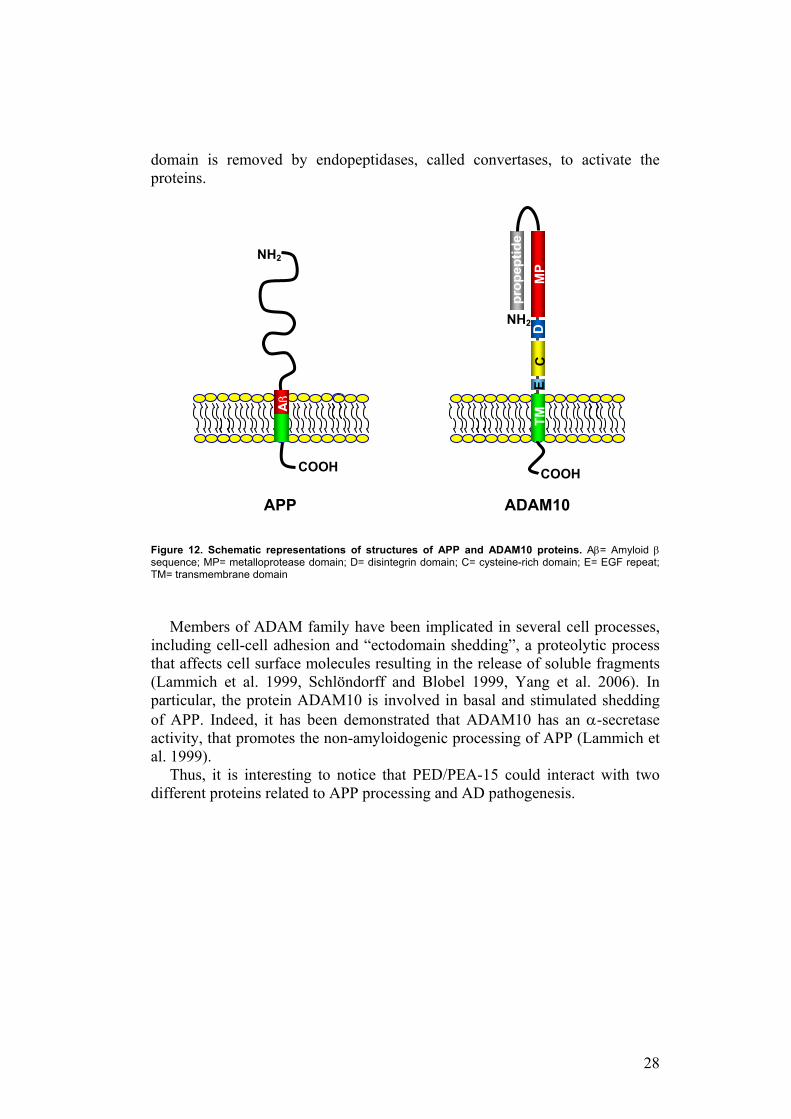
28
domain is removed by endopeptidases, called convertases, to activate the proteins.
Figure 12. Schematic representations of structures of APP and ADAM10 proteins. Aβ= Amyloid β sequence; MP= metalloprotease domain; D= disintegrin domain; C= cysteine-rich domain; E= EGF repeat; TM= transmembrane domain
Members of ADAM family have been implicated in several cell processes, including cell-cell adhesion and “ectodomain shedding”, a proteolytic process that affects cell surface molecules resulting in the release of soluble fragments (Lammich et al. 1999, Schlöndorff and Blobel 1999, Yang et al. 2006). In particular, the protein ADAM10 is involved in basal and stimulated shedding of APP. Indeed, it has been demonstrated that ADAM10 has an α-secretase activity, that promotes the non-amyloidogenic processing of APP (Lammich et al. 1999).
Thus, it is interesting to notice that PED/PEA-15 could interact with two different proteins related to APP processing and AD pathogenesis.
NH2
COOH
MP
prop
eptid
e
NH2
COOH
D
C
E TM
Aβ
APP ADAM10
29
APP and ADAM10 interact with PED/PEA-15 in vitro as well as in mammalian cells and mouse brain
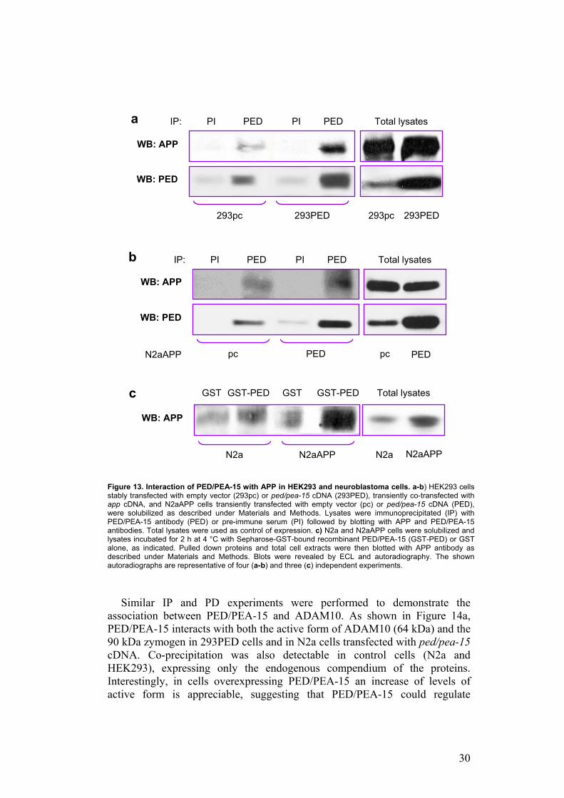
To verify the interaction of PED/PEA-15 with the two proteins identified by yeast two hybrid screening, I performed co-immunoprecipitation experiments (IP) in mammalian cells and pull-down assays (PD). HEK293 cells (Human Embryonic Kidney cell line) and rat neuroblastoma cells (N2a) were used because the first one are a simply transfectable cell line and N2a cells have a neuronal origin and so they constitute a more representative system. HEK293 cells were stably transfected with ped/pea-15 cDNA (293PED) or with the empty vector (293pc), and transiently co-transfected with App. N2a cells stably overexpressing App (N2aAPP), were transiently transfected with the empty vector (pc) or with Ped/Pea-15. As shown in Figures 13a/13b, precipitation of lysates from the different cell types with PED/PEA-15 antibodies followed by blotting with APP antibodies revealed that PED/PEA-15 is able to bind APP and this interaction is more evident when PED/PEA-15 is overexpressed. Pre-immune serum was used as control of specific immunoprecipitation. This interaction is also detectable in vitro by pull down experiments in N2a cells. Indeed, GST-fused recombinant PED/PEA-15 binds APP in N2a cell extracts and, more efficiently, in N2aAPP cells. Faint background signal was detectable using GST alone (Figure 13c).
30
Figure 13. Interaction of PED/PEA-15 with APP in HEK293 and neuroblastoma cells. a-b) HEK293 cells stably transfected with empty vector (293pc) or ped/pea-15 cDNA (293PED), transiently co-transfected with app cDNA, and N2aAPP cells transiently transfected with empty vector (pc) or ped/pea-15 cDNA (PED), were solubilized as described under Materials and Methods. Lysates were immunoprecipitated (IP) with PED/PEA-15 antibody (PED) or pre-immune serum (PI) followed by blotting with APP and PED/PEA-15 antibodies. Total lysates were used as control of expression. c) N2a and N2aAPP cells were solubilized and lysates incubated for 2 h at 4 °C with Sepharose-GST-bound recombinant PED/PEA-15 (GST-PED) or GST alone, as indicated. Pulled down proteins and total cell extracts were then blotted with APP antibody as described under Materials and Methods. Blots were revealed by ECL and autoradiography. The shown autoradiographs are representative of four (a-b) and three (c) independent experiments.
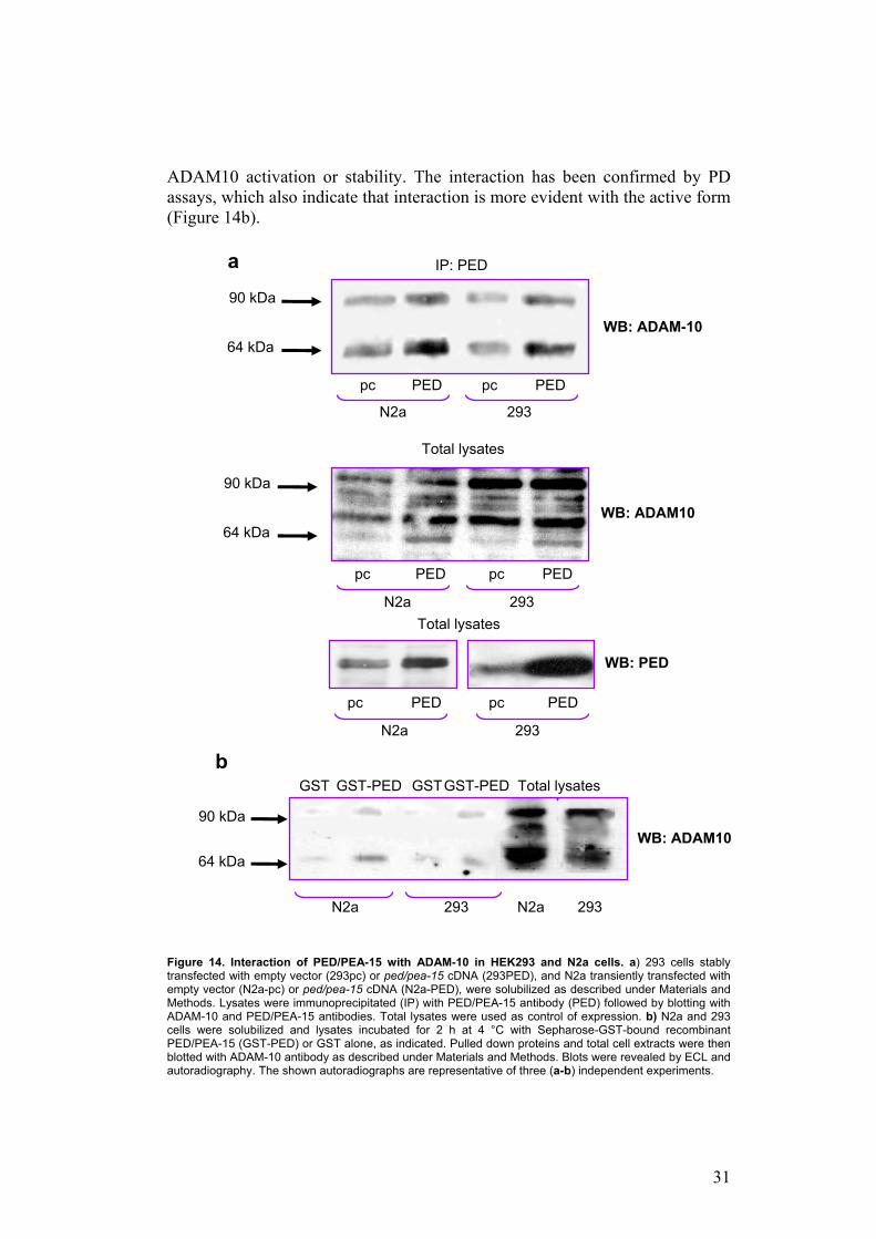
Similar IP and PD experiments were performed to demonstrate the association between PED/PEA-15 and ADAM10. As shown in Figure 14a, PED/PEA-15 interacts with both the active form of ADAM10 (64 kDa) and the 90 kDa zymogen in 293PED cells and in N2a cells transfected with ped/pea-15 cDNA. Co-precipitation was also detectable in control cells (N2a and HEK293), expressing only the endogenous compendium of the proteins. Interestingly, in cells overexpressing PED/PEA-15 an increase of levels of active form is appreciable, suggesting that PED/PEA-15 could regulate
N2a N2aAPP
WB: APP
N2aAPP N2a
Total lysates GST GST-PED GST GST-PED
PI PED PED PI IP:
293pc 293PED
WB: APP
WB: PED
293PED 293pc
Total lysates
PI PED PED PI IP:
pc PED
WB: APP
WB: PED
PED pc
Total lysates b
N2aAPP
c
a
31
ADAM10 activation or stability. The interaction has been confirmed by PD assays, which also indicate that interaction is more evident with the active form (Figure 14b).
Figure 14. Interaction of PED/PEA-15 with ADAM-10 in HEK293 and N2a cells. a) 293 cells stably transfected with empty vector (293pc) or ped/pea-15 cDNA (293PED), and N2a transiently transfected with empty vector (N2a-pc) or ped/pea-15 cDNA (N2a-PED), were solubilized as described under Materials and Methods. Lysates were immunoprecipitated (IP) with PED/PEA-15 antibody (PED) followed by blotting with ADAM-10 and PED/PEA-15 antibodies. Total lysates were used as control of expression. b) N2a and 293 cells were solubilized and lysates incubated for 2 h at 4 °C with Sepharose-GST-bound recombinant PED/PEA-15 (GST-PED) or GST alone, as indicated. Pulled down proteins and total cell extracts were then blotted with ADAM-10 antibody as described under Materials and Methods. Blots were revealed by ECL and autoradiography. The shown autoradiographs are representative of three (a-b) independent experiments.
90 kDa
64 kDa WB: ADAM-10
IP: PED
PED pc PED pc
pc pc PED PED
WB: PED
90 kDa
64 kDa
Total lysates
N2a 293 Total lysates
pc pc PED PED
N2a 293
WB: ADAM10
N2a
WB: ADAM10 90 kDa
64 kDa
GST GST-PED GST GST-PED Total lysates
N2a 293 293
N2a 293
b
a
32
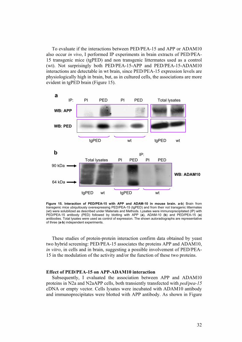
To evaluate if the interactions between PED/PEA-15 and APP or ADAM10 also occur in vivo, I performed IP experiments in brain extracts of PED/PEA-15 transgenic mice (tgPED) and non transgenic littermates used as a control (wt). Not surprisingly both PED/PEA-15-APP and PED/PEA-15-ADAM10 interactions are detectable in wt brain, since PED/PEA-15 expression levels are physiologically high in brain, but, as in cultured cells, the associations are more evident in tgPED brain (Figure 15).
Figure 15. Interaction of PED/PEA-15 with APP and ADAM-10 in mouse brain. a-b) Brain from transgenic mice ubiquitously overexpressing PED/PEA-15 (tgPED) and from their not transgenic littermates (wt) were solubilized as described under Materials and Methods. Lysates were immunoprecipitated (IP) with PED/PEA-15 antibody (PED) followed by blotting with APP (a), ADAM-10 (b) and PED/PEA-15 (a) antibodies. Total lysates were used as control of expression. The shown autoradiographs are representative of three (a-b) independent experiments.
These studies of protein-protein interaction confirm data obtained by yeast
two hybrid screening: PED/PEA-15 associates the proteins APP and ADAM10, in vitro, in cells and in brain, suggesting a possible involvement of PED/PEA-15 in the modulation of the activity and/or the function of these two proteins. Effect of PED/PEA-15 on APP-ADAM10 interaction
Subsequently, I evaluated the association between APP and ADAM10 proteins in N2a and N2aAPP cells, both transiently transfected with ped/pea-15 cDNA or empty vector. Cells lysates were incubated with ADAM10 antibody and immunoprecipitates were blotted with APP antibody. As shown in Figure
PI PED PED PI IP:
tgPED wt
WB: APP
WB: PED
wt tgPED
Total lysates
90 kDa
64 kDa
WB: ADAM10
Total lysates PI PED PED PI
tgPED wt wt tgPED
a
IP: b
33
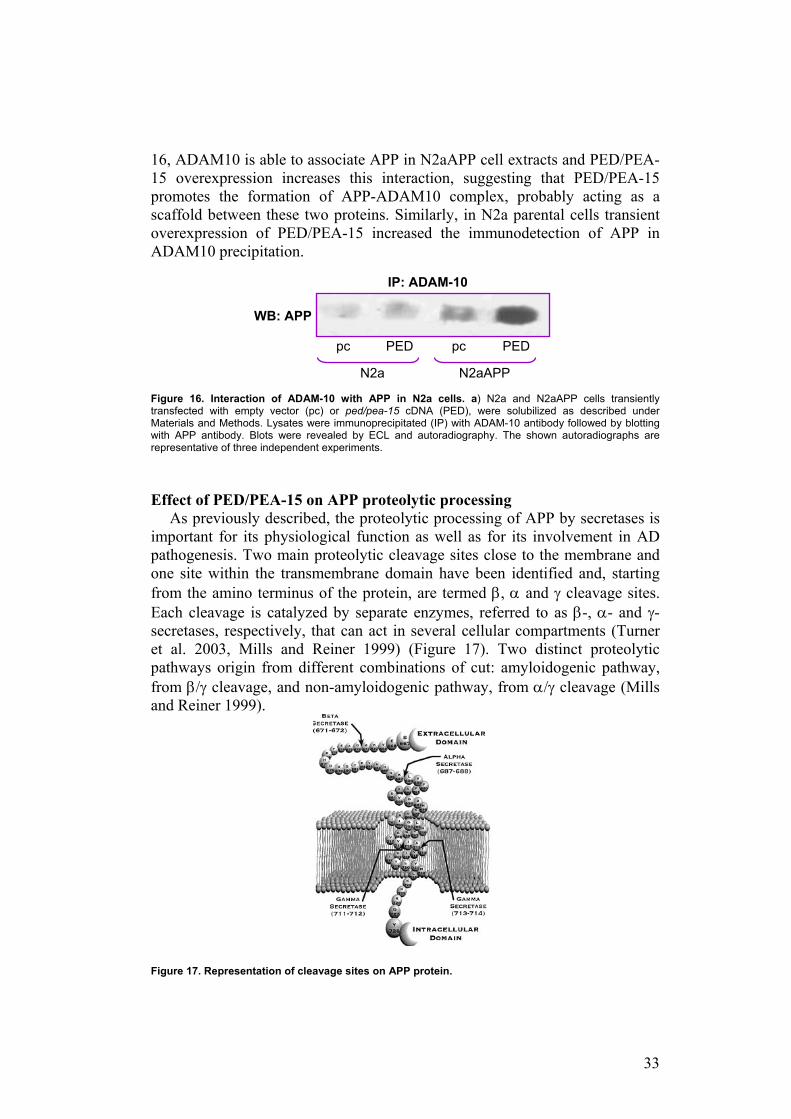
16, ADAM10 is able to associate APP in N2aAPP cell extracts and PED/PEA-15 overexpression increases this interaction, suggesting that PED/PEA-15 promotes the formation of APP-ADAM10 complex, probably acting as a scaffold between these two proteins. Similarly, in N2a parental cells transient overexpression of PED/PEA-15 increased the immunodetection of APP in ADAM10 precipitation.
Figure 16. Interaction of ADAM-10 with APP in N2a cells. a) N2a and N2aAPP cells transiently transfected with empty vector (pc) or ped/pea-15 cDNA (PED), were solubilized as described under Materials and Methods. Lysates were immunoprecipitated (IP) with ADAM-10 antibody followed by blotting with APP antibody. Blots were revealed by ECL and autoradiography. The shown autoradiographs are representative of three independent experiments.
Effect of PED/PEA-15 on APP proteolytic processing
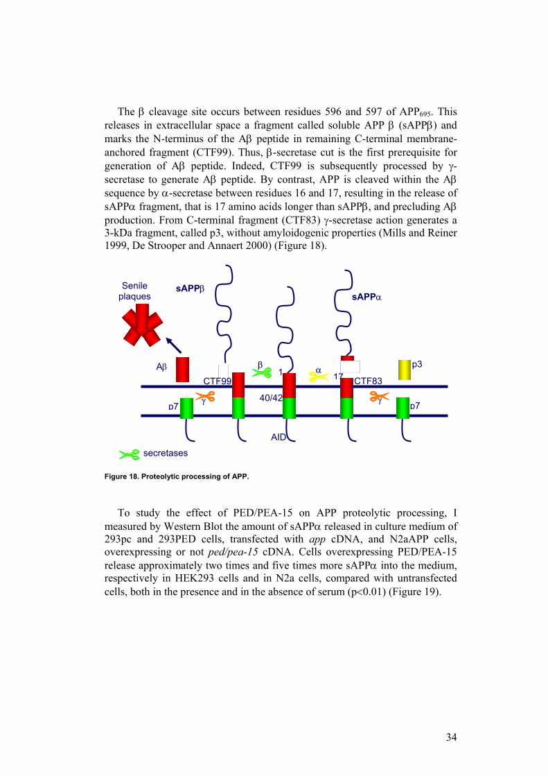
As previously described, the proteolytic processing of APP by secretases is important for its physiological function as well as for its involvement in AD pathogenesis. Two main proteolytic cleavage sites close to the membrane and one site within the transmembrane domain have been identified and, starting from the amino terminus of the protein, are termed β, α and γ cleavage sites. Each cleavage is catalyzed by separate enzymes, referred to as β-, α- and γ-secretases, respectively, that can act in several cellular compartments (Turner et al. 2003, Mills and Reiner 1999) (Figure 17). Two distinct proteolytic pathways origin from different combinations of cut: amyloidogenic pathway, from β/γ cleavage, and non-amyloidogenic pathway, from α/γ cleavage (Mills and Reiner 1999).
Figure 17. Representation of cleavage sites on APP protein.
IP: ADAM-10
N2a N2aAPP
WB: APP
pc PED pc PED
34
The β cleavage site occurs between residues 596 and 597 of APP695. This releases in extracellular space a fragment called soluble APP β (sAPPβ) and marks the N-terminus of the Aβ peptide in remaining C-terminal membrane-anchored fragment (CTF99). Thus, β-secretase cut is the first prerequisite for generation of Aβ peptide. Indeed, CTF99 is subsequently processed by γ-secretase to generate Aβ peptide. By contrast, APP is cleaved within the Aβ sequence by α-secretase between residues 16 and 17, resulting in the release of sAPPα fragment, that is 17 amino acids longer than sAPPβ, and precluding Aβ production. From C-terminal fragment (CTF83) γ-secretase action generates a 3-kDa fragment, called p3, without amyloidogenic properties (Mills and Reiner 1999, De Strooper and Annaert 2000) (Figure 18).
Figure 18. Proteolytic processing of APP.
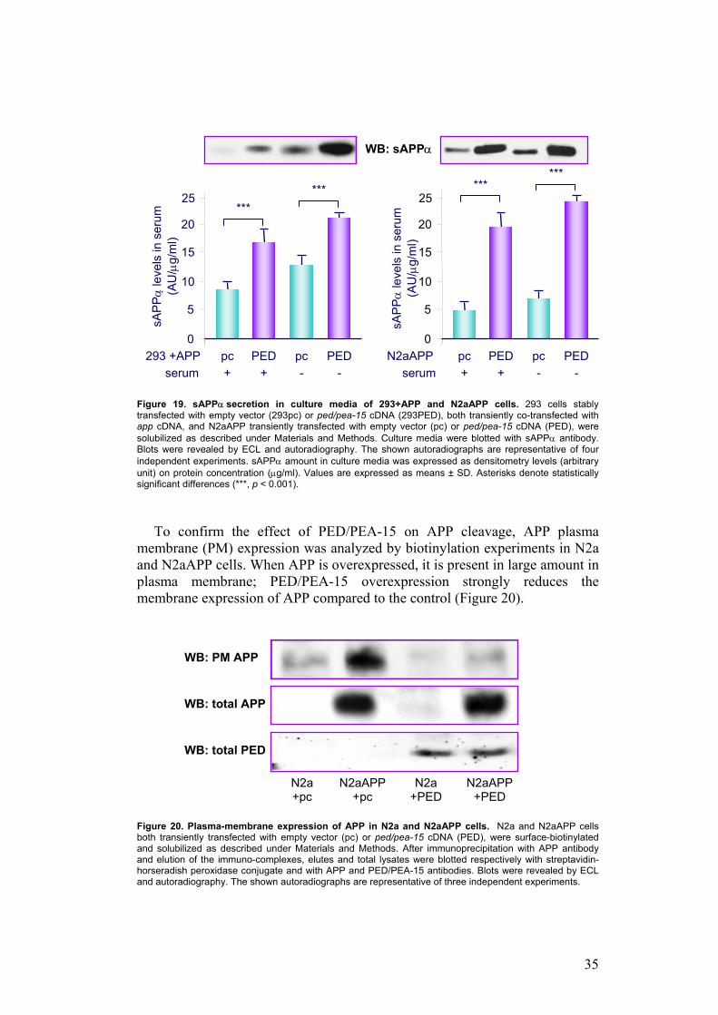
To study the effect of PED/PEA-15 on APP proteolytic processing, I measured by Western Blot the amount of sAPPα released in culture medium of 293pc and 293PED cells, transfected with app cDNA, and N2aAPP cells, overexpressing or not ped/pea-15 cDNA. Cells overexpressing PED/PEA-15 release approximately two times and five times more sAPPα into the medium, respectively in HEK293 cells and in N2a cells, compared with untransfected cells, both in the presence and in the absence of serum (p<0.01) (Figure 19).
1
40/42
AID
p7
Aβ
p7
p3 17
sAPPα
CTF83
β α
Senile plaques
sAPPβ
CTF99
secretases
γ γ
35
Figure 19. sAPPα secretion in culture media of 293+APP and N2aAPP cells. 293 cells stably transfected with empty vector (293pc) or ped/pea-15 cDNA (293PED), both transiently co-transfected with app cDNA, and N2aAPP transiently transfected with empty vector (pc) or ped/pea-15 cDNA (PED), were solubilized as described under Materials and Methods. Culture media were blotted with sAPPα antibody. Blots were revealed by ECL and autoradiography. The shown autoradiographs are representative of four independent experiments. sAPPα amount in culture media was expressed as densitometry levels (arbitrary unit) on protein concentration (µg/ml). Values are expressed as means ± SD. Asterisks denote statistically significant differences (***, p < 0.001).
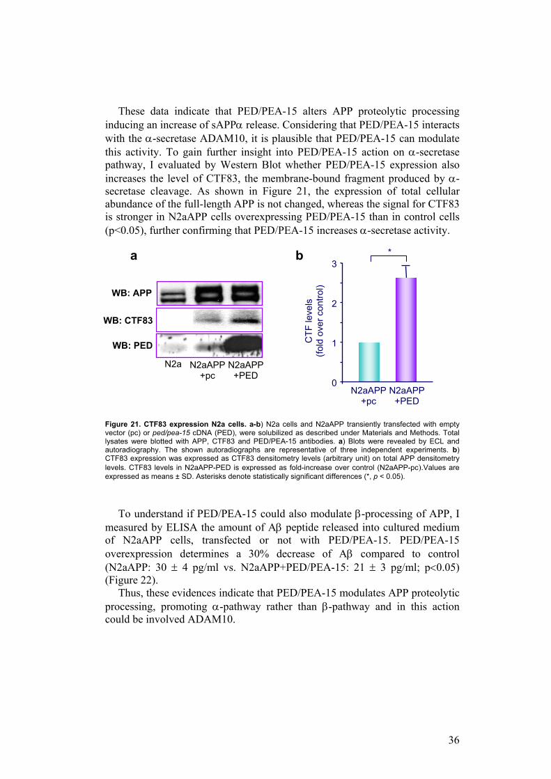
To confirm the effect of PED/PEA-15 on APP cleavage, APP plasma membrane (PM) expression was analyzed by biotinylation experiments in N2a and N2aAPP cells. When APP is overexpressed, it is present in large amount in plasma membrane; PED/PEA-15 overexpression strongly reduces the membrane expression of APP compared to the control (Figure 20).
Figure 20. Plasma-membrane expression of APP in N2a and N2aAPP cells. N2a and N2aAPP cells both transiently transfected with empty vector (pc) or ped/pea-15 cDNA (PED), were surface-biotinylated and solubilized as described under Materials and Methods. After immunoprecipitation with APP antibody and elution of the immuno-complexes, elutes and total lysates were blotted respectively with streptavidin-horseradish peroxidase conjugate and with APP and PED/PEA-15 antibodies. Blots were revealed by ECL and autoradiography. The shown autoradiographs are representative of three independent experiments.
0
5
10
15
20
+ + - - serum 293 +APP pc PED pc PED
sAPP
α le
vels
in s
erum
(A
U/µ
g/m
l)
*** ***
0
5
10
15
20
+ + - - serum N2aAPP pc PED pc PED
sAPP
α le
vels
in s
erum
(A
U/µ
g/m
l)
*** ***
2525
WB: sAPPα
WB: total APP
WB: total PED
WB: PM APP
N2a+pc
N2aAPP +pc
N2a +PED
N2aAPP +PED
36
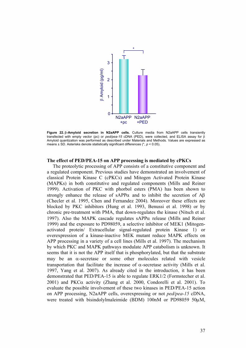
These data indicate that PED/PEA-15 alters APP proteolytic processing inducing an increase of sAPPα release. Considering that PED/PEA-15 interacts with the α-secretase ADAM10, it is plausible that PED/PEA-15 can modulate this activity. To gain further insight into PED/PEA-15 action on α-secretase pathway, I evaluated by Western Blot whether PED/PEA-15 expression also increases the level of CTF83, the membrane-bound fragment produced by α-secretase cleavage. As shown in Figure 21, the expression of total cellular abundance of the full-length APP is not changed, whereas the signal for CTF83 is stronger in N2aAPP cells overexpressing PED/PEA-15 than in control cells (p<0.05), further confirming that PED/PEA-15 increases α-secretase activity.
Figure 21. CTF83 expression N2a cells. a-b) N2a cells and N2aAPP transiently transfected with empty vector (pc) or ped/pea-15 cDNA (PED), were solubilized as described under Materials and Methods. Total lysates were blotted with APP, CTF83 and PED/PEA-15 antibodies. a) Blots were revealed by ECL and autoradiography. The shown autoradiographs are representative of three independent experiments. b) CTF83 expression was expressed as CTF83 densitometry levels (arbitrary unit) on total APP densitometry levels. CTF83 levels in N2aAPP-PED is expressed as fold-increase over control (N2aAPP-pc).Values are expressed as means ± SD. Asterisks denote statistically significant differences (*, p < 0.05).
To understand if PED/PEA-15 could also modulate β-processing of APP, I measured by ELISA the amount of Aβ peptide released into cultured medium of N2aAPP cells, transfected or not with PED/PEA-15. PED/PEA-15 overexpression determines a 30% decrease of Aβ compared to control (N2aAPP: 30 ± 4 pg/ml vs. N2aAPP+PED/PEA-15: 21 ± 3 pg/ml; p<0.05) (Figure 22).
Thus, these evidences indicate that PED/PEA-15 modulates APP proteolytic processing, promoting α-pathway rather than β-pathway and in this action could be involved ADAM10.
WB: PED
WB: APP
0
1
2
3
CTF
leve
ls
(fold
ove
r con
trol)
N2a N2aAPP +pc
N2aAPP +PED
a
N2aAPP +pc
N2aAPP +PED
b *
WB: CTF83
37
Figure 22. β-Amyloid secretion in N2aAPP cells. Culture media from N2aAPP cells transiently transfected with empty vector (pc) or ped/pea-15 cDNA (PED), were collected, and ELISA assay for β Amyloid quantization was performed as described under Materials and Methods. Values are expressed as means ± SD. Asterisks denote statistically significant differences (*, p < 0.05). The effect of PED/PEA-15 on APP processing is mediated by cPKCs
The proteolytic processing of APP consists of a constitutive component and a regulated component. Previous studies have demonstrated an involvement of classical Protein Kinase C (cPKCs) and Mitogen Activated Protein Kinase (MAPKs) in both constitutive and regulated components (Mills and Reiner 1999). Activation of PKC with phorbol esters (PMA) has been shown to strongly enhance the release of sAPPα and to inhibit the secretion of Aβ (Checler et al. 1995, Chen and Fernandez 2004). Moreover these effects are blocked by PKC inhibitors (Hung et al. 1993, Benussi et al. 1998) or by chronic pre-treatment with PMA, that down-regulates the kinase (Nitsch et al. 1997). Also the MAPK cascade regulates sAPPα release (Mills and Reiner 1999) and the exposure to PD98059, a selective inhibitor of MEK1 (Mitogen-activated protein/ Extracellular signal-regulated protein Kinase 1) or overexpression of a kinase-inactive MEK mutant reduce MAPK effects on APP processing in a variety of a cell lines (Mills et al. 1997). The mechanism by which PKC and MAPK pathways modulate APP catabolism is unknown. It seems that it is not the APP itself that is phosphorylated, but that the substrate may be an α-secretase or some other molecules related with vesicle transportation that facilitate the increase of α-secretase activity (Mills et al. 1997, Yang et al. 2007). As already cited in the introduction, it has been demonstrated that PED/PEA-15 is able to regulate ERK1/2 (Formstecher et al. 2001) and PKCα activity (Zhang et al. 2000, Condorelli et al. 2001). To evaluate the possible involvement of these two kinases in PED/PEA-15 action on APP processing, N2aAPP cells, overexpressing or not ped/pea-15 cDNA, were treated with bisindolylmaleimide (BDM) 100nM or PD98059 50µM,
0
1
2
3
β A
myl
oid
(pg/
ml)
N2aAPP +pc
N2aAPP +PED
*
38
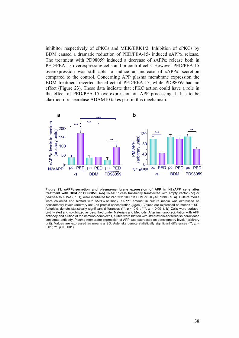
inhibitor respectively of cPKCs and MEK/ERK1/2. Inhibition of cPKCs by BDM caused a dramatic reduction of PED/PEA-15- induced sAPPα release. The treatment with PD98059 induced a decrease of sAPPα release both in PED/PEA-15 overexpressing cells and in control cells. However PED/PEA-15 overexpression was still able to induce an increase of sAPPα secretion compared to the control. Concerning APP plasma membrane expression the BDM treatment reverted the effect of PED/PEA-15, while PD98059 had no effect (Figure 23). These data indicate that cPKC action could have a role in the effect of PED/PEA-15 overexpression on APP processing. It has to be clarified if α-secretase ADAM10 takes part in this mechanism.
Figure 23. sAPPα secretion and plasma-membrane expression of APP in N2aAPP cells after treatment with BDM or PD98059. a-b) N2aAPP cells transiently transfected with empty vector (pc) or ped/pea-15 cDNA (PED), were incubated for 24h with 100 nM BDM or 50 µM PD98059. a) Culture media were collected and blotted with sAPPα antibody. sAPPα amount in culture media was expressed as densitometry levels (arbitrary unit) on protein concentration (µg/ml). Values are expressed as means ± SD. Asterisks denote statistically significant differences (**, p < 0.01; ***, p < 0.001). b) Cells were surface-biotinylated and solubilized as described under Materials and Methods. After immunoprecipitation with APP antibody and elution of the immuno-complexes, elutes were blotted with streptavidin-horseradish peroxidase conjugate antibody. Plasma-membrane expression of APP was expressed as densitometry levels (arbitrary unit). Values are expressed as means ± SD. Asterisks denote statistically significant differences (**, p < 0.01; ***, p < 0.001).
0
40
80
120PM
APP
(a
rbitr
ary
units
) *** **
BDM PD98059
0
50
100
150
200
pc PED pc PED pc PED
sAPP
α le
vels
in m
ediu
m
(arb
itrar
y un
its)
-s
***
**
*** ***
a b
BDM PD98059pc PED pc PED pc PED
-s N2aAPP N2aAPP
39
CONCLUSIONS
Phosphoprotein enriched in diabetes/phosphoprotein enriched in astrocytes (PED/PEA-15) is physiologically expressed at high levels in brain, in particular in hippocampus and in frontal and pre-frontal cortex. PED/PEA-15 gene is overexpressed in tissues from type 2 diabetics and the protein is involved in the regulation of insulin sensitivity (Condorelli et al. 1998, Valentino et al. 2006). Indeed, its overexpression causes insulin-resistance in cultured cells and in transgenic mice (tgPED) (Condorelli et al. 2001, Vigliotta et al. 2004). Nowadays it is known that insulin-resistance and neurodegenerative diseases are associated and they could share common pathogenetic mechanisms.
Based on these observations, I investigated the function of PED/PEA-15 in brain. I identified two new proteins able to interact with PED/PEA-15: APP (Amyloid Precursor Protein) and ADAM10 (A Disintegrin And Metalloprotease 10). Thus, PED/PEA-15 modulates APP proteolytic processing, promoting α-pathway rather than β-pathway and in this action could be involved the α-secretase ADAM10. The supposed model for molecular mechanism underlining PED/PEA-15 effect on APP proteolytic processing is the following: PED/PEA-15 promotes the formation of APP-ADAM10 complex, probably acting as a scaffold between these two proteins. In this way PED/PEA-15 induces α-proteolytic pathway facilitating the physical contact between the enzyme and its substrate. Moreover, PED/PEA-15 can directly regulate the α-secretase activity of ADAM10 activating cPKCs, which are known to phosphorylate and regulate the α-secretase (Mills et al. 1997, Yang et al. 2007).
It has been hypothesized that α-processing is protective for Alzheimer’s Disease, but it is not clear if an increase of α-cleavage and a conseguent accumulation of the α-fragment, or a decrease of the physiological levels of the β-amyloid fragment could be toxic for neuronal cells. Moreover, it might be also hypothesized that the increased processing of APP induced by PED/PEA-15, and the concomitant decrease of APP plasma-membrane abundance, causes a loss of the physiological function of the full-length APP protein. Thus, it remains to clarify how PED/PEA-15-mediated modulation of APP processing can be involved in cognitive functions.
40
REFERENCES Apelt J, Mehlhorn G, Schliebs R. Insulin-sensitive GLUT4 glucose transporters are colocalized with GLUT3-expressing cells and demonstrate a chemically distinct neuron-specific localization in rat brain. J Neurosci Res 1999; 57:693- 705. Araujo H, Danziger N, Cordier J, Glowinski J, Chneiweiss H. Characterization of PEA-15, a major substrate for protein kinase C in astrocytes. J Biol Chem 1993; 268:5911-20. Banks WA, Jaspan JB, Kastin AJ. Selective, physiological transport of insulin across the blood–brain barrier: novel demonstration by species-specific radioimmunoassays. Peptides 1997a;18:1257– 62 Banks WA, Jaspan JB, Huang W, Kastin AJ. Transport of insulin across the blood–brain barrier: saturability at euglycemic doses of insulin. Peptides 1997b;18:1423– 9. Baura GD, Foster DM, Porte Jr D, Kahn SE, Bergman RN, Cobelli C, Schwartz MW. Saturable transport of insulin from plasma into the central nervous system of dogs in vivo. A mechanism for regulated insulin delivery to the brain. J Clin Invest 1993; 92:1824–30. Benussi L, Govoni S, Gasparini I, Binetti G, Trabucchi M, Bianchetti A, Racchi M. Specific role for protein kinase Calpha in the costitutive and regulated secretion of amyloid precursor protein in human skin fibroblasts. Neurosci lett 1998; 240: 97-101. Blennow K. Cerebrospinal fluid protein biomarkers for Alzheimer's disease. NeuroRx 2004; 1:213-25. Blennow K, de Leon MJ, Zetterberg H. Alzheimer’s disease. Lancet 2006; 368:387–403. Bruning JC, Gautam D, Burks DJ, Gillette J, Schubert M, Orban PC, Klein R, Krone W, Muller-Wieland D, Kahn CR. Role of brain insulin receptor in control of body weight and reproduction. Science 2000; 289:2122-5. Burn DJ, Jaros E. Multiple system atrophy: cellular and molecular pathology. Mol Pathol 2001; 54:419-426. Checler F. Processing of the beta-amyloid precursor protein and its regulation in Alzheimer's disease. J Neurochem 1995; 65:1431-1444.
41
Chen M, Fernandez HL. Stimulation of beta-amyloid precursor protein alpha-processing by phorbol ester involves calcium and calpain activation. Biochem Biophys Res Commun 2004;316:332-340. Condorelli G, Vigliotta G, Iavarone C, Caruso M, Tocchetti CG, Andreozzi F, Cafieri A, Tecce MF, Formisano P, Beguinot L, Beguinot F. PED/ PEA-15 gene controls glucose transport and is overexpressed in type 2 diabetes mellitus. The EMBO Journal 1998; 17: 3858–3866. Condorelli G, Vigliotta G, Cafieri A, Trencia A, Andalo P, Oriente F, Miele C, Caruso M, Formisano P, Beguinot F. PED/PEA-15: an anti-apoptotic molecule that regulates FAS/TNFR1-induced apoptosis. Oncogene 1999; 18:4409-4415. Condorelli G, Vigliotta G, Trencia A, Maitan MA, Caruso M, Miele C, Oriente F, Santopietro S, Formisano P, Beguinot F: Protein kinase C (PKC)-alpha activation inhibits PKC-zeta and mediates the action of PED/PEA-15 on glucose transport in the L6 skeletal muscle cells. Diabetes 2001; 50:1244-1252. Condorelli G, Trencia A, Vigliotta G, Perfetti A, Goglia U, Cassese A, Musti AM, Miele C, Santopietro S, Formisano P, Beguinot F. Multiple members of the mitogen-activated protein kinase family are necessary for PED/PEA-15 anti-apoptotic function. J Biol Chem 2002.; 277: 11013–11018. Craft S, Asthana S, Schellenberg G, Cherrier M, Baker LD, Newcomer J, Plymate S, Latendresse S, Petrova A, Raskind M, Peskind E, Lofgreen C, Grimwood K. Insulin metabolism in Alzheimer's disease differs according to apolipoprotein E genotype and gender. Neuroendocrinology 1999; 70:146-52. De Strooper B, Annaert W. Proteolytic processing and cell biological functions of the amyloid precursor protein. J Cell Sci 2000; 113:1857-1870. Devaskar SU, Giddings SJ, Rajakumar PA, Carnaghi LR, Menon RK, Zahm DS. Insulin gene expression and insulin synthesis in mammalian neuronal cells. J Biol Chem 1994; 269:8445–54. Diabetes Atlas 2006, 3rd edition, International Diabetes Federation, 2006. Estelles A, Yokoyama M, Nothias F, Vincent JD, Glowinski J, Vernier P, Chneiweiss H. The major astrocytic phosphoprotein PEA-15 is encoded by two mRNAs conserved on their full length in mouse and human. J Biol Chem 1996; 271:14800-6. European Journal of Pharmacology 2004; 490 (1-3):1-203.
42
Formisano P, Perruolo G, Libertini S, Santopietro S, Troncone G, Raciti GA, Oriente F, Portella G, Miele C, Beguinot F. Raised expression of the antiapoptotic protein ped/pea-15 increases susceptibility to chemically induced skin tumor development. Oncogene 2005; 24:7012-7021. Formstecher E, Ramos JW, Fauquet M, Calderwood DA, Hsieh JC, Canton B, Nguyen XT, Barnier JV, Camonis J, Ginsberg MH, Chneiweiss H: PEA-15 mediates cytoplasmic sequestration of ERK MAP kinase. Dev Cell 2001; 1:239-250. Frolich L, Blum-Degen D, Bernstein HG, Engelsberger S, Humrich J, Laufer S, Muschner D, Thalheimer A, Turk A, Hoyer S, Zochling R, Boissl KW, Jellinger K, Riederer P. Brain insulin and insulin receptors in aging and sporadic Alzheimer's disease. J Neural Transm 1998; 105:423-38. Gietz D, St Jean A, Woods RA, Schiestl RH. Improved method for high efficiency transformation of intact yeast cells. Nucleic Acids Res 1992;20:1425. Gralle M, Ferreira ST. Structure and functions of the human amyloid precursor protein: the whole is more than the sum of its parts. Prog Neurobiol 2007;82:11-32. Hao C, Beguinot F, Condorelli G, Trencia A, Van Meir EG, Yong VW, Parney IF, Roa WH, Petruk KC. Induction and intracellular regulation of tumor necrosis factor-related apoptosis-inducing ligand (TRAIL) mediated apoptosis in human malignant glioma cells. Cancer Res 2001; 61:1–9. Hung AY, Haass C, Nitsch RM, Qiu WQ, Citron M, Wurtman RJ, Growdon JH, Selkoe D. Activation of protein kinase C inhibits cellular production of the amyloid beta-protein. J Biol Chem 1993;268:22959-22962. Janson J, Laedtke T, Parisi JE, O’Brien P, Petersen RC, Butler PC. Increased risk of type 2 diabetes in Alzheimer disease. Diabetes 2004; 53:474–481. Jones JM, Datta P, Srinivasula SM, Ji W, Gupta S, Zhang Z, Davies E, Hajnóczky G,Saunders TL, Van Keuren ML, Fernandes-Alnemri T, Meisler MH, Alnemri ES. Loss of Omi mitochondrial protease activity causes the neuromuscular disorder of mnd2 mutant mice. Nature 2003; 425:721-7. Kasper DL, Braunwald E, Fauci AS, Hauser SL, Longo DL, Jameson JL, editors. Harrison’s Principles of internal medicine. 16th ed. USA: Mc Graw-Hill; 2004.
43
Kubes M, Cordier J, Glowinski J, Girault JA, Chneiweiss H. Endothelin induces a calcium-dependent phosphorylation of PEA-15 in intact astrocytes: identification of Ser104 and Ser116 phosphorylated, respectively, by protein kinase C and calcium/calmodulin kinase II in vitro. J Neurochem 1998; 71:1303–1314. Laemmli UK: Cleavage of structural proteins during the assembly of the head of bacteriophage T4. Nature 1970; 227:680-685. Lammich S, Kojro E, Postina R, Gilbert S, Pfeiffer R, Jasionowski M, Haass C, Fahrenholz F. Constitutive and regulated alpha-secretase cleavage of Alzheimer's amyloid precursor protein by a disintegrin metalloprotease. Proc Natl Acad Sci USA 1999: 96:3922-3927. Leibson CL, Rocca WA, Hanson VA, Cha R, Kokmen E, O’Brien PC, Palumbo PJ. The risk of dementia among persons with diabetes mellitus: a population-based cohort study. Ann N Y Acad Sci 1997; 826: 422-7. Martins LM, Morrison A, Klupsch K, Fedele V, Moisoi N, Teismann P, Abuin A, Grau E, Geppert M, Livi GP, Creasy CL, Martin A, Hargreaves I, Heales SJ, Okada H, Brandner S, Schulz JB, Mak T, Downward J. Neuroprotective role of the Reaper-related serine protease HtrA2/Omi revealed by targeted deletion in mice. Mol Cell Biol 2004; 24:9848-62. Menken M, Munsat TL, Toole JF. The global burden of disease study: implications for neurology, Arch Neurol. 2000; 57:418-420. Messier C. Diabetes, Alzheimer’s disease and apolipoprotein genotype. Exp Gerontol 2003; 38:941– 6. Miele C, Raciti GA, Cassese A, Romano C, Giacco F, Oriente F, Paturzo F, Andreozzi F, Zabatta A, Troncone G, Bosch F, Pujol A, Chneiweiss H, Formisano P, Beguinot F. PED/PEA-15 regulates glucose-induced insulin secretion by restraining potassium channel expression in pancreatic beta-cells. Diabetes 2007; 56:622-33. Miller JH, editor. Experiments in Molecular Genetics, Cold Spring Harbor: Cold Spring Harbor Laboratory Press; 1972. Mills J, Laurent Charest D, Lam F, Beyreuther K, Ida N, Pelech SL, Reiner PB. Regulation of amyloid precursor protein catabolism involves the mitogen-activated protein kinase signal transduction pathway. J Neurosci 1997;17:9415-9422.
44
Mills J, Reiner PB. Mitogen-activated protein kinase is involved in N-methyl-D-aspartate receptor regulation of amyloid precursor protein cleavage. Neuroscience 1999; 94:1333-1338. Murray CJL, Lopez A., The Global Burden of Disease, World Health Organisation, Geneva, 1996. Myers AJ, Goate AM. The genetics of late-onset Alzheimer’s disease. Curr Opin Neurol 2001;14:433-40. Newman M, Musgrave IF, Lardelli M. Alzheimer disease: amyloidogenesis, the presenilins and animal models. Biochim Biophys Acta 2007; 1772:285-97. Nitsch RM, Deng A, Wurtman RJ, Growdon JH. Metabotropic glutamate receptor subtype mGluR1alpha stimulates the secretion of the amyloid beta-protein precursor ectodomain. J Neurochem 1997;69:704-712. Nussbaum RL, Ellis CE. Alzheimer's disease and Parkinson's disease. N Engl J Med 2003;348:1356-64. Ott A, Stolk RP, van Harskamp F, Pols HA, Hofman A, Breteler MM. Diabetes mellitus and the risk of dementia: the Rotterdam Study. Neurology 1999; 53:1937–42. Park CR, Seeley RJ, Craft S, Woods SC. Intracerebroventricular insulin enhances memory in a passive-avoidance task. Physiol Behav 2000; 68:509–14. Peila R, Rodriguez BL, Launer LJ. Type 2 diabetes, APOE gene, and the risk for dementia and related pathologies: the Honolulu– Asia Aging Study. Diabetes 2002; 51:1256–62. Pickup JC, Williams G, editors.Textbook of Diabetes: selected chapters. 3rd ed. Oxford: Blackwell Publishing; 2005. Pratico D, Delanty N. Oxidative injury in diseases of the central nervous system: focus on Alzheimer’s disease. Am J Med 2000; 109:577–585. Przedborski S, Vila M, Jackson-Lewis V. Neurodegeneration: what is it and where are we? J Clin Invest 2003;111:3-10. Razay G, Wilcock GK Hyperinsulinaemia and Alzheimer’s disease. Age Ageing 1994; 23:396–399. Reagan LP, Gorovits N, Hoskin EK, Alves SE, Katz EB, Grillo CA, Piroli GG, Mc Ewen BS, Charron MJ. Localization and regulation of GLUTx1 glucose
45
transporter in the hippocampus of streptozotocin diabetic rats. Proc Natl Acad Sci U S A 2001; 98:2820–5. Ristow M. Neurodegenerative disorders associated with diabetes mellitus. J Mol Med 2004; 82:510–529. Rulifson EJ, Kim SK, Nusse R. Ablation of insulin-producing neuronsin flies: growth and diabetic phenotypes. Science 2002; 296:1118–20. Saltiel AR, Kahn CR. Insulin signalling and the regulation of glucose and lipid metabolism. Nature 2001; 414:799-806. Schlöndorff J, Blobel CP. Metalloprotease-disintegrins: modular proteins capable of promoting cell-cell interactions and triggering signals by protein-ectodomain shedding. J Cell Sci 1999;112:3603-3617. Schulingkamp RJ, Pagano TC, Hung D, Raffa RB. Insulin receptors and insulin action in the brain: review and clinical implications. Neurosci Biobehav Rev 2000;24:855-72. Schwartz MW, Porte Jr D. Diabetes, obesity, and the brain. Science 2005; 307:375– 9. Selkoe DJ. Amyloid beta-protein and the genetics of Alzheimer's disease. J Biol Chem 1996; 271:18295-8. Singh BS, Rajakumar PA, Eves EM, Rosner MR, Wainer BH, Devaskar SU. Insulin gene expression in immortalized rat hippocampal and pheochromocytoma-12 cell lines. Regul Pept 1997; 69:7– 14. Strachan MW. Insulin and cognitive function. The Lancet 2003; 362:1253. Strauss KM, Martins LM, Plun-Favreau H, Marx FP, Kautzmann S, Berg D, Gasser T, Wszolek Z, Muller T, Bornemann A, Wolburg H, Downward J, Riess O, Schulz JB, Kruger R. Loss of function mutations in the gene encoding Omi/HtrA2 in Parkinson's disease. Hum Mol Genet 2005; 14:2099-111. Trencia A, Perfetti A, Cassese A, Vigliotta G, Miele C, Oriente F, Santopietro S, Giacco F, Condorelli G, Formisano P, Beguinot F: Protein kinase B/Akt binds and phosphorylates PED/PEA-15, stabilizing its antiapoptotic action. Mol Cell Biol 2003; 23:4511-4521. Trencia A, Fiory F, Maitan MA, Vito P, Barbagallo AP, Perfetti A, Miele C, Ungaro P, Oriente F, Cilenti L, Zervos AS, Formisano P, Beguinot
46
F.Omi/HtrA2 Promotes Cell Death by Binding and Degrading the Anti-apoptotic Protein ped/pea-15. J Biol Chem 2004; 279: 46566–46572. Turner PR, O'Connor K, Tate WP, Abraham WC. Roles of amyloid precursor protein and its fragments in regulating neural activity, plasticity and memory. Prog Neurobiol. 2003; 70:1-32. Vaidyanathan H, Ramos JW: RSK2 activity is regulated by its interaction with PEA-15. J Biol Chem 2003; 278:32367-32372. Valentino R, Luppoli GA, Raciti GA, Oriente F, Marinaro E, Della Valle E, Salomone M, Riccardi G, Vaccaro O, Donnarumma G, Sesti G, Hribal ML, Cardellini M, Miele C, Formisano P, Beguinot F. The PEA-15 gene is overexpressed and related to insulin resistance in healthy first-degree relatives of patients with type 2 diabetes. Diabetologia 2006; 49:3058-3066. Vanhanen M, Koivisto K, Kuusisto J, Mykkanen L, Helkala EL, Hanninen T, Riekkinen Sr P, Soininen H, Laakso M. Cognitive function in an elderly population with persistent impaired glucose tolerance. Diabetes Care 1998; 21:398–402. Vigliotta G, Miele C, Santopietro S, Portella G, Perfetti A, Maitan MA, Cassese A, Oriente F, Trencia A, Fiory F, Romano C, Tiveron C, Tatangelo L, Troncone G, Formisano P, Beguinot F: Overexpression of the ped/pea-15 gene causes diabetes by impairing glucose-stimulated insulin secretion in addition to insulin action. Mol Cell Biol 2004; 24:5005-5015. Watson GS, Peskind ER, Asthana S, Purganan K, Wait C, Chapman D, Schwartz MW, Plymate S, Craft S. Insulin increases CSF Abeta42 levels in normal older adults. Neurology 2003;60:1899–903. Watson GS, Craft S. Modulation of memory by insulin and glucose: neuropsychological observation in Alzheimer’s disease. European Journal of Pharmacology 2004; 490: 97–113. Watson GS, Craft S. Insulin resistance inflammation, and cognition in Alzheimer’s Disease: Lessons for multiple sclerosis. Journal of the Neurological Sciences 2006; 245:21-33. Yang HQ, Ba MW, Ren RJ, Zhang YH, Ma JF, Pan J, Lu GQ, Chen SD. Mitogen activated protein kinase and protein kinase C activation mediate promotion of sAPPalpha secretion by deprenyl. Neurochem Int 2007;50:74-82. Yang P, Baker KA, Hagg T. The ADAMs family: coordinators of nervous system development, plasticity and repair. Progress in neurobiology 2006; 79:73-94.

47
Zhang Y, Redina O, Altshuller YM, Yamazaki M, Ramos J, Chneiweiss H, Kanaho Y, Frohman MA. Regulation of expression of phospholipase D1 and D2 by PEA-15, a novel protein that interacts with them. J Biol Chem 2000; 275:35224-35232. Zimmet P, Alberti KG, Shaw J. Global and societal implications of the diabetesepidemic. Nature 2001; 414:782-787.
48
ACKNOWLEDGEMENTS Eccomi dopo 3 anni a raggiungere un nuovo traguardo… Innanzitutto GRAZIE a tutti coloro che hanno creduto in me e che, in un modo o nell’altro, hanno preso parte a questa storia. Il primo ringraziamento speciale va al Prof. Francesco Beguinot, per avermi dato l’opportunità di far parte del suo gruppo di ricerca. Pur essendo “il Professore” è sempre stato una Persona con cui parlare e confrontarsi in maniera semplice, istruttiva, costruttiva. Al Prof. Pietro Formisano, grazie per aver avuto così tanta fiducia in me! E’ esemplare e stimolante per me - per noi - il tuo modo fuori dal comune di affrontare la ricerca con passione, entusiasmo, professionalità e originalità. E poi c’è Lei! Grazie! Grazie!Grazie! Grazie per tutto quello che ti devo, per l’infinita attenzione e le innumerevoli ore dedicate a me, per i tuoi preziosi e continui suggerimenti che hanno contribuito ad arricchire la mia formazione scientifica e non. Grazie per i tuoi grandi abbracci che mi hanno sorretto nelle mie piccole cadute e grazie per i tanti meritati rimproveri. Amica e consigliera spirituale, grazie per aver sopportato i miei momenti di rabbia e delusione e per aver gioito dei miei successi. Grazie per ogni splendido attimo passato insieme. Grazie per avermi dimostrato che un amico, se lo è, è un secondo io. Lo so che in fondo sei fiera di me. Grazie a mio “fratello” Gregory, per aver condiviso le situazioni della vita, gioie e dolori, sogni e pensieri. Grazie per il lungo cammino professionale e personale percorso insieme, dal principio alla fine, ancora lontana! Grazie per essere stato in tutti questi anni il mio porto sicuro. Grazie...perchè senza di te non ce l’avrei mai fatta! A te, Vincenzo, la mia stella polare; costante, fissa, inamovibile; che mi guida nel buio e che c’è anche quando è giorno e non si riesce a vedere. Grazie! Grazie a tutti gli amici del DiabLab, i miei compagni d’avventura. Vi ringrazio per tutto ciò che ho imparato in questa “vita” insieme, tra soddisfazioni ed insuccessi, tra sorrisi e tensioni, perché nonostante tutto...we believe! Tra tutti, grazie a te Ferdinando, per essere sempre stato un importante punto di riferimento, per la tua infinita pazienza nel sopportarmi, e per la tua...grande testa! E poi, grazie Teresa! Per la spontaneità e la trascinante allegria con cui affronti ogni giornata e che inevitabilmente rende tutto più piacevole, e per tutto ciò che di “mitico” abbiamo combinato insieme dentro e fuori dal lab.
49
Vi ringrazio di cuore, miei “vecchi” amici “Macca Liotru”; il legame solido, duraturo, sincero; gli amici di sempre e per sempre, perché “lu veru amicu è comu lu suli, ca quannu manca ogni cosa mori”. Un grazie speciale ad Alessandro, depositario dei miei sogni, dei miei pianti, dei miei sorrisi; per ogni tua parola d’incoraggiamento, per ogni sorriso che riesci a rubarmi anche nei momenti più duri. Grazie per essere così come sei. E ovviamente Simona, incondizionata presenza e custode di piccoli-grandi segreti. Ti ringrazio Amica mia per l’entusiasmo contagioso che mi da la forza di non mollare e per essere la prova concreta che “volere è potere”. Ai miei nuovi amici, che con sorrisi e brio hanno saputo alleggerire il peso della stanchezza. Non si finisce mai di imparare quanto l’amicizia può e sa donare. Un pensiero ad una persona speciale, Graziano. Pur in così poco tempo la tua presenza è stata determinante per il mio cammino. Grazie per essere così come vorrei essere io. Un grazie particolare a Emilia, a Mauro, a Gianluca, a Friedman, la mia famiglia napoletana, per le serate passate simpaticamente insieme, per i teatrali pranzi della domenica, e per avermi accolto così affettuosamente. Mamma e Papà, la mia ragion d’essere, grazie! E non aggiungo altro; ci vorrebbero infinite parole. A voi dedico il mio lavoro, il mio presente, il mio futuro. E’ tutto merito vostro. E tutta la mia famiglia, per essere sempre stata la mia miglior tifoseria. E infine un ringraziamento particolare a tutti coloro che in questi anni mi hanno ostacolato e contrastato, perché “senza un avversario la virtù marcisce”.
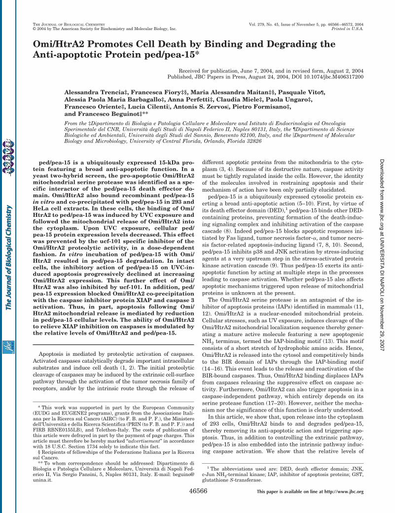
Omi/HtrA2 Promotes Cell Death by Binding and Degrading theAnti-apoptotic Protein ped/pea-15*
Received for publication, June 7, 2004, and in revised form, August 2, 2004Published, JBC Papers in Press, August 24, 2004, DOI 10.1074/jbc.M406317200
Alessandra Trencia‡, Francesca Fiory‡§, Maria Alessandra Maitan‡§, Pasquale Vito¶,Alessia Paola Maria Barbagallo‡, Anna Perfetti‡, Claudia Miele‡, Paola Ungaro‡,Francesco Oriente‡, Lucia Cilenti�, Antonis S. Zervos�, Pietro Formisano‡,and Francesco Beguinot‡**
From the ‡Dipartimento di Biologia e Patologia Cellulare e Molecolare and Istituto di Endocrinologia ed OncologiaSperimentale del CNR, Universita degli Studi di Napoli Federico II, Naples 80131, Italy, the ¶Dipartimento di ScienzeBiologiche ed Ambientali, Universita degli Studi del Sannio, Benevento 82100, Italy, and the �Department of MolecularBiology and Microbiology, University of Central Florida, Orlando, Florida 32826
ped/pea-15 is a ubiquitously expressed 15-kDa pro-tein featuring a broad anti-apoptotic function. In ayeast two-hybrid screen, the pro-apoptotic Omi/HtrA2mitochondrial serine protease was identified as a spe-cific interactor of the ped/pea-15 death effector do-main. Omi/HtrA2 also bound recombinant ped/pea-15in vitro and co-precipitated with ped/pea-15 in 293 andHeLa cell extracts. In these cells, the binding of Omi/HtrA2 to ped/pea-15 was induced by UVC exposure andfollowed the mitochondrial release of Omi/HtrA2 intothe cytoplasm. Upon UVC exposure, cellular ped/pea-15 protein expression levels decreased. This effectwas prevented by the ucf-101 specific inhibitor of theOmi/HtrA2 proteolytic activity, in a dose-dependentfashion. In vitro incubation of ped/pea-15 with Omi/HtrA2 resulted in ped/pea-15 degradation. In intactcells, the inhibitory action of ped/pea-15 on UVC-in-duced apoptosis progressively declined at increasingOmi/HtrA2 expression. This further effect of Omi/HtrA2 was also inhibited by ucf-101. In addition, ped/pea-15 expression blocked Omi/HtrA2 co-precipitationwith the caspase inhibitor protein XIAP and caspase 3activation. Thus, in part, apoptosis following Omi/HtrA2 mitochondrial release is mediated by reductionin ped/pea-15 cellular levels. The ability of Omi/HtrA2to relieve XIAP inhibition on caspases is modulated bythe relative levels of Omi/HtrA2 and ped/pea-15.
Apoptosis is mediated by proteolytic activation of caspases.Activated caspases catalytically degrade important intracellularsubstrates and induce cell death (1, 2). The initial proteolyticcleavage of caspases may be induced by the extrinsic cell-surfacepathway through the activation of the tumor necrosis family ofreceptors, and/or by the intrinsic route through the release of
different apoptotic proteins from the mitochondria to the cyto-plasm (3, 4). Because of its destructive nature, caspase activitymust be tightly regulated inside the cells. However, the identityof the molecules involved in restraining apoptosis and theirmechanism of action have been only partially elucidated.
ped/pea-15 is a ubiquitously expressed cytosolic protein ex-erting a broad anti-apoptotic action (5–10). First, by virtue ofits death effector domain (DED),1 ped/pea-15 binds other DED-containing proteins, preventing formation of the death-induc-ing signaling complex and inhibiting activation of the caspasecascade (8). Indeed ped/pea-15 blocks apoptotic responses ini-tiated by Fas ligand, tumor necrosis factor-�, and tumor necro-sis factor-related apoptosis-inducing ligand (7, 8, 10). Second,ped/pea-15 inhibits p38 and JNK activation by stress-inducingagents at a very upstream step in the stress-activated proteinkinase activation cascade (9). Thus ped/pea-15 exerts its anti-apoptotic function by acting at multiple steps in the processesleading to caspase activation. Whether ped/pea-15 also affectsapoptotic mechanisms triggered upon release of mitochondrialproteins is unknown at the present.
The Omi/HtrA2 serine protease is an antagonist of the in-hibitor of apoptosis proteins (IAPs) identified in mammals (11,12). Omi/HtrA2 is a nuclear-encoded mitochondrial protein.Cellular stresses, such as UV exposure, induces cleavage of theOmi/HtrA2 mitochondrial localization sequence thereby gener-ating a mature active molecule featuring a new apoptogenicNH2 terminus, termed the IAP-binding motif (13). This motifconsists of a short stretch of hydrophobic amino acids. Hence,Omi/HtrA2 is released into the cytosol and competitively bindsto the BIR domain of IAPs through the IAP-binding motif(14–16). This event leads to the release and reactivation of theBIR-bound caspases. Thus, Omi/HtrA2 binding displaces IAPsfrom caspases releasing the suppressive effect on caspase ac-tivity. Furthermore, Omi/HtrA2 can also trigger apoptosis in acaspase-independent pathway, which entirely depends on itsserine protease function (17–20). However, neither the mecha-nism nor the significance of this function is clearly understood.
In this article, we show that, upon release into the cytoplasmof 293 cells, Omi/HtrA2 binds to and degrades ped/pea-15,thereby removing its anti-apoptotic action and triggering apo-ptosis. Thus, in addition to controlling the extrinsic pathway,ped/pea-15 is also embedded into the intrinsic pathway induc-ing caspase activation. We show that the relative levels of
* This work was supported in part by the European Community(EUDG and EUGENE2 programs), grants from the Associazione Itali-ana per la Ricerca sul Cancro (AIRC) (to F. B. and P. F.), the Ministerodell’Universita e della Ricerca Scientifica (PRIN (to F. B. and P. F.)) andFIRB RBNE0155LB), and Telethon-Italy. The costs of publication ofthis article were defrayed in part by the payment of page charges. Thisarticle must therefore be hereby marked “advertisement” in accordancewith 18 U.S.C. Section 1734 solely to indicate this fact.
§ Recipients of fellowships of the Federazione Italiana per la Ricercasul Cancro.
** To whom correspondence should be addressed: Dipartimento diBiologia e Patologia Cellulare e Molecolare, Universita di Napoli Fed-erico II, Via Sergio Pansini, 5, Naples 80131, Italy. E-mail: [email protected].
1 The abbreviations used are: DED, death effector domain; JNK,c-Jun NH2-terminal kinase; IAP, inhibitor of apoptosis proteins; GST,glutathione S-transferase.
THE JOURNAL OF BIOLOGICAL CHEMISTRY Vol. 279, No. 45, Issue of November 5, pp. 46566–46572, 2004© 2004 by The American Society for Biochemistry and Molecular Biology, Inc. Printed in U.S.A.
This paper is available on line at http://www.jbc.org46566
at UN
IVE
RS
ITA
DI N
AP
OLI on N
ovember 29, 2007
ww
w.jbc.org
Dow
nloaded from
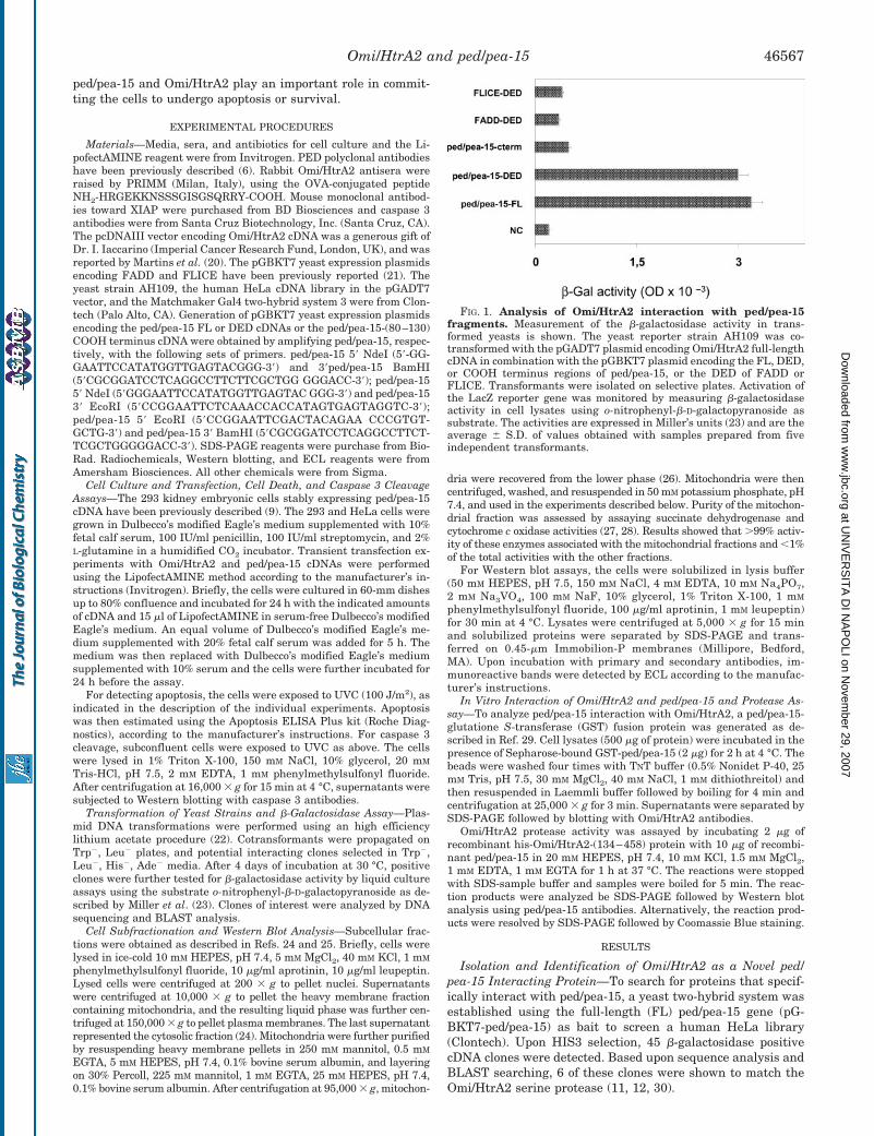
ped/pea-15 and Omi/HtrA2 play an important role in commit-ting the cells to undergo apoptosis or survival.
EXPERIMENTAL PROCEDURES
Materials—Media, sera, and antibiotics for cell culture and the Li-pofectAMINE reagent were from Invitrogen. PED polyclonal antibodieshave been previously described (6). Rabbit Omi/HtrA2 antisera wereraised by PRIMM (Milan, Italy), using the OVA-conjugated peptideNH2-HRGEKKNSSSGISGSQRRY-COOH. Mouse monoclonal antibod-ies toward XIAP were purchased from BD Biosciences and caspase 3antibodies were from Santa Cruz Biotechnology, Inc. (Santa Cruz, CA).The pcDNAIII vector encoding Omi/HtrA2 cDNA was a generous gift ofDr. I. Iaccarino (Imperial Cancer Research Fund, London, UK), and wasreported by Martins et al. (20). The pGBKT7 yeast expression plasmidsencoding FADD and FLICE have been previously reported (21). Theyeast strain AH109, the human HeLa cDNA library in the pGADT7vector, and the Matchmaker Gal4 two-hybrid system 3 were from Clon-tech (Palo Alto, CA). Generation of pGBKT7 yeast expression plasmidsencoding the ped/pea-15 FL or DED cDNAs or the ped/pea-15-(80–130)COOH terminus cDNA were obtained by amplifying ped/pea-15, respec-tively, with the following sets of primers. ped/pea-15 5� NdeI (5�-GG-GAATTCCATATGGTTGAGTACGGG-3�) and 3�ped/pea-15 BamHI(5�CGCGGATCCTCAGGCCTTCTTCGCTGG GGGACC-3�); ped/pea-155� NdeI (5�GGGAATTCCATATGGTTGAGTAC GGG-3�) and ped/pea-153� EcoRI (5�CCGGAATTCTCAAACCACCATAGTGAGTAGGTC-3�);ped/pea-15 5� EcoRI (5�CCGGAATTCGACTACAGAA CCCGTGT-GCTG-3�) and ped/pea-15 3� BamHI (5�CGCGGATCCTCAGGCCTTCT-TCGCTGGGGGACC-3�). SDS-PAGE reagents were purchase from Bio-Rad. Radiochemicals, Western blotting, and ECL reagents were fromAmersham Biosciences. All other chemicals were from Sigma.
Cell Culture and Transfection, Cell Death, and Caspase 3 CleavageAssays—The 293 kidney embryonic cells stably expressing ped/pea-15cDNA have been previously described (9). The 293 and HeLa cells weregrown in Dulbecco’s modified Eagle’s medium supplemented with 10%fetal calf serum, 100 IU/ml penicillin, 100 IU/ml streptomycin, and 2%L-glutamine in a humidified CO2 incubator. Transient transfection ex-periments with Omi/HtrA2 and ped/pea-15 cDNAs were performedusing the LipofectAMINE method according to the manufacturer’s in-structions (Invitrogen). Briefly, the cells were cultured in 60-mm dishesup to 80% confluence and incubated for 24 h with the indicated amountsof cDNA and 15 �l of LipofectAMINE in serum-free Dulbecco’s modifiedEagle’s medium. An equal volume of Dulbecco’s modified Eagle’s me-dium supplemented with 20% fetal calf serum was added for 5 h. Themedium was then replaced with Dulbecco’s modified Eagle’s mediumsupplemented with 10% serum and the cells were further incubated for24 h before the assay.
For detecting apoptosis, the cells were exposed to UVC (100 J/m2), asindicated in the description of the individual experiments. Apoptosiswas then estimated using the Apoptosis ELISA Plus kit (Roche Diag-nostics), according to the manufacturer’s instructions. For caspase 3cleavage, subconfluent cells were exposed to UVC as above. The cellswere lysed in 1% Triton X-100, 150 mM NaCl, 10% glycerol, 20 mM
Tris-HCl, pH 7.5, 2 mM EDTA, 1 mM phenylmethylsulfonyl fluoride.After centrifugation at 16,000 � g for 15 min at 4 °C, supernatants weresubjected to Western blotting with caspase 3 antibodies.
Transformation of Yeast Strains and �-Galactosidase Assay—Plas-mid DNA transformations were performed using an high efficiencylithium acetate procedure (22). Cotransformants were propagated onTrp�, Leu� plates, and potential interacting clones selected in Trp�,Leu�, His�, Ade� media. After 4 days of incubation at 30 °C, positiveclones were further tested for �-galactosidase activity by liquid cultureassays using the substrate o-nitrophenyl-�-D-galactopyranoside as de-scribed by Miller et al. (23). Clones of interest were analyzed by DNAsequencing and BLAST analysis.
Cell Subfractionation and Western Blot Analysis—Subcellular frac-tions were obtained as described in Refs. 24 and 25. Briefly, cells werelysed in ice-cold 10 mM HEPES, pH 7.4, 5 mM MgCl2, 40 mM KCl, 1 mM
phenylmethylsulfonyl fluoride, 10 �g/ml aprotinin, 10 �g/ml leupeptin.Lysed cells were centrifuged at 200 � g to pellet nuclei. Supernatantswere centrifuged at 10,000 � g to pellet the heavy membrane fractioncontaining mitochondria, and the resulting liquid phase was further cen-trifuged at 150,000 � g to pellet plasma membranes. The last supernatantrepresented the cytosolic fraction (24). Mitochondria were further purifiedby resuspending heavy membrane pellets in 250 mM mannitol, 0.5 mM
EGTA, 5 mM HEPES, pH 7.4, 0.1% bovine serum albumin, and layeringon 30% Percoll, 225 mM mannitol, 1 mM EGTA, 25 mM HEPES, pH 7.4,0.1% bovine serum albumin. After centrifugation at 95,000 � g, mitochon-
dria were recovered from the lower phase (26). Mitochondria were thencentrifuged, washed, and resuspended in 50 mM potassium phosphate, pH7.4, and used in the experiments described below. Purity of the mitochon-drial fraction was assessed by assaying succinate dehydrogenase andcytochrome c oxidase activities (27, 28). Results showed that �99% activ-ity of these enzymes associated with the mitochondrial fractions and �1%of the total activities with the other fractions.
For Western blot assays, the cells were solubilized in lysis buffer(50 mM HEPES, pH 7.5, 150 mM NaCl, 4 mM EDTA, 10 mM Na4PO7,2 mM Na3VO4, 100 mM NaF, 10% glycerol, 1% Triton X-100, 1 mM
phenylmethylsulfonyl fluoride, 100 �g/ml aprotinin, 1 mM leupeptin)for 30 min at 4 °C. Lysates were centrifuged at 5,000 � g for 15 minand solubilized proteins were separated by SDS-PAGE and trans-ferred on 0.45-�m Immobilion-P membranes (Millipore, Bedford,MA). Upon incubation with primary and secondary antibodies, im-munoreactive bands were detected by ECL according to the manufac-turer’s instructions.
In Vitro Interaction of Omi/HtrA2 and ped/pea-15 and Protease As-say—To analyze ped/pea-15 interaction with Omi/HtrA2, a ped/pea-15-glutatione S-transferase (GST) fusion protein was generated as de-scribed in Ref. 29. Cell lysates (500 �g of protein) were incubated in thepresence of Sepharose-bound GST-ped/pea-15 (2 �g) for 2 h at 4 °C. Thebeads were washed four times with TNT buffer (0.5% Nonidet P-40, 25mM Tris, pH 7.5, 30 mM MgCl2, 40 mM NaCl, 1 mM dithiothreitol) andthen resuspended in Laemmli buffer followed by boiling for 4 min andcentrifugation at 25,000 � g for 3 min. Supernatants were separated bySDS-PAGE followed by blotting with Omi/HtrA2 antibodies.
Omi/HtrA2 protease activity was assayed by incubating 2 �g ofrecombinant his-Omi/HtrA2-(134–458) protein with 10 �g of recombi-nant ped/pea-15 in 20 mM HEPES, pH 7.4, 10 mM KCl, 1.5 mM MgCl2,1 mM EDTA, 1 mM EGTA for 1 h at 37 °C. The reactions were stoppedwith SDS-sample buffer and samples were boiled for 5 min. The reac-tion products were analyzed be SDS-PAGE followed by Western blotanalysis using ped/pea-15 antibodies. Alternatively, the reaction prod-ucts were resolved by SDS-PAGE followed by Coomassie Blue staining.
RESULTS
Isolation and Identification of Omi/HtrA2 as a Novel ped/pea-15 Interacting Protein—To search for proteins that specif-ically interact with ped/pea-15, a yeast two-hybrid system wasestablished using the full-length (FL) ped/pea-15 gene (pG-BKT7-ped/pea-15) as bait to screen a human HeLa library(Clontech). Upon HIS3 selection, 45 �-galactosidase positivecDNA clones were detected. Based upon sequence analysis andBLAST searching, 6 of these clones were shown to match theOmi/HtrA2 serine protease (11, 12, 30).
FIG. 1. Analysis of Omi/HtrA2 interaction with ped/pea-15fragments. Measurement of the �-galactosidase activity in trans-formed yeasts is shown. The yeast reporter strain AH109 was co-transformed with the pGADT7 plasmid encoding Omi/HtrA2 full-lengthcDNA in combination with the pGBKT7 plasmid encoding the FL, DED,or COOH terminus regions of ped/pea-15, or the DED of FADD orFLICE. Transformants were isolated on selective plates. Activation ofthe LacZ reporter gene was monitored by measuring �-galactosidaseactivity in cell lysates using o-nitrophenyl-�-D-galactopyranoside assubstrate. The activities are expressed in Miller’s units (23) and are theaverage � S.D. of values obtained with samples prepared from fiveindependent transformants.
Omi/HtrA2 and ped/pea-15 46567
at UN
IVE
RS
ITA
DI N
AP
OLI on N
ovember 29, 2007
ww
w.jbc.org
Dow
nloaded from
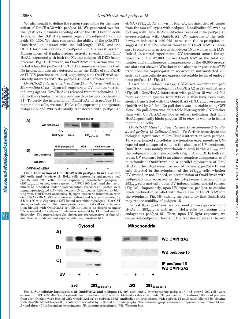
We also sought to define the region responsible for the inter-action of Omi/HtrA2 with ped/pea-15. We generated two fur-ther pGBKT7 plasmids encoding either the DED (amino acids1–80), or the COOH terminus region of ped/pea-15 (aminoacids 80–130). We then compared the ability of the pGADT7-Omi/HtrA2 to interact with the full-length, DED, and theCOOH terminus regions of ped/pea-15 in the yeast system.Measurement of �-galactosidase activity revealed that Omi/HtrA2 interacted with both the FL and ped/pea-15 DED fusionproteins (Fig. 1). However, no Omi/HtrA2 interaction was de-tected when the ped/pea-15 COOH terminus was used as bait.No interaction was also detected when the DEDs of the FADDor FLICE proteins were used, suggesting that Omi/HtrA2 spe-cifically interacts with the ped/pea-15 death effector domain.
Omi/HtrA2 Interacts with ped/pea-15 in Vitro as Well as inMammalian Cells—Upon cell exposure to UV and other stress-inducing agents, Omi/HtrA2 is released from mitochondria (18,30) to the cytoplasm, where ped/pea-15 is largely localized (5,31). To verify the interaction of Omi/HtrA2 with ped/pea-15 inmammalian cells, we used HeLa cells expressing endogenousped/pea-15 and 293 cells stably transfected with ped/pea-15
cDNA (293PED). As shown in Fig. 2A, precipitation of lysatesfrom the two cell types with ped/pea-15 antibodies followed byblotting with Omi/HtrA2 antibodies revealed little ped/pea-15co-precipitation with Omi/HtrA2. UV exposure of the cells,however, induced a �20-fold increase in the co-precipitation,suggesting that UV-induced cleavage of Omi/HtrA2 is neces-sary to enable interaction with ped/pea-15 as well as with IAPs.Indeed, in control experiments, UV treatment caused the ap-pearance of the 37,000 mature Omi/HtrA2 in the total celllysates and simultaneous disappearance of the 49,000 precur-sor (data not shown). Whether in the absence or presence of UVirradiation no co-precipitation occurred in untransfected 293cells, as these cells do not express detectable levels of endoge-nous ped/pea-15 (Fig. 2A).
Based on pull-down assays, GST-fused recombinant ped/pea-15 bound to the endogenous Omi/HtrA2 in 293 cell extracts(Fig. 2B). Omi/HtrA2 interaction with ped/pea-15 was �2-foldmore evident in lysates from 293 cells that have been tran-siently transfected with the Omi/HtrA2 cDNA and overexpressOmi/HtrA2 by 2.5-fold. No pull-down was detectable using GSTalone. No pull-down was detectable blotting with JNK ratherthan with Omi/HtrA2 antibodies either, indicating that Omi/HtrA2 specifically binds ped/pea-15 in vitro as well as in intactmammalian cells.
Omi/HtrA2 Mitochondrial Release Is Accompanied by Re-duced ped/pea-15 Cellular Levels—To further investigate thebiological significance of Omi/HtrA2 interaction with ped/pea-15, we performed subcellular fractionation experiments in UV-exposed and unexposed cells. In the absence of UV treatment,Omi/HtrA2 was mainly mitochondrial both in the 293PED andthe ped/pea-15 untransfected cells (Fig. 3, A and B). In both celltypes, UV exposure led to an almost complete disappearance ofmitochondrial Omi/HtrA2 and a parallel appearance of Omi/HtrA2 in the cytoplasmic fraction. At variance, ped/pea-15 wasonly detected in the cytoplasm of the 293PED cells, whetherUV-treated or not. Indeed, co-precipitation of Omi/HtrA2 withped/pea-15 only occurred in the cytoplasmic fraction of the293PED cells and only upon UV-induced mitochondrial release(Fig. 3C). Importantly, upon UV exposure, ped/pea-15 cellularlevels declined in parallel with the release of Omi/HtrA2 intothe cytoplasm (Fig. 3B), raising the possibility that Omi/HtrA2may reduce stability of ped/pea-15.
To test this hypothesis, we transiently overexpressed Omi/HtrA2 in 293PED as well as in HeLa cells (expressing onlyendogenous ped/pea-15). Then, upon UV light exposure, wecompared ped/pea-15 levels in the transfected versus the un-
FIG. 2. Interaction of Omi/HtrA2 with ped/pea-15 in HeLa and293 cells and in vitro. A, HeLa cells expressing endogenous ped/pea-15 and 293 cells, either expressing transfected ped/pea-15(293ped/pea-15) or not, were exposed to UVC (100 J/m2) and then solu-bilized as described under “Experimental Procedures.” Lysates wereimmunoprecipitated (IP) with ped/pea-15 antibodies followed by blot-ting with Omi/HtrA2 antibodies. B, upon transient transfection withOmi/HtrA2 cDNA, 293 cells were solubilized and lysates incubated for2 h at 4 °C with Sepharose-GST-bound recombinant ped/pea-15 or GSTalone, as indicated. Pulled down proteins and total cell extracts werethen blotted with Omi/HtrA2 or JNK antibodies as described under“Experimental Procedures.” Blots were revealed by ECL and autora-diography. The autoradiographs shown are representative of four (A)and three (B) independent experiments. WB, Western blot.
FIG. 3. Subcellular localization of Omi/HtrA2 and ped/pea-15. 293 cells stably overexpressing ped/pea-15 and control 293 cells wereexposed to UVC (100 J/m2) and cytosolic and mitochondrial fractions obtained as described under “Experimental Procedures.” 80 �g of proteinsfrom each fraction were blotted with Omi/HtrA2 (A) or ped/pea-15 (B) antibodies or, precipitated with ped/pea-15 antibodies followed by blottingwith Omi/HtrA2 antibodies (C). Blots were revealed by ECL and autoradiography. The autoradiographs shown are representative of four (A andB) and three (C) independent experiments. IP, immunoprecipitated; WB, Western blot.
Omi/HtrA2 and ped/pea-1546568
at UN
IVE
RS
ITA
DI N
AP
OLI on N
ovember 29, 2007
ww
w.jbc.org
Dow
nloaded from
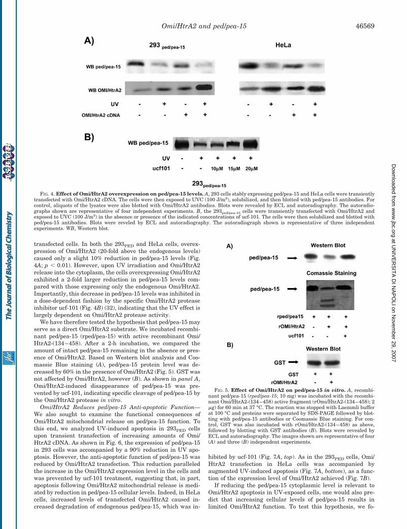
transfected cells. In both the 293PED and HeLa cells, overex-pression of Omi/HtrA2 (20-fold above the endogenous levels)caused only a slight 10% reduction in ped/pea-15 levels (Fig.4A; p � 0.01). However, upon UV irradiation and Omi/HtrA2release into the cytoplasm, the cells overexpressing Omi/HtrA2exhibited a 2-fold larger reduction in ped/pea-15 levels com-pared with those expressing only the endogenous Omi/HtrA2.Importantly, this decrease in ped/pea-15 levels was inhibited ina dose-dependent fashion by the specific Omi/HtrA2 proteaseinhibitor ucf-101 (Fig. 4B) (32), indicating that the UV effect islargely dependent on Omi/HtrA2 protease activity.
We have therefore tested the hypothesis that ped/pea-15 mayserve as a direct Omi/HtrA2 substrate. We incubated recombi-nant ped/pea-15 (rped/pea-15) with active recombinant Omi/HtrA2-(134–458). After a 2-h incubation, we compared theamount of intact ped/pea-15 remaining in the absence or pres-ence of Omi/HtrA2. Based on Western blot analysis and Coo-massie Blue staining (A), ped/pea-15 protein level was de-creased by 60% in the presence of Omi/HtrA2 (Fig. 5). GST wasnot affected by Omi/HtrA2, however (B). As shown in panel A,Omi/HtrA2-induced disappearance of ped/pea-15 was pre-vented by ucf-101, indicating specific cleavage of ped/pea-15 bythe Omi/HtrA2 protease in vitro.
Omi/HtrA2 Reduces ped/pea-15 Anti-apoptotic Function—We also sought to examine the functional consequences ofOmi/HtrA2 mitochondrial release on ped/pea-15 function. Tothis end, we analyzed UV-induced apoptosis in 293PED cellsupon transient transfection of increasing amounts of Omi/HtrA2 cDNA. As shown in Fig. 6, the expression of ped/pea-15in 293 cells was accompanied by a 90% reduction in UV apo-ptosis. However, the anti-apoptotic function of ped/pea-15 wasreduced by Omi/HtrA2 transfection. This reduction paralleledthe increase in the Omi/HtrA2 expression level in the cells andwas prevented by ucf-101 treatment, suggesting that, in part,apoptosis following Omi/HtrA2 mitochondrial release is medi-ated by reduction in ped/pea-15 cellular levels. Indeed, in HeLacells, increased levels of transfected Omi/HtrA2 caused in-creased degradation of endogenous ped/pea-15, which was in-
hibited by ucf-101 (Fig. 7A, top). As in the 293PED cells, Omi/HtrA2 transfection in HeLa cells was accompanied byaugmented UV-induced apoptosis (Fig. 7A, bottom), as a func-tion of the expression level of Omi/HtrA2 achieved (Fig. 7B).
If reducing the ped/pea-15 cytoplasmic level is relevant toOmi/HtrA2 apoptosis in UV-exposed cells, one would also pre-dict that increasing cellular levels of ped/pea-15 results inlimited Omi/HtrA2 function. To test this hypothesis, we fo-
FIG. 5. Effect of Omi/HtrA2 on ped/pea-15 in vitro. A, recombi-nant ped/pea-15 (rped/pea-15; 10 mg) was incubated with the recombi-nant Omi/HtrA2-(134–458) active fragment (rOmi/HtrA2-(134–458); 2�g) for 60 min at 37 °C. The reaction was stopped with Laemmli bufferat 100 °C and proteins were separated by SDS-PAGE followed by blot-ting with ped/pea-15 antibodies or Coomassie Blue staining. For con-trol, GST was also incubated with rOmi/HtrA2-(134–458) as above,followed by blotting with GST antibodies (B). Blots were revealed byECL and autoradiography. The images shown are representative of four(A) and three (B) independent experiments.
FIG. 4. Effect of Omi/HtrA2 overexpression on ped/pea-15 levels. A, 293 cells stably expressing ped/pea-15 and HeLa cells were transientlytransfected with Omi/HtrA2 cDNA. The cells were then exposed to UVC (100 J/m2), solubilized, and then blotted with ped/pea-15 antibodies. Forcontrol, aliquots of the lysates were also blotted with Omi/HtrA2 antibodies. Blots were revealed by ECL and autoradiography. The autoradio-graphs shown are representative of four independent experiments. B, the 293ped/pea-15 cells were transiently transfected with Omi/HtrA2 andexposed to UVC (100 J/m2) in the absence or presence of the indicated concentrations of ucf-101. The cells were then solubilized and blotted withped/pea-15 antibodies. Blots were reveled by ECL and autoradiography. The autoradiograph shown is representative of three independentexperiments. WB, Western blot.
Omi/HtrA2 and ped/pea-15 46569
at UN
IVE
RS
ITA
DI N
AP
OLI on N
ovember 29, 2007
ww
w.jbc.org
Dow
nloaded from
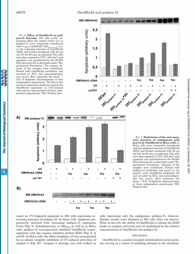
cused on UV-triggered apoptosis in 293 cells expressing in-creasing amounts of ped/pea-15. In these cells, apoptosis pro-gressively declined with increasing ped/pea-15 expressionlevels (Fig. 8). Simultaneously, in 293PED as well as in HeLacells, ped/pea-15 overexpression inhibited Omi/HtrA2 co-pre-cipitation with the caspase inhibitor protein XIAP (Fig. 9, Aand B). In HeLa cells, the effect of ped/pea-15 was accompaniedby an almost complete inhibition of UV-induced activation ofcaspase 3 (Fig. 9C). Caspase 3 cleavage was well evident in
cells expressing only the endogenous ped/pea-15, however.Similar results were obtained in 293 cells (data not shown).Thus, in the cell, the ability of Omi/HtrA2 to release the XIAPbrake on caspase activation may be modulated by the relativeconcentrations of Omi/HtrA2 and ped/pea-15.
DISCUSSION
Omi/HtrA2 is a nuclear-encoded mitochondrial serine prote-ase serving as a sensor of unfolding stresses in the mitochon-
FIG. 6. Effect of Omi/HtrA2 on ped/pea-15 function. 293 cells stably ex-pressing either the control vector (pc) orped/pea-15 were transiently transfectedwith 8 �g of pcDNAIII (293ped/pea-15�pc)or the indicated amounts of Omi/HtrA2cDNA and further incubated with 20 �M
ucf-101 for 60 min, as indicated. The cellswere then exposed to UVC (100 J/m2) andapoptosis was quantitated by the ELISAPlus detection kit as described under “Ex-perimental Procedures.” For control, ali-quots of the samples were solubilized,blotted with Omi/HtrA2 antibodies, andrevealed by ECL and autoradiography(top panel). Bars represent the mean �S.D. of duplicate determinations in fourindependent experiments. The blot in thetop panel represents duplicate controls forOmi/HtrA2 expression in UVC-treatedcells and are representative of three inde-pendent experiments. WB, Western blot.
FIG. 7. Modulation of the anti-apop-totic function of endogenous ped/pea-15 by Omi/HtrA2 in HeLa cells. A,HeLa cells were transiently transfectedwith the indicated amounts of Omi/HtrA2cDNA and further incubated with 20 �M
ucf-101 for 60 min, as indicated. The cellswere then exposed to UVC (100 J/m2) andapoptosis was quantitated by the ELISAPlus detection kit as described under “Ex-perimental Procedures.” Aliquots of thesamples were solubilized, blotted withped/pea-15 antibodies (top panel) or, forcontrol, with Omi/HtrA2 antibodies (B)and revealed by ECL and autoradiogra-phy (top panel). Bars represent themean � S.D. of duplicate determinationsin three independent experiments. WB,Western blot.
Omi/HtrA2 and ped/pea-1546570
at UN
IVE
RS
ITA
DI N
AP
OLI on N
ovember 29, 2007
ww
w.jbc.org
Dow
nloaded from
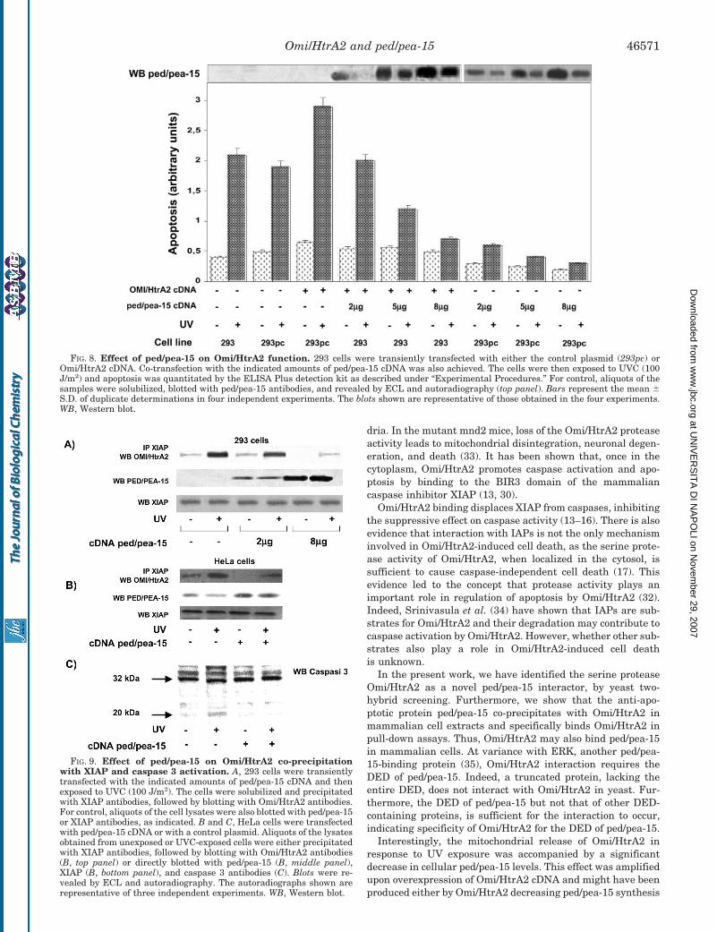
dria. In the mutant mnd2 mice, loss of the Omi/HtrA2 proteaseactivity leads to mitochondrial disintegration, neuronal degen-eration, and death (33). It has been shown that, once in thecytoplasm, Omi/HtrA2 promotes caspase activation and apo-ptosis by binding to the BIR3 domain of the mammaliancaspase inhibitor XIAP (13, 30).
Omi/HtrA2 binding displaces XIAP from caspases, inhibitingthe suppressive effect on caspase activity (13–16). There is alsoevidence that interaction with IAPs is not the only mechanisminvolved in Omi/HtrA2-induced cell death, as the serine prote-ase activity of Omi/HtrA2, when localized in the cytosol, issufficient to cause caspase-independent cell death (17). Thisevidence led to the concept that protease activity plays animportant role in regulation of apoptosis by Omi/HtrA2 (32).Indeed, Srinivasula et al. (34) have shown that IAPs are sub-strates for Omi/HtrA2 and their degradation may contribute tocaspase activation by Omi/HtrA2. However, whether other sub-strates also play a role in Omi/HtrA2-induced cell deathis unknown.
In the present work, we have identified the serine proteaseOmi/HtrA2 as a novel ped/pea-15 interactor, by yeast two-hybrid screening. Furthermore, we show that the anti-apo-ptotic protein ped/pea-15 co-precipitates with Omi/HtrA2 inmammalian cell extracts and specifically binds Omi/HtrA2 inpull-down assays. Thus, Omi/HtrA2 may also bind ped/pea-15in mammalian cells. At variance with ERK, another ped/pea-15-binding protein (35), Omi/HtrA2 interaction requires theDED of ped/pea-15. Indeed, a truncated protein, lacking theentire DED, does not interact with Omi/HtrA2 in yeast. Fur-thermore, the DED of ped/pea-15 but not that of other DED-containing proteins, is sufficient for the interaction to occur,indicating specificity of Omi/HtrA2 for the DED of ped/pea-15.
Interestingly, the mitochondrial release of Omi/HtrA2 inresponse to UV exposure was accompanied by a significantdecrease in cellular ped/pea-15 levels. This effect was amplifiedupon overexpression of Omi/HtrA2 cDNA and might have beenproduced either by Omi/HtrA2 decreasing ped/pea-15 synthesis
FIG. 9. Effect of ped/pea-15 on Omi/HtrA2 co-precipitationwith XIAP and caspase 3 activation. A, 293 cells were transientlytransfected with the indicated amounts of ped/pea-15 cDNA and thenexposed to UVC (100 J/m2). The cells were solubilized and precipitatedwith XIAP antibodies, followed by blotting with Omi/HtrA2 antibodies.For control, aliquots of the cell lysates were also blotted with ped/pea-15or XIAP antibodies, as indicated. B and C, HeLa cells were transfectedwith ped/pea-15 cDNA or with a control plasmid. Aliquots of the lysatesobtained from unexposed or UVC-exposed cells were either precipitatedwith XIAP antibodies, followed by blotting with Omi/HtrA2 antibodies(B, top panel) or directly blotted with ped/pea-15 (B, middle panel),XIAP (B, bottom panel), and caspase 3 antibodies (C). Blots were re-vealed by ECL and autoradiography. The autoradiographs shown arerepresentative of three independent experiments. WB, Western blot.
FIG. 8. Effect of ped/pea-15 on Omi/HtrA2 function. 293 cells were transiently transfected with either the control plasmid (293pc) orOmi/HtrA2 cDNA. Co-transfection with the indicated amounts of ped/pea-15 cDNA was also achieved. The cells were then exposed to UVC (100J/m2) and apoptosis was quantitated by the ELISA Plus detection kit as described under “Experimental Procedures.” For control, aliquots of thesamples were solubilized, blotted with ped/pea-15 antibodies, and revealed by ECL and autoradiography (top panel). Bars represent the mean �S.D. of duplicate determinations in four independent experiments. The blots shown are representative of those obtained in the four experiments.WB, Western blot.
Omi/HtrA2 and ped/pea-15 46571
at UN
IVE
RS
ITA
DI N
AP
OLI on N
ovember 29, 2007
ww
w.jbc.org
Dow
nloaded from
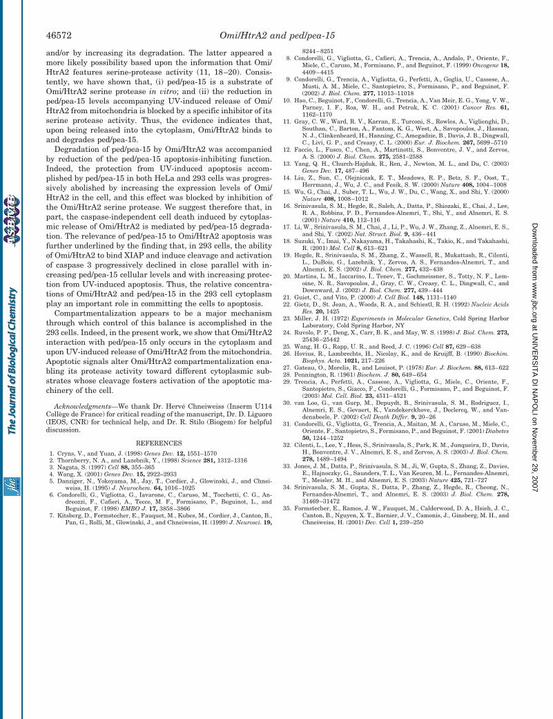
and/or by increasing its degradation. The latter appeared amore likely possibility based upon the information that Omi/HtrA2 features serine-protease activity (11, 18–20). Consis-tently, we have shown that, (i) ped/pea-15 is a substrate ofOmi/HtrA2 serine protease in vitro; and (ii) the reduction inped/pea-15 levels accompanying UV-induced release of Omi/HtrA2 from mitochondria is blocked by a specific inhibitor of itsserine protease activity. Thus, the evidence indicates that,upon being released into the cytoplasm, Omi/HtrA2 binds toand degrades ped/pea-15.
Degradation of ped/pea-15 by Omi/HtrA2 was accompaniedby reduction of the ped/pea-15 apoptosis-inhibiting function.Indeed, the protection from UV-induced apoptosis accom-plished by ped/pea-15 in both HeLa and 293 cells was progres-sively abolished by increasing the expression levels of Omi/HtrA2 in the cell, and this effect was blocked by inhibition ofthe Omi/HtrA2 serine protease. We suggest therefore that, inpart, the caspase-independent cell death induced by cytoplas-mic release of Omi/HtrA2 is mediated by ped/pea-15 degrada-tion. The relevance of ped/pea-15 to Omi/HtrA2 apoptosis wasfurther underlined by the finding that, in 293 cells, the abilityof Omi/HtrA2 to bind XIAP and induce cleavage and activationof caspase 3 progressively declined in close parallel with in-creasing ped/pea-15 cellular levels and with increasing protec-tion from UV-induced apoptosis. Thus, the relative concentra-tions of Omi/HtrA2 and ped/pea-15 in the 293 cell cytoplasmplay an important role in committing the cells to apoptosis.
Compartmentalization appears to be a major mechanismthrough which control of this balance is accomplished in the293 cells. Indeed, in the present work, we show that Omi/HtrA2interaction with ped/pea-15 only occurs in the cytoplasm andupon UV-induced release of Omi/HtrA2 from the mitochondria.Apoptotic signals alter Omi/HtrA2 compartmentalization ena-bling its protease activity toward different cytoplasmic sub-strates whose cleavage fosters activation of the apoptotic ma-chinery of the cell.
Acknowledgments—We thank Dr. Herve Chneiweiss (Inserm U114College de France) for critical reading of the manuscript, Dr. D. Liguoro(IEOS, CNR) for technical help, and Dr. R. Stilo (Biogem) for helpfuldiscussion.
REFERENCES
1. Cryns, V., and Yuan, J. (1998) Genes Dev. 12, 1551–15702. Thornberry, N. A., and Lazebnik, Y., (1998) Science 281, 1312–13163. Nagata, S. (1997) Cell 88, 355–3654. Wang, X. (2001) Genes Dev. 15, 2922–29335. Danziger, N., Yokoyama, M., Jay, T., Cordier, J., Glowinski, J., and Chnei-
weiss, H. (1995) J. Neurochem. 64, 1016–10256. Condorelli, G., Vigliotta, G., Iavarone, C., Caruso, M., Tocchetti, C. G., An-
dreozzi, F., Cafieri, A., Tecce, M. F., Formisano, P., Beguinot, L., andBeguinot, F. (1998) EMBO J. 17, 3858–3866
7. Kitsberg, D., Formstecher, E., Fauquet, M., Kubes, M., Cordier, J., Canton, B.,Pan, G., Rolli, M., Glowinski, J., and Chneiweiss, H. (1999) J. Neurosci. 19,
8244–82518. Condorelli, G., Vigliotta, G., Cafieri, A., Trencia, A., Andalo, P., Oriente, F.,
Miele, C., Caruso, M., Formisano, P., and Beguinot, F. (1999) Oncogene 18,4409–4415
9. Condorelli, G., Trencia, A., Vigliotta, G., Perfetti, A., Goglia, U., Cassese, A.,Musti, A. M., Miele, C., Santopietro, S., Formisano, P., and Beguinot, F.(2002) J. Biol. Chem. 277, 11013–11018
10. Hao, C., Beguinot, F., Condorelli, G., Trencia, A., Van Meir, E. G., Yong, V. W.,Parney, I. F., Roa, W. H., and Petruk, K. C. (2001) Cancer Res. 61,1162–1170
11. Gray, C. W., Ward, R. V., Karran, E., Turconi, S., Rowles, A., Viglienghi, D.,Southan, C., Barton, A., Fantom, K. G., West, A., Savopoulos, J., Hassan,N. J., Clinkenbeard, H., Hanning, C., Amegadzie, B., Davis, J. B., Dingwall,C., Livi, G. P., and Creasy, C. L. (2000) Eur. J. Biochem. 267, 5699–5710
12. Faccio, L., Fusco, C., Chen, A., Martinotti, S., Bonventre, J. V., and Zervos,A. S. (2000) J. Biol. Chem. 275, 2581–2588
13. Yang, Q. H., Church-Hajduk, R., Ren, J., Newton, M. L., and Du, C. (2003)Genes Dev. 17, 487–496
14. Liu, Z., Sun, C., Olejniczak, E. T., Meadows, R. P., Betz, S. F., Oost, T.,Herrmann, J., Wu, J. C., and Fesik, S. W. (2000) Nature 408, 1004–1008
15. Wu, G., Chai, J., Suber, T. L., Wu, J. W., Du, C., Wang, X., and Shi, Y. (2000)Nature 408, 1008–1012
16. Srinivasula, S. M., Hegde, R., Saleh, A., Datta, P., Shiozaki, E., Chai, J., Lee,R. A., Robbins, P. D., Fernandes-Alnemri, T., Shi, Y., and Alnemri, E. S.(2001) Nature 410, 112–116
17. Li, W., Srinivasula, S. M., Chai, J., Li, P., Wu, J. W., Zhang, Z., Alnemri, E. S.,and Shi, Y. (2002) Nat. Struct. Biol. 9, 436–441
18. Suzuki, Y., Imai, Y., Nakayama, H., Takahashi, K., Takio, K., and Takahashi,R. (2001) Mol. Cell 8, 613–621
19. Hegde, R., Srinivasula, S. M., Zhang, Z., Wassell, R., Mukattash, R., Cilenti,L., DuBois, G., Lazebnik, Y., Zervos, A. S., Fernandes-Alnemri, T., andAlnemri, E. S. (2002) J. Biol. Chem. 277, 432–438
20. Martins, L. M., Iaccarino, I., Tenev, T., Gschmeissner, S., Totty, N. F., Lem-oine, N. R., Savopoulos, J., Gray, C. W., Creasy, C. L., Dingwall, C., andDownward, J. (2002) J. Biol. Chem. 277, 439–444
21. Guiet, C., and Vito, P. (2000) J. Cell Biol. 148, 1131–114022. Gietz, D., St. Jean, A., Woods, R. A., and Schiestl, R. H. (1992) Nucleic Acids
Res. 20, 142523. Miller, J. H. (1972) Experiments in Molecular Genetics, Cold Spring Harbor
Laboratory, Cold Spring Harbor, NY24. Ruvolo, P. P., Deng, X., Carr, B. K., and May, W. S. (1998) J. Biol. Chem. 273,
25436–2544225. Wang, H. G., Rapp, U. R., and Reed, J. C. (1996) Cell 87, 629–63826. Hovius, R., Lambrechts, H., Nicolay, K., and de Kruijff, B. (1990) Biochim.
Biophys. Acta. 1021, 217–22627. Gateau, O., Morelis, R., and Louisot, P. (1978) Eur. J. Biochem. 88, 613–62228. Pennington, R. (1961) Biochem. J. 80, 649–65429. Trencia, A., Perfetti, A., Cassese, A., Vigliotta, G., Miele, C., Oriente, F.,
Santopietro, S., Giacco, F., Condorelli, G., Formisano, P., and Beguinot, F.(2003) Mol. Cell. Biol. 23, 4511–4521
30. van Loo, G., van Gurp, M., Depuydt, B., Srinivasula, S. M., Rodriguez, I.,Alnemri, E. S., Gevaert, K., Vandekerckhove, J., Declercq, W., and Van-denabeele, P. (2002) Cell Death Differ. 9, 20–26
31. Condorelli, G., Vigliotta, G., Trencia, A., Maitan, M. A., Caruso, M., Miele, C.,Oriente, F., Santopietro, S., Formisano, P., and Beguinot, F. (2001) Diabetes50, 1244–1252
32. Cilenti, L., Lee, Y., Hess, S., Srinivasula, S., Park, K. M., Junqueira, D., Davis,H., Bonventre, J. V., Alnemri, E. S., and Zervos, A. S. (2003) J. Biol. Chem.278, 1489–1494
33. Jones, J. M., Datta, P., Srinivasula, S. M., Ji, W, Gupta, S., Zhang, Z., Davies,E., Hajnoczky, G., Saunders, T. L., Van Keuren, M. L., Fernandes-Alnemri,T., Meisler, M. H., and Alnemri, E. S. (2003) Nature 425, 721–727
34. Srinivasula, S. M., Gupta, S., Datta, P., Zhang, Z., Hegde, R., Cheong, N.,Fernandes-Alnemri, T., and Alnemri, E. S. (2003) J. Biol. Chem. 278,31469–31472
35. Formstecher, E., Ramos, J. W., Fauquet, M., Calderwood, D. A., Hsieh, J. C.,Canton, B., Nguyen, X. T., Barnier, J. V., Camonis, J., Ginsberg, M. H., andChneiweiss, H. (2001) Dev. Cell 1, 239–250
Omi/HtrA2 and ped/pea-1546572
at UN
IVE
RS
ITA
DI N
AP
OLI on N
ovember 29, 2007
ww
w.jbc.org
Dow
nloaded from
Phorbol Esters Induce Intracellular Accumulation of theAnti-apoptotic Protein PED/PEA-15 by PreventingUbiquitinylation and Proteasomal Degradation*
Received for publication, August 31, 2006, and in revised form, December 21, 2006 Published, JBC Papers in Press, January 16, 2007, DOI 10.1074/jbc.M608359200
Anna Perfetti, Francesco Oriente, Salvatore Iovino, A. Teresa Alberobello, Alessia P. M. Barbagallo,Iolanda Esposito, Francesca Fiory, Raffaele Teperino, Paola Ungaro, Claudia Miele, Pietro Formisano1,and Francesco BeguinotFrom the Dipartimento di Biologia e Patologia cellulare e Molecolare (DBPCM) and Istituto di Endocrinologia ed OncologiaSperimentale del Consiglio Nazionale delle Ricerche, Federico II University of Naples, Via Pansini 5, 80131 Naples, Italy
Phosphoprotein enriched in diabetes/phosphoprotein en-riched in astrocytes (PED/PEA)-15 is an anti-apoptotic proteinwhose expression is increased in several cancer cells and follow-ing experimental skin carcinogenesis. Exposure of untrans-fected C5N keratinocytes and transfected HEK293 cells tophorbol esters (12-O-tetradecanoylphorbol-13-acetate (TPA))increased PED/PEA-15 cellular content and enhanced itsphosphorylation at serine 116 in a time-dependent fashion.Ser-116 3 Gly (PEDS116G) but not Ser-1043 Gly (PEDS104G)substitution almost completely abolished TPA regulation ofPED/PEA-15 expression. TPA effect was also prevented by anti-sense inhibition of protein kinase C (PKC)-� and by the expres-sion of a dominant-negative PKC-� mutant cDNA in HEK293cells. Similar to long term TPA treatment, overexpression ofwild-type PKC-� increased cellular content and phosphoryla-tion of WT-PED/PEA-15 and PEDS104G but not of PEDS116G.These events were accompanied by the activation of Ca2�-cal-modulin kinase (CaMK) II and prevented by the CaMK blocker,KN-93. At variance, the proteasome inhibitor lactacystin mim-icked TPA action on PED/PEA-15 intracellular accumulationand reverted the effects of PKC-� and CaMK inhibition. More-over, we show that PED/PEA-15 bound ubiquitin in intact cells.PED/PEA-15 ubiquitinylation was reduced by TPA and PKC-�overexpression and increased by KN-93 and PKC-� block. Fur-thermore, in HEK293 cells expressing PEDS116G, TPA failed toprevent ubiquitin-dependent degradation of the protein.Accordingly, in the same cells, TPA-mediated protection fromapoptosis was blunted. Taken together, our results indicate thatTPA increasesPED/PEA-15 expression at thepost-translationallevel by inducing phosphorylation at serine 116 and preventingubiquitinylation and proteosomal degradation.
Cancer cells feature both excessive proliferation and aban-donment of the ability to die (1, 2). Thus, alterations of genesinvolved in the control of apoptosis have been implicated in anumber of human malignancies. In certain lymphomas, forexample, cell death is blocked by excessive production of theanti-apoptotic factor Bcl-2 (2). Similarly, some tumors preventapoptosis by up-regulating the expression of anti-apoptoticdeath effector domain (DED)2-containing proteins, which, inturn, inhibit Fas from conveying signals to the deathmachinery(3).Phosphoprotein enriched in diabetes/phosphoprotein
enriched in astrocytes (PED/PEA)-15 is a DED-containing pro-tein originally identified in astrocytes as a protein kinase C(PKC) substrate (4–6) and found overexpressed in insulin tar-get tissues of patients with type 2 diabetes (7). Raised PED/PEA-15 levels have also been detected in several human tumorcell lines (7–9). A growing body of evidence indicates thatincreased PED/PEA-15 expression may provide a mechanismto escape cell death upon a number of pro-apoptotic stimuli(10–14).Moreover, in transgenicmice, overexpression of PED/PEA-15 enhances the susceptibility to develop experimentallyinduced skin tumors (15). The molecular mechanism of PED/PEA-15 anti-apoptotic action has been extensively investi-gated. In several cell types, PED/PEA-15 blocks Fas- and tumornecrosis factor-�-induced apoptosis by competing with itsDEDwith the interaction between FADD and caspase 8 (10). Inaddition, in several cell lines of human glioma, PED/PEA-15inhibits tumor necrosis factor-related apoptosis-inducingligand (TRAIL)-induced apoptosis, thereby generating resist-ance to this anti-neoplastic agent (9). At variance with otheranti-apoptotic proteins inhibiting caspase 8 activation viaFADD trapping (3), PED/PEA-15 overexpression also preventsapoptosis induced by growth factors deprivation, UV exposure,and osmotic stimuli (11, 13).Besides the anti-apoptotic function, a role for PED/PEA-15
in restraining cell proliferation has been proposed (16–20). It* This work was supported by Grant LSHM-CT-2004-512013 from the Euro-
pean Community FP6 EUGENE2, grants from the Associazione Italiana perla Ricerca sul Cancro (AIRC) (to F. B. and P. F.) and the Ministerodell’Universita e della Ricerca Scientifica Grant PRIN (to F. B. and P. F.) andGrant FIRB RBNE0155LB (to F. B.), and by a grant from Telethon – Italy. Thecosts of publication of this article were defrayed in part by the payment ofpage charges. This article must therefore be hereby marked “advertise-ment” in accordance with 18 U.S.C. Section 1734 solely to indicate this fact.
1 To whom correspondence should be addressed. Dipartimento di Biologia ePatologia Cellulare e Molecolare, Universita di Napoli “Federico II,” Via S.Pansini 5, 80131 Naples, Italy. Tel.: 39-081-7463608; Fax: 39-081-7463235;E-mail: [email protected].
2 The abbreviations used are: DED, death effector domain; PED/PEA, phos-phoprotein enriched in diabetes/phosphoprotein enriched in astrocytes;TPA, 12-O-tetradecanoylphorbol-13-acetate; PKB, protein kinase B; PKC,protein kinase C; CaMK, calmodulin kinase; FADD, Fas-associated deathdomain; HA, hemagglutinin; DMEM, Dulbecco’s modified Eagle’s medium;RT, reverse transcription; PBS, phosphate-buffered saline; Ab, antibody;ASO, antisense oligonucleotides; DN, dominant-negative; WT, wild type;SO, scrambled.
THE JOURNAL OF BIOLOGICAL CHEMISTRY VOL. 282, NO. 12, pp. 8648 –8657, March 23, 2007© 2007 by The American Society for Biochemistry and Molecular Biology, Inc. Printed in the U.S.A.
8648 JOURNAL OF BIOLOGICAL CHEMISTRY VOLUME 282 • NUMBER 12 • MARCH 23, 2007
at UN
IVE
RS
ITA
DI N
AP
OLI on N
ovember 29, 2007
ww
w.jbc.org
Dow
nloaded from
has been described that PED/PEA-15 directly binds extracellu-lar signal-regulated kinase 2 (ERK2) and RSK2 and preventstheir nuclear translocation and transduction of biologicaleffects (16–19). Together with the anti-apoptotic effect, thisaction may expand cellular senescence (20).PED/PEA-15 is a phosphorylated protein (4, 5). It has
recently been shown that PED/PEA-15 phosphorylation at spe-cific sites controls the ability of the protein to form complexeswith specific intracellular interactors (21). PED/PEA-15 serinephosphorylation has also been shown to enhance protein sta-bility (22). Several kinases were evidenced to phosphorylatePED/PEA-15 at specific serines. Ser-104 represents the maintarget for PKC phosphorylation (4, 5, 23), whereas Ser-116 hasbeen shown to be a target site for both Ca2�-calmodulin kinase(CaMK) II (23) and protein kinase B (PKB)/Akt (22). However,the precise function of these phosphorylation sites in control-ling PED/PEA-15 expression is currently unknown. Recent evi-dence indicates that abnormal accumulation of PED/PEA-15maylead to derangement of cell growth andmetabolism (15, 24, 25).In this study, we have shown that phorbol esters, which are
tumor promoters and inhibitors of insulin action, up-regulatePED/PEA-15 expression by inhibiting its ubiquitinylation andproteasomal targeting. This effect involves activation ofCaMKII and subsequent phosphorylation of PED/PEA-15 atSer-116. PKC-� activity is required for phorbol ester-inducedactivation of CaMKII and for the regulation of PED/PEA-15degradation.
EXPERIMENTAL PROCEDURES
Materials—Media, sera, and antibiotics for cell culture andthe Lipofectamine reagent were purchased from Invitrogen(Paisley, UK). Rabbit polyclonal PKC-�, PKC-�, PKC-�, PKC-�,and phospho-PKC antibodies were from Santa Cruz Biotech-nology (Santa Cruz, CA). PED/PEA-15, p-Ser-104PED, andp-Ser-116PEDand antibodies have been previously reported (7,22). Mouse monoclonal polyubiquitinylated protein antibodies(FK1) were from Biomol International. Mouse monoclonal HAantibody were from Roche Applied Science. PhosphorothioatePKC-�, PKC-�, PKC-�, PKC-� antisense and scrambled con-trol oligonucleotides have been previously described (26–28)and were synthesized by PRIMM (Milan, Italy). PKC-� wild-type and dominant-negative constructs were kindly providedbyDr.M. S.Marber (St. ThomasHospital, London,UK) andDr.S.Gutkind (NCI, National Institutes of Health, Bethesda, MD),respectively. SDS-PAGE reagents were purchased from Bio-Rad. Western blotting and ECL reagents and radiochemicalswere from Amersham Biosciences. All other reagents werefrom Sigma.Plasmid Preparation, Cell Culture, and Transfection—The
PEDS104G andPEDS116Gmutant cDNAswere prepared by usingpcDNAIIIPEDY1 cDNA (pcDNAIII containing His6-Myc-tagged PED/PEA-15) as template with the site-directedmutagenesis kit by Promega according to the manufacturer’sinstructions. Stable expression of the mutants and wild-typePEDY1 cDNAs in HEK293 cells (293PEDY1) was achieved asreported in Condorelli et al. (11). The cells were cultured inDulbecco’s modified Eagle’s medium (DMEM) supplementedwith 10% fetal calf serum, 100 IU of penicillin/ml, 100 IU of
streptomycin/ml, and 2% L-glutamine in a humidified CO2incubator. Transient transfection of phosphorothioate oligo-nucleotides and plasmid DNA in HEK293 cells was accom-plished by using the Lipofectamine method according to themanufacturer’s instructions. Briefly, the cells were cultured in60-mm-diameter dishes and incubated for 24 h in serum-freeDMEM supplemented with 3 �g of cDNA and 15 �l of Lipo-fectamine reagent. An equal volume of DMEM supplementedwith 20% fetal calf serum was then added for 5 h followed byreplacement with DMEM supplemented with 10% serum for24 h before the assays.Western Blot Analysis—ForWestern blotting, cells were sol-
ubilized in lysis buffer (50mMHEPES (pH 7.5), 150mMNaCl, 4mM EDTA, 10 mM Na4PO7, 2 mM Na3VO4, 100 mM NaF, 10%glycerol, 1%TritonX-100, 1mMphenylmethylsulfonyl fluoride,100mg of aprotinin/ml, 1mM leupeptin) for 60min at 4 °C. Celllysates were clarified at 5,000 � g for 15 min. Solubilized pro-teins were then separated by SDS-PAGE and transferred onto0.45-�m-pore size Immobilon-P membranes (Millipore, Bed-ford, MA). Upon incubation with the primary (PED, etc.) anti-body and secondary antibodies, immunoreactive bands weredetected by ECL according to the manufacturer’s instructions.Real-time RT-PCR Analysis—Total cellular RNA was iso-
lated from C5N cells by the use of RNeasy kit (Qiagen) accord-ing to the manufacturer’s instructions. For real-time RT-PCR analysis, 1 �g of cell RNA was reverse-transcribed usingSuperScript II reverse transcriptase (Invitrogen). PCR reactionmixes were analyzed using SYBRGreenmix (Invitrogen). Reac-tions were performed using Platinum SYBRGreen quantitativePCR Super-UDG using an iCycler IQmulticolor real-time PCRdetection system (Bio-Rad). All reactions were performed intriplicate, and �-actin was used as an internal standard. Primersequences used were as follows: PED/PEA-15, forward, 5�-TTCCCGCTGTTCCCTTAGG-3�, and PED/PEA-15, reverse5�-TCTGGCTCATCCGCATCC-3�.Immunoprecipitation of PED/PEA-15—Cells grown in
100-mmPetri disheswere treated as indicated andwashed oncewith ice-cold PBS and solubilized in lysis buffer for 2 h at 4 °C.Then, lysates were clarified by centrifugation at 5,000� g for 20min. 500 �g of protein lysates was immunoprecipitated withMyc or PED antibodies for 16 h. The precipitates were incu-bated with protein G-Sepharose beads at 4 °C for 1 h with shak-ing. Beads were precipitated by centrifugation at 1,000� g for 5min at 4 °C and washed five times with ice-cold washing buffer.After the final wash, the pellets were resuspended in 30�l of 1�SDS electrophoresis buffer and heated to 95 °C for 5 min priorto protein separation by 15% SDS-PAGE.Western blot analysiswas performed as described above.Purification of Ubiquitin-PED/PEA-15 Conjugates—Cellular
ubiquitinylation assay was performed as described by Musti etal. (29). Briefly, 24 h after transfection, cells expressing His-tagged-PED/PEA-15 (PED/PEA-15Y1) and HA-ubiquitin wereharvested in 2ml of 6M guanidium-HCl, 0.1 MNa2HPO4/NaH2PO4 (pH 8) plus 5 mM imidazole/100-mm dish and sonicatedwith a Branson micro-tipped sonifier for 30 s to reduce viscos-ity. Lysatesweremixed on a rotatorwith 0.2ml (settled volume)of Ni2�-nitrilotriacetic acid-agarose (Qiagen) for 4 h at roomtemperature. The slurry was applied to a Bio-Rad Econo-Col-
Regulation of PED/PEA-15 Ubiquitinylation
MARCH 23, 2007 • VOLUME 282 • NUMBER 12 JOURNAL OF BIOLOGICAL CHEMISTRY 8649
at UN
IVE
RS
ITA
DI N
AP
OLI on N
ovember 29, 2007
ww
w.jbc.org
Dow
nloaded from
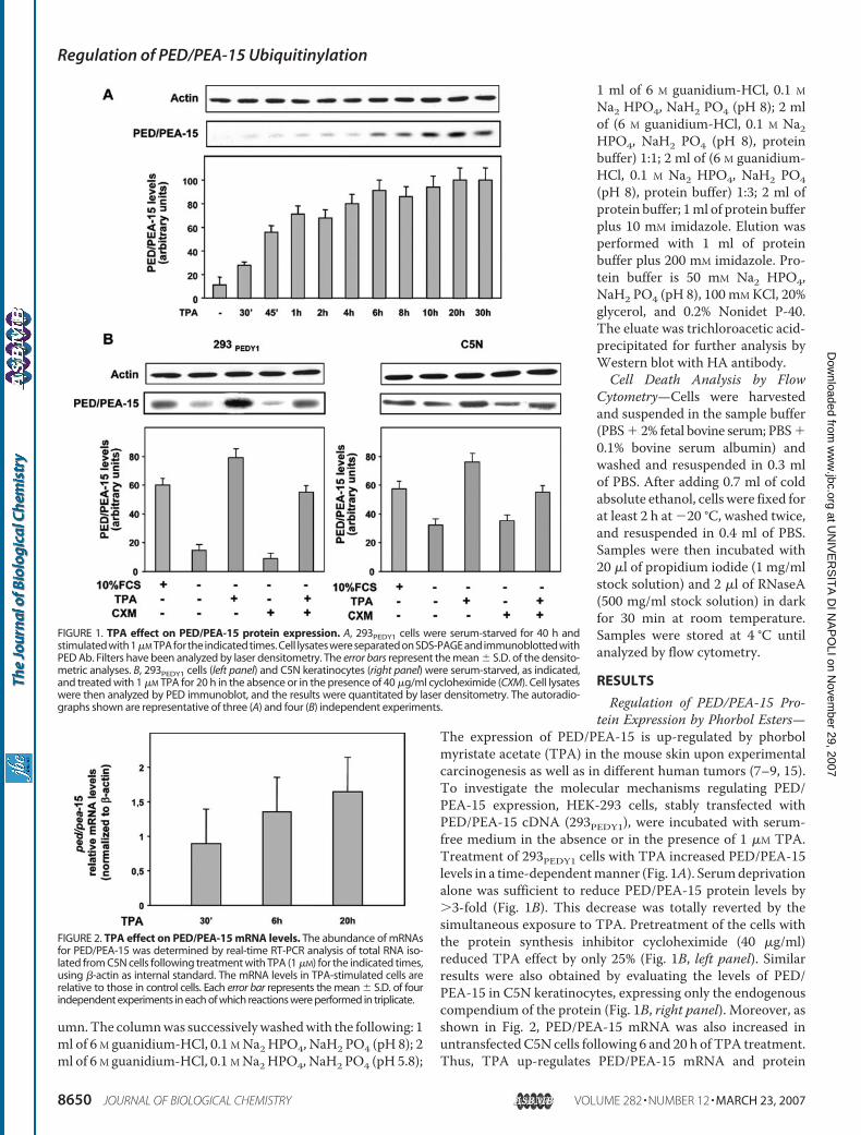
umn.The columnwas successivelywashedwith the following: 1ml of 6 M guanidium-HCl, 0.1 MNa2HPO4, NaH2 PO4 (pH 8); 2ml of 6 M guanidium-HCl, 0.1 MNa2HPO4, NaH2 PO4 (pH 5.8);
1 ml of 6 M guanidium-HCl, 0.1 MNa2 HPO4, NaH2 PO4 (pH 8); 2 mlof (6 M guanidium-HCl, 0.1 M Na2HPO4, NaH2 PO4 (pH 8), proteinbuffer) 1:1; 2 ml of (6 M guanidium-HCl, 0.1 M Na2 HPO4, NaH2 PO4(pH 8), protein buffer) 1:3; 2 ml ofprotein buffer; 1ml of protein bufferplus 10 mM imidazole. Elution wasperformed with 1 ml of proteinbuffer plus 200 mM imidazole. Pro-tein buffer is 50 mM Na2 HPO4,NaH2 PO4 (pH 8), 100 mMKCl, 20%glycerol, and 0.2% Nonidet P-40.The eluate was trichloroacetic acid-precipitated for further analysis byWestern blot with HA antibody.Cell Death Analysis by Flow
Cytometry—Cells were harvestedand suspended in the sample buffer(PBS� 2% fetal bovine serum; PBS�0.1% bovine serum albumin) andwashed and resuspended in 0.3 mlof PBS. After adding 0.7 ml of coldabsolute ethanol, cells were fixed forat least 2 h at �20 °C, washed twice,and resuspended in 0.4 ml of PBS.Samples were then incubated with20 �l of propidium iodide (1 mg/mlstock solution) and 2 �l of RNaseA(500 mg/ml stock solution) in darkfor 30 min at room temperature.Samples were stored at 4 °C untilanalyzed by flow cytometry.
RESULTS
Regulation of PED/PEA-15 Pro-tein Expression by Phorbol Esters—
The expression of PED/PEA-15 is up-regulated by phorbolmyristate acetate (TPA) in the mouse skin upon experimentalcarcinogenesis as well as in different human tumors (7–9, 15).To investigate the molecular mechanisms regulating PED/PEA-15 expression, HEK-293 cells, stably transfected withPED/PEA-15 cDNA (293PEDY1), were incubated with serum-free medium in the absence or in the presence of 1 �M TPA.Treatment of 293PEDY1 cells with TPA increased PED/PEA-15levels in a time-dependentmanner (Fig. 1A). Serumdeprivationalone was sufficient to reduce PED/PEA-15 protein levels by�3-fold (Fig. 1B). This decrease was totally reverted by thesimultaneous exposure to TPA. Pretreatment of the cells withthe protein synthesis inhibitor cycloheximide (40 �g/ml)reduced TPA effect by only 25% (Fig. 1B, left panel). Similarresults were also obtained by evaluating the levels of PED/PEA-15 in C5N keratinocytes, expressing only the endogenouscompendium of the protein (Fig. 1B, right panel). Moreover, asshown in Fig. 2, PED/PEA-15 mRNA was also increased inuntransfectedC5N cells following 6 and 20 h of TPA treatment.Thus, TPA up-regulates PED/PEA-15 mRNA and protein
FIGURE 1. TPA effect on PED/PEA-15 protein expression. A, 293PEDY1 cells were serum-starved for 40 h andstimulated with 1�M TPA for the indicated times. Cell lysates were separated on SDS-PAGE and immunoblotted withPED Ab. Filters have been analyzed by laser densitometry. The error bars represent the mean � S.D. of the densito-metric analyses. B, 293PEDY1 cells (left panel) and C5N keratinocytes (right panel) were serum-starved, as indicated,and treated with 1 �M TPA for 20 h in the absence or in the presence of 40 �g/ml cycloheximide (CXM). Cell lysateswere then analyzed by PED immunoblot, and the results were quantitated by laser densitometry. The autoradio-graphs shown are representative of three (A) and four (B) independent experiments.
FIGURE 2. TPA effect on PED/PEA-15 mRNA levels. The abundance of mRNAsfor PED/PEA-15 was determined by real-time RT-PCR analysis of total RNA iso-lated from C5N cells following treatment with TPA (1 �M) for the indicated times,using �-actin as internal standard. The mRNA levels in TPA-stimulated cells arerelative to those in control cells. Each error bar represents the mean � S.D. of fourindependent experiments in each of which reactions were performed in triplicate.
Regulation of PED/PEA-15 Ubiquitinylation
8650 JOURNAL OF BIOLOGICAL CHEMISTRY VOLUME 282 • NUMBER 12 • MARCH 23, 2007
at UN
IVE
RS
ITA
DI N
AP
OLI on N
ovember 29, 2007
ww
w.jbc.org
Dow
nloaded from
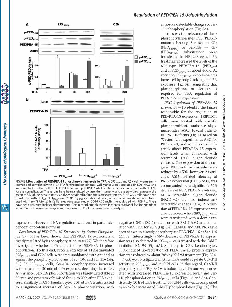
expression. However, TPA regulation is, at least in part, inde-pendent of protein synthesis.Regulation of PED/PEA-15 Expression by Serine Phosphor-
ylation—It has been shown that PED/PEA-15 expression istightly regulated by its phosphorylation state (22).We thereforeinvestigated whether TPA could induce PED/PEA-15 phos-phorylation. To this end, protein extracts of TPA-stimulated293PEDY1 and C5N cells were immunoblotted with antibodiesagainst the phosphorylated forms of Ser-104 and Ser-116 (Fig.3A). In 293PEDY1 cells, Ser-104 phosphorylation increasedwithin the initial 30 min of TPA exposure, declining thereafter.At variance, Ser-116 phosphorylation was barely detectable at30 min and progressively raised for up to 20 h after TPA expo-sure. Similarly, inC5Nkeratinocytes, 20 h of TPA treatment ledto a significant increase of Ser-116 phosphorylation, with
almost undetectable changes of Ser-104 phosphorylation (Fig. 3A).To assess the relevance of those
phosphorylation sites, PED/PEA-15mutants bearing Ser-104 3 Gly(PEDS104G) or Ser-116 3 Gly(PEDS116G) substitutions weretransfected in HEK293 cells. TPAtreatment increased the levels of thewild-type PED/PEA-15 (PEDWT)and of PEDS104G by about 4-fold. Atvariance, PEDS116G expression wasincreased by only 2-fold upon TPAexposure (Fig. 3B), suggesting thatphosphorylation of Ser-116 isrequired for TPA regulation ofPED/PEA-15 expression.PKC Regulation of PED/PEA-15
Expression—To identify the kinaseresponsible for the regulation ofPED/PEA-15 expression, 293PEDY1cells were treated with specificphosphorothioate antisense oligo-nucleotides (ASO) toward individ-ual PKC isoforms (Fig. 4). Based onWestern blot experiments, ASO forPKC-�, -�, and -� did not signifi-cantly affect PED/PEA-15 expres-sion levels when compared withscrambled (SO) oligonucleotidecontrols. The expression of the tar-geted PKC isoform was selectivelyreduced by�50%, however. At vari-ance, ASO-mediated silencing ofPKC-� expression (PKC�-ASO) wasaccompanied by a significant 70%decrease of PED/PEA-15 levels (Fig.4). A scrambled oligonucleotide(PKC�-SO) did not induce anydetectable change (Fig. 4). A reduc-tion of PED/PEA-15 expression wasalso observed when 293PEDY1 cellswere transfected with a dominant-
negative (DN) PKC-� mutant or with PKC�-ASO and stimu-lated with TPA for 20 h (Fig. 5A). CaMKII and Akt/PKB havebeen shown to directly phosphorylate PED/PEA-15 at Ser-116(22, 23). Interestingly, a 75% decrease of PED/PEA-15 expres-sion was also detected in 293PEDY1 cells treated with the CaMKinhibitor, KN-93 (Fig. 5A). Similarly, in C5N keratinocytes,TPA-induced up-regulation of PED/PEA-15 protein expres-sion was reduced by about 70% by KN-93 treatment (Fig. 5B).Next, we investigated whether TPA could regulate CaMKII
activity in 293PEDY1 and in C5N cells. In this regard, CaMKIIphosphorylation (Fig. 6A) was induced by TPA and well corre-lated with increased PED/PEA-15 expression levels and Ser-116 phosphorylation in 293PEDY1 cells (Figs. 1A and 3A). Con-sistently, 20 h of TPA treatment of C5N cells was accompaniedby a 2.5-fold increase ofCaMKII phosphorylation (Fig. 6A). The
FIGURE 3. Regulation of PED/PEA-15 phosphorylation levels by TPA. A, 293PEDY1 and C5N cells were serum-starved and stimulated with 1 �M TPA for the indicated times. Cell lysates were separated on SDS-PAGE andimmunoblotted either with p-PEDS104 Ab or with p-PEDS116 Ab. Each filter has been reprobed with PED Abfor the normalization. The results have been analyzed by laser densitometry, and the error bars represent themean � S.D. of the densitometric analyses obtained in four duplicate experiments. B, HEK293 cells have beentransfected with PEDWT, PEDS104G, and 293PEDS116G, as indicated. Next, cells were serum-starved and stimu-lated with 1 �M TPA for 20 h. Cell lysates were separated on SDS-PAGE and immunoblotted with PED Ab. Filtershave been analyzed by laser densitometry. The autoradiograph shown is representative of five independentexperiments. The error bars represent the mean � S.D. of the densitometric analysis.
Regulation of PED/PEA-15 Ubiquitinylation
MARCH 23, 2007 • VOLUME 282 • NUMBER 12 JOURNAL OF BIOLOGICAL CHEMISTRY 8651
at UN
IVE
RS
ITA
DI N
AP
OLI on N
ovember 29, 2007
ww
w.jbc.org
Dow
nloaded from
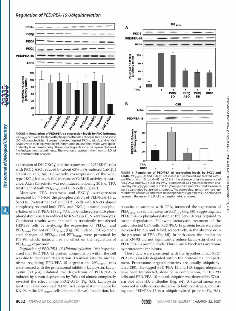
expression of DN-PKC-� and the treatment of 293PEDY1 cellswith PKC�-ASO reduced by about 65% TPA-induced CaMKIIactivation (Fig. 6B). Conversely, overexpression of the wild-type PKC-� led to �3-fold increase of CaMKII activity. At vari-ance, Akt/PKB activity was not induced following 20 h of TPAtreatment of both 293PEDY1 and C5N cells (Fig. 6C).Moreover, TPA treatment and PKC-� overexpression
increased by �5-fold the phosphorylation of PED/PEA-15 atSer-116. Pretreatment of 293PEDY1 cells with KN-93 almostcompletely reverted both TPA- and PKC-�-induced phospho-rylation of PED/PEA-15 (Fig. 7A). TPA-induced Ser-116 phos-phorylation was also reduced by KN-93 in C5N keratinocytes.Consistent results were obtained in transiently transfectedHEK293 cells by analyzing the expression of PEDWT andPEDS104G but not of PEDS116G (Fig. 7B). Indeed, PKC-�-medi-ated changes of PEDWT and PEDS104G were prevented byKN-93, which, instead, had no effect on the regulation ofPEDS116G expression.Regulation of PED/PEA-15 Ubiquitinylation—We hypothe-
sized that PED/PEA-15 protein accumulation within the cellwas due to decreased degradation. To investigate the mecha-nisms regulating PED/PEA-15 degradation, 293PEDY1 cellswere treated with the proteasomal inhibitor lactacystin. Lacta-cystin (30 �M) inhibited the degradation of PED/PEA-15induced by serum deprivation by 70% and almost completelyreverted the effect of the PKC�-ASO (Fig. 8A). Lactacystintreatment also prevented PED/PEA-15 degradation induced byKN-93 in the 293PEDY1 cells (data not shown). In addition, lac-
tacystin, at variance with TPA, increased the expression ofPEDS116G at a similar extent as PEDWT (Fig. 8B), suggesting thatPED/PEA-15 phosphorylation at the Ser-116 was required toescape degradation. Following lactacystin treatment of theuntransfected C5N cells, PED/PEA-15 protein levels were alsoincreased by 2.5- and 3-fold, respectively, in the absence or inthe presence of TPA (Fig. 8B). In both cases, the incubationwith KN-93 did not significantly reduce lactacystin effect onPED/PEA-15 protein levels. Thus, CaMK block was overcomeby proteasome inhibitors.These data were consistent with the hypothesis that PED/
PEA-15 is largely degraded within the proteasomal compart-ment. Proteasome-targeted proteins are usually ubiquitiny-lated (30). His-tagged PED/PEA-15 and HA-tagged ubiquitinhave been transfected, alone or in combination, in HEK293cells, and PED/PEA-15-bound ubiquitin was detected byWest-ern blot with HA antibodies (Fig. 9A). A typical smear wasobserved in cells co-transfected with both constructs, indicat-ing that PED/PEA-15 is a ubiquitinylated protein (Fig. 9A).
FIGURE 4. Regulation of PED/PEA-15 expression levels by PKC isoforms.293PEDY1 cells were treated with phosphorothioate antisense (ASO) and sense(SO) oligonucleotides (3 �g/ml) directed against PKC-�, -�, -�, and -�. Celllysates were then analyzed by PED immunoblot, and the results were quan-titated by laser densitometry. The autoradiograph shown is representative offive independent experiments. The error bars represent the mean � S.D. ofthe densitometric analysis.
FIGURE 5. Regulation of PED/PEA-15 expression levels by PKC� andCaMK. 293PEDY1 (A) and C5N (B) cells were serum-starved and treated with 1�M TPA or with 10 �M KN-93 for 20 h in the absence or in the presence ofPKC�-ASO and PKC�-SO or DN-PKC�, as indicated. Cell lysates were then ana-lyzed by PKC-� (upper part) or PED Ab (lower part) immunoblot, and the resultswere quantitated by laser densitometry. The autoradiographs shown are rep-resentative of four (A) and three (B) independent experiments. The error barsrepresent the mean � S.D. of the densitometric analyses.
Regulation of PED/PEA-15 Ubiquitinylation
8652 JOURNAL OF BIOLOGICAL CHEMISTRY VOLUME 282 • NUMBER 12 • MARCH 23, 2007
at UN
IVE
RS
ITA
DI N
AP
OLI on N
ovember 29, 2007
ww
w.jbc.org
Dow
nloaded from
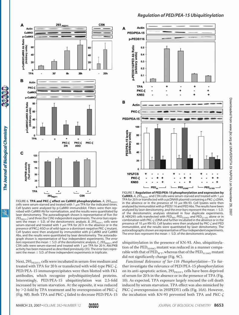
Next, 293PEDY1 cells were incubated in serum-freemedium andtreated with TPA for 20 h or transfected with wild-type PKC-�.PED/PEA-15 immunoprecipitates were then blotted with FK1antibodies, which recognize polyubiquitinylated proteins.Interestingly, PED/PEA-15 ubiquitinylation was 2.5-foldincreased by serum starvation. At the opposite, it was reducedby �2-fold by TPA treatment and by overexpression of PKC-�(Fig. 9B). Both TPA and PKC-� failed to decrease PED/PEA-15
ubiquitinylation in the presence of KN-93. Also, ubiquitinyla-tion of the PEDS104G mutant was reduced in a manner compa-rable with that of PEDWT, whereas that of the PEDS116Gmutantdid not significantly change (Fig. 9C).Functional Relevance of Ser-116 Phosphorylation—To fur-
ther investigate the relevance of PED/PEA-15 phosphorylationon its anti-apoptotic action, 293PEDY1 cells have been deprivedof serum for 20 h in the absence or in the presence of TPA (Fig.10). As expected, TPA exposure largely rescued the cell deathinduced by serum starvation. TPA effect was also mimicked byPKC-� overexpression in 293PEDY1 cells (Fig. 10A). However,the incubation with KN-93 prevented both TPA and PKC-�
FIGURE 6. TPA and PKC-� effect on CaMKII phosphorylation. A, 293PEDY1cells were serum-starved and treated with 1 �M TPA for the indicated times.Cell lysates were analyzed by p-CaMKII immunoblot. Filters were then rep-robed with CaMKII Ab for normalization, and the results were quantitated bylaser densitometry. The autoradiograph shown is representative of five (for293PEDY1) and three (for C5N) independent experiments. The error bars repre-sent the mean � S.D. of the densitometric analysis. B, 293PEDY1 cells wereserum-starved and treated with 1 �M TPA for 20 h in the absence or in thepresence of PKC�-ASO or of wild-type or a dominant-negative PKC-� mutant.Cell lysates were then analyzed by immunoblot with p-CaMKII and CaMKIIAbs, and the results were quantitated by laser densitometry. The autoradio-graph shown is representative of four independent experiments. The errorbars represent the mean � S.D. of the densitometric analysis. C, 293PEDY1 andC5N cells were serum-starved and treated with 1 �M TPA for 20 h. Akt/PKBactivity has been measured as described previously (35). The error bars repre-sent the mean � S.D. of three independent experiments in triplicate.
FIGURE 7. Regulation of PED/PEA-15 phosphorylation and expression byCaMKII. A, 293PEDY1 and C5N cells were serum-starved and treated with 1 �M
TPA for 20 h or transfected with a pcDNAIII plasmid containing a PKC-� cDNA,in the absence or in the presence of 10 �M KN-93. Cell lysates were thenanalyzed by immunoblot with p-PEDS116 and PED Abs. The results have beenanalyzed by laser densitometry, and the error bars represent the mean � S.D.of the densitometric analyses obtained in four duplicate experiments.B, HEK293 cells transfected with PEDWT, PEDS104G, and PEDS116G alone or incombination with PKC-� cDNA and further incubated in the absence or in thepresence of 10 �M KN-93. Cell lysates were then analyzed by PKC-� and PEDimmunoblot, and the results were quantitated by laser densitometry. Theautoradiographs shown are representative of four independent experiments.The error bars represent the mean � S.D. of the densitometric analyses.
Regulation of PED/PEA-15 Ubiquitinylation
MARCH 23, 2007 • VOLUME 282 • NUMBER 12 JOURNAL OF BIOLOGICAL CHEMISTRY 8653
at UN
IVE
RS
ITA
DI N
AP
OLI on N
ovember 29, 2007
ww
w.jbc.org
Dow
nloaded from
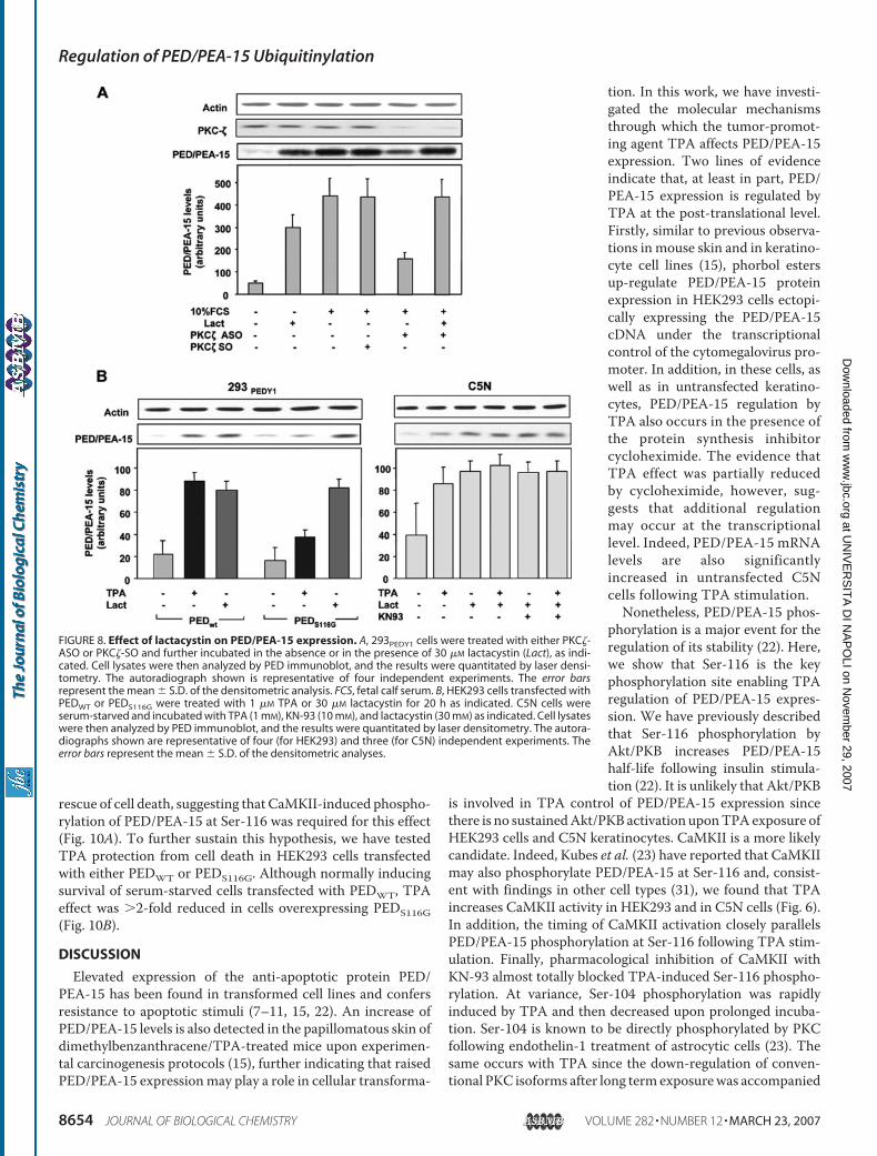
rescue of cell death, suggesting that CaMKII-induced phospho-rylation of PED/PEA-15 at Ser-116 was required for this effect(Fig. 10A). To further sustain this hypothesis, we have testedTPA protection from cell death in HEK293 cells transfectedwith either PEDWT or PEDS116G. Although normally inducingsurvival of serum-starved cells transfected with PEDWT, TPAeffect was �2-fold reduced in cells overexpressing PEDS116G(Fig. 10B).
DISCUSSION
Elevated expression of the anti-apoptotic protein PED/PEA-15 has been found in transformed cell lines and confersresistance to apoptotic stimuli (7–11, 15, 22). An increase ofPED/PEA-15 levels is also detected in the papillomatous skin ofdimethylbenzanthracene/TPA-treated mice upon experimen-tal carcinogenesis protocols (15), further indicating that raisedPED/PEA-15 expressionmay play a role in cellular transforma-
tion. In this work, we have investi-gated the molecular mechanismsthrough which the tumor-promot-ing agent TPA affects PED/PEA-15expression. Two lines of evidenceindicate that, at least in part, PED/PEA-15 expression is regulated byTPA at the post-translational level.Firstly, similar to previous observa-tions in mouse skin and in keratino-cyte cell lines (15), phorbol estersup-regulate PED/PEA-15 proteinexpression in HEK293 cells ectopi-cally expressing the PED/PEA-15cDNA under the transcriptionalcontrol of the cytomegalovirus pro-moter. In addition, in these cells, aswell as in untransfected keratino-cytes, PED/PEA-15 regulation byTPA also occurs in the presence ofthe protein synthesis inhibitorcycloheximide. The evidence thatTPA effect was partially reducedby cycloheximide, however, sug-gests that additional regulationmay occur at the transcriptionallevel. Indeed, PED/PEA-15 mRNAlevels are also significantlyincreased in untransfected C5Ncells following TPA stimulation.Nonetheless, PED/PEA-15 phos-
phorylation is a major event for theregulation of its stability (22). Here,we show that Ser-116 is the keyphosphorylation site enabling TPAregulation of PED/PEA-15 expres-sion. We have previously describedthat Ser-116 phosphorylation byAkt/PKB increases PED/PEA-15half-life following insulin stimula-tion (22). It is unlikely that Akt/PKB
is involved in TPA control of PED/PEA-15 expression sincethere is no sustainedAkt/PKB activation uponTPAexposure ofHEK293 cells and C5N keratinocytes. CaMKII is a more likelycandidate. Indeed, Kubes et al. (23) have reported that CaMKIImay also phosphorylate PED/PEA-15 at Ser-116 and, consist-ent with findings in other cell types (31), we found that TPAincreases CaMKII activity in HEK293 and in C5N cells (Fig. 6).In addition, the timing of CaMKII activation closely parallelsPED/PEA-15 phosphorylation at Ser-116 following TPA stim-ulation. Finally, pharmacological inhibition of CaMKII withKN-93 almost totally blocked TPA-induced Ser-116 phospho-rylation. At variance, Ser-104 phosphorylation was rapidlyinduced by TPA and then decreased upon prolonged incuba-tion. Ser-104 is known to be directly phosphorylated by PKCfollowing endothelin-1 treatment of astrocytic cells (23). Thesame occurs with TPA since the down-regulation of conven-tional PKC isoforms after long termexposurewas accompanied
FIGURE 8. Effect of lactacystin on PED/PEA-15 expression. A, 293PEDY1 cells were treated with either PKC�-ASO or PKC�-SO and further incubated in the absence or in the presence of 30 �M lactacystin (Lact), as indi-cated. Cell lysates were then analyzed by PED immunoblot, and the results were quantitated by laser densi-tometry. The autoradiograph shown is representative of four independent experiments. The error barsrepresent the mean � S.D. of the densitometric analysis. FCS, fetal calf serum. B, HEK293 cells transfected withPEDWT or PEDS116G were treated with 1 �M TPA or 30 �M lactacystin for 20 h as indicated. C5N cells wereserum-starved and incubated with TPA (1 mM), KN-93 (10 mM), and lactacystin (30 mM) as indicated. Cell lysateswere then analyzed by PED immunoblot, and the results were quantitated by laser densitometry. The autora-diographs shown are representative of four (for HEK293) and three (for C5N) independent experiments. Theerror bars represent the mean � S.D. of the densitometric analyses.
Regulation of PED/PEA-15 Ubiquitinylation
8654 JOURNAL OF BIOLOGICAL CHEMISTRY VOLUME 282 • NUMBER 12 • MARCH 23, 2007
at UN
IVE
RS
ITA
DI N
AP
OLI on N
ovember 29, 2007
ww
w.jbc.org
Dow
nloaded from
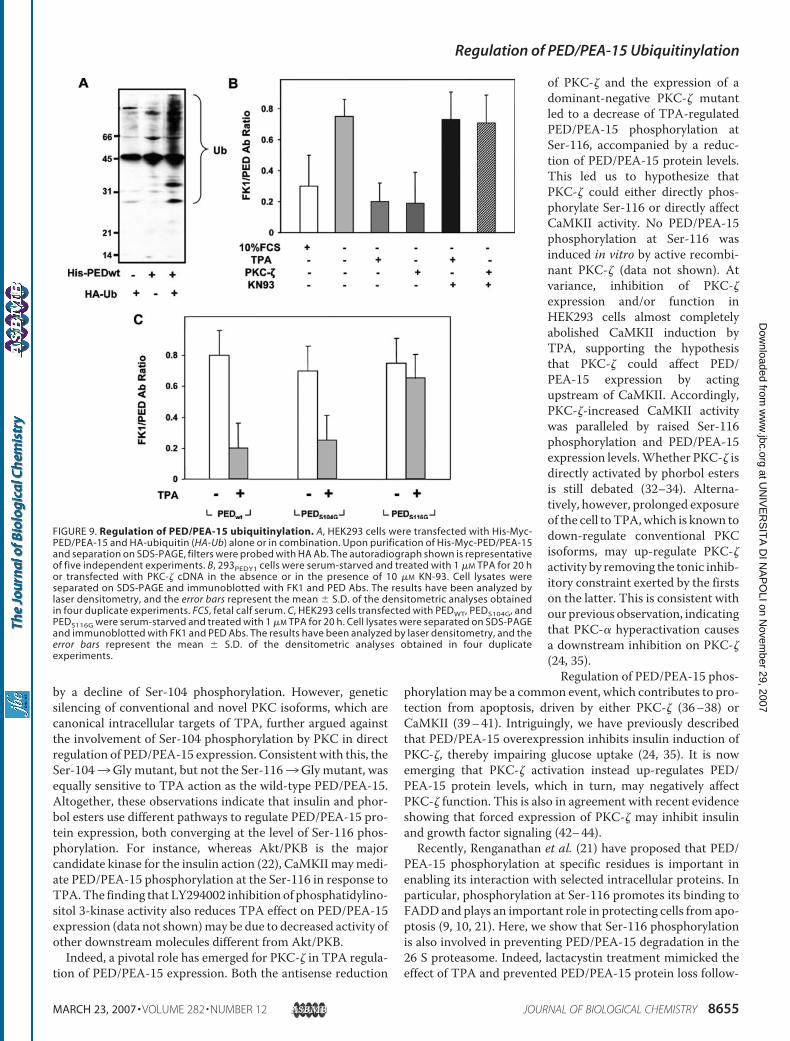
by a decline of Ser-104 phosphorylation. However, geneticsilencing of conventional and novel PKC isoforms, which arecanonical intracellular targets of TPA, further argued againstthe involvement of Ser-104 phosphorylation by PKC in directregulation of PED/PEA-15 expression. Consistent with this, theSer-1043Gly mutant, but not the Ser-1163Gly mutant, wasequally sensitive to TPA action as the wild-type PED/PEA-15.Altogether, these observations indicate that insulin and phor-bol esters use different pathways to regulate PED/PEA-15 pro-tein expression, both converging at the level of Ser-116 phos-phorylation. For instance, whereas Akt/PKB is the majorcandidate kinase for the insulin action (22), CaMKIImaymedi-ate PED/PEA-15 phosphorylation at the Ser-116 in response toTPA.The finding that LY294002 inhibition of phosphatidylino-sitol 3-kinase activity also reduces TPA effect on PED/PEA-15expression (data not shown)may be due to decreased activity ofother downstream molecules different from Akt/PKB.Indeed, a pivotal role has emerged for PKC-� in TPA regula-
tion of PED/PEA-15 expression. Both the antisense reduction
of PKC-� and the expression of adominant-negative PKC-� mutantled to a decrease of TPA-regulatedPED/PEA-15 phosphorylation atSer-116, accompanied by a reduc-tion of PED/PEA-15 protein levels.This led us to hypothesize thatPKC-� could either directly phos-phorylate Ser-116 or directly affectCaMKII activity. No PED/PEA-15phosphorylation at Ser-116 wasinduced in vitro by active recombi-nant PKC-� (data not shown). Atvariance, inhibition of PKC-�expression and/or function inHEK293 cells almost completelyabolished CaMKII induction byTPA, supporting the hypothesisthat PKC-� could affect PED/PEA-15 expression by actingupstream of CaMKII. Accordingly,PKC-�-increased CaMKII activitywas paralleled by raised Ser-116phosphorylation and PED/PEA-15expression levels.Whether PKC-� isdirectly activated by phorbol estersis still debated (32–34). Alterna-tively, however, prolonged exposureof the cell toTPA,which is known todown-regulate conventional PKCisoforms, may up-regulate PKC-�activity by removing the tonic inhib-itory constraint exerted by the firstson the latter. This is consistent withour previous observation, indicatingthat PKC-� hyperactivation causesa downstream inhibition on PKC-�(24, 35).Regulation of PED/PEA-15 phos-
phorylationmay be a common event, which contributes to pro-tection from apoptosis, driven by either PKC-� (36–38) orCaMKII (39–41). Intriguingly, we have previously describedthat PED/PEA-15 overexpression inhibits insulin induction ofPKC-�, thereby impairing glucose uptake (24, 35). It is nowemerging that PKC-� activation instead up-regulates PED/PEA-15 protein levels, which in turn, may negatively affectPKC-� function. This is also in agreement with recent evidenceshowing that forced expression of PKC-� may inhibit insulinand growth factor signaling (42–44).Recently, Renganathan et al. (21) have proposed that PED/
PEA-15 phosphorylation at specific residues is important inenabling its interaction with selected intracellular proteins. Inparticular, phosphorylation at Ser-116 promotes its binding toFADDand plays an important role in protecting cells from apo-ptosis (9, 10, 21). Here, we show that Ser-116 phosphorylationis also involved in preventing PED/PEA-15 degradation in the26 S proteasome. Indeed, lactacystin treatment mimicked theeffect of TPA and prevented PED/PEA-15 protein loss follow-
FIGURE 9. Regulation of PED/PEA-15 ubiquitinylation. A, HEK293 cells were transfected with His-Myc-PED/PEA-15 and HA-ubiquitin (HA-Ub) alone or in combination. Upon purification of His-Myc-PED/PEA-15and separation on SDS-PAGE, filters were probed with HA Ab. The autoradiograph shown is representativeof five independent experiments. B, 293PEDY1 cells were serum-starved and treated with 1 �M TPA for 20 hor transfected with PKC-� cDNA in the absence or in the presence of 10 �M KN-93. Cell lysates wereseparated on SDS-PAGE and immunoblotted with FK1 and PED Abs. The results have been analyzed bylaser densitometry, and the error bars represent the mean � S.D. of the densitometric analyses obtainedin four duplicate experiments. FCS, fetal calf serum. C, HEK293 cells transfected with PEDWT, PEDS104G, andPEDS116G were serum-starved and treated with 1 �M TPA for 20 h. Cell lysates were separated on SDS-PAGEand immunoblotted with FK1 and PED Abs. The results have been analyzed by laser densitometry, and theerror bars represent the mean � S.D. of the densitometric analyses obtained in four duplicateexperiments.
Regulation of PED/PEA-15 Ubiquitinylation
MARCH 23, 2007 • VOLUME 282 • NUMBER 12 JOURNAL OF BIOLOGICAL CHEMISTRY 8655
at UN
IVE
RS
ITA
DI N
AP
OLI on N
ovember 29, 2007
ww
w.jbc.org
Dow
nloaded from
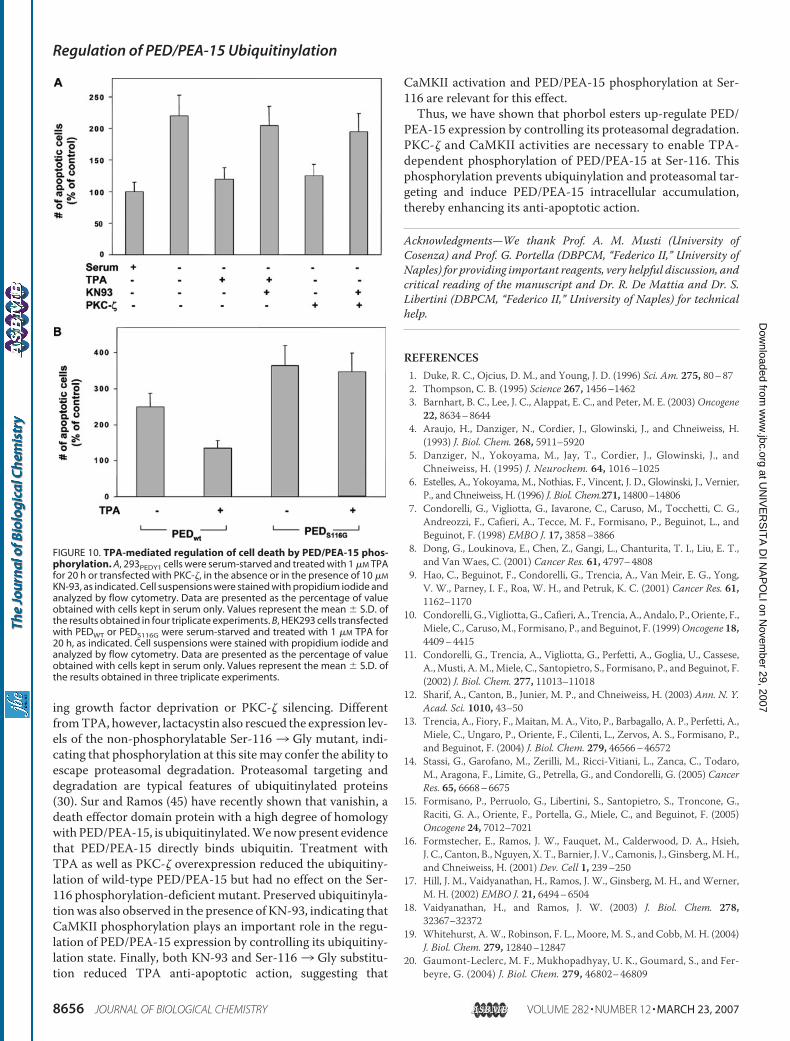
ing growth factor deprivation or PKC-� silencing. DifferentfromTPA, however, lactacystin also rescued the expression lev-els of the non-phosphorylatable Ser-116 3 Gly mutant, indi-cating that phosphorylation at this sitemay confer the ability toescape proteasomal degradation. Proteasomal targeting anddegradation are typical features of ubiquitinylated proteins(30). Sur and Ramos (45) have recently shown that vanishin, adeath effector domain protein with a high degree of homologywithPED/PEA-15, is ubiquitinylated.Wenowpresent evidencethat PED/PEA-15 directly binds ubiquitin. Treatment withTPA as well as PKC-� overexpression reduced the ubiquitiny-lation of wild-type PED/PEA-15 but had no effect on the Ser-116 phosphorylation-deficient mutant. Preserved ubiquitinyla-tionwas also observed in the presence of KN-93, indicating thatCaMKII phosphorylation plays an important role in the regu-lation of PED/PEA-15 expression by controlling its ubiquitiny-lation state. Finally, both KN-93 and Ser-1163 Gly substitu-tion reduced TPA anti-apoptotic action, suggesting that
CaMKII activation and PED/PEA-15 phosphorylation at Ser-116 are relevant for this effect.Thus, we have shown that phorbol esters up-regulate PED/
PEA-15 expression by controlling its proteasomal degradation.PKC-� and CaMKII activities are necessary to enable TPA-dependent phosphorylation of PED/PEA-15 at Ser-116. Thisphosphorylation prevents ubiquinylation and proteasomal tar-geting and induce PED/PEA-15 intracellular accumulation,thereby enhancing its anti-apoptotic action.
Acknowledgments—We thank Prof. A. M. Musti (University ofCosenza) and Prof. G. Portella (DBPCM, “Federico II,” University ofNaples) for providing important reagents, very helpful discussion, andcritical reading of the manuscript and Dr. R. De Mattia and Dr. S.Libertini (DBPCM, “Federico II,” University of Naples) for technicalhelp.
REFERENCES1. Duke, R. C., Ojcius, D. M., and Young, J. D. (1996) Sci. Am. 275, 80–872. Thompson, C. B. (1995) Science 267, 1456–14623. Barnhart, B. C., Lee, J. C., Alappat, E. C., and Peter, M. E. (2003)Oncogene
22, 8634–86444. Araujo, H., Danziger, N., Cordier, J., Glowinski, J., and Chneiweiss, H.
(1993) J. Biol. Chem. 268, 5911–59205. Danziger, N., Yokoyama, M., Jay, T., Cordier, J., Glowinski, J., and
Chneiweiss, H. (1995) J. Neurochem. 64, 1016–10256. Estelles, A., Yokoyama, M., Nothias, F., Vincent, J. D., Glowinski, J., Vernier,
P., and Chneiweiss, H. (1996) J. Biol. Chem.271, 14800–148067. Condorelli, G., Vigliotta, G., Iavarone, C., Caruso, M., Tocchetti, C. G.,
Andreozzi, F., Cafieri, A., Tecce, M. F., Formisano, P., Beguinot, L., andBeguinot, F. (1998) EMBO J. 17, 3858–3866
8. Dong, G., Loukinova, E., Chen, Z., Gangi, L., Chanturita, T. I., Liu, E. T.,and Van Waes, C. (2001) Cancer Res. 61, 4797–4808
9. Hao, C., Beguinot, F., Condorelli, G., Trencia, A., Van Meir, E. G., Yong,V. W., Parney, I. F., Roa, W. H., and Petruk, K. C. (2001) Cancer Res. 61,1162–1170
10. Condorelli, G., Vigliotta,G., Cafieri, A., Trencia, A., Andalo, P.,Oriente, F.,Miele, C., Caruso,M., Formisano, P., and Beguinot, F. (1999)Oncogene 18,4409–4415
11. Condorelli, G., Trencia, A., Vigliotta, G., Perfetti, A., Goglia, U., Cassese,A.,Musti, A.M.,Miele, C., Santopietro, S., Formisano, P., and Beguinot, F.(2002) J. Biol. Chem. 277, 11013–11018
12. Sharif, A., Canton, B., Junier, M. P., and Chneiweiss, H. (2003) Ann. N. Y.Acad. Sci. 1010, 43–50
13. Trencia, A., Fiory, F., Maitan, M. A., Vito, P., Barbagallo, A. P., Perfetti, A.,Miele, C., Ungaro, P., Oriente, F., Cilenti, L., Zervos, A. S., Formisano, P.,and Beguinot, F. (2004) J. Biol. Chem. 279, 46566–46572
14. Stassi, G., Garofano, M., Zerilli, M., Ricci-Vitiani, L., Zanca, C., Todaro,M., Aragona, F., Limite, G., Petrella, G., and Condorelli, G. (2005) CancerRes. 65, 6668–6675
15. Formisano, P., Perruolo, G., Libertini, S., Santopietro, S., Troncone, G.,Raciti, G. A., Oriente, F., Portella, G., Miele, C., and Beguinot, F. (2005)Oncogene 24, 7012–7021
16. Formstecher, E., Ramos, J. W., Fauquet, M., Calderwood, D. A., Hsieh,J. C., Canton, B., Nguyen, X. T., Barnier, J. V., Camonis, J., Ginsberg,M.H.,and Chneiweiss, H. (2001) Dev. Cell 1, 239–250
17. Hill, J. M., Vaidyanathan, H., Ramos, J. W., Ginsberg, M. H., andWerner,M. H. (2002) EMBO J. 21, 6494–6504
18. Vaidyanathan, H., and Ramos, J. W. (2003) J. Biol. Chem. 278,32367–32372
19. Whitehurst, A.W., Robinson, F. L., Moore, M. S., and Cobb, M. H. (2004)J. Biol. Chem. 279, 12840–12847
20. Gaumont-Leclerc, M. F., Mukhopadhyay, U. K., Goumard, S., and Fer-beyre, G. (2004) J. Biol. Chem. 279, 46802–46809
FIGURE 10. TPA-mediated regulation of cell death by PED/PEA-15 phos-phorylation. A, 293PEDY1 cells were serum-starved and treated with 1 �M TPAfor 20 h or transfected with PKC-�, in the absence or in the presence of 10 �M
KN-93, as indicated. Cell suspensions were stained with propidium iodide andanalyzed by flow cytometry. Data are presented as the percentage of valueobtained with cells kept in serum only. Values represent the mean � S.D. ofthe results obtained in four triplicate experiments. B, HEK293 cells transfectedwith PEDWT or PEDS116G were serum-starved and treated with 1 �M TPA for20 h, as indicated. Cell suspensions were stained with propidium iodide andanalyzed by flow cytometry. Data are presented as the percentage of valueobtained with cells kept in serum only. Values represent the mean � S.D. ofthe results obtained in three triplicate experiments.
Regulation of PED/PEA-15 Ubiquitinylation
8656 JOURNAL OF BIOLOGICAL CHEMISTRY VOLUME 282 • NUMBER 12 • MARCH 23, 2007
at UN
IVE
RS
ITA
DI N
AP
OLI on N
ovember 29, 2007
ww
w.jbc.org
Dow
nloaded from
21. Renganathan, H., Vaidyanathan, H., Knapinska, A., and Ramos, J. W.(2005) Biochem. J. 390, 729–735
22. Trencia, A., Perfetti, A., Cassese, A., Vigliotta, G., Miele, C., Oriente, F.,Santopietro, S., Giacco, F., Condorelli, G., Formisano, P., and Beguinot, F.(2003)Mol. Cell Biol. 23, 4511–4521
23. Kubes, M., Cordier, J., Glowinski, J., Girault, J. A., and Chneiweiss, H.(1998) J. Neurochem. 71, 1307–1314
24. Vigliotta, G., Miele, C., Santopietro, S., Portella, G., Perfetti, A., Maitan,M. A., Cassese, A., Oriente, F., Trencia, A., Fiory, F., Romano, C., Tiveron,C., Tatangelo, L., Troncone, G., Formisano, P., and Beguinot, F. (2004)Mol. Cell Biol. 24, 5005–5015
25. Sharif, A., Renault, F., Beuvon, F., Castellanos, R., Canton, B., Barbeito, L.,Junier, M. P., and Chneiweiss, H. (2004) Neuroscience 126, 263–275
26. Formisano, P., Oriente, F., Fiory, F., Caruso, M., Miele, C., Maitan, M. A.,Andreozzi, F., Vigliotta, G., Condorelli, G., and Beguinot, F. (2000) Mol.Cell Biol. 20, 6323–6333
27. Caruso, M., Maitan, M. A., Bifulco, G., Miele, C., Vigliotta, G., Oriente, F.,Formisano, P., and Beguinot, F. (2001) J. Biol. Chem. 276, 45088–45097
28. Oriente, F., Formisano, P., Miele, C., Fiory, F., Maitan, M. A., Vigliotta, G.,Trencia, A., Santopietro, S., Caruso,M., VanObberghen, E., and Beguinot,F. (2001) J. Biol. Chem. 276, 37109–37119
29. Musti, A. M., Treier, M., and Bohmann, D. (1997) Science 275, 400–40230. Ciechanover, A. (2005)Mol. Cell Biol. 6, 79–8731. Hughes, K., Edin, S., Antonsson, A., and Grundstrom, T. (2001) J. Biol.
Chem. 276, 36008–3601332. Ways, D. K., Cook, P. P.,Webster, C., and Parker, P. J. (1992) J. Biol. Chem.
267, 4799–4805
33. Kim, S. J., Chang, Y. Y., Kang, S. S., andChun, J. S. (1997)Biochem. Biophys.Res. Commun. 237, 336–339
34. Hirai, T., and Chida, K. (2003) J Biochem. (Tokyo) 133, 1–7; CorrectionJ. Biochem. (Tokyo) 133, 395
35. Condorelli, G., Vigliotta, G., Trencia, A.,Maitan,M.A., Caruso,M.,Miele,C., Oriente, F., Santopietro, S., Formisano, P., and Beguinot, F. (2001)Diabetes 50, 1244–1252
36. Spitaler, M.,Wiesenhofer, B., Biedermann, V., Seppi, T., Zimmermann, J.,Grunicke, H., and Hofmann, J. (1999) Anticancer Res. 19, 3969–3976
37. de Thonel, A., Bettaieb, A., Jean, C., Laurent, G., and Quillet-Mary, A.(2001) Blood 98, 3770–3777
38. Leroy, I., de Thonel, A., Laurent, G., and Quillet-Mary, A. (2005) CellSignal. 17, 1149–1157
39. Tombes, R.M., Grant, S.,Westin, E. H., andKrystal, G. (1995)Cell GrowthDiffer. 6, 1063–1070
40. Yang, B. F., Xiao, C., Roa,W.H., Krammer, P. H., andHao, C. (2003) J. Biol.Chem. 278, 7043–7050
41. Xiao, C., Yang, B. F., Song, J. H., Schulman, H., Li, L., and Hao, C. (2005)Exp. Cell Res. 304, 244–255
42. Liu, Y. F., Paz, K., Herschkovitz, A., Alt, A., Tennenbaum, T., Sampson,S. R., Ohba, M., Kuroki, T., LeRoith, D., and Zick, Y. (2001) J. Biol. Chem.276, 14459–14465
43. Liu, Y. F., Herschkovitz, A., Boura-Halfon, S., Ronen, D., Paz, K., Leroith,D., and Zick, Y. (2004)Mol. Cell Biol. 24, 9668–9681
44. Zick, Y. (2005) Sci. STKE. 2005 25, 26845. Sur, R., and Ramos, J. W. (2005) Biochem. J. 387, 315–324
Regulation of PED/PEA-15 Ubiquitinylation
MARCH 23, 2007 • VOLUME 282 • NUMBER 12 JOURNAL OF BIOLOGICAL CHEMISTRY 8657
at UN
IVE
RS
ITA
DI N
AP
OLI on N
ovember 29, 2007
ww
w.jbc.org
Dow
nloaded from