Embed Size (px)
Citation preview
Allosteric Inhibition of Human Immunodeficiency VirusIntegraseLATE BLOCK DURING VIRAL REPLICATION AND ABNORMAL MULTIMERIZATIONINVOLVING SPECIFIC PROTEIN DOMAINS*□S
Received for publication, January 28, 2014, and in revised form, June 3, 2014 Published, JBC Papers in Press, June 5, 2014, DOI 10.1074/jbc.M114.551119
Kushol Gupta‡, Troy Brady§, Benjamin M. Dyer§, Nirav Malani§, Young Hwang§, Frances Male§, Robert T. Nolte¶,Liping Wang¶, Emile Velthuisen�, Jerry Jeffrey�1, Gregory D. Van Duyne‡2, and Frederic D. Bushman§3
From the §Department of Microbiology, Perelman School of Medicine, University of Pennsylvania, Philadelphia, Pennsylvania19104-6076, the ‡Department of Biochemistry and Biophysics, University of Pennsylvania, Philadelphia, Pennsylvania 19104-6059,and ¶Chemical Sciences and the �HIV Discovery Performance Unit, Infectious Disease Therapy Area Unit, GlaxoSmithKline,Research Triangle Park, North Carolina 27709
Background: New antiviral agents bind to a site on HIV integrase protein also bound by the cellular protein LEDGF/p75.Results: Compound GSK1264 binds to this site, but it has surprising properties; it inhibits late during HIV replication, not earlyduring integration, and it promotes abnormal multimerization.Conclusion: GSK1264 provides new insight into HIV replication.Significance: These observations inform the design of improved antiviral agents.
HIV-1 replication in the presence of antiviral agents results inevolution of drug-resistant variants, motivating the search foradditional drug classes. Here we report studies of GSK1264,which was identified as a compound that disrupts the interac-tion between HIV-1 integrase (IN) and the cellular factor lensepithelium-derived growth factor (LEDGF)/p75. GSK1264 dis-played potent antiviral activity and was found to bind at the siteoccupied by LEDGF/p75 on IN by x-ray crystallography. Assaysof HIV replication in the presence of GSK1264 showed onlymodest inhibition of the early infection steps and little effect onintegration targeting, which is guided by the LEDGF/p75�INinteraction. In contrast, inhibition of late replication steps wasmore potent. Particle production was normal, but particlesshowed reduced infectivity. GSK1264 promoted aggregation ofIN and preformed LEDGF/p75�IN complexes, suggesting amechanism of inhibition. LEDGF/p75 was not displaced fromIN during aggregation, indicating trapping of LEDGF/p75 inaggregates. Aggregation assays with truncated IN variantsrevealed that a construct with catalytic and C-terminal domainsof IN only formed an open polymer associated with efficientdrug-induced aggregation. These data suggest that the allosteric
inhibitors of IN are promising antiviral agents and provide newinformation on their mechanism of action.
Early steps of HIV-1 replication involve reverse transcrip-tion, which produces a double-stranded cDNA copy of the viralRNA genome, and integration, which results in the incorpora-tion of the viral DNA into host chromosomal DNA (1). Both ofthese steps have been targeted by clinically useful inhibitors (2,3). The integrase strand transfer inhibitors bind to the activesite of the virus-encoded integrase (IN)4 enzyme and block theinitial strand transfer step that incorporates viral DNA into thehost chromosome. In this report, we describe the properties ofa small molecule, GSK1264, that functions as an allostericinhibitor of IN and inhibits late during the viral replicationcycle.
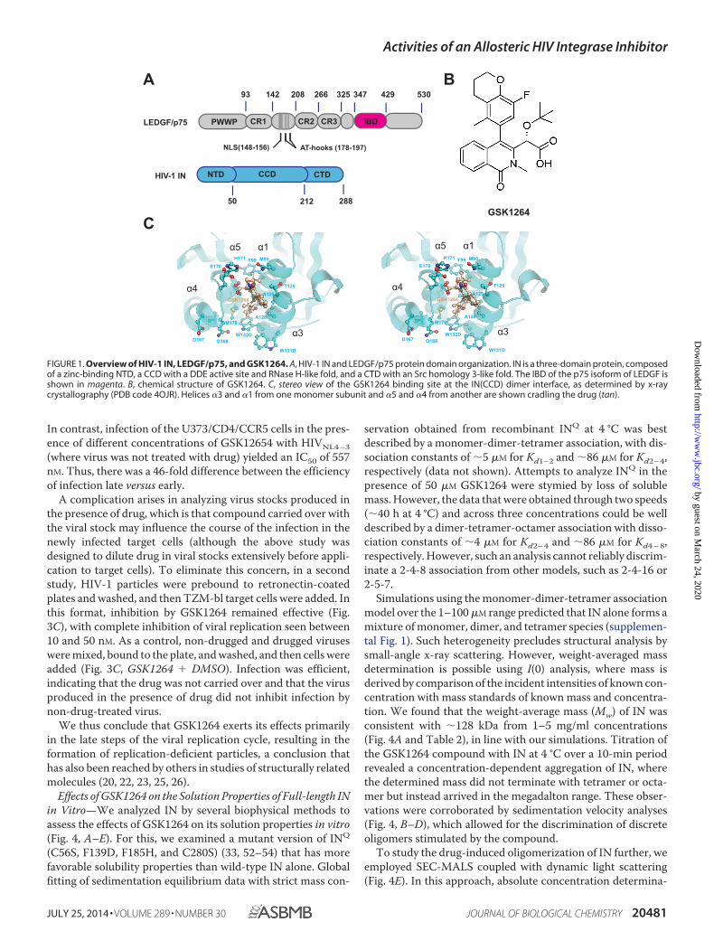
In infected cells, HIV-1 IN binds the cellular host factor,LEDGF/p75, which is a transcriptional co-activator that is theproduct of the PSIP1 gene (Fig. 1A). Following reverse tran-scription, IN binds the double-stranded viral DNA ends to formthe intasome. LEDGF/p75 primarily binds to the catalytic coredomain (CCD) and, to a lesser extent, the N-terminal domain(NTD) of integrase (4) via the LEDGF/p75 C-terminal integrasebinding domain (IBD) (5). This interaction promotes efficientintegration and the targeting of the intasome to active tran-scription units (6 – 8). LEDGF/p75 acts as a simple tether, asshown by experiments in which the LEDGF/p75 chromatinbinding domain is substituted with new chromatin bindingdomains, resulting in retargeting of HIV-1 integration (9 –11).Additionally, the LEDGF/p75�IN interaction protects IN from
* This work was supported, in whole or in part, by National Institutes of HealthGrant R01 AI 052845 (to F. D. B.). GlaxoSmithKline owns the patent on thecompound used in this study.
□S This article contains supplemental Table S1 and Figs. 1 and 2.The atomic coordinates and structure factors (code 4OJR) have been deposited in
the Protein Data Bank (http://wwpdb.org/).1 To whom correspondence may be addressed: HIV DPU, Infectious Disease
Therapy Area Unit, GlaxoSmithKline, Research Triangle Park, NC 27709.E-mail: [email protected].
2 To whom correspondence may be addressed: Dept. of Biochemistry andBiophysics, Perelman School of Medicine, University of Pennsylvania, 810Stellar-Chance Bldg., 422 Curie Blvd., Philadelphia, PA 19104-6059. Tel.:215-573-7260; Fax: 215-573-4764; E-mail: [email protected].
3 To whom correspondence may be addressed: Dept. of Microbiology, Perel-man School of Medicine, University of Pennsylvania, 426A Johnson Pavil-ion, 3610 Hamilton Walk, Philadelphia, PA 19104. Tel.: 215-573-8732; Fax:215-573-4856; E-mail: [email protected].
4 The abbreviations used are: IN, integrase; INQ, quadramutated IN (C56S/F139D/F185H/C280S); NTD, N-terminal domain; CCD, catalytic coredomain; CTD, C-terminal domain; LEDGF, lens epithelium-derived growthfactor; SEC-MALS, size-exclusion chromatography in-line with multianglelight scattering; SAXS, small-angle X-ray scattering; IBD, integrase bindingdomain.
THE JOURNAL OF BIOLOGICAL CHEMISTRY VOL. 289, NO. 30, pp. 20477–20488, July 25, 2014© 2014 by The American Society for Biochemistry and Molecular Biology, Inc. Published in the U.S.A.
JULY 25, 2014 • VOLUME 289 • NUMBER 30 JOURNAL OF BIOLOGICAL CHEMISTRY 20477
by guest on March 24, 2020
http://ww
w.jbc.org/
Dow
nloaded from
proteolysis and stimulates catalysis both in vitro and in vivo(4, 12–16).
The structure of the LEDGF/p75 IBD�IN(CCD) complex wasdetermined by Cherepanov and colleagues (17, 18). The IBDbinds at the interface of the IN(CCD) dimer, occupying apocket at the interface. Earlier work has demonstrated that thissite can be bound by small molecules (19), and subsequentscreening efforts have yielded several small molecule classesthat interfere with binding of the LEDGF/p75 IBD and displayantiviral activity (reviewed in Refs. 20 and 21).
Here we present a detailed study of one such molecule,GSK1264 (Fig. 1B). Co-crystallization with the IN(CCD)showed that GSK1264 indeed bound the LEDGF/p75 bindingsite. GSK1264 inhibited most potently in the late part of theviral replication cycle, after integration. As this work was beingcompleted, several other groups made similar findings withstructurally related inhibitors (20, 22–26). Potency of GSK1264early during infection was only modest, and there was littleeffect of GSK1264 on integration target site selection. In thepresence of GSK1264, viral particles were produced normallylate during infection, but they were reduced in infectivity.Potent inhibition correlated with increased oligomerization ofIN in the presence of drug in vitro. Drug-induced oligomeriza-tion of preformed LEDGF�IN complexes was not associatedwith detectable displacement of LEDGF/p75. Study of trun-cated derivatives of IN in vitro demonstrated that drug-inducedpolymerization was most potent in variants containing the CCDand CTD only. Thus, compounds that bind the LEDGF/p75 site onIN are effective inhibitors whose primary effects occur at the lateststeps of replication, and inhibition correlates with abnormal INpolymerization involving specific protein domains.
EXPERIMENTAL PROCEDURES
Cell Lines—The TZM-bl, 293T, and U373/CD4/CCR5 (27)cell lines were obtained through the National Institutes ofHealth AIDS Research and Reference Reagent Program(ARRRP) and grown as directed (28). A1953 chronic HIV pro-ducer cells were a gift from James Hoxie.
HIV-1 Infection and Integration Target Site Analysis—Infec-tions were carried out in TZM-bl cells using standard methodsand the HIV-1 strain HIV89.6 (29). Analysis of HIV-1 integra-tion targeting was carried out as described previously (6,30 –32). All sites common among samples (including thereporter construct in the TZM-bl cells) were removed prior toanalysis.
For the study of LEDGF/p75 knockdown cells, an shRNAconstruct (Sigma-Aldrich, TRCN0000074819) was transducedinto a 293T-derived cell line, and cells were subjected to puro-mycin selection (1 �g/ml), yielding KD19 cells. In parallel, amatched construct encoding a GFP-targeting shRNA wasintroduced into the 293T cell line and compared. Knockdownwas confirmed to reduce LEDGF/p75 mRNA levels by 92%, andprotein was undetectable by Western blot analysis.
Protein Purification—The CCD of HIV-1 INF185K used forTR-FRET binding experiments and x-ray crystallography wasexpressed and purified as described in the supplemental Meth-ods. Recombinant proteins were expressed and purified asdescribed previously (7, 33). Complexes between LEDGF(326 –
530) or LEDGF(IBD) (residues 347– 471) and quadramutatedIN (C56S/F139D/F185H/C280S, referred to as “INQ”) or wild-type HIV-1 IN were obtained by co-expression from pETDuet(Novagen Inc., Madison, WI) in BL21 (DE3) cells (Novagen) at37 °C. LEDGF constructs were inserted into the vector in-framewith a C-terminal Mxe intein (New England Biolabs, Ipswich, MA)containing chitin-binding domain and hexahistidine affinity tags.The domain truncations INF185H(NTD-CCD), INF185H(CCD-CTD), and INF185H(CCD) were similarly purified.
Proteins were purified using nickel-nitrilotriacetic acid (Qia-gen, Valencia, CA) and chitin (New England Biolabs) resins.Fusion proteins were released by intein cleavage in 50 mM DTTovernight at 4 °C. Preparations of full-length INQ alone andLEDGF(326 –530) were further purified using SP-Sepharosechromatography (GE Healthcare). Proteins were concentratedat 4 °C in YM-10 Centricons (Millipore, Billerica, MA), andaliquots were flash-frozen in liquid nitrogen with 20% glycerolfor storage at �80 °C. All preparations used for this study werestored in 20 mM HEPES-NaOH, pH 7.5, 450 mM NaCl, 0.1 mM
EDTA, 10 �M ZnOAc2, 5 mM CHAPS, 10 mM DTT, and 20%glycerol. All biophysical analyses were performed in 0.1-�mfiltered buffer composed of 20 mM HEPES-NaOH, pH 7.5, 450mM NaCl, 0.1 mM EDTA, 10 �M ZnOAc2, 1–10 mM DTT, withor without 5 mM CHAPS.
The detergent was confirmed to be at submicellar concentra-tions at this ionic strength (450 mM NaCl) using both a colori-metric assay and small-angle x-ray scattering (SAXS) analysis(34, 35) (data not shown). It has been reported that detergentssuch as CHAPS can attenuate IN oligomerization (36). Thus,this model system provides a hypo-oligomeric backgroundagainst which drug-induced multimerization is measured.
IN-LEDGF FRET Binding Assay (48)—GSK1264 was dis-solved in DMSO to 10 mM and serially diluted in DMSO forassays. Reactions were performed at a 10-�l final volume in a384-well plate format (Greiner Bio-One, San Diego, CA). Thereaction buffer contained 50 mM HEPES, pH 7.5, 150 mM NaCl,20 mM MgCl2, 0.1 mg/ml bovine serum albumin (BSA), 50 �M
CHAPS, and fresh 2 mM DTT. After the addition of drug in a0.1-�l volume, hexahistidine-tagged INF185K(CCD) was addedto a final concentration of 5 nM using a Multidrop Combireagent dispenser (Thermo Fisher Scientific). After 30 minof incubation at room temperature, another addition wasmade with the dispenser to deliver GST-tagged LEDGF(IBD)(residues 347– 429) to a final concentration of 5 nM alongsidethe time-resolved FRET reagents allophycocyanin-conju-gated �-hexahistidine monoclonal antibody and �-GST europi-um-labeled monoclonal antibody (PerkinElmer Life Sci-ences) at 5 nM final concentrations. After an additional 1 h ofincubation at room temperature, the time-resolved FRETsignal at 665 nm was recorded with a ViewLux microplateimager (PerkinElmer Life Sciences).
Size-exclusion Chromatography and Multiangle Light Scat-tering (SEC-MALS)—Absolute molecular weights were deter-mined by multiangle light scattering coupled with refractiveinterferometric detection (Wyatt Technology Corp., Santa Bar-bara, CA) and a Superdex 200 10/300 GL column (GE Health-care) at room temperature, as described previously (33).
Activities of an Allosteric HIV Integrase Inhibitor
20478 JOURNAL OF BIOLOGICAL CHEMISTRY VOLUME 289 • NUMBER 30 • JULY 25, 2014
by guest on March 24, 2020
http://ww
w.jbc.org/
Dow
nloaded from
Sedimentation Equilibrium Analysis—Sedimentation equi-librium analytical ultracentrifugation experiments were per-formed at 4 °C with an XL-A analytical ultracentrifuge (Beck-man-Coulter, Brea, CA) and a TiAn60 rotor with two-channelcharcoal-filled Epon centerpieces and quartz windows. Datawere collected at 4 °C with detection at 280 nm for 5, 7.5, and10 �M samples. Analysis was carried out using global fits toall concentrations with data acquired at 18,000, 22,000, and24,000 rpm, with strict mass conservation using the programSEDPHAT (70).
Sedimentation Velocity Analysis—Sedimentation velocity ultra-centrifugation experiments were performed at 4 °C with anXL-A analytical ultracentrifuge (Beckman-Coulter, Brea, CA)and a TiAn60 rotor with two-channel charcoal-filled Epon cen-terpieces and quartz windows. Samples were analyzed at anA280 of 0.5–1.2. Complete sedimentation velocity profiles wererecorded every 30 s for 50 –200 boundaries at 45,000 rpm. Datawere fit using the c(S) distribution model of the Lamm equationas implemented in the program SEDFIT (37). After optimizingmeniscus position and fitting limits, the sedimentation coeffi-cient (S) and best fit frictional ratio (f/f0) was determined byiterative least squares analysis, and final values were correctedto 20 °C in water (s20,w). The partial specific volume (v�) of theprotein studied, including molar extinction coefficients, sol-vent density (� � 1.01 g/ml), and viscosity (� � 0.01002 poise)were derived from chemical composition by the programSEDNTERP (38).
SAXS—Data were recorded on a Rigaku S-Max3000 small-angle x-ray scattering system equipped with Osmic mirroroptics (Osmic Inc., Troy, MI), a three-pinhole enclosed pre-flight path, an evacuated sample chamber with customizedsample holder cryostated at 4 °C, and a gas-filled multiwiredetector; the instrument is served by a Rigaku MicroMax-007HF microfocus rotating anode generator (Rigaku America,Woodland, TX). Protein samples were spun at 45,000 RPM for5 min at 4 °C in a tabletop centrifuge prior to the addition ofDMSO or GSK1264 and immediate 10-min x-ray exposures.The forward scattering from the samples studied was recordedon a CCD detector and circularly averaged to yield one-dimen-sional intensity profiles as a function of q (q � 4�sin�/�, where2� is the scattering angle, in units of Å�1). Data were reducedusing SAXSGui version 2.05.02 (JJ X-Ray Systems ApS, Lyngby,Denmark), and matching buffers were subtracted to yield thefinal scattering profile. The sample-to-detector distance andbeam center were calibrated using silver behenate and intensityconverted to absolute units (cm�1) using a known polymerstandard.
SAXS Data Analysis—All of the preparations analyzed weremonodisperse, as evidenced by linearity in the Guinier region ofthe scattering data and agreement of the I(0) and Rg valuesdetermined with inverse Fourier transform analysis by the pro-grams GNOM (39). Guinier analyses were performed whereqRg 1.4. Molecular mass was derived from I(0) measure-ments, using the forward scatter from the following series ofprotein standards of known mass at a 5 mg/ml concentrationfor a standard curve: cytochrome c (12.2 kDa), RNase A (13.7kDa), myoglobin (17.7 kDa), soybean trypsin inhibitor (20.1kDa), chymotrypsin (25 kDa), horseradish peroxidase (44 kDa),
catalase (dimer of 125 kDa), -globulin (151 kDa), and thyro-globulin (a dimer of 670 kDa).
When fitting manually, the maximum diameter of the parti-cle (Dmax) was adjusted in 5–10-Å increments in GNOM tomaximize the goodness of fit parameter, to minimize the dis-crepancy between the fit and the experimental data, and tooptimize the visual qualities of the distribution profile.
Turbidity Assays—Assays were performed using a Tecan96-well plate reader (Tecan Group Ltd., Männedorf, Switzer-land), which monitors absorbance at 405 nm in 1-min intervalsover 10 – 60 min at 27 °C. Reactions were initiated by addingprotein solutions to a final concentration of 9 –30 �M to a buffercontaining 20 mM HEPES, pH 7.5, 450 mM NaCl, 0.1 mM EDTA,10 �M ZnOAc2, 5 mM CHAPS, 10 mM DTT, and inhibitor at5–100 �M final concentrations or DMSO control. Experimentsperformed on preformed LEDGF�IN complexes were per-formed in the absence of CHAPS.
Structure Determination—The CCD of HIV-1 INF185K wasdiluted to 10 mg/ml in 10 mM HEPES, pH 7.0, 500 mM NaCl,and 3 mM DTT. Apocrystals were grown by hanging drop vapordiffusion at 4 °C. 2-�l drops were set by combining 1 �l of pro-tein and 1 �l of 7% PEG 8000, 0.2 M ammonium sulfate, 0.1 M
sodium cacodylate, pH 6.5, 5 mM manganese chloride, 5 mM
magnesium chloride, and 5 mM DTT. The apocrystals wereharvested and soaked with 2.5 mM GSK1264 for 3 days at 4 °C.The soaked crystals were transferred to 30% ethylene glycol inmother liquor and subsequently flash-frozen in liquid nitrogen.
X-ray diffraction data were collected at the Advanced PhotonSource at Argonne National Laboratories on beam line 21-ID-G.Data collection statistics are summarized in Table 1. The structurewas determined by molecular replacement using the programPhaser (40). AutoBuster (41) was used for the initial rounds ofrefinement and map calculations. The structure was rebuilt usingthe program Coot (42). Final refinements were done using theprogram Refmac (43).
RESULTS
GSK1264 Inhibits Binding of LEDGF(IBD) to INF185K(CCD)—GSK1264 (Fig. 1B) was identified as a disruptor of the LEDGF/p75IBD interactions with IN(CCD) in vitro.5 GSK1264 inhibited IN3�-end processing in vitro (data not shown), comparable withother compounds that interfere with LEDGF/IN interactions invitro (23, 44–46).
GSK1264 was tested in an INF185K(CCD) versus LEDGF(IBD)time-resolved FRET binding assay (47). The pIC50 was deter-mined to be 8.2 � 0.14 nM (n � 7, where pIC50 is the negativelog of the IC50 in moles/liter; IC50 is the concentration requiredto achieve 50% inhibition). In multicycle viral replication assays(MT4 cell assay), GSK1264 inhibited HIV-1 replication with anIC50 of �38 nM.
X-ray Crystallography Reveals That GSK1264 Binds to theIN(CCD) Dimer Interface—Crystals of INF185K(CCD) weresoaked with GSK1264, and the structure was determined by
5 Patent filing: M. DeLa Rosa, S. N. Haydar, B. A. Johns, and E. J. Velthuisen(January 23, 2012) Isoquinline Compounds and Methods for Treating HIV.WO 2012/102985. The compound referred to here as GSK1264 is com-pound 159 in the filing.
Activities of an Allosteric HIV Integrase Inhibitor
JULY 25, 2014 • VOLUME 289 • NUMBER 30 JOURNAL OF BIOLOGICAL CHEMISTRY 20479
by guest on March 24, 2020
http://ww
w.jbc.org/
Dow
nloaded from
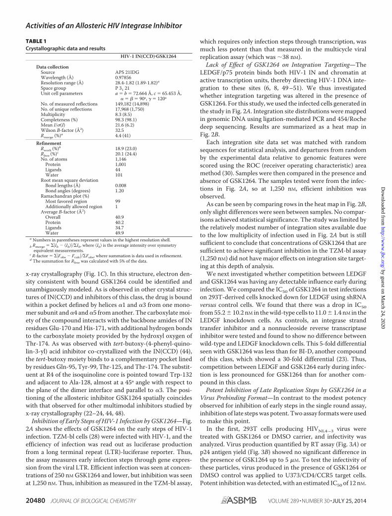
x-ray crystallography (Fig. 1C). In this structure, electron den-sity consistent with bound GSK1264 could be identified andunambiguously modeled. As is observed in other crystal struc-tures of IN(CCD) and inhibitors of this class, the drug is boundwithin a pocket defined by helices �1 and �3 from one mono-mer subunit and �4 and �5 from another. The carboxylate moi-ety of the compound interacts with the backbone amides of INresidues Glu-170 and His-171, with additional hydrogen bondsto the carboxylate moiety provided by the hydroxyl oxygen ofThr-174. As was observed with tert-butoxy-(4-phenyl-quino-lin-3-yl) acid inhibitor co-crystallized with the IN(CCD) (44),the tert-butoxy moiety binds to a complementary pocket linedby residues Gln-95, Tyr-99, Thr-125, and Thr-174. The substit-uent at R4 of the isoquinoline core is pointed toward Trp-132and adjacent to Ala-128, almost at a 45º angle with respect tothe plane of the dimer interface and parallel to �3. The posi-tioning of the allosteric inhibitor GSK1264 spatially coincideswith that observed for other multimodal inhibitors studied byx-ray crystallography (22–24, 44, 48).
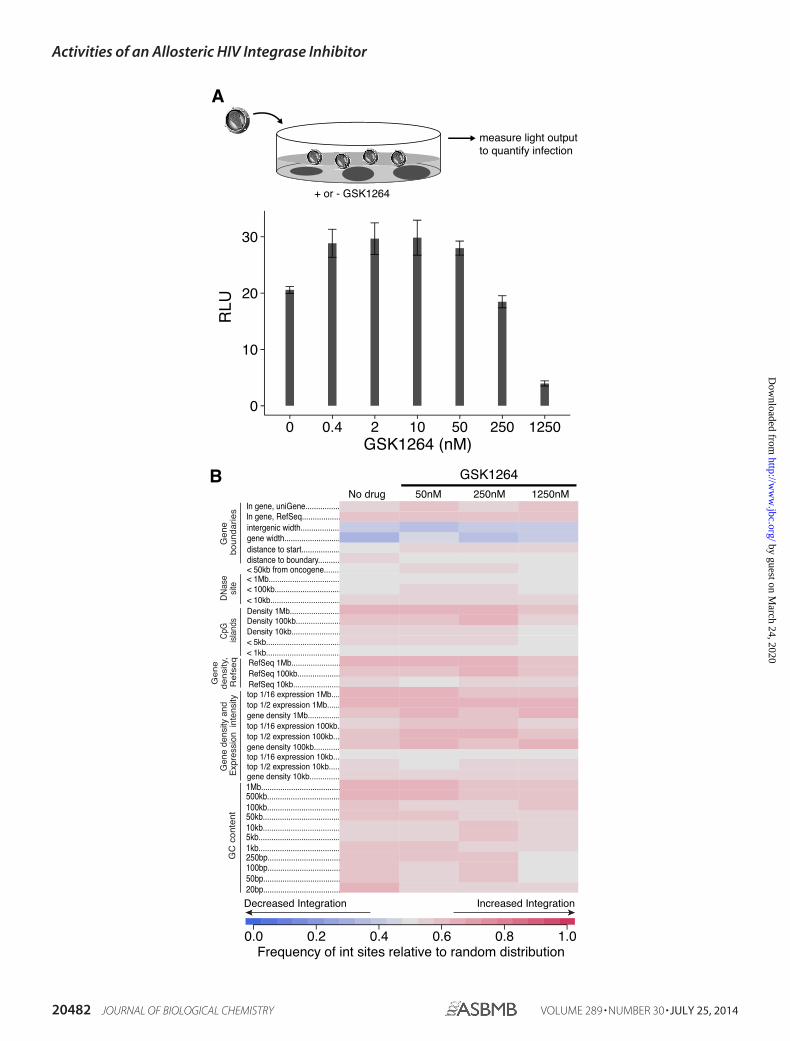
Inhibition of Early Steps of HIV-1 Infection by GSK1264—Fig.2A shows the effects of GSK1264 on the early steps of HIV-1infection. TZM-bl cells (28) were infected with HIV-1, and theefficiency of infection was read out as luciferase productionfrom a long terminal repeat (LTR)-luciferase reporter. Thus,the assay measures early infection steps through gene expres-sion from the viral LTR. Efficient infection was seen at concen-trations of 250 nM GSK1264 and lower, but inhibition was seenat 1,250 nM. Thus, inhibition as measured in the TZM-bl assay,
which requires only infection steps through transcription, wasmuch less potent than that measured in the multicycle viralreplication assay (which was �38 nM).
Lack of Effect of GSK1264 on Integration Targeting—TheLEDGF/p75 protein binds both HIV-1 IN and chromatin atactive transcription units, thereby directing HIV-1 DNA inte-gration to these sites (6, 8, 49 –51). We thus investigatedwhether integration targeting was altered in the presence ofGSK1264. For this study, we used the infected cells generated inthe study in Fig. 2A. Integration site distributions were mappedin genomic DNA using ligation-mediated PCR and 454/Rochedeep sequencing. Results are summarized as a heat map inFig. 2B.
Each integration site data set was matched with randomsequences for statistical analysis, and departures from randomby the experimental data relative to genomic features werescored using the ROC (receiver operating characteristic) areamethod (30). Samples were then compared in the presence andabsence of GSK1264. The samples tested were from the infec-tions in Fig. 2A, so at 1,250 nM, efficient inhibition wasobserved.
As can be seen by comparing rows in the heat map in Fig. 2B,only slight differences were seen between samples. No compar-isons achieved statistical significance. The study was limited bythe relatively modest number of integration sites available dueto the low multiplicity of infection used in Fig. 2A but is stillsufficient to conclude that concentrations of GSK1264 that aresufficient to achieve significant inhibition in the TZM-bl assay(1,250 nM) did not have major effects on integration site target-ing at this depth of analysis.
We next investigated whether competition between LEDGFand GSK1264 was having any detectable influence early duringinfection. We compared the IC50 of GSK1264 in test infectionson 293T-derived cells knocked down for LEDGF using shRNAversus control cells. We found that there was a drop in IC50from 55.2 � 10.2 nM in the wild-type cells to 11.0 � 1.4 nM in theLEDGF knockdown cells. As controls, an integrase strandtransfer inhibitor and a nonnucleoside reverse transcriptaseinhibitor were tested and found to show no difference betweenwild-type and LEDGF knockdown cells. This 5-fold differentialseen with GSK1264 was less than for BI-D, another compoundof this class, which showed a 30-fold differential (23). Thus,competition between LEDGF and GSK1264 early during infec-tion is less pronounced for GSK1264 than for another com-pound in this class.
Potent Inhibition of Late Replication Steps by GSK1264 in aVirus Prebinding Format—In contrast to the modest potencyobserved for inhibition of early steps in the single round assay,inhibition of late steps was potent. Two assay formats were usedto make this point.
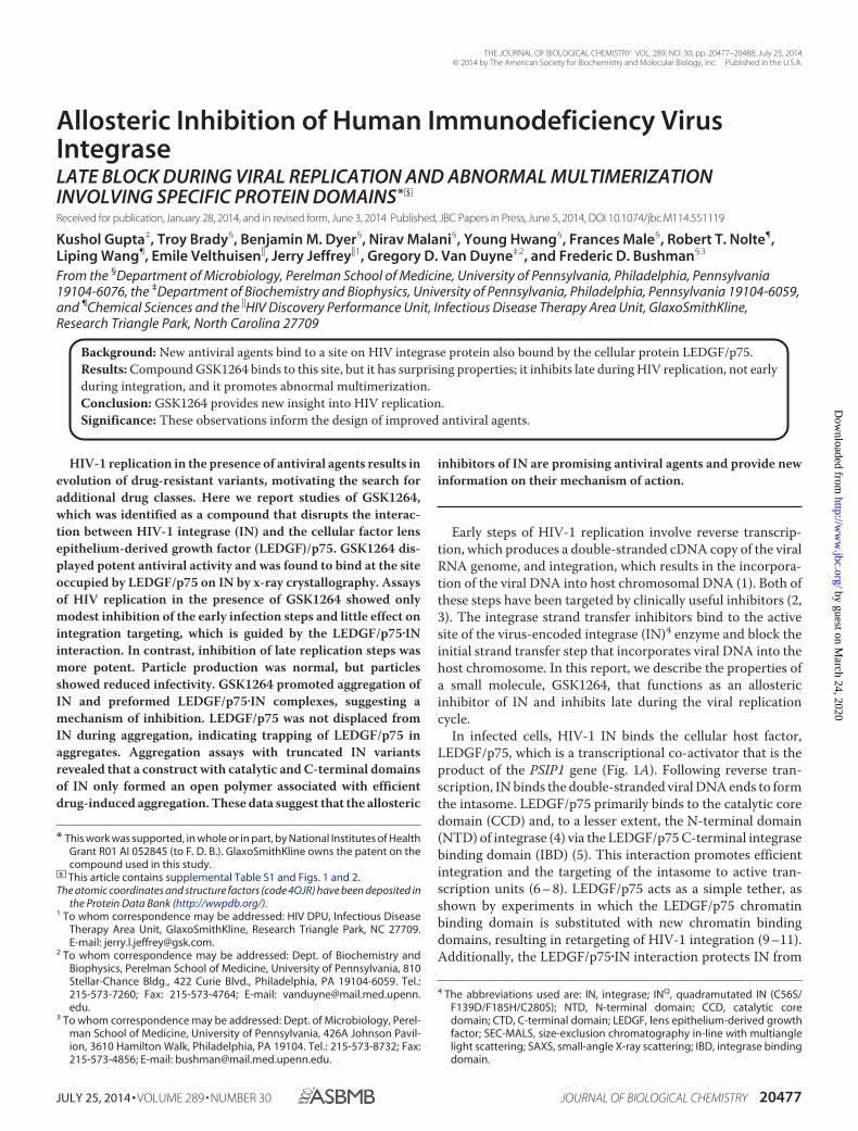
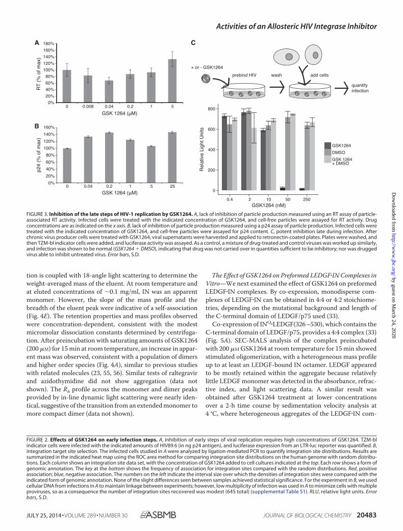
In the first, 293T cells producing HIVNL4 –3 virus weretreated with GSK1264 or DMSO carrier, and infectivity wasanalyzed. Virus production quantified by RT assay (Fig. 3A) orp24 antigen yield (Fig. 3B) showed no significant difference inthe presence of GSK1264 up to 5 �M. To test the infectivity ofthese particles, virus produced in the presence of GSK1264 orDMSO control was applied to U373/CD4/CCR5 target cells.Potent inhibition was detected, with an estimated IC50 of 12 nM.
TABLE 1Crystallographic data and results
HIV-1 IN(CCD)�GSK1264
Data collectionSource APS 21IDGWavelength (Å) 0.97856Resolution range (Å) 28.4-1.82 (1.89-1.82)a
Space group P 31 21Unit cell parameters a � b � 72.664 Å, c � 65.453 Å,
� � � � 90º, � 120ºNo. of measured reflections 149,182 (14,898)No. of unique reflections 17,968 (1,750)Multiplicity 8.3 (8.5)Completeness (%) 98.3 (98.1)Mean I/�(I) 21.6 (6.2)Wilson B-factor (Å2) 32.5Rmerge (%)a 4.4 (41)
RefinementRwork (%)b 18.9 (23.0)Rfree (%)c 20.1 (24.4)No. of atoms 1,146
Protein 1,001Ligands 44Water 101
Root mean square deviationBond lengths (Å) 0.008Bond angles (degrees) 1.20
Ramachandran plot (%)Most favored region 99Additionally allowed region 1
Average B-factor (Å2)Overall 40.9Protein 40.2Ligands 34.7Water 49.9
a Numbers in parentheses represent values in the highest resolution shell.b Rmerge � ��Ih � �Ih�/�Ih, where �Ih is the average intensity over symmetry
equivalent measurements.c R-factor � ��Fobs � Fcalc�/�Fobs, where summation is data used in refinement.d The summation for Rfree was calculated with 5% of the data.
Activities of an Allosteric HIV Integrase Inhibitor
20480 JOURNAL OF BIOLOGICAL CHEMISTRY VOLUME 289 • NUMBER 30 • JULY 25, 2014
by guest on March 24, 2020
http://ww
w.jbc.org/
Dow
nloaded from
In contrast, infection of the U373/CD4/CCR5 cells in the pres-ence of different concentrations of GSK12654 with HIVNL4 –3(where virus was not treated with drug) yielded an IC50 of 557nM. Thus, there was a 46-fold difference between the efficiencyof infection late versus early.
A complication arises in analyzing virus stocks produced inthe presence of drug, which is that compound carried over withthe viral stock may influence the course of the infection in thenewly infected target cells (although the above study wasdesigned to dilute drug in viral stocks extensively before appli-cation to target cells). To eliminate this concern, in a secondstudy, HIV-1 particles were prebound to retronectin-coatedplates and washed, and then TZM-bl target cells were added. Inthis format, inhibition by GSK1264 remained effective (Fig.3C), with complete inhibition of viral replication seen between10 and 50 nM. As a control, non-drugged and drugged viruseswere mixed, bound to the plate, and washed, and then cells wereadded (Fig. 3C, GSK1264 DMSO). Infection was efficient,indicating that the drug was not carried over and that the virusproduced in the presence of drug did not inhibit infection bynon-drug-treated virus.
We thus conclude that GSK1264 exerts its effects primarilyin the late steps of the viral replication cycle, resulting in theformation of replication-deficient particles, a conclusion thathas also been reached by others in studies of structurally relatedmolecules (20, 22, 23, 25, 26).
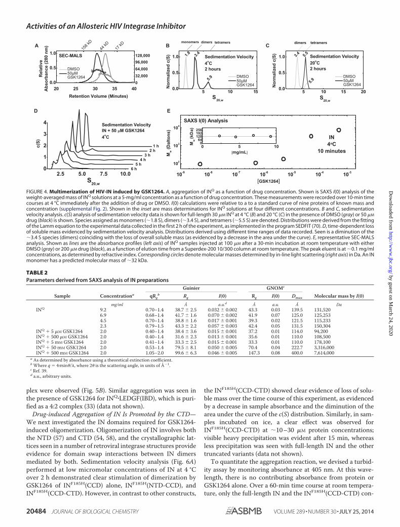
Effects of GSK1264 on the Solution Properties of Full-length INin Vitro—We analyzed IN by several biophysical methods toassess the effects of GSK1264 on its solution properties in vitro(Fig. 4, A–E). For this, we examined a mutant version of INQ
(C56S, F139D, F185H, and C280S) (33, 52–54) that has morefavorable solubility properties than wild-type IN alone. Globalfitting of sedimentation equilibrium data with strict mass con-
servation obtained from recombinant INQ at 4 °C was bestdescribed by a monomer-dimer-tetramer association, with dis-sociation constants of �5 �M for Kd1–2 and �86 �M for Kd2– 4,respectively (data not shown). Attempts to analyze INQ in thepresence of 50 �M GSK1264 were stymied by loss of solublemass. However, the data that were obtained through two speeds(�40 h at 4 °C) and across three concentrations could be welldescribed by a dimer-tetramer-octamer association with disso-ciation constants of �4 �M for Kd2– 4 and �86 �M for Kd4 – 8,respectively. However, such an analysis cannot reliably discrim-inate a 2-4-8 association from other models, such as 2-4-16 or2-5-7.
Simulations using the monomer-dimer-tetramer associationmodel over the 1–100 �M range predicted that IN alone forms amixture of monomer, dimer, and tetramer species (supplemen-tal Fig. 1). Such heterogeneity precludes structural analysis bysmall-angle x-ray scattering. However, weight-averaged massdetermination is possible using I(0) analysis, where mass isderived by comparison of the incident intensities of known con-centration with mass standards of known mass and concentra-tion. We found that the weight-average mass (Mw) of IN wasconsistent with �128 kDa from 1–5 mg/ml concentrations(Fig. 4A and Table 2), in line with our simulations. Titration ofthe GSK1264 compound with IN at 4 °C over a 10-min periodrevealed a concentration-dependent aggregation of IN, wherethe determined mass did not terminate with tetramer or octa-mer but instead arrived in the megadalton range. These obser-vations were corroborated by sedimentation velocity analyses(Fig. 4, B–D), which allowed for the discrimination of discreteoligomers stimulated by the compound.
To study the drug-induced oligomerization of IN further, weemployed SEC-MALS coupled with dynamic light scattering(Fig. 4E). In this approach, absolute concentration determina-
FIGURE 1. Overview of HIV-1 IN, LEDGF/p75, and GSK1264. A, HIV-1 IN and LEDGF/p75 protein domain organization. IN is a three-domain protein, composedof a zinc-binding NTD, a CCD with a DDE active site and RNase H-like fold, and a CTD with an Src homology 3-like fold. The IBD of the p75 isoform of LEDGF isshown in magenta. B, chemical structure of GSK1264. C, stereo view of the GSK1264 binding site at the IN(CCD) dimer interface, as determined by x-raycrystallography (PDB code 4OJR). Helices �3 and �1 from one monomer subunit and �5 and �4 from another are shown cradling the drug (tan).
Activities of an Allosteric HIV Integrase Inhibitor
JULY 25, 2014 • VOLUME 289 • NUMBER 30 JOURNAL OF BIOLOGICAL CHEMISTRY 20481
by guest on March 24, 2020
http://ww
w.jbc.org/
Dow
nloaded from
Activities of an Allosteric HIV Integrase Inhibitor
20482 JOURNAL OF BIOLOGICAL CHEMISTRY VOLUME 289 • NUMBER 30 • JULY 25, 2014
by guest on March 24, 2020
http://ww
w.jbc.org/
Dow
nloaded from
tion is coupled with 18-angle light scattering to determine theweight-averaged mass of the eluent. At room temperature andat eluted concentrations of �0.1 mg/ml, IN was an apparentmonomer. However, the slope of the mass profile and thebreadth of the eluent peak were indicative of a self-association(Fig. 4E). The retention properties and mass profiles observedwere concentration-dependent, consistent with the modestmicromolar dissociation constants determined by centrifuga-tion. After preincubation with saturating amounts of GSK1264(200 �M) for 15 min at room temperature, an increase in appar-ent mass was observed, consistent with a population of dimersand higher order species (Fig. 4A), similar to previous studieswith related molecules (23, 55, 56). Similar tests of raltegravirand azidothymidine did not show aggregation (data notshown). The Rh profile across the monomer and dimer peaksprovided by in-line dynamic light scattering were nearly iden-tical, suggestive of the transition from an extended monomer tomore compact dimer (data not shown).
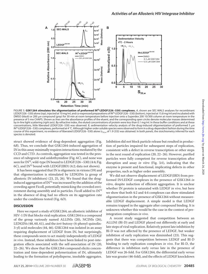
The Effect of GSK1264 on Preformed LEDGF�IN Complexes inVitro—We next examined the effect of GSK1264 on preformedLEDGF�IN complexes. By co-expression, monodisperse com-plexes of LEDGF�IN can be obtained in 4:4 or 4:2 stoichiome-tries, depending on the mutational background and length ofthe C-terminal domain of LEDGF/p75 used (33).
Co-expression of INQ�LEDGF(326 –530), which contains theC-terminal domain of LEDGF/p75, provides a 4:4 complex (33)(Fig. 5A). SEC-MALS analysis of the complex preincubatedwith 200 �M GSK1264 at room temperature for 15 min showedstimulated oligomerization, with a heterogeneous mass profileup to at least an LEDGF-bound IN octamer. LEDGF appearedto be mostly retained within the aggregate because relativelylittle LEDGF monomer was detected in the absorbance, refrac-tive index, and light scattering data. A similar result wasobtained after GSK1264 treatment at lower concentrationsover a 2-h time course by sedimentation velocity analysis at4 °C, where heterogeneous aggregates of the LEDGF�IN com-
FIGURE 2. Effects of GSK1264 on early infection steps. A, inhibition of early steps of viral replication requires high concentrations of GSK1264. TZM-blindicator cells were infected with the indicated amounts of HIV89.6 (in ng p24 antigen), and luciferase expression from an LTR-luc reporter was quantified. B,integration target site selection. The infected cells studied in A were analyzed by ligation-mediated PCR to quantify integration site distributions. Results aresummarized in the indicated heat map using the ROC area method for comparing integration site distributions on the human genome with random distribu-tions. Each column shows an integration site data set, with the concentration of GSK1264 added to cell cultures indicated at the top. Each row shows a form ofgenomic annotation. The key at the bottom shows the frequency of association for integration sites compared with the random distributions. Red, positiveassociation; blue, negative association. The numbers on the left indicate the interval size over which the densities of integration sites were compared with theindicated form of genomic annotation. None of the slight differences seen between samples achieved statistical significance. For the experiment in B, we usedcellular DNA from infections in A to maintain linkage between experiments; however, low multiplicity of infection was used in A to minimize cells with multipleproviruses, so as a consequence the number of integration sites recovered was modest (645 total) (supplemental Table S1). RLU, relative light units. Errorbars, S.D.
FIGURE 3. Inhibition of the late steps of HIV-1 replication by GSK1264. A, lack of inhibition of particle production measured using an RT assay of particle-associated RT activity. Infected cells were treated with the indicated concentration of GSK1264, and cell-free particles were assayed for RT activity. Drugconcentrations are as indicated on the x axis. B, lack of inhibition of particle production measured using a p24 assay of particle production. Infected cells weretreated with the indicated concentration of GSK1264, and cell-free particles were assayed for p24 content. C, potent inhibition late during infection. Afterchronic virus producer cells were treated with GSK1264, viral supernatants were harvested and applied to retronectin-coated plates. Plates were washed, andthen TZM-bl indicator cells were added, and luciferase activity was assayed. As a control, a mixture of drug-treated and control viruses was worked up similarly,and infection was shown to be normal (GSK1264 DMSO), indicating that drug was not carried over in quantities sufficient to be inhibitory; nor was druggedvirus able to inhibit untreated virus. Error bars, S.D.
Activities of an Allosteric HIV Integrase Inhibitor
JULY 25, 2014 • VOLUME 289 • NUMBER 30 JOURNAL OF BIOLOGICAL CHEMISTRY 20483
by guest on March 24, 2020
http://ww
w.jbc.org/
Dow
nloaded from
plex were observed (Fig. 5B). Similar aggregation was seen inthe presence of GSK1264 for INQ�LEDGF(IBD), which is puri-fied as a 4:2 complex (33) (data not shown).
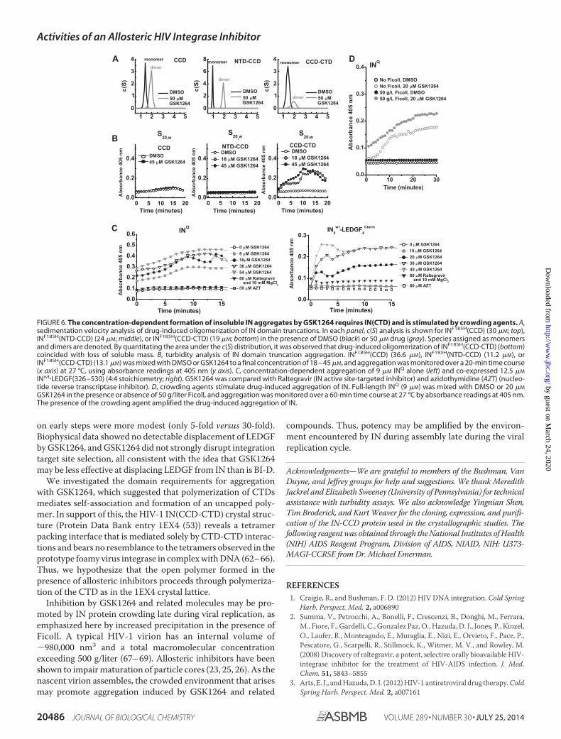
Drug-induced Aggregation of IN Is Promoted by the CTD—We next investigated the IN domains required for GSK1264-induced oligomerization. Oligomerization of IN involves boththe NTD (57) and CTD (54, 58), and the crystallographic lat-tices seen in a number of retroviral integrase structures provideevidence for domain swap interactions between IN dimersmediated by both. Sedimentation velocity analysis (Fig. 6A)performed at low micromolar concentrations of IN at 4 °Cover 2 h demonstrated clear stimulation of dimerization byGSK1264 of INF185H(CCD) alone, INF185H(NTD-CCD), andINF185H(CCD-CTD). However, in contrast to other constructs,
the INF185H(CCD-CTD) showed clear evidence of loss of solu-ble mass over the time course of this experiment, as evidencedby a decrease in sample absorbance and the diminution of thearea under the curve of the c(S) distribution. Similarly, in sam-ples incubated on ice, a clear effect was observed forINF185H(CCD-CTD) at �10 –30 �M protein concentrations;visible heavy precipitation was evident after 15 min, whereasless precipitation was seen with full-length IN and the othertruncated variants (data not shown).
To quantitate the aggregation reaction, we devised a turbid-ity assay by monitoring absorbance at 405 nm. At this wave-length, there is no contributing absorbance from protein orGSK1264 alone. Over a 60-min time course at room tempera-ture, only the full-length IN and the INF185H(CCD-CTD) con-
FIGURE 4. Multimerization of HIV-IN induced by GSK1264. A, aggregation of INQ as a function of drug concentration. Shown is SAXS I(0) analysis of theweight-averaged mass of INQ solutions at a 5-mg/ml concentration as a function of drug concentration. These measurements were recorded over 10-min timecourses at 4 °C immediately after the addition of drug or DMSO. I(0) calculations were relative to a to a standard curve of nine proteins of known mass andconcentration (supplemental Fig. 2). Shown in the inset are mass determinations for INQ solutions at four different concentrations. B and C, sedimentationvelocity analysis. c(S) analysis of sedimentation velocity data is shown for full-length 30 �M INQ at 4 °C (B) and 20 °C (C) in the presence of DMSO (gray) or 50 �M
drug (black) is shown. Species assigned as monomers (�1.8 S), dimers (�3.4 S), and tetramers (�5.5 S) are denoted. Distributions were derived from the fittingof the Lamm equation to the experimental data collected in the first 2 h of the experiment, as implemented in the program SEDFIT (70). D, time-dependent lossof soluble mass evidenced by sedimentation velocity analysis. Distributions derived using different time ranges of data recorded. Seen is a diminution of the�3.4 S species (dimers) coinciding with the loss of overall soluble mass (as evidenced by a decrease in the area under the curve). E, representative SEC-MALSanalysis. Shown as lines are the absorbance profiles (left axis) of INQ samples injected at 100 �M after a 30-min incubation at room temperature with eitherDMSO (gray) or 200 �M drug (black), as a function of elution time from a Superdex-200 10/300 column at room temperature. The peak eluent is at �0.1 mg/mlconcentrations, as determined by refractive index. Corresponding circles denote molecular masses determined by in-line light scattering (right axis) in Da. An INmonomer has a predicted molecular mass of �32 kDa.
TABLE 2Parameters derived from SAXS analysis of IN preparations
Sample Concentrationa
Guinier GNOMc
Molecular mass by I(0)qRgb Rg I(0) Rg I(0) Dmax
mg/ml Å a.u.d Å a.u. Å DaINQ 9.2 0.70–1.4 38.7 � 2.5 0.032 � 0.002 43.3 0.03 139.5 131,520
6.9 0.68–1.4 41.7 � 1.6 0.070 � 0.002 41.9 0.07 125.0 125,2534.5 0.70–1.4 38.8 � 1.6 0.017 � 0.001 39.3 0.02 121.5 115,2332.3 0.79–1.5 43.3 � 2.2 0.057 � 0.003 42.4 0.05 131.5 150,304
INQ 5 �M GSK1264 2.0 0.40–1.4 38.4 � 3.6 0.015 � 0.001 37.2 0.01 114.0 94,200INQ 500 �M GSK1264 2.0 0.40–1.4 31.6 � 2.3 0.013 � 0.001 35.6 0.01 110.0 108,500INQ 5 mM GSK1264 2.0 0.41–1.4 33.3 � 2.5 0.015 � 0.001 33.3 0.01 110.0 178,100INQ 50 mM GSK1264 2.0 0.53–1.4 79.5 � 8.1 0.050 � 0.005 70.4 0.04 222.7 3,316,000INQ 500 mM GSK1264 2.0 1.05–2.0 99.6 � 6.3 0.046 � 0.005 147.3 0.08 400.0 7,614,000
a As determined by absorbance using a theoretical extinction coefficient.b Where q � 4�sin�/�, where 2� is the scattering angle, in units of �1.c Ref. 39.d a.u., arbitrary units.
Activities of an Allosteric HIV Integrase Inhibitor
20484 JOURNAL OF BIOLOGICAL CHEMISTRY VOLUME 289 • NUMBER 30 • JULY 25, 2014
by guest on March 24, 2020
http://ww
w.jbc.org/
Dow
nloaded from
struct showed evidence of drug-dependent aggregation (Fig.6B). Thus, we conclude that GSK1264-induced aggregation ofIN in this assay minimally requires interactions mediated by theCCD and CTD. As controls, aggregation was tested in the pres-ence of raltegravir and azidothymidine (Fig. 6C), and none wasseen for INQ, wild-type IN bound to LEDGF(326 –530) (4:4; Fig.6C), and INQ bound with LEDGF(IBD) (4:2; data not shown).
It has been suggested that IN is oligomeric in virions (59) andthat oligomerization is stimulated by LEDGINs (a group ofallosteric IN inhibitors) (23, 25, 26). We found that the drug-induced aggregation of INQ was increased by the addition of thecrowding agent Ficoll, potentially mimicking the crowded envi-ronment during assembly and in particles. Ficoll added to INQ
in the absence of drug had no effects on its aggregation rateunder the conditions tested (Fig. 6D).
DISCUSSION
Here we report a study of GSK1264, an allosteric inhibitor ofHIV-1 IN that blocks viral replication. GSK1264 is a compoundof the group variously named ALLINIs (20), NCINIs (26),LEDGINs (48, 60, 61), and the tert-butoxy-(4-phenyl-quinolin-3-yl) acid molecules (44, 46). GSK1264 was isolated in an assayrequiring displacement of LEDGF from IN, but surprisingly,these compounds seem to act mainly independently of LEDGFin vivo. Instead, these inhibitors have been linked to post-inte-gration effects associated with the self-association of IN (20,22–26). We show that the GSK1264 compound elicits concen-tration and time-dependent polymerization of IN, ultimatelyleading to the formation of polydisperse, insoluble aggregates.
Inhibition did not block particle release but resulted in produc-tion of particles impaired for subsequent steps of replication,consistent with a defect in reverse transcription or other stepsin the next round of replication (20, 22–26). However, purifiedparticles were fully competent for reverse transcription afterdisruption and assay in vitro (Fig. 3A), indicating that theenzyme is present and functional, implicating defects in otherproperties, such as higher order assembly.
We did not observe displacement of LEDGF(IBD) from pre-formed IN�LEDGF complexes in the presence of GSK1264 invitro, despite induction of efficient aggregation. It is unclearwhether IN protein is saturated with LEDGF in vivo, but herewe show that both 4:2 and 4:4 complexes can show stimulatedoligomerization in the presence of GSK1264 without measure-able LEDGF displacement. A simple model is that LEDGFremains trapped in the aggregate after compound binding. It isunknown whether this would be the case in the context of pre-integration complexes in vivo.
A recent study suggested that competition between anALLINI (BI-D) and LEDGF played out differently at early andlate steps of viral replication. Relatively potent late inhibition byBI-D was not affected by the presence of LEDGF, but weakerinhibition of early replication was diminished (23). This sug-gests that there was competition between LEDGF and BI-Dbinding to early replication complexes in vivo. For BI-D, thedifference in inhibition early versus late in the presence ofLEDGF was 26-fold. For GSK1264, the differential early versuslate was greater (46-fold), and the effects of LEDGF knockdown
FIGURE 5. GSK1264 stimulates the oligomerization of preformed INQ�LEDGF(326 –530) complexes. A, shown are SEC-MALS analyses for recombinantLEDGF(326 –530) alone (top), injected at 10 mg/ml, and co-expressed preparations of INQ�LEDGF(326 –530) (bottom), injected at 15.8 mg/ml and incubated withDMSO (black) or 200 �M compound (gray) for 30 min at room temperature before injection onto a Superdex 200 10/300 column at room temperature in thepresence of 5 mM CHAPS. Shown as lines are the absorbance profiles of the eluent, and the corresponding open circles denote molecular masses determinedby in-line light scattering (right axis). By refractive index, the eluted concentrations of protein were less than 0.1 mg/ml. In these buffer conditions and at theseconcentrations, little liberated LEDGF(326 –530) was observed. B, sedimentation velocity analysis of the drug-induced oligomerization of preformed 5 �M
INQ�LEDGF(326 –530) complexes, performed at 4 °C. Although higher order soluble species were observed to form in a drug-dependent fashion during the timecourse of this experiment, no evidence of liberated LEDGF(326 –530) alone (s20,w of 1.9 (33)) was obtained. In both panels, the stoichiometry inferred for eachspecies is denoted.
Activities of an Allosteric HIV Integrase Inhibitor
JULY 25, 2014 • VOLUME 289 • NUMBER 30 JOURNAL OF BIOLOGICAL CHEMISTRY 20485
by guest on March 24, 2020
http://ww
w.jbc.org/
Dow
nloaded from
on early steps were more modest (only 5-fold versus 30-fold).Biophysical data showed no detectable displacement of LEDGFby GSK1264, and GSK1264 did not strongly disrupt integrationtarget site selection, all consistent with the idea that GSK1264may be less effective at displacing LEDGF from IN than is BI-D.
We investigated the domain requirements for aggregationwith GSK1264, which suggested that polymerization of CTDsmediates self-association and formation of an uncapped poly-mer. In support of this, the HIV-1 IN(CCD-CTD) crystal struc-ture (Protein Data Bank entry 1EX4 (53)) reveals a tetramerpacking interface that is mediated solely by CTD-CTD interac-tions and bears no resemblance to the tetramers observed in theprototype foamy virus integrase in complex with DNA (62– 66).Thus, we hypothesize that the open polymer formed in thepresence of allosteric inhibitors proceeds through polymeriza-tion of the CTD as in the 1EX4 crystal lattice.
Inhibition by GSK1264 and related molecules may be pro-moted by IN protein crowding late during viral replication, asemphasized here by increased precipitation in the presence ofFicoll. A typical HIV-1 virion has an internal volume of�980,000 nm3 and a total macromolecular concentrationexceeding 500 g/liter (67– 69). Allosteric inhibitors have beenshown to impair maturation of particle cores (23, 25, 26). As thenascent virion assembles, the crowded environment that arisesmay promote aggregation induced by GSK1264 and related
compounds. Thus, potency may be amplified by the environ-ment encountered by IN during assembly late during the viralreplication cycle.
Acknowledgments—We are grateful to members of the Bushman, VanDuyne, and Jeffrey groups for help and suggestions. We thank MeredithJackrel and Elizabeth Sweeney (University of Pennsylvania) for technicalassistance with turbidity assays. We also acknowledge Yingnian Shen,Tim Broderick, and Kurt Weaver for the cloning, expression, and purifi-cation of the IN-CCD protein used in the crystallographic studies. Thefollowing reagent was obtained through the National Institutes of Health(NIH) AIDS Reagent Program, Division of AIDS, NIAID, NIH: U373-MAGI-CCR5E from Dr. Michael Emerman.
REFERENCES1. Craigie, R., and Bushman, F. D. (2012) HIV DNA integration. Cold Spring
Harb. Perspect. Med. 2, a0068902. Summa, V., Petrocchi, A., Bonelli, F., Crescenzi, B., Donghi, M., Ferrara,
M., Fiore, F., Gardelli, C., Gonzalez Paz, O., Hazuda, D. J., Jones, P., Kinzel,O., Laufer, R., Monteagudo, E., Muraglia, E., Nizi, E., Orvieto, F., Pace, P.,Pescatore, G., Scarpelli, R., Stillmock, K., Witmer, M. V., and Rowley, M.(2008) Discovery of raltegravir, a potent, selective orally bioavailable HIV-integrase inhibitor for the treatment of HIV-AIDS infection. J. Med.Chem. 51, 5843–5855
3. Arts, E. J., and Hazuda, D. J. (2012) HIV-1 antiretroviral drug therapy. ColdSpring Harb. Perspect. Med. 2, a007161
FIGURE 6. The concentration-dependent formation of insoluble IN aggregates by GSK1264 requires IN(CTD) and is stimulated by crowding agents. A,sedimentation velocity analysis of drug-induced oligomerization of IN domain truncations. In each panel, c(S) analysis is shown for INF185H(CCD) (30 �M; top),INF185H(NTD-CCD) (24 �M; middle), or INF185H(CCD-CTD) (19 �M; bottom) in the presence of DMSO (black) or 50 �M drug (gray). Species assigned as monomersand dimers are denoted. By quantitating the area under the c(S) distribution, it was observed that drug-induced oligomerization of INF185H(CCD-CTD) (bottom)coincided with loss of soluble mass. B, turbidity analysis of IN domain truncation aggregation. INF185H(CCD) (36.6 �M), INF185H(NTD-CCD) (11.2 �M), orINF185H(CCD-CTD) (13.1 �M) was mixed with DMSO or GSK1264 to a final concentration of 18 – 45 �M, and aggregation was monitored over a 20-min time course(x axis) at 27 °C, using absorbance readings at 405 nm (y axis). C, concentration-dependent aggregation of 9 �M INQ alone (left) and co-expressed 12.5 �M
INwt�LEDGF(326 –530) (4:4 stoichiometry; right). GSK1264 was compared with Raltegravir (IN active site-targeted inhibitor) and azidothymidine (AZT) (nucleo-tide reverse transcriptase inhibitor). D, crowding agents stimulate drug-induced aggregation of IN. Full-length INQ (9 �M) was mixed with DMSO or 20 �M
GSK1264 in the presence or absence of 50 g/liter Ficoll, and aggregation was monitored over a 60-min time course at 27 °C by absorbance readings at 405 nm.The presence of the crowding agent amplified the drug-induced aggregation of IN.
Activities of an Allosteric HIV Integrase Inhibitor
20486 JOURNAL OF BIOLOGICAL CHEMISTRY VOLUME 289 • NUMBER 30 • JULY 25, 2014
by guest on March 24, 2020
http://ww
w.jbc.org/
Dow
nloaded from
4. Maertens, G., Cherepanov, P., Pluymers, W., Busschots, K., De Clercq, E.,Debyser, Z., and Engelborghs, Y. (2003) LEDGF/p75 is essential for nu-clear and chromosomal targeting of HIV-1 integrase in human cells.J. Biol. Chem. 278, 33528 –33539
5. Cherepanov, P., Devroe, E., Silver, P. A., and Engelman, A. (2004) Identi-fication of an evolutionarily conserved domain in human lens epithelium-derived growth factor/transcriptional co-activator p75 (LEDGF/p75) thatbinds HIV-1 integrase. J. Biol. Chem. 279, 48883– 48892
6. Schroder, A. R., Shinn, P., Chen, H., Berry, C., Ecker, J. R., and Bushman, F.(2002) HIV-1 integration in the human genome favors active genes andlocal hotspots. Cell 110, 521–529
7. Ciuffi, A., Diamond, T. L., Hwang, Y., Marshall, H. M., and Bushman, F. D.(2006) Modulating target site selection during human immunodeficiencyvirus DNA integration in vitro with an engineered tethering factor. Hum.Gene Ther. 17, 960 –967
8. Shun, M. C., Raghavendra, N. K., Vandegraaff, N., Daigle, J. E., Hughes, S.,Kellam, P., Cherepanov, P., and Engelman, A. (2007) LEDGF/p75 func-tions downstream from preintegration complex formation to effect gene-specific HIV-1 integration. Genes Dev. 21, 1767–1778
9. Silvers, R. M., Smith, J. A., Schowalter, M., Litwin, S., Liang, Z., Geary, K.,and Daniel, R. (2010) Modification of integration site preferences of anHIV-1-based vector by expression of a novel synthetic protein. Hum. GeneTher. 21, 337–349
10. Ferris, A. L., Wu, X., Hughes, C. M., Stewart, C., Smith, S. J., Milne, T. A.,Wang, G. G., Shun, M. C., Allis, C. D., Engelman, A., and Hughes, S. H.(2010) Lens epithelium-derived growth factor fusion proteins redirectHIV-1 DNA integration. Proc. Natl. Acad. Sci. U.S.A. 107, 3135–3140
11. Gijsbers, R., Ronen, K., Vets, S., Malani, N., De Rijck, J., McNeely, M.,Bushman, F. D., and Debyser, Z. (2010) LEDGF hybrids efficiently retargetlentiviral integration into heterochromatin. Mol. Ther. 18, 552–560
12. Cherepanov, P., Maertens, G., Proost, P., Devreese, B., Van Beeumen, J.,Engelborghs, Y., De Clercq, E., and Debyser, Z. (2003) HIV-1 integraseforms stable tetramers and associates with LEDGF/p75 protein in humancells. J. Biol. Chem. 278, 372–381
13. Hendrix, J., Gijsbers, R., De Rijck, J., Voet, A., Hotta, J., McNeely, M.,Hofkens, J., Debyser, Z., and Engelborghs, Y. (2011) The transcriptionalco-activator LEDGF/p75 displays a dynamic scan-and-lock mechanismfor chromatin tethering. Nucleic Acids Res. 39, 1310 –1325
14. Llano, M., Vanegas, M., Fregoso, O., Saenz, D., Chung, S., Peretz, M., andPoeschla, E. M. (2004) LEDGF/p75 determines cellular trafficking of di-verse lentiviral but not murine oncoretroviral integrase proteins and is acomponent of functional lentiviral preintegration complexes. J. Virol. 78,9524 –9537
15. Turlure, F., Maertens, G., Rahman, S., Cherepanov, P., and Engelman, A.(2006) A tripartite DNA-binding element, comprised of the nuclear local-ization signal and two AT-hook motifs, mediates the association ofLEDGF/p75 with chromatin in vivo. Nucleic Acids Res. 34, 1653–1665
16. Pandey, K. K., Sinha, S., and Grandgenett, D. P. (2007) Transcriptionalcoactivator LEDGF/p75 modulates human immunodeficiency virus type1 integrase-mediated concerted integration. J. Virol. 81, 3969 –3979
17. Hare, S., Shun, M. C., Gupta, S. S., Valkov, E., Engelman, A., and Che-repanov, P. (2009) A novel co-crystal structure affords the design of gain-of-function lentiviral integrase mutants in the presence of modifiedPSIP1/LEDGF/p75. PLoS Pathog. 5, e1000259
18. Cherepanov, P., Sun, Z. Y., Rahman, S., Maertens, G., Wagner, G., andEngelman, A. (2005) Solution structure of the HIV-1 integrase-bindingdomain in LEDGF/p75. Nat. Struct. Mol. Biol. 12, 526 –532
19. Molteni, V., Greenwald, J., Rhodes, D., Hwang, Y., Kwiatkowski, W., Bush-man, F. D., Siegel, J. S., and Choe, S. (2001) Identification of a small mol-ecule binding site at the dimer interface of the HIV integrase catalyticdomain. Acta Crystallogr. D Biol. Crystallogr. 57, 536 –544
20. Engelman, A., Kessl, J. J., and Kvaratskhelia, M. (2013) Allosteric inhibitionof HIV-1 integrase activity. Curr. Opin. Chem. Biol. 17, 339 –345
21. Christ, F., and Debyser, Z. (2013) The LEDGF/p75 integrase interaction, anovel target for anti-HIV therapy. Virology 435, 102–109
22. Le Rouzic, E., Bonnard, D., Chasset, S., Bruneau, J. M., Chevreuil, F., LeStrat, F., Nguyen, J., Beauvoir, R., Amadori, C., Brias, J., Vomscheid, S.,Eiler, S., Levy, N., Delelis, O., Deprez, E., Saıb, A., Zamborlini, A., Emiliani,
S., Ruff, M., Ledoussal, B., Moreau, F., and Benarous, R. (2013) Dual inhi-bition of HIV-1 replication by integrase-LEDGF allosteric inhibitors ispredominant at the post-integration stage. Retrovirology 10, 144
23. Jurado, K. A., Wang, H., Slaughter, A., Feng, L., Kessl, J. J., Koh, Y., Wang,W., Ballandras-Colas, A., Patel, P. A., Fuchs, J. R., Kvaratskhelia, M., andEngelman, A. (2013) Allosteric integrase inhibitor potency is determinedthrough the inhibition of HIV-1 particle maturation. Proc. Natl. Acad. Sci.U.S.A. 110, 8690 – 8695
24. Feng, L., Sharma, A., Slaughter, A., Jena, N., Koh, Y., Shkriabai, N., Larue,R. C., Patel, P. A., Mitsuya, H., Kessl, J. J., Engelman, A., Fuchs, J. R., andKvaratskhelia, M. (2013) The A128T resistance mutation reveals aberrantprotein multimerization as the primary mechanism of action of allostericHIV-1 integrase inhibitors. J. Biol. Chem. 288, 15813–15820
25. Desimmie, B. A., Schrijvers, R., Demeulemeester, J., Borrenberghs, D.,Weydert, C., Thys, W., Vets, S., Van Remoortel, B., Hofkens, J., De Rijck, J.,Hendrix, J., Bannert, N., Gijsbers, R., Christ, F., and Debyser, Z. (2013)LEDGINs inhibit late stage HIV-1 replication by modulating integrasemultimerization in the virions. Retrovirology 10, 57
26. Balakrishnan, M., Yant, S. R., Tsai, L., O’Sullivan, C., Bam, R. A., Tsai, A.,Niedziela-Majka, A., Stray, K. M., Sakowicz, R., and Cihlar, T. (2013) Non-catalytic site HIV-1 integrase inhibitors disrupt core maturation and in-duce a reverse transcription block in target cells. PloS One 8, e74163
27. Vodicka, M. A., Goh, W. C., Wu, L. I., Rogel, M. E., Bartz, S. R., Schweick-art, V. L., Raport, C. J., and Emerman, M. (1997) Indicator cell lines fordetection of primary strains of human and simian immunodeficiency vi-ruses. Virology 233, 193–198
28. Platt, E. J., Wehrly, K., Kuhmann, S. E., Chesebro, B., and Kabat, D. (1998)Effects of CCR5 and CD4 cell surface concentrations on infections bymacrophagetropic isolates of human immunodeficiency virus type 1. J. Vi-rol. 72, 2855–2864
29. Doranz, B. J., Rucker, J., Yi, Y., Smyth, R. J., Samson, M., Peiper, S. C.,Parmentier, M., Collman, R. G., and Doms, R. W. (1996) A dual-trophicprimary HIV-1 isolate that uses Fusin and the �-chemokine receptorsCKR-5, CKR-3 and CKR-2b as fusion cofactors. Cell 85, 1149 –1158
30. Berry, C., Hannenhalli, S., Leipzig, J., and Bushman, F. D. (2006) Selectionof target sites for mobile DNA integration in the human genome. PLoSComput. Biol. 2, e157
31. Wang, G. P., Ciuffi, A., Leipzig, J., Berry, C. C., and Bushman, F. D. (2007)HIV integration site selection: analysis by massively parallel pyrosequenc-ing reveals association with epigenetic modifications. Genome Res. 17,1186 –1194
32. Ciuffi, A., Ronen, K., Brady, T., Malani, N., Wang, G., Berry, C. C., andBushman, F. D. (2009) Methods for integration site distribution analysesin animal cell genomes. Methods 47, 261–268
33. Gupta, K., Diamond, T., Hwang, Y., Bushman, F., and Van Duyne, G. D.(2010) Structural properties of HIV integrase. Lens epithelium-derivedgrowth factor oligomers. J. Biol. Chem. 285, 20303–20315
34. Urbani, A., and Warne, T. (2005) A colorimetric determination for glyco-sidic and bile salt-based detergents: applications in membrane proteinresearch. Anal. Biochem. 336, 117–124
35. Lipfert, J., Columbus, L., Chu, V. B., Lesley, S. A., and Doniach, S. (2007)Size and shape of detergent micelles determined by small-angle x-ray scat-tering. J. Phys. Chem. B 111, 12427–12438
36. Deprez, E., Tauc, P., Leh, H., Mouscadet, J. F., Auclair, C., and Brochon,J. C. (2000) Oligomeric states of the HIV-1 integrase as measured by time-resolved fluorescence anisotropy. Biochemistry 39, 9275–9284
37. Schuck, P. (2000) Size-distribution analysis of macromolecules by sedi-mentation velocity ultracentrifugation and Lamm equation modeling.Biophys. J. 78, 1606 –1619
38. Laue, T. M., Shah, B. D., Ridgeway, T. M., and Pelletier, S. L. (1992) Com-puter-aided interpretation of analytical sedimentation data for proteins inAnalytical Ultracentrifugation in Biochemistry and Polymer Science (Har-ding, S. E., Rowe, A. J., and Horton, J. C. eds), pp. 90 –125. The RoyalSociety of Chemistry, Cambridge, UK
39. Semenyuk, A. V., and Svergun, D. I. (1991) GNOM: a program package forsmall-angle scattering data-processing. J. Appl. Crystallogr. 24, 537–540
40. McCoy, A. J., Grosse-Kunstleve, R. W., Adams, P. D., Winn, M. D., Sto-roni, L. C., and Read, R. J. (2007) Phaser crystallographic software. J. Appl.
Activities of an Allosteric HIV Integrase Inhibitor
JULY 25, 2014 • VOLUME 289 • NUMBER 30 JOURNAL OF BIOLOGICAL CHEMISTRY 20487
by guest on March 24, 2020
http://ww
w.jbc.org/
Dow
nloaded from
Crystallogr. 40, 658 – 67441. Smart, O. S., Womack, T. O., Flensburg, C., Keller, P., Paciorek, W., Sharff,
A., Vonrhein, C., and Bricogne, G. (2012) Exploiting structure similarity inrefinement: automated NCS and target-structure restraints in BUSTER.Acta Crystallogr. D 68, 368 –380
42. Emsley, P., Lohkamp, B., Scott, W. G., and Cowtan, K. (2010) Features anddevelopment of Coot. Acta Crystallogr. D 66, 486 –501
43. Murshudov, G. N., Vagin, A. A., and Dodson, E. J. (1997) Refinement ofmacromolecular structures by the maximum-likelihood method. ActaCrystallogr. D 53, 240 –255
44. Tsiang, M., Jones, G. S., Niedziela-Majka, A., Kan, E., Lansdon, E. B.,Huang, W., Hung, M., Samuel, D., Novikov, N., Xu, Y., Mitchell, M., Guo,H., Babaoglu, K., Liu, X., Geleziunas, R., and Sakowicz, R. (2012) New classof HIV-1 integrase (IN) inhibitors with a dual mode of action. J. Biol.Chem. 287, 21189 –21203
45. Kessl, J. J., Jena, N., Koh, Y., Taskent-Sezgin, H., Slaughter, A., Feng, L., deSilva, S., Wu, L., Le Grice, S. F., Engelman, A., Fuchs, J. R., and Kvaratskhe-lia, M. (2012) Multimode, cooperative mechanism of action of allostericHIV-1 integrase inhibitors. J. Biol. Chem. 287, 16801–16811
46. Fenwick, C. W., Tremblay, S., Wardrop, E., Bethell, R., Coulomb, R., El-ston, R., Faucher, A. M., Mason, S., Simoneau, B., Tsantrizos, Y., andYoakim, C. (2011) Resistance studies with HIV-1 non-catalytic site inte-grase inhibitors. Antivir. Ther. 16, Suppl. 1, A9
47. Hou, Y., McGuinness, D. E., Prongay, A. J., Feld, B., Ingravallo, P., Ogert,R. A., Lunn, C. A., and Howe, J. A. (2008) Screening for antiviral inhibitorsof the HIV integrase-LEDGF/p75 interaction using the AlphaScreen lu-minescent proximity assay. J. Biomol. Screen. 13, 406 – 414
48. Christ, F., Voet, A., Marchand, A., Nicolet, S., Desimmie, B. A., Marchand,D., Bardiot, D., Van der Veken, N. J., Van Remoortel, B., Strelkov, S. V., DeMaeyer, M., Chaltin, P., and Debyser, Z. (2010) Rational design of small-molecule inhibitors of the LEDGF/p75-integrase interaction and HIV rep-lication. Nat. Chem. Biol. 6, 442– 448
49. Mitchell, R. S., Beitzel, B. F., Schroder, A. R. W., Shinn, P., Chen, H. M.,Berry, C. C., Ecker, J. R., and Bushman, F. D. (2004) Retroviral DNA inte-gration: ASLV, HIV, and MLV show distinct target site preferences. PLoSBiol. 2, 1127–1137
50. Ciuffi, A., Llano, M., Poeschla, E., Hoffmann, C., Leipzig, J., Shinn, P.,Ecker, J. R., and Bushman, F. (2005) A role for LEDGF/p75 in targetingHIV DNA integration. Nat. Med. 11, 1287–1289
51. Marshall, H. M., Ronen, K., Berry, C., Llano, M., Sutherland, H., Saenz, D.,Bickmore, W., Poeschla, E., and Bushman, F. D. (2007) Role of PSIP1/LEDGF/p75 in lentiviral infectivity and integration targeting. PloS One 2,e1340
52. Alian, A., Griner, S. L., Chiang, V., Tsiang, M., Jones, G., Birkus, G., Gelezi-unas, R., Leavitt, A. D., and Stroud, R. M. (2009) Catalytically-active com-plex of HIV-1 integrase with a viral DNA substrate binds anti-integrasedrugs. Proc. Natl. Acad. Sci. U.S.A. 106, 8192– 8197
53. Chen, J. C., Krucinski, J., Miercke, L. J., Finer-Moore, J. S., Tang, A. H.,Leavitt, A. D., and Stroud, R. M. (2000) Crystal structure of the HIV-1integrase catalytic core and C-terminal domains: a model for viral DNAbinding. Proc. Natl. Acad. Sci. U.S.A. 97, 8233– 8238
54. Bischerour, J., Leh, H., Deprez, E., Brochon, J. C., and Mouscadet, J. F.(2003) Disulfide-linked integrase oligomers involving C280 residues areformed in vitro and in vivo but are not essential for human immunodefi-ciency virus replication. J. Virol. 77, 135–141
55. Demeulemeester, J., Tintori, C., Botta, M., Debyser, Z., and Christ, F.(2012) Development of an AlphaScreen-based HIV-1 integrase dimeriza-
tion assay for discovery of novel allosteric inhibitors. J. Biomol. Screen. 17,618 – 628
56. Tintori, C., Demeulemeester, J., Franchi, L., Massa, S., Debyser, Z., Christ,F., and Botta, M. (2012) Discovery of small molecule HIV-1 integrasedimerization inhibitors. Bioorg. Med. Chem. Lett. 22, 3109 –3114
57. Zheng, R., Jenkins, T. M., and Craigie, R. (1996) Zinc folds the N-terminaldomain of HIV-1 integrase, promotes multimerization, and enhances cat-alytic activity. Proc. Natl. Acad. Sci. U.S.A. 93, 13659 –13664
58. Jenkins, T. M., Engelman, A., Ghirlando, R., and Craigie, R. (1996) A sol-uble active mutant of HIV-1 integrase: involvement of both the core andcarboxyl-terminal domains in multimerization. J. Biol. Chem. 271,7712–7718
59. Petit, C., Schwartz, O., and Mammano, F. (1999) Oligomerization withinvirions and subcellular localization of human immunodeficiency virustype 1 integrase. J. Virol. 73, 5079 –5088
60. Christ, F., Shaw, S., Demeulemeester, J., Desimmie, B. A., Marchand, A.,Butler, S., Smets, W., Chaltin, P., Westby, M., Debyser, Z., and Pickford, C.(2012) Small-molecule inhibitors of the LEDGF/p75 binding site of inte-grase block HIV replication and modulate integrase multimerization. An-timicrob. Agents Chemother. 56, 4365– 4374
61. Peat, T. S., Rhodes, D. I., Vandegraaff, N., Le, G., Smith, J. A., Clark, L. J.,Jones, E. D., Coates, J. A., Thienthong, N., Newman, J., Dolezal, O., Mul-der, R., Ryan, J. H., Savage, G. P., Francis, C. L., and Deadman, J. J. (2012)Small molecule inhibitors of the LEDGF site of human immunodeficiencyvirus integrase identified by fragment screening and structure based de-sign. PloS One 7, e40147
62. Hare, S., Gupta, S. S., Valkov, E., Engelman, A., and Cherepanov, P. (2010)Retroviral intasome assembly and inhibition of DNA strand transfer. Na-ture 464, 232–236
63. Hare, S., Maertens, G. N., and Cherepanov, P. (2012) 3�-processing andstrand transfer catalysed by retroviral integrase in crystallo. EMBO J. 31,3020 –3028
64. Hare, S., Smith, S. J., Metifiot, M., Jaxa-Chamiec, A., Pommier, Y.,Hughes, S. H., and Cherepanov, P. (2011) Structural and functionalanalyses of the second-generation integrase strand transfer inhibitordolutegravir (S/GSK1349572). Mol. Pharmacol. 80, 565–572
65. Hare, S., Vos, A. M., Clayton, R. F., Thuring, J. W., Cummings, M. D., andCherepanov, P. (2010) Molecular mechanisms of retroviral integrase in-hibition and the evolution of viral resistance. Proc. Natl. Acad. Sci. U.S.A.107, 20057–20062
66. Yin, Z., Lapkouski, M., Yang, W., and Craigie, R. (2012) Assembly of pro-totype foamy virus strand transfer complexes on product DNA bypassingcatalysis of integration. Protein Sci. 21, 1849 –1857
67. del Alamo, M., Rivas, G., and Mateu, M. G. (2005) Effect of macromolec-ular crowding agents on human immunodeficiency virus type 1 capsidprotein assembly in vitro. J. Virol. 79, 14271–14281
68. Benjamin, J., Ganser-Pornillos, B. K., Tivol, W. F., Sundquist, W. I., andJensen, G. J. (2005) Three-dimensional structure of HIV-1 virus-like par-ticles by electron cryotomography. J. Mol. Biol. 346, 577–588
69. Briggs, J. A., Simon, M. N., Gross, I., Krausslich, H. G., Fuller, S. D., Vogt,V. M., and Johnson, M. C. (2004) The stoichiometry of Gag protein inHIV-1. Nat. Struct. Mol. Biol. 11, 672– 675
70. Vistica, J., Dam, J., Balbo, A., Yikilmaz, E., Mariuzza, R. A., Rouault, T. A.,and Schuck, P. (2004) Sedimentation equilibrium analysis of protein in-teractions with global implicit mass conservation constraints and system-atic noise decomposition. Anal. Biochem. 326, 234 –256
Activities of an Allosteric HIV Integrase Inhibitor
20488 JOURNAL OF BIOLOGICAL CHEMISTRY VOLUME 289 • NUMBER 30 • JULY 25, 2014
by guest on March 24, 2020
http://ww
w.jbc.org/
Dow
nloaded from
Duyne and Frederic D. BushmanMale, Robert T. Nolte, Liping Wang, Emile Velthuisen, Jerry Jeffrey, Gregory D. Van Kushol Gupta, Troy Brady, Benjamin M. Dyer, Nirav Malani, Young Hwang, Frances
INVOLVING SPECIFIC PROTEIN DOMAINSDURING VIRAL REPLICATION AND ABNORMAL MULTIMERIZATION
Allosteric Inhibition of Human Immunodeficiency Virus Integrase: LATE BLOCK
doi: 10.1074/jbc.M114.551119 originally published online June 5, 20142014, 289:20477-20488.J. Biol. Chem.
10.1074/jbc.M114.551119Access the most updated version of this article at doi:
Alerts:
When a correction for this article is posted•
When this article is cited•
to choose from all of JBC's e-mail alertsClick here
Supplemental material:
http://www.jbc.org/content/suppl/2014/06/05/M114.551119.DC1
http://www.jbc.org/content/289/30/20477.full.html#ref-list-1
This article cites 69 references, 27 of which can be accessed free at
by guest on March 24, 2020
http://ww
w.jbc.org/
Dow
nloaded from