Embed Size (px)
Citation preview

Bull. Egypt. Soc. Physiol. Sci. 27 (1) 2007 Abdel Gawad et al.
221
Amelioration of Acetic Acid-Induced Colitis in Rats by Oral Administration of Ginger Extract
Hala S. Abdel Gawad1, Lamiaa N.A. Hammad2, Hanan S. El-Abhar3
1Department of Physiology, Faculty of Medicine, Alexandria University; 2Department of Biochemistry, Faculty of Pharmacy (Girl),
Al-Azhar University, Cairo; 3Department of Pharmacology & Toxicology, Faculty of Pharmacy, Cairo University
ABSTRACT
Ulcerative colitis (UC) is a chronically recurrent inflammatory bowel disease of unknown origin. The aim of the present study is to evaluate possible protective effects of ginger extract (GE) on the extent and severity of UC caused by intracolonic administration of acetic acid in rats. Animals received either GE (100, 200 and 400 mg/kg) or sulphasalazine (500 mg/kg), for 3 consecutive days before intra-rectal acetic acid administration (1 ml, 4% v/v), and continued for another 7 days after the induction. The degree of tissue injuries was assessed by macroscopical and histopathological scores of the colonic mucosa. the biochemical studies involve the redox state including colon mucosal content of malondialdehyde (MDA) as an index of lipid peroxidation, glutathione (GSH) and protein carbonyl content (PCO) as indexes of protein oxidation as well as the activity of catalase and superoxide dismutase (SOD) enzymes in addition to some indicators of the inflammatory response myeloperoxidase (MPO) activity, index of neutrophilic infiltration, and the tissue contents of tumor necrosis factor (TNF-α), and prostaglandin E2 (PGE2). Oral pretreatment with ginger extract and sulphasalazine were able to correct altered parameters significantly. Moreover, ginger extract attenuated the macroscopic colonic damage and the histopathological changes-induced by acetic acid. These results suggest a beneficial protective effect of ginger extract against acetic acid-induced colitis possibly by its antioxidant and anti-inflammatory effects. Key words: ulcerative colitis, ginger extract, TNF-α, PGE 2, MPO, oxidative stress, rats.
INTRODUCTION
Ulcerative colitis and Crohn's disease are chronic, relapsing, immunologically mediated disorders that are collectively referred to as inflammatory bowel diseases (IBD). The prevalence of IBD rapidly increased in Europe and North America in the second half of the twentieth century and is becoming
more common in the rest of the world as different countries adopt a Western lifestyle[1]. Etiology and pathogenesis of inflammatory bowel disease, however, remain obscure, although environmental factors in combination with genetic factors are suggested to be involved in its pathogenesis[2]. Even with this knowledge, the etiologies of these diseases remain an enigma. In the past few years,

Bull. Egypt. Soc. Physiol. Sci. 27 (1) 2007 Abdel Gawad et al.
222
however, work in animal models, human genetics, basic science and clinical trials have provided new insights into the pathogenesis of these diseases[3].
The role of oxidative stress in the pathogenesis of ulcerative colitis has been investigated, where oxygen-centered radicals, such as superoxide and hydroxyl radicals generated by phagocytes, have been suggested to be involved in the pathogenesis of chronic inflammations of the bowel, such as Crohn's disease and colitis ulcerosa [4]; [5], due to its major tissue-destructive force.
Generation of reactive oxygen species (ROS) by inflammatory cells is a major microbiocidal mechanism and may mediate important components of the inflammatory response[6]. Prolonged or inadequate activation of the intestinal immune system plays an important role in the pathophysiology of chronic mucosal inflammation[7], and infiltration of neutrophils, macrophages, lymphocytes and mast cells, ultimately give rise to mucosal disruption and ulceration[2]. The infiltered neutrophils represent an important source of reactive oxygen species (ROS), which are cytotoxic agents, inducing cellular oxidative stress by cross-linking proteins, lipids, and nucleic acids, leading to cellular dysfunction and damage[8]. The disturbed redox system can be reflected by the elevation of protein carbonyls levels (PCO)[9], and lipid peroxidation[10], [11].
Oxidative stress occurs when there is an imbalance between generation of ROS and inadequate antioxidant defense systems. This fact
holds true in IBD, since both levels of non-enzymatic endogenous antioxidants, such as reduced glutathione (GSH), and the activity of the enzymatic ones, for example, catalase (CAT) and superoxide dismutase (SOD), are all significantly decreased in these patients, thus augmenting the deleterious effect of ROS [12]; [13]. Most currently used therapeutic drugs for IBD, in particular sulphasalazine and its active moiety 5-aminosalicylic acid, control the disease partly by their property as potent ROS scavengers [14]; [15].
In addition to free radicals, neutrophils can also release proteases and lipid mediators that can contribute to intestinal injury[16]. Inflammatory bowel disease (IBD) is associated with increased activation of intestinal immune cells, whose overproduction of pro-inflammatory cytokines, such as tumor necrosis factor-alpha (TNF-alpha) and interleukin-1 beta (IL-1 beta) is implicated in mediating the sustained inflammatory response[17]. TNF-α level is often increased [18] both in animal models [19] and patients with ulcerative colitis[20]. This cytokine is a key immunoregulatory cytokine that amplifies the inflammatory response by activating a cascade of immune cells. TNF-α, secreted by activated macrophages, stimulate production of cytokines, arachidonic acid metabolites, and proteases by intestinal macrophages, neutrophils, smooth muscle cells, fibroblast, and epithelial cells [21]. Moreover, myeloperoxidase (MPO), which is an enzyme mainly found in azurophilic granules of neutrophils, is a good marker of neutrophil infiltration[22]; [5].
Bull. Egypt. Soc. Physiol. Sci. 27 (1) 2007 Abdel Gawad et al.
223
Experimental models in which colitis is induced by chemicals or haptens have been widely used to understand the mechanisms involved in the disease state with the goal of discovering new therapeutic agents[23];[24]. Among these experimental models, intrarectal administration of organic acids (acetic acid) is known to create initial injurious effects to the colonic mucosa, a process that produces acute colitis[25], [5].
Ginger (Zingiber officinale Roscoe), is a well-known herbaceous species, which is consumed in most parts of the world and are rich in gingerols, shogaols, and other constituents, which exhibit antioxidant[26],antihyperlipidemic[27;28] antiproliferative[29], and anticarcinogenic proprieties under ‘‘in vitro’’ and ‘‘in vivo’’ systems [30];[31]. Furthermore, in folk medicine, ginger has been used traditionally as a carminative, diaphoretic, anti-spasmodic and anti-emetic agent against motion sickness [32].
However, the possible role of ginger in modulating colon inflammation has not been verified yet, thus we aimed in the current investigation to evaluate the effect of ginger extract on acetic acid-induced ulcerative colitis in rats and its possible mechanism(s) of action. MATERIALS & METHODS
Chemicals
Acetic acid, sodium sulfate, Triton X-100, 2-thiobarbituric acid (TBA), trichloroacetic acid (TCA), Tris base, phosphate buffer, n-butanol were purchased from Merck
(Germany), 1,1,3,3-tetraethoxypropane, sulphasalazine, 5,5-dithiobis-(2-nitrobenzoic acid) (DTNB), reduced glutathione (GSH), adenosine diphosphate, agents for myeloperoxidase assay were procured from Sigma-Aldrich (St Louis. MO, USA). TNF- and PGE2 ELISA kits were obtained from Amersham (Pharmacia Biotech, Little Chalfont, UK) and R&D Systems Inc. (Minneapolis, USA), respectively. All other chemicals used were of analytical grade. Animals
Male Sprague-Dawley rats (190 - 210 g) were exposed to the same environmental conditions and were maintained on a proper diet chow and water ad libitum. Animals were randomly divided into six groups (8 animals each) and the study was conducted in accordance with ethical procedures and policies approved by the Animal Care and Use Committee of Faculty of Pharmacy, Al-Azhar University Cairo, Egypt. Preparation of the extract
Fresh rhizome of ginger (Zingiber officinale Roscoe) was purchased from a local market in Egypt and authenticated by a pharmacognosist (Pharmacognosy Department, Faculty of Pharmacy, Al-Azhar University, Cairo) according to Langner et al. [32]. The plant was dried in the shade and the dried rhizome was powdered mechanically and extracted by cold percolation with 95% ethanol in a soxhlet apparatus for 72 h. The process was repeated three times and the three extracts were pooled together and the combined extract was concentrated under reduced pressure (22–26 mmHg) at 45–60°C. The
Bull. Egypt. Soc. Physiol. Sci. 27 (1) 2007 Abdel Gawad et al.
224
solvent was removed under reduced pressure to give a dry extract, 9.5% yield, w/w (with respect to the crude material) and doses equivalent to 100, 200 and 400 mg of the crude drug per kg body weight, were calculated and suspended in 0.5 % carboxy methyl cellulose (CMC) solution for the experiment. Induction of experimental colitis in rats
Colitis was induced using the method previously described by Millar et al., [23], where animals fasted for 24 h with free access to water ad libitum before induction of colitis, and each rat was sedated by phenobarbitone (35 mg/kg, i.p). One milliliter of acetic acid (4 %, v/v, in 0.9 % saline) was infused for 30s using a polyethylene tube (2 mm in diameter), which was inserted through the rectum into the colon to a distance of 8 cm. The acetic acid was retained in the colon for another 30s after which the fluid was withdrawn and animals' head were kept in a downward position for 30s then returned to cages. Animals were divided in 6 groups, where group 1 served as normal control receiving 0.9% saline, group 2 were subjected to ulcerative colitis model and used as positive control, groups 3, 4, and 5 received ginger extract in doses of 100, 200 and 400 mg/kg, respectively, and the last group was used as reference group, where animals received sulphasalazine (500 mg/kg). Drugs were given once daily (0.5 ml/100 g body weight, p.o) for 3 days before the induction of colitis and continued for another 7 consecutive days. Drugs were suspended in 0.5% carboxymethyl cellulose (CMC),
which does not affect the severity of acetic acid-induced ulcerative colitis[24]. Following completion of the experiment, rats were ether anesthetized and colonic biopsies were taken for macroscopic scoring, histopathological examination and biochemical studies. Assessment of colitis - Macroscopic scoring
At post-mortem laparotomy, 6 cm of colon extending proximally for 2 cm above the anal orifice was removed. The tissue was first split longitudinally, pinned out onto card, and the macroscopic appearance of the colonic mucosa was scored by an independent observer according to Millar et al. [23] , a scale ranging from 0 to 4 as follows: 0 =No macroscopic changes, 1 =Mucosal erythema only, 2 =Mild mucosal edema, slight bleeding or small erosions, 3 =Moderate edema, bleeding ulcers or erosions, 4 = Severe ulceration, erosions, edema and tissue necrosis - Histopathological study
Sections of colon were obtained from the same areas of the large intestine from four representative animals in each of the treatment groups during autopsy. They were fixed in 8phosphate buffered formaldehyde, embedded in paraffin, and routine 5 µm sections were prepared. Tissues were routinely stained with haematoxylin and eosin and were evaluated by light microscopy by a pathologist . -Biochemical studies
Myeloperoxidase (MPO, EC 1.11.1.7) activity was determined as an index of neutrophil accumulation[33], and the rate of change in absorbance was measured

Bull. Egypt. Soc. Physiol. Sci. 27 (1) 2007 Abdel Gawad et al.
225
spectrophotometrically at 650 nm. MPO activity was defined as the quantity of enzyme degrading 1 mmol of peroxide/min at 37°C and expressed in units per gram weight of wet tissue. Catalase (CAT) (EC 1.11.1.6) activity was measured according to the Aebi[34], where one unit of CAT activity was defined as the amount of enzyme required to decompose 1 mol of H2O2 in 1 min. The rate of decomposition of H2O2 was measured spectrophotometrically at 240 nm for 1 min. Superoxide dismutase (SOD) (EC 1.15.1.1) activity was determined according to Marklund and Marklund [35]. A colorimetric assay involving generation of superoxide by pyrogallol auto-oxidation was measured at 570 nm. Protein content was measured according to Lowry et al.[36].
Lipid peroxidation in colon tissue homogenate was evaluated by measuring MDA using thiobarbituric method described by Ohkawa et al.[37], and the absorbance of the supernatant was measured spectrophotometrically at 532 nm. PCO content in colon homogenates were determined spectrophotometrically by the method of Levine et al.[38], based on the reaction of the carbonyl group with 2,4-dinitrophenylhydrazine to form 2,4 dinitrophenylhydrazone. GSH content was determined as previously described by Owens and Belcher[39] based on the reaction of 5,5-dithiobis-(2-nitrobenzoic acid) (DTNB) with the GSH present and the absorbance was measured by a double beam spectrophotometer at 412 nm.
TNF-α was assessed according to the method of Reinecker et al.[40], and was quantified by enzyme-linked
immunoabsorbent assay [ELISA] (Amersham Pharmacia Biotech, Little Chalfont, UK) and the results were expressed as picograms /gram of wet tissue. PGE2 was also measured by ELISA kits (R&D Systems, USA)[41]. Statistical analysis
All data are expressed as mean ± standard error of the mean (SEM) for eight rats per experimental group. Statistical analysis was performed with SPSS 10.0 statistical software. Parametric one-way analysis of variance (ANOVA) followed by Tukey-Kramer Multiple Comparisons test was used to compare the mean values of quantitative variables among the groups. The minimal level of significance was identified at p < 0.05. Correlation coefficient was determined by linear regression analysis. The non-parametric ANOVA, followed by Kruskal-Wallis Test was used for analyzing the macroscopical test.
RESULTS
Table 1 depicts the macroscopic changes related to the 3 dose levels of ginger extract and the reference drug, sulfasalazine, in comparison with vehicle-treated rats. Acetic acid caused severe macroscopic edematous inflammation in the colon, 24 h after rectal administration, as assessed by the high score of colonic damage and the increased colon wet weight. Ginger extract ameliorated the acetic acid effect as reflected by the significant dose dependant anti-edematous effect and the percentage reduction in the lesion scores. Treatment with sulphasalazine showed similar effect. However,
Bull. Egypt. Soc. Physiol. Sci. 27 (1) 2007 Abdel Gawad et al.
226
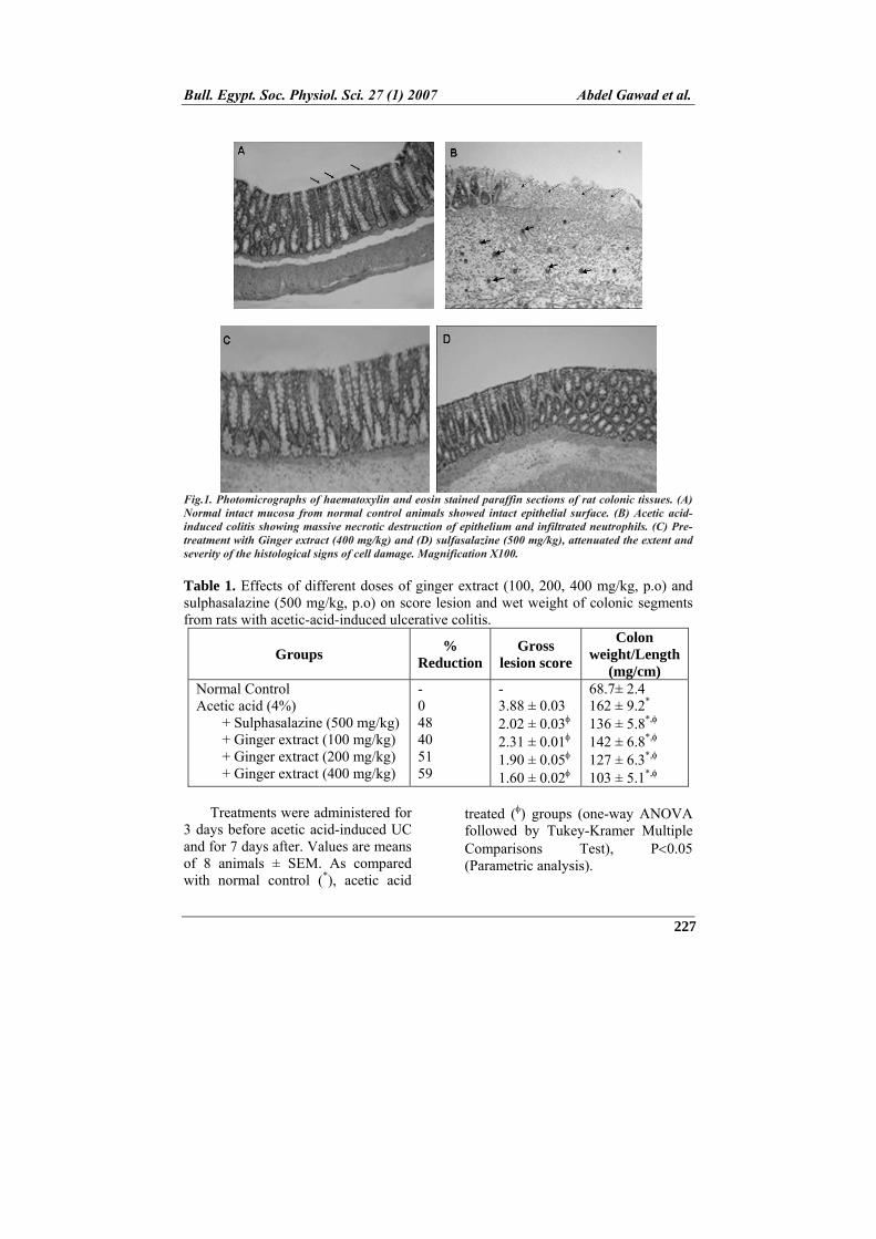
ginger extract (100 mg/kg) did not show any improvement regarding score difference from acetic acid. These findings were emphasized by the histopathological studies present in figure1, where acetic acid treated rats showed trasmural necrosis, edema and diffuse inflammatory cell infil-tration in the mucosa. There was focal ulceration of the colonic mucosa extending through the muscularis mucosa, desquamated areas with loss of the epithelium. The architecture of the crypts was distorted and the lamina propria was thickened in peripheral areas of distorted crypts especially in basal areas. An infiltrate consisting of mixed inflammatory cells was observed (Fig. 1B). Treatment of rats with sulphasalazine (Fig. 1C) or GE (Fig. 1D) significantly attenuated the extent and severity of the histological signs of cell damage. Histological studies confirmed the intestinal anti-inflammatory effect exerted by the two drugs used. This effect was mirrored by a 4 fold increase in mucosal MPO activity following acetic acid administration. Pre-treatment with either GE (100, 200 and 400 mg/kg) or sulphasalazine produced a significant reduction in MPO activity, however, none was able to reach the control value (Figure 3). Ulcerated non-treated group showed a vast elevation in the colonic TNF-α, an effect which leveled off in
groups treated with either GE (100, 200 and 400 mg/kg) or sulphasalazine (Figure 4). Effect of acetic acid alone or with either GE (100, 200 and 400 mg/kg) or sulphasalazine show the same pattern as illustrated in figure 5, and none of the treatment agents could normalize the PGE2 level. Using pair-wise comparisons among the groups. Ginger extract in a dose of 400 mg kg-1 was the most potent at reducing PGE2 level. Regarding the redox state, acetic acid group caused a significant decrease in colonic non-enzymatic (figure 2) and enzymatic (table 3) defense systems, however, these effects were altered in animals pre-treated with either treatment regimen, to reach nearly the normal values with the highest dose of GE. The overwhelmed defense systems result in a significant increase in concentration of both MDA (5.9 fold) and PCO (2.6 fold) in acetic acid non-treated animals (table 2). Pretreatment with GE produced a marked decrease in MDA and PCO levels in a dose-dependant manner, and so did sulphasalazine (table 2). Analysis of the correlation coefficients between gross lesion score and PGE2, TNF-, and MPO is shown in Fig. 6a–c. A significant positive correlation was observed between gross lesion score and colonic PGE2, TNF- and MPO (r2 = 0.994, 0.987 and 0.968, respectively, P < 0.001).
Bull. Egypt. Soc. Physiol. Sci. 27 (1) 2007 Abdel Gawad et al.
227
Fig.1. Photomicrographs of haematoxylin and eosin stained paraffin sections of rat colonic tissues. (A) Normal intact mucosa from normal control animals showed intact epithelial surface. (B) Acetic acid-induced colitis showing massive necrotic destruction of epithelium and infiltrated neutrophils. (C) Pre-treatment with Ginger extract (400 mg/kg) and (D) sulfasalazine (500 mg/kg), attenuated the extent and severity of the histological signs of cell damage. Magnification X100. Table 1. Effects of different doses of ginger extract (100, 200, 400 mg/kg, p.o) and sulphasalazine (500 mg/kg, p.o) on score lesion and wet weight of colonic segments from rats with acetic-acid-induced ulcerative colitis.
Groups %
Reduction Gross
lesion score
Colon weight/Length
(mg/cm) Normal Control Acetic acid (4%) + Sulphasalazine (500 mg/kg) + Ginger extract (100 mg/kg) + Ginger extract (200 mg/kg) + Ginger extract (400 mg/kg)
- 0 48 40 51 59
- 3.88 ± 0.03 2.02 ± 0.03 2.31 ± 0.01 1.90 ± 0.05 1.60 ± 0.02
68.7± 2.4 162 ± 9.2* 136 ± 5.8*, 142 ± 6.8*, 127 ± 6.3*, 103 ± 5.1*,
Treatments were administered for
3 days before acetic acid-induced UC and for 7 days after. Values are means of 8 animals ± SEM. As compared with normal control (*), acetic acid
treated () groups (one-way ANOVA followed by Tukey-Kramer Multiple Comparisons Test), P0.05 (Parametric analysis).

Bull. Egypt. Soc. Physiol. Sci. 27 (1) 2007 Abdel Gawad et al.
228
Table 2. Effects of different doses of ginger extract (100, 200 and 400 mg/kg, p.o) and sulphasalazine (500 mg/kg, p.o) on colonic levels of protein oxidation (PCO) and lipid peroxidation (MDA) in rats with acetic acid-induced ulcerative colitis.
Groups Protein carbonyl
content (nmol/mg protein)
Lipid peroxidation (nmol/g wet tissue)
Normal Control Acetic acid (4%)
+ Sulphasalazine + Ginger extract (100 mg/kg) + Ginger extract (200 mg/kg) + Ginger extract (400 mg/kg)
2.21 ± 0.08 5.78 ± 0.09* 3.41 ± 0.07 *, 3.93 ± 0.03*, 3.10 ± 0.05*, 2.78 ± 0.02*,
9.71 ± 0.22 57.6 ± 3.3* 31.5 ± 1.3*, 34.6 ± 1.2*, 25.2 ± 0.38*, 18.7 ± 0.23*,
Treatments were administered for 3 days before acetic acid-induced UC and for 7 days after. Values are means of 8 animals ± SEM. As compared with normal control(*), and acetic acid
treated () groups (one-way ANOVA followed by Tukey-Kramer Multiple Comparisons Test), P0.05.
Table 3. Effects of different doses of ginger extract (100, 200 and 400 mg/kg) and sulphasalazine (500 mg/kg, p.o) on the activity of colonic catalase and SOD of rats with acetic acid-induced ulcerative colitis.
Groups Catalase
(U/mg protein) SOD
(U/mg protein) Normal Control Acetic acid (4%) + Sulphasalazine + Ginger extract (100 mg/kg) + Ginger extract (200 mg/kg) + Ginger extract (400 mg/kg)
21.6 ± 0.23 10.23 ± 0.12* 14.75 ± 0.18*, 14.0 ± 0.25*, 17.2 ± 0.28*, 20.8 ± 0.31
6.23 ± 0.06 3.52 ± 0.02* 4.20 ± 0.05 *, 4.41 ± 0.03*, 6.22 ± 0.08 8.23 ± 0.04*,
Treatments were administered for 3 days before acetic acid-induced UC and for 7 days after. Values are means of 8 animals ± SEM. As compared with normal control (*), and acetic
acid treated () groups (one-way ANOVA followed by Tukey-Kramer Multiple Comparisons Test), P0.05.
Bull. Egypt. Soc. Physiol. Sci. 27 (1) 2007 Abdel Gawad et al.
229
GS
H (
nm
ol/
g w
et t
issu
e)0
400
800
1200
1600Controlacetic acid+ sulphasal.+ GE 100+ GE 200+ GE 400
*
* ***
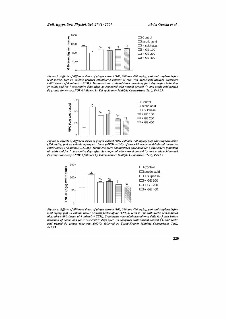
Figure 2: Effects of different doses of ginger extract (100, 200 and 400 mg/kg, p.o) and sulphasalazine (500 mg/kg, p.o) on colonic reduced glutathione content of rats with acetic acid-induced ulcerative colitis (mean of 8 animals ± SEM.). Treatments were administered once daily for 3 days before induction of colitis and for 7 consecutive days after. As compared with normal control (*), and acetic acid treated () groups (one-way ANOVA followed by Tukey-Kramer Multiple Comparisons Test), P0.05.
MP
O (
U/g
wet
tis
sue)
0
25
50
75Controlacetic acid+ sulphasal.+ GE 100+ GE 200+ GE 400
*
*
**
*
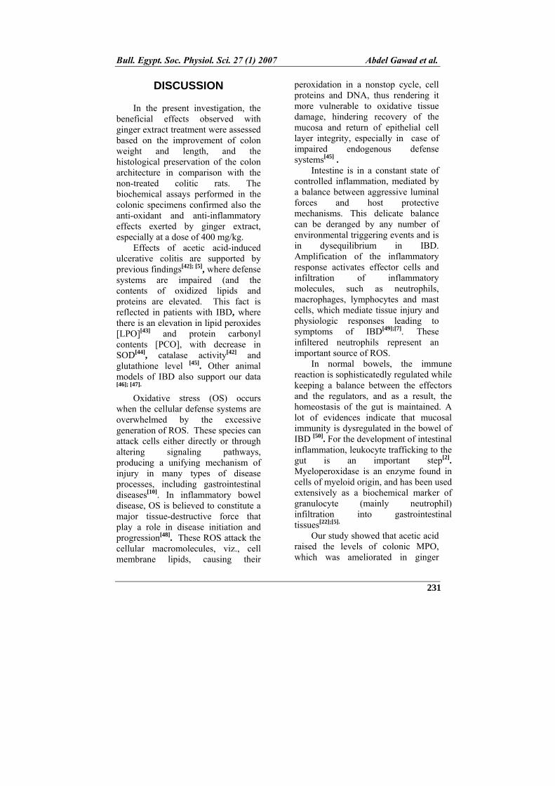
Figure 3: Effects of different doses of ginger extract (100, 200 and 400 mg/kg, p.o) and sulphasalazine (500 mg/kg, p.o) on colonic myeloperoxidase (MPO) activity of rats with acetic acid-induced ulcerative colitis (mean of 8 animals ± SEM.). Treatments were administered once daily for 3 days before induction of colitis and for 7 consecutive days after. As compared with normal control (*), and acetic acid treated () groups (one-way ANOVA followed by Tukey-Kramer Multiple Comparisons Test), P0.05.
TN
F-
(p
g/g
wet
tis
sue)
0
50
100
150Controlacetic acid+ sulphasal.+ GE 100+ GE 200+ GE 400
*
*
*
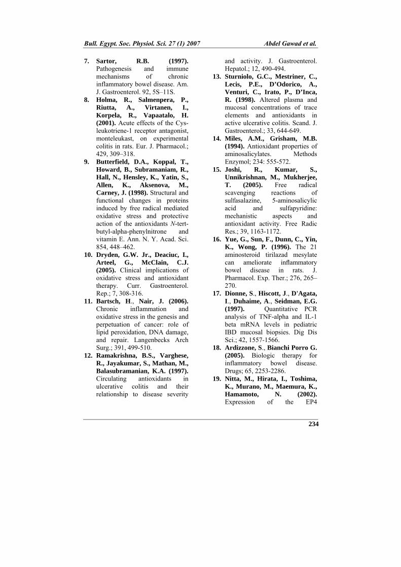
Figure 4: Effects of different doses of ginger extract (100, 200 and 400 mg/kg, p.o) and sulphasalazine (500 mg/kg, p.o) on colonic tumor necrosis factor-alpha (TNF-α) level in rats with acetic acid-induced ulcerative colitis (mean of 8 animals ± SEM). Treatments were administered once daily for 3 days before induction of colitis and for 7 consecutive days after. As compared with normal control (*), and acetic acid treated () groups (one-way ANOVA followed by Tukey-Kramer Multiple Comparisons Test), P0.05.

Bull. Egypt. Soc. Physiol. Sci. 27 (1) 2007 Abdel Gawad et al.
230
PG
E2 (
pg
/mg
tis
sue)
0
500
1000
1500
2000Controlacetic acid+ sulphasal.+ GE 100+ GE 200+ GE 400
*
*
**
*
Figure 5: Effects of different doses of ginger extract (100, 200 and 400 mg/kg, p.o) and sulphasalazine (500 mg/kg, p.o) on colonic prostaglandine E2 (PGE2) content in rats with acetic acid-induced ulcerative colitis (mean of 8 animals ± SEM.). Treatments were administered once daily for 3 days before induction of colitis and for 7 consecutive days after. As compared with normal control (*), and acetic acid treated () groups (one-way ANOVA followed by Tukey-Kramer Multiple Comparisons Test), P0.05.
Gross lesion score
PG
E2
(pg
/mg
pro
tein
)
0.0 2.5 5.0 7.5 10.00
500
1000
1500
2000
r2=0.994p 0.0001
(A)
Gross lesion score
TN
F-
(p
g/g
tis
sue)
0.0 2.5 5.0 7.5 10.00
25
50
75
100
125
150r2=0.987p 0.0001
(B)
Gross lesion score
MP
O (
U/g
tis
sue)
0.0 2.5 5.0 7.5 10.00
25
50
75r2=0.968p 0.001
(D)
Figure 6: Linear regression analysis of gross lesion score and (a) PGE2 (r
2 = 0.994, P < 0.0001), (b) TNF- (r2 = 0.987, P < 0.0001), and (c) MPO (r2 = 0.968, P < 0.001).
Bull. Egypt. Soc. Physiol. Sci. 27 (1) 2007 Abdel Gawad et al.
231
DISCUSSION
In the present investigation, the beneficial effects observed with ginger extract treatment were assessed based on the improvement of colon weight and length, and the histological preservation of the colon architecture in comparison with the non-treated colitic rats. The biochemical assays performed in the colonic specimens confirmed also the anti-oxidant and anti-inflammatory effects exerted by ginger extract, especially at a dose of 400 mg/kg.
Effects of acetic acid-induced ulcerative colitis are supported by previous findings[42]; [5], where defense systems are impaired (and the contents of oxidized lipids and proteins are elevated. This fact is reflected in patients with IBD, where there is an elevation in lipid peroxides [LPO][43] and protein carbonyl contents [PCO], with decrease in SOD[44], catalase activity[42] and glutathione level [45]. Other animal models of IBD also support our data [46]; [47].
Oxidative stress (OS) occurs when the cellular defense systems are overwhelmed by the excessive generation of ROS. These species can attack cells either directly or through altering signaling pathways, producing a unifying mechanism of injury in many types of disease processes, including gastrointestinal diseases[10]. In inflammatory bowel disease, OS is believed to constitute a major tissue-destructive force that play a role in disease initiation and progression[48]. These ROS attack the cellular macromolecules, viz., cell membrane lipids, causing their
peroxidation in a nonstop cycle, cell proteins and DNA, thus rendering it more vulnerable to oxidative tissue damage, hindering recovery of the mucosa and return of epithelial cell layer integrity, especially in case of impaired endogenous defense systems[45] .
Intestine is in a constant state of controlled inflammation, mediated by a balance between aggressive luminal forces and host protective mechanisms. This delicate balance can be deranged by any number of environmental triggering events and is in dysequilibrium in IBD. Amplification of the inflammatory response activates effector cells and infiltration of inflammatory molecules, such as neutrophils, macrophages, lymphocytes and mast cells, which mediate tissue injury and physiologic responses leading to symptoms of IBD[49];[7]. These infiltered neutrophils represent an important source of ROS.
In normal bowels, the immune reaction is sophisticatedly regulated while keeping a balance between the effectors and the regulators, and as a result, the homeostasis of the gut is maintained. A lot of evidences indicate that mucosal immunity is dysregulated in the bowel of IBD [50]. For the development of intestinal inflammation, leukocyte trafficking to the gut is an important step[2]. Myeloperoxidase is an enzyme found in cells of myeloid origin, and has been used extensively as a biochemical marker of granulocyte (mainly neutrophil) infiltration into gastrointestinal tissues[22];[5].
Our study showed that acetic acid raised the levels of colonic MPO, which was ameliorated in ginger

Bull. Egypt. Soc. Physiol. Sci. 27 (1) 2007 Abdel Gawad et al.
232
extract and sulfasalazine treated groups. Activation of MPO indicates infiltration of neutrophils and perturbation of the inflammatory system[33], which is documented both in animal models[42];[5]; [51] and patients with IBD [43]. The neutrophils may contribute to the production of ROS via activation of their NADPH oxidase and secretion of MPO into extracellular space. Although we did not measure NADPH oxidase activity in our experiment, it was speculated that this enzyme could also be activated by acetic acid enema since NADPH oxidase and MPO are two enzymes that are activated almost simultaneously [52]. In this study, however, the amount of MDA was associated with catalase activation and neutrophilic myeloperoxidase activity, which suggests a hydrogen peroxide (H2O2) - and/or hypochlorous acid (HOCl-)-mediated mechanism [43].
Under normal situations, the intestinal mucosa is in a state of 'controlled' inflammation regulated by a delicate balance of proinflammatory (tumour necrosis factor [TNF]-alpha, interferon [IFN]-gamma, interleukin [IL-1, IL-6, IL-12) and anti-inflammatory cytokines (IL-4, IL-10, IL-11). The mucosal immune system is the central effector of intestinal inflammation and injury, with cytokines playing a central role in modulating inflammation [18];[53]. TNF- is a proinflammatory cytokine which is abundantly expressed in the gut of IBD patients [53], this information is evidenced in this work.
Promotion of TNF-α is mediated by a new transcription factor termed lipopolysaccharide-induced TNF-alpha factor (LITAF)[54]. This factor was shown
to mediate TNF-alpha expression in human macrophages by direct binding to specific sequences in the promoter region of the TNF-alpha gene. LITAF is readily detectable in ileal and colonic tissues from patients with either CD or UC. It is significantly elevated above controls, and is localized to macrophages, a major source of TNF-alpha[54].
Another inflammatory mediator, viz., PGE2 was increased in rats with ulcerative colitis caused by acetic acid installation rectally, this goes with the fact that high amounts of this mediator are detected in the inflamed mucosa of patients with inflammatory bowel disease (IBD) [55]. The increased level of PGE2 is attributed to enhanced synthesis rather than reduced catabolism as reported by Otani et al.[55]. They found also that TNF-α affects the expression of 15-hydroxyprostaglandin dehydrogenase
(15-PGDH), which plays a major role in the catabolism of PGE2. TNF-α suppressed the transcription of 15-PGDH in human colonocytes, resulting in reduced amounts of 15-PGDH mRNA and protein and enzyme activity. In contrast, TNF-α induced two enzymes, cyclooxygenase-2 and microsomal prostaglandin E synthase-1 that contribute to increased synthesis of PGE2.
Overexpressing 15-PGDH blocked the increase in PGE2 production mediated by TNF-α.[55]. Our results show increased levels of both TNF-α and PGE2, which are known to produce epithelial cell necrosis, edema, neutrophil infiltration and, global cell depletion, effects which are documented by the histopathological study of this work.

Bull. Egypt. Soc. Physiol. Sci. 27 (1) 2007 Abdel Gawad et al.
233
Ginger extract (Zingiber officinale Roscoe) used in this study significantly reduced the wet weight of distal colon segments and the gross lesion score, compared with controls that received the vehicle. Further, they effectively reduced the histological signs of inflammation such as leukocyte infiltration, edema, and tissue injury and improved the acetic acid-induced biochemical alterations both in redox parameters and inflammatory ones. Ginger is ranked one of the plants with highest anti-oxidant values [56]; [26], thus, one of the mechanisms involved could be its antioxidant properties and its ability to inhibit free radical generation, properties which can guard against cellular macromolecules attack and depletion of the colon antioxidant defense systems.
The antiinflammatory properties of Z. officinale present in our study offers another mechanism of action to this plant. This action was reported previously in other inflammatory models [57]; [58]; [46]; [59]. In these studies, ginger extract affected the level of TNF-α and PGE2
[57], reported that ginger extract significantly inhibited the activation of TNF-α and COX-2 expression in human synoviocytes and suppressed production of TNF-α and PGE-2. Using the in vitro assay, these authors proved that ginger extract blocks activation of proinflammatory mediators and its transcriptional regulator suggesting its mode of action.
This study showed that the antioxidant and atiinflammatory properties of ginger extract are comparable to those reported for sulfasalazine[15];[4];[60]. These
observations indicate that ginger extract could be beneficial as a complementary agent and offers an alternative approach to modulate the inflammatory process involved in IBD.
REFERENCES
1. Loftus E.V. Jr. (2004). Clinical
epidemiology of inflammatory bowel disease: incidence, prevalence, and environmental influences. Gastroenterology; 126, 1504–1517.
2. Fiocchi, C. (1998). Inflammatory bowel disease: etiology and pathogenesis. Gastroenterology 115:182–205.
3. Sartor, R.B. (2006). Mechanisms of Disease: pathogenesis of Crohn's disease and ulcerative colitis. Nature Clinical Practice Gastroenterology & Hepatology; 3, 390-407.
4. Pronai, L., Yukinobu, I., Lang, I., Feher, J. (1992). The oxygen-centered radicals scavenging activity of sulfasalazine and its metabolites. A direct protection of the bowel. Acta Physiol. Hung.; 80, 317-323. [Abstract].
5. Cetinkaya, A., Bulbuloglu, E., Kantarceken, B., Ciralik, H., Kurutas, E.B., Buyukbese, M.A., Gumusalan, Y. (2006). Effects of L-carnitine on oxidant/antioxidant status in acetic acid-induced colitis. Dig. Dis. Sci.; 51, 488-494.
6. Rachmilewitz, D., Simon, P.L., Schwartz, L.W., Griswold, D.E., Fondacaro, J.D., Wasserman, M.A. (1989). Inflammatory mediators of experimental colitis in rats. Gastroenterology; 97:326–37.
Bull. Egypt. Soc. Physiol. Sci. 27 (1) 2007 Abdel Gawad et al.
234
7. Sartor, R.B. (1997). Pathogenesis and immune mechanisms of chronic inflammatory bowel disease. Am. J. Gastroenterol. 92, 5S–11S.
8. Holma, R., Salmenpera, P., Riutta, A., Virtanen, I., Korpela, R., Vapaatalo, H. (2001). Acute effects of the Cys-leukotriene-1 receptor antagonist, monteleukast, on experimental colitis in rats. Eur. J. Pharmacol.; 429, 309–318.
9. Butterfield, D.A., Koppal, T., Howard, B., Subramaniam, R., Hall, N., Hensley, K., Yatin, S., Allen, K., Aksenova, M., Carney, J. (1998). Structural and functional changes in proteins induced by free radical mediated oxidative stress and protective action of the antioxidants N-tert-butyl-alpha-phenylnitrone and vitamin E. Ann. N. Y. Acad. Sci. 854, 448–462.
10. Dryden, G.W. Jr., Deaciuc, I., Arteel, G., McClain, C.J. (2005). Clinical implications of oxidative stress and antioxidant therapy. Curr. Gastroenterol. Rep.; 7, 308-316.
11. Bartsch, H., Nair, J. (2006). Chronic inflammation and oxidative stress in the genesis and perpetuation of cancer: role of lipid peroxidation, DNA damage, and repair. Langenbecks Arch Surg.; 391, 499-510.
12. Ramakrishna, B.S., Varghese, R., Jayakumar, S., Mathan, M., Balasubramanian, K.A. (1997). Circulating antioxidants in ulcerative colitis and their relationship to disease severity
and activity. J. Gastroenterol. Hepatol.; 12, 490-494.
13. Sturniolo, G.C., Mestriner, C., Lecis, P.E., D’Odorico, A., Venturi, C., Irato, P., D’Inca, R. (1998). Altered plasma and mucosal concentrations of trace elements and antioxidants in active ulcerative colitis. Scand. J. Gastroenterol.; 33, 644-649.
14. Miles, A.M., Grisham, M.B. (1994). Antioxidant properties of aminosalicylates. Methods Enzymol; 234: 555-572.
15. Joshi, R., Kumar, S., Unnikrishnan, M., Mukherjee, T. (2005). Free radical scavenging reactions of sulfasalazine, 5-aminosalicylic acid and sulfapyridine: mechanistic aspects and antioxidant activity. Free Radic Res.; 39, 1163-1172.
16. Yue, G., Sun, F., Dunn, C., Yin, K., Wong, P. (1996). The 21 aminosteroid tirilazad mesylate can ameliorate inflammatory bowel disease in rats. J. Pharmacol. Exp. Ther.; 276, 265–270.
17. Dionne, S., Hiscott, J., D'Agata, I., Duhaime, A., Seidman, E.G. (1997). Quantitative PCR analysis of TNF-alpha and IL-1 beta mRNA levels in pediatric IBD mucosal biopsies. Dig Dis Sci.; 42, 1557-1566.
18. Ardizzone, S., Bianchi Porro G. (2005). Biologic therapy for inflammatory bowel disease. Drugs; 65, 2253-2286.
19. Nitta, M., Hirata, I., Toshima, K., Murano, M., Maemura, K., Hamamoto, N. (2002). Expression of the EP4
Bull. Egypt. Soc. Physiol. Sci. 27 (1) 2007 Abdel Gawad et al.
235
prostaglandin E2 receptor subtype with rat dextran sodium sulphate colitis; suppression by a selective agonist, ONO-AEI-329. Scand. J. Immunol.; 56, 66-75.
20. Elsasser-Beile, U., von Kleist, S., Gerlach, S., Gallati, H., Monting, J.S. (1994). TNF Cytokine production in whole blood cell cultures of patients with Crohn's disease and ulcerative colitis. J Clin Lab Anal.; 8, 447-451.
21. Jainu, M., Mohan, K., Devi, C. (2006). Protective effect of Cissus quadrangularis on neutrophil mediated tissue injury induced by aspirin in rats. J. Ethnopharmacol.; 104, 302-305.
22. Morris, G.P., Beck, P.L., Herridge, M.S., Depew, W.T., Szewezuk, M.R., Wallace, J.L. (1989). Hapten-induced model of chronic inflammation and ulceration in the rat colon. Gastroenterology; 96: 795-803.
23. Millar, A.D., Rampton, D.S., Chander, C.L., Claxson, A.W.D., Blake, D.R. (1996). Evaluating the antioxidant potential of new treatments for inflammatory bowel disease in a rat model of colitis. Gut; 39, 407–15.
24. Mustafa, A., El-Medany, A., Hagar, H., El-Medany, G. (2006). Ginkgo biloba attenuates mucosal damage in a rat model of ulcerative colitis. Pharmacol. Res.; 53, 324–330.
25. Noa, M., Más, R., Carbajal, D., Valdés, S. (2000). Effect of D-002 on acetic acid-induced colitis in rats at single and repeated
doses. Pharmacol. Res.; 41, 391–395.
26. Masuda, Y., Kikuzaki, H., Hisamoto, M., Nakatani, N. (2004). Antioxidant properties of gingerol related compounds from ginger. Biofactors; 21, 293-296.
27. Bhandari, U., Sharma, J. N., Zafar, R. (1998). The protective action of ethanolic ginger extract in cholesterol-fed rabbits. J. Ethnopharmacol.; 61, 167–171.
28. Bhandari, U., Kanojia, R., Pillai, K.K. (2005). Effect of ethanolic extract of Zingiber officinale on dyslipidaemia in diabetic rats. J Ethnopharmacol.; 97, 227-230.
29. Hofbauer S, Kainz V, Golser L, Klappacher M, Kiesslich T, Heidegger W, Krammer B, Hermann A, Weiger TM. (2006). Antiproliferative properties of Padma Lax and its components ginger and elecampane. Forsch Komplementarmed.;13 Suppl 1:18-22.
30. Katiyar, S.K., Agarwal, R., Mukhtar, H. (1996). Inhibition of tumor promotion in SENCAR mouse skin by ethanol extract of Zingiber officinale rhizome. Cancer Res.; 56, 1023–1030.
31. Keum, Y., Kim, J., Lee, K., Park, K., Surh, Y, Lee, J., Lee, S., Yoon, J., Joo, S., Cha, I., Yook, J. (2002). Induction of apoptosis and caspase-3 activation by chemopreventive [6]-paradol and structurally related compounds in KB cells. Cancer Lett.; 177, 41–47.
32. Langner, E., Greifenberg, S., Gruenwald, J. (1998). Ginger:
Bull. Egypt. Soc. Physiol. Sci. 27 (1) 2007 Abdel Gawad et al.
236
history and use. Adv. Ther.; 15, 25–44.
33. Krawisz, J.E., Sharon, P., Stenson, W.F. (1984). Quantitative assay for acute intestinal inflammation based on myeloperoxidase activity. Assessment of inflammation in rat and hamster models. Gastroenterology.; 87, 1344-1350.
34. Aebi H. (1983). Catalase. In: Bergmeyer HU (ed.). Methods in Enzymatic Analysis, Vol. 3. Academic Press, New York.; pp 276–86.
35. Marklund, S., Marklund, G. (1974). Involvement of superoxide anion radical in the autoxidation of pyrogallol and convenient assay for superoxide dismutase. Eur J Biochem.; 47:69–74.
36. Lowry, O.H., Rosenbrough, M.S., Farr, A.L., Randall, R.J. (1951). Protein measurement with the folin phenol reagent. Journal Biological Chemistry; 193, 265–267.
37. Ohkawa, H., Ohishi, N., Yagi, K. (1979). Assay for lipid peroxides in animal tissues by thiobarbituric acid reaction. Anal. Biochem.; 95, 351–358.
38. Levine, R.L., Garland, D., Oliver, C.N., Amici, A., Climent, I., Lenz, A.G., Ahn, B.W., Shaltiel S, Stadtman ER. (1990). Determination of carbonyl content in oxidatively modified proteins. Methods Enzymol.; 186, 464-478.
39. Owens, C.W.J., Belcher, R.V. (1965). A colorimetric micromethod for determination of
glutathione. Biochem J.; 94:705–11.
40. Reinecker, H.C., Steffen, M., Witthoeft, T., Pflueger, I., Schreibe, S., Mac-Dermatt, R.P. (1993). Enhanced secretion of tumor necrosis factor-alpha, IL-6 and IL-1 beta by isolated lamina propria mononuclear cells from patients with ulcerative colitis and crohn’s disease. Clin Exp Immunol; 94:174–81.
41. Chard, T. (1990). “An introduction to Radioimmunoassay and related Techniques, 4 th edition”. Amsterdam: Elsevier.
42. Cetinkaya, A., Bulbuloglu, E., Kurutas, E.B., Ciralik, H., Kantarceken, B., Buyukbese, M.A. (2005). Beneficial effects of N-acetylcysteine on acetic acid-induced colitis in rats. Tohoku J Exp Med; 206(2):131–139.
43. Kruidenier, L., Kuiper, I., Lamers, C., Verspaget, H.W. (2003). Intestinal oxidative damage in inflammatory bowel disease: semi-quantification, localization, and association with mucosal antioxidants. J. Pathol.; 201, 28-36.
44. Lih-Brody L, Powell SR, Collier KP, Reddy GM, Cerchia R, Kahn E, Weissman GS, Katz S, Floyd RA, McKinley MJ, Fisher SE, Mullin GE. (1996). Increased oxidative stress and decreased antioxidant defenses in mucosa of inflammatory bowel disease. Dig. Dis. Sci.; 41, 2078-2086.
45. Buffinton, G.D., Doe, W.F. (1995). Depleted mucosal

Bull. Egypt. Soc. Physiol. Sci. 27 (1) 2007 Abdel Gawad et al.
237
antioxidant defences in inflammatory bowel disease. Free Radic. Biol. Med.; 19, 911-918.
46. Murakami, A., Hayashi, R., Tanaka, T., Kwon, K.H., Ohigashi, H., Safitri, R. (2003). Suppression of dextran sodium sulfate-induced colitis in mice by zerumbone, a subtropical ginger sesquiterpene, and nimesulide: separately and in combination. Biochem Pharmacol.; 66, 1253-1261.
47. Kohno, H., Suzuki, R., Sugie, S., Tanaka, T. (2005). Suppression of colitis-related mouse colon carcinogenesis by a COX-2 inhibitor and PPAR ligands. B.M.C. Cancer; 5, 46-58.
48. Kruidenier, L., Verspaget, H.W. (2002). Review article: oxidative stress as a pathogenic factor in inflammatory bowel disease--radicals or ridiculous? Aliment. Pharmacol. Ther.; 16, 1997-2015.
49. Sartor, R.B. (1995). Current concepts of the etiology and pathogenesis of ulcerative colitis and Crohn's disease. Gastroenterol. Clin. North Am.; 24, 475-507.
50. Bouma, G., Strober, W. (2003). The immunological and genetic basis of inflammatory bowel disease. Nat. Rev. Immunol.; 3, 521-533.
51. Akgun, E., Caliskan, C., Celik, H.A., Ozutemiz, A.O., Tuncyurek, M., Aydin, H.H. (2005). Effects of N-acetylcysteine treatment on oxidative stress in acetic acid-induced experimental colitis in
rats. J. Int. Med. Res.; 33, 196-206.
52. Weiss, S.J. (1989). Tissue destruction by neutrophils. N Engl J Med; 320, 365-376.
53. Nakamura, K., Honda, K., Mizutani, T., Akiho, H., Harada, N. (2006). Novel strategies for the treatment of inflammatory bowel disease: Selective inhibition of cytokines and adhesion molecules. World J Gastroenterol.; 12, 4628-4635.
54. Stucchi, A., Reed, K., O'Brien, M., Cerda, S., Andrews, C., Gower, A., Bushell, K., Amar, S., Leeman, S., Becker, J. (2006). A new transcription factor that regulates TNF-alpha gene expression, LITAF, is increased in intestinal tissues from patients with CD and UC. Inflamm. Bowel Dis.; 12, 581-587.
55. Otani, T., Yamaguchi, K., Scherl, E., Du, B., Tai, H., Greifer, M., Petrovic, L., Daikoku, T., Dey, S.K., Subbaramaiah, K., Dannenberg, A.J. (2006). Levels of NAD+-dependent 15-hydroxyprostaglandin dehydrogenase are reduced in inflammatory bowel disease: evidence for involvement of TNF-α. Am. J. Physiol. Gastrointest. Liver Physiol.; 290, G361-G368.
56. Blomhoff, R. (2004). Antioxidants and oxidative stress. Tidsskr Nor Laegeforen.; 124, 1643-1645. [Abstract].
57. Frondoza, C.G., Sohrabi, A., Polotsky, A., Phan, P.V., Hungerford, D.S., Lindmark,

Bull. Egypt. Soc. Physiol. Sci. 27 (1) 2007 Abdel Gawad et al.
238
L. (2004). An in vitro screening assay for inhibitors of proinflammatory mediators in herbal extracts using human synoviocyte cultures. In Vitro Cell Dev. Biol. Anim.; 40, 95-101.
58. Jolad, S.D., Lantz, R.C., Solyom, A.M., Chen, G.J., Bates, R.B., Timmermann, B.N. (2005). Commercially processed dry ginger (Zingiber officinale): composition and effects on LPS-stimulated PGE2 production. Phytochemistry; 66, 1614-1635.
59. Young, H.Y., Luo, Y.L., Cheng, H.Y., Hsieh, W.C., Liao, J.C., Peng, W.H. (2005). Analgesic and anti-inflammatory activities of [6]-gingerol. J. Ethnopharmacol.; 96, 207–210.
60. Singh, V.P., Patil, C.S., Kulkarni, S.K. (2004). Effect of 5-lipoxygenase inhibition on events associated with inflammatory bowel disease in rats. Indian J. Exp. Biol.; 42, 667-673.
Bull. Egypt. Soc. Physiol. Sci. 27 (1) 2007 Abdel Gawad et al.
239
التأثير المحسن لخلاصة الزنجبيل لالتهاب القولون التقرحي المحدث
في الجرذان
التهاب القولون التقرحي من الأمراض المزمنة متكررة الحدوث وغير معروفة تهدف الدراسة تقيم فاعلية خلاصه الزنجبيل للحد من التهابات القولون التقيحية . السبب
من ٤٨اشتملت الدراسة علي %). ٤(ض الخليك المحدثة بواسطة الحقن الشرجي لحممجموعات، المجموعة الأولي هي المجموعة الضابطة وتم ٦الجرذان تم تقسيمهم إلي
حقنها عن طريق الشرج بمحلول الملح، المجموعة الثانية وتم حقنها بحمض الخليك، نجبيل والمجموعات من الثالثة إلي السادسة فتم إعطائهم عن طريق الفم خلاصة الز
أيام قبل ٣) كج/مللجرام ٥٠٠(وعقار السلفاسلازين ) كج/مللجرام ٤٠٠، ٢٠٠، ١٠٠(تم قياسه المؤشر الالتهابي . أيام بعد حقنهم بحمض الخليك عن طريق الشرج ٧و
التقرحي للقولون بواسطة الهيستوباثولوجي وبعض القياسات البيوكيميائية مثل قياس الكتاليز والسوبر أكسيد (ذ والإنزيمات المضادة للأكسدة مستوي إنزيم المايلوبيروكسيدا
ومستوي الجلوتاثيون وفوق أكسدة الدهون ومحتوي الكربونيل للبروتين ) ديسميوتاذفي أنسجة ) PGE2( ٢ھ-وبروستاجلاندين) TNF-α(ومعامل التنكرز التورمي ألفا
التهاب تقرحي في أظهرت النتائج أن حقن الجرذان بحمض الخليك يودي الي . القولون) TNF-α(القولون أدي إلي زيادة جوهرية في معامل التنكرز التورمي ألفا
، وفوق أكسدة الدهون ومحتوي الكربونيل للبروتين )PGE2( ٢ھ-وبروستاجلاندينونشاط إنزيم المايلوبيروكسيداذ والي انخفاض في مستوي الجلوتاثيون والإنزيمات
وأثبتت النتائج أن إعطاء ). سوبر اكسيد ديسميوتاذالكتاليز وال(المضادة للأكسدة أيام بعد الحقن بحمض الخليك أدت إلي ٧أيام قبل و ٣الجرذان خلاصة الزنجبيل
انخفاضا جوهريا في مؤشرات الالتهاب مثل معامل التنكرز التورمي ألفا دى مثل فوق وانزيم المايلوبيروكسيداذ وفي مؤشرات الإجهاد التأكس ھ-والبروستاجلاندين
كما أعطت زيادة ذو دلالة إحصائية في . أكسدة الدهون ومحتوي الكربونيل للبروتين

Bull. Egypt. Soc. Physiol. Sci. 27 (1) 2007 Abdel Gawad et al.
240
ولهذا من الممكن أن نستخلص من . مستوي الجلوتاثيون والإنزيمات المضادة للأكسدةهذا البحث أن إعطاء خلاصة الزنجبيل عن طريف الفم يؤدي إلي حماية القولون من
مزمنة وأن هذه الحماية لخلاصة الزنجبيل ترجع إلي احتوائها علي الالتهابات التقرحية ال .مضادات للأكسدة