Embed Size (px)
Citation preview
CentralBringing Excellence in Open Access
JSM Dental Surgery
Cite this article: Zilberman U (2017) Amelogenesis Imperfecta in Deciduous, Mixed and Permanent Dentition- Diagnosis and Treatment, Case Series. JSM Dent Surg 2(1): 1009.
*Corresponding authorUri Zilberman, Barzilai Medical Center, 2nd Hahistadrut st, Head of the pediatric dental unit, Barzilai Medical University Center, Ashkelon, Israel, Tel: 97286745854; Fax: 97286745238; Email: [email protected]
Submitted: 27 October 2016
Accepted: 07 December 2016
Published: 02 January 2017
Copyright© 2017 Zilberman
OPEN ACCESS
Keywords•Amelogenesis imperfect•Enamel•Deciduous dentition•Permanent dentition•Mixed dentition
Case Series
Amelogenesis Imperfecta in Deciduous, Mixed and Permanent Dentition- Diagnosis and Treatment, Case SeriesUri Zilberman*Head of the pediatric dental unit, Barzilai Medical University Center, Israel
Abstract
Amelogenesis imperfecta represents a broad spectrum of genetic diseases affecting enamel formation in both deciduous and permanent dentition. This paper describes different phenotypes of amelogenesis imperfecta in deciduous, mixed and permanent dentition and treatment options, including a novel treatment possibility for extracted anterior deciduous teeth. The results showed that diagnosis and treatment of amelogenesis imperfecta cases needs a multidisciplinary team of dentists.
INTRODUCTIONAmelogenesis Imperfecta (AI) (MIM #301200) represents a
broad spectrum of genetic diseases affecting enamel formation in both primary and permanent dentition, characterized by a yellow to brown discoloration of teeth. It results from defects disrupting ameloblasts during amelogenesis, causing either thin or pitted enamel, or reduction in mineral content of enamel with higher, up to 40%, content of enamel proteins. AI is the oldest hereditary disorder affecting enamel, observed in early hominids. It has been described in a Homo erectus child from Melka Kunture Ethiopia (Garba IV) dated to circa 1.5 MY [1]. AI has been classified into 14 different subtypes according to the clinical appearance of the enamel and the Mendelian mode of inheritance [2], however, the molecular genetic basis for only some of the phenotypes has been defined. The prevalence of AI has been reported to be 1:14000 in the USA [2], 1:8000 in Israel [3], 1:4000 in Sweden [4], and as high as 1:700 in the Vasterbotten country of Sweden [5]. The enamel abnormalities have been categorized into four major groups (hypo calcified, hypo maturation, hypo plastic and hypomaturation-hypoplastic with taurodontism) [6], and the inheritance patterns reported include autosomal dominant or recessive as well as X-linked dominant or recessive heredity [2]. Distinctive clinical features may be observed in each variant [6]. However, regardless of the mode of heredity, all AI patients are afflicted with clinical problems of poor aesthetics, teeth sensitivity and loss of occlusal vertical dimensions. The mildest problems were found in the pitted hypo plastic type, whereas the most severe problems were encountered in the hypo calcified type of AI [7]. The mean enamel mineral content in hypo maturation and hypo calcified AI is reduced, while in the hypo plastic variant, enamel mineral content varies from normal to reduced compared
to normal enamel. The decreased enamel content is associated with increased protein content in AI teeth [8]. In the hypo maturation type, the enamel shows increased proline content compared with normal enamel or other AI types, while the enamel in hypocalcified AI is characterized by increased tyrosine content [8]. The occurrence of hypo calcified AI may be in part due to malfunction of matrix protein degradation (amelogen in and enamel in) during the maturation phase [9]. Amelogenin is associated with X-linked chromosome. The gene is found on the X and Y chromosome but only an X-linked pattern of inheritance exist. Enamel in is associated with autosomal chromosomes. In the hypomaturation-hypoplastic type with taurodontism the enamel is characterized by pitting and mottling on the buccal surfaces together with large pulp chambers, elongated crowns and apically positioned furcation.
Autosomal recessive (homozygous and compound heterozygous), autosomal dominant, or X-linked forms of isolated AI have been reported to be caused by mutations in AMELX (MIM #300391), ENAM (MIM #606585), MMP20 (MIM #604629), KLK4 (MIM #603767), FAM83H (MIM #611927), WDR72 (MIM #613214), C4orf26 (MIM # 614829) and FAM20A (MIM #611062) and a yet undefined gene at the AIH3 (MIM #301201) locus. Based on a genome-wide linkage analysis in a large Bedouin consanguineous kindred of the Negev area of Israel a novel FAM20A mutation was described leading to autosomal recessive hypo plastic AI with un-erupted and absorbed permanent molars and no renal impairment [10].
Associated dental anomalies include congenitally missing teeth, pulpal calcifications, crown resorbtion, delayed tooth
CentralBringing Excellence in Open Access
Zilberman (2017)Email:
JSM Dent Surg 2(1): 1009 (2017) 2/6
eruption, follicular cysts, hypercementosis, anterior open bite and taurodontism.
CLINICAL CASES
Deciduous dentition
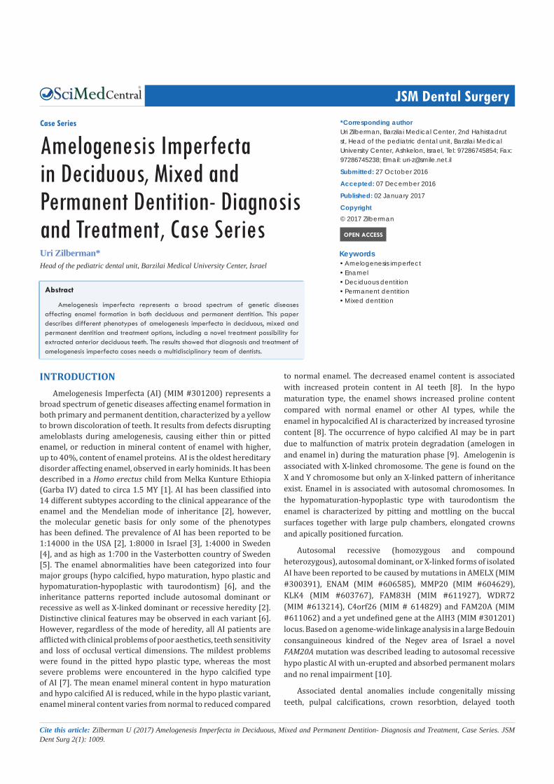
a. Hypocalcified AI. A three years old girl referred to the pediatric clinic at Barzilai Medical University center at Ashkelon due to poor esthetic of her teeth and difficulty of eating and drinking cold or hot liquids. Her medical hystory included Ichthyosis vulgaris (OMIM 146700), (Figure 1) a heterogeneous family of at least 28 generalized, mostly genetic disorders showing dry, thickened, scaly or flaky skin [11]. Treatment for ichthyosis contained of topical application of cream and emollient oils in an attempt to hydrate the skin. The clinical appearance shows deciduous dentition with abnormal morphology and marked attrition (Figures 2-4). The upper incisors show severe attrition and brown discoloration. The upper incisors are the most affected while the lower incisors show normal morphology and light brownish discoloration. The first deciduous molars show attrition and missing enamel while the erupting second deciduous molars show altered morphology with regions of missing enamel. On the upper anterior and bite wing radiographs (Figures 5-7) no distinction can be observed between enamel and dentin on deciduous incisors and molars or on the developing upper permanent incisors and first permanent molars. In comparison with normal radiographs of unaffected child hypo calcified enamel and altered morphology of the deciduous molars can be observed (Figure 8). The soft and rapidly attritioned enamel together with the altered morphology suggested hypocalcified AI. The treatment was performed under generalized anesthesia and the deciduous molars were covered with prefabricated crowns (Figures 9,10), and the upper incisors were extracted.
b. Hypo plastic and hypo mineralized AI- L.O., a 2.5 year old healthy boy. The clinical appearance shows deciduous dentition affected by AI (Figures 11-13). The gross tooth morphology is normal with moth-eaten appearance and with a yellowish discoloration due to very thin and missing enamel.
Figure 1 The forehead of ES, showing the thick scaly skin.
Figure 2 Anterior view.
Figure 3 Lower deciduous teeth.
Figure 4 Upper deciduous teeth.
Figure 5-7 Radiographic analysis of ES deciduous dentition.
Figure 8 Bite-wing radiograph of normal deciduous molars.
Figure 9-10 Post treatment (lower and upper jaws).
Figure 11 Anterior clinical view.
CentralBringing Excellence in Open Access
Zilberman (2017)Email:
JSM Dent Surg 2(1): 1009 (2017) 3/6
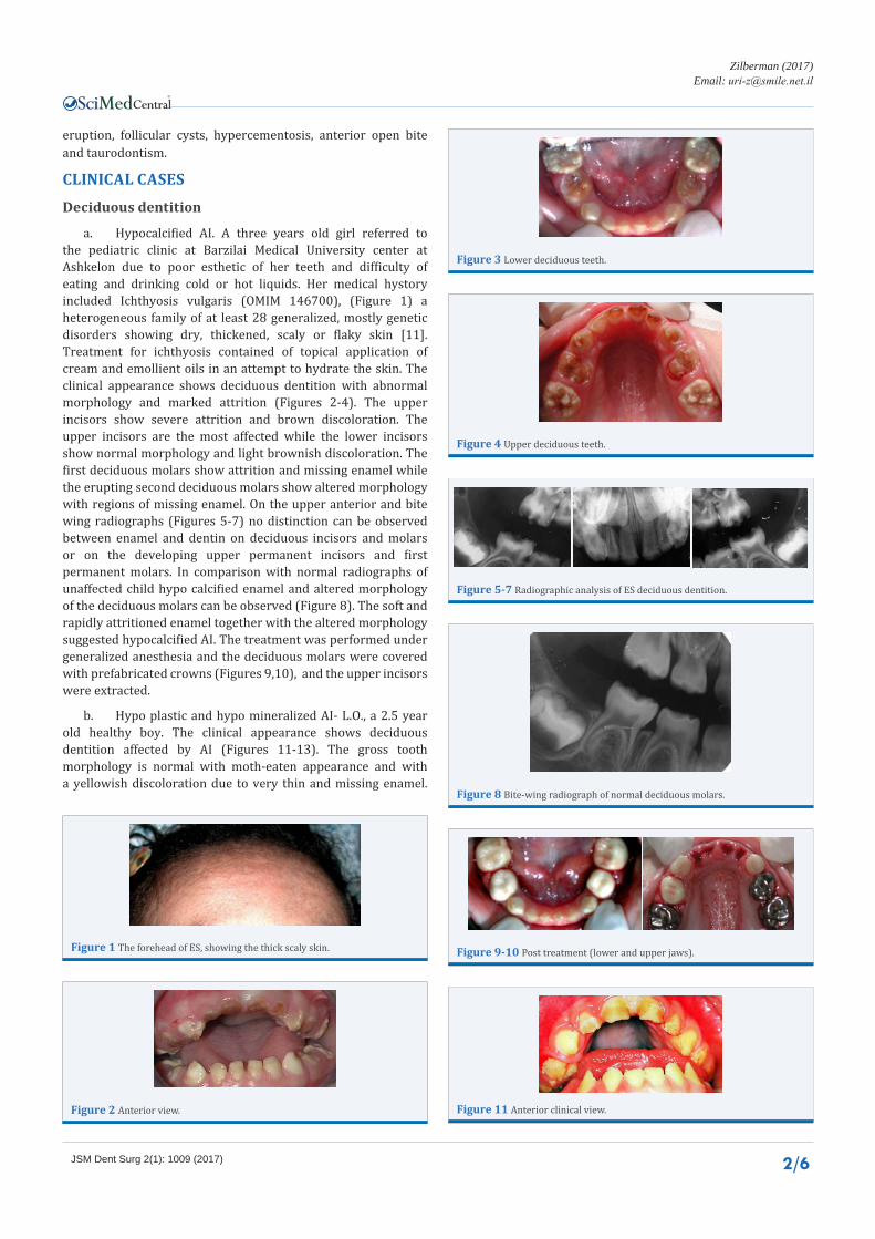
The most affected teeth are the upper incisors while the lower incisors show minimal effect. Remnants of the thin enamel can be observed on the buccal aspect of the upper incisors. The primary molars show no attrition. A class II div 1 occlusion can be observed with marked open bite. On the X-ray of the upper permanent incisors no distinction was observed between the dentin and the enamel. The enamel on the primary incisors is broken on the mesial and distal surfaces. On the bite-wing roentgen graphs (Figures 14,15) the very thin enamel can be observed on the primary molars and permanent molars. The morphology of the pulp chambers is normal. The developing permanent first molar shows similar affected enamel as the primary molars with moth-eaten appearance and thin enamel. The treatment included coverage of all deciduous molars with preformed crowns and restoration of the upper incisors with composites under general anesthesia (Figures 16-19).
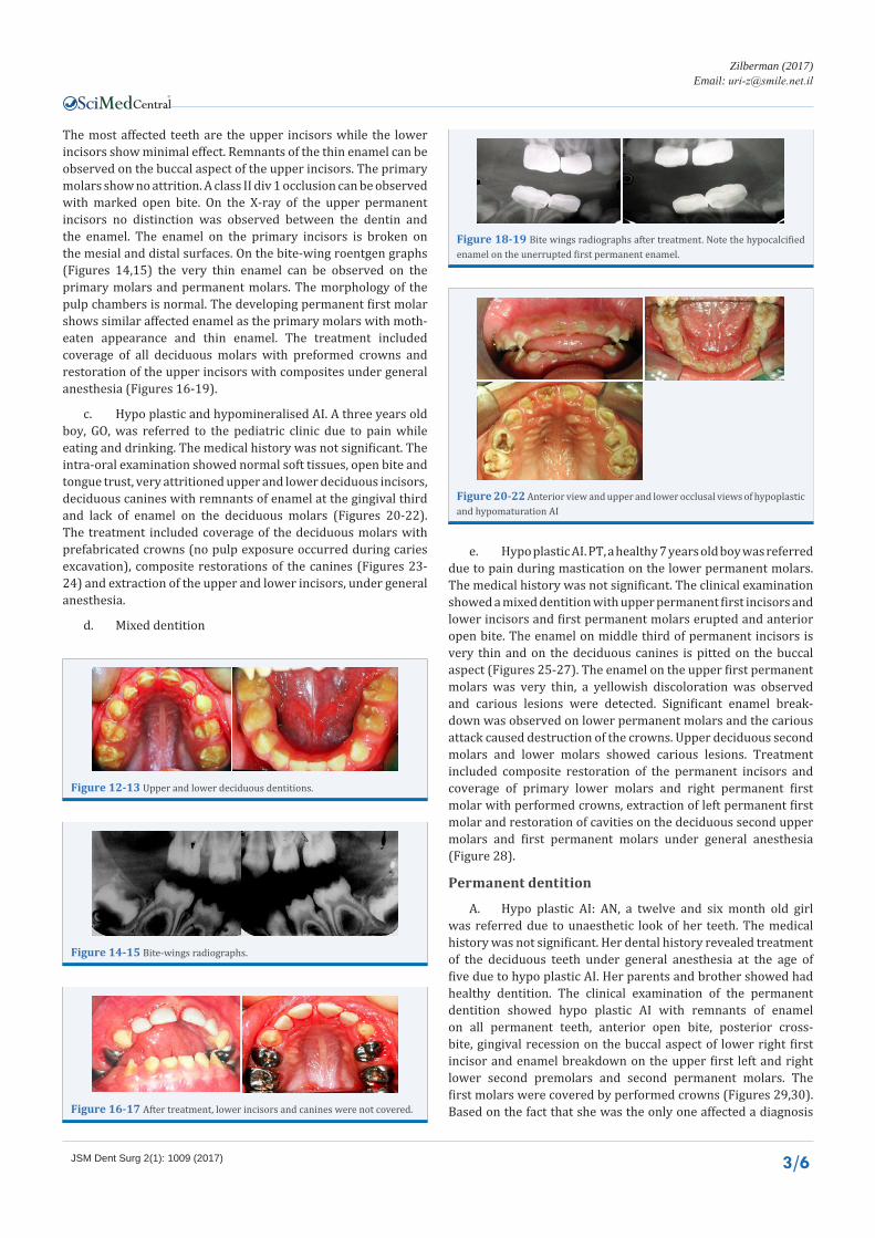
c. Hypo plastic and hypomineralised AI. A three years old boy, GO, was referred to the pediatric clinic due to pain while eating and drinking. The medical history was not significant. The intra-oral examination showed normal soft tissues, open bite and tongue trust, very attritioned upper and lower deciduous incisors, deciduous canines with remnants of enamel at the gingival third and lack of enamel on the deciduous molars (Figures 20-22). The treatment included coverage of the deciduous molars with prefabricated crowns (no pulp exposure occurred during caries excavation), composite restorations of the canines (Figures 23-24) and extraction of the upper and lower incisors, under general anesthesia.
d. Mixed dentition
e. Hypo plastic AI. PT, a healthy 7 years old boy was referred due to pain during mastication on the lower permanent molars. The medical history was not significant. The clinical examination showed a mixed dentition with upper permanent first incisors and lower incisors and first permanent molars erupted and anterior open bite. The enamel on middle third of permanent incisors is very thin and on the deciduous canines is pitted on the buccal aspect (Figures 25-27). The enamel on the upper first permanent molars was very thin, a yellowish discoloration was observed and carious lesions were detected. Significant enamel break-down was observed on lower permanent molars and the carious attack caused destruction of the crowns. Upper deciduous second molars and lower molars showed carious lesions. Treatment included composite restoration of the permanent incisors and coverage of primary lower molars and right permanent first molar with performed crowns, extraction of left permanent first molar and restoration of cavities on the deciduous second upper molars and first permanent molars under general anesthesia (Figure 28).
Permanent dentition
A. Hypo plastic AI: AN, a twelve and six month old girl was referred due to unaesthetic look of her teeth. The medical history was not significant. Her dental history revealed treatment of the deciduous teeth under general anesthesia at the age of five due to hypo plastic AI. Her parents and brother showed had healthy dentition. The clinical examination of the permanent dentition showed hypo plastic AI with remnants of enamel on all permanent teeth, anterior open bite, posterior cross-bite, gingival recession on the buccal aspect of lower right first incisor and enamel breakdown on the upper first left and right lower second premolars and second permanent molars. The first molars were covered by performed crowns (Figures 29,30). Based on the fact that she was the only one affected a diagnosis
Figure 12-13 Upper and lower deciduous dentitions.
Figure 14-15 Bite-wings radiographs.
Figure 16-17 After treatment, lower incisors and canines were not covered.
Figure 18-19 Bite wings radiographs after treatment. Note the hypocalcified enamel on the unerrupted first permanent enamel.
Figure 20-22 Anterior view and upper and lower occlusal views of hypoplastic and hypomaturation AI

CentralBringing Excellence in Open Access
Zilberman (2017)Email:
JSM Dent Surg 2(1): 1009 (2017) 4/6
Figure 23-24 Upper and lower occlusal views after treatment, before extraction of the deciduous incisors.
Figure 25-27 Hypoplastic AI in mixed dentition.
Figure 28 Anterior view after treatment.
Figure 29-30 Clinical views of hypoplastic AI in permanent dentition.
Figure 31 Panoramic radiograph.
Figure 32 Clinical view after 23 years of follow-up.
of X-linked hypoplastic AI was made. The panoramic radiograph revealed very thin enamel on the teeth (Figure 31). The initial treatment included extraction of the upper right second molar and coverage of the remaining second molars with performed crowns, root canal treatment of right second lower premolar and composite restorations of all incisors, canines and premolars. The posterior cross-bite was treated orthodontically and at the age of sixteen all the anterior teeth and premolars were covered by ceramic fussed to semi precious metal crowns. During the follow-up period the upper third molars were extracted. After 23 years of follow-up, at the age of 39, the ceramic and performed crowns showed very good clinical and radiographic results, both esthetically and functional (Figure 32,33). The roots of the first molars are short and the crowns of the impacted lower third molars showed resorption.
Her husband and two children showed no signs of AI in the deciduous or permanent dentition.
B. Autosomal recessive hypo plastic AI with unerupted and resorbed permanent teeth due to mutation in the FAM20A gene.
Two sixteen year old Bedouin Israeli girls from the same tribe were examined at Barzilai pediatric dental clinic with a major complaint of “ugly teeth”. Apart from the dentition pathology, the medical history and physical exam were normal. Clinical examination demonstrated brown upper anterior and lower teeth with hypoplastic enamel and missing permanent molars and premolars (Figures 34,35). The panoramic view showed impacted and crown absorbed permanent molars and un-erupted canines and premolars (Figures 36,37). Very thin enamel on the erupted teeth was observed. Based on the clinical and radiographic examination, a diagnosis of hypo plastic amelogenesis imperfecta with un-erupted and absorbed permanent teeth was made. Due to the complexity and extent of the treatment, it was performed under general anesthesia for both girls. The stage one treatment included extraction of deciduous teeth, coverage of all erupted teeth with composite material on anterior teeth and stainless steel crowns [SSC] on the premolars. Girl A- the upper left first molar was exposed and covered with SSC. Root canal treatment was performed when necessary. Girl B- two premolars (upper right first and lower left second) were extracted and repositioned so that the crowns were exposed and the apical part of the socket was filled with artificial bone. On the lower incisors orthodontic brackets were bonded for up-righting so that parallelism can be obtained (Figures 38-41). After six months 24 ceramic crowns were bonded to both girls (Figures 42,43).
DISCUSSIONAI has many forms and affects the well-being of children at a
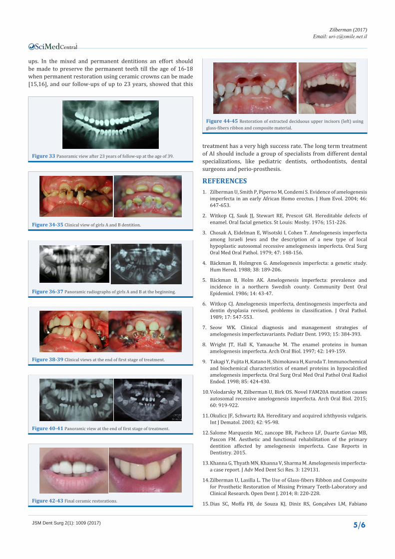
very early stage of development. As early as two to three years of age AI may cause masticatory malfunction and esthetic problems. The treatment of AI in primary dentition should include full coverage of the molar teeth and if possible esthetic restoration of the anterior teeth [12,13]. If the anterior teeth cannot be restored and extractions are needed, a restorative procedure can be using glass-fibers ribbons to produce an esthetic bridge to be bonded to the canines [14], but this procedure should take into consideration the age of the child and periodical follow-
CentralBringing Excellence in Open Access
Zilberman (2017)Email:
JSM Dent Surg 2(1): 1009 (2017) 5/6
Figure 33 Panoramic view after 23 years of follow-up at the age of 39.
Figure 34-35 Clinical view of girls A and B dentition.
Figure 36-37 Panoramic radiographs of girls A and B at the beginning.
Figure 38-39 Clinical views at the end of first stage of treatment.
Figure 40-41 Panoramic view at the end of first stage of treatment.
Figure 42-43 Final ceramic restorations.
ups. In the mixed and permanent dentitions an effort should be made to preserve the permanent teeth till the age of 16-18 when permanent restoration using ceramic crowns can be made [15,16], and our follow-ups of up to 23 years, showed that this
treatment has a very high success rate. The long term treatment of AI should include a group of specialists from different dental specializations, like pediatric dentists, orthodontists, dental surgeons and perio-prosthesis.
REFERENCES1. Zilberman U, Smith P, Piperno M, Condemi S. Evidence of amelogenesis
imperfecta in an early African Homo erectus. J Hum Evol. 2004; 46: 647-653.
2. Witkop CJ, Sauk JJ, Stewart RE, Prescot GH. Hereditable defects of enamel. Oral facial genetics. St Louis: Mosby. 1976; 151-226.
3. Chosak A, Eidelman E, Wisotski I, Cohen T. Amelogenesis imperfecta among Israeli Jews and the description of a new type of local hypoplastic autosomal recessive amelogenesis imperfecta. Oral Surg Oral Med Oral Pathol. 1979; 47: 148-156.
4. Bäckman B, Holmgren G. Amelogenesis imperfecta: a genetic study. Hum Hered. 1988; 38: 189-206.
5. Bäckman B, Holm AK. Amelogenesis imperfecta: prevalence and incidence in a northern Swedish county. Community Dent Oral Epidemiol. 1986; 14: 43-47.
6. Witkop CJ. Amelogenesis imperfecta, dentinogenesis imperfecta and dentin dysplasia revised, problems in classification. J Oral Pathol. 1989; 17: 547-553.
7. Seow WK. Clinical diagnosis and management strategies of amelogenesis imperfectavariants. Pediatr Dent. 1993; 15: 384-393.
8. Wright JT, Hall K, Yamauche M. The enamel proteins in human amelogenesis imperfecta. Arch Oral Biol. 1997; 42: 149-159.
9. Takagi Y, Fujita H, Katano H, Shimokawa H, Kuroda T. Immunochemical and biochemical characteristics of enamel proteins in hypocalcified amelogenesis imperfecta. Oral Surg Oral Med Oral Pathol Oral Radiol Endod. 1998; 85: 424-430.
10. Volodarsky M, Zilberman U, Birk OS. Novel FAM20A mutation causes autosomal recessive amelogenesis imperfecta. Arch Oral Biol. 2015; 60: 919-922.
11. Okulicz JF, Schwartz RA. Hereditary and acquired ichthyosis vulgaris. Int J Dematol. 2003; 42: 95-98.
12. Salome Marquezin MC, zancope BR, Pacheco LF, Duarte Gaviao MB, Pascon FM. Aesthetic and functional rehabilitation of the primary dentition affected by amelogenesis imperfecta. Case Reports in Dentistry. 2015.
13. Khanna G, Thyath MN, Khanna V, Sharma M. Amelogenesis imperfecta- a case report. J Adv Med Dent Sci Res. 3: 129131.
14. Zilberman U, Lasilla L. The Use of Glass-fibers Ribbon and Composite for Prosthetic Restoration of Missing Primary Teeth-Laboratory and Clinical Research. Open Dent J. 2014; 8: 220-228.
15. Dias SC, Moffa FB, de Souza KJ, Diniz RS, Gonçalves LM, Fabiano
Figure 44-45 Restoration of extracted deciduous upper incisors (left) using glass-fibers ribbon and composite material.

CentralBringing Excellence in Open Access
Zilberman (2017)Email:
JSM Dent Surg 2(1): 1009 (2017) 6/6
Zilberman U (2017) Amelogenesis Imperfecta in Deciduous, Mixed and Permanent Dentition- Diagnosis and Treatment, Case Series. JSM Dent Surg 2(1): 1009.
Cite this article
Perez, et al. Full mouth rehabilitation of a patient with amelogenesis imperfecta: a case report. J Int Oral Health. 2016; 8: 385-388.
16. Sabandal MM, Schafer A, Edgar B. Amelogenesis imperfecta: review of diagnostic findings and treatment concepts. Odontol. 2016; 104: 245-256.