Embed Size (px)
Citation preview

AMERICAN THORACIC SOCIETYDOCUMENTS
Evaluating Molecular Biomarkers for the Early Detection of LungCancer: When Is a Biomarker Ready for Clinical Use?An Official American Thoracic Society Policy StatementPeter J. Mazzone, Catherine Rufatto Sears, Doug A. Arenberg, Mina Gaga, Michael K. Gould, Pierre P. Massion,Vish S. Nair, Charles A. Powell, Gerard A. Silvestri, Anil Vachani, and Renda Soylemez Wiener; on behalf of the ATSAssembly on Thoracic Oncology
THIS OFFICIAL POLICY STATEMENT OF THE AMERICAN THORACIC SOCIETY WAS APPROVED JULY 2017
Background:Molecular biomarkers have the potential to improve thecurrent stateof early lungcancerdetection.Thegoal of this projectwas todevelop a policy statement that provides guidance about the level ofevidence required to determine that a molecular biomarker, used tosupport early lung cancer detection, is appropriate for clinical use.
Methods: An ad hoc project steering committee was formed, toinclude individualswith expertise in the early detectionof lung cancerand molecular biomarker development, from inside and outside ofthe Assembly on Thoracic Oncology. Key questions, generated fromthe results of a survey of the project steering committee, werediscussed at an in-person meeting. Results of the discussion weresummarized in a policy statement that was circulated to the steeringcommittee and revised multiple times to achieve consensus.
Results:With a focus on the clinical applications of lung cancerscreening and lung nodule evaluation, the policy statement outlinescategories of results that should be reported in the early phases of
molecular biomarker development, discusses the level of evidencethat would support study of the clinical utility, describes theoutcomes that should be proven to consider a molecularbiomarker clinically useful, and suggests study designs capable ofassessing these outcomes.
Conclusions:The applicationofmolecular biomarkers to assistwiththe early detection of lung cancer has the potential to substantiallyimprove our ability to select patients for lung cancer screening,and to assist with the characterization of indeterminate lungnodules. We have described relevant considerations and havesuggested standards to apply when determining whether a molecularbiomarker for the early detection of lung cancer is ready forclinical use.
Keywords: clinical utility; outcomes; study design; lung cancerscreening; lung nodules
ContentsOverviewIntroductionMethodsBackground
Clinical Utility Phase ofBiomarker Development
Cost-Effectiveness
ResultsDiscussion: Clinical Applications ofMolecular Biomarkers for the EarlyDetection of Lung Cancer
Lung Cancer ScreeningLung Nodule Evaluation
ConclusionsAppendix 1: DefinitionsAppendix 2: Additional ReferencesUsed during the Project
Overview
Molecular biomarkers, developed to assistwith the early detection of lung cancer, maybe applied in the settings of lung cancerscreening and lung nodule evaluation. True-positive and -negative results may benefitpatients, whereas false-positive and-negative results may lead to harm. To haveclinical utility, the molecular biomarkermust affect clinical management decisions
An Executive Summary of this document is available at http://www.atsjournals.org/doi/suppl/10.1164/rccm.201708-1678ST
Correspondence and requests for reprints should be addressed to Peter J. Mazzone, M.D., M.P.H., Respiratory Institute, Cleveland Clinic, 9500 Euclid Avenue,A90, Cleveland, OH 44195. E-mail: [email protected]
Am J Respir Crit Care Med Vol 196, Iss 7, pp e15–e29, Oct 1, 2017
Copyright © 2017 by the American Thoracic Society
DOI: 10.1164/rccm.201708-1678ST
Internet address: www.atsjournals.org
American Thoracic Society Documents e15

in a manner that improves clinicaloutcomes. This policy statement describesitems to consider when determiningwhether the evidence supports clinical use ofan early lung cancer detection molecularbiomarker. Key points made in thestatement include the following:
d Key results from studies in all phases ofbiomarker development may influence theinterpretation of clinical utility. A list ofresults that should be reported is provided.
d Calculations are available to helpdetermine the minimal accuracy of amolecular biomarker that could lead to afavorable clinical impact, and thus justifyinvestment in a clinical utility study. Anexample is provided.
d A clinically useful molecular biomarkerapplied as the initial test for lung cancerscreening may improve the balance ofbenefit to harm by identifying those mostlikely to benefit from screening whileminimizing exposure to the harmsamong those least likely to benefit.
d To be considered clinically useful, amolecular biomarker used to identifypatients eligible for lung cancer screeningmust lead to:∘ Fewer lung cancer deaths in thepopulation tested compared with thecurrent standard of care for thatpopulation, without substantiallyincreasing harms and expense, or
∘ A similar number of lung cancer deathsin the population tested compared withthe current standard of care for thatpopulation, with fewer harms or lessexpense.
d A clinically useful molecular biomarkerapplied to the evaluation of lung nodulesmay lead to expedited therapy for earlylung cancer and/or fewer aggressiveinterventions in patients with benignlung nodules.
d To be considered clinically useful, amolecular biomarker used to assist withlung nodule management must lead to:∘ Earlier diagnosis of malignant noduleswithout substantially increasing thenumber of procedures performed onpatients with benign nodules, or
∘ Fewer procedures for patients withbenign nodules without substantiallydelaying the diagnosis of cancer inpatients with malignant nodules.
d Biomarker-stratified, enrichment, andbiomarker strategy study designs mayprovide the evidence required to assess
the outcomes of interest whendetermining whether a lung cancerscreening or lung nodule managementmolecular biomarker is clinically useful.
Introduction
Molecular biomarkers have the potentialto improve the current state of early lungcancer detection. Biomarkers capable ofidentifying the presence of presymptomaticlung cancer may help to optimize patientselection for lung cancer screening.Biomarkers capable of characterizingpulmonary nodules may help to expeditetherapy of early-stage lung cancerswhile minimizing the harms of evaluatingpatients with benign disease.
A new lung cancer biomarkerwill be clinically useful if it fulfills anunmet clinical need or provides anadvantage over standard practice (e.g.,more accurate, simpler to use, providesresults more rapidly, lowers costs). Theultimate measure of lung cancer biomarkerperformance is whether and how the resultaffects clinical management decisionsand clinical outcomes (1). Even biomarkersthat are sensitive and specific enough tobe considered accurate by most cliniciansmay not impact clinical care, or mayadversely impact clinical care (e.g., Table 1).
The goal of this project was to developa policy statement that provides guidanceabout the evidence required to determinewhether a molecular biomarker for theearly detection of lung cancer isappropriate for clinical use.
Methods
During the first stage of the project, theproject co-chairs (P.J.M. and C.R.S.)developed an overview of biomarker
development principles and the currentstate of lung cancer risk prediction, earlydetection, and diagnosis, as well as a surveywith questions related to each of the phasesof biomarker development. A conferencecall was held to introduce the project to thesteering committee. The documents werethen circulated to the steering committee forreview. The survey was completed andreturned by 11 of the 12 steering committeemembers, and the results were collated. Thesurvey responses were used to generatediscussion questions.
The project ad hoc steering committeewas selected to include individuals withan interest in lung cancer biomarkerdevelopment and expertise in the variousphases of biomarker development.Representatives of American Thoracic Societyinternational partner societies (ChineseThoracic Society, European RespiratorySociety, and Japanese Respiratory Society)and the U.S. Food and Drug Administrationwere invited to participate on the steeringcommittee. Potential conflicts of interest weredisclosed and managed in accordance withthe policies and procedures of the AmericanThoracic Society. The committee had a face-to-face meeting on May 13, 2016 at theannual international conference of theAmerican Thoracic Society. The meetingincluded a presentation from a patientadvocate, and presentations related to thephases of biomarker development. The formalpresentations were followed by discussionsguided by questions generated from thesurvey responses (see below).
Separately, the steering committeereviewed definitions, reportingconsiderations, target conditions, targetpopulations, reference standards, and thepotential impact of true-positive/-negativeand false-positive/-negative results withineach potential clinical application. A draftof the current document was developed by
Table 1. Potential for Harm from an Accurate Biomarker
Nodule Malignant Nodule Benign Total
Test result positive 9 99 108Test result negative 1 891 892Total 10 990 1,000
If a molecular biomarker is 90% sensitive and 90% specific for the detection of a malignant lungnodule, it would generally be considered an accurate test. If applied to a population of patients withlung nodules with a 1% probability of malignancy (e.g., solid nodules 4–8 mm in diameter), 92% of allpositive test results would be false positive, potentially leading to more aggressive evaluation of manypatients without lung cancer, with physical, social, and behavioral consequences.
AMERICAN THORACIC SOCIETY DOCUMENTS
e16 American Journal of Respiratory and Critical Care Medicine Volume 196 Number 7 | October 1 2017
the project co-chairs and then circulated forreview. The draft was modified severaltimes, based on written feedback from theproject steering committee and on feedbackreceived during a phone conference. Relevantdefinitions and additional references arepresented in Appendices 1 and 2.
Background
Clinical Utility Phase of BiomarkerDevelopmentThe clinical utility phase of biomarkerdevelopment follows successful completionof biomarker discovery, analytical validationof the biomarker assay, and clinicalvalidation of the accuracy of the biomarker(Figure 1). A biomarker should be used inclinical practice only if it reliably adds to aclinician’s judgment, resulting in a morefavorable clinical outcome for the targetpopulation. Biomarker accuracy is not enoughto imply clinical utility. Clinical utility isdependent on how the test result affectssubsequent clinical decisions and outcomes.Both the benefit of clinical decisions influencedby true-positive and true-negative results, aswell as the harms of clinical decisionsinfluenced by false-positive and false-negativeresults, must be considered. Clinical decisionsbased on misleading biomarker results mayexpose the patient to adverse consequencesand increase the cost of care.
Outcomes of relevance in clinical utilitytesting include the frequency and manner inwhich the biomarker impacts clinicaldecisions (use of other testing, treatmentdecisions) and the consequences of thosedecisions (complications from testing andtreatment, patient quality of life, timelinessof accurate diagnosis, survival). There aremany challenges to evaluating the clinicalutility of a biomarker in randomizedcontrolled trials, as is typically required fordetermination of the therapeutic efficacy ofa drug or intervention.
Research designs capable ofdetermining the clinical utility of abiomarker have been described (2–5)(Figures 2 and 4):
1. Biomarker stratified: All patients arerandomly assigned to one of two ormore management options regardless ofbiomarker status. This design is themost efficient way to determine the bestmanagement option for each biomarkersubgroup when there is no evidence thatone option is preferred. Biomarker-stratified designs include the following:a. Prospective controlled clinical trials
where the test result is used tostratify patients who are thenrandomized to the managementoptions being studied. Thebiomarker is not used for patientmanagement, but the primaryobjective of the clinical trial is toassess the clinical utility of the testfor its intended use. Limitations ofthis pathway include the potential forbias from randomly missing testresults (can be minimized in thestatistical analysis), limited statisticalpower to detect small effects, and aninability to test strategies with a largenumber of management options.
b. Prospective–retrospective studiesusing archived specimens frompreviously conducted controlledclinical trials that address theintended use of the test. Thispathway is the least time andresource intensive. It is appropriatefor biomarkers that can be evaluatedretrospectively in a reliable way.Limitations of this pathway includethe difficulty in finding adequatearchived specimens (quality andnumber) and the potential for biasfrom randomly missing test results.Because missing specimens may notbe equally distributed between therandomized management arms, the
study design must ensure that anadequate number of patient samples isavailable for each biomarker subgroup.
2. Enrichment: Patients in one biomarker-defined subgroup (e.g., biomarkerpositive) are randomly assigned to oneof two or more management options,and the other biomarker-definedsubgroup (e.g., biomarker negative) ismanaged on the basis of standard of carefor the population. This design isapplicable in settings where there isevidence to suggest that the benefit of amanagement option would be limited toone biomarker-defined subgroup. Theenrichment study design can onlyaddress questions about the bestmanagement strategy within thebiomarker-defined subgroup that israndomized to the studied managementoptions. It is an efficient study designwhen the assumption that benefit willonly be seen in one biomarker-definedsubgroup is correct, and when theincidence of the condition in question islow. This design cannot completelyaddress clinical utility as it does notassess the potential utility of themanagement strategies in the otherbiomarker-defined subgroup.
3. Biomarker strategy: Patients arerandomly assigned to a biomarker-directed arm where the managementstrategy is based on the biomarkerresult, or to a control arm. Patients inthe control arm may be assigned tostandard of care management, or berandomly assigned to one of twoor more management strategies,independent of the biomarker result.This design determines whetherbiomarker-directed management isbetter than standard of care management.This design most directly assesses theclinical utility of a biomarker but isthe most resource and time intensiveand carries the highest risk to the studyparticipants. This design may notcompletely assess clinical utility asit does not determine whether themanagement strategies assessed in thebiomarker-directed arm are better thanstandard of care regardless of thebiomarker result.
Evidence of clinical utility may notresult in widespread adoption of abiomarker, particularly if measurement ofthe biomarker is costly, requires nonroutine
Clinical UtilityClinical ValidationDiscovery Analytical Validation
Pre-Analytical Analytical Post-Analytical
Figure 1. Phases of biomarker development. (Top row) Phases of biomarker development. (Bottomrow) Process considerations. This applies to the development of all categories of biomarkers: riskprediction, cancer detection, and diagnosis.
AMERICAN THORACIC SOCIETY DOCUMENTS
American Thoracic Society Documents e17
sample collection, is technically difficult, orrequires a change in the culture of clinicalpractice.
Cost-EffectivenessCost-effectiveness analysis (CEA) is animportant tool in determining the impact ofthe use of a biomarker. CEA measures maybe used to guide the development ofrecommendations for clinical use of abiomarker from professional societies,government, and industry payer sources.Some regulatory authorities (e.g., the U.S.Food and Drug Administration) do notordinarily consider monetary costs (e.g.,medical bills, societal or insurer costs), buthave considered health costs (e.g., mortalityor morbidity) (6). This practice is likely toevolve, with monetary costs more routinelyconsidered by payers.
The measurement and interpretation ofcost-effectiveness is complex. It requiresaccurate estimates of the net cost ofimplementing a biomarker for a givenoutcome. CEA is only relevant if clinicalutility has been proven (e.g., reducedmortality in a cancer screening trial). CEAuses data from published studies todetermine measures such as sensitivity,specificity, and disease prevalence. It isimperative that CEA be measured in thecorrect clinical context across diverse ethnicand social groups, and be compared withaccepted clinical practices (incremental cost-effectiveness ratio). It is difficult to comparethe cost-effectiveness of one biomarker orintervention with another if differentmeasures are used. For that reason, uniformmeasurement and reporting of cost-effectiveness using quality-adjusted life-years(QALYs) or similar measures arerecommended. For example, sensitivityanalyses should be performed and cost-effectiveness acceptability curves published toaccount for variability in the costs andoutcomes by location, population, andclinical context (7–11).
Results
The following questions were discussed,with group consensus summarized aftereach question:
1. Should we organize our comments bybiomarker category (risk assessment,cancer detection, and diagnosis) or bypotential clinical use (risk mitigation,
screening, and symptom and lungnodule evaluation)?
The project steering committee recognizedthat biomarkers within a given biomarkercategory may have more than one clinicaluse (Table 2). As this statement is intendedto be a guide to help determine when amolecular biomarker is ready for clinicaluse for the early detection of lung cancer,the committee determined that it wasbest to organize our comments by themost relevant clinical applications (lungcancer screening and lung noduleevaluation).
2. What level of evidence would supportthe clinical use of a validated lungcancer biomarker? Does the level ofevidence differ based on the intendedclinical use?
Large, well-designed and conductedstudies, capable of determining the impactof testing on clinical decisions andoutcomes, are required to confirm theclinical utility of a molecular biomarker.The clinical application, intended usepopulation, consequences of true and falseresults, and current state of clinical care inthe field can influence the required level ofevidence. The steering committee hasprovided guidance about the level ofevidence (study outcomes and study design)that would support the clinical use of abiomarker for lung cancer screening andlung nodule evaluation (fit for purpose) inthe clinical application sections of theDISCUSSION portion of this document.
3. a. What amount of time before adiagnosis of lung cancer wouldbe considered adequate toseparate a risk predictionbiomarker from a cancerdetection biomarker?
b. Should a biomarker of risk of“being diagnosed” with lung canceror of “dying of” lung cancer bedefined?
c. Is “being diagnosed” with lungcancer or “developing” lung cancera better way to define a riskprediction biomarker?
The steering committee recognized thatit is difficult to distinguish between abiomarker that predicts the risk of lungcancer developing over a period of time anda cancer detection biomarker. A biomarkerwhose intended use is to determine the risk
of developing lung cancer, may identify lungcancer that is present but cannot be detectedby currently available means. To be definedas a biomarker of risk, the referencestandard available at the time the biomarkersample is collected should be used to excludethe presence of lung cancer. A time intervalbetween when the biomarker sample wascollected and lung cancer was identified maybe established to add further reassurancethat the cancer was not present at the timeof sample collection, or the accuracy ofthe biomarker at various time intervalsfrom collection to diagnosis can beassessed.
There are different arguments for andagainst defining a risk prediction biomarkeras one that would predict the risk ofdeveloping, being diagnosed with, or dyingof lung cancer. The accuracy of thebiomarker would be influenced by thedefinition. Not all lung cancers will bediagnosed, making it impossible to confirmhow many cancers develop (althoughstatistical models can be used to estimatethe number). A definition that requiresdeath from lung cancer would excludeoverdiagnosed cancers but would not be ableto address the influence of treatment orcompeting risks of death on the accuracy ofthe biomarker. The steering committeeconcluded that it is more important that thedefinition of a risk prediction biomarker beclearly described when it is used or studiedthan it is to mandate a single standardizeddefinition. A general definition is providedin Table 2.
4. a. How do we define early detection oflung cancer?
b. Should we add language aboutpotential lethality of the cancer?Should biomarkers applied for theearly detection of lung cancer beconsidered only for asymptomaticindividuals or include a group withsymptoms undergoing evaluation?
The World Health Organization describestwo components of early detection ofcancer (12). The first is early diagnosisthrough prompt action when symptoms orsigns of cancer are present. The second isthrough screening someone at risk forhaving cancer but who is free of symptomsor signs. The steering committee recognizedthese and other acceptable definitions, suchas diagnosing lung cancer at a stage that ismore amenable to successful treatment(e.g., stage I, localized or locoregional) or
AMERICAN THORACIC SOCIETY DOCUMENTS
e18 American Journal of Respiratory and Critical Care Medicine Volume 196 Number 7 | October 1 2017
diagnosing lung cancer earlier than it wouldotherwise have been diagnosed. There wassubstantial debate about whether thedefinition of early detection of lung cancershould include lung cancer diagnosis whensymptoms or signs of cancer are present, oronly include lung cancer diagnosis inasymptomatic individuals. A consensusdefinition of early detection of lung cancerwas not developed. We concluded that it ismore important that the definition of earlydetection be clearly described within studiesassessing the individual biomarkers, withdistinction between biomarker-detectedlung cancer in patients with and withoutsymptoms.
To be clinically useful, an earlydetection biomarker should detect lungcancer that could be lethal if not detectedand treated early. This should be consideredwhen interpreting studies of early detectionbiomarkers. For the reasons described inquestion 3 above, the steering committee didnot believe it was practical to mandatethat lethality of the cancer be includedin the definition of an early detectionbiomarker.
5. Should we include guidance about howaccurate a clinically validatedbiomarker should be to considerevaluating it for clinical utility for each
potential clinical use of the biomarker?Do we compare potentially usefulaccuracies with current standardpractice, prediction tools, and otherbiomarkers currently in use (e.g.,positron emission tomography [PET]for lung nodule management)?
To justify the investment required tocomplete the clinical utility phase ofbiomarker development, the steeringcommittee members agreed that it would behelpful to provide guidance about theminimal accuracy, as assessed in the clinicalvalidation phase, that could lead to a positiveclinical impact. This minimal accuracywould vary by clinical application, and beguided by an understanding of theconsequences of true and false results (13).Several methods have been described toassist with the estimate of minimalaccuracy, such as calculation of theoptimal slope of the receiver operatingcharacteristic curve (14). Here we describea formula to help with this estimate (15).
Biomarkers are frequently optimizedfor sensitivity or specificity based on theirintended clinical application. For testswhere a positive biomarker result leads toan action whereas a negative biomarkerresult is associated with standard of carefor the population, the formula states:
sensitivity/(12 specificity)> [(12prevalence)/prevalence]3 harm/benefit,where harm/benefit is the ratio of the netharm of a falsely positive test result to thenet benefit of a true-positive test result.
For tests where a negative result leads toan action other than standard of care for thepopulation, the formula states:specificity/(12 sensitivity)>[prevalence/(12 prevalence)]3harm/benefit, where harm/benefit is theratio of the net harm of a falsely negativetest result to the net benefit of a true-negative test result.
Sensitivity/(12 specificity) is knownas the positive likelihood ratio, andspecificity/(12 sensitivity) is 1 divided bythe negative likelihood ratio. Prevalencerefers to the percentage of cases in theintended use population. The harm/benefitratio in the formulas can be articulated inone of two ways:
a. 1/N, where in the first scenario N is themaximum number of control subjectstesting positive that is tolerated to benefitone case subject testing positive (or in thesecond formula, the maximum numberof case subjects testing negative that isworth the benefit of one control subjecttesting negative). For example, if thebiomarker is used as an upfront lung
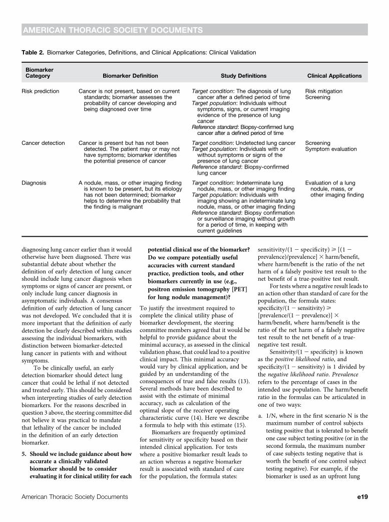
Table 2. Biomarker Categories, Definitions, and Clinical Applications: Clinical Validation
BiomarkerCategory Biomarker Definition Study Definitions Clinical Applications
Risk prediction Cancer is not present, based on currentstandards; biomarker assesses theprobability of cancer developing andbeing diagnosed over time
Target condition: The diagnosis of lungcancer after a defined period of time
Risk mitigation
Target population: Individuals withoutsymptoms, signs, or current imagingevidence of the presence of lungcancer
Screening
Reference standard: Biopsy-confirmed lungcancer after a defined period of time
Cancer detection Cancer is present but has not beendetected. The patient may or may nothave symptoms; biomarker identifiesthe potential presence of cancer
Target condition: Undetected lung cancer ScreeningTarget population: Individuals with orwithout symptoms or signs of thepresence of lung cancer
Symptom evaluation
Reference standard: Biopsy-confirmedlung cancer
Diagnosis A nodule, mass, or other imaging findingis known to be present, but its etiologyhas not been determined; biomarkerhelps to determine the probability thatthe finding is malignant
Target condition: Indeterminate lungnodule, mass, or other imaging finding
Evaluation of a lungnodule, mass, orother imaging findingTarget population: Individuals with
imaging showing an indeterminate lungnodule, mass, or other imaging finding
Reference standard: Biopsy confirmationor surveillance imaging without growthfor a period of time, in keeping withcurrent guidelines
AMERICAN THORACIC SOCIETY DOCUMENTS
American Thoracic Society Documents e19

cancer screening test, N could representthe largest acceptable number of patientswithout lung cancer who have a positivetest result and therefore undergocomputed tomography (CT) screeningfor every one patient with lung cancerwho tests positive.
b. R/(12 R), where R is the risk thresholdabove which procedures consequent topositive testing, and below whichavoidance of procedures consequent tonegative testing, seem worthwhile. Forexample, when evaluating a lung nodulewith a biomarker, the risk threshold Rcould be the probability or risk that alung nodule is malignant at which one isindifferent to choosing surveillanceimaging versus active investigation.
It is important to compare andcombine biomarker accuracies with testingavailable in current practice. The accuracyrequired to impact clinical care is dependenton the needs within a clinical applicationand the potential consequences of theresults. This will vary by clinicalapplication, highlighting the need tojudge the utility of the biomarker on afit-for-purpose basis.
6. a. Should we include guidance aboutcost-effectiveness or leave this tosociety and regulators to decide?
b. Is affordability an adequate outcomeat the expense of accuracy?
It is important that cost-effectivenessanalysis be performed and reported within
each clinical application. Ideally, athird-party independent analysis wouldbe performed to avoid the potential forbias. The steering committee membersdecided that defining a threshold of cost-effectiveness that is acceptable to society isbeyond the scope of this statement.
7. Should we include guidance about thecomponents of study design anddetails of study results that should bereported for all phases of biomarkerdevelopment?
As the focus of this project is on clinicalutility of early detection biomarkers, thesteering committee agreed that we shouldsuggest components of the study design anddetails of the study results that should bereported for the phases of biomarkerdevelopment that most directly influencethe interpretation of clinical utility. Thisincludes clinical validation, clinical utility,and cost-effectiveness (Table 3).
Discussion: ClinicalApplications of MolecularBiomarkers for the EarlyDetection of Lung Cancer
The goal of this project is to provideguidance about the evidence required todetermine that a molecular biomarker forearly lung cancer detection is ready forclinical use. The most relevant clinicalapplications of a biomarker for the earlydetection of lung cancer are the selectionof individuals for further lung cancer
Table 3. Results That Should Be Reported in Various Phases of Biomarker Evaluation
Clinical validationd The sensitivity and specificity of a technically validated biomarker, with fixedinterpretation of the biomarker result, applied to the intended use population, ascompared with the reference standard for the clinical application
d The clinical features of the cancer and control groups in clinical validation studies of thebiomarker compared with the intended use population
d Biomarker results for relevant clinical subgroupsd Biomarker performance compared with and combined with clinical calculators,standard practice, and/or clinician judgment
Clinical utilityd The frequency with which the biomarker result impacts a clinical decisiond The impact (benefit and harm) of patient management decisions on patient outcomeswhen the biomarker is used
Cost-effectivenessd The cost-effectiveness of the biomarker compared with the currently acceptedstandards for the clinical application
Table 4. Models of the Risk of Developing Lung Cancer
Bach et al. (19)Spitz
et al. (20)Cassidyet al. (21)
Tammemagiet al. (22)
Hoggartet al. (23)
Katkiet al. (24)
Source CARET MDA LLP PLCO EPIC PLCOSubjects 18,172 3,852 never- and
ever-smokers1,736 never-and ever-smokers
80,375ever-smokers
169,035ever-smokers
105,556ever-smokers10–60 cpd
for 25–55 yrAge, yr 50–75 20–80 20–80 55–74 35–65 55–74Variables Age Age Age Age Age Age
Asbestos Dust Asbestos BMI Smoking BMISex Emphysema Family history Chest X-ray EducationSmoking Family history Pneumonia COPD Emphysema
Sex Prior cancer Education Family historySmoking Sex Family history Race
Smoking Smoking SexSmoking
Definition of abbreviations: BMI = body mass index; CARET =Carotene and Retinol Efficacy Trial; COPD = chronic obstructive pulmonary disease; cpd =cigarettes per day; EPIC = European Prospective Investigation into Cancer and Nutrition; LLP = Liverpool Lung Project; MDA =M. D. Anderson; PLCO =Prostate, Lung, Colorectal, and Ovarian Screening Trial.Reprinted by permission from Reference 39.
AMERICAN THORACIC SOCIETY DOCUMENTS
e20 American Journal of Respiratory and Critical Care Medicine Volume 196 Number 7 | October 1 2017
screening, and assistance with thecharacterization and management of lungnodules. For these two clinical applications,we discuss currently accepted practice, andthen outline (1) the potential clinical utilityand category of the biomarker, (2) thepotential impact of applying thebiomarker, (3) the level of evidence andaccuracy that could support assessmentof clinical utility, and (4) the level ofevidence required to confirm the clinicalutility of the biomarker.
Lung Cancer Screening
Current state. The National Lung ScreeningTrial (NLST) randomized more than 53,000people at high risk of developing lung cancer(ages, 55–74 yr; active or former smokers ofat least 30 pack-years who had smokedwithin the past 15 yr) to receive a baselineand two annual low-radiation-dose chestCT scans or a baseline and two annualchest radiographs. Fewer people in thechest CT arm died of lung cancer (16).
Several potential harms from lungcancer screening have been described. Forexample, lung nodules are frequentlyidentified. Although usually benign, theiridentification leads to patient distress,additional imaging, and nonsurgical andsurgical biopsies, all with potentialcomplications. Radiation exposure duringchest imaging and the evaluation andtreatment of overdiagnosed lung cancers areother harms that have been reported (17, 18).
Multiple models exist to help estimatethe risk of developing lung cancer (Table 4)(19–24). One model, PLCO (Prostate,Lung, Colorectal, and Ovarian ScreeningTrial, 2012), was evaluated in comparisonwith the NLST criteria, showing marginallyimproved sensitivity with similar specificityfor identifying patients with lung cancer(25). At this time, it is not clear whetherhaving a risk of developing lung cancerequal to that of the cohort obtained usingNLST criteria, based on factors included ina risk model, will result in a similar balanceof benefit to harm from lung cancerscreening.
The cost-effectiveness of lung cancerscreening by low-dose CT, based on theNLST data, was estimated to be $81,000 perQALY gained, which is within the rangetypically considered cost-effective.Sensitivity analysis showed a range of$32,000–$615,000. Factors associated withthis variability included the patient’s lung
cancer risk, sex, age, and smoking status atthe time of screening (26).
Potential clinical utility of a molecularbiomarker applied as an initial test in ascreening context. A clinically usefulmolecular biomarker applied as the initialtest for lung cancer screening may improvethe balance of benefit to harm of lung cancerscreening by identifying those most likely tobenefit from screening while minimizingexposure to harm among those least likely tobenefit.
Category of biomarker. A molecularbiomarker applied as an initial test in ascreening context would be either a riskprediction or cancer detection biomarker.
Potential impact of applying amolecular biomarker as the initial test in ascreening context.d True-positive results—more individuals
with lung cancer could be identified atcurable stages.
d True-negative results—individuals withoutlung cancer (or at low risk of developinglung cancer) could avoid the harmsassociated with low-dose CT screening.
d False-positive results—individualswithout lung cancer (or at low risk ofdeveloping lung cancer) could be enrolledin a low-dose CT screening program andbe exposed to the associated harms.
d False-negative results—individuals with(or who will develop) lung cancer maynot be enrolled in, and thus not have anopportunity to benefit from, a low-doseCT–based lung cancer screening program.Level of evidence suggested to determine
whether a biomarker applied to lung cancerscreening justifies an assessment of clinicalutility.d The biomarker should be more accurate
at identifying patients with (or who willdevelop) potentially curable lung cancer
than current eligibility criteria andavailable clinical risk predictioncalculators, alone or in combination.
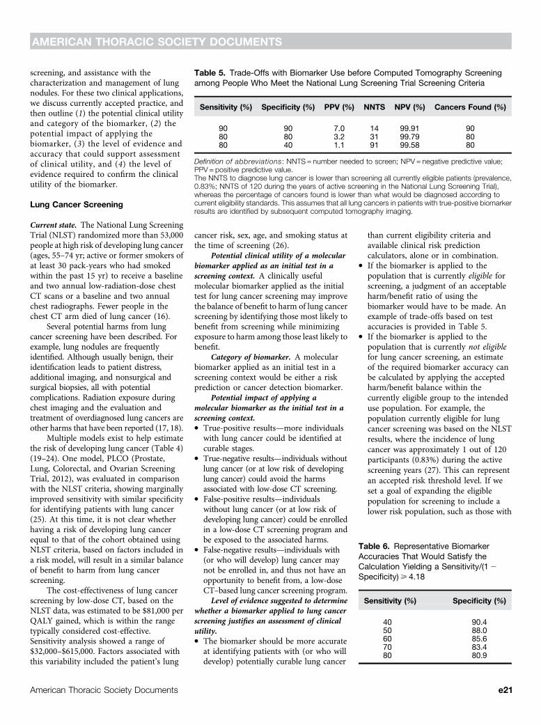
d If the biomarker is applied to thepopulation that is currently eligible forscreening, a judgment of an acceptableharm/benefit ratio of using thebiomarker would have to be made. Anexample of trade-offs based on testaccuracies is provided in Table 5.
d If the biomarker is applied to thepopulation that is currently not eligiblefor lung cancer screening, an estimateof the required biomarker accuracy canbe calculated by applying the acceptedharm/benefit balance within thecurrently eligible group to the intendeduse population. For example, thepopulation currently eligible for lungcancer screening was based on the NLSTresults, where the incidence of lungcancer was approximately 1 out of 120participants (0.83%) during the activescreening years (27). This can representan accepted risk threshold level. If weset a goal of expanding the eligiblepopulation for screening to include alower risk population, such as those with
Table 5. Trade-Offs with Biomarker Use before Computed Tomography Screeningamong People Who Meet the National Lung Screening Trial Screening Criteria
Sensitivity (%) Specificity (%) PPV (%) NNTS NPV (%) Cancers Found (%)
90 90 7.0 14 99.91 9080 80 3.2 31 99.79 8080 40 1.1 91 99.58 80
Definition of abbreviations: NNTS = number needed to screen; NPV = negative predictive value;PPV = positive predictive value.The NNTS to diagnose lung cancer is lower than screening all currently eligible patients (prevalence,0.83%; NNTS of 120 during the years of active screening in the National Lung Screening Trial),whereas the percentage of cancers found is lower than what would be diagnosed according tocurrent eligibility standards. This assumes that all lung cancers in patients with true-positive biomarkerresults are identified by subsequent computed tomography imaging.
Table 6. Representative BiomarkerAccuracies That Would Satisfy theCalculation Yielding a Sensitivity/(12Specificity)> 4.18
Sensitivity (%) Specificity (%)
40 90.450 88.060 85.670 83.480 80.9
AMERICAN THORACIC SOCIETY DOCUMENTS
American Thoracic Society Documents e21
a clinical risk of 1 out of 500 participants(0.2%) having lung cancer, the formuladescribed in question 5 of RESULTS showsthe minimum positive likelihood ratioas sensitivity/(12 specificity)> [(1 –0.002)/0.002]3 0.0083/(1 – 0.0083) =4.18. Representative biomarker accuraciesthat would meet this standard areshown in Table 6.Evidence required for a molecular
biomarker to be considered clinically usefulin the context of lung cancer screening. Tobe considered clinically useful, a molecularbiomarker used to identify patients eligiblefor lung cancer screening must lead to:
d Fewer lung cancer deaths in thepopulation tested compared with thecurrent standard of care for thatpopulation, without substantiallyincreasing harms and expense, or
d A similar number of lung cancer deathsin the population tested compared withthe current standard of care for thatpopulation, with fewer harms or less expense.
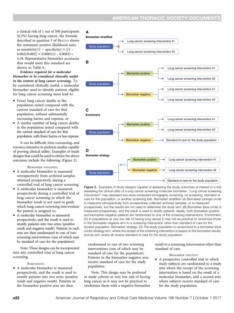
It can be difficult, time consuming, andresource intensive to perform studies capableof proving clinical utility. Examples of studydesigns that could be used to obtain the aboveoutcomes include the following (Figure 2):
BIOMARKER STRATIFIED.d A molecular biomarker is measured
retrospectively from archived samplesobtained prospectively during acontrolled trial of lung cancer screening.
d A molecular biomarker is measuredprospectively during a controlled trial oflung cancer screening in which thebiomarker result is not used to guidewhich lung cancer screening interventionthe patient is assigned to.
d A molecular biomarker is measuredprospectively, and the result is used tostratify patients into two arms (positiveresult and negative result). Patients in eacharm are then randomized to one of twoscreening interventions (one of which maybe standard of care for the population).
Note: These designs can be incorporatedinto any controlled trial of lung cancerscreening.
ENRICHMENT.d A molecular biomarker is measured
prospectively, and the result is used tostratify patients into two arms (positiveresult and negative result). Patients inthe biomarker-positive arm are then
randomized to one of two screeninginterventions (one of which may bestandard of care for the population).Patients in the biomarker-negative armreceive standard of care for the studypopulation.
Note: This design may be preferredin study cohorts at very low risk of havinglung cancer as it may not be practical torandomize those with a negative biomarker
result to a screening intervention other thanstandard of care.
BIOMARKER STRATEGY.d A prospective controlled trial in which
study subjects are randomized to a studyarm where the receipt of the screeningintervention is based on the result of amolecular biomarker, and a second armwhere subjects receive standard of carefor the study population.
Biomarker stratified
Study population
Lung cancer screening intervention #2
Lung cancer screening intervention #1
A
Lung cancer screening intervention #1
Lung cancer screening intervention #2
Lung cancer screening intervention #1
Lung cancer screening intervention #2
Biomarker positive
Biomarker negative
Study population
B
Enrichment
Lung cancer screening intervention #1
Lung cancer screening intervention #2
Standard of care for the study population
Biomarker positive
Biomarker negative
Study population
C
Biomarker strategy
Lung cancer screening intervention #1
Lung cancer screening intervention #2
Standard of care for the study population
Biomarker positive
Biomarker negativeStudy population
D
Figure 2. Examples of study designs capable of assessing the study outcomes of interest in a trialassessing the clinical utility of a lung cancer screening molecular biomarker. “Lung cancer screeningintervention” may represent low-dose computed tomography screening, no screening, standard ofcare for the population, or another screening test. Biomarker stratified: (A) Biomarker (orange circle)is measured retrospectively from prospectively collected archived samples, or is measuredprospectively, but the results are not used to determine the study arm. (B) Biomarker (blue circle) ismeasured prospectively, and the result is used to stratify patients. Ideally, both biomarker-positiveand biomarker-negative patients are randomized to one of the screening interventions. Enrichment:(C) In populations at very low risk of having lung cancer it may not be practical to randomize thosein the biomarker-negative arm to a screening intervention other than standard of care for thestudied population. Biomarker strategy: (D) The study population is randomized to a biomarker (bluecircle)–strategy arm, where the receipt of the screening intervention is based on the biomarker results,and an arm where all receive standard of care for the study population.
AMERICAN THORACIC SOCIETY DOCUMENTS
e22 American Journal of Respiratory and Critical Care Medicine Volume 196 Number 7 | October 1 2017
Note: This design can be used incurrently eligible or ineligible populations.It is less efficient than biomarker-stratifieddesigns and cannot address whether thescreening intervention is effective regardlessof biomarker status.
Cost-effectiveness considerations for alung cancer screening biomarker.d It is important to consider whether the
additional costs of the test would resultin enough benefit to prove cost-effective(e.g., based on QALYs or incrementalcost-effectiveness ratio) in comparison withcurrently practiced screening strategiesalone within the intended use population.
d To be cost-effective, a test applied to a largepopulation with a relatively low incidenceof lung cancer (such as in screening) wouldneed to be relatively inexpensive.
Lung Nodule Evaluation
Current state. Lung nodule managementalgorithms, based on the probability ofmalignancy, are available for solidsubcentimeter nodules, solid larger (1- to 3-cm)nodules, and for subsolid nodules (28–31).
Solid subcentimeter lung nodules have alow probability of being malignant and aredifficult to characterize by additional imagingor nonsurgical biopsies. Thus, surveillanceimaging is the most appropriate managementstrategy. The interval and duration ofsurveillance are based on the size of the nodule.
Solid nodules larger than 1 cm have ahigher probability of malignancy. Lungnodule risk calculators have been developedfor this group (32, 33). Additional imagingand nonsurgical biopsies are more helpfulfor characterizing these nodules as benignor malignant. Very low–risk nodules entera surveillance strategy, low- to moderate-risknodules can be further characterized by PETimaging and/or nonsurgical biopsy, whereashigh-risk nodules may proceed directly tosurgical resection. PET imaging has asensitivity for malignancy near 90%, whereasthe specificity is lower and more variable(61–77%) (34). Nonsurgical biopsies have ayield of 60–80% and carry risks of bleedingand pneumothorax (35, 36). Approximatelyone out of four surgical biopsies is performedfor a benign nodule (37).
Subsolid nodules have a higher baselinerisk of malignancy than solid nodules ofequal size, but are generally more indolentin their behavior when malignant. The higherprobability of malignancy and less aggressivebehavior inform the management algorithm
for subsolid nodules. Growth in the total sizeof a subsolid nodule, or growth of the solidcomponent, strongly suggests the noduleis malignant. The low metabolic activityleads to a low yield from PET imaging.Nonsurgical biopsies also have a relativelylow yield (38).
Potential clinical utility of a molecularbiomarker applied to assist with thecharacterization of a lung nodule. Aclinically useful molecular biomarker applied tothe evaluation of lung nodules may lead toexpedited therapy for early lung cancer and/or
fewer aggressive interventions in patients withbenign lung nodules.
Category of biomarker. A molecularbiomarker applied to assist with thecharacterization of a lung nodule would bein the diagnosis biomarker category.
Potential impact of applying amolecular biomarker for lung nodulemanagement.d True-positive results—individuals with
malignant lung nodules could beidentified sooner or with fewer costlyand/or invasive interventions.
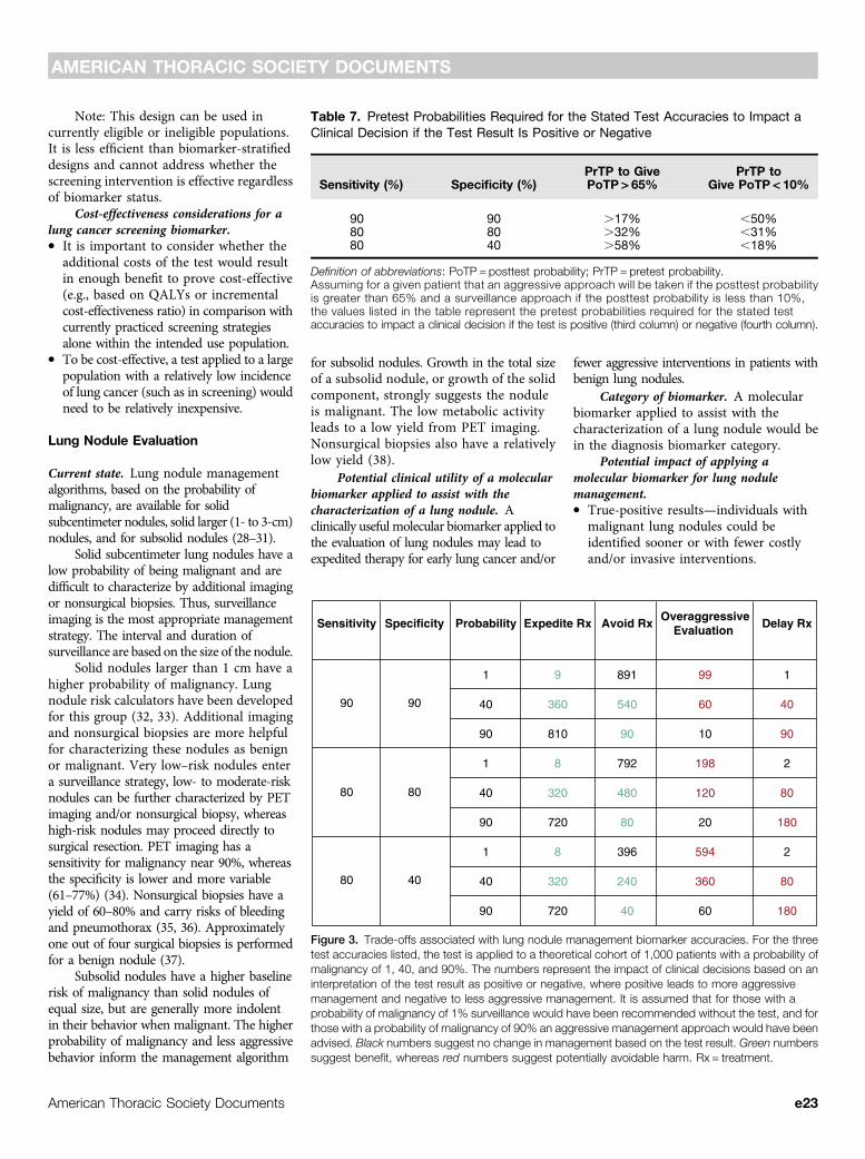
Table 7. Pretest Probabilities Required for the Stated Test Accuracies to Impact aClinical Decision if the Test Result Is Positive or Negative
Sensitivity (%) Specificity (%)PrTP to GivePoTP > 65%
PrTP toGive PoTP < 10%
90 90 .17% ,50%80 80 .32% ,31%80 40 .58% ,18%
Definition of abbreviations: PoTP = posttest probability; PrTP = pretest probability.Assuming for a given patient that an aggressive approach will be taken if the posttest probabilityis greater than 65% and a surveillance approach if the posttest probability is less than 10%,the values listed in the table represent the pretest probabilities required for the stated testaccuracies to impact a clinical decision if the test is positive (third column) or negative (fourth column).
Sensitivity Specificity Probability Expedite Rx Avoid RxOveraggressive
Evaluation Delay Rx
90
80
80
90
80
40
1
40
90
1
40
90
1
40
90
9
360
810
8
320
720
8
320
720
891
540
90
792
480
80
396
240
40
99
60
10
198
120
20
594
360
60
1
40
90
2
80
180
2
80
180
Figure 3. Trade-offs associated with lung nodule management biomarker accuracies. For the threetest accuracies listed, the test is applied to a theoretical cohort of 1,000 patients with a probability ofmalignancy of 1, 40, and 90%. The numbers represent the impact of clinical decisions based on aninterpretation of the test result as positive or negative, where positive leads to more aggressivemanagement and negative to less aggressive management. It is assumed that for those with aprobability of malignancy of 1% surveillance would have been recommended without the test, and forthose with a probability of malignancy of 90% an aggressive management approach would have beenadvised. Black numbers suggest no change in management based on the test result. Green numberssuggest benefit, whereas red numbers suggest potentially avoidable harm. Rx = treatment.
AMERICAN THORACIC SOCIETY DOCUMENTS
American Thoracic Society Documents e23
d True-negative results—individuals withbenign lung nodules could avoid costlyand/or invasive testing.
d False-positive results—individuals withbenign lung nodules could undergocostly and/or invasive testing includingsurgical resection.
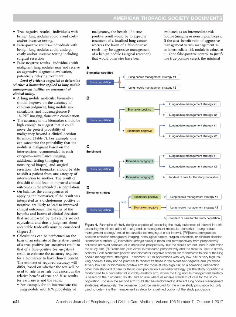
d False-negative results—individuals withmalignant lung nodules may not receivean aggressive diagnostic evaluation,potentially delaying treatment.Level of evidence suggested to determine
whether a biomarker applied to lung nodulemanagement justifies an assessment ofclinical utility.d A lung nodule molecular biomarker
should improve on the accuracy ofclinician judgment, lung nodule riskcalculators, and fludeoxyglucose F18–PET imaging, alone or in combination.
d The accuracy of the biomarker should behigh enough to suggest that it couldmove the pretest probability ofmalignancy beyond a clinical decisionthreshold (Table 7). For example, onecan categorize the probability that thenodule is malignant based on theinterventions recommended in eachcategory—surveillance imaging,additional testing (imaging ornonsurgical biopsy), and surgicalresection. The biomarker should be ableto shift a patient from one category ofintervention to another. The result ofthis shift should lead to improved clinicaloutcomes in the intended use population.
d On balance, the consequences ofapplying the biomarker, if the result wasinterpreted as a dichotomous positive ornegative, are likely to lead to improvedclinical outcomes. The values of thebenefits and harms of clinical decisionsthat are impacted by test results are notequivalent, and thus a judgment aboutacceptable trade-offs must be considered(Figure 3).
d Calculations can be performed on thebasis of an estimate of the relative benefitof a true-positive (or -negative) result tothat of a false-positive (or -negative)result to estimate the accuracy requiredfor a biomarker to have clinical benefit.The estimate of required accuracy willdiffer, based on whether the test will beused to rule in or rule out cancer, as therelative benefit of true and false resultsfor each use is not the same.∘ For example, for an intermediate-risklung nodule with 40% probability of
malignancy, the benefit of a true-positive result would be to expeditetreatment of a localized lung cancer,whereas the harm of a false-positiveresult may be aggressive managementof a benign nodule (surgical resection)that would otherwise have been
evaluated as an intermediate-risknodule (imaging or nonsurgical biopsy).If the cost–benefit ratio of aggressivemanagement versus management asan intermediate-risk nodule is valued at5:1 (one false-positive control to justifyfive true-positive cases), the minimal
Biomarker stratified
Study population
Lung nodule management strategy #2
Lung nodule management strategy #1
A
Lung nodule management strategy #1
Lung nodule management strategy #2
Lung nodule management strategy #1
Lung nodule management strategy #2
Biomarker positive
Biomarker negative
Study population
B
Enrichment
Lung nodule management strategy #1
Lung nodule management strategy #2
Standard of care for the study population
Biomarker category 1
Biomarker category 2
Study population
C
Biomarker strategy
Lung nodule management strategy #1
Lung nodule management strategy #2
Standard of care for the study population
Biomarker positive
Biomarker negativeStudy population
D
Figure 4. Examples of study designs capable of assessing the study outcomes of interest in a trialassessing the clinical utility of a lung nodule management molecular biomarker. “Lung nodulemanagement strategy” could be surveillance imaging at a set interval, [18F]fluorodeoxyglucose–positron emission tomography imaging, nonsurgical biopsy, surgical resection, or clinician decision.Biomarker stratified: (A) Biomarker (orange circle) is measured retrospectively from prospectivelycollected archived samples, or is measured prospectively, but the results are not used to determinethe study arm. (B) Biomarker (blue circle) is measured prospectively and the result is used to stratifypatients. Both biomarker-positive and biomarker-negative patients are randomized to one of the lungnodule management strategies. Enrichment: (C) In populations with very low–risk or very high–risklung nodules it may not be practical to randomize those in the biomarker-negative arm (for thoseat very low risk) or biomarker-positive arm (for those at very high risk) to a screening interventionother than standard of care for the studied population. Biomarker strategy: (D) The study population israndomized to a biomarker (blue circle)–strategy arm, where the lung nodule management strategyis based on the biomarker results, and an arm where all receive standard of care for the studypopulation. Those in the second arm could also be randomized to different lung nodule managementstrategies. Alternatively, the biomarker could be measured for the entire study population but onlyused to determine the management strategy for a defined portion of the study population.
AMERICAN THORACIC SOCIETY DOCUMENTS
e24 American Journal of Respiratory and Critical Care Medicine Volume 196 Number 7 | October 1 2017

positive likelihood ratio, calculatedusing the above formula, would be asfollows: sensitivity/12 specificity>[(1 – 0.4)/0.4]3 5 = 7.5. For example, asensitivity of 75% and specificity of90% (12 specificity = 10%) wouldequal 7.5.
∘ Conversely, the benefit of a true-negativeresult may be to avoid standardevaluation of an intermediate-risknodule in favor of surveillance, and theharm of a false-negative result may be todelay the diagnosis of a localized lungcancer. If the cost–benefit ratio ofsurveillance versus evaluation of anintermediate-risk nodule is valued at 3:1(one false-negative case to justify threetrue-negative controls), a calculationusing the above formula would show arequirement that the specificity/(12sensitivity)> [0.4/(1 – 0.4)]3 3 = 2. Forexample, a specificity of 40% andsensitivity of 80% (12 sensitivity =20%) would equal 2.Level of evidence required for a
molecular biomarker to be consideredclinically useful in the context of lung nodulemanagement. To be considered clinicallyuseful, a molecular biomarker used to assistwith lung nodule management must lead to:
d Earlier diagnosis of malignant noduleswithout substantially increasing thenumber of procedures performed onpatients with benign nodules, or
d Fewer procedures for patients withbenign nodules without substantiallydelaying the diagnosis of cancer inpatients with malignant nodules.
Examples of study designs that could beused to obtain the above outcomes includethe following (Figure 4):
BIOMARKER STRATIFIED.d A molecular biomarker is measured
retrospectively from archived samplesobtained prospectively during a controlledtrial of lung nodule management strategies.
d A molecular biomarker is measuredprospectively during a controlled trial oflung nodule management strategies inwhich the biomarker result is not used toguide which lung nodule managementstrategy the patient is assigned to.
d A molecular biomarker is measuredprospectively, and the result is used tostratify patients into two arms (positiveresult and negative result). Patients ineach arm are then randomized to one oftwo lung nodule management strategies.
Note: These designs can beincorporated into any controlled trial oflung nodule management strategies.
ENRICHMENT.d A molecular biomarker is measured
prospectively, and the result is used tostratify patients into two arms (positiveresult and negative result). Patients in one ofthe arms (biomarker positive or biomarkernegative) are then randomized to one oftwo lung nodule management strategies.Patients in the other biomarker arm receivestandard of care for the study population.
Note: This design may be preferredin study cohorts with very low– or veryhigh–risk lung nodules as it may not bepractical to randomize those with a negativebiomarker result (in a very low–risk nodulecohort) or positive biomarker result (in a veryhigh–risk cohort) to a nodule managementstrategy other than standard of care.
BIOMARKER STRATEGY.d A prospective controlled trial in which study
subjects are randomized to one of two studyarms. In the first, the nodule managementstrategy is based on the result of a molecularbiomarker. In the second, subjects couldeither receive standard of care for the studypopulation or be randomized to nodulemanagement strategies without use of thebiomarker. Alternatively, the biomarkercould be measured for the entire studypopulation but only used to determine the
management strategy for a defined portionof the study population.
Note: This design can be used toaddress whether a biomarker-drivenstrategy is better than a standardmanagement strategy. It is less efficientthan biomarker-stratified designs andcannot address whether the nodulemanagement strategy is effectiveregardless of biomarker status.
Cost-effectiveness considerations for abiomarker used for the evaluation of lungnodules.d The costs related to nodule evaluation
may vary, based on differences inpractice location, resources, clinicaljudgment, and patient populations.
d Cost-effectiveness analyses shouldinclude a description of the study setting,costs, benefits, and how they werecalculated. A sensitivity analysis isparticularly important.
Conclusions
The application of molecular biomarkers toassist with the early detection of lung cancerhas the potential to substantially improveour ability to select patients for lung cancerscreening, and to assist with thecharacterization of indeterminate lungnodules. To support the application ofmolecular biomarkers in these clinicalsettings there must be evidence that themolecular biomarker leads to clinicaldecisions whose benefits outweigh theirharms. Although it is tempting to applynovel testing based on promisingdiscovery or validation level studies, thelung cancer community should insist onadditional evidence of clinical utilitybefore changing practice. We havedescribed relevant considerations and havesuggested standards to apply whendetermining whether a molecular biomarkerfor the early detection of lung cancer is readyfor clinical use. n
This official statement was prepared by an ad hoc subcommittee of the Assembly on Thoracic Oncology.
Members of the subcommittee are asfollows:
PETER J. MAZZONE, M.D., M.P.H. (Co-Chair)CATHERINE RUFATTO SEARS, M.D. (Co-Chair)
DOUG A. ARENBERG, M.D.
MINA GAGA, M.D., Ph.D.
MICHAEL K. GOULD, M.D., M.S.PIERRE P. MASSION, M.D.
VISH S. NAIR, M.D., M.S.CHARLES A. POWELL, M.D.GERARD A. SILVESTRI, M.D., M.S.ANIL VACHANI, M.D.
RENDA SOYLEMEZ WIENER, M.D., M.P.H.
Author disclosures: P.J.M. received researchsupport from InDi, 20/20 Genesystems,Metabolomx, and Oncimmune; and served onan advisory committee for Genentech USA,Grail, InDi, Nucleix, and Oncimmune. D.A.A.served on an advisory committee for Nucleix
AMERICAN THORACIC SOCIETY DOCUMENTS
American Thoracic Society Documents e25
and VisionGate. M.G. served on an advisorycommittee for AstraZeneca, Chiesi,GlaxoSmithKline, Menarini, Novartis, andPharmaten; served as a consultant forBoehringer Ingelheim; served as a speaker forTeva; and received research support fromAstraZeneca, Boehringer Ingelheim, Chiesi,Elpen, GlaxoSmithKline, Novartis, and Teva.M.K.G. received author royalties fromUpToDate and received research support fromMedial. C.A.P. served on the advisory
committee for Genentech USA; and served asa consultant for Genentech USA, Nucleix,Siemen, Chugai Pharmaceutical, and TakedaPharmaceutical. G.A.S. received researchsupport from Integrated Diagnostics,Oncocyte, and Exact Sciences. A.V. servedon an advisory committee for AllegroDiagnostics and Veracyte; received researchsupport from Allegro Diagnostics, IntegratedDiagnostics, Janssen Research andDevelopment, MagArray, and Viomics; and
provided expert testimony for Ford MotorCompany and Honeywell International.C.R.S., P.P.M., V.S.N., and R.S.W. reportedno relationships with relevant commercialinterests.
Acknowledgment: The writing committee isgrateful for the guidance of Gene Pennello andStuart Baker and the inspiration provided byDennis O’Brien.
References
1. Fryback DG, Thornbury JR. The efficacy of diagnostic imaging. MedDecis Making 1991;11:88–94.
2. Freidlin B, McShane LM, Korn EL. Randomized clinical trials withbiomarkers: design issues. J Natl Cancer Inst 2010;102:152–160.
3. Simon R. Clinical trial designs for evaluating the medical utility of prognosticand predictive biomarkers in oncology. Per Med 2010;7:33–47.
4. Simon RM, Paik S, Hayes DF. Use of archived specimens in evaluationof prognostic and predictive biomarkers. J Natl Cancer Inst 2009;101:1446–1452.
5. Micheel CM, Nass SJ, Omenn GS, editors. Committee on the Review ofOmics-Based Tests for Predicting Patient Outcomes in Clinical Trials;Board on Health Care Services; Board on Health Sciences Policy;Institute of Medicine. Chapter 4: Evaluation of omics-based tests forclinical utility and use. In: Evolution of translational omics: lessonslearned and the path forward. Washington, DC: National AcademiesPress; 2012 [accessed 2015 Dec 15]. Available from: https://www.ncbi.nlm.nih.gov/books/NBK202169/
6. McNeil BJ, Keller E, Adelstein SJ. Primer on certain elements of medicaldecision making. N Engl J Med 1975;293:211–215.
7. Russell LB, Gold MR, Siegel JE, Daniels N, Weinstein MC; Panel onCost-Effectiveness in Health and Medicine. The role of cost-effectiveness analysis in health and medicine. JAMA 1996;276:1172–1177.
8. Siegel JE, Weinstein MC, Russell LB, Gold MR; Panel on Cost-Effectiveness in Health and Medicine. Recommendations for reportingcost-effectiveness analyses. JAMA 1996;276:1339–1341.
9. Weinstein MC, Siegel JE, Gold MR, Kamlet MS, Russell LB.Recommendations of the panel on cost-effectiveness in health andmedicine. JAMA 1996;276:1253–1258.
10. Edejer TTT, Baltussen R, Adam T, Hutubessy R, Acharya A, Evans DB,Murray CJL. Part 1: Methods for generalized cost-effectivenessanalysis. In: Making choices in health: WHO guide to cost-effectiveness analysis. Geneva, Switzerland: World HealthOrganization; 2003 [accessed 2015 Dec 15]. pp. 3–97. Available from:http://www.who.int/choice/publications/p_2003_generalised_cea.pdf
11. Sanders GD, Neumann PJ, Basu A, Brock DW, Feeny D, Krahn M,Kuntz KM, Meltzer DO, Owens DK, Prosser LA, et al.Recommendations for conduct, methodological practices, andreporting of cost-effectiveness analyses: second panel on cost-effectiveness in health and medicine. JAMA 2016;316:1093–1103.
12. World Health Organization. Cancer: early detection of cancer. 2017[accessed 2015 Dec 15]. Available from: http://www.who.int/cancer/detection/en/
13. Ost DE, Gould MK. Decision making in patients with pulmonarynodules. Am J Respir Crit Care Med 2012;185:363–372.
14. Baker SG. Improving the biomarker pipeline to develop and evaluatecancer screening tests. J Natl Cancer Inst 2009;101:1116–1119.
15. Pepe MS, Janes H, Li CI, Bossuyt PM, Feng Z, Hilden J. Early-phasestudies of biomarkers: what target sensitivity and specificity valuesmight confer clinical utility? Clin Chem 2016;62:737–742.
16. Aberle DR, Adams AM, Berg CD, Black WC, Clapp JD, Fagerstrom RM,Gareen IF, Gatsonis C, Marcus PM, Sicks JD; National LungScreening Trial Research Team. Reduced lung-cancer mortality withlow-dose computed tomographic screening. N Engl J Med 2011;365:395–409.
17. Agency for Healthcare Research and Quality. Benefits and harms ofcomputed tomography lung cancer screening programs for high-riskpopulations. AHRQ Publication No. 13-05196-EF-2. Rockville, MD:Agency for Healthcare Research and Quality; 2013.
18. Harris RP, Sheridan SL, Lewis CL, Barclay C, Vu MB, Kistler CE, Golin CE,DeFrank JT, Brewer NT. The harms of screening: a proposedtaxonomy and application to lung cancer screening. JAMA InternMed 2014;174:281–285.
19. Bach PB, Kattan MW, Thornquist MD, Kris MG, Tate RC, Barnett MJ,Hsieh LJ, Begg CB. Variations in lung cancer risk among smokers.J Natl Cancer Inst 2003;95:470–478.
20. Spitz MR, Hong WK, Amos CI, Wu X, Schabath MB, Dong Q, Shete S,Etzel CJ. A risk model for prediction of lung cancer. J Natl CancerInst 2007;99:715–726.
21. Cassidy A, Myles JP, van Tongeren M, Page RD, Liloglou T, Duffy SW,Field JK. The LLP risk model: an individual risk prediction model forlung cancer. Br J Cancer 2008;98:270–276.
22. Tammemagi MC, Katki HA, Hocking WG, Church TR, Caporaso N,Kvale PA, Chaturvedi AK, Silvestri GA, Riley TL, Commins J, et al.Selection criteria for lung-cancer screening. N Engl J Med 2013;368:728–736.
23. Hoggart C, Brennan P, Tjonneland A, Vogel U, Overvad K, Østergaard JN,Kaaks R, Canzian F, Boeing H, Steffen A, et al. A risk model for lungcancer incidence. Cancer Prev Res (Phila) 2012;5:834–846.
24. Katki HA, Kovalchik SA, Berg CD, Cheung LC, Chaturvedi AK.Development and validation of risk models to select ever-smokersfor CT lung cancer screening. JAMA 2016;315:2300–2311.
25. Tammemagi MC, Church TR, Hocking WG, Silvestri GA, Kvale PA,Riley TL, Commins J, Berg CD. Evaluation of the lung cancer risks atwhich to screen ever- and never-smokers: screening rules applied tothe PLCO and NLST cohorts. PLoS Med 2014;11:e1001764.
26. Black WC, Gareen IF, Soneji SS, Sicks JD, Keeler EB, Aberle DR, Naeim A,Church TR, Silvestri GA, Gorelick J, et al.; National Lung ScreeningTrial Research Team. Cost-effectiveness of CT screening in theNational Lung Screening Trial. N Engl J Med 2014;371:1793–1802.
27. Aberle DR, DeMello S, Berg CD, Black WC, Brewer B, Church TR,Clingan KL, Duan F, Fagerstrom RM, Gareen IF, et al.; National LungScreening Trial Research Team. Results of the two incidence screeningsin the National Lung Screening Trial. N Engl J Med 2013;369:920–931.
28. Gould MK, Donington J, Lynch WR, Mazzone PJ, Midthun DE,Naidich DP, Wiener RS. Evaluation of individuals with pulmonarynodules: when is it lung cancer? In: Diagnosis and management of lungcancer, 3rd ed: American College of Chest Physicians evidence-basedclinical practice guidelines. Chest 2013;143(5 Suppl):e93S–e120S.
29. MacMahon H, Austin JH, Gamsu G, Herold CJ, Jett JR, Naidich DP,Patz EF Jr, Swensen SJ; Fleischner Society. Guidelines formanagement of small pulmonary nodules detected on CT scans:a statement from the Fleischner Society. Radiology 2005;237:395–400.
30. Naidich DP, Bankier AA, MacMahon H, Schaefer-Prokop CM, Pistolesi M,Goo JM, Macchiarini P, Crapo JD, Herold CJ, Austin JH, et al.Recommendations for the management of subsolid pulmonarynodules detected at CT: a statement from the Fleischner Society.Radiology 2013;266:304–317.
31. Callister MEJ, Baldwin DR, Akram AR, Barnard S, Cane P, Draffan J,Franks K, Gleeson F, Graham R, Malhotra P, et al.; British ThoracicSociety Pulmonary Nodule Guideline Development Group; BritishThoracic Society Standards of Care Committee. British Thoracic
AMERICAN THORACIC SOCIETY DOCUMENTS
e26 American Journal of Respiratory and Critical Care Medicine Volume 196 Number 7 | October 1 2017
Society guidelines for the investigation and management ofpulmonary nodules. Thorax 2015;70:ii1–ii54.
32. Swensen SJ, Silverstein MD, Ilstrup DM, Schleck CD, Edell ES. Theprobability of malignancy in solitary pulmonary nodules: applicationto small radiologically indeterminate nodules. Arch Intern Med 1997;157:849–855.
33. Herder GJ, van Tinteren H, Golding RP, Kostense PJ, Comans EF, Smit EF,Hoekstra OS. Clinical prediction model to characterize pulmonarynodules: validation and added value of 18F-fluorodeoxyglucosepositron emission tomography. Chest 2005;128:2490–2496.
34. Deppen SA, Blume JD, Kensinger CD, Morgan AM, Aldrich MC,Massion PP, Walker RC, McPheeters ML, Putnam JB Jr, Grogan EL.Accuracy of FDG-PET to diagnose lung cancer in areas withinfectious lung disease: a meta-analysis. JAMA 2014;312:1227–1236.
35. Wang Memoli JS, Nietert PJ, Silvestri GA. Meta-analysis of guidedbronchoscopy for the evaluation of the pulmonary nodule. Chest2012;142:385–393.
36. Ost DE, Ernst A, Lei X, Kovitz KL, Benzaquen S, Diaz-Mendoza J,Greenhill S, Toth J, Feller-Kopman D, Puchalski J, et al.; AQuIREBronchoscopy Registry. Diagnostic yield and complications ofbronchoscopy for peripheral lung lesions: results of the AQuIRERegistry. Am J Respir Crit Care Med 2016;193:68–77.
37. Bach PB, Mirkin JN, Oliver TK, Azzoli CG, Berry DA, Brawley OW, Byers T,Colditz GA, Gould MK, Jett JR, et al. Benefits and harms of CT screeningfor lung cancer: a systematic review. JAMA 2012;307:2418–2429.
38. Detterbeck FC, Homer RJ. Approach to the ground-glass nodule. ClinChest Med 2011;32:799–810.
39. Mazzone PJ. Obstacles to and solutions for a successful lung cancerscreening program. Semin Respir Crit Care Med 2016;37:659–669.
Appendix 1: Definitions
Analytical performance: The ability of a biomarker assay to measure the underlying biomarker quantity under a variety of conditions.
Analytical specificity: The ability of a biomarker assay to measure solely the biomarker of interest without interference by othersubstances or cross-reactivity with other analytes.
Analytical validity: Acceptable performance in the measurement or detection of characteristics of a biomarker; how well the test measuresor identifies molecular changes in a person.
Bias: A systematic difference between the compared groups that impacts results in a manner that does not reflect an underlying reality.The systematic erroneous association of a characteristic with a group in a way that distorts a comparison with another group.
Biomarker: A characteristic that is objectively measured and evaluated as an indicator of normal biological processes, pathogenicprocesses, or pharmacological responses to a therapeutic intervention; a biological molecule found in blood, other body fluids, or tissuesthat is a sign of a normal or abnormal process, or of a condition or disease.
Clinical performance: The ability of the biomarker to inform about a clinical condition of interest.
Clinical reference standard: The best available method for establishing the presence or absence of the target condition.
Clinical utility: A biomarker’s ability to improve clinical outcomes when measured and used as directed for its intended use.
Clinical validity: The demonstrated association of a test result with the presence or absence of the target condition.
Companion diagnostic: A biomarker that is essential for the safe and effective use of a therapy.
Confidence intervals: An interval about a point estimate that quantifies the statistical uncertainty in the true value being estimated (e.g.,an accuracy metric) due to variability in the subject/sample selection process. A 1 – a level confidence interval contains the true value in100(1 – a)% of applications (but in any given application either contains it or does not).
Context of use: What the test measures, why, and in what population(s) it should be used.
Cost-effectiveness analysis: An evaluation that compares the net cost of an intervention with the benefits gained by that intervention.
Diagnostic accuracy: The extent of agreement between the outcome of the new test and the reference standard.
Diagnostic marker: A test used in people with signs or symptoms to aid in assessing whether they have a condition.
External validity: The generalizability of the comparison results to persons outside of the study.
Fundamental comparison: The process of arranging and analyzing groups of subjects and specimens to learn whether a differenceobserved in the compared groups is related to a particular condition.
Independent validation: The assessment of analytical and clinical performance on a set of subjects that is independent of the data set usedin the development of the test.
Index test: The test under evaluation.
Intended use of the test: The population, condition, and question for which the biomarker is being developed (e.g., diagnosis, staging,screening, surveillance, prediction, prognosis).
Intention to diagnose analysis: An analysis that includes all study subjects, whether or not all of their test results are available.
Intermediate precision: Precision when some conditions vary and others are held constant.
Internal validity: The strength or fairness of the comparison of groups within the study.
Lead time: The length of time the diagnosis is advanced by testing with the biomarker.
AMERICAN THORACIC SOCIETY DOCUMENTS
American Thoracic Society Documents e27
Limit of detection: The lowest assay level at which the presence of the analyte is detected with reliability in repeated measurement.
Linearity: The ability to provide results directly proportional to the amount of analyte in the test sample within a given measuring range.
Measurement accuracy: The closeness of agreement between a measurement result and an accepted reference value; an aggregate oftrueness and precision.
Measurement trueness: The closeness of agreement between the average of an infinite number of replicate test results and the referencevalue. Trueness is usually expressed numerically by the statistical measure bias.
Medical tests: Results of a clinical, imaging, or laboratory-based assay that are used alone or with other information to help assess asubject’s health condition of interest, or target condition.
Negative predictive value (NPV): The predictive value of a negative result; the proportion of subjects with a negative test result who donot have the target condition.
Performance around the cutoff: The measurement accuracy of an assay at biomarker levels near the threshold chosen to distinguish apositive and negative result for the intended use of the test.
Positive predictive value (PPV): The predictive value of a positive result; the proportion of subjects with a positive test result who havethe target condition.Precision: The closeness of agreement of replicate test results under stipulated conditions.
Predictive marker: A biomarker that assesses the safety or efficacy of a specific therapy.
Prognostic marker: A biomarker used in subjects diagnosed with a condition to predict subsequent outcomes, such as disease recurrenceor progression.
Qualitative result: A biomarker result consisting of a set number of possible responses (often two).
Quantitative result: A biomarker result that is numerical in amount or level of a physical quantity.
Repeatability: Precision when replicate measurements are taken under the same conditions (within a run).
Reproducibility: Precision when one of the conditions being varied is the laboratory for an in vitro diagnostic measurement.
Role of the test: The position of the index test relative to other tests for the same condition.
Screening marker: A biomarker used in asymptomatic people to detect a disease or condition at an early stage.
Semiquantitative: Results of a test that fall into an approximate range of values.
Sensitivity: The proportion of subjects with the target condition in whom the test result is positive.
Signature: Multiple variables combined to provide a single result.
Specificity: The proportion of subjects without the target condition in whom the test result is negative.
Target condition: The disease or condition that the index test is expected to detect.
Tumor marker: A qualitative or quantitative alteration or deviation from normal of a molecule, substance, or process that can be detectedby some type of assay; surrogate indicators that increase or decrease the clinician’s suspicion that future clinically important events, suchas cancer onset, recurrence, progression or patient death, will or will not happen, and/or that a specific treatment will decrease the risk ofsuch events.
Appendix 2: Additional References Used during the Project
Baker SG. Biomarker evaluation in randomized trials: addressing different research questions. Stat Med 2014;33:4139–4140.Baker SG, Kramer BS. Peirce, Youden, and receiver operating characteristic curves. Am Stat 2007;61:343–346.Baker SG, Kramer BS, Srivastava S. Markers for early detection of cancer: statistical guidelines for nested case–control studies. BMC Med Res Methodol
2002;2:4.Beaver JA, Tzou A, Blumenthal GM, McKee AE, Kim G, Pazdur R, Philip R. An FDA perspective on the regulatory implications of complex signatures to
predict response to targeted therapies. Clin Cancer Res 2017;23:1368–1372.Biomarkers Definitions Working Group. Biomarkers and surrogate endpoints: preferred definitions and conceptual framework. Clin Pharmacol Ther 2001;
69:89–95.Bossuyt PM, Reitsma JB, Bruns DE, Gatsonis CA, Glasziou PP, Irwig L, Lijmer JG, Moher D, Remmie D, de Vet HCW, et al.; STARD Group. STARD 2015:
an updated list of essential items for reporting diagnostic accuracy studies. BMJ 2015;351:h5527.Center for Devices and Radiological Health, Food and Drug Administration, U.S. Department of Health and Human Services. Guidance for industry and
FDA staff: statistical guidance on reporting results from studies evaluating diagnostic tests. 2007 March 3 [accessed 2015 Dec 15]. Available from:https://www.fda.gov/RegulatoryInformation/Guidances/ucm071148.htm
Collins GS, Reitsma JB, Altman DG, Moons KGM. Transparent reporting of a multivariable prediction model for individual prognosis or diagnosis(TRIPOD): the TRIPOD statement. BMC Med 2015;13:1.
Duffy MJ, Sturgeon CM, Soletormos G, Barak V, Molina R, Hayes DF, Diamandis EP, Bossuyt PM. Validation of new cancer biomarkers: a positionstatement from the European Group on Tumor Markers. Clin Chem 2015;61:809–820.
Gluud C, Gluud LL. Evidence based diagnostics. BMJ 2005;330:724–726.
AMERICAN THORACIC SOCIETY DOCUMENTS
e28 American Journal of Respiratory and Critical Care Medicine Volume 196 Number 7 | October 1 2017
Hayes DF, Bast RC, Desch CE, Fritsche H Jr, Kemeny NE, Jessup JM, Locker GY, Macdonald JS, Mennel RG, Norton L, et al. Tumor marker utility gradingsystem: a framework to evaluate clinical utility of tumor markers. J Natl Cancer Inst 1996;88:1456–1466.
Henry NL, Hayes DF. Cancer biomarkers. Mol Oncol 2012;6:140–146.Kovalchik SA, Tammemagi M, Berg CD, Caporaso NE, Riley TL, Korch M, Silvestri GA, Chaturvedi AK, Katki HA. Targeting of low-dose CT screening according to the
risk of lung-cancer death. N Engl J Med 2013;369:245–254.Mazzone P, Powell CA, Arenberg D, Bach P, Detterbeck F, Gould MK, Jaklitsch MT, Jett J, Naidich D, Vachani A, et al. Components necessary for high-
quality lung cancer screening: American College of Chest Physicians and American Thoracic Society policy statement. Chest 2015;147:295–303.McMahon PM, Kong CY, Bouzan C, Weinstein MC, Cipriano LE, Tramontano AC, Johnson BE, Weeks JC, Gazelle GS. Cost-effectiveness of computed
tomography screening for lung cancer in the United States. J Thorac Oncol 2011;6:1841–1848.Moore HM, Kelly A, Jewell SD, McShane LM, Clark DP, Greenspan R, Hainaut P, Hayes DF, Kim P, Mansfield E, et al. Biospecimen reporting for improved
study quality. Biopreserv Biobank 2011;9:57–70.Moyer VA; U.S. Preventive Services Task Force. Screening for lung cancer: U.S. Preventive Services Task Force recommendation statement. Ann Intern
Med 2014;160:330–338.Office of Public Health Strategy and Analysis, Office of the Commissioner, U.S. Food and Drug Administration. The public health evidence for FDA
oversight of laboratory developed tests: 20 case studies. 2015 Nov 16 [accessed 2015 Dec 15]. Available from: https://www.fda.gov/downloads/AboutFDA/ReportsManualsForms/Reports/UCM472777.pdf
Oken MM, Hocking WG, Kvale PA, Andriole GL, Buys SS, Church TR, Crawford ED, Fouad MN, Isaacs C, Reding DJ, et al.; PLCO Project Team.Screening by chest radiograph and lung cancer mortality: the Prostate, Lung, Colorectal, and Ovarian (PLCO) randomized trial. JAMA 2011;306:1865–1873.
Pataky R, Gulati R, Etzioni R, Black P, Chi KN, Coldman AJ, Pickles T, Tyldesley S, Peacock S. Is prostate cancer screening cost-effective? Amicrosimulation model of prostate-specific antigen-based screening for British Columbia, Canada. Int J Cancer 2014;135:939–947.
Pennello GA. Analytical and clinical evaluation of biomarkers assays: when are biomarkers ready for prime time? Clin Trials 2013;10:666–676.Pepe MS, Etzioni R, Feng Z, Potter JD, Thompson ML, Thornquist M, Winget M, Yasui Y. Phases of biomarker development for early detection of cancer.
J Natl Cancer Inst 2001;93:1054–1061.Pepe MS, Feng Z, Janes H, Bossuyt PM, Potter JD. Pivotal evaluation of the accuracy of a biomarker used for classification or prediction: standards for
study design. J Natl Cancer Inst 2008;100:1432–1438.Pepe MS, Fan J, Seymour CW, Li C, Huang Y, Feng Z. Biases introduced by choosing controls to match risk factors of cases in biomarker research. Clin
Chem 2012;58:1242–1251.Ransohoff DF, Gourlay ML. Sources of bias in specimens for research about molecular markers for cancer. J Clin Oncol 2010;28:698–704.Romanus D, Cardarella S, Cutler D, Landrum MB, Lindeman NI, Gazelle GS. Cost-effectiveness of multiplexed predictive biomarker screening in non–small-
cell lung cancer. J Thorac Oncol 2015;10:586–594.Sackett DL, Haynes RB. The architecture of diagnostic research. BMJ 2002;324:539–541.Schully SD, Carrick DM, Mechanic LE, Srivastava S, Anderson GL, Baron JA, Berg CD, Cullen J, Diamandis EP, Doria-Rose VP, et al. Leveraging
biospecimen resources for discovery or validation of markers for early cancer detection. J Natl Cancer Inst 2015;107:djv012.Sharaf RN, Ladabaum U. Comparative effectiveness and cost-effectiveness of screening colonoscopy vs. sigmoidoscopy and alternative strategies. Am J
Gastroenterol 2013;108:120–132.Sturgeon CM, Hoffman BR, Chan DW, Ch’ng SO, Hammond E, Hayes DF, Liotta LA, Petricoin EF, Schmitt M, Semmes OJ, et al.; National Academy of
Clinical Biochemistry. National Academy of Clinical Biochemistry laboratory medicine practice guidelines for use of tumor markers in clinical practice:quality requirement. Clin Chem 2008;54:e1–e10.
Villanti AC, Jiang Y, Abrams DB, Pyenson BS. A cost–utility analysis of lung cancer screening and the additional benefits of incorporating smokingcessation interventions. PLoS One 2013;8:e71379.
Wu D, Rosner GL, Broemeling LD. Bayesian inference for the lead time in periodic cancer screening. Biometrics 2007;63:873–880.Zauber AG. Cost-effectiveness of colonoscopy. Gastrointest Endosc Clin N Am 2010;20:751–770.
AMERICAN THORACIC SOCIETY DOCUMENTS
American Thoracic Society Documents e29