Embed Size (px)
Citation preview
GE HealthcareLife Sciences
Amersham cAMP Biotrak Enzymeimmunoassay (EIA) SystemProduct bookletCodes: RPN225 RPN2251 RPN2255
2
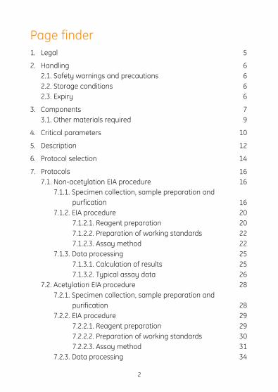
Page finder1. Legal 5
2. Handling 6 2.1. Safety warnings and precautions 6 2.2. Storage conditions 6 2.3. Expiry 6
3. Components 7 3.1. Other materials required 9
4. Critical parameters 10
5. Description 12
6. Protocol selection 14
7. Protocols 16 7.1. Non-acetylation EIA procedure 16 7.1.1. Specimen collection, sample preparation and purfication 16 7.1.2. EIA procedure 20 7.1.2.1. Reagent preparation 20 7.1.2.2. Preparation of working standards 22 7.1.2.3. Assay method 22 7.1.3. Data processing 25 7.1.3.1. Calculation of results 25 7.1.3.2. Typical assay data 26 7.2. Acetylation EIA procedure 28 7.2.1. Specimen collection, sample preparation and purification 28 7.2.2. EIA procedure 29 7.2.2.1. Reagent preparation 29 7.2.2.2. Preparation of working standards 30 7.2.2.3. Assay method 31 7.2.3. Data processing 34

7.2.3.1. Calculation of results 34 7.2.3.2. Typical assay data 34 7.3. Intracellular cAMP measurement using the non–acetylation EIA procedure with the novel lysis reagents 36 7.3.1. EIA procedure 36 7.3.1.1. Reagent preparation 36 7.3.1.2. Cell lysis methods - intracellular cAMP measurement 38 7.3.1.3. Preparation of working standards 40 7.3.1.4. Assay method 40 7.3.2. Data processing 43 7.3.2.1. Calculation of results 43 7.3.2.2. Typical assay data 43 7.4. Total cellular cAMP measurement using the non–acetylation EIA procedure with the novel lysis reagents 45 7.4.1. EIA procedure 45 7.4.1.1. Reagent preparation 45 7.4.1.2. Cell lysis method - total cellular cAMP measurement 47 7.4.1.3. Preparation of working standards 48 7.4.1.4. Assay protocol 49 7.4.2. Data processing 51 7.4.2.1. Calculation of results 51 7.4.2.2. Typical assay data 51
8. Additional information 53 8.1. Expected values 53 8.2. Limitations of use 53 8.3. Specificity 53 8.4. Sensitivity 57 8.5. Precision 57
3

4
9. Troubleshooting guide 60
10. Background and references 61
11. Related products 64

5
1. LegalGE, imagination at work and GE monogram are trademarks of General Electric Company.
Amersham and Biotrak are trademarks of GE Healthcare companies.
All third party trademarks are the property of their respective owners.
© 1994-2011 General Electric Company – All rights reserved. Previously published 1994
All goods and services are sold subject to the tems and conditions of sale of the company within GE Healthcare which supplies them. A copy of these terms and conditions is available on request.Contact your local GE Healthcare representative for the most current information.
http//www.gelifesciences.com
GE Healthcare UK Limited. Amersham Place, Little Chalfont, Buckinghamshire, HP7 9NA UK
5
6
2. Handling
2.1. Safety warnings and precautionsWarning: For research use only. Not recommended or intended for diagnosis of disease in humans or animals. Do not use internally or externally in humans or animals.
All chemicals should be considered as potentially hazardous. We therefore recommend that this product is handled only by those persons who have been trained in laboratory techniques and that it is used in accordance with the principles of good laboratory practice. Wear suitable protective clothing such as laboratory overalls, safety glasses and gloves. Care should be taken to avoid contact with skin or eyes. In the case of contact with skin or eyes wash immediately with water. See material safety data sheet(s) and/or safety statement(s) for specific advice.
Note: The assay protocol may require the use of Sulfuric acid.
Warning: Sulfuric acid is corrosive.
Please follow the manufacturer’s safety data sheet relating to the safe handling and use of this material.
2.2. Storage Store at 2–8°C.
2.3. ExpiryThe expiry date is stated on the package and will be at least 4 weeks from the date of despatch.

3. ComponentsRPN225 The pack contains the following assay components, sufficient material for 96 wells. All components for this kit should be stored at 2–8°C.
Microplate: The plate contains 12 x 8 well strips coated with donkey anti-rabbit IgG, ready for use.
Assay buffer: Assay buffer concentrate. On dilution this bottle contains 0.05 M Sodium Acetate buffer, pH 6.0 containing 0.002%(w/v) Bovine Serum Albumin and 0.01%(w/v) preservative.
Standard (for non-acetylation assay): cAMP standard for non-acetylation assays in the range 25–6400 fmol/well, lyophilised. On reconstitution this bottle contains 64 pmol cAMP/ml.
Standard (for acetylation assay): cAMP standard 10.24 pmol, for acetylation assays in the range 2–128 fmol/well, lyophilised.
On reconstitution this bottle contains 2.56 pmol cAMP/ml.
Antibody: Rabbit anti-cAMP, lyophilised.
Peroxidase conjugate: cAMP- Horseradish Peroxidase, lyophilised.
Wash buffer concentrate: Wash buffer concentrate. On dilution the reagent contains 0.01 M Phosphate buffer, pH 7.5 containing 0.05% Tween™ 20.
TMB substrate: Enzyme substrate containing 3,3’,5,5’ -Tetramethylbenzidine (TMB)/< 1% Hydrogen Peroxide. Ready for use.
Acetic Anhydride: Ready for use. Caution: flammable, corrosive, causes burns.
Triethylamine: Ready for use. Caution: flammable, harmful vapour.
Lysis reagent 1: Dodecyltrimethylammonium Bromide, 2 g, solid. Caution: harmful
Lysis reagent 2: 5 g, Solid.
7

8
RPN2251 The pack contains the following assay components, sufficient material for 96 wells. All components for this kit should be stored at 2–8°C.
Microplate: The plate contains 12 x 8 well strips coated with donkey anti-rabbit IgG, ready for use.
Assay buffer: Assay buffer concentrate. On dilution this bottle contains 0.05 M Sodium Acetate buffer, pH 6.0 containing 0.002%(w/v) Bovine Serum Albumin and 0.01%(w/v) preservative.
Standard (for non-acetylation assay): cAMP standard for non-acetylation assays in the range 25–6400 fmol/well, lyophilised. On reconstitution this bottle contains 64 pmol cAMP/ml.
Antibody: Rabbit anti-cAMP, lyophilised.
Peroxidase conjugate: cAMP- Horseradish Peroxidase, lyophilised.
Wash buffer concentrate: Wash buffer concentrate. On dilution the reagent contains
0.01 M Phosphate buffer, pH 7.5 containing 0.05% Tween™ 20.
TMB substrate: Enzyme substrate containing 3,3’,5,5’ -Tetramethylbenzidine (TMB)/Hydrogen Peroxide. Ready for use.
Lysis reagent 1:Dodecyltrimethylammonium Bromide, 2 g, solid. Caution: harmful
Lysis reagent 2: 5 g, Solid.
RPN2255 The pack contains the following assay components, sufficient material for 480 wells. All components for this kit should be stored at 2–8°C.
Microplate x5: The plate contains 12 x 8 well strips coated with donkey anti-rabbit IgG, ready for use.
Assay buffer: Assay buffer concentrate. On dilution this bottle contains 0.05 M Sodium Acetate buffer, pH 6.0 containing 0.002%(w/v) Bovine Serum Albumin and 0.01%(w/v) preservative.

9
Standard (for non-acetylation assay) x5: cAMP standard for non-acetylation assays in the range 25–6400 fmol/well, lyophilised. On reconstitution this bottle contains 64 pmol cAMP/ml.
Antibody: Rabbit anti-cAMP, lyophilised.
Peroxidase conjugate: cAMP- Horseradish Peroxidase, lyophilised.
Wash buffer concentrate: Wash buffer concentrate. On dilution the reagent contains 0.01 M Phosphate buffer, pH 7.5 containing 0.05% Tween™ 20.
TMB substrate: Enzyme substrate containing 3,3’,5,5’ -Tetramethylbenzidine (TMB)/Hydrogen Peroxide. Ready for use.
3.1. Other materials requiredThe following materials and equipment are required but not supplied:
• Pipettesorpipettingequipment with disposable
polypropylene tips (50 µl, 100 µl, 500 µl, 1 ml and 5 ml)
• Disposablepolypropylenetest tubes
• Vortexmixer
• Refrigerator
• Glassmeasuringcylinders (50 ml, 100 ml, 500 ml)
• Distilledordeionisedwater
• Spectrophotometricplatereader capable of measuring at 450 nm
• 1.0MSulphuricacid
• Microplateshaker
• Magneticstirrerandstirrerbars
• 0.4%Trypanbluesolution
• Centrifugeandmicroplateholders for centrifuge (if using suspension cells).
• Automaticplatewasher(optional)

10
4. Critical parametersImportant The following points are critical:
When carrying out RPN225, RPN2251 and RPN2255 assays, please take particular note of the following instructions regarding critical steps:
• Itisessentialtoreadthecompleteinstructionbookletbeforestarting work.
• Thisisadelayedadditionassay.Donotemptyandwashtheplatebefore adding the Peroxidase conjugate.
• Allowsamplesandallreagentstoreachroomtemperaturepriortoperforming the assay.
• Mixsamplesandallreagentsthoroughlybeforeuse.
• Avoidexcessivefoamingofreagents.
• Avoidhandlingthetopsofthewellsbothbeforeandafterfilling.
• Keepthewellscoveredwithlidsexceptwhenaddingreagentsandreading.
• Standardsandsamplesshouldbeassayedinduplicate.
• Runaseparatestandardcurveforeachmicroplate.
• Carryoutamicroscopeevaluation,with0.4%Trypanblue,beforeand after lysing cells.
• Thoroughly wash the plate before adding the substrate. You can do this either manually or automatically provided that the following points are noted:
Manual plate washing • Useawashbottle.
• Completelyfilleachwellwithwashbuffer.

11
• Completelyemptyeachwellbetweenwashes.
• Afterthefinalwashitisessential that all wells are emptied. Tap the plate briskly on a pad of tissues to effect this.
Automatic plate washing • Ensurethatallwellsarefilledandemptiedcompletely with each
cycle.
• Unevenwashingwillcausepoorresults.Ifyoudoubttheeffectiveness of your instrument, wash manually as described above.
• Thereisnodifferenceinresultswhenusingeitherautomatedorhand washing procedures, if the instrument is carefully maintained.
• Toaidefficientwashinginautomatedplatewashers,thestripsshould be levelled with the edge of the microplate lid before each washing stage.

5. DescriptionThe cAMP Biotrak™ competitive enzymeimmunoassay system from GE Healthcare is specifically designed for research purposes. RPN225 and RPN2251 includes protocols using novel lysis reagents in order to facilitate simple and rapid extraction of cAMP from cell cultures. These components avoid the requirement for traditional, time-consuming extraction procedures and obviate the need for removal of extraction reagents prior to measurement. It combines the use of a Peroxidase-labelled cAMP conjugate, a specific antiserum which can be immobilised on to pre-coated microplates, and a one-pot stabilised substrate solution.
Each pack of RPN225 and RPN2251 contains sufficient material for 96 wells. Each pack of RPN2255 contains sufficient material for 480 wells. Each plate allows the construction of one standard curve and the measurement of at least 36 unknowns in duplicate. The procedure may be carried out in one of four ways depending on the kit used. Please see section on protocol selection (see pages 14–15).
• Eliminationofinconvenient,time-consumingextractionprocedures
• Flexiblemethod-choiceofassayprotocols(see pages 14–15)
• Rapidassayprotocol
• Dualrange25–6400fmol/well(non-acetylationprotocol)2–128 fmol/well (acetylation protocol) with RPN225
• Non-radioactive
• SpecificforcAMP
• Preciseandaccuratemeasurement
• Readytousesubstrate
• Colourcodedreagents
12

Lysis reagent 1 hydrolyses cell membranes to release intra-cellular cAMP. Lysis reagent 2 sequesters the key component in lysis reagent 1 and ensures cAMP is free for subsequent analysis. The detergent/sequestrant complex does not interfere with antigen:antibody binding. Lysis reagent 1 is simply added to cultured cells, followed by a 5–10 minute incubation before assay (figure 1). The antiserum is reconstituted with lysis reagent 2. The assay is based on competition between unlabelled cAMP and a fixed quantity of Peroxidase-labelled cAMP, for a limited number of binding sites on a cAMP specific antibody (figure 2).
13
Fig 1. Cell lysis-protocol 3 intracellular method
Donkeyanti-rabbitIg
Rabbitanti-cAMP cAMP-peroxidase
cAMP TMB
Stop reactionmeasure OD
+
Well
Incubation Incubation 60 minutes
Solid phase Assay reagent Standard or unknown Substrate
Fig 2. EIA principle
1. culture cells 3. lyse cells transfer tonew plate
perform assay
2. decant

6. Protocol SelectionRPN225 suitable for use with protocols one, two, three or four. RPN2251 suitable for use with protocols one, three or four. RPN2255 suitable for use with protocol one.
Protocol 1. The normal non-acetylation assay (range 25–6400 fmol/well) is used for the measurement of cAMP in Urine and tissue extracts and from cell cultures prepared with traditional sample extraction methods such as acid, solvent and solid-phase methods.
Protocol 2. The acetylation assay (range 2–128 fmol/well) is used for the measurement cAMP in plasma, or in tissue extracts where higher sensitivity is required.
Protocol 3. This method describes a non-acetylation assay using the novel lysis reagents enabling the simple, direct measurement of cAMP in cultured cells where cells are grown in flasks, vessels or on plates. Cells are lysed for 10 minutes (with the reagents provided in the kit), and an aliquot is transferred to a second plate for assay. cAMP is measured in the range 25–6400 fmol/well.
Protocol 4. This method also uses the non-acetylation assay with the lysis reagents. Here the combined amount of intracellular and cell supernatant cAMP is measured. This fraction is referred to as ‘total’ cellular cAMP and has the additional benefit of not requiring decantation of the cell culture supernatant. cAMP is measured in the range 25–6400 fmol/well.
14

Table 1. Summary of protocols
15
Curve range Urine samples, Plasma and Plasma and Cell cultures Cell cultures Cell cultures No sample tissue samples, tissues samples Using Intracellular Total extraction Sample where higher traditional cAMP cellular cAMP needed extraction sensitivity is extraction measurement. measurement. required needed. methods Lysis reagents Lysis reagents Sample and protocols and protocols extraction provided. provided. required.
25–6400 Protocol 1 Protocol 1 Protocol 1 Protocol 3 Protocol 4fmol/well (See page 16) (See page 16) (See page 16) (See page 36) (See page 45)
2–128 Protocol 2 fmol/well (See page 28)

7. Protocols
7.1. Non–Acetylation EIA Procedure (Curve range 25–6400 fmol/well, for measurement of cAMP in Urine and tissue samples and extraction of cAMP from cell cultures using traditional methods)
7.1.1. Specimen collection, sample preparation and purification • Numerousprocedureshavebeendescribedfortheextractionof
cAMP from biological samples. These include acidic extraction procedures using Trichloroacetic acid, Perchloric acid, dilute Hydrochloric acid and extraction with aqueous Ethanol (25–28).
• Someinvestigatorsrecommendtheuseofionexchangechromatography following an extraction technique (5). However, it remains the responsibility of the investigator to validate the chosen extraction procedure.
• Representativeproceduresaredescribedbelowfortheextractionof cAMP from Urine and tissues. This information is provided for guidance only.
Urine Random, timed or 24-hour Urine collections may be analysed. If 24-hour samples are collected, it may be necessary to include a bacteriostat (2 ml 6 M Hydrochloric acid per 100 ml Urine is sufficient for this purpose). Samples analysed within 24 hours of collection may be stored at 2–8°C until assayed. If analysis is not performed within 24 hours, all samples should be stored at -15°C to -30°C. If Urine contains particulate matter this should be removed by centrifugation prior to assay. It is not necessary to extract or deproteinise Urine before analysis. Urine should be diluted 1:1000 with assay buffer.
16

Tissue Tissue sections must be rapidly frozen immediately after collection so as to prevent alteration to the cAMP and associated enzymes before analysis. This is usually achieved by immersion of the fresh tissue in liquid Nitrogen at -196°C.
Methods of freezing biological samples for cyclic nucleotide assays are reviewed by Mayer et al (24).
Samples should be stored at -15°C to -30°C until the assay is carried out.
Liquid phase extraction method 1. Homogenise frozen tissue in cold 6% (w/v) Trichloroacetic acid at
2–8°C to give a 10% (w/v) homogenate.
2. Centrifuge at 2000 x g for 15 minutes at 4°C.
3. Recover the supernatant and discard the pellet.
4. Wash the supernatant 4 times with 5 volumes of water saturated Diethyl Ether. The upper Ether layer should be discarded after each wash.
5. The aqueous extract remaining should be lyophilised or dried under a stream of Nitrogen at 60°C.
6. Dissolve the dried extract in a suitable volume of assay buffer prior to analysis.
Solid phase extraction method GE Healthcare has developed a simple protocol for the extraction of cAMP from biological samples by ion–exchange chromatography using disposable Amprep™ minicolumns. Maximum recovery of cyclic AMP is obtained using columns containing anion exchange silca sorbents, for example Amprep SAX, code RPN1918 (500 mg) which are available from GE Healthcare. These columns provide
17

a rapid sample clean up and effectively reduce sample handling compared with solvent extraction.
Representative procedures are described below for the extraction of cAMP from biological samples using Amprep minicolumns. However, it remains the responsibility of the investigators to validate the chosen extraction procedure for their own application.
Amprep extraction of cyclic AMPa. Column conditioning1. Rinse an Amprep SAX 500 mg minicolumn (code RPN1918) with
2 ml Methanol.
2. Rinse the column with 2 ml distilled water.
Note: Do not allow the sorbent in the column to dry. The flow rate should not exceed 5 ml/minute.
b. Sample treatment1. Homogenise 1 g (wet weight) tissue in 10 mls Hank’s balanced
Salt solution (without Calcium and Magnesium) containing 5mM EDTA.
2. Centrifuge the homogenate for 10 minutes at 1000 x g at 4°C.
3. Dilute homogenate supernatant 1:10 with Hank’s and apply 1 ml directly to the conditioned SAX column. Alternatively, mix 1 ml of supernatantwith1mlundilutedAcetonitrile.Vortexmixfor 20 seconds, centrifuge for 10 minutes at 1500 x g at 4°C. Apply 1 ml of supernatant to the column.
c. Interference removal1. Wash the column with 3 ml Methanol.
d. Analyte elution1. Pass 3 ml acidified Methanol through the column and collect the
eluate. Prepare the acidified Methanol by diluting concentrated HCl to 0.1 M with absolute Methanol.
18
2. The eluate can be dried under Nitrogen and reconstituted in assay buffer then assayed directly.
Note: If lyophilisation is the preferred method of drying samples. 0.1 M HCl diluted in distilled water rather than Methanol can be used to elute the analyte.
Cell culture (See alternative procedures – protocol 3 or 4 using proprietary lysis reagents)
1. Add ice-cold Ethanol to cell suspension to give a final suspension volume of 65% (v/v) Ethanol. Allow to settle.
2. Draw off the supernatant into test tubes.
3. Wash the remaining precipitate with ice cold 65% (v/v) Ethanol and add the washings to the appropriate tubes.
4. Centrifuge the extracts at 2000 x g for 15 minutes at 4°C and transfer the supernatant to fresh tubes.
5. Dry the combined extracts under a stream of Nitrogen at 60°C or in a vacuum oven.
6. Dissolve the dried extracts in a suitable volume of assay buffer prior to analysis.
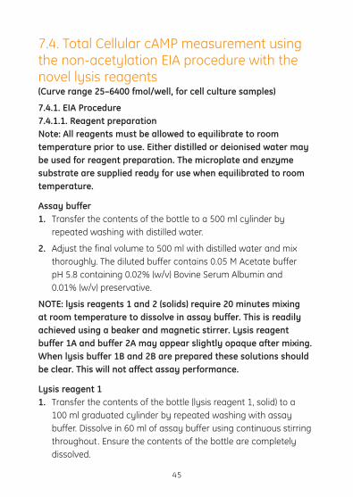
7.1.2. EIA Procedure7.1.2.1. Reagent preparationNote: All reagents must be allowed to equilibrate to room temperature prior to use. Either distilled or deionised water may be used for reagent preparation. The microplate and enzyme substrate are supplied ready for use when equilibrated to room temperature.
19

Assay buffer ForRPN225andRPN2251 1. Transfer the contents of the bottle to a 500 ml graduated cylinder
by repeated washing with distilled water.
2. Adjust the final volume to 500 ml with distilled water and mix thoroughly. The diluted buffer contains 0.05 M Acetate buffer pH 5.8, 0.02% (w/v) Bovine Serum Albumin and 0.01% (w/v) preservative.
ForRPN2255 1. Transfer the contents of the bottle to a suitable vessel by
repeated washing with distilled water.
2. Adjust the final volume to 2500 ml with distilled water and mix thoroughly.
Standard (for non-acetylation assay) 1. Carefully add 2.0 ml of diluted assay buffer and replace the
stopper.
2. Mix the contents of the bottle until completely dissolved. The final solution should contain cAMP at a concentration of 64 pmol/ml in 0.05 M Acetate buffer containing 0.02% (w/v) Bovine Serum Albumin and 0.01% (w/v) preservative.
Antiserum ForRPN225andRPN2251 1. Carefully add 11 ml diluted assay buffer and replace the stopper.
2. Gently mix the contents of the bottle by inversion and swirling untilacompletesolutionisobtained.Vigorousagitationandfoaming should be avoided. The contents will contain anti-cAMP serum in 0.05 M Acetate buffer containing 0.02% (w/v) Bovine Serum Albumin and 0.01% (w/v) preservative.
20

ForRPN2255 1. Reconstitiute with 55 ml of assay buffer and gently mix until a
complete solution is obtained..
cAMP Peroxidase conjugate ForRPN225andRPN2251 1. Carefully add 11 ml diluted assay buffer and replace the stopper.
2. Mix until the contents are completely dissolved. The solution will contain cAMP-horseradish Peroxidase in 0.05 M Acetate buffer pH 5.8, 0.02% (w/v) Bovine Serum Albumin and 0.01% (w/v) preservative.
ForRPN2255 1. Reconstitiute with 55 ml of assay buffer and gently mix until the
contents are completely dissolved.
Wash buffer ForRPN225andRPN2251 1. Transfer the contents of the bottle to a 500 ml graduated
cylinder by repeated washings with distilled water
2. Adjust the final volume to 500 ml with distilled water and mix thoroughly. The diluted wash buffer contains 0.01 M Phosphate buffer pH 7.5 containing 0.05% (v/v) Tween 20.
ForRPN2255 1. Transfer the contents of the bottle to a suitable vessel by
repeated washing with distilled water.
7.1.2.2. Preparation of working standardsNote: It is important to use a clean pipette tip for each dilution. Standards should be used within 1 hour of preparation.
1. Label 8 polypropylene or polystyrene tubes (12 x 75 mm), 25 fmol, 50 fmol, 100 fmol, 200 fmol, 400 fmol, 800 fmol, 1600 fmol and 3200 fmol.
21

2. Pipette 500 µl diluted assay buffer into these tubes.
3. Into the 3200 fmol tube pipette 500 µl of stock non-acetylation standard (64 pmol/ml) and mix thoroughly.
4. Transfer 500 µl from the 3200 fmol tube to the 1600 fmol tube and vortex mix thoroughly.
5. Repeat this doubling dilution successively with the remaining tubes and vortex after each dilution.
6. 100 µl aliquots from each of the serial dilutions will give rise to 8 standard levels of cAMP from 25–3200 fmol.
Note: One hundred microlitres (100 µl) of the reconstituted stock standard provided, serves as the top standard (6400 fmol/well).
7.1.2.3. Assay method Note: It is important that all reagents are equilibrated to room temperature before use. This is particularly important with the enzyme substrate, TMB. It is very important that the refrigerator temperature does not rise above 5°C during the course of the assay. An alternative method for achieving low assay temperatures is to place the microplate on crushed ice during the course of the assay.
1. Prepare assay components and standards ranging from 25–6400 fmol as described in the previous sections.
2. Equilibrate reagents to room temperature and mix before use.
3. Set up the microplate with sufficient wells for running of all blanks, standards and samples. Recommended positioning of blank (B), non-specific binding (NSB) wells, standard (0–6400 fmol) and sample (S) is shown in figure 3.
4. Pipette 200 µl of diluted assay buffer into the non-specific binding (NSB) wells.
5. Pipette 100 µl of diluted assay buffer into the zero standard wells (0).
22
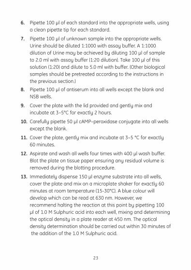
6. Pipette 100 µl of each standard into the appropriate wells, using a clean pipette tip for each standard.
7. Pipette 100 µl of unknown sample into the appropriate wells. Urine should be diluted 1:1000 with assay buffer. A 1:1000 dilution of Urine may be achieved by diluting 100 µl of sample to 2.0 ml with assay buffer (1:20 dilution). Take 100 µl of this solution (1:20) and dilute to 5.0 ml with buffer. (Other biological samples should be pretreated according to the instructions in the previous section.)
8. Pipette 100 µl of antiserum into all wells except the blank and NSB wells.
9. Cover the plate with the lid provided and gently mix and incubate at 3–5°C for exactly 2 hours.
10. Carefully pipette 50 µl cAMP–peroxidase conjugate into all wells except the blank.
11. Cover the plate, gently mix and incubate at 3–5 °C for exactly 60 minutes.
12. Aspirate and wash all wells four times with 400 µl wash buffer. Blot the plate on tissue paper ensuring any residual volume is removed during the blotting procedure.
13. Immediately dispense 150 µl enzyme substrate into all wells, cover the plate and mix on a microplate shaker for exactly 60 minutes at room temperature (15-30°C). A blue colour will develop which can be read at 630 nm. However, we recommend halting the reaction at this point by pipetting 100 µl of 1.0 M Sulphuric acid into each well, mixing and determining the optical density in a plate reader at 450 nm. The optical density determination should be carried out within 30 minutes of the addition of the 1.0 M Sulphuric acid.
23

24
Fig 3. Recommended positioning of standard (25–6400 fmol/well) and sample (S) wells.
* Since the non–specific binding in the assay is so low, typically less than 1% it is possible to omit the substrate blank determination, enabling the assay of an extra unknown sample. If this option is taken, the non–specific wells should be used to blank the plate reader.
BA
B
C
D
E
F
G
H
B S S S S S S S S
1 2 3 4 5 6 7 8 9 10 11 12
NSB NSB
800 800
S S S S S S S S1600 1600
S S S S S S S S3200 3200
6400 6400 S S S S S S S S
S S S S S S S S S S
S S S S S S S S S S
S S S S S S S S S S200 200
400 400 S S S S S S S S S S
0 0
25 25
50 50
100 100

** Reaction can be read at 630 nm before acidification but halting reaction prior to end point determination is recommended.
7.1.3. Data Processing7.1.3.1. Calculation of results The calculation is illustrated using representative data and is the same for all protocols.
The assay data should be similar to that shown in table 2.
1. Calculate the average optical density (OD) for each set of replicate wells.
2. Calculate the percent bound for each standard and sample using the following relationship:
%B/B0 = (standard or sample OD-NSB OD) x 100
(zero standard OD-NSB OD)
A standard curve may be generated by plotting the percent B/B0 as a function of the log cAMP concentration. Plot %B/B0 (y axis) against fmol cAMP standard per well (x axis). The curve shape should be similar to figure 4, if plotted on semi–log paper. The fmol/well value ofsamplescanbereaddirectlyfromthegraph.Figure4showsastandard curve generated from the data in table 2.
25

26
7.1.3.2. Typical assay data
Standard Optical density Mean OD- %B/B0 fmol/well (OD) at 450 nm NSB Blank 0.046 0.045 NSB 0.103 0.105 0 1.612 1.521 1.637 25 1.463 1.384 91 1.512 50 1.423 1.352 89 1.488 100 1.301 1.208 79 1.322 200 1.097 1.016 67 1.143 400 1.064 0.987 65 1.118 800 0.848 0.748 49 0.855 1600 0.651 0.544 36 0.645 3200 0.451 0.348 23 0.452 6400 0.299 0.305 20 0.310
Table 2. Typical assay data, protocol 1 (non-acetylation procedure)

Fig 4. Typical standard curve for protocol 1 (non-acetylation procedure)
27

7.2. Acetylation EIA Procedure (Curve range 2–128 fmol/well, for measurement of cAMP in plasma and tissue samples)
7.2.1. Specimen collection, sample preparation and purification• Representativeproceduresaredescribedonpages15–18
for the extraction of cAMP from tissues. Procedures for the measurement of cAMP from plasma samples are described below. This information is provided for guidance only.
Tissue Tissue sections must be rapidly frozen immediately after collection so as to prevent alteration to the cAMP and associated enzymes before analysis. This is usually achieved by immersion of the fresh tissue in liquid Nitrogen at -196°C. See pages 15–18 for more information on preparation of tissue samples.
Methods of freezing biological samples for cyclic nucleotide assays are reviewed by Mayer et al(24).
Samples should be stored at -15°C to -30°C until the assay is carried out.
Plasma Measurements should be made in plasma not serum. Blood should be collected into tubes containing 7.5 mM EDTA. Blood should be immediately centrifuged to remove cells and the plasma stored at -15°C to -30°C prior to analysis. If blood samples cannot be rapidly processed they should be stored in ice until it is possible to centrifuge. It is not necessary to extract or deproteinise plasma samples before analysis. Plasma should be diluted 1:100 with assay buffer.
28

7.2.2. EIA Procedure7.2.2.1. Reagent preparation Note: All reagents must be allowed to equilibrate to room temperature prior to use. Either distilled or deionised water may be used for reagent preparation. The microplate, Acetic Anhydride, Triethylamine and enzyme substrate are supplied ready for use when equilibrated to room temperature.
Assay buffer 1. Transfer the contents of the bottle to a 500 ml cylinder by
repeated washing with distilled water.
2. Adjust the final volume to 500 ml with distilled water and mix thoroughly. The diluted buffer contains 0.05 M Acetate buffer pH 5.8, 0.02% (w/v) Bovine Serum Albumin and 0.01% (w/v) preservative.
Standard (for acetylation assay) 1. Carefully add 4.0 ml diluted assay buffer and replace the stopper.
2. Mix until the contents are completely dissolved. The final solution should contain cAMP at a concentration of 2.56 pmol/ml in 0.05 M Acetate buffer containing 0.02% (w/v) Bovine Serum Albumin and 0.01% (w/v) preservative.
Antiserum 1. Carefully add 11 ml diluted assay buffer and replace the stopper.
2. Gently mix the contents of the bottle by inversion and swirling untilacompletesolutionisobtained.Vigorousagitationandfoaming should be avoided. The contents will contain anti-cAMP serum in 0.05 M Acetate buffer containing 0.02% (w/v) Bovine Serum Albumin and 0.01% (w/v) preservative.
cAMP Peroxidase conjugate 1. Carefully add 11 ml diluted assay buffer and replace the stopper.
29

2. Mix until the contents are completely dissolved. The solution will contain cAMP-horseradish Peroxidase in 0.05 M Acetate buffer pH 5.8, 0.02% (w/v) Bovine Serum Albumin and 0.01% (w/v) preservative.
Wash buffer 1. Transfer the contents of the bottle to a 500 ml graduated
cylinder by repeated washings with distilled water.
2. Adjust the volume to 500 ml with distilled water and mix thoroughly. The diluted wash buffer contains 0.01 M Phosphate buffer pH 7.5 containing 0.05%(v/v) Tween 20.
Reconstituted reagents should be stored at 2–8°C and re-used within 2 weeks.
7.2.2.2. Preparation of working standardsNote: It is important to use a clean pipette tip for each dilution. Standards should be used within 1 hour of preparation.
1. Label 7 polypropylene or glass tubes (12 x 75 mm) 2 fmol, 4 fmol, 8 fmol, 16 fmol, 32 fmol, 64 fmol and 128 fmol.
2. Pipette 1 ml diluted assay buffer into all tubes, except the 128 fmol.
3. Pipette 1 ml of stock acetylation standard (2.56 pmol/ml) into the 128 fmol tube.
4. Pipette 1 ml of stock acetylation standard (2.56 pmol/ml) into the 64 fmol tube and mix thoroughly.
5. Transfer 1 ml from the 64 fmol tube to the 32 fmol tube and mix thoroughly
6. Repeat this doubling dilution successively with the remaining tubes.
7. Remove 1 ml from the 2 fmol standard and discard. All tubes should now contain 1 ml.
30
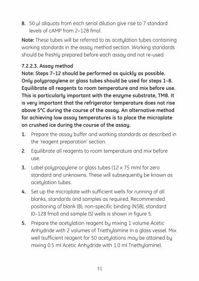
8. 50 µl aliquots from each serial dilution give rise to 7 standard levels of cAMP from 2–128 fmol.
Note: These tubes will be referred to as acetylation tubes containing working standards in the assay method section. Working standards should be freshly prepared before each assay and not re-used.
7.2.2.3. Assay method Note: Steps 7–12 should be performed as quickly as possible. Only polypropylene or glass tubes should be used for steps 1–8. Equilibrate all reagents to room temperature and mix before use. This is particularly important with the enzyme substrate, TMB. It is very important that the refrigerator temperature does not rise above 5°C during the course of the assay. An alternative method for achieving low assay temperatures is to place the microplate on crushed ice during the course of the assay.
1. Prepare the assay buffer and working standards as described in the ‘reagent preparation’ section.
2. Equilibrate all reagents to room temperature and mix before use.
3. Label polypropylene or glass tubes (12 x 75 mm) for zero standard and unknowns. These will subsequently be known as acetylation tubes.
4. Set up the microplate with sufficient wells for running of all blanks, standards and samples as required. Recommended positioning of blank (B), non-specific binding (NSB), standard (0–128 fmol) and sample (S) wells is shown in figure 5.
5. Prepare the acetylation reagent by mixing 1 volume Acetic Anhydride with 2 volumes of Triethylamine in a glass vessel. Mix well (sufficient reagent for 50 acetylations may be attained by mixing 0.5 ml Acetic Anhydride with 1.0 ml Triethylamine).
31

6. Pipette 1 ml of diluted assay buffer into the zero standard acetylation tube.
7. Pipette 1 ml of each unknown (see sample preparation section) into the appropriately labelled acetylation tubes. Plasma should be diluted 1:100 with assay buffer. A 1:100 dilution of plasma may be achieved by diluting 100 µl of sample to 2.0 ml with assay buffer (1:20 dilution). Take 200 µl of this solution (1:20) and dilute to 1 ml. Tubes containing 1 ml of each working standard should have been prepared (see p.26).
8. Carefully add 25 µl of the acetylation reagent to all acetylation tubes containing standards and unknowns. Optimum precision is attained by placing the pipette tip in contact with the test tube wall above the aqueous layer and allowing the acetylation reagent to run down the test tube wall into the liquid. Each tube should be vortexed immediately following addition of the acetylating reagents.
9. Pipette 100 µl of antiserum into all wells except the blank and non-specific binding (NSB) wells.
10. Pipette duplicate 50 µl aliqouts from all aceylation tubes including the zero standard into the appropriate wells.
11. Pipette 150 µl of assay buffer into the non-specific binding wells.
12. Cover the plate with the lid provided, gently mix and incubate at 3–5°C for exactly 60 minutes.
13. Pipette 100 µl of cAMP Peroxidase conjugate into all wells except the blank.
14. Cover the plate, gently mix and incubate at 3-5°C for exactly 60 minutes.
15. Aspirate and wash all wells four times with 400 µl wash buffer. Blot the plate on tissue paper ensuring any residual volume is
32
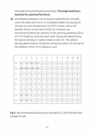
removed during the blotting procedure. Thorough washing is essential for good performance.
16. Immediately dispense 150 µl enzyme substrate into all wells, cover the plate and mix on a microplate shaker for exactly 60 minutes at room temperature (15-30°C). A blue colour will develop which can be read at 630 nm. However, we recommend halting the reaction at this point by pipetting 100 µl of 1.0 M Sulphuric acid into each well, mixing and determining the optical density in a plate reader at 450 nm. The optical density determination should be carried out within 30 minutes of the addition of the 1.0 M Sulphuric acid.
Fig 5. Recommended positioning of standard (2–128 fmol/well) and sample (S) well.
33
BA
B
C
D
E
F
G
H
B 64 64 S S S S S S S S
1 2 3 4 5 6 7 8 9 10 11 12
NSB NSB 128 128 S S S S S S S S
S S S S S S S S S S
S S S S S S S S S S
S S S S S S S S S S
S S S S S S S S S S
S S S S S S S S S S
32 32 S S S S S S S S S S
0 0
2 2
4 4
8 8
16
16
16
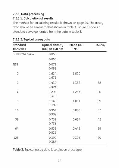
7.2.3. Data processing7.2.3.1. Calculation of results The method for calculating results is shown on page 25. The assay datashouldbesimilartothatshownintable3.Figure6showsastandard curve generated from the data in table 3.
7.2.3.2. Typical assay data
Standard Optical density Mean OD- %B/B0fmol/well (OD) at 450 nm NSB
Substrate blank 0.050
0.050
NSB 0.078 0.082
0 1.624 1.570 1.675
2 1.430 1.382 88 1.493
4 1.296 1.253 80 1.370
8 1.140 1.081 69 1.182
16 0.954 0.888 57 0.982
32 0.739 0.654 42 0.729
64 0.532 0.449 29 0.525
128 0.390 0.308 20 0.386
Table 3. Typical assay data (acetylation procedure)
34

Fig 6. Typical standard curve for protocol 2 (acetylation procedure)
35
*Forthecalculationof%B/B0 values, non-specific binding values have been subtracted from the appropriate optical densities.
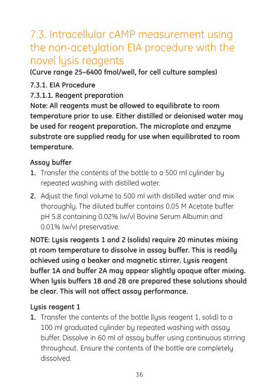
7.3. Intracellular cAMP measurement using the non-acetylation EIA procedure with the novel lysis reagents (Curve range 25–6400 fmol/well, for cell culture samples)
7.3.1. EIA Procedure7.3.1.1. Reagent preparation Note: All reagents must be allowed to equilibrate to room temperature prior to use. Either distilled or deionised water may be used for reagent preparation. The microplate and enzyme substrate are supplied ready for use when equilibrated to room temperature.
Assay buffer 1. Transfer the contents of the bottle to a 500 ml cylinder by
repeated washing with distilled water.
2. Adjust the final volume to 500 ml with distilled water and mix thoroughly. The diluted buffer contains 0.05 M Acetate buffer pH 5.8 containing 0.02% (w/v) Bovine Serum Albumin and 0.01% (w/v) preservative.
NOTE: Lysis reagents 1 and 2 (solids) require 20 minutes mixing at room temperature to dissolve in assay buffer. This is readily achieved using a beaker and magnetic stirrer. Lysis reagent buffer 1A and buffer 2A may appear slightly opaque after mixing. When lysis buffers 1B and 2B are prepared these solutions should be clear. This will not affect assay performance.
Lysis reagent 1 1. Transfer the contents of the bottle (lysis reagent 1, solid) to a
100 ml graduated cylinder by repeated washing with assay buffer. Dissolve in 60 ml of assay buffer using continuous stirring throughout. Ensure the contents of the bottle are completely dissolved.
36
2. Adjust the final volume to 80 ml with assay buffer and mix thoroughly. The final solution contains a 2.5% solution of Dodecyltrimethylammonium Bromide in assay buffer. This is lysis buffer 1A. Stir continuously when used.
3. Take 10 ml of lysis buffer 1A and make up to 100 ml with assay buffer to give a final 0.25% solution of Dodecyltrimethylammonium Bromide in assay buffer. This is lysis reagent 1B. It is used for the intracellular measurement of cAMP and for the preparation of standards.
Lysis reagent 2 1. Transfer the contents of the bottle (lysis reagent 2, solid) to a
100 ml graduated cylinder. Dissolve in 80 ml of assay buffer using continuous stirring throughout. Ensure the contents of the bottle are completely dissolved.
2. Adjust the final volume to 100 ml with assay buffer and mix thoroughly. This is lysis buffer 2A.
3. Take 10 ml of lysis buffer 2A and make up to a final volume of 40 ml with assay buffer and mix thoroughly. This is lysis reagent 2B.
Standard (for non-acetylation assay) Add 2 ml diluted lysis reagent 1B (prepared as described above) and replace the stopper. Gently mix until the contents are completely dissolved. The final solution contains cAMP at a concentration of 64 pmol/ml in lysis reagent 1B.
Antiserum Carefully add 11.0 ml of lysis reagent 2B replace the stopper. Gently mix the contents of the bottle by inversion and swirling until a completesolutionisobtained.Vigorousagitationandfoamingshould be avoided. The contents will contain anti-cAMP serum in lysis reagent 2B.
37
cAMP Peroxidase conjugate Do not add lysis reagent 1 or 2 to the conjugate.
Carefully add 11.0 ml of diluted assay buffer and replace the stopper. Mix the contents until completely dissolved. The solution will contain cAMP-horseradish Peroxidase in 0.05 M Acetate buffer pH 5.8, 0.02% (w/v) Bovine Serum Albumin and 0.01% (w/v) preservative.
Wash buffer 1. Transfer the contents of the bottle to a 500 ml cylinder by
repeated washing with distilled water.
2. Adjust the final volume to 500 ml with distilled water and mix thoroughly. The diluted wash buffer contains 0.01 M Phosphate buffer pH 7.5 containing 0.05% (w/v) Tween 20.
7.3.1.2. Cell lysis methods – intracellular cAMP measurement (Note:ForcelllysismethodfortotalcellularcAMPmeasurement,see protocol 4). Two lysis methods are described for intracellular cAMP measurement.
Adherent cells 1. Culture cells (100 µl volumes) in standard 96-well microplates
(tissue-culture grade), with cell concentrations of between 104–106 cells/ml.
2. Incubate plate overnight at 37°C (5% CO2 and 95% humidity). Note: do not use cell cultures that are over-confluent (e.g. at 107 cells/ml) as cells may be lost during decantation.
3. Add 100 µl of drug, agonist etc. under study. Incubate for suitable time period.
4. Decant or aspirate excess culture media.
5. Add 200 µl/well of diluted lysis reagent 1B
38

6. Agitate cells after lysis reagent 1B is added. This is to facilitate cell lysis and can be achieved by shaking the plate on a microplate shaker for 10 minutes after adding the lysis reagent.
7. Carry out a microscopic evaluation using Trypan blue to check cells have lysed. Cell membranes may still be visible after cell lysis. Lysed cells are now ready for use in the enzymeimmunoassay protocol and should be processed immediately in the immunoassay (see ‘assay method’).
Suspension cells Note: If suspension cells are used, special microplate adapters are needed for the centrifugation step.
1. Culture cells (100 µl) in standard 96-well microplates (tissue-culture grade) with cell concentrations of between 104–106 cells/ml.
2. Incubate plate overnight at 37°C (5% CO2 and 95% humidity).
3. Add 100 µl of drug, agonist etc. under study. Incubate for a suitable time period.
4. Using a centrifugal microplate adapter, centrifuge the microplate at 1000–1500 x g for 3 minutes to form a pellet in each well. Note: the actual centrifugal speed required is dependent on the cells under study and should be validated by the investigator.
5. Gently decant or aspirate excess media and resuspend pellet in 200 µl of lysis reagent 1B.
6. Agitate cells after lysis reagent 1B is added. This is to facilitate cell lysis and can be achieved by shaking the plate on a microplate shaker for 10 minutes after adding the lysis reagent.
7. Carry out a microscopic evaluation using Trypan blue to check cells have lysed. Cell membranes may still be visible after cell lysis. Lysed cells are now ready for use in the enzymeimmunoassay protocol and should be immediately processed in the immunoassay (see ‘assay method’).
39

7.3.1.3. Preparation of working standards Note: It is important to use a clean pipette tip for each dilution. Standards should be used within 1 hour of preparation.
1. Label 8 polypropylene or polystyrene tubes (12 x 75 mm), 25, 50, 100, 200, 400, 800, 1600 and 3200 fmol.
2. Pipette 500 µl of lysis reagent 1B into all tubes.
3. Into the 3200 fmol tube pipette 500 µl of stock non-acetylation standard (64 pmol/ml) and mix thoroughly.
4. Transfer 500 µl from the 3200 fmol tube to the 1600 fmol tube and vortex mix thoroughly.
5. Repeat this doubling dilution successively with the remaining tubes and vortex after each dilution.
6. 100 µl aliquots from each of the serial dilutions will give rise to 8 standard levels of cAMP from 25–3200 fmol.
Note: One hundred microlitres (100 µl) of the reconstituted stock standard provided, serves as the top standard (6400 fmol/well).
7.3.1.4. Assay method It is very important that the refrigerator temperature does not rise above 5°C during the course of the assay. An alternative method for achieving low assay temperatures is to place the microplate on crushed ice during the course of the assay.
1. Prepare assay buffer, lysis reagents and standards ranging from 25–6400 fmol as described in the previous section.
2. Equilibrate all reagents to room temperature and mix before use. This is particularly important with the enzyme substrate, TMB.
3. Set up the microplate with sufficient wells to enable the running of all blanks, standards and samples as required. Recommended
40

positioning of substrate blank (B), non-specifi c binding (NSB), standard (0–6400 fmol) and sample (S) wells is shown in figure 2.
4. Pipette 100 µl of lysis reagent 1B and 100 µl of lysis reagent 2B into the NSB wells.
5. Pipette 100 µl of lysis reagent 1B into the zero standard (0) wells.
6. Pipette 100 µl of each standard into the appropriate wells, using a clean pipette for each standard.
7. Pipette 100 µl of unknown sample into the appropriate wells. (See previous section for sample preparation).
8. Pipette 100 µl of antiserum into all wells except the blank and NSB wells.
9. Cover the plate with the lid provided, gently mix and incubate at 3–5°C for exactly 2 hours.
10. Carefully pipette 50 µl cAMP-peroxidase conjugate into all wells except the blank.
11. Cover the plate, gently mix and incubate at 3–5°C for exactly 60 minutes.
12. Aspirate and wash all wells four times with 400 µl wash buffer. Blot the plate on tissue paper ensuring any residual volume is removed during the blotting procedure. Thorough and careful washing is essential for good performance.
13. Immediately dispense 150 µl enzyme substrate into all wells, cover the plate and mix on a microplate shaker for exactly 60 minutes at room temperature (15-30°C). A blue colour will develop which can be read at 630 nm. However, we recommend halting the reaction at this point by pipetting 100 µl of 1.0 M Sulphuric acid into each well, mixing and determining the optical density in a plate reader at 450 nm. The optical density determination should be carried out within 30 minutes of the addition of the 1.0 M Sulphuric acid.
41

Fig 7. Recommended positioning of standard (25–6400 fmol/well) and sample (S) wells
42
BA
B
C
D
E
F
G
H
B S S S S S S S S
1 2 3 4 5 6 7 8 9 10 11 12
NSB NSB
800 800
S S S S S S S S1600 1600
S S S S S S S S3200 3200
6400 6400 S S S S S S S S
S S S S S S S S S S
S S S S S S S S S S
S S S S S S S S S S200 200
400 400 S S S S S S S S S S
0 0
25 25
50 50
100 100
7.3.2. Data processing7.3.2.1. Calculation of results The method for calculating results is shown on page 25. The assay datashouldbesimilartothatshownintable4.Figure8showsastandard curve generated from the data in table 4.
7.3.2.2. Typical assay data
Standard Optical density Mean OD- %B/B0fmol/well (OD) at 450 nm NSB Substrate 0.045blank 0.043NSB 0.112 0.112 0 1.533 1.422 1.534 25 1.492 1.379 97 1.489 50 1.447 1.319 93 1.415 100 1.345 1.255 88 1.388 200 1.322 1.182 83 1.266 400 1.057 0.946 66 1.058 800 0.78 0.656 46 0.759 1600 0.594 0.473 33 0.576 3200 0.406 0.293 20 0.404 6400 0.285 0.173 12 0.284
Table 4. Typical assay data for the intra- and ‘total’ cellular cAMP assays (protocols 3 and 4).
43
Fig 8. Typical standard curve (for intra- and total cellular cAMP protocols)
44
7.4. Total Cellular cAMP measurement using the non-acetylation EIA procedure with the novel lysis reagents (Curve range 25–6400 fmol/well, for cell culture samples)
7.4.1. EIA Procedure7.4.1.1. Reagent preparation Note: All reagents must be allowed to equilibrate to room temperature prior to use. Either distilled or deionised water may be used for reagent preparation. The microplate and enzyme substrate are supplied ready for use when equilibrated to room temperature.
Assay buffer 1. Transfer the contents of the bottle to a 500 ml cylinder by
repeated washing with distilled water.
2. Adjust the final volume to 500 ml with distilled water and mix thoroughly. The diluted buffer contains 0.05 M Acetate buffer pH 5.8 containing 0.02% (w/v) Bovine Serum Albumin and 0.01% (w/v) preservative.
NOTE: lysis reagents 1 and 2 (solids) require 20 minutes mixing at room temperature to dissolve in assay buffer. This is readily achieved using a beaker and magnetic stirrer. Lysis reagent buffer 1A and buffer 2A may appear slightly opaque after mixing. When lysis buffer 1B and 2B are prepared these solutions should be clear. This will not affect assay performance.
Lysis reagent 1 1. Transfer the contents of the bottle (lysis reagent 1, solid) to a
100 ml graduated cylinder by repeated washing with assay buffer. Dissolve in 60 ml of assay buffer using continuous stirring throughout. Ensure the contents of the bottle are completely dissolved.
45

2. Adjust the final volume to 80 ml with assay buffer and mix thoroughly. The final solution contains a 2.5% solution of Dodecyltrimethylammonium Bromide in assay buffer. This is lysis buffer 1A, which is used in the cell lysis method for the ‘total’ cellular cAMP assay. Stir continuously when used. Stir for 30 minutes after storage.
3. Take 10 ml of lysis buffer 1A and make up to 100 ml with assay buffer to give a final 0.25% solution of Dodecyltrimethylammonium Bromide in assay buffer. This is lysis reagent 1B and is used for the preparation of working standards only.
Lysis reagent 2 1. Transfer the contents of the bottle (lysis reagent 2, solid) to a
100 ml graduated cylinder. Dissolve in 80 ml of assay buffer using continuous stirring throughout. Ensure the contents of the bottle are completely dissolved.
2. Adjust the final volume to 100 ml with assay buffer and mix thoroughly. This is lysis buffer 2A.
3. Take 10 ml of lysis buffer 2A and make up to a final volume of 40 ml with assay buffer and mix thoroughly. This is lysis reagent 2B.
Standard (for non-acetylation assay) Add 2 ml diluted lysis reagent 1B and replace the stopper. Gently mix until the contents are completely dissolved. The final solution contains cAMP at a concentration of 64 pmol/ml in lysis reagent 1B.
Antiserum Carefully add 11.0 ml of lysis reagent 2B and replace the stopper. Gently mix the contents of the bottle by inversion and swirling until acompletesolutionisobtained.Vigorousagitationandfoamingshould be avoided. The contents will contain anti-cAMP serum in lysis reagent 2B.
46
cAMP Peroxidase conjugate Do not add lysis reagent 1 or 2 to the conjugate.
Carefully add 11.0 ml of diluted assay buffer and replace the stopper. Mix the contents until completely dissolved. The solution will contain cAMP-horseradish Peroxidase in 0.05 M Acetate buffer pH 5.8, 0.02% (w/v) Bovine Serum Albumin and 0.01% (w/v) preservative.
Wash buffer 1. Transfer the contents of the bottle to a 500 ml cylinder by
repeated washing with distilled water.
2. Adjust the final volume to 500 ml with distilled water and mix thoroughly.
Reconstituted reagents should be stored at 2–8°C and re-used within two weeks.
7.4.1.2. Cell lysis method – total cellular cAMP measurement (Note:ForcelllysismethodsandintracellularcAMPmeasurement,see page 36)
The following method measures the combined amount of intracellular and cell supernatant cAMP.
Suspension and adherent cells 1. Culture adherent or suspension cells (160 µl/well) in flat-
bottomed 96-well microplates (tissue-culture grade), with cell concentrations between 104 –106 cells/well.
2. Incubate the plate overnight at 37°C, (5% CO2 and 95% humidity).
3. Add 20 µl aliquots of agonist or cell stimulant directly to the cell culture samples. Do not decant or aspirate the culture media. Incubate agonist/cell stimulant with cultures depending on required experimental conditions.
47

4. Add 20 µl of lysis reagent 1A (2.5% Dodecyltrimethylammonium Bromide in assay buffer). The final volume in the wells should be 200 µl, each containing 0.25% Dodecyltrimethylammonium Bromide (final concentration) which is equivalent to the lysis reagent 1B.
5. Followingtheadditionoflysisreagent1A,agitatecellstofacilitate cell lysis. This can be readily achieved by vigorous, successive pipetting. Incubate the plate for 10 minutes at room temperature (in order to achieve cell lysis).
6. Carry out a microscopic evaluation with Trypan blue. Lysed cells are now ready for use in the assay (see ‘assay protocol’). Cell membranes may still be visible after lysis.
7. Transfer 100 µl aliquots of cell lysate to the donkey anti-rabbit Ig coated plate for assay.
7.4.1.3. Preparation of working standards Note: It is important to use a clean pipette tip for each dilution. Standards should be used within 1 hour of preparation.
1. Label 8 polypropylene or polystyrene tubes (12 x 75 mm), 25, 50, 100, 200, 400, 800, 1600 and 3200 fmol.
2. Pipette 500 µl lysis reagent 1B into these tubes.
3. Into the 3200 fmol tube pipette 500 µl of stock non-acetylation standard (64 pmol/ml) and mix thoroughly.
4. Transfer 500 µl from the 3200 fmol tube to the 1600 fmol tube and vortex mix thoroughly.
5. Repeat this doubling dilution successively with the remaining tubes and vortex after each dilution.
6. 100 µl aliquots from each of the serial dilutions will give rise to 8 standard levels of cAMP from 25–3200 fmol.
Note: One hundred microlitres (100 µl) of the reconstituted stock standard provided, serves as the top standard (6400 fmol/well).
48

7.4.1.4. Assay protocolIt is very important that the refrigerator temperature does not rise above 5°C during the course of the assay. An alternative method for achieving low assay temperatures is to place the microplate on crushed ice during the course of the assay.
1. Prepare assay buffer, lysis reagents and standards ranging from 25–6400 fmol as described in the previous section.
2. Equilibrate all reagents to room temperature and mix before use. This is particularly important with the enzyme substrate, TMB.
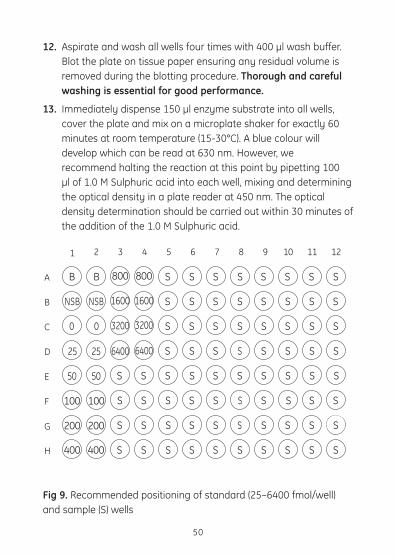
3. Set up the microplate with sufficient wells to enable the running of all blanks, standards and samples as required. Recommended positioning of substrate blank (B), non-specific binding (NSB), standard (0–6400 fmol) and sample (S) wells is shown in figure 9.
4. Pipette 100 µl of lysis reagent 1B and 100 µl of lysis reagent 2B into the NSB wells.
5. Pipette 100 µl of lysis reagent 1B into the zero standard (0) wells.
6. Pipette 100 µl of each standard into the appropriate wells, using a clean pipette for each standard.
7. Pipette 100 µl of unknown sample into the appropriate wells. (See previous section for sample preparation).
8. Pipette 100 µl of antiserum into all wells except the blank and NSB wells.
9. Cover the plate with the lid provided, gently mix and incubate at 3–5°C for exactly 2 hours.
10. Carefully pipette 50 µl cAMP-peroxidase conjugate into all wells except the blank.
11. Cover the plate, gently mix and incubate at 3–5°C for exactly 60 minutes.
49
12. Aspirate and wash all wells four times with 400 µl wash buffer. Blot the plate on tissue paper ensuring any residual volume is removed during the blotting procedure. Thorough and careful washing is essential for good performance.
13. Immediately dispense 150 µl enzyme substrate into all wells, cover the plate and mix on a microplate shaker for exactly 60 minutes at room temperature (15-30°C). A blue colour will develop which can be read at 630 nm. However, we recommend halting the reaction at this point by pipetting 100 µl of 1.0 M Sulphuric acid into each well, mixing and determining the optical density in a plate reader at 450 nm. The optical density determination should be carried out within 30 minutes of the addition of the 1.0 M Sulphuric acid.
Fig 9. Recommended positioning of standard (25–6400 fmol/well) and sample (S) wells
50
BA
B
C
D
E
F
G
H
B S S S S S S S S
1 2 3 4 5 6 7 8 9 10 11 12
NSB NSB
800 800
S S S S S S S S1600 1600
S S S S S S S S3200 3200
6400 6400 S S S S S S S S
S S S S S S S S S S
S S S S S S S S S S
S S S S S S S S S S200 200
400 400 S S S S S S S S S S
0 0
25 25
50 50
100 100
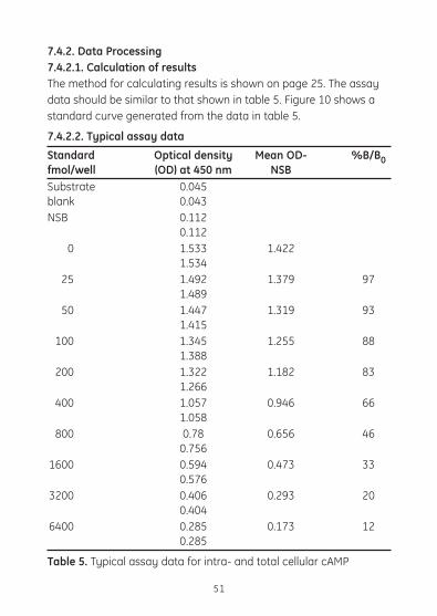
7.4.2. Data Processing7.4.2.1. Calculation of results The method for calculating results is shown on page 25. The assay datashouldbesimilartothatshownintable5.Figure10showsastandard curve generated from the data in table 5.
7.4.2.2. Typical assay data
Standard Optical density Mean OD- %B/B0fmol/well (OD) at 450 nm NSB Substrate 0.045blank 0.043NSB 0.112 0.112 0 1.533 1.422 1.534 25 1.492 1.379 97 1.489 50 1.447 1.319 93 1.415 100 1.345 1.255 88 1.388 200 1.322 1.182 83 1.266 400 1.057 0.946 66 1.058 800 0.78 0.656 46 0.756 1600 0.594 0.473 33 0.576 3200 0.406 0.293 20 0.404 6400 0.285 0.173 12 0.285
Table 5. Typical assay data for intra- and total cellular cAMP
51
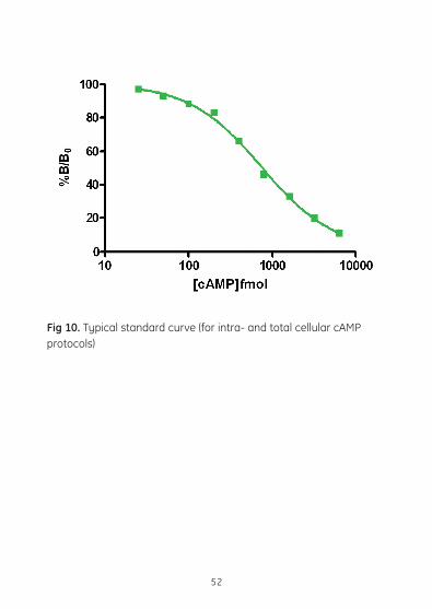
Fig 10. Typical standard curve (for intra- and total cellular cAMP protocols)
52
8. Additional Information
8.1. Expected valuesIndividual laboratories should establish their own normal ranges. GE Healthcare has achieved the following values of cAMP in normal subjects using the enzyme immunoassay system:
Urine 1.96 ± 0.94 nmol/ml (n = 18)
Plasma 13.96 ± 3.8 pmol/ml (n = 20)
8.2. Limitations of useDo not use lipaemic, haemolysed or turbid specimens. Avoid repeated freezing and thawing of specimens.
Renal function and diseases which alter PTH concentrations can influence cAMP concentration in Urine and plasma. Other factors that have been reported to alter plasma cAMP concentrations and urinary excretion are pregnancy, certain drugs (for example adrenalin) and exercise.
8.3. SpecificityThe cross-reactivity, as determined by the concentration giving 50% B/B0, is shown in the following table and graphically in figures 11 and 12.
53

54
Compound % Cross-reactivity (50% B/B0 displacement)
Non-acetylation Acetylation (protocols 1, 3, 4) (protocol 2)
cIMP 1 <0.005cGMP <0.05 <0.005cCMP <0.05 <0.005cTMP <0.05 <0.005AMP <0.05 <0.005ADP <0.05 <0.005ATP <0.05 <0.005EDTA <0.05 <0.005Theophylline <0.05 <0.005cAMP 100 100
Table 6. Cross-reactivity

55
Fig 11.
(a) Cross–reactivity profile for protocols 1,3,4 (non-acetylation assay)
(b) Cross–reactivity profile for protocols 1,3,4 (non-acetylation assay)
0
20
40
60
80
100
120
10 100 1000 104 105 106 107 108 109
cAMP (fmol/well )
%B/
B0
= cAMP= cGMP= cIMP= cTMP= ATP= cCMP
▲
★
◗
◆
▲ ▲ ▲ ▲
▲
▲
▲
▲★
★
★
★★★
★★
◗
◗
◗
◗
◗◗
◗◗
◆
◆
◆◆
◆◆
0
20
40
60
80
100
120
10 100 1000 104 105 106 107 108 109
cAMP (fmol/well )
%B/
B0
= cAMP= AM P= ADP= EDT A= Theophylline
�
�
�
� � � �� �
�
�
�
�
�
�
�
�
��
�
�
�
���
�
� � � �
�
�
10 100 1000 104 105 106 107 108 109
cAMP (fmol/well)
120
100
80
60
40
20
0
%B/
B0

56
Fig 12.
(a) Cross–reactivity profile for protocol 2 (acetylation assay)
(b) Cross–reactivity profile for protocol 2 (acetylation assay)
0
20
40
60
80
100
120
cAMP (fmol/well )
%B/
B0
s s s s s s
s
H
H
HHHHH wwwwwww
u
u
uuuu
1 10 100 1000 104 105 106 107
= cAMP= cGMP= cIMP= cTMP= ATP= cCMP
s
H
w
u
01 10 100 1000 104 105 106 107 108
cAMP (fmol/well )
%B/
B0
��
�
�
20
40
60
80
100
= cAMP= AM P= ADP= EDT A= Theophylline
�
�
�
��
�
� ��
�
� ��
�
� ��
�
� ��
�
� ��
�
�
��
�
�
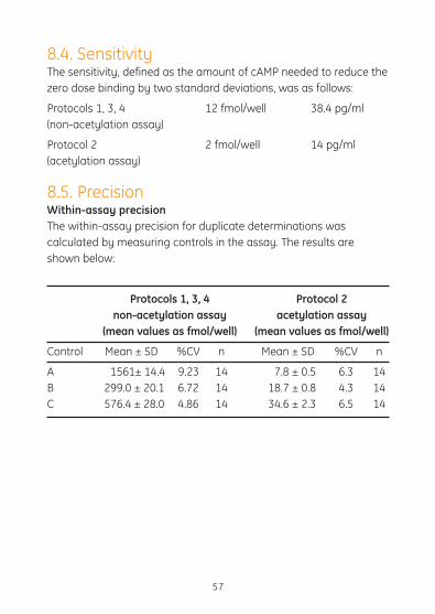
8.4. SensitivityThe sensitivity, defined as the amount of cAMP needed to reduce the zero dose binding by two standard deviations, was as follows:
Protocols 1, 3, 4 12 fmol/well 38.4 pg/ml(non-acetylation assay)
Protocol 2 2 fmol/well 14 pg/ml(acetylation assay)
8.5. PrecisionWithin-assay precision The within-assay precision for duplicate determinations was calculated by measuring controls in the assay. The results are shown below:
Protocols 1, 3, 4 Protocol 2 non-acetylation assay acetylation assay (mean values as fmol/well) (mean values as fmol/well)
Control Mean±SD %CV n Mean±SD %CV n
A 1561± 14.4 9.23 14 7.8 ± 0.5 6.3 14B 299.0 ± 20.1 6.72 14 18.7 ± 0.8 4.3 14C 576.4 ± 28.0 4.86 14 34.6 ± 2.3 6.5 14
57

Between-assay precision The between-assay precision was assessed by repeated measurement of the same controls in successive assays. The results are shown below:
Table 7.
Protocols 1, 3, 4 Protocol 2 non-acetylation assay acetylation assay (mean values as fmol/well) (mean values as fmol/well)
Control Mean±SD %CV n Mean±SD %CV n
A 11.4 ± 9.2 08 8 06.2 ± 0.73 11.7 11B 295 ± 26.8 09.1 8 10.9 ± 0.89 08.2 11C 739 ± 143 15 8 21.8 ± 3.79 17.4 11
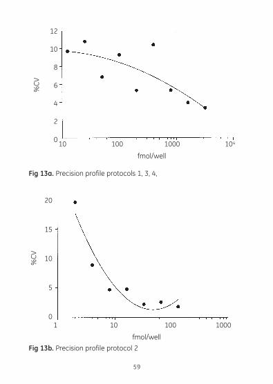
Precision profile A precision profile was generated by preparing ten replicates of each of the standards and calculating the standard deviation and percent coefficient of variation at each concentration.
Protocols 1, 3, 4 Protocol 2 non-acetylation assay acetylation assay
Standard Standard %CV Standard Standard %CV deviation deviation
12.5 1.2 9.7 2 0.4 19.6 25 2.4 10.8 4 0.4 8.9 50 3.4 6.8 8 0.4 4.7 100 9.6 9.3 16 0.8 4.8 200 10.7 5.3 32 0.7 2.2 400 38.9 10.4 64 1.7 2.6 800 42.8 5.3 128 2.3 1.8 1600 65.9 3.9 3200 106 3.3
58
59
Fig 13a. Precision profile protocols 1, 3, 4,
Fig 13b. Precision profile protocol 2
12
10
8
6
4
2
0
%CV
10 100 1000 104
fmol/well
20
15
10
5
0
%CV
1 10 100 1000
fmol/well
60
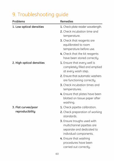
Problems
1. Low optical densities
2. High optical densities
3. Flat curves/poor reproducibility
Remedies
1. Check plate reader wavelength.2. Check incubation time and
temperature.3. Check that reagents are
equilibrated to room temperature before use.
4. Check that the kit reagents have been stored correctly.
1. Ensure that every well is completely filled and emptied at every wash step.
2. Ensure that automatic washers are functioning correctly.
3. Check incubation times and temperatures.
4. Ensure that plates have been blotted on tissue paper after washing.
1. Check pipette calibration.2. Check preparation of working
standards.3. Ensure troughs used with
multichannel pipettes are separate and dedicated to individual components.
4. Ensure that washing procedures have been carried out correctly.
9. Troubleshooting guide

10. Background and referencesThe physiological responses to many biologically important compounds are mediated through ‘second messengers’. This is a term described by Sutherland for molecules which are able to transmit intracellularly, the biological effects of compounds not able to enter the target cells themselves (1).
cAMP was identified as playing a major role in the mode of action of Adrenaline 30 years ago (2–5). In response to receptor binding, the enzyme adenylate cyclase converts ATP to cAMP, which exerts its effect by activating a protein kinase capable of phosphorylating specfic substrates. Numerous hormones are known to act through this mechanism including Corticotrophin (ATCH), luteinising hormone (LH),folliclestimulatinghormone(FSH),thyroidstimulatinghormone(TSH), calcitonin, glucagon, vasopressin and parathyroid hormone (PTH).
cAMP has now been shown to be involved in the cardiovascular(6) and nervous system (7), in immune mechanisms (8), cell growth and differentiation (9) and general metabolism (10). There remains considerable interest in the measurement of intracellular cAMP in tissues and cell cultures, and this may help to provide an understanding of the physiology and pathology of many disease states.
The assay system may be used in adenylate cyclase assays which determine cAMP formation from unlabelled ATP (11–14). The method allows high sensitivity without the interference from ATP to which other adenylate cyclase assays are prone (14). In recent years there has been great interest in a new generation of phosphoinositide-derived second messengers (15–20).
Receptor stimulation triggers the Phospholipase C catalysed Hydrolytic cleavage of membrane Phosphatidylinositol
61

4,5-biphosphate to yield two second messenger molecules viz inositol 1,4,5-trisphosphate (IP3) and sn-1,2-diacylglycerol (DAG).
It is now well established that IP3 acts as a second messenger of Ca2+mobilised hormones in a variety of cell types (21). DAG appears to be an essential co-factor for the enzyme protein kinase C which plays a crucial role in signal transduction (17,22).
Levels of IP3 and DAG can be determined using GE Healthcare assay system TRK1000.
1. Sutherland, E.W., et al., Circulation. 37, 279, (1968).
2. Rall, T.W., et al., J.Biol. Chem. 224, 463, (1957).
3. Cook, W.H., et al., Am. Chem. Soc. 79, 3607, (1957).
4. Suthgerland, E.W. and Rall, T.W., J. Am. Chem. Soc. 79, 3608, (1957).
5. Lipkin, D., et al., Am. Chem. Soc. 81, 6198, (1959).
6. Hamet, P. et al, Advances in Cyclic Nucleotide Research. 12, 11, Hamet, P. and Sands, H., eds, Raven Press, New York, (1980).
7. Drummond, G., Advances in Nucleotide Research. 15, 373, Greenard, P. and Robinson, G.A., eds, Raven Press, New York, (1983).
8. Plaut, M. et al., Advances in Nucleotide Research. 12, 161, Hamet, P. and Sands, H., eds, Raven Press, New York, (1980).
9. Boyton,A.L.andWhitefield,J.F.,Advances in Cyclic Nucleotide Research. 15, 193, Greenard, P. and Robinson, G.A., eds, Raven Press, New York, (1983).
10. Exton, J.H., Advances in Cyclic Nucleotide Research. 12, 319, Hamet, P. and Sands, H., eds, Raven Press, New York, (1980).
11. Albano, J.D.M. et al., Analyt. Biochem. 60, 130, (1974).
12. Albano, J.D.M. et al., Analyt. Biochem. Soc. Trans. 1, 477, (1973).
62

13. Thomas, J.A. and Singhal, R.L., Biochem. Pharmacol. 22, 507, (1973).
14. Volker,T.T.,et al., Analyt.Biochem. 144, 347, (1985).
15. Berridge, M.J., Biochem. J. 220, 345-360, (1984).
16. Berridge, M.J. and Irvine, R.F., Nature. 312, 315-321, (1984).
17. Nishizuka, Y., Nature. 308, 693-698, (1984).
18. Majerus, P.W. et al., Science. 234, 1519-1526, (1986).
19. Michell, R.H., Phosphoinositides and Receptor Mechanisms, Putney, J. W. Jnr., Alan R., eds. Liss Inc., New York. 1-24, (1986).
20. Berridge, M.J., Phosphoinositides and Receptor Mechanisms. 24-45, Putney, J. W. Jnr., Alan R., eds, Liss Inc., New York, (1986).
21. Michell, R.H., Bicochem. Biophys. Acta. 415, 81-147, (1975).
22. Nishizuka, Y., Science. 223, 305-312, (1986).
23. Bos, E.S. et al, J. Immunoassay 2, 187-204, (1981).
24. Mayer, S.E.., et al., Methods in Enzymology. 38, 3. Hardman, J.C. and O’Malley, B.W., eds, Academic Press, New York, (1974).
25. Goldburg, N.D. and O’Toole, A.G., Methods of Biological Analysis. 20, 1-39. Glick, D., ed Interscience Publishers, John Wiley and Sons
Inc., London, (1971).
26. Harper,J.F.H.andBrooker,G.,J.Cyclic Nucleotide Res. 1, 207, (1975).
27. Steiner, A.L., Methods of Hormone Radioimmunoassay. 3, Jaffe, B.M. and Behrman, H.R., eds, Academic Press, New York, (1979).
28. Rosenburg, N., et al. FEBS Letts. 137, 105, (1982).
63

11. Related productsBiotrak signal transduction assay range
Cyclic GMP EIA RPN226
Amprep SAX 500 mg (pack of 50) RPN1918
Amprep SAX 100 mg (pack of 100) RPN1908
64

65

66

67

imagination at work
Forcontactinformationforyourlocaloffice, please visit: www.gelifesciences.com/contact
GE Healthcare UK Limited Amersham Place
Little Chalfont, Buckinghamshire,
HP7 9NA, UK
http://www.gelifesciences.com
RPN225PL AG 10-2012
GE Healthcare offices:
GE Healthcare Bio-Sciences AB Björkgatan 30 751 84 Uppsala Sweden
GE Healthcare Europe GmbH MunzingerStrasse5D-79111Freiburg Germany
GE Healthcare Bio-Sciences Corp. 800 Centennial Avenue P.O. Box 1327 Piscataway NJ 08855-1327 USA
GE Healthcare Bio-Sciences KK Sanken Bldg. 3-25-1 Hyakunincho Shinjuku-ku Tokyo 169-0073 Japan
2895
3249