Embed Size (px)
Citation preview

Progress in Biological Sciences
Vol. 6, Number 1, Winter / Spring 2016/95-106
An efficient and simple CTAB based method for
total genomic DNA isolation from low amounts of
aquatic plants with a high level of secondary
metabolites
Shabnam Abbasi1, Saeed Afsharzadeh
1*
1 Department of Biology, Faculty of Science, University of Isfahan, Iran
Received: April 12, 2016; Accepted: July 23, 2016
An efficient DNA isolation protocol specifically modified to get pure quality DNA required for molecular studies
has been reported in this paper. Some aquatic plants (Potamogeton spp., Ceratophyllum demersum and
Myriophyllum spicatum) were used for the study. The protocol developed will be useful in getting high and pure
DNA. Instead of using the available DNA extraction kits, this protocol can be used to get pure quality DNA, free
from proteins and polysaccharide compounds. The absorbance rate A260/A280 was 1.92 ± 0.069 and A260/A230
was 1.73 by spectrophotometer and NanoDrop machines which showed the sample genomic DNA is pure, free from
contaminant proteins and polyphenolics/polysaccharides compound. The highest concentration of DNA was 640 ±
340.58 ng/μl when measured at 260 nm. When we run on agarose gel also, the isolated DNA gave a clear and sharp
band. Thus, the DNA does not need any additional purification before proceeding for molecular analysis of the
isolated DNA samples. This protocol is very simple and economical which will find wide applications in genomic
studies of aquatic plants.
Keywords: DNA extraction; Potamogeton; Ceratophyllum; Contaminations; Purity.
Introduction
Many protocols have been used in plant DNA
isolation, but because of chemical heterogeneity,
many of them could be applied to a limited number of
species, or even closely related species in some cases
fail to respond to the same protocol (1). Molecular
techniques require isolation of genomic DNA of
suitable purity that is the prerequisite for molecular
research. Also, successful using of PCR based
downstream applications requires efficient recovery of
good quality and quantity of DNA. To isolate pure
and intact DNA from plant tissues, numerous
protocols have been reported (1-7). However, plant
* Corresponding author: s. [email protected]

DNA isolation from aquatic plants
Progress in Biological Sciences / Vol. 6 (1) 2016 / 95-106
96
species belonging to the same or related genera can
exhibit enormous variability in the complexity of
pathways of dispensable functions. Thus, these DNA
extraction protocols cannot be reproduced for all plant
species (8). The cetyl trimethylammonium bromide
(CTAB) method and their modifications are reported
by various authors (9-11). Some studies for DNA
extraction from aquatic plants used CTAB method
(12, 13). These protocols sometimes need
modification to obtain good quality total DNA for
polymerase chain reaction (PCR). Various
commercial extraction kits such as DNeasy Plant Mini
kits (Qiagen Valencia, CA, USA) are available, but
the main problem with these commercially available
kits is their high cost per sample (14). There are many
major methods for DNA isolation from plant and
marine species. One of them is phenol-chloroform-
based extraction. Using of phenol-chloroform
isoamylalchol, would remove protein impurities
successfully without affecting DNA yield. Some
researchers also indicated that phenol in the extraction
buffer significantly increased the yield of DNA (15).
The disadvantages of this method include generation
of toxic products (16) and differentiation of some
amount of extracted DNA among analyses (17).
Another method is based on Chelex-100, which is
effectively applied for bacteria, Chlamydomonas and
animals and rarely used for plant species due to the
need for additional time consuming and tedious steps
(16, 18). Salt-based protocols are universal and rapid
for extraction of high quality DNA (19). Heat based
procedures make cell lysis in different tissues
necessary (20). Aquatic plants are reported to have
great economical values and health benefits. They
have low amounts of DNA. The current protocol
development aimed to make this technique readily
available in poorly equipped laboratories. In the
present study, there is no need for commonly used
expensive extraction kits. This method leads to getting
very high concentrations of permanent DNA from
aquatic plants. It also bypasses RNAase and using of
poisonous materials such as phenolchloroform and
PVP treatment. The protocol was efficiently employed
in aquatic plants including Potamogeton spp.
(Potamogetonaceae), Ceratophyllum
(Ceratophyllaceae) and Myriophyllum (Holarogaceae).
The method does not require expensive and hazardous
reagents. The quantity and the quality of the DNA
extracted by this method are high enough for long
distance transport and performing thousands
successful PCR-based reactions, RAPD (Random
amplified Polymorphic DNA), ISSR (Inter simple
sequence repeat), SSR (Simple sequence repeat),
SRAP (Sequence-related amplified polymorphism),
and amplification of plant barcode genes (ITS, trnH-
psbA, matK and rbcL), with reduced cost. The
efficiency and requirement of less expensive as well
as non-hazardous chemicals make the present method
an attractive alternative to the existing methods of
genomic DNA isolations in aquatic plants. Using this
protocol we were able to isolate DNA even from
herbal leaves. One of the advantages of this protocol
is physical grinding treatment undertaken with either
dry ice or liquid nitrogen that can prevent DNA
oxidation (21) The current study was taken up to gain
quality DNA from aquatic plants for molecular
biology studies with some modifications in the CTAB
method.
Materials and Methods
We used several protocols for DNA extraction from
plants:
1) Phenol-chloroform isoamyl alcohol: 50 mg of
lyophilized material was mixed with 900 µl of CTAB
lysis buffer. All samples were incubated at 65°C for
60 min before being centrifuged at 12000 g for 5 min
at 4°C. Supernatants were transferred to 2-ml
microfuge tubes and 900 µl of phenol: chloroform:
isoamyl alcohol (25: 24: 1, pH = 6.7) added for each
extraction. Samples were mixed thoroughly prior to
being incubated at room temperature for 10 min.
Phase separation occurred by centrifugation at 12000
g for 15 min at 4°C, and the upper aqueous phase was
re-extracted with a further 900 µl of phenol:
chloroform:isoamyl alcohol. Next, samples were
centrifuged at 12000 g for 10 min at 4°C, and the
upper aqueous phases were transferred to fresh 2-ml
microfuge tubes. The final extraction was performed
with 900 µl of chloroform: isoamyl alcohol (24: 1),
and layer separation occurred by centrifugation at
12000 g for 15 min at 4°C. Precipitation of DNA was
achieved by adding the upper phase from the last
extraction step to 450 µl of isopropanol containing 50

Shabnam Abbasi and Saeed Afsharzadeh
Progress in Biological Sciences / Vol. 6 (1) 2016 / 95-106
97
µl of 7.5 M ammonium acetate. Samples were
incubated at 20°C overnight. Samples were
centrifuged at 7500 g for 10 min at 4°C, and
supernatants were discarded. Finally, DNA pellets
were washed three times in 1 ml of 70% (v/v) ethanol.
The final pellet was air-dried and re-suspended in 200
µl of 75 mM TE buffer (15).
2) Chelex-based isolation for transgenic plant and
algal species: In a 1.5-ml microfuge tube, shoot or leaf
tissue (10-15 mg; one or two leaf discs with a
diameter of 1 cm) is homogenized with a pestle for 1
min in 150-300 µl of 5% Chelex 100 (Bio-Rad, USA).
The tissue is vortexed for 10 s, incubated in boiling
water for 5 min, then vortexed again for 10 s, and
finally centrifuged at 13000 rpm for 1 min. The
supernatant can then be used as template for PCR
amplification (18).
3) A protocol for DNA extraction from plants with
high amount of polysaccharides and secondary
metabolites: 1 g of plant tissue was submerged in 5 ml
of absolute alcohol for 5 min and alcohol to was
allowed to evaporate. The tissue was grinded in
presence of 1% PVP and the extraction buffer was
pre-warmed by using a pre-chilled mortar and pestle
(−40°C/−80°C) at room temperature. The ground
material was transferred into 2-ml centrifuge tubes
and incubated in water bath at 60°C for 1h. The tubes
were centrifuged at 10,000 rpm for 10 min at 4°C and
the supernatant was collected in 1.5 ml centrifuge tube
using bored tip. To the supernatant we added equal
volume of chloroform: isoamyl alcohol (24:1) and
then mixed it by inversion for 15 min. The tubes were
centrifuged at 10000 rpm for 10 min at 4°C and the
supernatant was collected in 1.5 ml tubes. The
previous step was repeated. The tubes were
centrifuges at 10000 rpm for 10 min at 4°C and the
supernatant was collected. To the supernatant we
added twice the volume of chilled isopropanol to
precipitate the DNA and incubate it at −20°C for 30
min. The tubes were centrifuge at 10000 rpm for 10
min at 4°C and the pellet was collected. The pellet
was washed with 70% ethanol and air dried in room
temperature. 50-100 μl of TE buffer was added to
dissolve the DNA (14). The DNA was stored for
further use at −20°C (22).
4) A universal protocol for DNA extraction from
marine species and Human blood using CTAB: The
leaves were crushed with help of mortarpestle using
PVP to remove phenolic contamination, then we
added adequate amount of CTAB into leaves to make
fine slurry, and took 1.5 ml of sample into 2ml
eppendorf tube. 10μl of β-mercaptoethanol was added
into each eppendorf tube, shaken well to mix properly.
Incubate at 65°C for 1h in water bath. Centrifuge at
13000 rpm for 15 min, collect supernatant in new
eppendorf tube and add equal amount of Chroloform:
isoamyl alcohol (CIA) to the supernatant. We
collected the upper layer in a fresh eppendorf tube
then added equal volume of chilled isopropanol. The
mixture was incubated for 20 min at 20°C so as to
precipitate the DNA. Centrifuge at 13000 rpm for 15
min. The supernatant was discarded and 500μl of 70%
ethanol was added to the pellet obtained for washing.
Mix well and centrifuge at 8000 rpm for 5 min. The
supernatant was discarded and 70 μl of TE buffer was
added (23).
5) A simple DNA extraction protocol for plant
biological systems: WTake half of a young dry leaf
and cut it into small pieces then grind it using a
porcelainmortar and pestle in 400 μl of the extraction
buffer (1% SDS, 0.5 M NaCl (no EDTA, Tris-HCl).
Add more buffer until it reaches a final volume of
1200 μl. Harvest the homogenate into 1.7 ml
microfuge tubes. Spin (13500 rpm, 4 min, RT) using a
microfuge. Transfer the supernatant into a new
microfuge tube and add an equal volume of
isopropanol (500 μl in our study) and mix gently by
inversion. Place the mixture on ice for 5 min. Spin
(13500 rpm, 4 min, RT) using a microfuge. Discard
the supernatant and wash the DNA pellet with 500 μl
70% (v/v) ethanol. Spin (13,500 rpm, 2 min, RT).
Discard the ethanol. Let the pellet air-dry. Dissolve
the DNA in 50 μl ddH2O and store it at 4°C for
immediate use or −20°C (24).
Plant material
Representatives of some common genera of aquatic
plants including Potamogeton, Ceratophyllum and
Myriophyllum were collected from a vast area of Iran.
Potamogeton comprise 14 species in Iran (25). In this
research we extracted DNA from 12 species of them

DNA isolation from aquatic plants
Progress in Biological Sciences / Vol. 6 (1) 2016 / 95-106
98
(P. pectinatus L., P. perfoliatus L., P. nodosus Poir.,
P. lucens L., P. crispus L., P. natans L., P.
amblyphyllus L., P. friesii Rupr., P. filiformis Pers., P.
pusillus L., P. berchtoldii Fieber and P. alpinus
Balb.). We identified the species according to credible
resources (26, 27). Also the species checked by the
specimens in herbarium of VUB (Vrije Universiteit
Brussel). Ceratophyllum comprises one species in Iran
(28). We tested C. demersum L. for this experiment.
Myriophyllum comprises two species in Iran (29) and
we used M. spicatum L.
Required solutions:
- CTAB Extraction buffer: 2% CTAB (4 g), 100 mM
Tris-HCL (20 ml Tris-HCL, pH8), 1.4 M NaCl (56 ml
NaCl, 5 M), 20 mM EDTA (16 ml EDTA, 0.25 M)
adding distilled water to 200 ml with pH8).
Tris-Hcl 1 M: 12.1 g Tris-base, 800 ml water, 35
ml HCL 1 M.
EDTA 0.5 M: 10 g NaCl, 800 ml water, 186.1 g
EDTA.
Chroloform:isoamyl alcohol (CIA) (Merck,
Darmstadt, Germany) 24:1
Protocol for DNA extraction from aquatic
plants:
1- Pour Extraction Buffer (CTAB: 1-1.5 ml for each
sample+2-3 µl of Beta mercaptoethanol for each
sample in falcon tube and heated and incubated at
65°c.
Note: If you want to extract from 4 samples you
should pour 4 ml of CTAB and 8 µl of Beta
mercaptoethanol in falcon tube and put it in water
bath).
2- Grind leaf tissue (1 g) in liquid N2 with a mortar
and pestle or with a grinding mill.
Note: all extractions done from a homogenized
pool of the exact same tissue of the same individuals.
3- Add ground leaf to a 2 ml tube with a spatula
and add 1.5 ml of hot Extraction Buffer from water
bath.
4- Put the tube in water bath.
5- Continue for other samples.
6- After 3 h, take the samples from water bathand
add 750 µl Chloroform/Isoamylalchol (24:1) to each
tube and invert many times for 15 min by hand.
Note: in this stage don’t use vortex because it
induces DNA denaturation.
7- Centrifuge the tubes for 5 min at 10000 g.
8-Take the supernatant phase carefully and pour to
another tube
9- Add 1 ml of Isopropanol and 700 µl of sodium
acetate 3 M (pH=5.5).
10- Put the sample tubes on the ice and put them in
the refrigerator overnight.
11-Centrifuge for 10 min at 10000 g
12-Take out the upper phase and wash the DNA
pellet in ethanol 70% then put invert the tube for
drying DNA pellet during 1 day
13- Dissolve DNA pellet in distilled water.
Note: in this stage do not use TE buffer for DNA
dilution because TE results in DNA chelation that can
prevent from PCR performance a long time after
extraction.
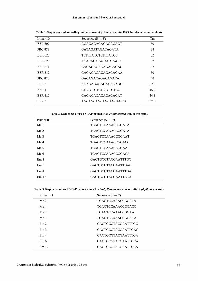
We used 11 ISSR primers for PCR of all species of
Potamogeton, Ceratophyllum and Myriophyllum. The
primer sequences used in this study are listed in Table 1.
ISSR PCR: The PCRs were carried out in a 15 µl
volume with 250 nM of each primer (Table 2), 0.2
mM of each dNTP, 1.5 mM MgCl2, 1 U Taq
polymerase, and 50-100 ng of genomic DNA. After 4
min at 95°C, PCR was followed by 40 cycles of 1 min
at 95°C, 1 min at annealing temperature (Table 2), 2
min at 72°C, followed by a final extension step of 10
min at 72°C. PCR products were detected by 2 %
agarose and ethidiumbromide staining under UV light
(Fig. 2.4).
We also used 10 pairs of primers of SRAP for all
of species of Potamogeton named above,
Ceratophyllum demersum and Myriophyllum
spicatum. The primer sequences for examined taxa are
showed in Table 2 and 3, respectively.
Shabnam Abbasi and Saeed Afsharzadeh
Progress in Biological Sciences / Vol. 6 (1) 2016 / 95-106
99
Table 1. Sequences and annealing temperatures of primers used for ISSR in selected aquatic plants
Primer ID Sequence (5'→ 3') Tm
ISSR 807 AGAGAGAGAGAGAGAGT 50
UBC 872 GATAGATAGATAGATA 38
ISSR 823 TCTCTCTCTCTCTCTCC 52
ISSR 826 ACACACACACACACACC 52
ISSR 811 GAGAGAGAGAGAGAGAC 52
ISSR 812 GAGAGAGAGAGAGAGAA 50
UBC 873 GACAGACAGACAGACA 48
ISSR 2 AGAGAGAGAGAGAGAGG 52.6
ISSR 4 CTCTCTCTCTCTCTCTGG 45.7
ISSR 810 GAGAGAGAGAGAGAGAT 54.3
ISSR 3 AGCAGCAGCAGCAGCAGCG 52.6
Table 2. Sequences of used SRAP primers for Potamogeton spp. in this study
Primer ID Sequence (5'→ 3')
Me 1 TGAGTCCAAACCGGATA
Me 2 TGAGTCCAAACCGGATA
Me 3 TGAGTCCAAACCGGAAT
Me 4 TGAGTCCAAACCGGACC
Me 5 TGAGTCCAAACCGGAA
Me 6 TGAGTCCAAACCGGACA
Em 2 GACTGCGTACGAATTTGC
Em 3 GACTGCGTACGAATTGAC
Em 4 GACTGCGTACGAATTTGA
Em 17 GACTGCGTACGAATTCCA
Table 3. Sequences of used SRAP primers for Ceratophyllum demersum and Myriophyllum spicatum
Primer ID Sequence (5'→3')
Me 2 TGAGTCCAAACCGGATA
Me 4 TGAGTCCAAACCGGACC
Me 5 TGAGTCCAAACCGGAA
Me 6 TGAGTCCAAACCGGACA
Em 2 GACTGCGTACGAATTTGC
Em 3 GACTGCGTACGAATTGAC
Em 4 GACTGCGTACGAATTTGA
Em 6 GACTGCGTACGAATTGCA
Em 17 GACTGCGTACGAATTCCA

DNA isolation from aquatic plants
Progress in Biological Sciences / Vol. 6 (1) 2016 / 95-106
100
SRAP PCR: The PCRs were performed in 25 μL
reaction volumes containing Taq 2× Master Mix Red
(Amplicon), 0.1 μM of each forward and reverse
primer, 50 ng DNA template, and nuclease-free water
to 20 μL. The PCR program conducted with the
following cycle profile in a Eppendorf Thermal
Cycler (Mastercycler Gradient): 5 min of initial
denaturation at 94°C followed by 5 cycles of 1 min
denaturing, 1 min annealing at 35°C and 1 min of
elongation at 72°C, after these, 35 cycles of 1 min
denaturing, 1 min annealing at 50°C ending with an
elongation step of 5 min at 72°C. The PCR products
were 2 % agarose and ethidiumbromide staining under
UV light (Fig 2,3,5).
SSR analyses were down only for all populations
of P. pectinatus with 9 primers. The primer sequences
used for study are indicated in Table 4.
Table 4. sequences of SSR primers for Potamogeton pectinatus
Locus Primer sequence Repeat motif
Potpect 24 F Ned- TCAGTGAAAGAAAGCCAGGA
R GGGCTTATGGCGTTATCAA
(GA)n
Potpect 26 F Fam-GTATAGGCGAGGTGCGAGAG
R CTTCATGTCGACCACCTTCC
(CT)n
Potpect 28 F Fam-TCGTTTCCTCCATTCGTAGG
R AATAAAAAGGGCCCAGACC
(GA)n
Potpect 32 F Hex-CAGCAAACGAAACAACCAAA
R AAAAGAAGCCGTTGTTTACAGAG
(GA)n
Potpect 34 F Fam-GTAAGGCAAGCAGCGTCAAC
R GTTTGTGAGCTAGCGGGAAG
(GA)n
Potpect 37 F Hex-CACTTCCTCTGTGCTGCTTG
R GCGTGCTCTTCCTGAGTTCT
(CT)n
Potpect 39 F Hex-TCACAACACCTCACCCAGAA
R CCATTTCCATTCCTCACTGC
(GA)n
Potpect 40 F Ned-AAATCTCCAAATATTTCCACTGTTG
R CAAAGATTGAGCTCCCCAAA
(GA)n
Potpect 42 F Ned-TTAGCAAGTGGGTGGGTTTC
R TGCACTCGTGTGTCTCTTCC
(CT)n
This single amplification was made possible by the
use of the QIAGEN Multiplex PCR Kit (QIAGEN) in
a final volume of 10.5 µl, as follow: 25 ng of DNA
template, 5 µl 2× QIAGEN Multiplex PCR Master
Mix [QIAGEN Multiplex PCR Buffer, pH 8.7,
containing dNTPs, QIAGEN HotStar Taq DNA
Polymerase, and 6 mM MgCl2 (for a final
concentration of 3 mM)], 1 µl Q-Solution (59
concentrated proprietary QIAGEN PCR additive), 1
µl of a primer mix with 2 µM of each primer (for a 0.2
µM final concentration of each primer) and 1 µl of
highly pure water obtained from a Milli-Q Synthesis
A10 (Millipore, Molsheim, France). PCR were carried
out in 96-well plates on a MyCycler TM thermal
cycler (BIO-RAD) under the following conditions: 15
min denaturing at 95°C, [3000 denaturing at 94°C, 1.5
min annealing at 57°C and 1 min extension at 72°C] ×
30 cycles and a final extension step at 72°C for 10
min. PCR were carried out in 96-well plates on a
MyCycler TM thermal cycler (BIO-RAD) under the
following conditions: 4 min denaturing at 94°C, [3000
denaturing at 94°C, 1 min annealing at 57°C and 1
min extension at 72°C] × 30 cycles and a final
extension step at 72°C for 30 min.
Shabnam Abbasi and Saeed Afsharzadeh
Progress in Biological Sciences / Vol. 6 (1) 2016 / 95-106
101
For barcoding the species and study of
interspecific diversity of Potamogeton species in Iran,
we used four fragments ITS, trnH-psbA, matk and
rbcL. The primer pairs used for amplifying each locus
were as follows: rbcl-a-F and rbcl-a-R; matK, 390F
and 1326R; ITS1, ITS2, ITS3, ITS4, trnHf-05, psbA3-
f. The PCR amplification for ITS was performed in 30
µl reaction mixture containing 3 µl DNA (50 ng ),
17.8 µl water, 6 µl PCR buffer 5 mM, 0.6 µl dNTP
10mM, 1.8 µl MgCl2 25 mM, 0.06 µl forward primer
0.1 mM, 0.06 µl reverse primer 0.1 mM, 0.6 µl BSA
(10mg/ml), 0.2 µl Taq (5u/ µl). The PCR
amplification for ccmp10, ccmp2 and trnH-psbA was
performed in 12.5 µl reaction mixture containing 2.5
µl water, 6.25 µl MasterMix, 1.25 µl primer mix and
2.5 µl DNA. The PCR amplification for rbcL, trnH-
psbA and matK was performed in 25 µl reaction
mixture containing 5 µl water, 12.5 µl MasterMix, 2.5
µl Primer Mix, 5 µl DNA. The PCR amplification
conditions for the ITS region were as follows: an
initial predenaturation step at 95ºC for 4 min,
followed by 35 cycles of 1 min at 95ºC, 1 min at 54ºC,
and 1 min at 72ºC, with a final extension step of 10
min at 72ºC. The PCR amplification conditions for the
TrnH-psbA region were as follows: an initial
predenaturation step at 95ºC for 4 min, followed by
35 cycles of 1 min at 94ºC, 1 min at 50ºC, and 1΄.30΄΄
at 72ºC, with a final extension step of 20 min at 72ºC.
The PCR amplification conditions for the matK region
were as follows: an initial predenaturation step at
95ºC for 4 min, followed by 35 cycles of 1 min at
94ºC, 1 min at 52ºC, and 1΄.30΄΄ at 72ºC, with a final
extension step of 20 min at 72ºC. The PCR
amplification conditions for the rbcl region were as
follows: an initial predenaturation step at 95ºC for 4
min, followed by 35 cycles of 1 min at 94ºC, 1 min at
50ºC, and 1΄.30΄΄ at 72ºC, with a final extension step
of 20 min at 72ºC.
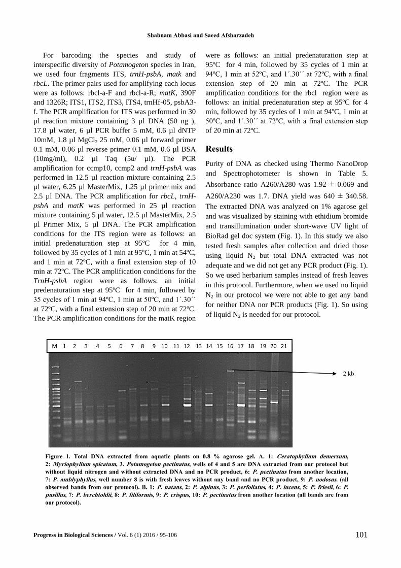
Results
Purity of DNA as checked using Thermo NanoDrop
and Spectrophotometer is shown in Table 5.
Absorbance ratio A260/A280 was 1.92 ± 0.069 and
A260/A230 was 1.7. DNA yield was 640 ± 340.58.
The extracted DNA was analyzed on 1% agarose gel
and was visualized by staining with ethidium bromide
and transillumination under short-wave UV light of
BioRad gel doc system (Fig. 1). In this study we also
tested fresh samples after collection and dried those
using liquid N2 but total DNA extracted was not
adequate and we did not get any PCR product (Fig. 1).
So we used herbarium samples instead of fresh leaves
in this protocol. Furthermore, when we used no liquid
N2 in our protocol we were not able to get any band
for neither DNA nor PCR products (Fig. 1). So using
of liquid N2 is needed for our protocol.
Figure 1. Total DNA extracted from aquatic plants on 0.8 % agarose gel. A. 1: Ceratophyllum demersum,
2: Myriophyllum spicatum, 3. Potamogeton pectinatus, wells of 4 and 5 are DNA extracted from our protocol but
without liquid nitrogen and without extracted DNA and no PCR product, 6: P. pectinatus from another location,
7: P. amblyphyllus, well number 8 is with fresh leaves without any band and no PCR product, 9: P. nodosus. (all
observed bands from our protocol). B. 1: P. natans, 2: P. alpinus, 3: P. perfoliatus, 4: P. lucens, 5: P. friesii, 6: P.
pusillus, 7: P. berchtoldii, 8: P. filiformis, 9: P. crispus, 10: P. pectinatus from another location (all bands are from
our protocol).

DNA isolation from aquatic plants
Progress in Biological Sciences / Vol. 6 (1) 2016 / 95-106
102
Table 5. Purity of total DNA extracted from aquatic plants in this study (the values are mean value in triplicates). Mean values
and standard deviation were also calculated in triplicates
Taxon A260/A280 A260/A230 DNA concentration (ng)?
Potamogeton pectinatus 1.92 ± 0.069 1.7 640 ± 340.58
P. perfoliatus 1.82 ± 0.067 1.7 640 ± 340.58
P. nodosus 1.92 ± 0.069 1.7 640 ± 340.58
P. lucens 1.92 ± 0.069 1.7 640 ± 340.58
P. crispus 1.92 ± 0.069 1.7 640 ± 340.58
P. natans 1.92 ± 0.069 1.7 640 ± 340.58
P. amblyphyllus 1.92 ± 0.069 1.7 640 ± 340.58
P. friesii 1.92 ± 0.069 1.7 640 ± 340.58
P. filiformis 1.80 ± 0.050 1.7 640 ± 340.58
P. pusillus 1.92 ± 0.069 1.7 640 ± 340.58
P. berchtoldii 1.92 ± 0.059 1.7 640 ± 340.58
P. alpinus 1.92 ± 0.069 1.7 640 ± 340.58
Ceratophyllum demersum 1.82 ± 0.070 1.7 400 ± 340.58
Myriophyllum spicatum 1.88 ± 0.060 1.7 540 ± 340.58
Discussion
DNA extraction from plants with high secondary
metabolits is very challenging (30, 31). Some methods
for DNA extraction from aquatic plants used CTAB
method (12). There is another method for DNA
extraction from plants with high concentration of
secondary metabolites (32). It is however expensive
and time consuming because of using suspension
buffer with extraction buffer. There are only few
modifications from the CTAB method by Doyle &
Doyle (2, 33) methodology. Here we have added up
sodium acetate for DNA purification. Our protocol
does not need proteinase in isolation step. Some
aquatic plant plants have relatively high polyphenol
such as Potamogeton spp.and Myriophyllum spp. (34,
35). We can remove the polyphenols by using high
levels of β-mercaptoethanol as in other protocols (7).
The addition of NaCl with concentration higher than
0.5 M to CTAB is known as removing factor for
polysaccharides during extraction (36). In this study
we used higher concentration of NaCl (1.4 M). Abu-
Romman used PVP in CTAB extraction for removing
polysaccharides and polyphenol (37). We used it but
in case of our experiment we had not adequate amount
of good quality DNA. In our protocol DNA is
precipitated by using isopropanol and sodium acetate,
because isopropanol gives rise to precipitation of
DNA and other phenolic and secondary metabolites
solved in sodium acetate. Simultaneous using of two
components (isopropanol and sodium acetate) can
make a DNA in an intact form. After this step DNA
pellet washed with 70% ethanol to remove salts.
Previously we used only isopropanol without sodium
acetate for precipitation of DNA but in final step we
had a black pellet of DNA. These secondary
metabolites provide colorful DNA that can inhibit
PCR. Sodium acetate was also used previously in
isopropanol step for efficient DNA extraction from
Passiflora foetida (38). DNA yield is important in
molecular studies. DNA extracted from aquatic plants
in our study was pure with adequate concentration
which can be stored for further use in molecular studies
like polymerase chain reaction amplifications (Figs. 2-
5) and long distance transport. Also Absorption ratio
(A260/A280) of extracted DNA samples was 1.92 ±
0.069 indicating that the DNA was free from proteins
and polyphenols. A260/A230 was 1.7 which showed
the sample genomic DNA is pure; free from
contaminants protein, polyphenolic and polysaccharide
compounds. The highest length PCR product of DNA
extracted by our protocol was 2-3 kb for ISSR and
SRAP (Figs. 3-5). This method can be considered as a
universal one, because in addition to its effective
application in many aquatic plant genera, it can be used
for DNA extraction from microbial organisms such as
cyanobacteria (Oscillatoria).

Shabnam Abbasi and Saeed Afsharzadeh
Progress in Biological Sciences / Vol. 6 (1) 2016 / 95-106
103
Figure 2. PCR products used extracted DNA by our protocol; A: DNA was amplified with barcoding markers, M: 100 bp DNA
Ladder 1: Potamogeton pectinatus, 2: P. nodosus; B: with SRAP markers using primer of Me2-Em 4; M: 100 bp DNA Ladder, 1:
P. natans, 2: P. alpinus, 3: P. perfoliatus, 4: P. lucens, 5: P. friesii, 6: P. pusillus, 7: P. berchtoldii, 8: P. filiformis, 9: P. crispus, 10:
P. pectinatus, 11. Ceratophyllum demersum, 12. Myriophyllum spicatum; C: with ISSR markers using primer of ISSR 810, M:
100 bp DNA Ladder, 1-20 are different populations of P. pectinatus.
Figure 3. PCR products of SRAP marker with primer of Me 1, Em 17 used extracted DNA by our protocol M: 100 bp
DNA ladder, 1: Potamogeton natans, 2: P. alpinus, 3: P. perfoliatus, 4: P. lucens, 5: P. friesii, 6: P. pusillus, 7: P.
berchtoldii, 8: P. filiformis, 9: P. crispus, 10: P. pectinatus, 11. Ceratophyllum demersum, 12. Myriophyllum spicatum; 13-
22: other populations of P. pectinatus.
Figure 4. PCR products of ISSR marker with primer of ISSR 4 used extracted DNA by our protocol, 1: Potamogeton
natans, 2: P. alpinus, 3: P. perfoliatus, 4: P. lucens, 5: P. friesii, 6: P. pusillus, 7: P. berchtoldii, 8: P. filiformis, 9: P.
crispus, 10: P. pectinatus, 11. Ceratophyllum demersum, 12. Myriophyllum spicatum; 13-21: other populations of P.
pectinatus.

DNA isolation from aquatic plants
Progress in Biological Sciences / Vol. 6 (1) 2016 / 95-106
104
Figure 5. PCR products of SRAP marker with primer of Me5,Em 17,1: P. natans, 2: P. alpinus, 3: P. perfoliatus,
4: P. lucens, 5: P. friesii, 6: P. pusillus, 7: P. berchtoldii, 8: P. filiformis, 9: P. crispus, 10: P. pectinatus, 11. C.
demersum, 12. M. spicatum; 13,14: other populations of P. pectinatus.
Aknowledgement: We are grateful for the financial
support provided by the Department of Biology,
University of Isfahan
1. Sharma, K. K., Lavanya, M. and Anjaiah, V. (2000) A method for isolation and purification of peanut genomic
DNA suitable for analytical applications. Plant Mol. Biol. Rep., 18, 393.
2. Doyle, J.J. and Doyle, J.L. (1990) Isolation of plant DNA from fresh tissue. Focus, 12, 13-15.
3. Drobkov, L., Kirschner, J. and Vlcek, C. (2002) Comparison of seven DNA extraction and amplification
protocols in historical herbarium specimens of Juncaceae. Plant Mol. Biol. Rep., 20,161-175.
4. Mogg, R.J. and Bond, J.M.A. (2003) Cheap, reliable and rapid method of extracting high quality DNA from
plants. Mol. Ecol. Notes, 3, 666-668.
5. Pirttil, M.A., Hirsikorpi, M., Kamarainen, T., Jaakola, L. and Hohtola, A. (2001) DNA isolation methods for
medicinal and aromatic plants. Plant Mol. Biol. Rep., 19, 273.
6. Saghai-Maroof, M.A., Soliman, K.M., Jorgensen, R.A. and Allard, R.W. (1984) Ribosomal DNA spacer-length
polymorphism in barley: Mendelian inheritance, chromosomal location, and population dynamics. Proc. Natl.
Acad. Sci., 81, 8014-8019.
7. Scott, K.D. and Playford, J. (1996) DNA extraction technique for PCR in rain forest plant species.
Biotechniques, 20, 974-979.
8. Porebski, S., Bailey, L.G. and Baum, B.R. (1997) Modification of a CTAB DNA extraction protocol for plants
containing high polysaccharide and polyphenol components. Plant Mol. Biol. Rep., 15, 8-15.
9. Zhu, H., Qu, F. and Zhu, L.H. (1993) Isolation of genomic DNAs from plants, fungi and bacteria using benzyl
chloride. Nucl. Acids Res., 21, 5279-5280.
10. Liu, Y.G., Mitsukawa, N., Oosumi, T. and Whittier, R.F. (1995) Efficient isolation and mapping of
Arabidopsis thaliana T-DNA insert junctions by thermal asymmetric interlaced PCR. Plant J., 8, 457-463.
11. Huang, J., Ge, X., Sun, M. (2000) Modified CTAB protocol using a silica matrix for isolation of plant genomic
DNA. Biotechniques, 28, 432-434.
12. Kaplan, Z., Fehrer, J. and Hellquist, B. (2009) New hybrid combinations revealed by molecular analysis: the
unknown side of North American pondweed diversity (Potamogeton ). Syst. Bot., 34, 625-642.

Shabnam Abbasi and Saeed Afsharzadeh
Progress in Biological Sciences / Vol. 6 (1) 2016 / 95-106
105
13. Zhang, T., Wang, Q., Li, W., Cheng, Y. and Wang, I. (2008) Analysis of phylogenetic relationships of
Potamogeton species in China based on chloroplast trnT-trnF sequences. Aquat. Bot., 89, 34-42.
14. Ahmed, I., Islam, M., Arshad, W., Mannan, A., Ahmad, W. and Mirza, B. (2009) High quality plant DNA
extraction for PCR: an easy approach. J. Appl. Genet., 50, 105-107.
15. Minas, K., McEwan, N.R., Newbold, C.J. and Scott., K.P. (2011) Optimization of a high-throughput CTAB-
based protocol for the extraction of qPCR-grade DNA from rumen fluid, plant and bacterial pure cultures.
FEMS Microbiol. L., 325, 162-169.
16. Pereira, J.C., Chaves, R., Bastos, E., Leitao, A. and Guedes-Pinto, H. (2011) An efficient method for genomic
DNA extraction from different Molluscs species. Int. J. Mol. Sci., 12, 8086-8095.
17. Hwang, C., Ling, F., Anderson, G. L., Lechevallier, M.W. and Liu, W.T. (2012) Evaluation of methods for the
extraction of DNA from drinking water distribution system biofilms. Microbes Environ., 27, 9-18.
18. Hwang Bo, K., Son, S. H., Lee, J. S., Min, S. R., Ko, S. M., Liu, J. R., Choi, D. and Jeong, W. J. (2010) Rapid
and simple method for DNA extraction from plant and algal species suitable for PCR amplification using a
chelating resin Chelex 100. Plant Biotechnol. Rep., 4, 49-52.
19. Aljanabi, S. M. and Martinez, I. (1997) Universal and rapid salt-extraction of high qualitymgenomic DNA for
PCR-based techniques. Nucleic Acids Res., 25, 1-2.
20. Simonelli, P., Troedsson, C., Nejstgaard, J. C., Zech, K., Larsen, J. B. and Frischer, M. (2009) Evaluation of
DNA extraction and handling procedures for PCR-based copepod feeding studies. J. Plankton Res., 31, 12,
1465-1474.
21. Al-Samarrai, T. H. and Schmid, J. A. (2000) Simple method for extraction of fungal genomic DNA. Lett. Appl.
Microbiol, 30, 53-56.
22. Devi, K.D., Punyarani, K., Singh, N.S. and Dev, H.S. (2013) An efficient protocol for total DNA extraction
from the members of order Zingiberales- suitable for diverse PCR based downstream applications. Springer
Plus, 2, 1-9.
23. Kumar, M.S., Kaur, G. and Sandhu, A.K. (2012) Genomic DNA Isolation from Fungi, Algae, Plant, Bacteria
and Human Blood using CTAB. IJSR, 3, 617-618.
24. Sika, K.C., Kefela, T., Adoukonou-Sagbadja, H., Ahoton, L., Saidou, A., Abu-Moussa, L., Baptiste, L.J.,
Kotconi, S.O. and Gachomo, E.W. (2015) A simple and efficient genomic DNA extraction protocol for large
scale genetic analyses of plant biological systems. Plant Gene, 1, 43-45.
25. Abbasi, S., Afsharzadeh, S. and Dinarvand, M. (2015) Potamogeton friesii (Potamogetonaceae), a new aquatic
plant record for the Flora Iranica area. Iran. J. Bot., 21, 39-42.
26. Dandy, J.E. (1971) Potamogetonaceae. In: Rechinger, K.H. (ed.) Flora Iranica, Vol 83. Akademisch Druk- und
Veragsanatalt. Graz, Austria.
27. Wiegleb, G. and Kaplan, Z. (1998) An account of the species of Potamogeton L. (Potamogetonaceae). Folia
Geobot., 33, 241-316.
28. Rechinger, K. H. (1966) Ceratophyllaceae. In: Rechinger, K.H. (ed.) Flora Iranica, Vol 28. Akademisch Druk-
und Veragsanatalt. Graz, Austria.
29. Rechinger, K.H. (1966) Haloragaceae. In: Rechinger, K.H. (ed.) Flora Iranica, Vol. 18. Akademisch Druk- und
Veragsanatalt. Graz, Austria.
30. Barnwell, P., Blanchard, A.N., Bryant, J.A., Smirnoff, F.N. and Weir, A.F. (1998) Isolation of DNA from the
highly mucilaginous succulent plant Sedum telephium. Mol. Biol. Rep., 16, 133-138.
31. Barzegari, A., Vahed, S. Z., Atashpaz, S. Khani, S. and Omidi, Y. (2010) Rapid and simple methodology for
isolation of high quality genomic DNA from coniferous tissues (Taxus baccata). Mol. Biol. Rep., 37, 833-837.
32. Sahu, S. K., Thangaraj, M. and Kathiresan, K. (2012) DNA extraction protocol for plants with high levels of
secondary metabolites and polysaccharides without using liquid nitrogen and phenol. Int. Schol. Res. Net.
http://dx.doi.org/10.5402/2012/205049

DNA isolation from aquatic plants
Progress in Biological Sciences / Vol. 6 (1) 2016 / 95-106
106
33. Doyle, J.J. and Doyle, J.L. (1987) A rapid DNA isolation procedure for small quantities of fresh leaf tissue.
Phytochem. Bull., 19, 11-15.
34. Lupoae, P., Cristea, V., Borda, D., Lupoae, M., Gurau, G. and Dinica, R.M. (2015) Phytochemical screening:
Antioxidant and antibacterial properties of Potamogeton species in order to obtain valuable feed additives. J
Oleo Sci., 1-13.
35. Hempel, M., Grossart, H. P. and Gross, E. M. (2009) Community composition of bacterial biofilms on two
submerged macrophytes and an artificial substrate in a pre-alpine lake. Aquat. Microb. Ecol., 58, 79-94.
36. Paterson, A.H., Brubaker, C.L. and Wendel, J.F.A. (1993) rapid method for extraction of cotton (Gossypium
spp.) genomic DNA suitable for RFLP or PCR analysis. Plant Mol. Biol. Rep., 11, 122-127.
37. Abu-Romman, S. (2011) Comparison of methods for isolating high quality DNA from sage (Salvia officinalis).
J. Med. Plants Res., 5, 938-941.
38. Lade, B. D., Patil, A. S. and Paikrao, H.M. (2014) Efficient genomic DNA extraction protocol from medicinal
rich Passiflora foetida containing high level of polysaccharide and polyphenol. Springer Plus, 3, 1-7.