Embed Size (px)
Citation preview
An Evaluation of Resting Tidal Volume Using a Biopac
System Spirometer in Comparison to the Benchmark
Avery Sipe, Sara Busche, Jamie Goetzinger,
Tori Gallichio, Alexander Pitts, Bea Angela Carvajal
University of Wisconsin-Madison, Department of Physiology
Lab 602, Group 14
Word Count: 4371
Key Words: Audio-visual, distraction, electrodermal activity (EDA), Hawthorne effect,
respiration, resting tidal volume, spirometry
2
Introduction
Spirometers are devices that are commonly used to determine lung function by measuring
tidal volume (TV) and airflow (Gildea et al. 2010). TV is the volume of air that enters and exits
the lungs in a single inhalation and subsequent exhalation during any point of physical activity.
Resting tidal volume (RTV), however, is a measurement of respiration that is collected when a
person is breathing normally in a relaxed, homeostatic state (Widmaier et al. 2016). Thus,
measurements of RTV are typically lower than total TV because total TV is subject to
fluctuations during periods of physical activity when the body requires more oxygen.
Comparisons in TV are made in relation to the accepted standard value for RTV in human adults,
500 milliliters (mL), depending on body size (Widmaier et al. 2016).
Though spirometers are commonly used in a clinical setting to support diagnosis of
respiratory diseases (Gildea et al. 2010), this paper addresses the use of spirometers in
conducting research studies. A previous experiment at the University of Wisconsin-Madison was
conducted on human subjects to test the ability of water-filled spirometers to measure RTVs that
were close to the benchmark value of 500 mL. Average RTV was calculated for 181 male and
267 female participants and determined to be 735.2 mL with a standard deviation of 365.2 mL
and 609.3 mL with a standard deviation of 302.4 mL, respectively (courtesy of Dr. Andrew
Lokuta, unpublished data). The results of this study suggested a large deviation from the
accepted RTV of 500 mL and variability among participants using the water-filled spirometer.
One suggested explanation may be that the water-filled spirometer created a physical resistance
against the participant’s airflow due to the unequal pressure within the water-filled device
compared to the air pressure of the testing environment, resulting in more forceful respirations
(Mottram, 2018, in personal communication with Dr. Lokuta). Another explanation could be that
the participants were aware their respiration was being tested, causing them to consciously
control their breathing. In modification of this previous experiment, the following study was
3
conducted to measure RTV in human subjects using a Biopac spirometer and an experimental
design that presented the participants with a distraction to minimize conscious respiratory
control. This spirometer was utilized in this comparative study as a water-filled spirometer was
unavailable for experimentation. Additionally, the Biopac spirometer is an improvement from
the water-filled spirometer because the device has an aperture that allows for equal pressure
within the device compared to the air pressure in the testing environment, thus allowing for less
resistance to airflow (Rebuck et al. 1996, in personal communication with Dr. Andrew Lokuta).
An examination of the Hawthorne effect prompted the decision to include a distraction in
this experimental design. This effect states that expected outcomes of an experiment can change
depending on an individual’s knowledge of the variable of interest (McCarney et al. 2007). Many
previous studies have indicated that intrusive techniques, such as the utilization of spirometers,
focus an individual’s attention on their breathing. This behavioral control has led to significant
effects on breathing patterns (Etzel, 2006).
Therefore, in regard to the following study examining RTV, it was assumed that
participants were aware that their breathing was being monitored because they were interacting
with devices used to measure their respiration. Thus, it was expected that this awareness would
result in conscious control of breathing and abnormal fluctuations in RTV, which could result in
deviations from the benchmark RTV of 500 mL. To increase the likelihood for spontaneous,
resting-state breathing, studies have suggested the use of task engagement or distraction (Boiten,
1998). Hence, the following study utilized an auditory and visual distraction to divert the
participant’s focus from controlling their breathing in order to reduce the Hawthorne effect.
In the following study, it was hypothesized that the use of an auditory and visual
distraction would reduce the effect of confounding variables on the data, resulting in a RTV that
more closely reflects the benchmark value of 500 mL. The ability to observe a RTV that is closer
to this value than in previous experimentation using the water-filled spirometer would suggest

4
that the Biopac System spirometer better reflects the benchmark for measuring RTV. This
hypothesis was tested by studying three physiological measurements: TV, skin conductance and
respiration rate (RR).
TV was the main physiological measurement that was evaluated in this study, whereas
RR and skin conductance were supporting variables that could provide an explanation for large
deviations in RTV. These measurements were deemed important because variations in
respiration reflect changes in mood, which is also a causal factor in skin conductance changes
(Etzel, 2006). For example, mood disorders, such as anxiety, have been associated with
hyperventilation and increased levels of electrodermal activity (EDA) (Wilhelm and Roth, 2001).
Sweat contains saltwater which conducts electricity well, so stimulation of sympathetic nervous
system activity in mood disorders causes increased sweat secretion, resulting in higher EDA
values, as well as altered respiration patterns (Etzel, 2006). Thus, the relationship between skin
conductance and respiration measures was utilized in the following study.
Measuring these supporting variables throughout both the baseline and distraction periods
was done to examine how a distraction period could potentially influence skin conductance and
RR, resulting in a more relaxed state. Furthermore, the results of this study allow for a
comparison of the Biopac System spirometer in relation to the benchmark, which will be helpful
information for future studies using this Biopac technology.
Materials
Tidal volume (TV), respiration rate (RR), and skin conductance were all examined using
three different measurement devices. A spirometer and its separate 2.0 L calibration syringe
(Model: SS11LA, SN: 12128333, Biopac Systems, Inc. Goleta, CA) were used for calibration
and measurement of TV in milliliters (mL). An additional supply of mouth pieces, air filters and
nose clamps were utilized for sanitary purposes and accurate TV measurements through mouth
5
exhalation. RR, in breaths per minute (BPM), was studied using a respiratory belt (Model:
SS5LB, SN: 1602007558, Biopac Systems, Inc. Goleta, CA). BSL EDA finger electrodes were
used in conjunction with a Xdcr lead set (Model: SS3LA, SN: 11053677, Biopac Systems, Inc.
Goleta, CA) to measure skin conductance in microsiemens (µS). Data recording and analysis was
conducted using the Biopac Student Lab System (BSL 4 software, MP36) as well as consultation
from the Biopac Systems, Inc. Student Manual (Biopac Systems Inc. ISO 9001:2008) for
equipment setup.
A video was presented for the distraction period of the experiment. The Small Thing Big
Idea video, “Why the Pencil is Perfect,” was played in order to observe any measurable
differences in TV that occurred upon distraction by this auditory and visual stimulus. This
distraction was presented to divert the participants’ attention away from the physiological
measurements being collected to determine if spontaneous breathing could be induced, resulting
in data reflecting the accepted literature TV value.
Methods
Participants
Participants, ages 20-25 were recruited on a voluntary basis from the University of
Wisconsin-Madison. Physiological measurements were collected at the UW-Madison Medical
Sciences Center. A consent form was signed by all participants describing the confidentiality
measures utilized in the study and alerting them of any potential discomfort.
Procedure
Participants were eligible to participate in this study if they met the following criteria.
The inclusion criteria included the willingness and ability to consent as well as falling into the
age range of 20 to 25 years. The exclusion criteria of participants were determined based on the
questionnaire the participants filled out following experimentation. If they indicated that they
6
smoked, the data was excluded from the results. Participants with asthma were still included as
they were not required to participate in physical activity nor were they having an asthma attack
during the experiment.
The following Biopac System preparations were made prior to testing. The Biopac
equipment – spirometer, respiration belt, and BSL EDA finger electrodes – was attached to the
Biopac Student Lab System via channels 1, 2 and 3 respectively. “Airflow” (SSL11LA, SN) was
selected for TV, measured in liters (L), on channel 1, “Respiration” (SS5LB) was selected for
RR, measured in millivolts (mV), using channel 2, and “Electrodermal activity” (EDA, SS3L,
SS3LA, SS57L, 0-35 Hz) was selected for skin conductance, measured in microsiemens (µS), on
channel 3.
A respiratory belt was secured on each participant’s upper chest with the monitor
centered on the sternum. The BSL EDA Finger Electrodes were strapped to the participant's
index and middle fingers on their right hand after applying electrode gel. The spirometer was
calibrated using the Biopac spirometer 2.0 L calibration syringe. Five full pumps of the syringe
were recorded, and the graph was scanned for distinct peaks indicating the plunger’s movement
to confirm that the system and spirometer were working properly. A disposable mouthpiece and
air filter were attached, and the spirometer was given to the participant’s free hand. Each
participant received a new mouthpiece and air filter in order to maintain consistency between
trials and prevent transfer of bacteria between participants. Calibration procedures were
conducted for the respiratory belt and BSL EDA Finger Electrodes as indicated by the Biopac
Systems Inc. Student Manual. After the participants were instructed to take three deep breaths,
the graphs were reviewed to identify peaks and troughs indicating a properly functioning Biopac
system.
Experimentation was conducted in a private room to ensure minimal external disruptions
that could alter the participant’s data. Two investigators stayed with the participant during the
7
entirety of the study in order to verify that the measurements were continually recorded and to
stop the trial if the participant felt uncomfortable at any time. The investigators were positioned
out of the participant’s view in order to reduce the risk of introducing a confounding variable that
could influence the data measurements. The participant was instructed to sit with both feet on the
floor facing the monitor. Data collection began at the start of a timed PowerPoint presentation
that the participant was told to watch for the duration of the experiment. The experimental
timeline included two baseline periods separated by a distraction period (Figure 1). An initial
one-minute baseline recording of TV, skin conductance, and RR occurred while a blank
PowerPoint slide was displayed. The “Why the Pencil is Perfect” video played for a duration of 3
minutes and 37 seconds and was followed by another minute of a blank PowerPoint slide. The
video served as a distraction for the participants to limit voluntary control of breathing, resulting
in what was expected to be a more accurate resting-state value for TV.
Upon completion, participants were assisted by an investigator to remove all Biopac
devices and were told experimentation had concluded. A post-trial questionnaire asking
participants to indicate their sex, asthma and smoking history, and height was collected. These
details were reviewed against the data collected in order to detect potential confounding
variables which may have provided reasoning for any outlier data observed. In addition, these
factors could alter the participant’s normal RTV, deviating from the accepted value of 500 mL.
Data Analysis
The average TV (mL) for each participant was measured before, during, and after the
distraction period (Figure 1) using the spirometer. TV was determined by collecting the “P-P”
(peak to peak) values of each inhalation and exhalation waveform, as calculated by the Biopac
software, and dividing this number by two (Figure 2). The TV values for each breath taken were
averaged to determine the individual’s overall TV average throughout the first baseline period.
This was repeated with the distraction period and the second baseline period. To determine the
8
average TV of the entire sample for each period, a weighted average of the participants’
individual averages was calculated.
Supplemental RR and skin conductance data was also collected using the Biopac
program. RR (BPM) was determined by counting the number of peak-trough pairs in each
participant’s trial and dividing this value by the respective amount of time (in minutes) for each
period. Sample averages for each period were also recorded. The average skin conductance for
each participant was measured before, during, and after the distraction period, and a weighted
average of the entire sample was calculated. The data collected from these calculations provided
insight into how the participant’s physiological responses reflected resting-state TV.
A one-sample, two-tailed t-test was conducted to compare the average TV data to the
published RTV value of 500 mL. This type of statistical analysis was used to account for the
potential for the sample mean to be greater than or less than the stated population mean. Multiple
two-tailed t-tests were conducted. For each physiological measurement, the participant averages
for the first baseline period were compared to those of the distraction period. Likewise, similar t-
tests were conducted between the distraction period and the second baseline period. These
statistical analyses were utilized to identify if the distraction period had an effect on the
physiological measures. A final t-test was performed to compare average TV during the
distraction period according to biological sex. This statistical value allowed for analysis of
potential inherent differences in TV. If p < 0.05 the results were determined to be statistically
significant. Skin conductance and respiration data were used to observe any potential correlation
between variables or attempt to explain any deviations. These data allowed for comparisons with
the TV data.
Positive Controls
9
To ensure functionality of the Biopac equipment, positive control tests were conducted
by the investigators. Baseline measurements for TV, RR and skin conductance were taken for 2
minutes. These resting averages were found to be 480 ± 117.38 mL, 12.48 ± 4.10 BPM, and 2.18
± 1.84 µS, respectively (n=2). Another set of measurements for TV, RR and EDA were taken
following 2 minutes of running in place. These averages were found to be 851.50 ± 243.95 mL,
13.53 ± 3.56 BPM, and 2.87 ± 2.54 µS, respectively (n=2). All variable measurements were
observed to increase as expected after a short period of activity. This indicated that equipment
was functional and that measurements tested in this experiment exist.
Negative Controls
The initial and final minute of experimentation for each participant functioned as the
negative control for their own data due to anatomical and physiological differences. These were
baseline measurements without visual stimuli or experimental manipulations. Physiological
changes observed during the distraction period of the experiment were compared against the two
baseline periods to understand how the presentation of a distraction potentially affected
participants’ respiration. Any discrepancies between these periods allowed for analysis of the
impact of video distraction on the physiological measurements studied in this experiment.
Results
Data was recorded from 30 participants, and all recorded participant data was included in
the analysis as zero participants met the exclusion criteria. This experimental sample contained
13 male and 17 female subjects. All TV data was collected in the following order and reported as
such; first baseline period, distraction period, and second baseline period. The average TVs were
calculated and found to be 509.60 ± 160.39 mL, 489 ± 117.54 mL, and 501.37 ± 123.83 mL,
respectively (n=30) (Figure 3). The ranges of average TV for the entire sample population were
10
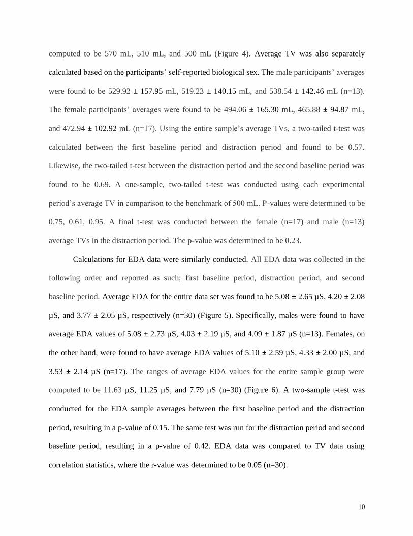
computed to be 570 mL, 510 mL, and 500 mL (Figure 4). Average TV was also separately
calculated based on the participants’ self-reported biological sex. The male participants’ averages
were found to be 529.92 ± 157.95 mL, 519.23 ± 140.15 mL, and 538.54 ± 142.46 mL (n=13).
The female participants’ averages were found to be 494.06 ± 165.30 mL, 465.88 ± 94.87 mL,
and 472.94 ± 102.92 mL (n=17). Using the entire sample’s average TVs, a two-tailed t-test was
calculated between the first baseline period and distraction period and found to be 0.57.
Likewise, the two-tailed t-test between the distraction period and the second baseline period was
found to be 0.69. A one-sample, two-tailed t-test was conducted using each experimental
period’s average TV in comparison to the benchmark of 500 mL. P-values were determined to be
0.75, 0.61, 0.95. A final t-test was conducted between the female (n=17) and male (n=13)
average TVs in the distraction period. The p-value was determined to be 0.23.
Calculations for EDA data were similarly conducted. All EDA data was collected in the
following order and reported as such; first baseline period, distraction period, and second
baseline period. Average EDA for the entire data set was found to be 5.08 ± 2.65 µS, 4.20 ± 2.08
µS, and 3.77 ± 2.05 µS, respectively (n=30) (Figure 5). Specifically, males were found to have
average EDA values of 5.08 ± 2.73 µS, 4.03 ± 2.19 µS, and 4.09 ± 1.87 µS (n=13). Females, on
the other hand, were found to have average EDA values of 5.10 ± 2.59 µS, 4.33 ± 2.00 µS, and
3.53 ± 2.14 µS (n=17). The ranges of average EDA values for the entire sample group were
computed to be 11.63 µS, 11.25 µS, and 7.79 µS (n=30) (Figure 6). A two-sample t-test was
conducted for the EDA sample averages between the first baseline period and the distraction
period, resulting in a p-value of 0.15. The same test was run for the distraction period and second
baseline period, resulting in a p-value of 0.42. EDA data was compared to TV data using
correlation statistics, where the r-value was determined to be 0.05 (n=30).
11
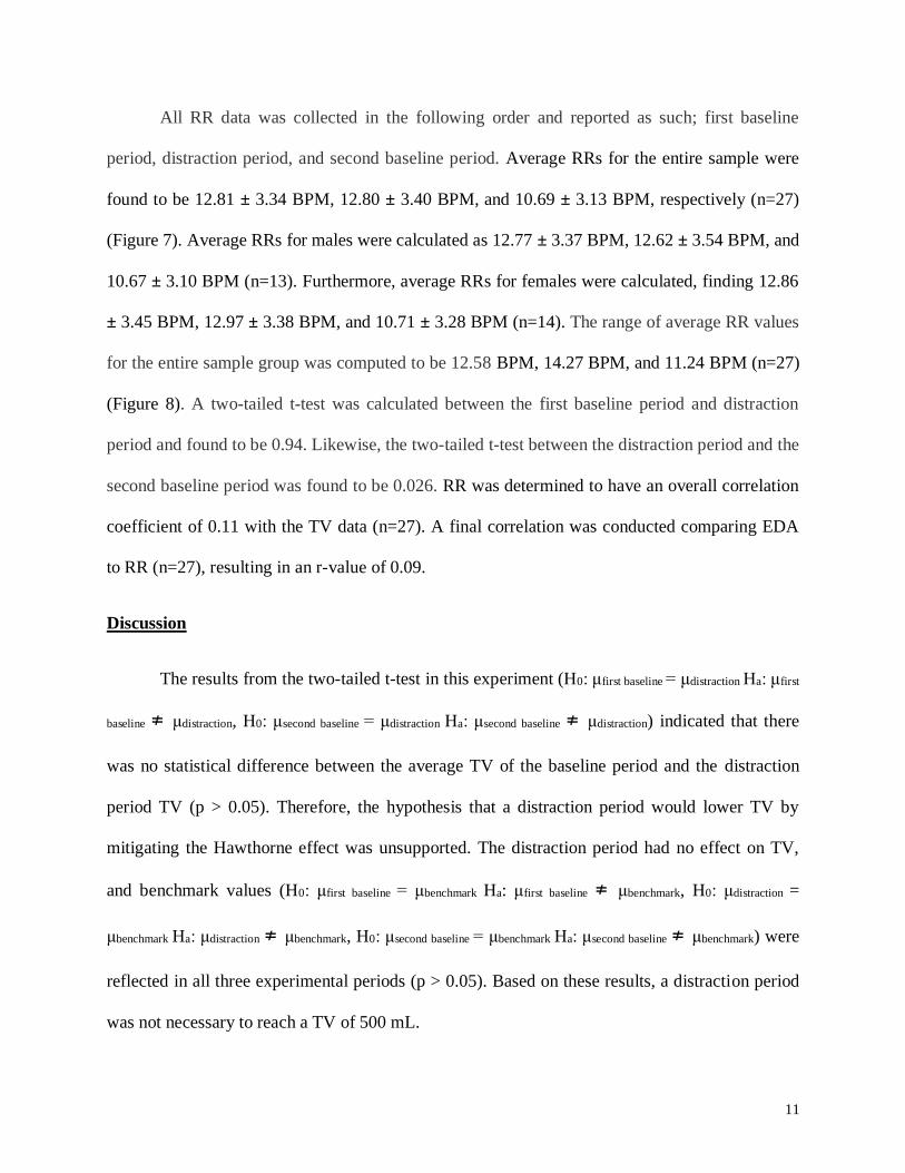
All RR data was collected in the following order and reported as such; first baseline
period, distraction period, and second baseline period. Average RRs for the entire sample were
found to be 12.81 ± 3.34 BPM, 12.80 ± 3.40 BPM, and 10.69 ± 3.13 BPM, respectively (n=27)
(Figure 7). Average RRs for males were calculated as 12.77 ± 3.37 BPM, 12.62 ± 3.54 BPM, and
10.67 ± 3.10 BPM (n=13). Furthermore, average RRs for females were calculated, finding 12.86
± 3.45 BPM, 12.97 ± 3.38 BPM, and 10.71 ± 3.28 BPM (n=14). The range of average RR values
for the entire sample group was computed to be 12.58 BPM, 14.27 BPM, and 11.24 BPM (n=27)
(Figure 8). A two-tailed t-test was calculated between the first baseline period and distraction
period and found to be 0.94. Likewise, the two-tailed t-test between the distraction period and the
second baseline period was found to be 0.026. RR was determined to have an overall correlation
coefficient of 0.11 with the TV data (n=27). A final correlation was conducted comparing EDA
to RR (n=27), resulting in an r-value of 0.09.
Discussion
The results from the two-tailed t-test in this experiment (H0: μfirst baseline = μdistraction Ha: μfirst
baseline ≠ μdistraction, H0: μsecond baseline = μdistraction Ha: μsecond baseline ≠ μdistraction) indicated that there
was no statistical difference between the average TV of the baseline period and the distraction
period TV (p > 0.05). Therefore, the hypothesis that a distraction period would lower TV by
mitigating the Hawthorne effect was unsupported. The distraction period had no effect on TV,
and benchmark values (H0: μfirst baseline = μbenchmark Ha: μfirst baseline ≠ μbenchmark, H0: μdistraction =
μbenchmark Ha: μdistraction ≠ μbenchmark, H0: μsecond baseline = μbenchmark Ha: μsecond baseline ≠ μbenchmark) were
reflected in all three experimental periods (p > 0.05). Based on these results, a distraction period
was not necessary to reach a TV of 500 mL.
12
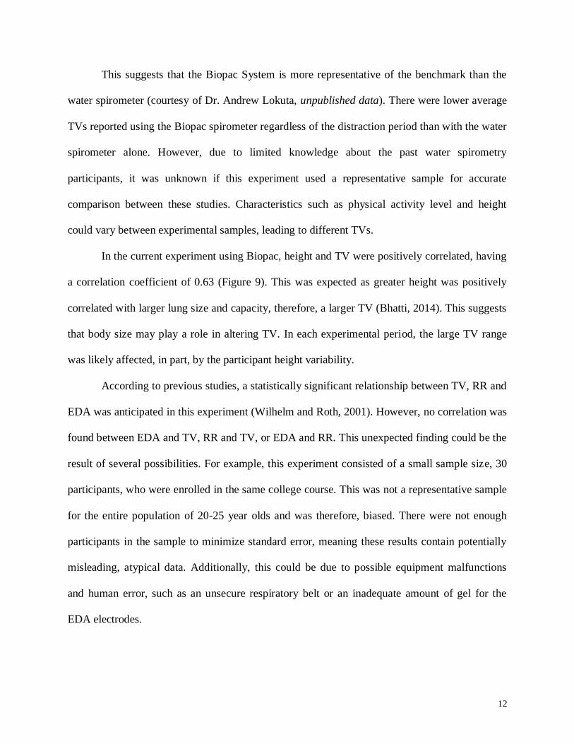
This suggests that the Biopac System is more representative of the benchmark than the
water spirometer (courtesy of Dr. Andrew Lokuta, unpublished data). There were lower average
TVs reported using the Biopac spirometer regardless of the distraction period than with the water
spirometer alone. However, due to limited knowledge about the past water spirometry
participants, it was unknown if this experiment used a representative sample for accurate
comparison between these studies. Characteristics such as physical activity level and height
could vary between experimental samples, leading to different TVs.
In the current experiment using Biopac, height and TV were positively correlated, having
a correlation coefficient of 0.63 (Figure 9). This was expected as greater height was positively
correlated with larger lung size and capacity, therefore, a larger TV (Bhatti, 2014). This suggests
that body size may play a role in altering TV. In each experimental period, the large TV range
was likely affected, in part, by the participant height variability.
According to previous studies, a statistically significant relationship between TV, RR and
EDA was anticipated in this experiment (Wilhelm and Roth, 2001). However, no correlation was
found between EDA and TV, RR and TV, or EDA and RR. This unexpected finding could be the
result of several possibilities. For example, this experiment consisted of a small sample size, 30
participants, who were enrolled in the same college course. This was not a representative sample
for the entire population of 20-25 year olds and was therefore, biased. There were not enough
participants in the sample to minimize standard error, meaning these results contain potentially
misleading, atypical data. Additionally, this could be due to possible equipment malfunctions
and human error, such as an unsecure respiratory belt or an inadequate amount of gel for the
EDA electrodes.

13
Further statistical testing indicated that there was no statistical difference in EDA values
between any of the experimental periods (p > 0.05). This means that the distraction had no effect
on EDA. Likewise, RR between the first baseline period and the distraction period was found to
have no statistical difference (p > 0.05). A statistical difference was found, however, in RR
between the distraction period and the second baseline period (p < 0.05). This was likely due to
participants’ adjustment to the experimental design and apparatus as the first and second baseline
periods utilized identical blank PowerPoint slides. While this could be a direct result of the
distraction period, it’s unlikely because there was no statistical difference found among any other
physiological measurements.
Conclusions were also made indicating that, based on this data, there was no difference in
TV based on biological sex. A two-tailed t-test (H0: μmale = μfemale Ha: μmale ≠ μfemale) was
conducted, indicating that there was no significant difference between male and female TV
during the distraction period (p > 0.05).
Following experimentation, possible limitations were addressed in relation to the
experimental results. Systematic errors, such as selection bias and confirmation bias, likely
occurred throughout the study. The participants were not randomly selected to participate in this
study, but instead, were selected from a specific college course, skewing the data. Also, it was
assumed that the investigators, knowing the hypothesis in question, subconsciously affected the
data collection process, altering it in favor of the anticipated outcome.
The data illustrated a large range of TVs with high standard deviations for each
experimental period. Tidal volume values were expected to be higher than the benchmark of 500
mL, but TVs as low as 300 mL were recorded. This does not necessarily suggest errors in data
collection but may simply reflect the accuracy of the Biopac spirometer in measuring the
14
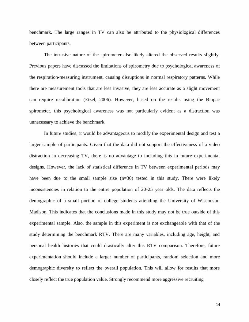
benchmark. The large ranges in TV can also be attributed to the physiological differences
between participants.
The intrusive nature of the spirometer also likely altered the observed results slightly.
Previous papers have discussed the limitations of spirometry due to psychological awareness of
the respiration-measuring instrument, causing disruptions in normal respiratory patterns. While
there are measurement tools that are less invasive, they are less accurate as a slight movement
can require recalibration (Etzel, 2006). However, based on the results using the Biopac
spirometer, this psychological awareness was not particularly evident as a distraction was
unnecessary to achieve the benchmark.
In future studies, it would be advantageous to modify the experimental design and test a
larger sample of participants. Given that the data did not support the effectiveness of a video
distraction in decreasing TV, there is no advantage to including this in future experimental
designs. However, the lack of statistical difference in TV between experimental periods may
have been due to the small sample size (n=30) tested in this study. There were likely
inconsistencies in relation to the entire population of 20-25 year olds. The data reflects the
demographic of a small portion of college students attending the University of Wisconsin-
Madison. This indicates that the conclusions made in this study may not be true outside of this
experimental sample. Also, the sample in this experiment is not exchangeable with that of the
study determining the benchmark RTV. There are many variables, including age, height, and
personal health histories that could drastically alter this RTV comparison. Therefore, future
experimentation should include a larger number of participants, random selection and more
demographic diversity to reflect the overall population. This will allow for results that more
closely reflect the true population value. Strongly recommend more aggressive recruiting

15
Vital capacity is a measurement of interest in future studies as it lends further insight into
physiological differences among participants. Average vital capacity measurements will be
useful in order to compare a participant’s tidal volume data to their inspiratory and expiratory
reserve volumes as well as residual volume. Therefore, it is strongly encouraged that future
studies utilize forced vital capacity maneuvers and standardize each participant’s data to their
chest volume and dimensions. This will provide an individualized normalization tool in order to
better understand if the tidal volume differences seen are indicative of participant variation or
experimental manipulation.
The results of this study indicated that the benchmark can be achieved using the Biopac
spirometer; however, there was a large amount of variance in average TV during each
experimental period. Therefore, future studies should focus on decreasing the variance across
participants through meticulous data collection. Testing a larger sample size would also likely
decrease the large standard deviations seen in this study.
Conclusion
In conclusion, this data shows that the Biopac spirometer is a more accurate tool to
measure benchmark RTV than the water spirometer. The participants were able to reach a RTV
of 500 mL using the Biopac spirometer, even without a distraction. Further, the experimental
periods had no effect on any of the physiological measures tested except for RR. The average
RRs between the distraction period and the second baseline period were determined to be
statistically significant. This indicates that the video either had an effect on the participants’
respirations immediately following the video or that the participants became accustomed to the
experimental environment. Future studies are required to determine which explanation applies to

16
this observed outcome. In addition, height and TV had a positive correlation, indicating that
controlling for body size in future experiments may be advantageous.
17
Figures
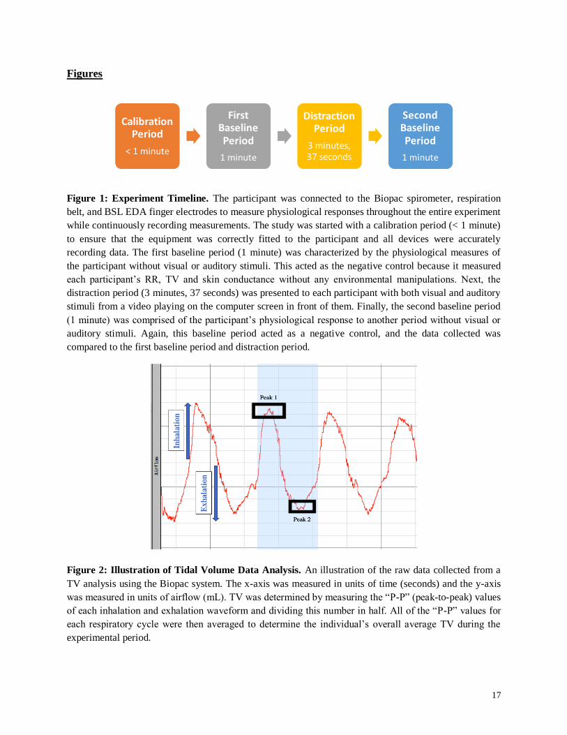
Figure 1: Experiment Timeline. The participant was connected to the Biopac spirometer, respiration
belt, and BSL EDA finger electrodes to measure physiological responses throughout the entire experiment
while continuously recording measurements. The study was started with a calibration period (< 1 minute)
to ensure that the equipment was correctly fitted to the participant and all devices were accurately
recording data. The first baseline period (1 minute) was characterized by the physiological measures of
the participant without visual or auditory stimuli. This acted as the negative control because it measured
each participant’s RR, TV and skin conductance without any environmental manipulations. Next, the
distraction period (3 minutes, 37 seconds) was presented to each participant with both visual and auditory
stimuli from a video playing on the computer screen in front of them. Finally, the second baseline period
(1 minute) was comprised of the participant’s physiological response to another period without visual or
auditory stimuli. Again, this baseline period acted as a negative control, and the data collected was
compared to the first baseline period and distraction period.
Figure 2: Illustration of Tidal Volume Data Analysis. An illustration of the raw data collected from a
TV analysis using the Biopac system. The x-axis was measured in units of time (seconds) and the y-axis
was measured in units of airflow (mL). TV was determined by measuring the “P-P” (peak-to-peak) values
of each inhalation and exhalation waveform and dividing this number in half. All of the “P-P” values for
each respiratory cycle were then averaged to determine the individual’s overall average TV during the
experimental period.

18
Figure 3: Average Tidal Volume by Experimental Period. The average TV for each participant was
used to find the overall average TV of participants (n=30) in each experimental period. The average TV
was 509.60 mL (σx̅= 29.28) for the First Baseline Period, 489.00 mL (σx̅ = 21.46) for the Distraction
Period, and 501.37 mL (σx̅ = 22.61) for the Second Baseline Period. The standard error for each
experimental period was indicated using error bars. A decrease in TV was observed from the First
Baseline Period to the Distraction Period, followed by an increase in TV from the Distraction Period to
the Second Baseline Period. No statistical significance was found between the average TVs of each
experimental period (p > 0.05). The benchmark was indicated using a red dashed line, illustrating the
comparison of the average TV for each experimental period in relation to the benchmark of 500 mL (p >
0.05).
Figure 4: Distribution of Averages for Tidal Volume by Experimental Period. The distribution of
average TV for each participant (n=30) was illustrated using box plots according to experimental period.
For the First Baseline Period, the median value was 482.5 mL, the upper and lower quartiles were 622.5
mL and 377.5 mL with an interquartile range of 245 mL. The highest and lowest observations were 830
mL and 260 mL with a range of 570 mL. For the Distraction Period, the median value was 480.0 mL, the
upper and lower quartiles were 562.5 mL and 410.0 mL with an interquartile range of 152.5 mL. The
highest and lowest observations were 750 mL and 240 mL with a range of 510 mL. For the Second

19
Baseline Period, the median value was 490.0 mL, the upper and lower quartiles were 576.75 mL and
407.5 mL with an interquartile range of 162.25 mL. The highest and lowest observations were 780 mL
and 280 mL with a range of 500 mL. The benchmark was indicated using a red dashed line, illustrating
the comparison of the average TV for each experimental period in relation to the benchmark of 500 mL.
No statistical significance was observed between the distributions of average EDA for each experimental
period (p > 0.05).
Figure 5: Average Electrodermal Activity by Experimental Period. The average EDA for each
participant was used to find the overall average EDA of participants (n=30) in each experimental period.
The average EDA was 5.08 µS (σx̅ = 0.48) for the First Baseline Period, 4.20 µS (σx̅ = 0.38) for the
Distraction Period, and 3.77 µS (σx̅ = 0.37) for the Second Baseline Period. The standard error for each
experimental period was indicated using error bars. A decrease in EDA was observed from the First
Baseline Period to the Distraction Period, followed by another decrease in EDA from the Distraction
Period to the Second Baseline Period. No statistical significance was found in the average EDAs between
the First Baseline Period and the Distraction Period as well as the Distraction Period and the Second
Baseline Period (p > 0.05).

20
Figure 6: Distribution of Averages for Electrodermal Activity by Experimental Period. The
distribution of average EDA for each participant (n=30) was illustrated using box plots according to
experimental period. For the First Baseline Period, the median value was 4.86 µS, the upper and lower
quartiles were 6.56 µS and 3.42 µS with an interquartile range of 3.14 µS. The highest and lowest
observations were 10.72 µS and 0.69 µS with a range of 10.03 µS. For the Distraction Period, the median
value was 4.12 µS, the upper and lower quartiles were 5.47 µS and 2.87 µS with an interquartile range of
2.6 µS. The highest and lowest observations were 8.4 µS and 0.22 µS with a range of 8.18 µS. For the
Second Baseline Period, the median value was 3.41 µS, the upper and lower quartiles were 4.67 µS and
2.26 with an interquartile range of 2.41 µS. The highest and lowest observations were 8.09 µS and 0.58
µS with a range of 7.51 µS. No statistical significance was observed between the distributions of average
EDA for each experimental period (p > 0.05).
Figure 7: Average Respiration Rate by Experimental Period. The average Respiration Rate (RR) for
each participant was used to find the overall average RR of participants (n=27) in each experimental
period. The average RR was 12.81 BPM (σx̅= 0.66) for the First Baseline Period, 12.80 BPM (σx̅ = 0.67)
for the Distraction Period, and 10.69 BPM (σx̅ = 0.61) for the Second Baseline Period. The standard error
for each experimental period was indicated using error bars. A slight decrease in RR was observed from
the First Baseline Period to the Distraction Period, followed by another decrease in RR from the
Distraction Period to the Second Baseline Period. No statistical significance was observed between the
First Baseline Period and Distraction Period. Statistical significance was found between the Distraction
Period and Second Baseline Period (p < 0.05), indicated by (**).
21
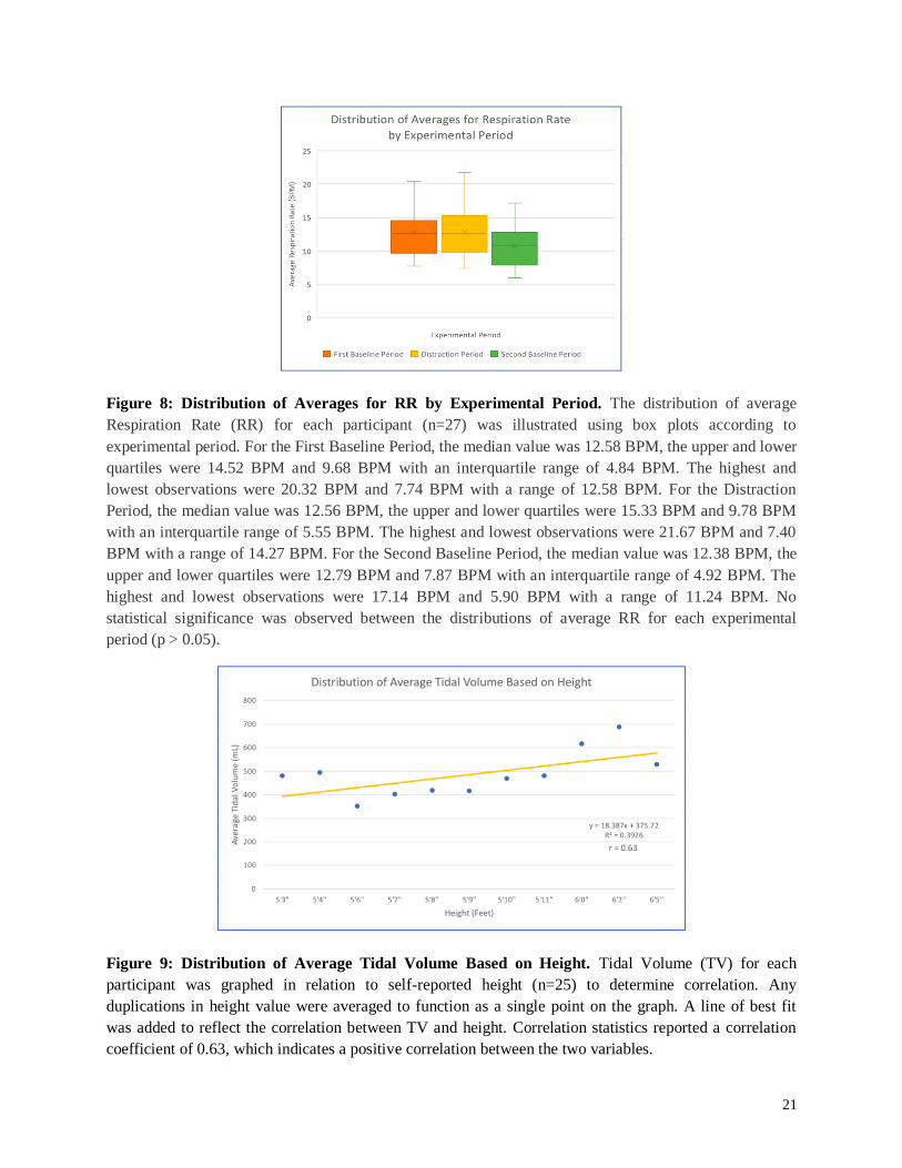
Figure 8: Distribution of Averages for RR by Experimental Period. The distribution of average
Respiration Rate (RR) for each participant (n=27) was illustrated using box plots according to
experimental period. For the First Baseline Period, the median value was 12.58 BPM, the upper and lower
quartiles were 14.52 BPM and 9.68 BPM with an interquartile range of 4.84 BPM. The highest and
lowest observations were 20.32 BPM and 7.74 BPM with a range of 12.58 BPM. For the Distraction
Period, the median value was 12.56 BPM, the upper and lower quartiles were 15.33 BPM and 9.78 BPM
with an interquartile range of 5.55 BPM. The highest and lowest observations were 21.67 BPM and 7.40
BPM with a range of 14.27 BPM. For the Second Baseline Period, the median value was 12.38 BPM, the
upper and lower quartiles were 12.79 BPM and 7.87 BPM with an interquartile range of 4.92 BPM. The
highest and lowest observations were 17.14 BPM and 5.90 BPM with a range of 11.24 BPM. No
statistical significance was observed between the distributions of average RR for each experimental
period (p > 0.05).
Figure 9: Distribution of Average Tidal Volume Based on Height. Tidal Volume (TV) for each
participant was graphed in relation to self-reported height (n=25) to determine correlation. Any
duplications in height value were averaged to function as a single point on the graph. A line of best fit
was added to reflect the correlation between TV and height. Correlation statistics reported a correlation
coefficient of 0.63, which indicates a positive correlation between the two variables.
22
References
Bhatti, U. et al. “Variations in lung volumes and capacities among young males in relation to
height.” US National Library of Medicine National Institute of Health. 2014.
Boiten, Frans. “The Effects of Emotional Behaviour on Components of the Respiratory
‘’’’’Cycle.” Biol Psychol (1998): 49(1-2), 29-51.
Etzel, Joset A. “Algorithms and Procedures to Analyze Physiological Signals in
‘’’’’Psychophysiological Research.” Retrospective Theses and Dissertations. Iowa State
‘’’’’University. 2006.
Evans, D. “The Hawthorne Effect: What Do We Really Learn Watching Teachers (and
‘’’’’Others)?”. The World Bank. 2014.
Gildea, Thomas R, McCarthy, Kevin. “Pulmonary Function Testing.” Cleveland Clinic: Center
‘’’’’Continuing Education, 2010.
McCarney, Rob et al. “The Hawthorne Effect: A Randomised, Controlled Trial.” BMC Medical
‘’’’’Research Methodology 7 (2007): 30. PMC. Web. 9 Mar. 2018.
Mottram, Carl. “Ruppel’s Manual of Pulmonary Function Testing, 11th Edition.” Elsevier Health
Sciences, 2017.
Rebuck, David et al. “The Accuracy of a Handheld Portable Spirometer.” CHEST Journal
(1996): 109(1), 152-157.
Widmaier, Eric P, Raff, Hershel, and Strang, Kevin T. Vander’s Human Physiology: The
‘’’’’Mechanisms of Body Function, 14th Edition. McGraw Hill Education, 2016. 454.
Wilhelm, F.H., & Roth, W.T. “The somatic symptom paradox in DSM-IV anxiety disorders:
‘’’’’suggestions for a clinical focus in psychophysiology.” Biol Psychol (2001): 57(1-3), 105-40.
Link to the video used in Distraction Period:
https://www.facebook.com/SmallThingBigIdea/videos/1344073369072021/