Embed Size (px)
Citation preview
NOTES:
An Evolving Concept: Individualized, Anatomic ACL
Reconstruction in 2017
Humza S. Shaikh, BA Marcio B.V. Albers, MD
Elmar Herbst, MD Daniel Guenther, MD
Sebastian Irarrázaval, MD Jeremy M. Burnham, MD
and Freddie H. Fu, MD, DSc (Hon.), DPs (Hon.)
AAOS Scientific Exhibit 71
Department of Orthopaedic Surgery,
University of Pittsburgh, Pittsburgh, PA, USA
Corresponding Author:
Freddie H. Fu, MD
E-Mail: [email protected]
2
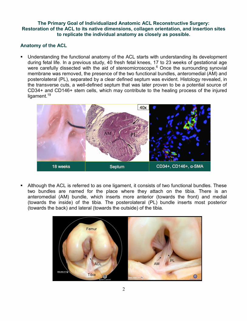
The Primary Goal of Individualized Anatomic ACL Reconstructive Surgery: Restoration of the ACL to its native dimensions, collagen orientation, and insertion sites
to replicate the individual anatomy as closely as possible. Anatomy of the ACL ▪ Understanding the functional anatomy of the ACL starts with understanding its development
during fetal life. In a previous study, 40 fresh fetal knees, 17 to 23 weeks of gestational age were carefully dissected with the aid of stereomicroscope.6 Once the surrounding synovial membrane was removed, the presence of the two functional bundles, anteromedial (AM) and posterolateral (PL), separated by a clear defined septum was evident. Histology revealed, in the transverse cuts, a well-defined septum that was later proven to be a potential source of CD34+ and CD146+ stem cells, which may contribute to the healing process of the injured ligament.19
▪ Although the ACL is referred to as one ligament, it consists of two functional bundles. These two bundles are named for the place where they attach on the tibia. There is an anteromedial (AM) bundle, which inserts more anterior (towards the front) and medial (towards the inside) of the tibia. The posterolateral (PL) bundle inserts most posterior (towards the back) and lateral (towards the outside) of the tibia.
3
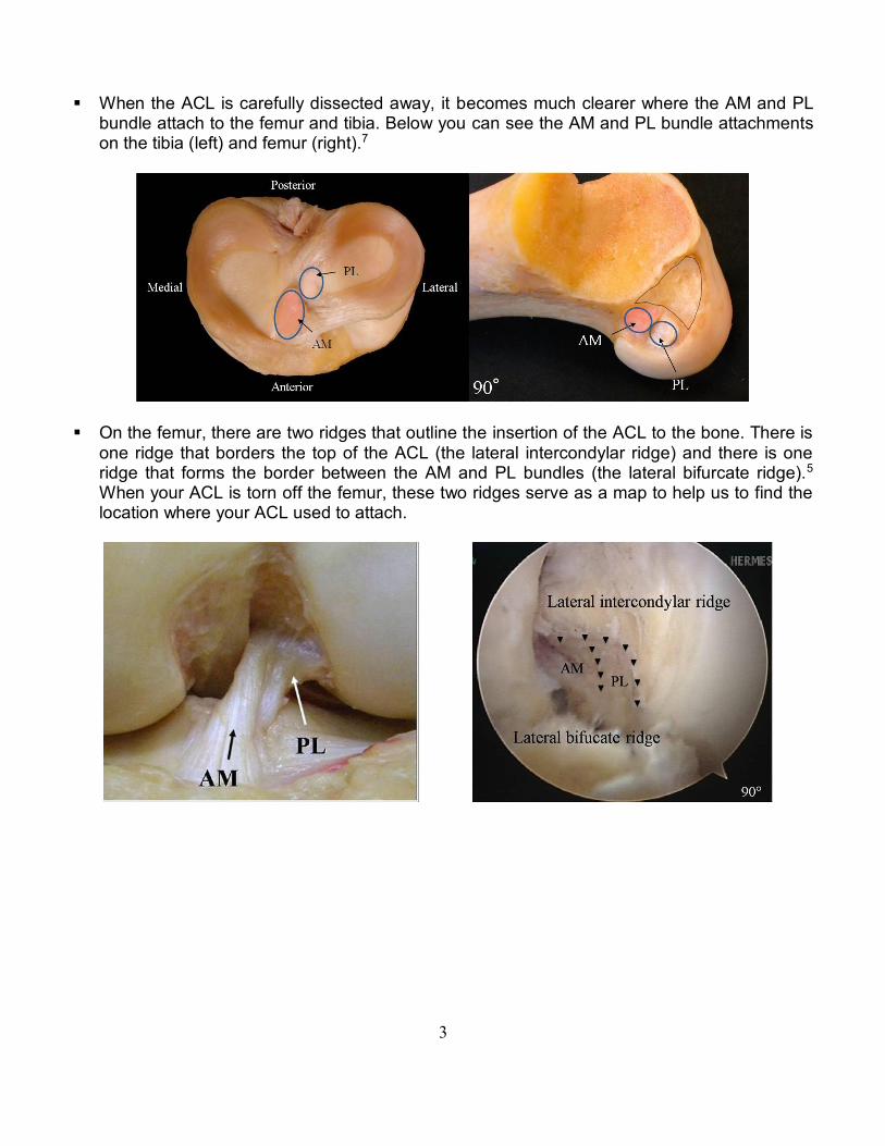
▪ When the ACL is carefully dissected away, it becomes much clearer where the AM and PL
bundle attach to the femur and tibia. Below you can see the AM and PL bundle attachments on the tibia (left) and femur (right).7
▪ On the femur, there are two ridges that outline the insertion of the ACL to the bone. There is one ridge that borders the top of the ACL (the lateral intercondylar ridge) and there is one ridge that forms the border between the AM and PL bundles (the lateral bifurcate ridge).5 When your ACL is torn off the femur, these two ridges serve as a map to help us to find the location where your ACL used to attach.
4
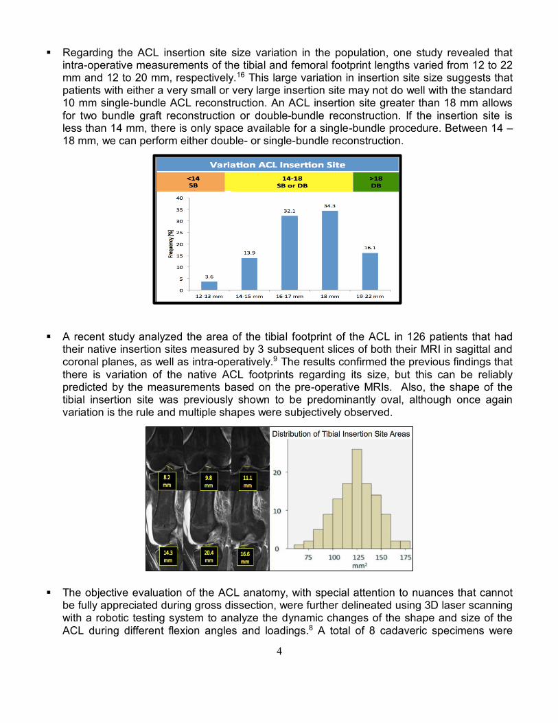
▪ Regarding the ACL insertion site size variation in the population, one study revealed that intra-operative measurements of the tibial and femoral footprint lengths varied from 12 to 22 mm and 12 to 20 mm, respectively.16 This large variation in insertion site size suggests that patients with either a very small or very large insertion site may not do well with the standard 10 mm single-bundle ACL reconstruction. An ACL insertion site greater than 18 mm allows for two bundle graft reconstruction or double-bundle reconstruction. If the insertion site is less than 14 mm, there is only space available for a single-bundle procedure. Between 14 – 18 mm, we can perform either double- or single-bundle reconstruction.
▪ A recent study analyzed the area of the tibial footprint of the ACL in 126 patients that had their native insertion sites measured by 3 subsequent slices of both their MRI in sagittal and coronal planes, as well as intra-operatively.9 The results confirmed the previous findings that there is variation of the native ACL footprints regarding its size, but this can be reliably predicted by the measurements based on the pre-operative MRIs. Also, the shape of the tibial insertion site was previously shown to be predominantly oval, although once again variation is the rule and multiple shapes were subjectively observed.
▪ The objective evaluation of the ACL anatomy, with special attention to nuances that cannot
be fully appreciated during gross dissection, were further delineated using 3D laser scanning with a robotic testing system to analyze the dynamic changes of the shape and size of the ACL during different flexion angles and loadings.8 A total of 8 cadaveric specimens were
5
studied with confirming that the ACL shape is complex, has an isthmus located at approximately the mid-portion between the tibial and femoral insertion sites and that compared to the projected area of the tibial insertion site to different planes, the isthmus measures from 35% to 50% of the tibial insertion site and the femoral insertion site measures 69% of the tibial insertion site.
Imaging: Special MRI Planes
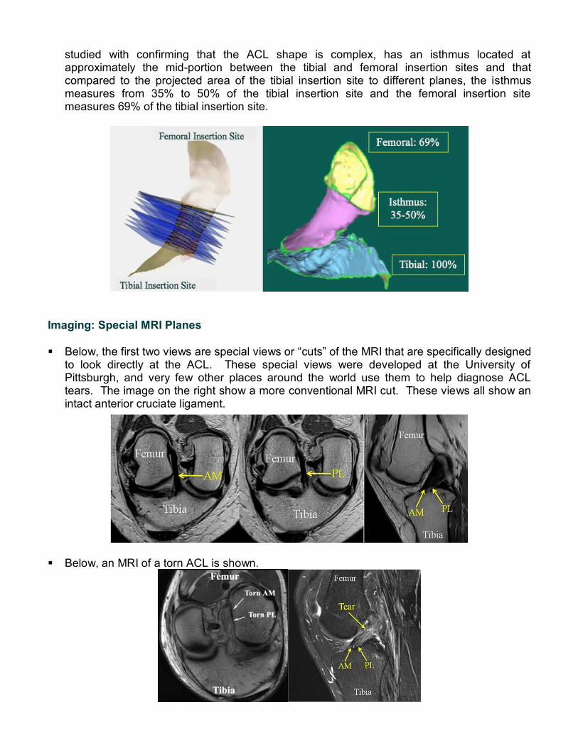
▪ Below, the first two views are special views or “cuts” of the MRI that are specifically designed to look directly at the ACL. These special views were developed at the University of Pittsburgh, and very few other places around the world use them to help diagnose ACL tears. The image on the right show a more conventional MRI cut. These views all show an intact anterior cruciate ligament.
▪ Below, an MRI of a torn ACL is shown.
6
Is Anatomic ACL Reconstruction Really Anatomic? “Evidence to Support the Interpretation and Use of the Anatomic Anterior Cruciate Ligament Reconstruction Checklist”. van Eck, Fu et al. JBJS Orthopaedic Forum 201325 ▪ Published papers on anatomic anterior cruciate ligament (ACL) reconstruction often lack
details in the description of the surgical procedure, and there are large variations in anatomic ACL reconstruction techniques. We aimed to develop a validated checklist to be used for anatomic ACL reconstruction. 34 ACL experts ranked 27 terms by importance, creating a list that was then verified by 959 academic orthopaedists. The final checklist underwent preliminary testing for internal consistency, intertester reliability, and validity. Cronbach’s alpha for internal consistency was 0.82, and the intraclass correlation coefficient (ICC) for intertester reliability was 0.65. This large survey-based study on anatomic ACL reconstruction resulted in the development of the Anatomic ACL Reconstruction Checklist.
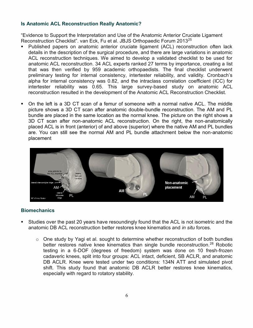
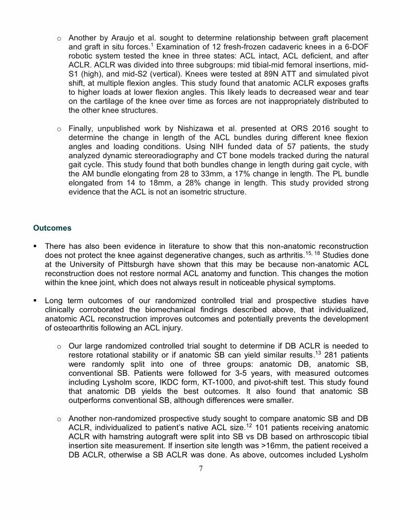
▪ On the left is a 3D CT scan of a femur of someone with a normal native ACL. The middle picture shows a 3D CT scan after anatomic double-bundle reconstruction. The AM and PL bundle are placed in the same location as the normal knee. The picture on the right shows a 3D CT scan after non-anatomic ACL reconstruction. On the right, the non-anatomically placed ACL is in front (anterior) of and above (superior) where the native AM and PL bundles are. You can still see the normal AM and PL bundle attachment below the non-anatomic placement
Biomechanics ▪ Studies over the past 20 years have resoundingly found that the ACL is not isometric and the
anatomic DB ACL reconstruction better restores knee kinematics and in situ forces.
o One study by Yagi et al. sought to determine whether reconstruction of both bundles better restores native knee kinematics than single bundle reconstruction.28 Robotic testing in a 6-DOF (degrees of freedom) system was done on 10 fresh-frozen cadaveric knees, split into four groups: ACL intact, deficient, SB ACLR, and anatomic DB ACLR. Knee were tested under two conditions: 134N ATT and simulated pivot shift. This study found that anatomic DB ACLR better restores knee kinematics, especially with regard to rotatory stability.
7
o Another by Araujo et al. sought to determine relationship between graft placement and graft in situ forces.1 Examination of 12 fresh-frozen cadaveric knees in a 6-DOF robotic system tested the knee in three states: ACL intact, ACL deficient, and after ACLR. ACLR was divided into three subgroups: mid tibial-mid femoral insertions, mid-S1 (high), and mid-S2 (vertical). Knees were tested at 89N ATT and simulated pivot shift, at multiple flexion angles. This study found that anatomic ACLR exposes grafts to higher loads at lower flexion angles. This likely leads to decreased wear and tear on the cartilage of the knee over time as forces are not inappropriately distributed to the other knee structures.
o Finally, unpublished work by Nishizawa et al. presented at ORS 2016 sought to
determine the change in length of the ACL bundles during different knee flexion angles and loading conditions. Using NIH funded data of 57 patients, the study analyzed dynamic stereoradiography and CT bone models tracked during the natural gait cycle. This study found that both bundles change in length during gait cycle, with the AM bundle elongating from 28 to 33mm, a 17% change in length. The PL bundle elongated from 14 to 18mm, a 28% change in length. This study provided strong evidence that the ACL is not an isometric structure.
Outcomes ▪ There has also been evidence in literature to show that this non-anatomic reconstruction
does not protect the knee against degenerative changes, such as arthritis.15, 18 Studies done at the University of Pittsburgh have shown that this may be because non-anatomic ACL reconstruction does not restore normal ACL anatomy and function. This changes the motion within the knee joint, which does not always result in noticeable physical symptoms.
▪ Long term outcomes of our randomized controlled trial and prospective studies have clinically corroborated the biomechanical findings described above, that individualized, anatomic ACL reconstruction improves outcomes and potentially prevents the development of osteoarthritis following an ACL injury.
o Our large randomized controlled trial sought to determine if DB ACLR is needed to restore rotational stability or if anatomic SB can yield similar results.13 281 patients were randomly split into one of three groups: anatomic DB, anatomic SB, conventional SB. Patients were followed for 3-5 years, with measured outcomes including Lysholm score, IKDC form, KT-1000, and pivot-shift test. This study found that anatomic DB yields the best outcomes. It also found that anatomic SB outperforms conventional SB, although differences were smaller.
o Another non-randomized prospective study sought to compare anatomic SB and DB ACLR, individualized to patient’s native ACL size.12 101 patients receiving anatomic ACLR with hamstring autograft were split into SB vs DB based on arthroscopic tibial insertion site measurement. If insertion site length was >16mm, the patient received a DB ACLR, otherwise a SB ACLR was done. As above, outcomes included Lysholm
8
score, IKDC form, KT-1000, and pivot-shift test. This study found that individualized, anatomic DB was no different than individualized, anatomic SB reconstruction.
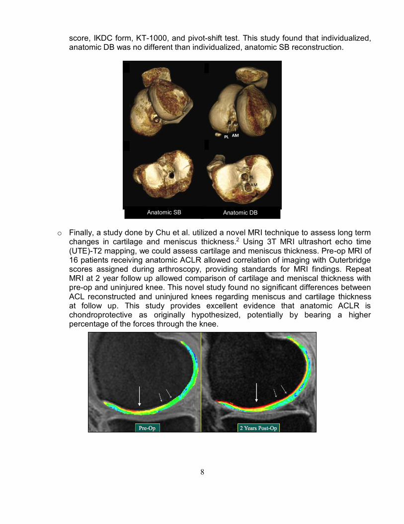
o Finally, a study done by Chu et al. utilized a novel MRI technique to assess long term changes in cartilage and meniscus thickness.2 Using 3T MRI ultrashort echo time (UTE)-T2 mapping, we could assess cartilage and meniscus thickness. Pre-op MRI of 16 patients receiving anatomic ACLR allowed correlation of imaging with Outerbridge scores assigned during arthroscopy, providing standards for MRI findings. Repeat MRI at 2 year follow up allowed comparison of cartilage and meniscal thickness with pre-op and uninjured knee. This novel study found no significant differences between ACL reconstructed and uninjured knees regarding meniscus and cartilage thickness at follow up. This study provides excellent evidence that anatomic ACLR is chondroprotective as originally hypothesized, potentially by bearing a higher percentage of the forces through the knee.
Anatomic SB Anatomic DB
9
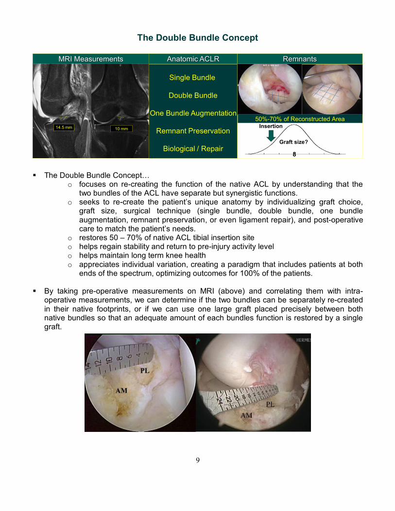
The Double Bundle Concept
▪ The Double Bundle Concept…
o focuses on re-creating the function of the native ACL by understanding that the two bundles of the ACL have separate but synergistic functions.
o seeks to re-create the patient’s unique anatomy by individualizing graft choice, graft size, surgical technique (single bundle, double bundle, one bundle augmentation, remnant preservation, or even ligament repair), and post-operative care to match the patient’s needs.
o restores 50 – 70% of native ACL tibial insertion site o helps regain stability and return to pre-injury activity level o helps maintain long term knee health o appreciates individual variation, creating a paradigm that includes patients at both
ends of the spectrum, optimizing outcomes for 100% of the patients. ▪ By taking pre-operative measurements on MRI (above) and correlating them with intra-
operative measurements, we can determine if the two bundles can be separately re-created in their native footprints, or if we can use one large graft placed precisely between both native bundles so that an adequate amount of each bundles function is restored by a single graft.
MRI Measurements Remnants
Single Bundle
Double Bundle
One Bundle Augmentation
Remnant Preservation
Biological / Repair
Anatomic ACLR
Insertion
Graft size?
8
50%-70% of Reconstructed Area10 mm14.5 mm
10
▪ Graft choice for ACLR is a key step in the surgical planning that is finalized after intra-operative evaluation of the joint. Graft individualization weighs the pros and cons of each commonly used graft, and accounts for the native ACL’s insertion site size to determine optimum predicted graft size.
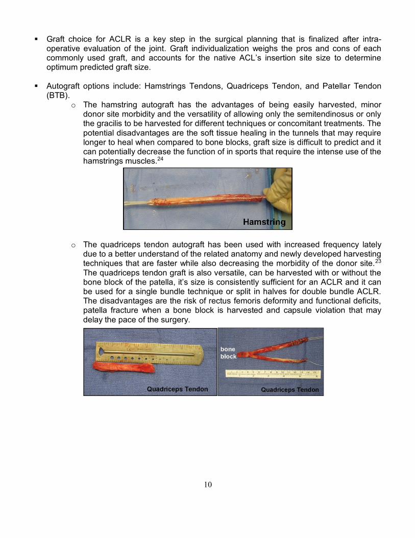
▪ Autograft options include: Hamstrings Tendons, Quadriceps Tendon, and Patellar Tendon
(BTB). o The hamstring autograft has the advantages of being easily harvested, minor
donor site morbidity and the versatility of allowing only the semitendinosus or only the gracilis to be harvested for different techniques or concomitant treatments. The potential disadvantages are the soft tissue healing in the tunnels that may require longer to heal when compared to bone blocks, graft size is difficult to predict and it can potentially decrease the function of in sports that require the intense use of the hamstrings muscles.24
o The quadriceps tendon autograft has been used with increased frequency lately
due to a better understand of the related anatomy and newly developed harvesting techniques that are faster while also decreasing the morbidity of the donor site.23 The quadriceps tendon graft is also versatile, can be harvested with or without the bone block of the patella, it’s size is consistently sufficient for an ACLR and it can be used for a single bundle technique or split in halves for double bundle ACLR. The disadvantages are the risk of rectus femoris deformity and functional deficits, patella fracture when a bone block is harvested and capsule violation that may delay the pace of the surgery.
11
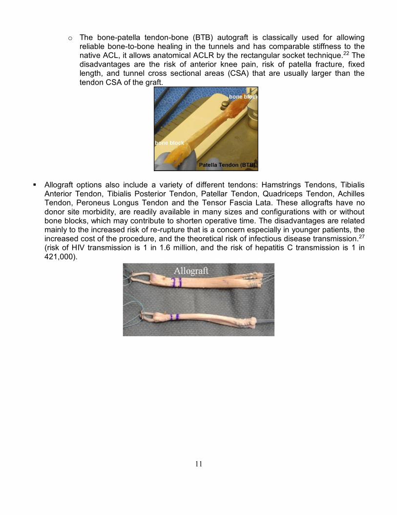
o The bone-patella tendon-bone (BTB) autograft is classically used for allowing reliable bone-to-bone healing in the tunnels and has comparable stiffness to the native ACL, it allows anatomical ACLR by the rectangular socket technique.22 The disadvantages are the risk of anterior knee pain, risk of patella fracture, fixed length, and tunnel cross sectional areas (CSA) that are usually larger than the tendon CSA of the graft.
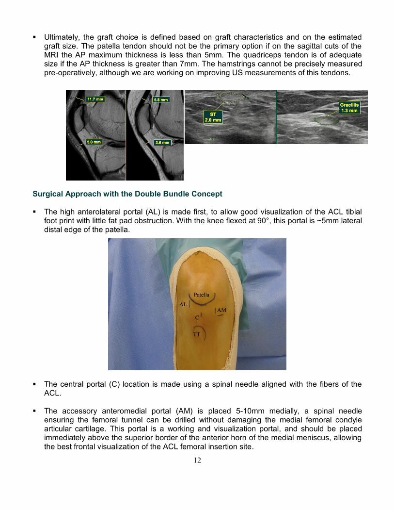
▪ Allograft options also include a variety of different tendons: Hamstrings Tendons, Tibialis Anterior Tendon, Tibialis Posterior Tendon, Patellar Tendon, Quadriceps Tendon, Achilles Tendon, Peroneus Longus Tendon and the Tensor Fascia Lata. These allografts have no donor site morbidity, are readily available in many sizes and configurations with or without bone blocks, which may contribute to shorten operative time. The disadvantages are related mainly to the increased risk of re-rupture that is a concern especially in younger patients, the increased cost of the procedure, and the theoretical risk of infectious disease transmission.27 (risk of HIV transmission is 1 in 1.6 million, and the risk of hepatitis C transmission is 1 in 421,000).
12
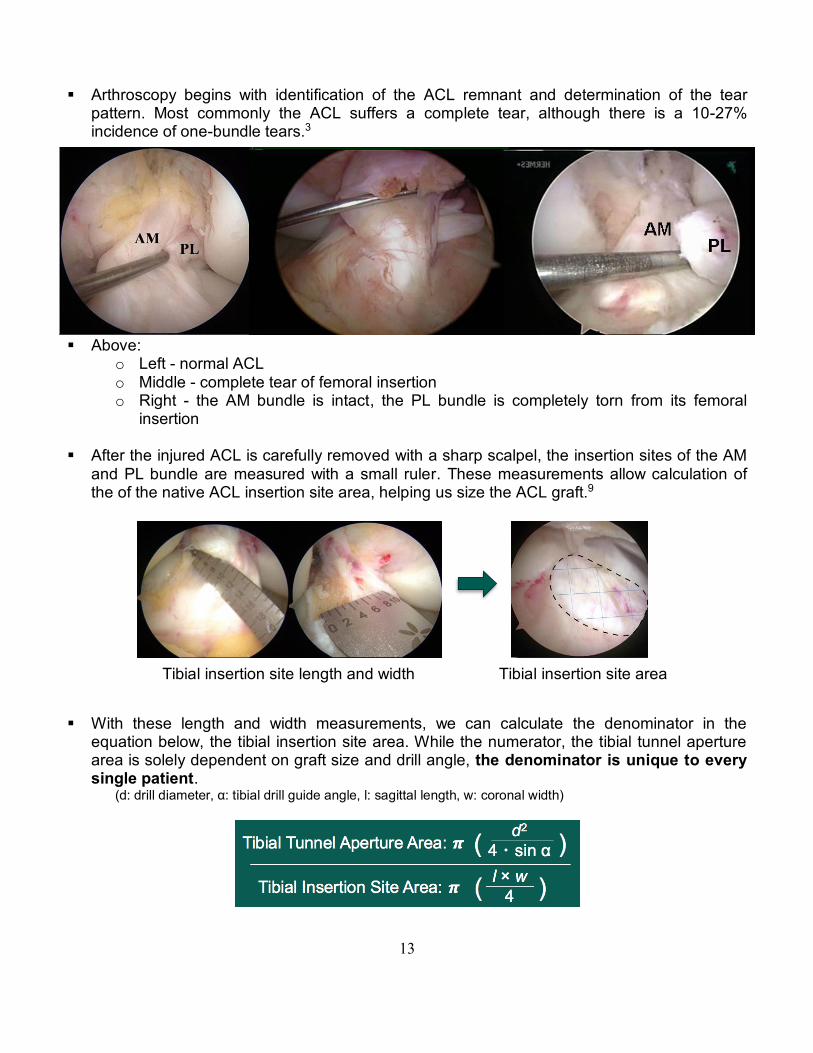
▪ Ultimately, the graft choice is defined based on graft characteristics and on the estimated graft size. The patella tendon should not be the primary option if on the sagittal cuts of the MRI the AP maximum thickness is less than 5mm. The quadriceps tendon is of adequate size if the AP thickness is greater than 7mm. The hamstrings cannot be precisely measured pre-operatively, although we are working on improving US measurements of this tendons.
Surgical Approach with the Double Bundle Concept ▪ The high anterolateral portal (AL) is made first, to allow good visualization of the ACL tibial
foot print with little fat pad obstruction. With the knee flexed at 90°, this portal is ~5mm lateral distal edge of the patella.
▪ The central portal (C) location is made using a spinal needle aligned with the fibers of the
ACL. ▪ The accessory anteromedial portal (AM) is placed 5-10mm medially, a spinal needle
ensuring the femoral tunnel can be drilled without damaging the medial femoral condyle articular cartilage. This portal is a working and visualization portal, and should be placed immediately above the superior border of the anterior horn of the medial meniscus, allowing the best frontal visualization of the ACL femoral insertion site.
13
▪ Arthroscopy begins with identification of the ACL remnant and determination of the tear
pattern. Most commonly the ACL suffers a complete tear, although there is a 10-27% incidence of one-bundle tears.3
▪ Above: o Left - normal ACL o Middle - complete tear of femoral insertion o Right - the AM bundle is intact, the PL bundle is completely torn from its femoral
insertion
▪ After the injured ACL is carefully removed with a sharp scalpel, the insertion sites of the AM and PL bundle are measured with a small ruler. These measurements allow calculation of the of the native ACL insertion site area, helping us size the ACL graft.9
▪ With these length and width measurements, we can calculate the denominator in the
equation below, the tibial insertion site area. While the numerator, the tibial tunnel aperture area is solely dependent on graft size and drill angle, the denominator is unique to every single patient.
(d: drill diameter, α: tibial drill guide angle, l: sagittal length, w: coronal width)
Tibial insertion site length and width Tibial insertion site area
14
▪ This equation allows calculation of the tibial insertion site Percent Reconstructed Area
(PRA).20 As mentioned earlier, based on our studies examining the morphology of the ACL, our goal is to reconstruct 50-70% of the native tibial insertion site.
▪ Notch size must also be measured. Wide and high notches do not pose any extra difficulty, but narrow and low notches can pose significant challenges. These notches can impede passage of grafts with bone block (i.e. technically difficult to rotate into femoral tunnel) and can increase the risk of graft impingement (ie. oversized grafts, DB reconstruction).26
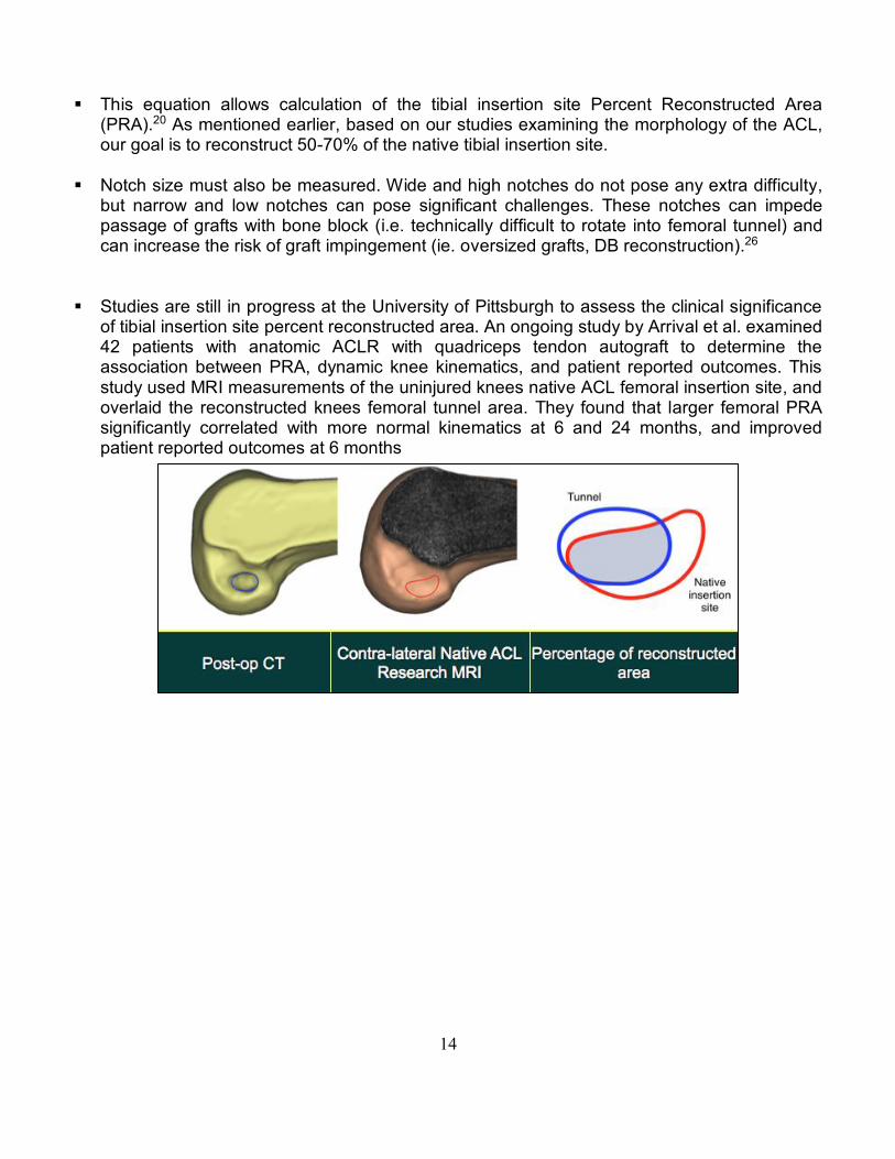
▪ Studies are still in progress at the University of Pittsburgh to assess the clinical significance
of tibial insertion site percent reconstructed area. An ongoing study by Arrival et al. examined 42 patients with anatomic ACLR with quadriceps tendon autograft to determine the association between PRA, dynamic knee kinematics, and patient reported outcomes. This study used MRI measurements of the uninjured knees native ACL femoral insertion site, and overlaid the reconstructed knees femoral tunnel area. They found that larger femoral PRA significantly correlated with more normal kinematics at 6 and 24 months, and improved patient reported outcomes at 6 months
15
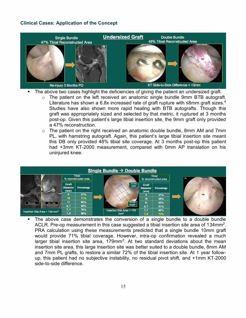
Clinical Cases: Application of the Concept
▪ The above two cases highlight the deficiencies of giving the patient an undersized graft. o The patient on the left received an anatomic single bundle 9mm BTB autograft.
Literature has shown a 6.8x increased rate of graft rupture with ≤8mm graft sizes.4 Studies have also shown more rapid healing with BTB autografts. Though this graft was appropriately sized and selected by that metric, it ruptured at 3 months post-op. Given this patient’s large tibial insertion site, the 9mm graft only provided a 47% reconstruction.
o The patient on the right received an anatomic double bundle, 8mm AM and 7mm PL, with hamstring autograft. Again, this patient’s large tibial insertion site meant this DB only provided 48% tibial site coverage. At 3 months post-op this patient had +3mm KT-2000 measurement, compared with 0mm AP translation on his uninjured knee.
▪ The above case demonstrates the conversion of a single bundle to a double bundle ACLR. Pre-op measurement in this case suggested a tibial insertion site area of 134mm2. PRA calculation using these measurements predicted that a single bundle 10mm graft would provide 71% tibial coverage. However, intra-op confirmation revealed a much larger tibial insertion site area, 179mm2. At two standard deviations about the mean insertion site area, this large insertion site was better suited to a double bundle, 8mm AM and 7mm PL grafts, to restore a similar 72% of the tibial insertion site. At 1 year follow-up, this patient had no subjective instability, no residual pivot shift, and +1mm KT-2000 side-to-side difference.
16
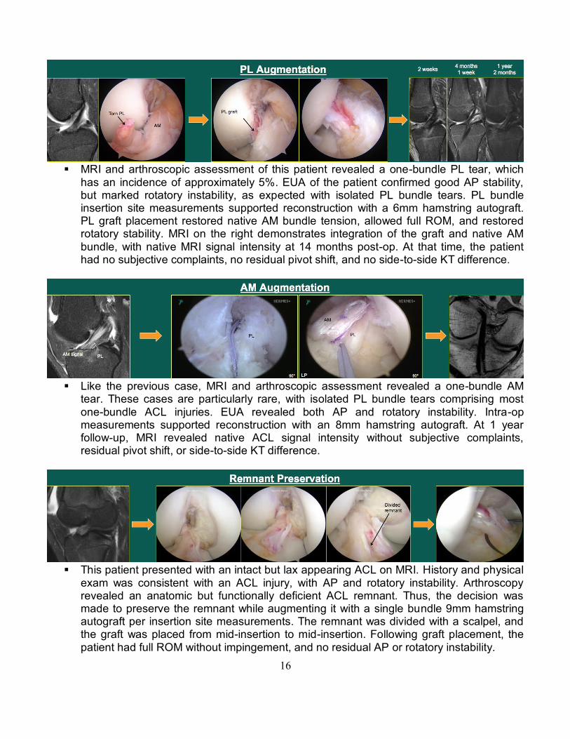
▪ MRI and arthroscopic assessment of this patient revealed a one-bundle PL tear, which has an incidence of approximately 5%. EUA of the patient confirmed good AP stability, but marked rotatory instability, as expected with isolated PL bundle tears. PL bundle insertion site measurements supported reconstruction with a 6mm hamstring autograft. PL graft placement restored native AM bundle tension, allowed full ROM, and restored rotatory stability. MRI on the right demonstrates integration of the graft and native AM bundle, with native MRI signal intensity at 14 months post-op. At that time, the patient had no subjective complaints, no residual pivot shift, and no side-to-side KT difference.
▪ Like the previous case, MRI and arthroscopic assessment revealed a one-bundle AM
tear. These cases are particularly rare, with isolated PL bundle tears comprising most one-bundle ACL injuries. EUA revealed both AP and rotatory instability. Intra-op measurements supported reconstruction with an 8mm hamstring autograft. At 1 year follow-up, MRI revealed native ACL signal intensity without subjective complaints, residual pivot shift, or side-to-side KT difference.
▪ This patient presented with an intact but lax appearing ACL on MRI. History and physical exam was consistent with an ACL injury, with AP and rotatory instability. Arthroscopy revealed an anatomic but functionally deficient ACL remnant. Thus, the decision was made to preserve the remnant while augmenting it with a single bundle 9mm hamstring autograft per insertion site measurements. The remnant was divided with a scalpel, and the graft was placed from mid-insertion to mid-insertion. Following graft placement, the patient had full ROM without impingement, and no residual AP or rotatory instability.
17
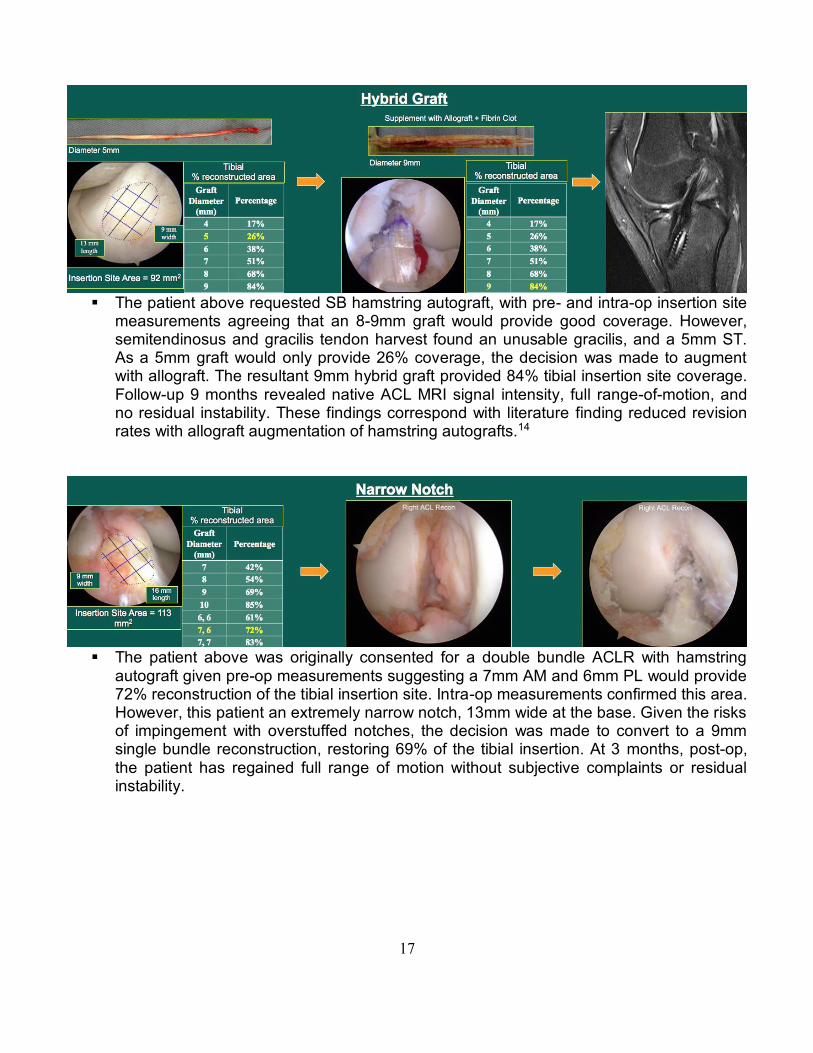
▪ The patient above requested SB hamstring autograft, with pre- and intra-op insertion site measurements agreeing that an 8-9mm graft would provide good coverage. However, semitendinosus and gracilis tendon harvest found an unusable gracilis, and a 5mm ST. As a 5mm graft would only provide 26% coverage, the decision was made to augment with allograft. The resultant 9mm hybrid graft provided 84% tibial insertion site coverage. Follow-up 9 months revealed native ACL MRI signal intensity, full range-of-motion, and no residual instability. These findings correspond with literature finding reduced revision rates with allograft augmentation of hamstring autografts.14
▪ The patient above was originally consented for a double bundle ACLR with hamstring autograft given pre-op measurements suggesting a 7mm AM and 6mm PL would provide 72% reconstruction of the tibial insertion site. Intra-op measurements confirmed this area. However, this patient an extremely narrow notch, 13mm wide at the base. Given the risks of impingement with overstuffed notches, the decision was made to convert to a 9mm single bundle reconstruction, restoring 69% of the tibial insertion. At 3 months, post-op, the patient has regained full range of motion without subjective complaints or residual instability.
18
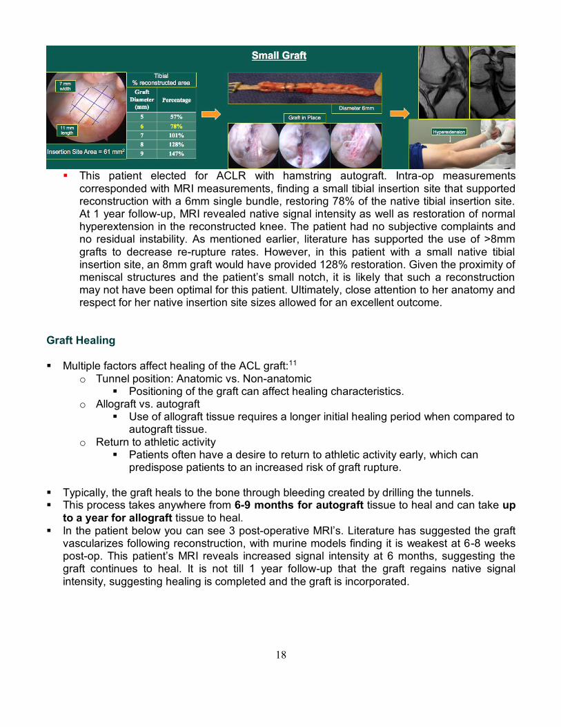
▪ This patient elected for ACLR with hamstring autograft. Intra-op measurements corresponded with MRI measurements, finding a small tibial insertion site that supported reconstruction with a 6mm single bundle, restoring 78% of the native tibial insertion site. At 1 year follow-up, MRI revealed native signal intensity as well as restoration of normal hyperextension in the reconstructed knee. The patient had no subjective complaints and no residual instability. As mentioned earlier, literature has supported the use of >8mm grafts to decrease re-rupture rates. However, in this patient with a small native tibial insertion site, an 8mm graft would have provided 128% restoration. Given the proximity of meniscal structures and the patient’s small notch, it is likely that such a reconstruction may not have been optimal for this patient. Ultimately, close attention to her anatomy and respect for her native insertion site sizes allowed for an excellent outcome.
Graft Healing ▪ Multiple factors affect healing of the ACL graft:11
o Tunnel position: Anatomic vs. Non-anatomic ▪ Positioning of the graft can affect healing characteristics.
o Allograft vs. autograft ▪ Use of allograft tissue requires a longer initial healing period when compared to
autograft tissue. o Return to athletic activity
▪ Patients often have a desire to return to athletic activity early, which can predispose patients to an increased risk of graft rupture.
▪ Typically, the graft heals to the bone through bleeding created by drilling the tunnels. ▪ This process takes anywhere from 6-9 months for autograft tissue to heal and can take up
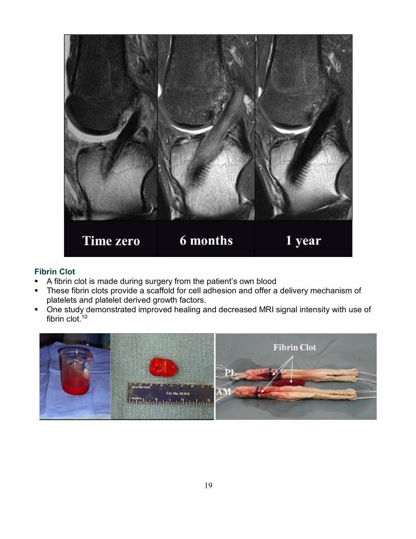
to a year for allograft tissue to heal. ▪ In the patient below you can see 3 post-operative MRI’s. Literature has suggested the graft
vascularizes following reconstruction, with murine models finding it is weakest at 6-8 weeks post-op. This patient’s MRI reveals increased signal intensity at 6 months, suggesting the graft continues to heal. It is not till 1 year follow-up that the graft regains native signal intensity, suggesting healing is completed and the graft is incorporated.
19
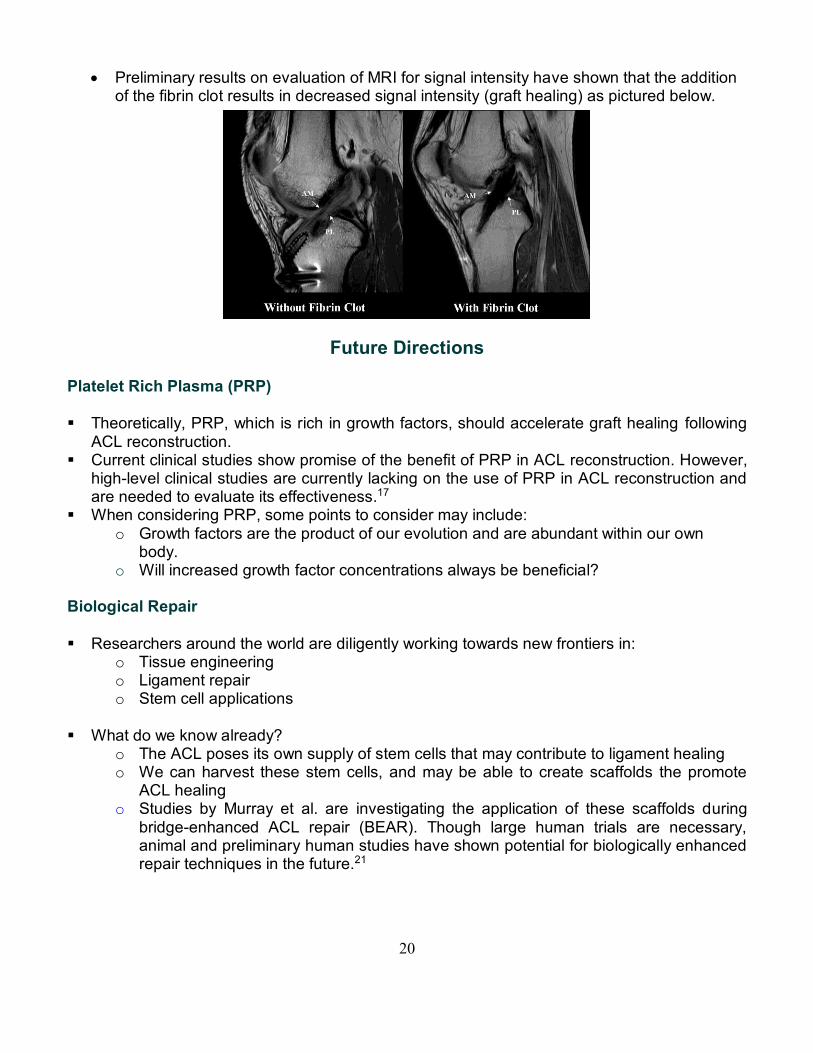
Fibrin Clot ▪ A fibrin clot is made during surgery from the patient’s own blood ▪ These fibrin clots provide a scaffold for cell adhesion and offer a delivery mechanism of
platelets and platelet derived growth factors. ▪ One study demonstrated improved healing and decreased MRI signal intensity with use of
fibrin clot.10
20
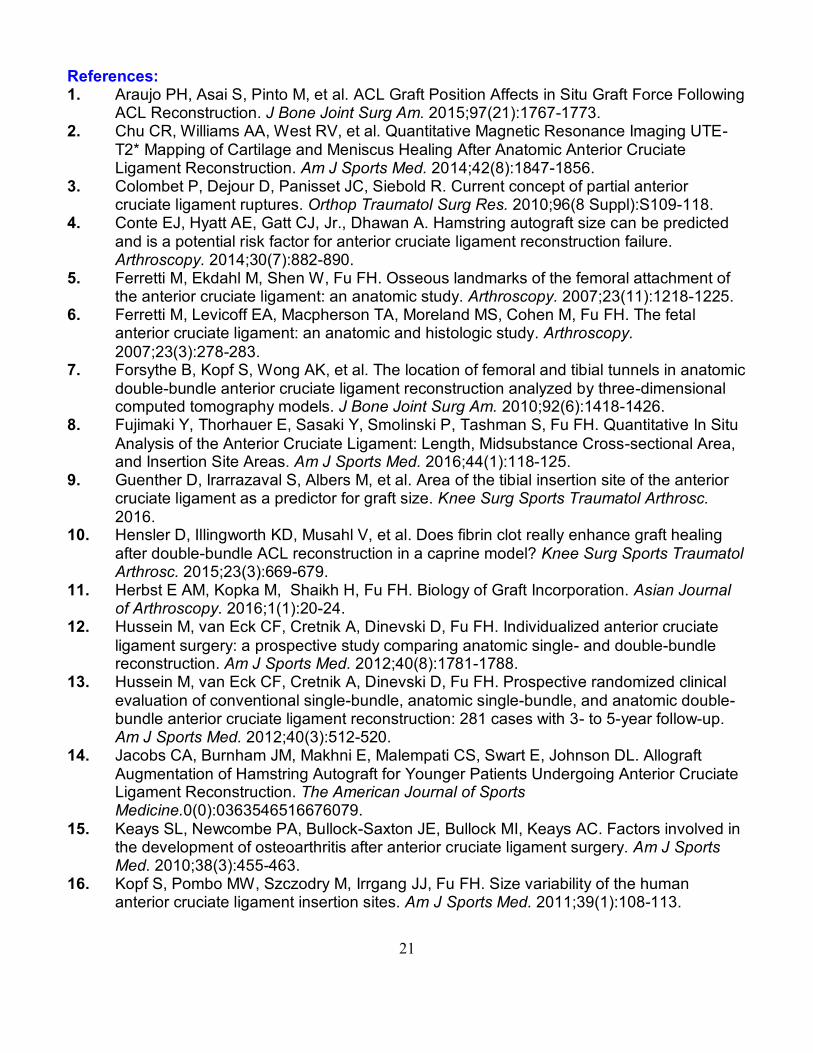
• Preliminary results on evaluation of MRI for signal intensity have shown that the addition of the fibrin clot results in decreased signal intensity (graft healing) as pictured below.
Future Directions
Platelet Rich Plasma (PRP) ▪ Theoretically, PRP, which is rich in growth factors, should accelerate graft healing following
ACL reconstruction. ▪ Current clinical studies show promise of the benefit of PRP in ACL reconstruction. However,
high-level clinical studies are currently lacking on the use of PRP in ACL reconstruction and are needed to evaluate its effectiveness.17
▪ When considering PRP, some points to consider may include: o Growth factors are the product of our evolution and are abundant within our own
body. o Will increased growth factor concentrations always be beneficial?
Biological Repair ▪ Researchers around the world are diligently working towards new frontiers in:
o Tissue engineering o Ligament repair o Stem cell applications
▪ What do we know already?
o The ACL poses its own supply of stem cells that may contribute to ligament healing o We can harvest these stem cells, and may be able to create scaffolds the promote
ACL healing o Studies by Murray et al. are investigating the application of these scaffolds during
bridge-enhanced ACL repair (BEAR). Though large human trials are necessary, animal and preliminary human studies have shown potential for biologically enhanced repair techniques in the future.21
21
References: 1. Araujo PH, Asai S, Pinto M, et al. ACL Graft Position Affects in Situ Graft Force Following
ACL Reconstruction. J Bone Joint Surg Am. 2015;97(21):1767-1773. 2. Chu CR, Williams AA, West RV, et al. Quantitative Magnetic Resonance Imaging UTE-
T2* Mapping of Cartilage and Meniscus Healing After Anatomic Anterior Cruciate Ligament Reconstruction. Am J Sports Med. 2014;42(8):1847-1856.
3. Colombet P, Dejour D, Panisset JC, Siebold R. Current concept of partial anterior cruciate ligament ruptures. Orthop Traumatol Surg Res. 2010;96(8 Suppl):S109-118.
4. Conte EJ, Hyatt AE, Gatt CJ, Jr., Dhawan A. Hamstring autograft size can be predicted and is a potential risk factor for anterior cruciate ligament reconstruction failure. Arthroscopy. 2014;30(7):882-890.
5. Ferretti M, Ekdahl M, Shen W, Fu FH. Osseous landmarks of the femoral attachment of the anterior cruciate ligament: an anatomic study. Arthroscopy. 2007;23(11):1218-1225.
6. Ferretti M, Levicoff EA, Macpherson TA, Moreland MS, Cohen M, Fu FH. The fetal anterior cruciate ligament: an anatomic and histologic study. Arthroscopy. 2007;23(3):278-283.
7. Forsythe B, Kopf S, Wong AK, et al. The location of femoral and tibial tunnels in anatomic double-bundle anterior cruciate ligament reconstruction analyzed by three-dimensional computed tomography models. J Bone Joint Surg Am. 2010;92(6):1418-1426.
8. Fujimaki Y, Thorhauer E, Sasaki Y, Smolinski P, Tashman S, Fu FH. Quantitative In Situ Analysis of the Anterior Cruciate Ligament: Length, Midsubstance Cross-sectional Area, and Insertion Site Areas. Am J Sports Med. 2016;44(1):118-125.
9. Guenther D, Irarrazaval S, Albers M, et al. Area of the tibial insertion site of the anterior cruciate ligament as a predictor for graft size. Knee Surg Sports Traumatol Arthrosc. 2016.
10. Hensler D, Illingworth KD, Musahl V, et al. Does fibrin clot really enhance graft healing after double-bundle ACL reconstruction in a caprine model? Knee Surg Sports Traumatol Arthrosc. 2015;23(3):669-679.
11. Herbst E AM, Kopka M, Shaikh H, Fu FH. Biology of Graft Incorporation. Asian Journal of Arthroscopy. 2016;1(1):20-24.
12. Hussein M, van Eck CF, Cretnik A, Dinevski D, Fu FH. Individualized anterior cruciate ligament surgery: a prospective study comparing anatomic single- and double-bundle reconstruction. Am J Sports Med. 2012;40(8):1781-1788.
13. Hussein M, van Eck CF, Cretnik A, Dinevski D, Fu FH. Prospective randomized clinical evaluation of conventional single-bundle, anatomic single-bundle, and anatomic double-bundle anterior cruciate ligament reconstruction: 281 cases with 3- to 5-year follow-up. Am J Sports Med. 2012;40(3):512-520.
14. Jacobs CA, Burnham JM, Makhni E, Malempati CS, Swart E, Johnson DL. Allograft Augmentation of Hamstring Autograft for Younger Patients Undergoing Anterior Cruciate Ligament Reconstruction. The American Journal of Sports Medicine.0(0):0363546516676079.
15. Keays SL, Newcombe PA, Bullock-Saxton JE, Bullock MI, Keays AC. Factors involved in the development of osteoarthritis after anterior cruciate ligament surgery. Am J Sports Med. 2010;38(3):455-463.
16. Kopf S, Pombo MW, Szczodry M, Irrgang JJ, Fu FH. Size variability of the human anterior cruciate ligament insertion sites. Am J Sports Med. 2011;39(1):108-113.
22
17. Kopka M, Bradley JP. The Use of Biologic Agents in Athletes with Knee Injuries. J Knee Surg. 2016;29(5):379-386.
18. Lohmander LS, Ostenberg A, Englund M, Roos H. High prevalence of knee osteoarthritis, pain, and functional limitations in female soccer players twelve years after anterior cruciate ligament injury. Arthritis Rheum. 2004;50(10):3145-3152.
19. Matsumoto T, Ingham SM, Mifune Y, et al. Isolation and characterization of human anterior cruciate ligament-derived vascular stem cells. Stem Cells Dev. 2012;21(6):859-872.
20. Middleton KK, Muller B, Araujo PH, et al. Is the native ACL insertion site "completely restored" using an individualized approach to single-bundle ACL-R? Knee Surg Sports Traumatol Arthrosc. 2015;23(8):2145-2150.
21. Murray MM, Flutie BM, Kalish LA, et al. The Bridge-Enhanced Anterior Cruciate Ligament Repair (BEAR) Procedure: An Early Feasibility Cohort Study. Orthop J Sports Med. 2016;4(11):2325967116672176.
22. Shino K, Nakata K, Nakamura N, et al. Rectangular tunnel double-bundle anterior cruciate ligament reconstruction with bone-patellar tendon-bone graft to mimic natural fiber arrangement. Arthroscopy. 2008;24(10):1178-1183.
23. Slone HS, Romine SE, Premkumar A, Xerogeanes JW. Quadriceps tendon autograft for anterior cruciate ligament reconstruction: a comprehensive review of current literature and systematic review of clinical results. Arthroscopy. 2015;31(3):541-554.
24. Vairo GL, Myers JB, Sell TC, Fu FH, Harner CD, Lephart SM. Neuromuscular and biomechanical landing performance subsequent to ipsilateral semitendinosus and gracilis autograft anterior cruciate ligament reconstruction. Knee Surg Sports Traumatol Arthrosc. 2008;16(1):2-14.
25. van Eck CF, Gravare-Silbernagel K, Samuelsson K, et al. Evidence to support the interpretation and use of the Anatomic Anterior Cruciate Ligament Reconstruction Checklist. J Bone Joint Surg Am. 2013;95(20):e153.
26. van Eck CF, Lesniak BP, Schreiber VM, Fu FH. Anatomic single- and double-bundle anterior cruciate ligament reconstruction flowchart. Arthroscopy. 2010;26(2):258-268.
27. van Eck CF, Schkrohowsky JG, Working ZM, Irrgang JJ, Fu FH. Prospective analysis of failure rate and predictors of failure after anatomic anterior cruciate ligament reconstruction with allograft. Am J Sports Med. 2012;40(4):800-807.
28. Yagi M, Wong EK, Kanamori A, Debski RE, Fu FH, Woo SL. Biomechanical analysis of an anatomic anterior cruciate ligament reconstruction. Am J Sports Med. 2002;30(5):660-666.


![PROFESSIONAL ARTICLES STRUČNI ČLANCI...tibial plateau, measured from the anterior edge of the tibia [16] in reference to the Amis-Jakob line [17]. Anatomic centre of the ACL tibial](https://img.pdfslide.net/doc/110x75/6115cddce123e03d306444ca/professional-articles-struoeni-oe-tibial-plateau-measured-from-the-anterior.jpg)