Embed Size (px)
Citation preview
To investigate the origin and postnatal changesof mouse mandibular angular cartilage, in situhybridization for cartilaginous marker proteins,histochemistry for alkaline phosphatase (ALP)and tartrate-resistant acid phosphatase (TRAP),and bromodeoxyuridine (BrDU) analyses wereperformed. Chondrocytes of the mandibular angu-lar cartilage were derived from ALP-positive prog-enitor cells and first detected at embryonic day (E)15.5. Newly formed chondrocytes rapidly differen-tiated into hypertrophic chondrocytes and hyper-trophic cell zone rapidly extended in subsequent afew days. During this period, bone sialoproteinmRNA was more widely expressed than osteo-pontin mRNA in cartilage. Endochondral bone for-mation started at E 17.5 with the resorption of thebone collar by osteoclasts. These characteristicswere consistent with those of the condylar carti-lage, although developmental process was 0.5-1.5day delayed relative to the condylar cartilage.During the postnatal period, contrast to thecondylar cartilage, the angular cartilage constantlydecreased in volume with advancing age.Reduction of proliferating activity estimated byBrDU incorporation accounts for this phenomenon.
We demonstrate new structural features of themandibular angular cartilage that may contribute toa coming research for the secondary cartilage.
Key words: secondary cartilage, angular carti-lage, in situ hybridization, alkalinephosphatase
Introduction
During the growing process of mandible, secondarycartilages are formed in condylar, angular and coronoidprocesses in human, rats and mice1-11. The chondro-cytes of the mouse mandibular condylar cartilagedevelop from alkaline phosphatase (ALP)-positiveprogenitor cells in the periosteum-like tissue afterossifying mandible has formed6-9. These progenitorcells then rapidly differentiate into hypertrophic chon-drocytes which express mRNAs for collagen Types I, IIand X simultaneously8,10,11. The hypertrophic cell zonethen rapidly extends in subsequent a few days7,8,10,12,13
and bone sialoprotein (BSP) mRNA is more widelyexpressed than osteopontin (OPN) mRNA during thisperiod13. The endochondral bone formation of thiscartilage starts with the resorption of bone collar at E167,14. During postnatal period, the mandibular condylarcartilage continues to grow and function as articularcartilage of the temporo-mandibular joint and some
Original Article
An in situ hybridization and histochemical study of development and postnatalchanges of mouse mandibular angular cartilage compared with condylar cartilage
Shunichi Shibata1, Tatsuya Fujimori2 and Yasuo Yamashita1
1) Maxillofacial Anatomy, Department of Maxillofacial Biology, Graduate School, Tokyo Medical and DentalUniversity2) Implantology, Department of Masticatory Function Rehabilitation, Graduate School, Tokyo Medical andDental University
J Med Dent Sci 2006; 53: 41–50
Corresponding Author: Shunichi ShibataMaxillofacial Anatomy, Department of Maxillofacial Biology,Graduate School, Tokyo Medical and Dental University1-5-45, Yushima, Bunkyo-ku, Tokyo 113-8549, JapanTel: 03-5803-5436 Fax: 03-5803-0185E-Mail: [email protected] October 12; Accepted December 2, 2005
immunohistochemical studies related to the postnatalchanges have been performed15,16.
Meanwhile, in the mandibular angular cartilage,several histological studies in rats2,3,17-19, in mice20 and inhuman21, and a few histochemical studies for ALP inrats3 and in mice9 have been performed. Although thiscartilage is also derived from ALP-positive progenitorcells9, detail developmental process from viewpoints ofin situ hybridization has not been described. First, wehypothesized that developmental process of the fetalangular cartilage is similar to that of the condylar carti-lage. Furthermore, although rat angular cartilage dis-appears in postnatal periods because likely of the lackof articular function2,18, definite reason for disappear-ance is still unknown. Second, we hypothesized thatreduction of proliferation activity and/or acceleration ofcartilage resorption may be important for this phenom-enon. To confirm these hypotheses, we investigateddevelopment and postnatal changes of the mousemandibular angular cartilage by in situ hybridization ofcartilage matrix proteins, incorporation of bromod-eoxyuridine (BrDU) and tartrate-resistant acid phos-phatase (TRAP) staining, as comparing with thecondylar cartilage.
Materials and Methods
Tissue preparation All animals were maintained in the animal research
center in the Tokyo Medical and Dental University andthe procedures conformed to the guidelines deter-mined by the University Animal Care Committee.Research protocols conformed to NIH guidelines asstated in the “Principles of Laboratory Animal Care”(NIH publication No. 86-23, revised 1985).
Ten fetal ICR mice, embryonic day (E) 13 - 18.5, andtwenty postnatal mice, 7 - 21 day after birth (d 7 - 21)were used for this study. BrDU was shot (2 ÒM /10g) toeach mouse 2 hrs before killing mice. At each timepoint, the pregnant and postnatal mice were killed bycervical dislocation under ether anesthetization, afterwhich each fetal mouse was killed by cervical disloca-tion. The heads were then taken and immersed in 4%paraformaldehyde (0.1 M phosphate buffer, pH 7.4) for1 day at 4°C or 95% ethanol for 3 d at room tempera-ture. Paraformaldehyde-fixed specimens were decalci-fied with 10% ethylenediamine tetraacetic acid(EDTA) for 7 days at 4°C then routinely embedded inparaffin. Sections of 5 Òm were cut in the coronal plane,perpendicular to the sagittal plane, and parallel to the
long axis of the angular or the condylar process of themandible. Sections were stained with 0.1% toluidineblue (0.1 M phosphate buffer, pH 7.4) for histologicalobservations. Some specimens were embedded inOCT compound (Miles, Elkhart, IN) and frozen with liq-uid nitrogen in preparation for cryosections.
Whole skeletal staining was performed according tothe methods of McLeod22, ethanol-fixed specimenswere stained with alcian blue and alizarin red S, thenimmersed in 1% KOH to dissolve soft tissues.
In situ hybridization and immunohistochemistry forBrDU
Digoxigenin-labeled cRNA probes for aggrecan,collagen Types II and X, BSP and OPN were used inprevious studies6,10,13. In situ hybridization using aNucleic Acid Detection Kit (Roche Diagnostics,Mannheim, Germany) was performed as previouslydescribed13,23. Sections were examined after counter-staining with nuclear fast red. Sense probes werereacted as negative controls.
Some in situ hybridization-treated sections werefurther utilized for immunohistochemistry for BrDUusing a HISTOFINE SAB kit (Nichirei, Tokyo, Japan)and a M. O. M kit (Vector Laboratories, Burlingame,CA). After reacting with NBT/BCIP in the Nucleic AcidDetection Kit, sections were then immersed inmethanol containing 1% hydrogen peroxide to blockendogenous peroxidase activity and further immersedin mouse IgG blocking reagent in the M. O. M kit.Sections were then reacted with anti-BrDU mousemonoclonal antibody (Roche Diagnostics, Mannheim,Germany) diluted (1:100) with phosphate bufferedsaline containing 1% bovine serum albumin Thestreptavidin-biotin method was then applied to thesections using the HISTOFINE SAB kit, as previouslydescribed8,13. Finally, sections were treated with 3-amino-9-ethylcarbazole (Nichirei, Tokyo, Japan) toreveal any reaction. Normal mouse IgG was reactedinstead of the primary antibody as negative controls.
Enzyme histochemistryTRAP activity was detected by the hexasotized
pararosaniline method described by Lewinson andSilbermann24. ALP activity was detected by routine azodye method. Cryosections (8 Òm) made by Cryostat1720 (Leitz, Wetzlar, Germany) were incubated in asolution with a mixture of Naphtol AS-MX sodium salt(Sigma, St Louis, MO) as a substrate and Fast-blue RRsalt (Sigma) diluted in 0.1M Tris-HCl buffer (pH 8.5) atroom temperature for 30 min. As negative controls, sec-
S. SHIBATA, T. FUJIMORI and Y. YAMASHITA J Med Dent Sci42
tions were incubated in working solution without sub-strate or containing 25mM levamisole.
Histomorphometry Sections cut through the middle plane of the angular
or the condylar process were used for histomorpho-metric analyses and four sets of three serial sections forin situ hybridization were used at each stage.According to Luder et al.25, the angular and thecondylar cartilages were classified as follows: fibrouscell zone (also known as articular zone), polymorphiccell zone, flattened cell zone, upper hypertrophic cellzone, lower hypertrophic cell zone. “Total cartilagi-nous area” in this study contained all these zonesbetween lateral and medial bone collars. Especially, thepolymorphic cell zone between lateral and medialbone collars was termed as “chondroproliferationarea”. In addition, the zone where chondrocyte lacunaewere open was regarded as erosion zone. The lengthof erosion zone was measured by N.I.H. Image 1.61 in
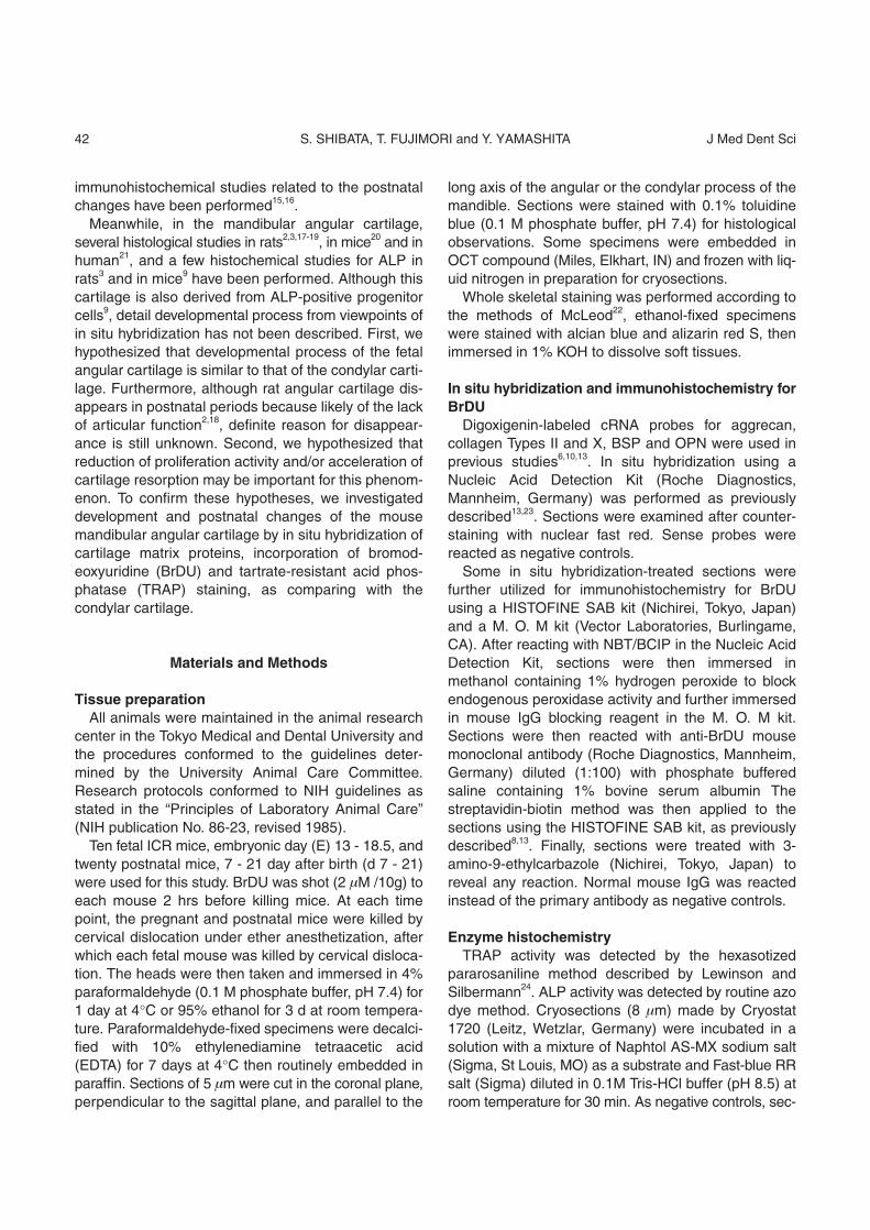
low magnified sections and termed as “erosion zonelength” (Figure 1).
The area of the mRNA-positive regions for Type IIcollagen (marker for mature chondrocytes), Type X col-lagen (marker for hypertrophic chondrocytes) andOPN (marker for deep layer of hypertrophic chondro-cytes) were measured using NIH Image 1.61. ThemRNA-positive region was easily distinguished from thenegative region by color difference. Since there weresome overlapping areas, the sum of each area occa-sionally exceeded the total cartilaginous area. Thenumber of BrDU-positive cells in the “chondroprolifera-tion area” was counted and normalized number by areawas estimated. Only a few chondrocytes in the flattenedcell layer were positive for BrDU as previouslydescribed25, but were not counted in this study. Thenumber of TRAP-positive, multinucleated chondro-cytes as discriminated from osteoclasts26 was countedand normalized number by “erosion zone length” wasestimated.
Results
Whole skeletal staining The mandibular angular cartilage and the condylar
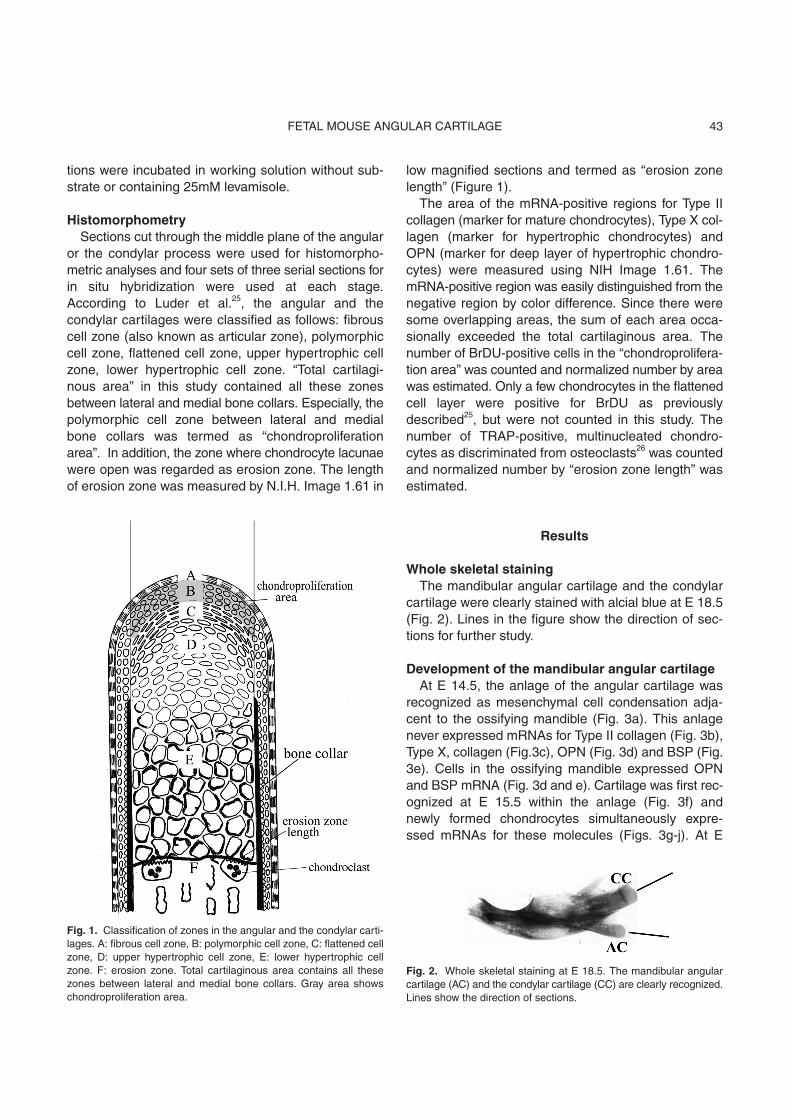
cartilage were clearly stained with alcial blue at E 18.5(Fig. 2). Lines in the figure show the direction of sec-tions for further study.
Development of the mandibular angular cartilageAt E 14.5, the anlage of the angular cartilage was
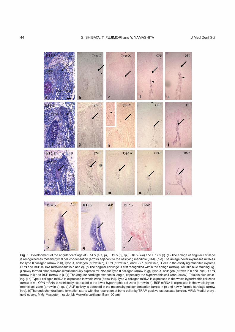
recognized as mesenchymal cell condensation adja-cent to the ossifying mandible (Fig. 3a). This anlagenever expressed mRNAs for Type II collagen (Fig. 3b),Type X, collagen (Fig.3c), OPN (Fig. 3d) and BSP (Fig.3e). Cells in the ossifying mandible expressed OPNand BSP mRNA (Fig. 3d and e). Cartilage was first rec-ognized at E 15.5 within the anlage (Fig. 3f) andnewly formed chondrocytes simultaneously expre-ssed mRNAs for these molecules (Figs. 3g-j). At E
43FETAL MOUSE ANGULAR CARTILAGE
Fig. 1. Classification of zones in the angular and the condylar carti-lages. A: fibrous cell zone, B: polymorphic cell zone, C: flattened cellzone, D: upper hypertrophic cell zone, E: lower hypertrophic cellzone. F: erosion zone. Total cartilaginous area contains all thesezones between lateral and medial bone collars. Gray area showschondroproliferation area.
Fig. 2. Whole skeletal staining at E 18.5. The mandibular angularcartilage (AC) and the condylar cartilage (CC) are clearly recognized.Lines show the direction of sections.
S. SHIBATA, T. FUJIMORI and Y. YAMASHITA J Med Dent Sci44
Fig. 3. Development of the angular cartilage at E 14.5 (a-e, p), E 15.5 (f-j, q), E 16.5 (k-o) and E 17.5 (r). (a) The anlage of angular cartilageis recognized as mesenchymal cell condensation (arrow) adjacent to the ossifying mandible (OM). (b-e) The anlage never expresses mRNAsfor Type II collagen (arrow in b), Type X, collagen (arrow in c), OPN (arrow in d) and BSP (arrow in e). Cells in the ossifying mandible expressOPN and BSP mRNA (arrowheads in d and e). (f) The angular cartilage is first recognized within the anlage (arrow). Toluidin blue staining. (g-j) Newly formed chondrocytes simultaneously express mRNAs for Type II collagen (arrow in g), Type X, collagen (arrows in h and inset), OPN(arrow in i) and BSP (arrow in j). (k) The angular cartilage extends in length, especially the hypertrophic cell zone (arrow). Toluidin blue stain-ing. (l-o) Type II collagen mRNA is expressed in whole zone (arrow in l). Type X collagen mRNA is expressed in the whole hypertrophic cell zone(arrow in m). OPN mRNA is restrictedly expressed in the lower hypertrophic cell zone (arrow in n). BSP mRNA is expressed in the whole hyper-trophic cell zone (arrow in o). (p, q) ALP activity is detected in the mesenchymal condensation (arrow in p) and newly formed cartilage (arrowin q). (r)The endochondral bone formation starts with the resorption of bone collar by TRAP-positive osteoclasts (arrow). MPM: Medial ptery-goid nuscle. MM: Masseter muscle. M: Meckel’s cartilage. Bar=100 Òm.
16.5, cartilage extended in length, especially thehypertrophic cell zone (Fig. 3k). Type II collagenmRNA was expressed in whole zone of this cartilage(Fig. 3l). Type X collagen mRNA was expressed in thewhole hypertrophic cell zone (Fig. 3m). OPN mRNAwas restrictedly expressed in the lower hypertrophic cellzone at this stage (Fig. 3n), while BSP mRNA wasexpressed in the whole hypertrophic cell zone (Fig. 3o).Further, ALP activity was detected both in the mes-enchymal condensation at E 14.5 (Fig. 3p) and in thenewly formed angular cartilage at E 15.5 (Fig. 3q). Theendochondral bone formation of this cartilage startedwith the resorption of bone collar by TRAP-positiveosteoclasts at E 17.5 (Fig. 3r).
Postnatal changes of the mandibular angular carti-lage
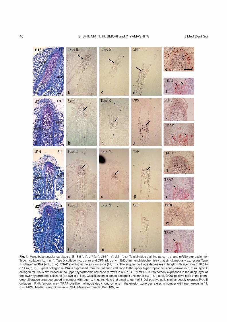
During postnatal period from E 18.5 to d 21, theangular cartilage decreased in length with advancingage and consequently classification of zones becameunclear at d 21. The medio-lateral width of this cartilagehardly increased with advancing age (Fig. 4a, g, m, s).
At E 18.5, Type II collagen mRNA was expressedfrom the flattened cell zone to the upper hypertrophiccell zone (Fig. 4b). Type X collagen mRNA was mainlyexpressed in the upper hypertrophic cell zone (Fig. 4c),while OPN mRNA was restrictedly expressed in thedeep layer of the lower hypertrophic cell zone (Fig. 4d).Although this expression pattern for each mRNA wasalmost similar at d 7 and d 14, each zone expressingmRNAs for these molecules reduced in volume (Fig.4h-j, n-p), and became undistinguishable at d 21 (Fig.4t-v). Results of histomorphometry for each zone in theangular cartilage were described in Figure 6a. BrDU-positive cells in the chondroproliferation area decreasedin number with advancing age (Fig. 4e, k, q, w).TRAP-positive, multinucleated chondroclasts in theerosion zone also decreased in number with advancingage (Fig. 4f, l, r, x). Results of cell counting normalizedwere described in Figure 6c and d.
Postnatal changes of the mandibular condylarcartilage
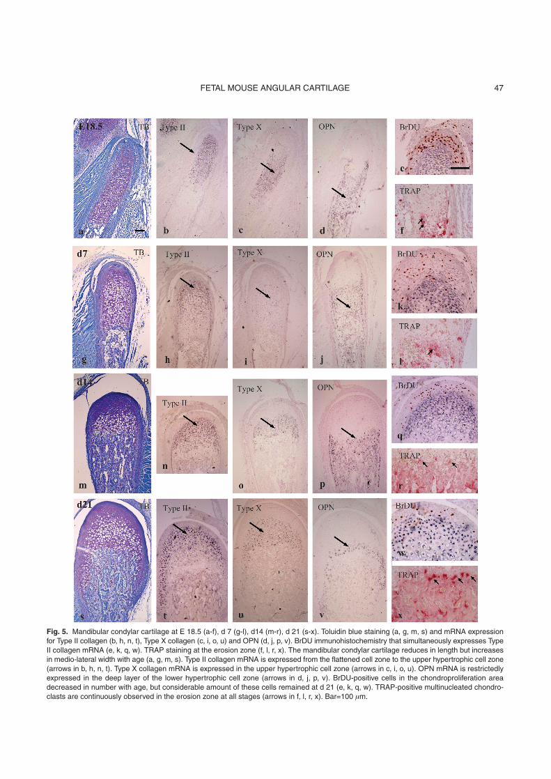
The mandibular condylar cartilage graduallyreduced in length but increased in medio-lateral widthwith advancing age, and consequently maintained itsvolume at d 21 (Fig. 5a, g, m, s). At E 18.5, Type II col-lagen mRNA was expressed from the flattened cellzone to the upper hypertrophic cell zone (Fig. 5b). TypeX collagen mRNA was mainly expressed in the upperhypertrophic cell zone (Fig. 5c), while OPN mRNA was
restrictedly expressed in the deep layer of the lowerhypertrophic cell zone (Fig. 5d). This expression patternfor each mRNA was almost similar at all stages exam-ined (Figs. 5h-j, n-p, t-v) and each zone expressingthese molecules maintained its volume and was clear-ly distinguishable at d 21. Results of histomorphometryfor each zone in the condylar cartilage weredescribed in Figure 6b. BrDU-positive cells in thechondroproliferation area decreased in number withadvancing age (Fig. 5e, k, q, w). Considerableamount of BrDU-positive cells remained at d 21 (Fig.5w), but since the chondroproliferation area alsoincreased in volume, the cell number per this arearemarkably reduced. To the contrary TRAP-positivemultinucleated chondroclasts were continuouslyobserved in the erosion zone at all stages (Fig. 5f, l, r,x), indicating endochondral bone formation wasactively progressing until d 21. Results of cell countingnormalized were described in Figure 6c and d.
Negative controls for in situ hybridization and histo-chemistry showed no positive reaction at any timepoints (data not shown), as previously described8,13,14.
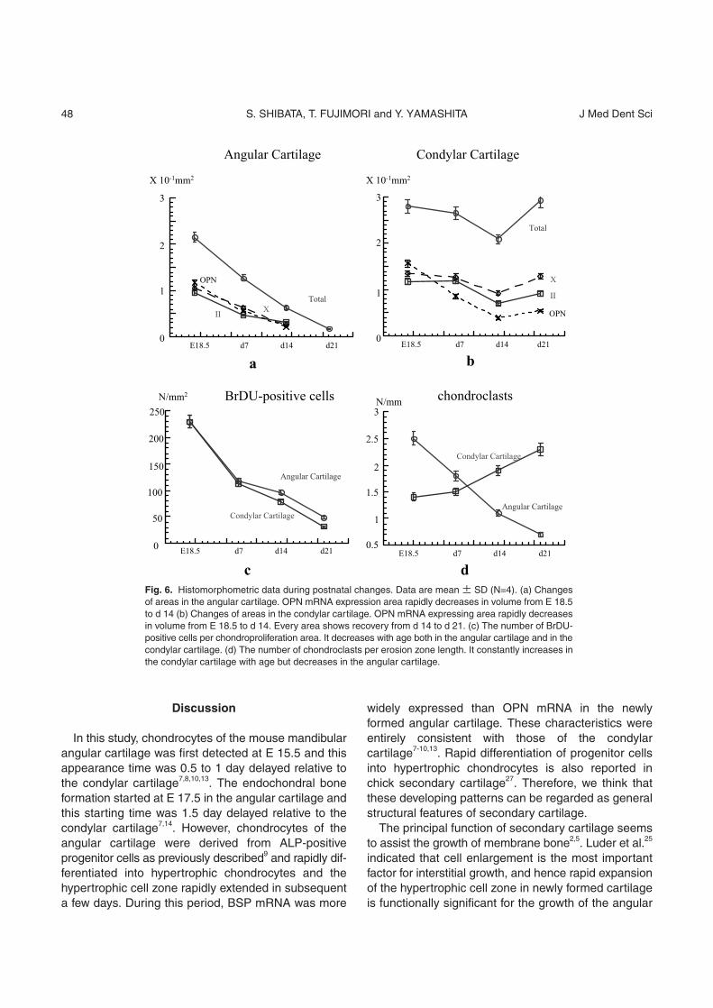
HistomorphometryThe total cartilaginous area and each zone con-
stantly decreased in volume in the angular cartilagefrom E 18.5 to d 21. While the OPN mRNA expressionarea rapidly decreased in volume from E 18.5 to d 14,the Type II collagen mRNA expression area tended todecrease slowly in volume as compared to other twoareas from d 7 to d 14 (Fig. 6a). Meanwhile, the totalcartilaginous area of the mandibular condylar cartilagedecreased in volume from E 18.5 to d 14 but recoveredfrom d 14 to d 21, and consequently large amount ofvolume remained at d 21. The Type II collagen andType X collagen mRNA expressing areas showedsimilar pattern of changes to the total cartilaginousarea, although the OPN expressing area rapidlydecreased in volume from E 18.5 to d 14 (Fig. 6b).
The number of BrDU-positive cells per chondropro-liferation area decreased with advancing age in theangular cartilage but this was similar in the condylarcartilage (Fig. 6c), indicating proliferating activity in theangular cartilage is reduced but still maintains tosome extent as the condylar cartilage goes at d 21.
The number of TRAP-positive multinucleated chon-droclasts per erosion zone length constantlyincreased in number in the condylar cartilage withadvancing age but constantly decreased in the angularcartilage (Fig. 6d).
45FETAL MOUSE ANGULAR CARTILAGE
S. SHIBATA, T. FUJIMORI and Y. YAMASHITA J Med Dent Sci46
Fig. 4. Mandibular angular cartilage at E 18.5 (a-f), d 7 (g-l), d14 (m-r), d 21 (s-x). Toluidin blue staining (a, g, m, s) and mRNA expression forType II collagen (b, h, n, t), Type X collagen (c, i, o, u) and OPN (d, j, p, v ). BrDU immunohistochemistry that simultaneously expresses TypeII collagen mRNA (e, k, q, w). TRAP staining at the erosion zone (f, l, r, x). The angular cartilage decreases in length with age from E 18.5 tod 14 (a, g, m). Type II collagen mRNA is expressed from the flattened cell zone to the upper hypertrophic cell zone (arrows in b, h, n). Type Xcollagen mRNA is expressed in the upper hypertrophic cell zone (arrows in c, i, o). OPN mRNA is restrictedly expressed in the deep layer ofthe lower hypertrophic cell zone (arrows in d, j, p). Classification of zones becomes unclear at d 21 (s, t, u, v). BrDU-positive cells in the chon-droproliferation area decreased in number with age (e, k, q, w). Note that small anount of BrDU-positive cells similtaneously express Type IIcollagen mRNA (arrows in e). TRAP-positive multinucleated chondroclasts in the erosion zone decreases in number with age (arrows in f, l,r, x). MPM: Medial pterygoid muscle. MM: Masseter muscle. Bar=100 Òm.
47FETAL MOUSE ANGULAR CARTILAGE
Fig. 5. Mandibular condylar cartilage at E 18.5 (a-f), d 7 (g-l), d14 (m-r), d 21 (s-x). Toluidin blue staining (a, g, m, s) and mRNA expressionfor Type II collagen (b, h, n, t), Type X collagen (c, i, o, u) and OPN (d, j, p, v). BrDU immunohistochemistry that simultaneously expresses TypeII collagen mRNA (e, k, q, w). TRAP staining at the erosion zone (f, l, r, x). The mandibular condylar cartilage reduces in length but increasesin medio-lateral width with age (a, g, m, s). Type II collagen mRNA is expressed from the flattened cell zone to the upper hypertrophic cell zone(arrows in b, h, n, t). Type X collagen mRNA is expressed in the upper hypertrophic cell zone (arrows in c, i, o, u). OPN mRNA is restrictedlyexpressed in the deep layer of the lower hypertrophic cell zone (arrows in d, j, p, v). BrDU-positive cells in the chondroproliferation areadecreased in number with age, but considerable amount of these cells remained at d 21 (e, k, q, w). TRAP-positive multinucleated chondro-clasts are continuously observed in the erosion zone at all stages (arrows in f, l, r, x). Bar=100 Òm.
Discussion
In this study, chondrocytes of the mouse mandibularangular cartilage was first detected at E 15.5 and thisappearance time was 0.5 to 1 day delayed relative tothe condylar cartilage7,8,10,13. The endochondral boneformation started at E 17.5 in the angular cartilage andthis starting time was 1.5 day delayed relative to thecondylar cartilage7,14. However, chondrocytes of theangular cartilage were derived from ALP-positiveprogenitor cells as previously described9 and rapidly dif-ferentiated into hypertrophic chondrocytes and thehypertrophic cell zone rapidly extended in subsequenta few days. During this period, BSP mRNA was more
widely expressed than OPN mRNA in the newlyformed angular cartilage. These characteristics wereentirely consistent with those of the condylar cartilage7-10,13. Rapid differentiation of progenitor cellsinto hypertrophic chondrocytes is also reported inchick secondary cartilage27. Therefore, we think thatthese developing patterns can be regarded as generalstructural features of secondary cartilage.
The principal function of secondary cartilage seemsto assist the growth of membrane bone2,5. Luder et al.25
indicated that cell enlargement is the most importantfactor for interstitial growth, and hence rapid expansionof the hypertrophic cell zone in newly formed cartilageis functionally significant for the growth of the angular
S. SHIBATA, T. FUJIMORI and Y. YAMASHITA J Med Dent Sci48
Fig. 6. Histomorphometric data during postnatal changes. Data are mean ± SD (N=4). (a) Changesof areas in the angular cartilage. OPN mRNA expression area rapidly decreases in volume from E 18.5to d 14 (b) Changes of areas in the condylar cartilage. OPN mRNA expressing area rapidly decreasesin volume from E 18.5 to d 14. Every area shows recovery from d 14 to d 21. (c) The number of BrDU-positive cells per chondroproliferation area. It decreases with age both in the angular cartilage and in thecondylar cartilage. (d) The number of chondroclasts per erosion zone length. It constantly increases inthe condylar cartilage with age but decreases in the angular cartilage.
process. In both cartilage types, the total cartilaginous area
and each zone decreased in volume during E 18.5 to d14. OPN is expressed in the deep layer of the hyper-trophic cell zone (zone of provisional mineralization) atthe site of the endochondral bone formation andinvolved in both provisional mineralization and cartilageresorption by recruiting chondroclasts13,28,29. Thusrapid reduction of OPN mRMA expression area duringthis period indicates that cartilage resorption rapidlyadvanced in both cartilage types through the endo-chondral bone formation. Constant increase of thenumber of chondroclasts per erosion zone length in thecondylar cartilage corresponds with this phenome-non. The condylar cartilage showed a recovery in vol-ume at d 21 despite the constant reduction of prolifer-ating activity in the chondroproliferation area and con-stant increase of the number of chondroclasts at theerosion zone. Increasing of the medio-lateral width mayaccount for this phenomenon.
Meanwhile the angular cartilage constantlydecreased in length and did not increase in medio-lat-eral width, leading to the constant decrease in volume.We have hypothesized two possible reasons for thedecrease of angular cartilage in volume; 1) Reductionof proliferating activity. 2) Acceleration of cartilageresorption at the erosion zone. The number of BrDU-positive cells per chondroproliferation area indeeddecreased with advancing age, indicating reducedproliferating activity is a reason. Teramoto et al.30
speculated similar reason for the decrease of condylarcartilage volume under the application of compressiveforce. However, similar reduced proliferating activity wasalso recognized in the condylar cartilage that main-tained its volume at d 21. The number of chondroclastsper erosion zone length increased in number in thecondylar cartilage with advancing age, but ratherdecreased in the angular cartilage, indicating cartilageresorption was not accelerated in the angular cartilagewith advancing age. Therefore, above two possible rea-sons cannot completely explain the disappearance ofthe angular cartilage.
Silbermann et al.31 insisted that the condylar cartilagedevelops from already differentiated progenitor cellscalled “skeletoblasts” which are differentiated fromembryonic mesenchymal cells, and work as osteo-chondro progenitor cells that can differentiate both intochondrocytes of secondary cartilage and matureosteoblasts according to circumstances. Several histo-logical/histochemical studies indicated that the condy-lar cartilage is derived from ALP-positive, Type I colla-
gen mRNA expressing progenitor cells continuous tothe ossifying mandible7,8,9,11, and hence we have fun-damentally supported Silberman’s hypothesis. In vivo,these “skeletoblasts” seem to differentiate into chon-drocytes of the mandibular condyle and osteoblasts ofthe bone collar32. Therefore, we hypothesize the possi-bility that the differentiation of osteochondro progenitorcells (skeletoblasts) into chondrocytes was graduallyinhibited in the angular cartilage with advancing age,while the bipotential activity was maintained in thecondylar cartilage. Another hypothesis is that thespeed of differentiation in every zone is remarkablyreduced in the angular cartilage. A pulse-chase study ofBrDU incorporation can possibly clarify thesehypotheses in the future.
Many experimental studies related to the mechanicalstress to the condylar cartilage30,33-35 indicate that car-tilage volume tends to decrease by the loss of physio-logical force, e.g., joint movement, masticatory forceand by static compressive force, whereas it tends toincrease by adding adequate force such as intermittedforce. Therefore, the volume of the condylar cartilageseems to be maintained by adequate mechanicalforce, since the occlusion of rodents is establishedaround 3 weeks after birth in and the condylar cartilagestarts to work as articular cartilage. Meanwhile theangular cartilage does not work as articular cartilage,loss of adequate mechanical force seems to lead thedisappearance of this cartilage and similar conceptshave been accepted by previous studies2,18,38.
We demonstrate new structural features of themandibular angular cartilage that may contribute to acoming research for the secondary cartilage.
Acknowledgment
This work is supported by Grant-in-Aid for ScientificResearch (No. 17591901) from Ministry of Education,Culture, Sports, Science and Technology of Japan.
References1. Stutzmann J, Petrovic A. Nature et apitudes des cellules du
compartiment mitotique des cartilages secondaires de lamandibule et du maxillaire dejeune rat. Expeeiences de culturecytotypique et d7 hormotransolantation. Bull Assoc Anat1975;59: 523-534.
2. Vinkka H. Secondary cartilages in the facial skeleton of the rat.Proc Finn Dent Soc [Supple 78] 1982;7:1-137.
3. Vilmann H. The mandibular angular cartilage in the rat. ActaAnat 1982;113:61-68.
4. Tomo S, Ogita M, Tomo I. Development of mandibular carti-
49FETAL MOUSE ANGULAR CARTILAGE
lages in the rat. Anat Rec 1997; 249: 233-9.5. Sperber GH. Craniofacial Development. BC Decker Inc,
Hamilton Canada. 2001;128-138.6. Shibata S, Suda N, Fukada K, et al. Mandibular coronoid
process in parathyroid hormone-related protein-deficientmice shows ectopic cartilage formation accompanied byabnormal bone modeling. Anat Embryol 2003;207:35-44.
7. Shibata S, Suzuki S, Tengan T, et al. A histological study of thedeveloping condylar cartilage of the fetal mouse mandibleusing coronal sections. Arch Oral Biol 1996;41:47-54.
8. Shibata S, Fukada K, Suzuki S, et al. Immunohistochemistry ofcollagen Types II and X, and enzyme-histochemistry of alka-line phosphatase in the developing condylar cartilage of thefetal mouse mandible. J Anat 1997;191:561-570.
9. Miyake T, Cameron AM, Hall BK. Stage-specific expressionpatterns of alkaline phosphatase during development of thefirst arch skeleton in inbred C57BL/6 mouse embryos. J Anat1997;190,239-260
10. Fukada K, Shibata S, Suzuki S, et al. In situ hybridisation studyof Type I, II, X collagens and aggrecan mRNAs in the devel-oping condylar cartilage of fetal mouse mandible. J Anat1999;195:321-329.
11. Ishii M, Suda N, Tengan T, et al. Immunohistochemical findingstype I and Type II collagen in prenatal mouse mandibularcondylar anlage compared with the tibial anlage. Arch Oral Biol1998;43:545-550.
12. Ogawa T, Shinokawa H, Fukada K, et al. Localization andinhibitory effect of basic fibroblast growth factor on chondro-genesis in cultured mouse mandibular condyle. J BoneMiner Metab 2003;21:145-153.
13. Shibata S, Fukada K, Suzuki S, et al. In situ hybridization andimmunohistochemistry of bone sialoprotein and secretedphosphoprotein 1 (osteopontin) in the developing mousemandibular condylar cartilage compared with limb bud carti-lage. J Anat 2002;200:309-320.
14. Shibata S, Suzuki S, Yamashita. An ultrastructural study of car-tilage resorption at the site of initial endochondral bone for-mation in the fetal mouse mandibular condyle. J Anat1997;191:65-76
15. Ashida T. Immunohistochemical studies of types I, II and X col-lagen in the mandibular cartilage of browing rats. (inJapanese) Jpn J Oral Biol 1996;38:80-88.
16. Ohashi N, Ejiri S, Hanada K, et al. Changes in type I, II, X col-lagen immunoreactivity of the mandibular condylar cartilage ina naturally aging rat madel. J Bone Miner Metab 1997;15:77-83.
17. Bhaskar SN. Growth pattern of the rat mandible from 13 daysinsemination age to 30 days after birth. Am J Anat1953;92:1-53.
18. Moss ML. Functional cranial analysis of the mandibularangular cartilage in the rat. Angle Orthodont 1969;39:209-214.
19. Duterloo HS, Jansen HWB. Chondrogenesis and osteogene-sis in the mandibular condylar blastema. Trans Eur orthod Soc1969 1970:109-118.
20. Frommer J. Prenatal development of the mandibular joint inmice. Anat Rec 1964;150:449-462.
21. Baume LJ. Ontogenesis of the human temporomandibularjoint: 1. Development of the condyles. J Dent Res1962;41:1327-1339.
22. McLeod MJ. Differential staining of cartilage and bone in wholemouse fetuses by alcian blue and alizarin red S. Teratology1980;22:299-301.
23. Shibata S, Fukada K, Imai H, et al. In situ hybridization andimmunohistochemistry of versican, aggrecan, and link proteinand histochemistry of hyaluronan in the developing mouselimb bud cartilage. J Anat 2003;203:425-43.
24. Lewinson D, Silbermann M. Chondroclasts and endothelialcells collaborate in the process of cartilage resorption. AnatRec 1992;233:504-514.
25. Luder HU, Leblond CP, von der Mark K. Cellular stages in car-tilage formation as revealed by morphometry, radioautographyand Type II collagen immunostaining of the mandibularcondyle from weanling rats. Am J Anat 1988;182:197-214.
26. Nordahl J, Anderson G, Reinholt FP. Chondroclasts andosteoclasts in bones of young rats: comparison of ultrastruc-tural and functional features. Calcif Tissue Int 1998;63:401-408.
27. Buxton PG, Hall B, Archer CW, et al. Secondary chondrocytes-derived Ihh stimulates proliferation of periosteal cells duringchick development. Development 2003;130:4729-4739.
28. Nomura S, Wills AJ, Edwards DR, et al. Developmentalexpression of 2ar (osteopontin) and SPARC (osteonectin) RNAas revealed by in situ hybridization. J Cell Biol 1988;106:441-450.
29. Chen J, Singh K, Mukherjee BB, et al. Developmentalexpression of osteopontin (OPN) mRNA in rat tissues: evi-dence for a role for OPN in bone formation and resorption.Matrix 1993;13:113-123.
30. Teramoto M, Kaneko S, Shibata S, et al. Effect of compressiveforces on extracellular matrix in rat mandibular condylar carti-lage. J Bone Miner Metab 2003;21:276-286.
31. Silbermsann M, Reddi AH, Hand AR, et al. Further charac-terization of the extracellular matrix in the mandibularcondyle in neonatal mice. J Anat 1987;151:169-188.
32. Terashima T. Observations on the structure of the mandibularcondyle of the prenatal rabbits (in Japanese). J Stomatol SocJpn 1985;52:348-96.
33. Copray JCVM, Jansen HWB, Duterloo HS. Effect of com-pressive force on proliferation and matrix synthesis inmandibular condylar cartilage of the rat in vitro. Arch Oral Biol1985;30:299-304.
34. Axelsson S, Björnsson S, Holmlund A, et al. Metabolicturnover of sulfated glycosaminoglycans and proteoglycans inrabbit temporomandibular joint cartilages with experimentallyinduced osteoarthrosis. Acta Odont Scand 1994;52:65-71.
35. Duterloo HS, Wolters JM. Experiments on the significance ofarticular function as a stimulating chondrogenic factor for thegrowth of the secondary cartilages of the rat mandible. TransEur Orthod Soc 1971 1972:103-115
S. SHIBATA, T. FUJIMORI and Y. YAMASHITA J Med Dent Sci50