Embed Size (px)
Citation preview

AN INTRODUCTION TO OPPORTUNISTIC INFECTIONS
Ellen Kitchell, M.D.
Associate Professor

Opportunistic • op·por·tun·is·tic/ˌäpərt(y)o͞oˈnistik/
• Adjective:
– Exploiting chances offered by
immediate circumstances without
reference to moral principle.
– (of a plant or animal) Able to spread
quickly in a previously unexploited
habitat.

Opportunistic Infections
• In resource-plentiful settings, with early
diagnosis of HIV, becoming much less
common
• However, in populations with low CD4
count (late diagnosis, interruptions in
care) you can still see opportunistic
infections
• Differential diagnosis driven by the CD4
count

Importance of CD4
Count and Viral Load
• Predictive of occurrence of
opportunistic infections

Guidelines
•http://www.aidsinfo.nih.gov/
guidelines

PULMONARY DISEASE

Case 1
Stable clinic patient, last CD4 1
month ago = 760 cells/uL
Yesterday developed onset of
fever/chills, cough productive of
purulent sputum

In high CD4 counts
– Bacterial pneumonia
– Tuberculosis
– Non-Hodgkin’s lymphoma
– Non-HIV associated disease
(pulmonary edema, hemorrhage,
rheumatologic)

Pulmonary Disease
• Bacterial pneumonia is
the most frequent HIV-
associated pulmonary
disease reported
– S. pneumoniae
– Haemophilus
– Pseudomonas aeruginosa
(CD4 < 50)

Case 2

Differential Diagnosis: Diffuse Interstitial Infiltrates in
patients with low CD4 • Pneumocystis jirovecii
• Fungal (Histoplasma,
Cryptococcus, Coccidioides)
• Mycobacterial disease
• Nonspecific/lymphocytic
interstitial pneumonitis
• Early pulmonary Kaposi’s
sarcoma or lymphoma

CXR Findings in Pneumocystis
• Interstitial, bilateral reticulonodular infiltrate
• Lobar infiltrates
• Pneumothorax, cysts
• Clear CXR
• Does NOT cause adenopathy or pleural effusions

CT Scan of Pneumocystis
• Even in patients with negative
CXR will see typical “ground glass
opacities,” and cysts
• Negative CT scan has high
negative predictive value
Microbe.org

Pneumocystis Pneumonia
• Caused by P.jirovecii (P.carinii is the rat pathogen)
• Universal exposure, airborne transmission
• 90% are in patients with CD4 <200 cells/uL, almost always off of medications
• Classically presents with 2-4 weeks of gradually progressive symptoms
• Non-productive cough
• “Poor-man’s” test (very sensitive for PCP): exercise desaturation test

Diagnosis of PCP
• Elevated serum LDH (>200 but usually
<500)
• Fungitell (beta-D-glucan) very sensitive,
not specific
• Quantitative PCR
• Sputum or induced sputum DFA
(variable sensitivity 50-92%)
• BAL/biopsy (90-99% sensitive)
• Open lung biopsy

Treatment of PCP:
Preferred
• Trimethoprim-sulfamethoxazole
• Dose: 5 mg/kg IV/PO q 8hr
• Adjust based on creatinine clearance
• Normal dose in mild-moderate disease:
2 DS (800 mg/160 mg) tabs TID x 21
days

When to Use
Steroids
• Patients with PCP often worsen in the first several days of therapy as the organisms die
• In patients with poor reserve, steroids are recommended to prevent clinical deterioration
• Criteria: PaO2 <70 or A-a gradient >30 on ABG
• Preferred steroid dose: prednisone 40 mg PO BID x 5 days, then 40 mg/day x 5 days, then 20 mg/day x 10 days; Solumedrol IV as alternate

PCP: Alternatives
for Severe Disease
• In patients with severe sulfa allergy, can
either perform desensitization
procedure or give IV pentamidine
• Pentamidine 4 mg/kg IV x 21 days
• Major side effects include:
nephrotoxicity, hypotension,
hypo/hyperglycemia, pancreatitis

PCP: Alternatives
in Mild-Moderate
Disease
•TMP-SMX is preferred agent with best outcomes
•In mild-moderate disease can use the following in order of activity if intolerance/toxicity:
1. Dapsone 100 mg PO daily + trimethoprim 15 mg/kg/day divided TID x 21 days
• Need to check G6PD status prior to using dapsone
2. Clindamycin 600 mg IV q8hr (or 450 mg PO q6hr) + primaquine base 15-30 mg PO daily
• Need to check G6PD status prior to using primaquine
3. Atovaquone suspension 750 mg PO BID
• GI intolerance, cost

Failure to Respond to Treatment
• Resistance to trimethoprim/sulfa is
extremely rare
• Confirm diagnosis of PCP, evaluate for
other coinfections
• Consider switch to trimethoprim/sulfa if
on alternate regimen; switch to
pentamidine if not responding to
trimethoprim/sulfa

PJP Primary
Prophylaxis
•Any patient with CD4 <200 cells/uL or CD4
percentage <14%
Preferred:
1. Trimethoprim-sulfamethoxazole 1 DS
tablet daily preferred (offers cross-
protection against Toxoplasma)

PJP Primary
Prophylaxis
Alternatives:
1. Dapsone (check G6PD
status)—does NOT
cover against
Toxoplasmosis
2. Inhaled pentamidine
monthly
3. Atovaquone

Discontinuation of Primary
Prophylaxis
• Patients who have responded to ART
with increased CD4 >200 for at least 3
months
• Low risk for infection if patient has
undetectable HIV viral load and CD4
100-200 cells/uL

Case 3
• CD4 100 cells/uL
• 3 month history of fever/chills,
night sweats, cough productive of
yellowish sputum

Tuberculosis
• Characteristic radiographic presentation of TB depends on
the CD4 cell count
– “High” CD4 count = upper lung zone disease, often with cavitation
– “Low” CD4 count = diffuse disease (including miliary), mid+lower lung
zone disease, cavitation less common, hilar and mediastinal
adenopathy

Tuberculosis—Key Points
• Diagnosis with AFB sputum samples
• HIV+ patients are more likely to develop
paucibacillary disease (smear-negative,
culture-positive)
• Recommend nucleic acid testing x 1 if
suspecting TB
– Rapid diagnosis of TB but sensitivity not
100%
– Xpert MTB/RIF assay

Evaluation for Latent
TB
• Interferon gamma-release assays
– T-spot
– Quantiferon Gold
• PPD
• Do not diagnose active TB
• Patients with advanced AIDS often have
false negative tests

Tuberculosis Treatment
• Daily DOT recommended with standard
therapy
• MAJOR drug interactions between HIV
and tuberculosis medications, usually
requiring dose adjustments

Immune Reconstitution
Syndrome
• Typically occurs in patients with low
CD4 count and high viral load after
being started on antiretroviral therapy
• Robust immune response to HIV
medications leads to inflammatory
changes which can cause the patient to
develop new and/or worsening
symptoms
• Response dependent on the organism
to which the immune system is reacting
• More common in mycobacterial, fungal
infections, CMV
• Rarely may need to delay ART initiation
but usually not

Case 3

Cavitary Disease in HIV+ with
Low CD4
• Pseudomonas aeruginosa
• Staphylococcus aureus
• Rhodococcus equi
• Nocardia spp.
• Cryptococcus and other endemic fungal
infections

Rhodococcus equi
•Gram-positive aerobic bacteria, can stain acid-fast
•Classical case is exposure to horses, but ~50% no history
•Primarily occurs in patients with CD4 <100 cells/uL
•Blood cultures may be positive (can be confused with Corynebacterium contaminant)
•Often requires invasive diagnosis
•Often disseminates to extrapulmonary sources
thehorse.com

Treatment of Rhodococcus
• Combination therapy preferred,
including addition of >1 antibiotic with
intracellular penetration
• Usually susceptible to macrolides,
vancomycin, ciprofloxacin, rifampin,
imipenem, linezolid
• Treat until resolves on imaging, at least
4 weeks, then oral maintenance therapy
with ciprofloxacin or
erythromycin/azithromycin

NON-INFECTIOUS PULMONARY
COMPLICATIONS

Respiratory Failure in
HIV
• Patients with HIV are hypercoagulable
• Can have antiphospholipid-
anticardiolipin antibodies, decreased
activities of natural anticoagulants
(especially protein S), and increased
platelet activation
• Don’t forget pulmonary embolism in
patients with clear CXR

Pulmonary Kaposi’s Sarcoma
• Pulmonary KS can present in the absence of mucocutaneous lesions, although most patients with pulmonary KS do have skin findings
• CXR: Bilateral perihilar infiltrates, reticulonodular, adenopathy, bloody pleural effusions
• Diagnosis: Bronchoscopy, BAL to r/o OI

Evaluation of Patient
with Pulmonary
Disease and Low
CD4
•Bacterial blood cultures
•Bacterial sputum cultures
•PCP DFA x 3
•Fungitell
•Fungal blood cultures
•Cryptococcal antigen in blood
•Histoplasma antigen in urine
•AFB blood and sputum cultures, consider MTB PCR
•Nocardia cultures
•Bronchoscopy!

NEUROLOGIC DISEASE


Cryptococcal Meningitis
• CD4 <100 cells/uL
• Subacute (weeks to months) onset of
fever, confusion, headache, occ seizures
• Cranial nerve (esp. II and VIII)
involvement
• Pulmonary disease, positive blood
cultures
Diagnosis of Cryptococcal
Meningitis
• Cryptococcal antigen is extremely
sensitive (>95%), in serum and CSF
• CSF may be bland (few WBC, may have
increased protein)
• Serum and CSF fungal cultures
(bacterial blood cultures also can be
positive)
• India ink exam not used
• If positive serum cryptococcal antigen,
recommended to perform LP even if no
neurologic symptoms

Treatment of Cryptococcal
Meningitis: Induction
•Liposomal amphotericin + 5-
flucytosine x at least 14 days
•If unable to tolerate
flucytosine consider
amphotericin + high dose
fluconazole
•Fluconazole + flucytosine
potential alternative but not
preferred

Consolidation • At least 14 days of induction therapy
• Patient clinically improved
• Negative CSF culture after repeat LP
(not helpful to monitor cryptococcal
antigens serially)
• Fluconazole 400 mg PO x 4 weeks
• Secondary prophylaxis with fluconazole
200 mg PO until CD4 count rises on
HAART
• Other azoles not preferred
• If just mild pneumonia and negative CSF
can use fluconazole x 12 months

Side Effects of
Cryptococcus Therapy
•Amphotericin:
–Reversible acute renal failure in most patients
–Mild azotemia often tolerated in severe disease
–Potassium, magnesium, phosphorus wasting
–Erythropoeitin-induced anemia common
–Other cytopenias uncommon
•5-flucytosine:
–Cytopenias common
–Renal failure
–Monitor levels

Cryptococcal Meningitis:
The Importance
of Controlling
Intracranial Pressure
•Advanced cryptococcal disease often associated with elevated ICP
•Only method of treating elevated ICP is through serial lumbar punctures until CSF opening pressure normalizes (steroids NOT effective)
•In severe cases, may require lumbar drain or VP shunt
•Untreated elevated ICP can lead to blindness, deafness, and death

CNS MASS LESIONS

CNS Mass Lesions in
Patients with AIDS
•Differential diagnosis is broad, top 2 are CNS toxoplasmosis and primary CNS lymphoma •Also consider: Tuberculoma, Brain abscess secondary to bacterial infection (Staphylococcus, Streptococcus, Salmonella), Septic emboli, Candida abscess, Nocardia, Rhodococcus, Listeria, Cryptococcoma, Syphilitic gumma, Neurocystercercosis; other parasitic diseases including Trypanosoma cruzi, Schistosoma, and Strongyloides stercoralis, Cytomegalovirus, other primary or metastatic brain neoplasm

Toxoplasma Encephalitis
• Caused by Toxoplasma gondii
• Acquired early in life, primarily from
undercooked meat, raw shellfish
• Reactivation disease in
immunocompromised patients (CD4
<100 cells/uL)
• Symptoms include: fever, headache,
focal neurologic deficits, seizures
• Other symptoms of disease rare in
patients with AIDS (e.g. pneumonia,
myocarditis, retinitis)

Toxoplasmosis Diagnosis
• Toxoplasma IgG in serum
indicates past exposure to
the parasite
– Very sensitive for encephalitis
(not IgM), not specific
– If negative, strongly consider
other causes
• Toxoplasma PCR in the CSF
is specific but not sensitive
for encephalitis (estimated
~70%)

Toxoplasma Appearance on Imaging
• Usually multiple lesions
• Located in parietal or frontal lobes, thalamus, basal ganglia, or at cortico-medullary junction
• Ring enhancement present in 90%
• Edema with mass effect often seen
• Rarely can present as diffuse encephalitis

Primary CNS Lymphoma
•>95% caused by reactivation of EBV
•Presents with headache, focal deficit and seizures
•Diagnose by brain biopsy
•Alternatively (not preferred): EBV PCR in CSF, SPECT
•Significantly improved survival in patients in HAART era
•Treated with palliative radiation

SPECT
• Helps differentiate between malignant
and non-malignant processes

Diagnosis of CNS
Mass Lesions
•Gold standard is brain biopsy
•Serum Toxoplasma IgG antibody, cryptococcal antigen, RPR, AFB, fungal, aerobic/anaerobic blood cultures, urine Histoplasma antigen
•CSF: Gram stain and bacterial culture, Mycobacterium tuberculosis PCR, AFB smear and culture, Cryptococcal antigen, Histoplasma antigen, fungal smear and culture, VDRL, EBV PCR; Toxoplasma PCR

Treatment of Toxoplasmosis:
Induction
• Pyrimethamine 200 mg PO x 1, then
50-75 mg/day + sulfadiazine 1-1.5 g
PO q6hr + leukovorin (folinic acid) 25
mg/day
• Alternatives to sulfadiazine, in order
of efficacy:
1. Clindamycin 600 mg IV/PO q6h
2. Atovaquone 1500 mg PO BID

Issues with Pyrimethamine
• Cost in the inpatient setting is high*
• If pyrimethamine is unavailable or delay
in obtaining it, use trimethoprim-
sulfamethoxazole (per guidelines TMP 5
mg/kg IV or PO BID, consider TID
dosing)
• Some small studies indicating similar
efficacy of TMP/SMX to pyrimethamine
but concerns about in vitro activity and
penetration into brain
Wall Street Journal
Other Alternatives
for Toxoplasma
Treatment
• Atovaquone 1500 mg PO BID +
sulfadiazine
• Atovaquone 1500 mg PO BID

Duration of Induction
Therapy
• At least 6 weeks
• Many clinicians repeat imaging,
continue therapy until lesions improved

Chronic Maintenance
Therapy for Toxoplasmosis
• Sulfadiazine 2000-4000 mg PO daily in 2
divided doses + pyrimethamine +
leukovorin
• Clindamycin 600 mg PO q8hr +
leukovorin + pyrimethamine (need
another agent to prevent PJP)
• TMP-SMX DS 1 tab PO BID
• Atovaquone + pyrimethamine +
leukovorin
• Atovaquone alone

Discontinuing Secondary
Prophylaxis for Toxoplasmosis
• Completed initial therapy
• Asymptomatic
• CD4 count >200 cells/uL for >6 months
• Consider repeat MRI to evaluate for
resolution of lesions

Toxoplasma Prevention
and Prophylaxis
• Wear gloves when changing litter boxes
for cats
• Avoid raw meats and seafood
• Sulfadiazine provides cross-prophylaxis
against PCP (do NOT use Bactrim +
sulfadiazine together)
• If patient on dapsone for PCP
prophylaxis and Toxoplasma IgG is
positive, needs weekly
pyrimethamine/leukovorin as well

WHITE MATTER ENCEPHALITIS

White Matter Encephalitis
• Presents with mental
slowing, eventual focal
neurologic symptoms,
seizures, altered mental
status/coma
• MRI shows
periventricular white
matter changes

Differential Diagnosis
• PML (progressive multifocal
leukoencephalopathy)
• CMV encephalitis
• VZV encephalitis
• HSV encephalitis
• HIV encephalitis (diagnosis of exclusion)
• Less likely Cryptococcus
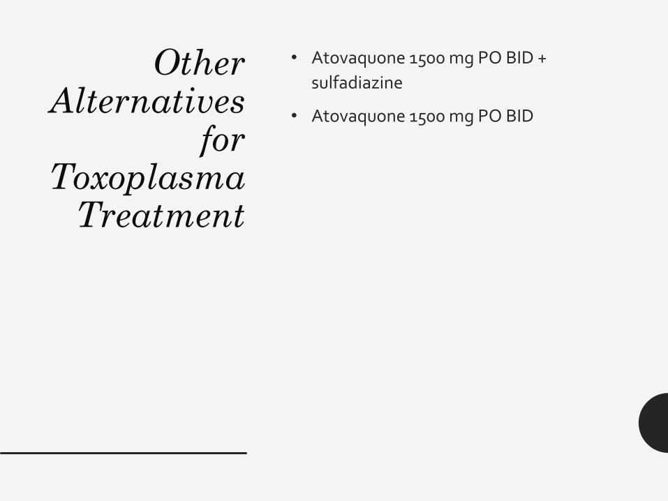
PML • Usually presents as slowly progressing
focal brain lesions, usually in frontal,
parietal, and cerebellar regions
• Do not have fevers/headaches
• Seizures occur in approximately 20%
• White matter lesions on T2 images, no
mass effect, rare to have contrast
enhancement

Diagnosis • Lumbar puncture:
– JC virus (causes PML), 70-90% sensitive
for this infection
– CMV, HSV, VZV PCRs
– HSV and VZV usually with abnormal CSF
(WBC)

Treatment • Specific therapies for viral encephalitis
(acyclovir, ganciclovir)
• Early HAART only treatment for PML
and HIV encephalopathy

OPHTHALMOLOGIC INFECTIONS

CMV Retinitis
• Occurs in patients with CD4 <100 cells/uL
• Commonly present with visual field loss, decreased visual acuity, floaters
• Characteristic perivascular white retinal infiltrates associated with retinal hemorrhage

Other Manifestations of CMV: GI
• May involve mouth, esophagus, stomach, colon, biliary
tract and pancreas
• Patients may present with odynophagia,
diarrhea/fever, colonic perforation, rarely mass lesions

Central Nervous
System Infections
• Encephalitis
– Subacute (3-4 weeks) progressive
disorientation, confusion, CN
palsies, nystagmus
– MRI shows periventricular
enhancement, diffuse white
matter changes
– Diagnosed with CMV PCR in CSF

Central Nervous
System Infections • CMV polyradiculopathy
– Progressive subacute flaccid
paralysis of lower extremities
– Associated with pain, parasthesias,
sphincter dysfunction
– Characteristic CSF: elevated protein,
low glucose, PMNs

CMV Pneumonia?
• Rare in the HIV population
• Usually reactivation in the setting of a
primary pathogen (e.g. PJP)
• Invasive disease uncommon, but
positive CMV PCR in BAL not
uncommon

Laboratory Diagnosis
• Culture
• Monoclonal antibody testing against CMV matrix protein pp65
• CMV DNA or RNA testing
• Histopathology
• Antibodies not useful (particularly if positive)

Diagnosis of CMV End-Organ Disease
•Relies on combination of clinical symptoms and histological findings from tissue biopsy and/or appearance
•Characteristic intranuclear and intracytoplasm inclusion bodies in infected cells (owl’s eye)
•Positive CMV PCR does not equal invasive disease

CMV PCR and pp65
• Can predict development of end-organ
disease as well as survival in patients
• However, a positive CMV PCR does not
mean that the patient has invasive
disease
• Need to obtain eye exam and evaluate
for colitis in HIV+ patients with CMV
viremia
• Not recommended to treat CMV viremia
without invasive disease
• Alternately, it is possible to have
invasive disease without a positive
serum CMV PCR, can use CMV PCR in
vitreous fluid

CMV Treatment
•Ganciclovir
–Major adverse effects of are neutropenia (extremely common)
and thrombocytopenia
–Valganciclovir (Valcyte) is the prodrug for ganciclovir
•Absolute oral bioavailability is approximately 60%
•FDA approved for Rx of non-sight threatening CMV retinitis
–Standard protocol is for 21 days

CMV Retinitis
Treatment
• Intravitreal ganciclovir and foscarnet
injections
• Previously intravitreal ganciclovir
implants, no longer commercially
available in US

Resistant CMV
• Foscarnet
• Cidofovir

Secondary Prophylaxis
• For retinitis, recommended to continue
900 mg/day valganciclovir until CD4
>100 cells/uL
• For colitis, secondary prophylaxis not
recommended unless recurrent or
severe episode
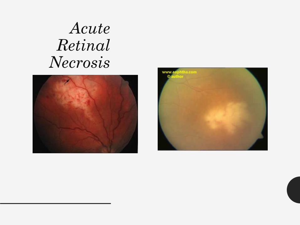
Acute Retinal
Necrosis

Treatment • Often caused by herpes simplex virus or
varicella zoster virus
• Can occur in immunocompetent
patients or at higher CD4 counts
• Inflammatory process
• Treatment: IV acyclovir + intravitreous
foscarnet, vitrectomy, laser, ?steroids

PORN
• Occurs in patients with CD4 <100 cells/uL
• Usually herpes zoster virus
• Minimal inflammation
• Multiple discrete peripheral lesions in outer retinal layer
• Rapid coalesence and full thickness retinal necrosis

Treatment – IV ganciclovir and/or foscarnet +
intravitreal ganciclovir/foscarnet twice
weekly
DIARRHEA IN HIV

Diarrhea in HIV+
• Evaluation for acute diarrhea similar to
that for non-immunocompromised
(stool culture, C. difficile toxins,
ova/parasite)
• For chronic diarrhea, also send
Cryptosporidium and Giardia antigens,
Microsporidia stain (modified
trichrome); consider medication-
induced diarrhea
• May need endoscopy if diagnosis
unclear

Cryptosporidium and
Microsporidia
•Cause chronic, watery, copious diarhea
•May cause cholangiopathy
•Diagnosis with ova/parasite exam, antigen test for Cryptosporidium
•Relatively refractory to treatment: need urgent consideration for HAART
•Strong anti-diarrheals indicated
•Can try nitazoxanide 100 mg PO BID x 2-4 weeks for cryptosporidiosis, albendazole 400 mg PO BID x 3 weeks for microsporidiosis

FEVER IN HIV+

Fever of Unknown Origin in
HIV
• Large differential diagnosis
• Send:
– Blood cultures
– Fungal blood cultures
– Fungal antibody complement fixation
panel
– AFB blood cultures
– Cryptococcal antigen
– Histoplasma antigen from urine
– Consider Bartonella testing

Mycobacterium avium
• Environmental organism found in soil, water
• Disseminated infection common in CD4 <50 cells/uL
• Fever, night sweats, wasting, diarrhea, abdominal pain, adenopathy, rarely HIV cholangiopathy
• Often causes pancytopenia and LFT abnormalities (obstructive pattern)

Diagnosis M. avium
• AFB blood cultures
• Isolation of MAI from respiratory and GI
tract often colonization

Treatment of M.avium
•Treatment: daily azithromycin +
ethambutol
•Some add rifabutin (watch out
for HIV drug interactions) in
severe disease or suspected
resistance (data from pre-
HAART era)
•12 months of therapy
•No signs or symptoms of MAC
•CD4 >100 cells/uL for at least 6
months

Prophylaxis of M.avium
•Primary prophylaxis with azithromycin
when CD4 < 50 cells/uL
•Recommended to start “after ruling out
disseminated MAC disease”
•Based on clinical assessment, consider
AFB blood cultures
•Alternative: rifabutin
•Can discontinue when CD4 > 100 cells/uL
for 3 months


Histoplasmosis • Caused by Histoplasma capsulatum
• Dallas is endemic area
• Symptoms include fever, adenopathy,
cough/shortness of breath,
pancytopenia
• CNS, GI, cutaneous involvement
• May present with septic-shock like
syndrome

Diagnosis of Histoplasmosis
• Urine Histoplasma antigen
• Fungal blood cultures, cultures of sterile
sites
• Fungal antibody by complement
fixation may be positive

Treatment of Histoplasmosis
•In severe disease, Ambisome
x 14 days, then itraconazole
•If mild disease, itraconazole
•Itraconazole solution is
preferred to capsules (poorly
absorbed)
–Check levels, preferred >1
mcg/mL
–Interacts with HIV medications

Prophylaxis • Primary prophylaxis consider if high risk
of occupational exposure and CD4 <150
cells/uL
• Secondary prophylaxis: 200 mg/day
itraconazole
• Can discontinue secondary prophylaxis
when: >1 year itraconazole, negative
fungal blood cultures and Histoplasma
antigen, CD4 >150 cells/uL, and ART > 6
months

Coccidioides • Not found in Dallas except for travelers
• Dissemination risk when CD4 <250
cells/uL
• Focal pneumonia, diffuse pulmonary
disease, meningitis, cutaneous disease,
lymph node, skin lesions
• Diagnosis with culture/biopsy of
specimen, complement fixation
CDC

Treatment • Mild infection: itraconazole or
fluconazole
• Severe infection: amphotericin B
• Meningitis: fluconazole, ?intrathecal
amphotericin
• Lifelong secondary prophylaxis if
meningitis; can discontinue in other
cases if CD4 >250 cells/uL


Bartonella • B. quintana (associated with with body
louse exposure) and B.henselae
(associated with cat exposure)
• Occurs when CD4 < 100 cells/uL
• Characteristic skin lesions can be
mistaken for KS
• Can cause FUO, culture negative
endocarditis

Bacillary Angiomatosis

Bacillary Peliosis Hepatis
• Multiple blood filled cavities throughout liver
• Usually asymptomatic but if severe can cause liver abnormalities, hemoperitoneum
• Occurs only with B.henselae

Diagnosis • Histopathology of skin lesion
– Modified silver stain (Warthin-Starry)
• Serology (~75% sensitivity)
• Culture
• PCR

Treatment • Erythromycin/azithromycin or
doxycycline for > 3 months
• Add rifamicin if severe infection
• Endocarditis: doxycycline + gentamicin
• No primary prophylaxis recommended
• No secondary prophylaxis
recommended unless relapsing disease
(doxycycline or macrolide)

Kaposi sarcoma
• Skin lesions
• Oral lesions
• Disseminated disease to GI tract and
lungs

Human Herpesvirus-8
Disease
• Highest prevalence in MSM, sub-
Saharan Africa
• Associated with Kaposi sarcoma,
primary effusion lymphoma,
multicentric Castleman disease
(angiofollicular lymph node
hyperplasia), KSHV inflammatory
cytokine syndrome (KICS)
• Usually occur when CD4 <200 cells/uL
but can occur at higher CD4

Lymphoproliferative
Disorders
• Primary effusion lymphoma: usually
presents with pleural, pericardial or
abdominal effusion
• Multicentric Castleman: generalized
lymphadenopathy, fever, peripheral
neuropathy. Can try antiviral therapy
for this (ganciclovir)

Diagnosis • Screening for HHV-8 by PCR in
peripheral blood not indicated
• Biopsy

Treatment of Kaposi sarcoma
• Institution of HAART alone often results
in regression of lesions
• Local radiation
• Intralesional vincristine or interferon-
alpha
• For disseminated disease, doxorubicin,
paclitaxel
• Antivirals not recommended
• Steroids often exacerbate this condition

Questions? • Call Amelia Court 214-590-2865 or 214-
590-4418, or page me (214-786-5293)
with questions


![[Micro] opportunistic mycosis](https://img.pdfslide.net/doc/110x75/55d6fc6bbb61ebfa2a8b47ec/micro-opportunistic-mycosis.jpg)