Embed Size (px)
Citation preview
i
AN INVESTIGATION OF THE NEUROPROTECTIVE EFFECTS OF PALM
VITAMIN E TOCOTRIENOLS USING WHITE MATTER LESIONS AS THE
HUMAN STUDY MODEL
by
YOGHESWARAN GOPALAN
Thesis submitted in fulfilment of the requirements for the degree of Doctor of Philosophy
September 2014
ii
DEDICATIONS
This thesis is dedicated to the Almighty God for giving me the strength to complete my
PhD, my beloved wife, Dr. Shubashini Gnanasan for her continuous support and
inspiration, my adorable sons, Prakaladhan and Sithaarthan who are always there to
cheer me up and to my wonderful parents, Mr. L. Gopalan and Madam M. Sathiamah for
their everlasting encouragement and blessings on me. Thank you.
iii
ACKNOWLEDGEMENTS
There are so many people without whom this project would not have been possible who
I would like to thank.
I would like to express my deepest and heartfelt gratitude to my supervisor, Professor
Dr. Yuen Kah Hay who has given me the opportunity to pursue this project under his
supervision. Indeed, I have gained tremendously in terms of academic excellence and
persistence to achieve goals under his guidance which I will treasure throughout my life.
My sincere appreciation to my co-supervisor Dr. Nurzalina Abdul Karim Khan for her
constant assistance, suggestions and encouragement to complete my project.
I owe a deep gratitude to Professor Dr. Ibrahim Lutfi Shuaib. Without his support, I
would not have been able to complete this study. I would also like to extend my heartfelt
gratitude to Dr. Mukhtar Alam Ansari and the staff of Advanced Medical and Dental
Institute, USM and Kepala Batas hospital for assisting me during the MRI imaging and
data collection process. Also not forgetting, Mr. Ong Kok Haur for the excellent work on
the MRI analysis.
I would like thank the Malaysian Palm Oil Board for the financial support provided to
conduct this project. Similar appreciation goes to Universiti Teknologi MARA for
providing me the scholarship to pursue my PhD. They have allowed me to fulfil my
dream by providing the much needed financial support and time that was required to
complete this study.
iv
I take this opportunity to thank my family members and friends for their abundance of
love, encouragement and support. My family has and always will be the greatest pillar of
support in my life. My colleagues who have been my best critiques, they were ever
willing to help and their assistance has been invaluable. I would like to acknowledge all
of them for their support, in particular Dr. Wong Jia Woei, Dr. Enrico Magosso and Mr.
Tommy Julianto. Thank you.
Last but not least, I would like to acknowledge all my dear friends Dr. Ng Bee Hong, Dr.
Sandy Ong, Dr. Jiayuddin Khan, Dr. Choon Wai Yee, Dr. Lim Sheau Chin, Dr. Kam Li
Ying, Dr. Mahmathi Karuppannan, Goh Song Thai, Cheah Mei Mei, Lim Ai Boey,
Cheah Phaik Chin, Siew siew, Belle, Mei Ching, Gan and the rest of the lab staff for
their constant support and encouragement and their hospitality throughout the study.
Finally, a deep appreciation goes out to all the participants of this study. Without them
this study would not have been possible.
v
TABLE OF CONTENT
DEDICATIONS ............................................................................................................................. ii
ACKNOWLEDGEMENTS .......................................................................................................... iii
TABLE OF CONTENT ..................................................................................................................v
LIST OF TABLE ............................................................................................................................x
LIST OF FIGURE ......................................................................................................................... xi
ABBREVIATIONS ..................................................................................................................... xii
PUBLICATIONS ........................................................................................................................ xiv
ABSTRAK .................................................................................................................................. xvi
ABSTRACT .............................................................................................................................. xviii
1. CHAPTER 1: INTRODUCTION ...........................................................................................1
1.1. White Matter Lesion .......................................................................................................1
1.1.1. Introduction .................................................................................................................1
1.1.2. Etiology of White Matter Lesion ................................................................................2
1.1.3. Clinical Implication of White Matter Lesions .............................................................4
1.2. Vitamin E ........................................................................................................................6
1.2.1. Tocotrienol ..................................................................................................................6
1.2.2. Neuroprotective Properties ..........................................................................................8
1.2.3. Anti-atherogenic Properties ........................................................................................9
1.2.4. Hypocholesterolemic effect of tocotrienols ..............................................................10
1.2.5. Hypo-apolipoprotein B effect of tocotrienols ...........................................................12
1.2.6. Hypo- lipoprotein Lp (a) effect of tocotrienols .........................................................13
1.2.7. Antioxidant properties of tocotrienols.......................................................................14
1.2.8. Inhibition of adhesion molecule expression and monocytic cell adherence by
tocotrienols ................................................................................................................................15
1.3. Clinical Evaluations: Basic Principles of Magnetic Resonance Imaging of the Brain .16
1.4. Scope of the study .........................................................................................................18
vi
1.4.1. Primary Objective .....................................................................................................18
1.4.2. Secondary Objectives ................................................................................................18
2. CHAPTER 2: MAGNETIC RESONANCE ANGIOGRAPHY OF THE CAROTID
ARTERY STENOSIS AMONG MILDLY HYPERCHOLESTEROLEMIC VOLUNTEERS ...19
2.1. Introduction ...................................................................................................................19
2.2. Materials and methods ..................................................................................................21
2.2.1. Study Population .......................................................................................................21
2.2.2. MRA Imaging Procedures .........................................................................................22
2.2.3. Image Analysis ..........................................................................................................23
2.2.4. Statistical Analysis ....................................................................................................24
2.3. Results ...........................................................................................................................25
2.3.1. Internal carotid artery stenosis ..................................................................................25
2.3.2. Baseline Characteristics ............................................................................................26
2.3.3. Demographic and disease profile ..............................................................................29
2.4. Discussion .....................................................................................................................30
2.5. Conclusion ....................................................................................................................31
3. CHAPTER 3: PREVALENCE OF WHITE MATTER LESION (WML) IN A LOCAL
HYPERCHOLESTEROLEMIC POPULATION IN NORTH –WEST PENINSULAR
MALAYSIA..................................................................................................................................32
3.1. Introduction ...................................................................................................................32
3.2. Methods .........................................................................................................................34
3.2.1. Study population .......................................................................................................34
3.2.2. MRI scanning protocol ..............................................................................................34
3.2.3. White matter lesion rating scale ................................................................................34
3.2.4. Statistical Analysis ....................................................................................................36
3.3. Results ...........................................................................................................................37
3.4. Discussion .....................................................................................................................46
3.5. Conclusion ....................................................................................................................48
vii
4. CHAPTER 4: THE NEUROPROTECTIVE EFFECTS OF PALM VITAMIN E
TOCOTRIENOLS IN WHITE MATTER LESION NAIVE (MRI -VE) VOLUNTEERS ..........49
4.1. Introduction ...................................................................................................................49
4.2. Methods .........................................................................................................................52
4.2.1. Study population .......................................................................................................52
4.2.2. MRI scanning protocol ..............................................................................................53
4.2.3. White matter lesion volumetric measurement ...........................................................53
4.2.4. Randomization and Treatment ..................................................................................54
4.2.5. Clinical evaluation ....................................................................................................55
4.2.6. Statistical Analysis ....................................................................................................56
4.3. Results ...........................................................................................................................57
4.3.1. Volunteers flow and follow up ..................................................................................57
4.3.2. Baseline characteristics and disease profile ..............................................................57
4.3.3. WML development in MRI –ve volunteers ..............................................................60
4.3.4. Clinical parameters ....................................................................................................62
4.3.5. Compliance ...............................................................................................................65
4.3.6. Adverse Events..........................................................................................................66
4.4. Discussion .....................................................................................................................67
4.5. Conclusion ....................................................................................................................69
5. CHAPTER 5: NEUROPROTECTIVE EFFECTS OF PALM VITAMIN E
TOCOTRIENOLS USING WHITE MATTER LESIONS AS THE HUMAN STUDY MODEL
70
5.1. Introduction ...................................................................................................................70
5.2. Methods .........................................................................................................................74
5.2.1. Study Protocol ...........................................................................................................74
5.2.2. Study Population .......................................................................................................75
5.2.3. Clinical Evaluation Procedures .................................................................................77
5.2.4. MRI Imaging Parameters ..........................................................................................78
viii
5.2.5. White matter lesion volumetric measurement ...........................................................79
5.2.6. Outcome measures ....................................................................................................80
5.2.7. Statistical Analysis ....................................................................................................81
5.3. Results ...........................................................................................................................83
5.3.1. Volunteers flow and follow-up .................................................................................83
5.3.2. Baseline Characteristics ............................................................................................85
5.3.3. WML volume changes ..............................................................................................87
5.3.4. Mean volume changes of WML for subjects with hypertension, diabetes or both
hypertension and diabetes. ........................................................................................................90
5.3.5. Clinical Parameters ...................................................................................................92
5.3.6. Compliance ...............................................................................................................94
5.3.7. Adverse Events..........................................................................................................95
5.4. Discussion .....................................................................................................................96
5.5. Conclusion ....................................................................................................................98
6. CHAPTER 6: SUMMARY AND GENERAL CONCLUSIONS .......................................100
7. CHAPTER 7: SUGGESTIONS FOR FURTHER RESEARCH ........................................104
REFERENCES............................................................................................................................107
APPENDICES ............................................................................................................................125
Appendix 1- Mean WML volume changes (Intention to treat analysis) .....................................126
Appendix 2: Mean WML volume changes (Per protocol analysis) ............................................129
Appendix 4 - Plasma Tocotrienol Concentration in White Matter Lesion (MRI+ve) volunteers
.....................................................................................................................................................133
Appendix 5 - Plasma Tocotrienol Concentration in White Matter Lesion Naïve (MRI-ve)
Volunteers ...................................................................................................................................136
Appendix 6- Ethics Approval......................................................................................................139
Appendix 7- Informed consent (Bahasa Malaysia) .....................................................................143
Appendix 8- Informed consent (English) ....................................................................................152
Appendix 9: Reference Range of Biochemistry Parameters .......................................................161

ix
Appendix 10: Previva certificate .................................................................................................163
Appendix 11: Research publication ............................................................................................165
x
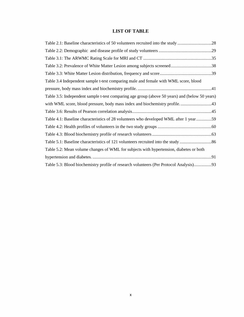
LIST OF TABLE
Table 2.1: Baseline characteristics of 50 volunteers recruited into the study ...............................28
Table 2.2: Demographic and disease profile of study volunteers ................................................29
Table 3.1: The ARWMC Rating Scale for MRI and CT ..............................................................35
Table 3.2: Prevalence of White Matter Lesion among subjects screened .....................................38
Table 3.3: White Matter Lesion distribution, frequency and score ...............................................39
Table 3.4 Independent sample t-test comparing male and female with WML score, blood
pressure, body mass index and biochemistry profile. ...................................................................41
Table 3.5: Independent sample t-test comparing age group (above 50 years) and (below 50 years)
with WML score, blood pressure, body mass index and biochemistry profile. ............................43
Table 3.6: Results of Pearson correlation analysis ........................................................................45
Table 4.1: Baseline characteristics of 28 volunteers who developed WML after 1 year ..............59
Table 4.2: Health profiles of volunteers in the two study groups .................................................60
Table 4.3: Blood biochemistry profile of research volunteers ......................................................63
Table 5.1: Baseline characteristics of 121 volunteers recruited into the study .............................86
Table 5.2: Mean volume changes of WML for subjects with hypertension, diabetes or both
hypertension and diabetes. ............................................................................................................91
Table 5.3: Blood biochemistry profile of research volunteers (Per Protocol Analysis) ................93
xi
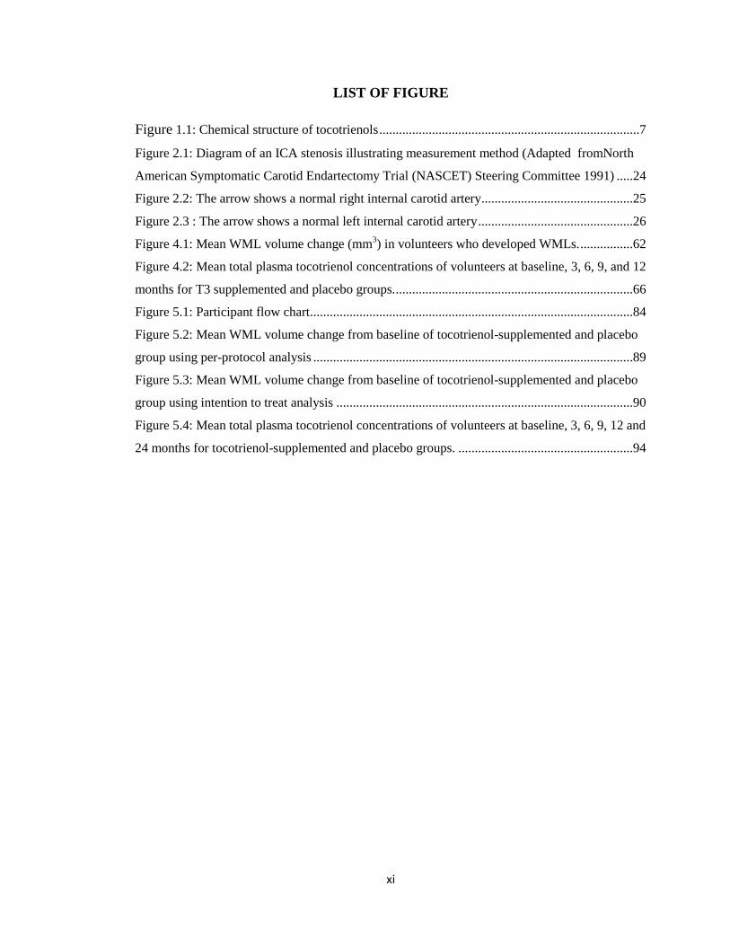
LIST OF FIGURE
Figure 1.1: Chemical structure of tocotrienols ...............................................................................7
Figure 2.1: Diagram of an ICA stenosis illustrating measurement method (Adapted fromNorth
American Symptomatic Carotid Endartectomy Trial (NASCET) Steering Committee 1991) .....24
Figure 2.2: The arrow shows a normal right internal carotid artery..............................................25
Figure 2.3 : The arrow shows a normal left internal carotid artery ...............................................26
Figure 4.1: Mean WML volume change (mm3) in volunteers who developed WMLs. ................62
Figure 4.2: Mean total plasma tocotrienol concentrations of volunteers at baseline, 3, 6, 9, and 12
months for T3 supplemented and placebo groups. ........................................................................66
Figure 5.1: Participant flow chart ..................................................................................................84
Figure 5.2: Mean WML volume change from baseline of tocotrienol-supplemented and placebo
group using per-protocol analysis .................................................................................................89
Figure 5.3: Mean WML volume change from baseline of tocotrienol-supplemented and placebo
group using intention to treat analysis ..........................................................................................90
Figure 5.4: Mean total plasma tocotrienol concentrations of volunteers at baseline, 3, 6, 9, 12 and
24 months for tocotrienol-supplemented and placebo groups. .....................................................94
xii
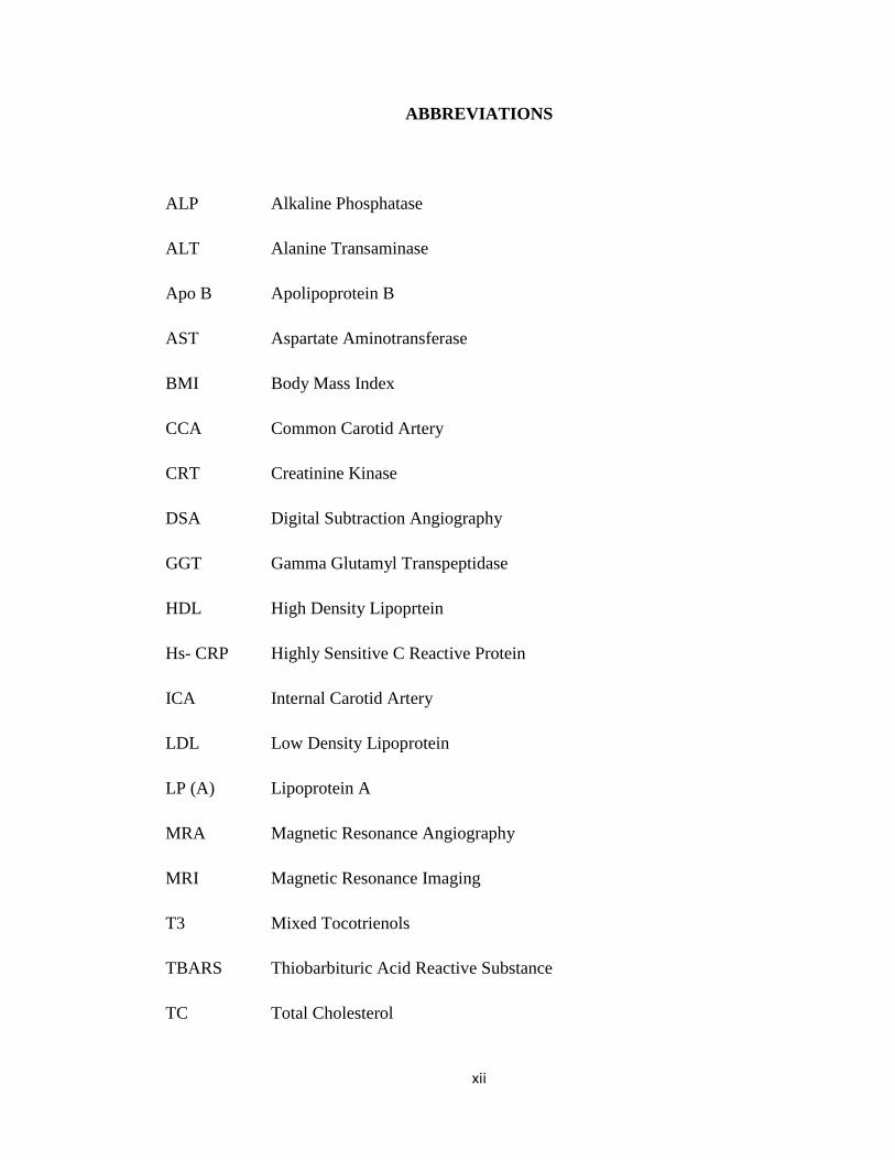
ABBREVIATIONS
ALP Alkaline Phosphatase
ALT Alanine Transaminase
Apo B Apolipoprotein B
AST Aspartate Aminotransferase
BMI Body Mass Index
CCA Common Carotid Artery
CRT Creatinine Kinase
DSA Digital Subtraction Angiography
GGT Gamma Glutamyl Transpeptidase
HDL High Density Lipoprtein
Hs- CRP Highly Sensitive C Reactive Protein
ICA Internal Carotid Artery
LDL Low Density Lipoprotein
LP (A) Lipoprotein A
MRA Magnetic Resonance Angiography
MRI Magnetic Resonance Imaging
T3 Mixed Tocotrienols
TBARS Thiobarbituric Acid Reactive Substance
TC Total Cholesterol
xiii
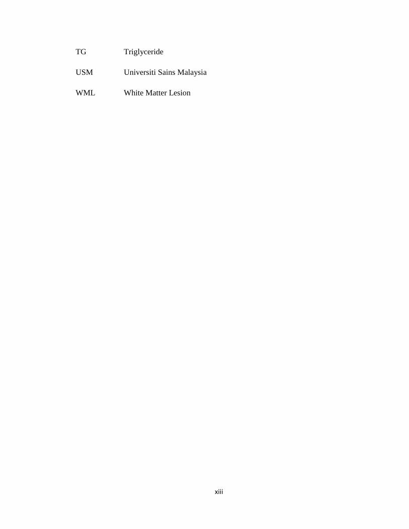
TG Triglyceride
USM Universiti Sains Malaysia
WML White Matter Lesion
xiv
PUBLICATIONS
Gopalan, Y., Shuaib, I.L., Magosso, E., Ansari, M.A., Rizal, M.A.B., Wong J.W.,
Khan, N.A.K., Leong, W.C., Sundram,K., Ng B.H., Chinna, K., & Yuen, K.H.
(2014).Clinical Investigation of the Protective Effects of Palm Vitamin E
Tocotrienols on Brain White Matter.Stroke; 45: 1422 – 1428.
Gopalan, Y., Magosso, E., Shuaib, I.L., Ansari, M.A., Rizal, M.A.B., Khan, N.A.K.,
Wong J.W., Ng B.H., Nesaretnam,K., Sundram,K, Chinna, K., & Yuen, K.H.
(2012). Neuroprotective Effects of Palm Vitamin E Tocotrienols in Brain White
Matter Lesion : Evidence from A Double Blind Placebo – Controlled Clinical
Trial. Neuroepidemiology, 39:193.
Gopalan, Y., Magosso, E., Shuaib, I.L., Ansari, M.A., Rizal, M.A.B., Khan, N.A.K.,
Wong J.W., Ng B.H., Nesaretnam,K., Sundram,K & Yuen, K.H. (2011).
Neuroprotective Effects of Tocotrienols in Brain White Matter Lesion :
Preliminary Findings From A Clinical Trial. Paper presented at Palm
International Nutra-Cosmeceutical Conference, Kuala Lumpur, Malaysia. (Poster
presentation)
Gopalan, Y., Magosso, E., Shuaib, I.L., Ansari, M.A., Rizal, M.A.B., Khan, N.A.K.,
Wong J.W., Ng, B.H., Nesaretnam,K., Sundram,K & Yuen, K.H. (2011). An
Insight into The Neuroprotective Effect of Tocotrienols Among White Matter
Lesion Naïve Volunteers: A Preliminary Findings From A Double Blind Placebo
Controlled Trial. Paper presented at 22nd
Annual Scientific Meeting of the
Malaysian Society of Neurosciences, Kuala Terengganu, Terengganu, Malaysia.
(Poster presentation)
xv
Magosso E, Gopalan Y, Yuen K.H. (2009). Antioxidant Supplements for Prevention of
Mortality in Healthy Participants and Patients with Various Diseases (review) –
Letter to the Editor. The Cochrane Library, 1:259-262.
Gopalan, Y., Magosso, E., Shuaib, I.L., Ansari, M.A., Rizal, M.A.B., Khan, N.A.K.,
Wong J.W., Ng B.H., Nesaretnam,K., Sundram,K & Yuen, K.H. (2009). Double
Blind Placebo Controlled Study On The Neuroprotective And Anti -Atherogenic
Effects Of Palm Tocotrienol Rich Fraction (Palm Vitamin E). Paper presented at
7th
COSTAM / SFRR (ASIA /MALAYSIA) International Workshop and 4th
biennial meeting of SFRR ASIA, Langkawi, Malaysia (Poster presentation).
Gopalan, Y. (2009). Prevalence of White Matter Lesion Among Adults In The North
West Region Of Peninsular Malaysia. Paper presented at 9th Asian Conference
on Clinical Pharmacy, Seoul, Korea. (Oral presentation)
xvi
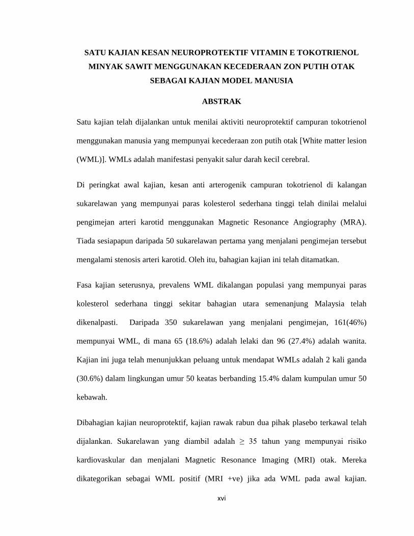
SATU KAJIAN KESAN NEUROPROTEKTIF VITAMIN E TOKOTRIENOL
MINYAK SAWIT MENGGUNAKAN KECEDERAAN ZON PUTIH OTAK
SEBAGAI KAJIAN MODEL MANUSIA
ABSTRAK
Satu kajian telah dijalankan untuk menilai aktiviti neuroprotektif campuran tokotrienol
menggunakan manusia yang mempunyai kecederaan zon putih otak [White matter lesion
(WML)]. WMLs adalah manifestasi penyakit salur darah kecil cerebral.
Di peringkat awal kajian, kesan anti arterogenik campuran tokotrienol di kalangan
sukarelawan yang mempunyai paras kolesterol sederhana tinggi telah dinilai melalui
pengimejan arteri karotid menggunakan Magnetic Resonance Angiography (MRA).
Tiada sesiapapun daripada 50 sukarelawan pertama yang menjalani pengimejan tersebut
mengalami stenosis arteri karotid. Oleh itu, bahagian kajian ini telah ditamatkan.
Fasa kajian seterusnya, prevalens WML dikalangan populasi yang mempunyai paras
kolesterol sederhana tinggi sekitar bahagian utara semenanjung Malaysia telah
dikenalpasti. Daripada 350 sukarelawan yang menjalani pengimejan, 161(46%)
mempunyai WML, di mana 65 (18.6%) adalah lelaki dan 96 (27.4%) adalah wanita.
Kajian ini juga telah menunjukkan peluang untuk mendapat WMLs adalah 2 kali ganda
(30.6%) dalam lingkungan umur 50 keatas berbanding 15.4% dalam kumpulan umur 50
kebawah.
Dibahagian kajian neuroprotektif, kajian rawak rabun dua pihak plasebo terkawal telah
dijalankan. Sukarelawan yang diambil adalah ≥ 35 tahun yang mempunyai risiko
kardiovaskular dan menjalani Magnetic Resonance Imaging (MRI) otak. Mereka
dikategorikan sebagai WML positif (MRI +ve) jika ada WML pada awal kajian.
xvii
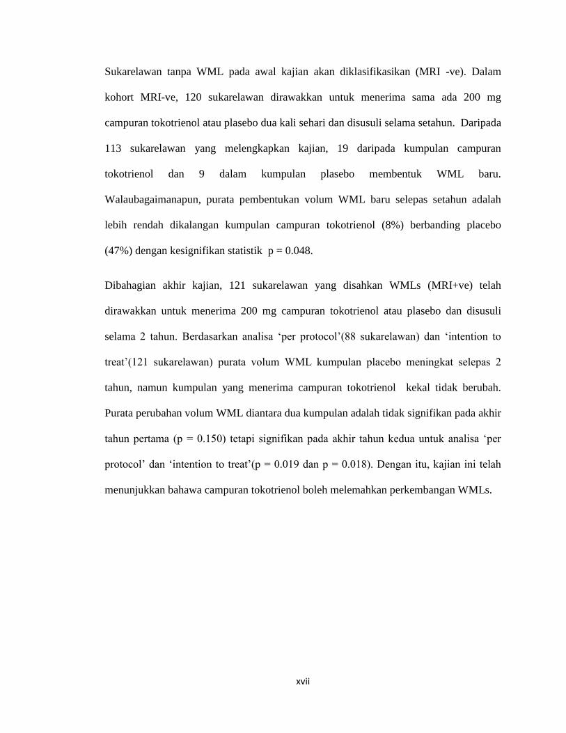
Sukarelawan tanpa WML pada awal kajian akan diklasifikasikan (MRI -ve). Dalam
kohort MRI-ve, 120 sukarelawan dirawakkan untuk menerima sama ada 200 mg
campuran tokotrienol atau plasebo dua kali sehari dan disusuli selama setahun. Daripada
113 sukarelawan yang melengkapkan kajian, 19 daripada kumpulan campuran
tokotrienol dan 9 dalam kumpulan plasebo membentuk WML baru.
Walaubagaimanapun, purata pembentukan volum WML baru selepas setahun adalah
lebih rendah dikalangan kumpulan campuran tokotrienol (8%) berbanding placebo
(47%) dengan kesignifikan statistik p = 0.048.
Dibahagian akhir kajian, 121 sukarelawan yang disahkan WMLs (MRI+ve) telah
dirawakkan untuk menerima 200 mg campuran tokotrienol atau plasebo dan disusuli
selama 2 tahun. Berdasarkan analisa „per protocol‟(88 sukarelawan) dan „intention to
treat‟(121 sukarelawan) purata volum WML kumpulan placebo meningkat selepas 2
tahun, namun kumpulan yang menerima campuran tokotrienol kekal tidak berubah.
Purata perubahan volum WML diantara dua kumpulan adalah tidak signifikan pada akhir
tahun pertama (p = 0.150) tetapi signifikan pada akhir tahun kedua untuk analisa „per
protocol‟ dan „intention to treat‟(p = 0.019 dan p = 0.018). Dengan itu, kajian ini telah
menunjukkan bahawa campuran tokotrienol boleh melemahkan perkembangan WMLs.
xviii
AN INVESTIGATION OF THE NEUROPROTECTIVE EFFECTS OF PALM
VITAMIN E TOCOTRIENOLS USING WHITE MATTER LESIONS AS THE
HUMAN STUDY MODEL
ABSTRACT
A study was conducted to evaluate the neuroprotective activity of mixed tocotrienols
using human volunteers with white matter lesion (WML). WMLs are regarded as
manifestations of cerebral small vessel disease.
Initially, the anti atherogenic effect of mixed tocotrienols was also evaluated in mildly
hypercholesterolemic volunteers via imaging the carotid arteries using Magnetic
Resonance Angiography (MRA). Of the first 50 consecutive volunteers who undergone
the carotid artery imaging, none of them were detected having carotid artery stenosis and
hence this part of the study was discontinued.
In the next phase of the study, the prevalence of white matter lesion (WML) in a local
hypercholesterolemic population in north –west peninsular Malaysia was determined. Of
the 350 research volunteers who were imaged, 161 (46%) of them had WMLs, of which
65 (18.6%) were males and 96 (27.4%) females. The study also demonstrated that the
chances of developing WMLs doubled (30.6%) in the age group of above 50 compared
to 15.4% in the age group of below 50.
In the neuroprotective part of the study, a randomized double-blind placebo-controlled
trial was conducted. Volunteers recruited were ≥ 35 years with cardiovascular risk
factors and undergo Magnetic Resonance Imaging (MRI) of the brain. They were
categorized as WML positive (MRI +ve) if they were present with WML at baseline.
xix
Volunteers without WMLs at baseline were classified as (MRI -ve). In the MRI –ve
cohort, 120 volunteers were randomized to receive either 200 mg mixed tocotrienols or
placebo twice daily and were followed up for 1 year. Out of 113 volunteers who
completed the study, 19 in the mixed tocotrienols and 9 in the placebo group developed
new WMLs. However, the mean volume of the WMLs developed after 1 year was lesser
in the mixed tocotrienol treated group (8%) compared to placebo (47%) with a statistical
significance of p = 0.048.
In the final part of the study, 121 volunteers with MRI confirmed WMLs (MRI +ve)
were randomized to receive 200 mg mixed tocotrienols or placebo twice daily and were
followed up for 2 years. According to per protocol (88 volunteers) and intention-to-treat
(121 volunteers) analyses, the mean WML volume of the placebo group increased after 2
years, whereas that of the mixed tocotrienol supplemented group remained essentially
unchanged. The mean WML volume change between the two groups was not
significantly different (p = 0.150) at the end of 1 year but was significant at the end of 2
years for both per protocol and intention-to-treat analyses (p = 0.019 and p = 0.018).
Thus, the present study found that mixed tocotrienols can attenuate the progression of
WMLs.
1
1. CHAPTER 1: INTRODUCTION
1.1. White Matter Lesion
1.1.1. Introduction
White matter lesions (WMLs) are areas of increased signal intensity detected on T2-
weighted magnetic resonance imaging (MRI) scans. These lesions are common among
older adults (Brickman, Schupf, Manly, & et al., 2008)(F.-E. de Leeuw et al., 2001) and
are thought to reflect small vessel vascular disease and neurodegeneration of nerve
bundles. WMLs are frequently observed in individuals with advanced age,
hypertension, prior ischemic stroke and other cerebrovascular risk factors (Hénon,
Godefroy, Lucas, Pruvo, & Leys, 1996; Kalaria & Erkinjuntti, 2006; Leys et al., 1999;
Räihä, Tarvonen, Kurki, Rajala, & Sourander, 1993; Wiszniewska, Devuyst,
Bogousslavsky, Ghika, & van Melle, 2000). The white matter of the brain accounts for
the 60% of the total brain volume. It includes major commissural tracts, the cortical
association fibers, and all the cortical afferent and efferent fibers. White matter consists
of nerve fibers, supporting cells, interstitial space and vascular structures. Axons are
major component of white matter and it is enveloped by myelin with two types of
neuroglia; oligodendrocytes and astrocytes. Myelin acts as an insulator of axons and its
structure facilitates rapid transmission of impulses (Valk & van der Knaap, 1989). The
role of white matter is in information transmission to connect various grey matters
(neuron bodies) which is mainly responsible for information processing.
2
1.1.2. Etiology of White Matter Lesion
Based on clinicopathological studies, WMLs can be classified into three different
categories which includes punctate, periventricular, and confluent WMLs and showed
that at least one of these is due to small vessel disease (Fazekas et al., 1993 ). This
affects arteries of 150μm in diameter and is generally associated with hypertension,
diabetes, or both. The major vascular pathological finding is the one that Fisher in the
1950s termed “segmental arterial wall disorganization” (Fisher, 1982) also widely
known by others as lipohyalinosis (Ogata, 1999), where the loss of the arterial
architecture consists of whorls, tangles or wisps of more or less fine connective tissue
that entirely replaces the vessel wall and obliterates the normal vascular coats. The
general outline of the vessel in less destructive lesions is preserved and the disintegrating
wall consists in a loose meshwork of collagenous strands separated by empty interstitial
clefts (Fisher, 1968). The foremost consequences of lipohyalinosis are small-vessel wall
thickening and luminal narrowing which leads to arteriolosclerosis (Leonardo Pantoni &
Julio H. Garcia, 1997; Wardlaw, Sandercock, Dennis, & Starr, 2003). The axonal
damage that occurs within the confluent lesions is however variable, which involves
demyelination with preservation of axonal integrity to complete axonal disruption with
loss of axonal function (Fazekas et al., 1988). As a result of the degeneration of the
neurons, axonal disruption leads to functional loss of the affected network, while
demyelination causes only slowing of stimulus speed conduction, hence at least in some
cases WMLs may lead to reduced efficiency of the affected network, but with preserved
function. On the other hand, imaging-pathological studies have shown that punctate
lesions can be related with variable pathological findings ranging from no detectable
3
pathology at all to enlargement of perivascular spaces (Fazekas, et al., 1993 ). In the case
of discontinuation of the ependymal lining which results in permeation of water into the
axons and chronic edema of the white matter adjacent to the ventricles, the condition is
known as periventricular caps and halo but with no axonal damage (Fazekas, et al., 1993
). Reduced blood flow was also evident in WMLs. Cerebral blood flow was lower in
WMH areas relative to normal appearing white matter, which in turn, was lower than
grey matter. Regions with consistently lower cerebral blood flow across individuals were
more likely to appear as WMLs (Brickman et al., 2009).

4
1.1.3. Clinical Implication of White Matter Lesions
White matter lesions are more common and widely detectable in patients with
cardiovascular risk factors and symptomatic cerebrovascular disease (Launer, 2004). The
presence of WMLs can be attributed to several factors. The Rotterdam Scan Study
(Sarah E. Vermeer et al., 2003) and Sydney Stroke Study (Wen & Sachdev, 2004)
documented that individuals with white matter lesions have an increased risk of clinical
stroke. In addition to that, larger white matter lesions can actually represent subclinical
brain infarct (Marshall, Bradley, Marshall, Bhoopat, & Rhodes, 1988). Subcortical silent
brain infarction which was presented as periventricular hyperintensity and focal WMLs
is associated with the presence cardiovascular risk factors such as hypertension, diabetes
and retinal artery sclerosis (Kobayashi, Okada, Koide, Bokura, & Yamaguchi, 1997).
Incidence of severe WMLs at baseline is an independent predictor of future stroke while
patients with initially mild WML may develop subsequent stroke as the WML
progresses (Yamauchi, Fukuda, & Oyanagi, 2002). Similar findings were also reported
that cerebral WMLs predict both ischemic strokes and myocardial infarctions in patients
with established atherosclerotic disease (Gerdes et al., 2006). Silent brain infarcts,
marked periventricular hyperintensity and distinct subcortical white matter lesions are
important risk factors of clinical stroke. Moreover, silent brain infarcts and marked
periventricular hyperintensities increase the risk of mortality (Bokura et al., 2006).
WMLs also contribute to the cognitive impairment of the patients. In the Rotterdam
Study (M. M. B. Breteler et al., 1994) elderly individuals with WMLs were associated
with lower scores on test of cognitive function and were significantly associated with
subjective mental decline. WMLs and periventricular hyperintensity were significantly
5
more extensive in dementia with lewy bodies than in controls in patients with
Alzheimers and vascular dementia. Presence of frontal WMLs was associated with
higher depression scores (Barber et al., 1999). Severity of dementia is directly correlated
with the amount and size of WML in the periventricular and subcortical regions of the
brain (Targosz-Gajniak, Siuda, Ochudło, & Opala, 2009b).
The above data clearly demonstrated that white matter hyperintensities as detected on
MRI is closely related to vascular events of the brain and in certain instances represent
subclinical infarcts. Moreover, the degenerative changes of the small vessel salso
contribute towards the mental and cognitive decline of the patients.
6
1.2. Vitamin E
1.2.1. Tocotrienol
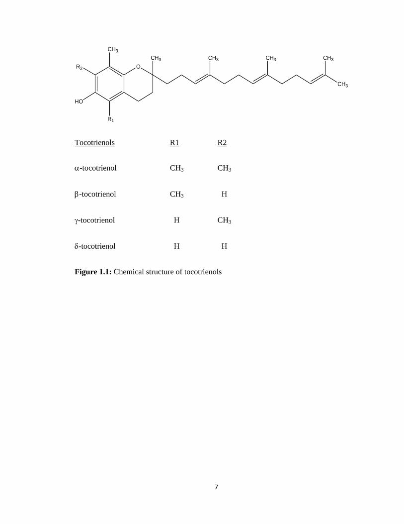
Vitamin E consists of eight isomers comprising four tocopherol and four tocotrienol
isomers, which share similar structural features of a chroman head and a 16-carbon
phytyl chain. Both tocopherols and tocotrienols are designated as , , and ,
depending on the number and positions of methyl groups on the chroman ring (Qureshi
& Qureshi, 1993). The difference between tocopherols and tocotrienols lie mainly in the
former having a saturated phytyl chain, while the latter is unsaturated, with 3 double
bonds at 3', 7' and 11' positions (A. Kamal-Eldin & L. A. Appelqvist, 1996).Figure 1.1
shows the general structure of α - tocotrienol. While tocopherols are generally present in
nuts and common vegetable oils, natural sources of tocotrienols are quite limited, with
palm oil and rice bran oil containing the highest concentrations of tocotrienols in nature
(Tan, 1989).
7
O
CH3
HO
R1
R2
CH3 CH3 CH3
CH3
CH3
Tocotrienols R1 R2
-tocotrienol CH3 CH3
-tocotrienol CH3 H
-tocotrienol H CH3
-tocotrienol H H
Figure 1.1: Chemical structure of tocotrienols
8
1.2.2. Neuroprotective Properties
Much investigation has been carried out to study the neuroprotective properties of
tocotrienols. Of special significance is the study by (Khanna et al., 2005) which
demonstrated that tocotrienols introduced into a neuronal cell at nanomolar
concentrations could prevent glutamate-induced death of neuron cells in mice. The
underlying mechanism behind the protective properties is through the inhibition of c-Src
kinase (Sen, Khanna, Roy, & Packer, 2000)and 12-lipoxygenase (Khanna et al., 2003). It
was also shown that the degeneration of the nerve cells was due to induced stress
mimicking clinical situations such as ischemia. Therefore, the findings of Khanna et al.
(2005) implicated that tocotrienols might have a role in the prevention of degenerative
diseases involving the nervous system. Khanna et al. (2005) showed that tocotrienols
have in vitro neuroprotective properties. In addition, tocotrienol-supplemented rats
showed more protection against stroke-inducedinjury compared with matched controls
(Khanna, et al., 2005). In another study, mongrel canines fed with tocotrienol-enriched
supplementation significantly attenuated ischemic stroke induced lesion volume.
Furthermore, it prevented loss of white matter fiber tract connectivity, improved
cerebrovascular collateral circulation to the ischemic mid cerebral artery territory during
mid cerebral artery occlusion(Rink et al., 2011). In another study, subjects supplemented
with self emulsifying preparation of tocotrienol rich vitamin E showed a trend towards
improvement of arterial compliance which would enable better elasticity and
sustainability of the blood vessels in the event of stroke (Rasool, Rahman, Yuen, &
Wong, 2008).
9
1.2.3. Anti-atherogenic Properties
The development of atherosclerosis involves a multitude of factors both genetic and
environmental in nature and these include elevated levels of cholesterol especially LDL,
apolipoprotien B and lipoprotein Lp(a), oxidation of LDL-cholesterol, adhesion
molecule expression and monocytic cell adherence. The anti-atherogenic activity of -
tocopherol has been linked to its ability to prevent the oxidation of LDL (Esterbauer,
1991; Salonen, Yia-Hertuala, & Yamamoto, 1992). Nevertheless, this theory remains
controversial with some studies failing to show association between plasma -
tocopherol levels and reduced mortality from cardiovascular disease(Hense, Stender,
Bors, & Keil, 1993). Despite the numerous studies carried out on -tocopherol, the
relationship between tocotrienols and atherosclerosis was investigated only recently.
These studies demonstrated various benefits of tocotrienols ranging from improving
lipid profiles [reduced total and LDL cholesterol, apolipoprotein B and lipoprotein
Lp(a)], lowering of thromboxane B2 and platelet factor 4 levels to reduce LDL oxidation
(Qureshi et al., 1995; Qureshi, Bradlow, Salser, & Brace, 1997; Qureshi, Qureshi,
Halser-Raspacz, et al., 1991; Tomeo, Gellar, Watkins, Gapor, & Bierenbaum, 1995)
10
1.2.4. Hypocholesterolemic effect of tocotrienols
Epidemiological studies have convincingly shown that plasma cholesterol is a major risk
factor in the development of atherosclerosis as well as cardiovascular diseases such as
myocardial infarction and coronary heart disease. A high plasma cholesterol
concentration is associated with a higher risk of cardiovascular diseases (Goldbourt,
Holtzman, & Nuefeld, 1985). Therefore, reduction of plasma cholesterol is essential in
lowering the risk (Iso, 1989) and hence decreasing the mortality associated with
cardiovascular diseases. Reduction of cholesterol can be achieved through restriction of
cholesterol intake (Chait et al., 1993) and administration of hypocholesterolemic agents.
The ability of the tocotrienols or tocotrienol rich fraction from palm oil to reduce plasma
cholesterol is a subject of much investigation (Qureshi, et al., 1995).
Unlike tocopherols, tocotrienols have been reported to inhibit cholesterol synthesis by
suppressing 3-hydroxy-3-methylglutaryl coenzyme A reductase (HMG-CoAR) activity.
This effect was ascribed to the unique ability of the isoprenoid side-chain to increase
cellular farnesol, a mevalonate-derived product. Farnesol in turn down-regulates HMG-
enzyme (Correll, Ng, & Edwards, 1994; Goldstein, 1990). The different isomers of
tocotrienols exhibit varying degrees of cholesterol lowering activity. In vitro models
have identified - and -tocotrienol to be more potent than -tocotrienol in suppressing
cholesterol biosynthesis (Pearce, Parker, Deason, Qureshi, & Wright, 1992).
A number of studies using animal models (Khor & Chieng, 1997; Qureshi, Qureshi,
Halser-Raspacz, et al., 1991)and human volunteers (Qureshi, et al., 1995; Qureshi, et al.,
1997; Qureshi, Qureshi, Wright, et al., 1991; Tan, Khor, Low, Ali, & Gapor, 1991)have
demonstrated the cholesterol-suppressive action of tocotrienols. However, there are
11
studies that show supplementation of tocotrienols had no marked favorable effects on the
serum lipoprotein profiles in human volunteers with elevated cholesterol levels
(Mensink, van Houwelingen, Kromhout, & Hornstra, 1999; Mustad, Smith, Ruey,
Edens, & DeMichele, 2002; Wahlqvist et al., 1992). Nevertheless, Qureshiet al. (1996)
reported that the relative alpha-tocopherol to the tocotrienol content of more than 30%
tend to attenuate the cholesterol lowering activity of the tocotrienols. In this regard, it
should be noted that the preparation used in the study by Wahlqvistet al. (1992) and
Mensinket al. (1999), had an alpha-tocopherol to tocotrienols ratio of approximately 4:6
and 3:7 respectively, and might indeed be the reason for the observed negative response.
In view of the conflicting results, more controlled studies using human volunteers need
to be carried out to establish the beneficial effects of tocotrienol on cholesterol profiles.
12
1.2.5. Hypo-apolipoprotein B effect of tocotrienols
Apolipoprotein B (Apo-B), the major structural component of VLDL (very low density
lipoprotein) and LDL, has been recognized as an independent risk factor for the
development of premature coronary artery disease (CAD) (Albers, Brunzell, & Med.,
1989). Apo-B appears to be directly involved in the atherosclerotic process as lowering
Apo-B levels in CAD subjects has resulted in the regression of atherosclerosis (Brown,
Albers, & Fisher, 1990). Tocotrienols have also been shown to reduce plasma
apolipoprotein B levels in hypercholesterolemic subjects (Qureshi, et al., 1995; Qureshi,
Qureshi, Wright, et al., 1991). It is believed that tocotrienols lower plasma Apo-B levels
partly by up-regulating LDL receptors in the liver, which facilitate the clearance of
LDL-ApoB from the blood stream (Parker, Pearce, Clark, Gordon, & Wright, 1993). It
has been suggested that tocotrienols in vitro, in particular -tocotrienol, increase the
intracellular proteolytic degradation of Apo-B which in turn alter the assembly of VLDL
with core lipids and its secretion from the liver (Theriault, Wang, Gapor, & Adeli, 1999;
Wang, Theriault, Gapor, & Adeli, 1998). In short, both the increased clearance rate of
LDL and the decreased production rate of VLDL cause a reduction of the plasma Apo-B
levels. Nevertheless, the beneficial reduction of apoB by tocotrienols merit further
investigation and confirmation.
13
1.2.6. Hypo- lipoprotein Lp (a) effect of tocotrienols
Lipoprotein (a) [Lp(a)], a plasma lipoprotein whose structure resembles that of LDL,
has been found to be the strongest predictor of coronary heart disease (Maher & Brown,
1995). Elevated plasma levels of Lp(a) is considered atherogenic. When modified by
lipid peroxidation, it is taken up by macrophages. As a result, cholesterol is deposited
into macrophages, which forms foam cells within the atherosclerotic lesion
(Naruszewicz, Selinger, & Davignon, 1992). However, Lp(a) association with
atherosclerosis is believed to be due to its interference with plasminogen activation
associated with thrombosis (Miles, Fless, Levin, Scanu, & Plow, 1989). Lowering of
Lp(a) has not been successful with diet and lipid lowering drugs. However, Qureshiet al.
(1997) have demonstrated that a novel tocotrienol rich fraction from rice bran oil
enriched with didesmethyl-tocotrienol (no methyl groups on the chromanol ring)
decreased plasma Lp(a) levels. This finding also merits further long-term clinical studies
to verify their beneficial effect in prevention of artherosclerosis.
14
1.2.7. Antioxidant properties of tocotrienols
The initial step in the pathogenesis of atherosclerosis is believed to be LDL lipid
peroxidation, which is followed by a cascade of events leading to the formation of foam
cells in the atherosclerotic lesion (Steinberg, Parthasarathy, Carew, Khoo, & Witztum,
1989). Tocotrienols, like tocopherols, are free radical scavengers, which owe their
antioxidative activity to their chain-breaking property that neutralizes peroxyl and
alkoxyl radicals generated during lipid peroxidation (Burton & Traber, 1990; A. Kamal-
Eldin & L. A. Appelqvist, 1996).
Until recently, of all the vitamin E isomers -tocopherol was generally regarded as the
most potent antioxidant protecting against lipid peroxidation. Serbinovaet al. (1991)
made remarkable observations that -tocotrienol possessed 40-60 times higher
antioxidant activity than -tocopherol against Fe2+
+ascorbate- and Fe2+
+NADPH-
induced lipid peroxidation in rat liver microsomal membranes and 6.5 times better
protection of cytochrome P-450 against oxidative damage. Their findings also suggested
that the higher antioxidant potency was due to -tocotrienol‟s higher recycling
efficiency from chromanoxyl radicals, its more uniform distribution in membrane bilayer
and its stronger disordering of membrane lipids, enabling better interaction of
chromanols with lipid radicals. In addition, Kamat and Devasagayam (1995)also
reported a significant inhibition of oxidative damage in vitro to both lipids and proteins
in rat brain mitochondria with -tocotrienol showing superior activity compared to the
other tocotrienols.
15
1.2.8. Inhibition of adhesion molecule expression and monocytic cell
adherence by tocotrienols
Monocytic adherence to endothelium of blood vessels, mediated by multiple cell
adhesion molecules, including ICAM-1, VCAM-1 and E-selectin(Carlos & Harlan,
1994)has been shown to be critical in the development of atherosclerosis. Enhanced
over-expression of these surface molecules has been shown to be stimulated by
oxidized LDL but down regulated by antioxidants (Khan, Parthasarthy, Alexander, &
Medford, 1995). -tocopherol has been shown to reduce endothelial adhesion
molecule expression and monocytic cell adherence (Devaraj, Li, & Jialal, 1996) but
the effect of tocotrienols was only recently investigated. Theriault et al. (2002)
reported that -tocotrienol was the most effective form of vitamin E for reducing
endothelial expression of adhesion molecules and adhesion to monocytes when they
compared 3 forms of vitamin E, -tocopherol, -tocopheryl succinate and -
tocotrienol.
In view of the neuroprotective and anti-atherogenic potential of tocotrienol rich
fraction through the various mechanisms mentioned above, the protective effects of
tocotrienols on progression of sub-clinical white matter lesions and carotid artery
stenosis in humans deserve further investigation and confirmation.
16
1.3. Clinical Evaluations: Basic Principles of Magnetic Resonance Imaging of the
Brain
An MRI is a noninvasive approach that produces cross-sectional images of the body.
Images of the brain, spine, joints, abdomen, and pelvis are generated using a strong
magnetic field and radio waves to produce very clear and detailed computerized
images of the inside of the body. Its efficacy as a clinical imaging modality is based
primarily upon humans being proton-rich; the tissues are composed of between 70%
and 90% water, which is concentrated hydrogen nuclei or protons. MRI images are
obtained by measuring how rapidly hydrogen nuclei of different tissues return to
their resting energy states after being excited by a strong magnetic field (McRobbie,
Moore, Graves, & Prince, 2003).
The properties and amount of water within a tissue can alter drastically with disease
or injury; MRI is very sensitive to the former and, therefore, a very sensitive
diagnostic modality. MRI images display a better definition between the lesion and
the adjacent normal tissue than other imaging modalities (Silvers, 2006). T1 scans
are often known as „anatomy scans‟, because their images display excellent contrast,
and most clearly show the boundaries between different tissues. T2 images take
longer to acquire than T1 images. T2 images are often termed „pathology scans‟
because collections of abnormal fluid are bright against the darker normal tissue
(McRobbie, et al., 2003). On a T2-weighted scan, water- and fluid-containing tissues
are bright and fat-containing tissues are dark. The reverse is true for T1-weighted
images. Damaged tissue tends to develop edema, which makes a T2-weighted
sequence sensitive for pathology, and generally able to distinguish pathologic tissue
17
from normal tissue. With the addition of an additional radio frequency pulse and
additional manipulation of the magnetic gradients, a T2-weighted sequence can be
converted to a FLAIR sequence, in which free water is now dark, but edematous
tissues remain bright. This sequence is currently the most sensitive way to evaluate
the brain for demyelinating diseases (Rinck, 2012).
MRI of the carotid blood vessels which is also referred as Magnetic Resonance
angiography (MRA). The blood vessels in the neck (carotid and vertebral arteries)
and brain are frequently studied by MRA to look for areas of constriction
(narrowing) or dilatation (widening).
18
1.4. Scope of the study
The nonexistence of a proper treatment for WMLs has led to the initiation of the
current clinical trial which is the first and largest human study to propose a
treatment for such condition. The objectives of the study are:
1.4.1. Primary Objective
To assess the neuroprotective properties of tocotrienols supplementation as
determined by white matter lesion load on serial MRI.
1.4.2. Secondary Objectives
a) To evaluate the anti-atherogenic effects of tocotrienols supplementation with
serial carotid artery MRA.
b) To determine the effects of tocotrienols on blood parameters including total lipid
profile (LDL and HDL subfractions), Apo-B, C-reactive protein, antioxidant
profile, Lp(a) and lipid peroxidation.
c) To determine the prevalence of WML in a local hypercholesterolemic population
in northwest peninsular Malaysia.

19
2. CHAPTER 2: MAGNETIC RESONANCE ANGIOGRAPHY OF THE
CAROTID ARTERY STENOSIS AMONG MILDLY
HYPERCHOLESTEROLEMIC VOLUNTEERS
2.1. Introduction
Carotid arteries are two large blood vessels in the neck that supplies brain with blood. In
the presence of carotid artery disease, the arteries become narrow, usually because of
atherosclerosis. Atherosclerosis (or arteriosclerotic vascular disease) is a condition
where the arteries become narrowed and hardened due to an excessive build up of plaque
around the artery wall. The disease disrupts the flow of blood around the body, posing
serious cardiovascular and cerebrovascular complications such as myocardial infarction
and stroke.
The current gold standard of imaging carotid artery stenosis is digital subtraction
angiography (DSA). However, DSA has a risk of morbidity and mortality which
includes transient ischaemic attack, minor stroke or a small risk of death (<1%) (Davies
& Humphrey, 1993; Hankey, Warlow, & Molyneux, 1990).The other option of
conducting the carotid artery imaging is by magnetic resonance angiography (MRA) or
duplex ultrasound (DUS). Both methods are non invasive. In a preoperative diagnostic
study by Nederkoorn et al. (2002) and a subsequent metanalysis by Nederkoorn et
al.(2003), MRA was shown to have a better accuracy than DUS in diagnosing carotid
artery stenosis. Thus, MRA of the carotid artery was preferred to assess the presence of
stenosis in our subject population. The aim of the study was to evaluate the anti-
atherogenic effects of mixed tocotrienols supplementation with serial carotid artery
20
MRA. A pilot study carried out by Kooyenga et al. (1997b) investigated the protective
effects of a tocotrienol and tocopherol enriched palm oil in patients with carotid stenosis.
Apart from measuring the various blood atherogenic indicators, they also monitored the
changes in the degree of carotid artery stenosis using duplex carotid ultrasonography.
After 2 years follow up, they found carotid atherosclerotic regression in 8 and
progression in 2 of the 25 patients receiving the palm vitamin E preparation, while none
of the control group exhibited regression and 10 of the 25 patients showed progression
(p<0.01). Moreover, the serum thiobarbituric acid reactive substance (TBARS)
decreased for patients in the treatment group (p<0.05) while there was no change for
patients in the placebo group, indicating that the anti-atherogenic effect of palm vitamin
E might be attributed to their antioxidant properties and protection against lipid
peroxidation. In view of this encouraging outcome, the MRA of the carotid artery was
conducted in this study to assess the anti atherogenic effect of mixed tocotrienols in a
mildly hypercholesterolemic volunteers using MRA as a preferred method of imaging.
21
2.2. Materials and methods
2.2.1. Study Population
Data sets were obtained from the initial 50 consecutively selected volunteers (mean age
48 ± 6.4 years, 27 males and 23 females) scheduled for the brain MRI WML study,
were also subjected to undergo the MRA of the carotid artery. Informed consent was
obtained from all volunteers, and the study was approved by the Research Ethics
Committee for Human Studies of Universiti Sains Malaysia
(http://www.crp.kk.usm.my/pages/jepem.htm). The volunteers were recruited if they had
one or more of the following criteria; total cholesterol level between 5.2 – 6.2 mmol/L
and low-density cholesterol level between 2.6 – 4.2 mmol/L, body mass index (BMI) of
more than 25kg/m2, hypertension (according to Joint National Committee 7 guidelines,
2003) or diabetes mellitus under medical supervision and treatment and their levels are
under control. Apart from the above criteria, the volunteers should display normal liver
and renal functions. Volunteers were excluded if they have consumed vitamin E
supplementation within the past 3 months at the time of recruitment, known history of
hypersensitivity to vitamin E, pregnant females, unable to comply to the study protocol,
history of drug dependence or drug abuse, undergoing antihyperlipidemic treatment and
contraindicated for the brain MRI and MRA screening such as claustrophobia and
presence of metal implants in their body. Baseline assessment of demographic profile
(age, race, present disease) and clinical parameters (TC, LDL, HDL, Apo B, LP (A), TG,
HsCRP, ALT, ALP, AST, CRT, GGT, BMI, fasting glucose and blood pressure) were
documented and are shown in table 2.1.
22
2.2.2. MRA Imaging Procedures
All patients were imaged on a 1.5T MRI scanner (Model Signa HDx, General Electric,
Milwaukee, USA) using a HD 8 ch NV array (In vivo corporation, Pewaukee
Wisconsin) radiofrequency coil using specially-designed, phased-array surface coils.
The examination included sequences of axial 2D spoiled gradient-recalled echo, T1-
weighted images of the carotid artery at 5 to 7 locations, centered on the carotid
bifurcation. The imaging parameters were as follows: repetition time (TR), 100 ms; echo
time (TE), 3.5 ms; flip, 60°; thickness, 3 mm; gap, 1 mm; field-of-view, 1612 cm; and
matrix, 256144. Each acquisition was repeated 10 times, with a repetition interval of 15
seconds. Coincident with the second image in the sequence, 0.1 mmol/kg (0.2 mL/kg)
gadolinium-based contrast agent (Omniscan, Amersham Health) was injected at a rate of
2 mL/s via a power injector. After acquisition, one location per patient was selected for
analysis. The primary criteria for selection were the presence of a large atherosclerotic
plaque and clear delineation of the lumen and outer wall boundaries of the artery. If
more than one location met these criteria, the one closest to the bifurcation was selected
to facilitate matching with histological specimens. Each selected image sequence was
then individually post processed with the Kalman Filtering Registration and Smoothing
(KFRS) algorithm, which reduced patient motion and noise in the image sequences.
Finally, contours were drawn around the carotid lumen and just inside the outer wall
boundary using the first image after contrast agent arrival in the carotid lumen. The outer
contour was carefully drawn to exclude the adventitia, thereby limiting the analysis
region to the plaque itself. Contrast agent kinetics was analyzed for the plaque region
between contours.
23
2.2.3. Image Analysis
One reader (consultant radiologist) with 16 years of experience, blinded to clinical
information and results of other diagnostic tests reviewed all contrast-enhanced MR
angiographic studies. Patient identifiers were masked. Image analysis was performed
and the subjects were randomized and evaluated. The contrast enhanced MR angiograms
were the evaluated using Osirix (Rosset, Spadola, & Ratib, 2004) and each left and right
internal carotid artery (ICA) was evaluated as an independent unit.
For each study, the reader first chose the projection that demonstrated any stenosis. It is
done initially by subjective visual impression. Four measurements were then made: (1)
luminal diameter of the normal distal ICA beyond the bulb where the artery wall
becomes parallel; (2) luminal diameter at the site of maximal narrowing; (3) luminal
diameter of the estimated original width of the artery at the site of maximal narrowing;
and, finally, (4) luminal diameter of the proximal disease-free common carotid artery
(CCA) where the artery wall becomes parallel. The calculation of percentage stenosis
was performed using North American Symptomatic Trial Collaborators (NASCET)
method as illustrated in figure 2.1. Stenosis was classified as mild (0% to49%), moderate
(50% to 69%), severe (70% to 99%), or complete occlusion (U-King-Im et al., 2004).
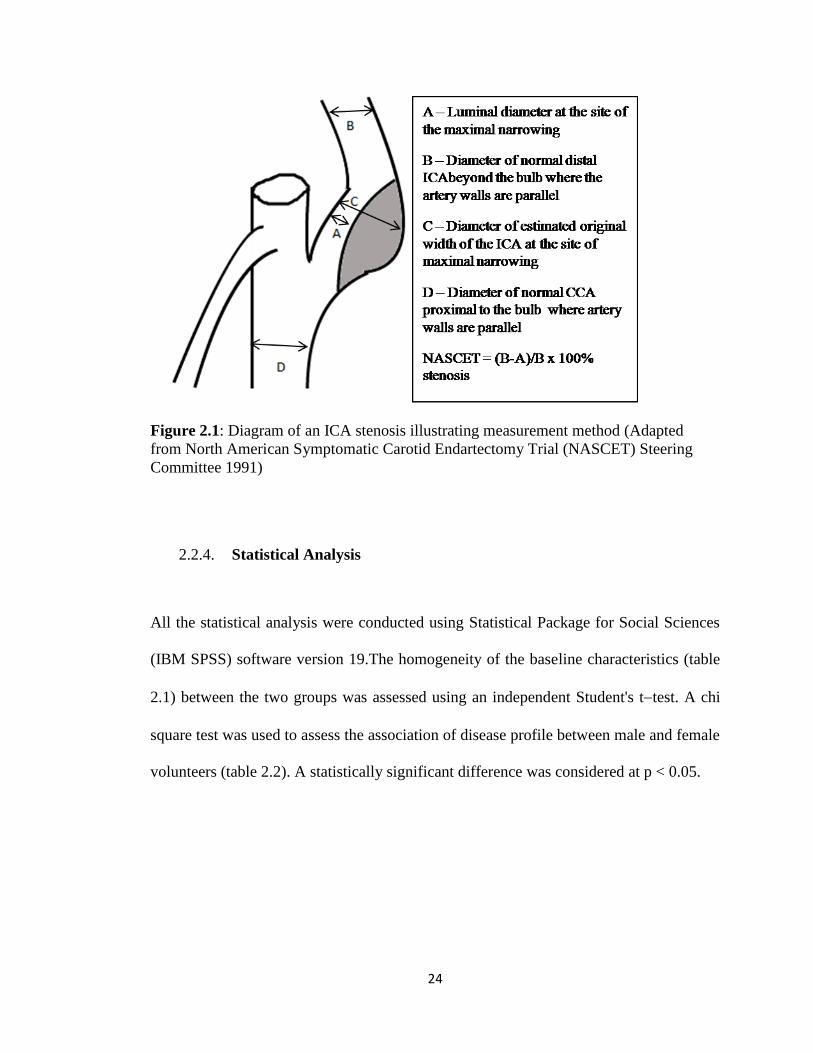
24
Figure 2.1: Diagram of an ICA stenosis illustrating measurement method (Adapted
from North American Symptomatic Carotid Endartectomy Trial (NASCET) Steering
Committee 1991)
2.2.4. Statistical Analysis
All the statistical analysis were conducted using Statistical Package for Social Sciences
(IBM SPSS) software version 19.The homogeneity of the baseline characteristics (table
2.1) between the two groups was assessed using an independent Student's ttest. A chi
square test was used to assess the association of disease profile between male and female
volunteers (table 2.2). A statistically significant difference was considered at p < 0.05.