Embed Size (px)
Citation preview
JOURNAL OF PATHOLOGY, VOL. 180: 102-105 (1996)
AN ORGAN-CULTURE METHOD FOR HUMAN COLORECTAL MUCOSA USING SERUM-FREE
MEDIUM M. MOORGHEN, M. CHAPMAN AND D. R. APPLETON*
Department of Pathology and Microbiology, University of Bristol, Bristol, U. K.; *Department of Medical Statistics, University o j Newcastle Upon Tyne, Newcastle Upon Tyne, U.K.
SUMMARY
This study describes an explant organ-culture system in which human colonic mucosa can be maintained for prolonged periods in serum-free medium. Following an initial phase of epithelial cell loss, there was intense regenerative activity, with the reformation of tubular crypts. Estimation of crypt lengths revealed a marked reduction after 5 and 9 days in culture with corresponding increases in labelling indices for the whole crypt. The shapes of bromodeoxyuridine (BrdU)-flash-labelling distribution curves were consistent with a proliferative compartment situated within the lower two-thirds of the crypt. We conclude that this is a useful in vitvo model for the study of the effects of growth factors and growth-inhibitory agents in respect of cell proliferation in human colonic mucosa.
KEY WORDsrgan-culture; colon; cell proliferation; bromodeoxyuridine
INTRODUCTION
Several experimental systems have been developed for the long-term maintenance of colonic mucosa in vitro. In explant organ-culture studies of rodent colonic mucosa, good structural and functional integrity can be main- tained for up to 3 In relation to human colonic mucosa, Autrup et U I . ~ , ~ described a system which en- abled explants to be maintained for up to 20 days, but this was associated with gross alterations in morphology with loss of normal crypt architecture. In another study by Senior e t a1.,6 explants of human colonic mucosa could be maintained for up to 14 days. In that study, a serum-containing formulation based on Waymouth MB752l1 medium, as first suggested by Hodges and Melcher,2 was employed. The presence of serum in the medium proved to be a serious drawback, in that serum contains a number of undefined components, many of which have growth-modulating properties; the presence of these would create difficulties in the interpretation of growth factor experiments. We therefore felt that it would be more appropriate to employ a model based on a serum-free preparation. In this study we propose a new organ-culture model which employs RPMI 1629 medium free of serum, which enables the successful maintenance of explants of human colorectal mucosa for up to 21 days.
MATERIALS AND METHODS Tissue explants
These were derived from surgical resection specimens from patients with colorectal cancer. A sheet of mucosa was removed from a site at least 6 cm from the tumour
Addressee for correspondence: Dr M. Moorghen, Department of Pathology, Level 9, Bristol Royal Infirmary, Marlborough Street, Bristol B22 8HW, U.K.
CCC 0022-34 17/96/090 102-04 0 1996 by John Wiley & Sons, Ltd.
and divided into small pieces, each measuring approxi- mately 2 mm2. Groups of four explants were laid on top of cellulose acetate filters placed in Petri dishes, each containing 2.5 ml of culture medium.
Medium formulation
The standard culture medium comprised RPMI 1629 (Gibco) with the addition of 300 pglml ascorbic acid, 3 pglml hydrocortisone-2 1 -sodium succinate, 0.45 pg/ ml ferrous sulphate, 100 U/ml penicillin/streptomycin solution, 50 pg/ml gentamicin, 100 U/ml mycostatin, 440 pglml L-glutamine, 1 pglml L-methionine, 2.7 mg/ml glucose, 50 nglml glucagon, 0.33 nglml T3, 6.25 pg/ml insulin, 1.25 mglml bovine serum albumin, 5 ng/ml sodium selenite, and 5.35pg/ml linoleic acid. The pH was adjusted to 7.4.
Incubation conditions
The explants were maintained at 37°C in 95 per cent oxygen, 5 per cent carbon dioxide on a rocking platform at 5 cycles per minute. The gas phase was renewed daily and the medium was changed every 2 days.
Cell prolifevation changes
In one experiment, bromodeoxyuridine (BrdU; 50 p ~ ) was added to the culture medium 1 h prior to the explants being fixed in formalin and subjected to im- munohistochemical staining using a monoclonal anti- body to BrdU purchased from Sera Labs, Crawley, Sussex, U.K. For each time-point ( 1 h, 5 days, and 9 days), the crypt lengths and the positions of BrdU- positive cells were determined by examining a total of 50 longitudinally sectioned hemi-crypts from different explants. Only explants which showed excellent tissue preservation were analysed. The edges of the explants,
Received I I October 1995 Accepted 30 January 1996
ORGAN-CULTURE OF COLORECTAL MUCOSA 103
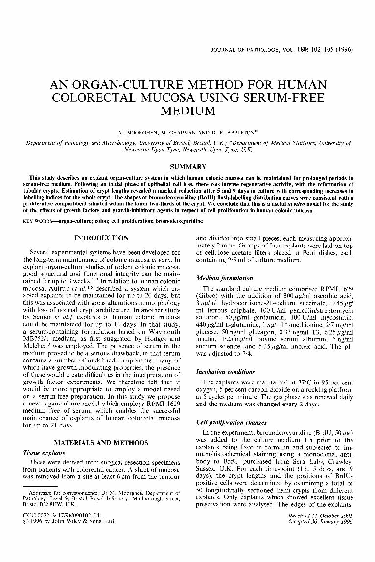
Fig. I-Explants of colonic mucosa showing (a) mild degenerative changes of epithelial cells at the surface and within the crypt after 1 day in culture, (b) severe degenerative changes after 3 days in culture, (c) well-formed tubular crypts after 5 days in culture, and (d) good preservation of mucosal architecture after 21 days in culture
arbitrarily defined as the three most peripheral crypts, were excluded for cell counting purposes.
RESULTS Histology
After 1 day in culture, a mild degree of goblet cell depletion and focal loss of epithelial cells became apparent (Fig. 1 a), followed by further degenerative changes (Fig. lb). After 5-6 days in culture, the crypts were gradually reconstituted (Fig. lc). Crypts situated at the periphery sometimes appeared hyperplastic, showing dilatation and apparent multilayering of the lining epi- thelium, which contained numerous mitoses. The overall appearances of these explants were maintained for a total of 11 days in culture in each of nine experiments that have been performed. Two experiments were allowed to run for a period of 21 days, at the end of
which 25 per cent of all the explants had survived and showed good tissue preservation throughout (Fig. 1 d).
BrdU labelling studies and crypt lengths For explants maintained in culture for 5 and 9 days,
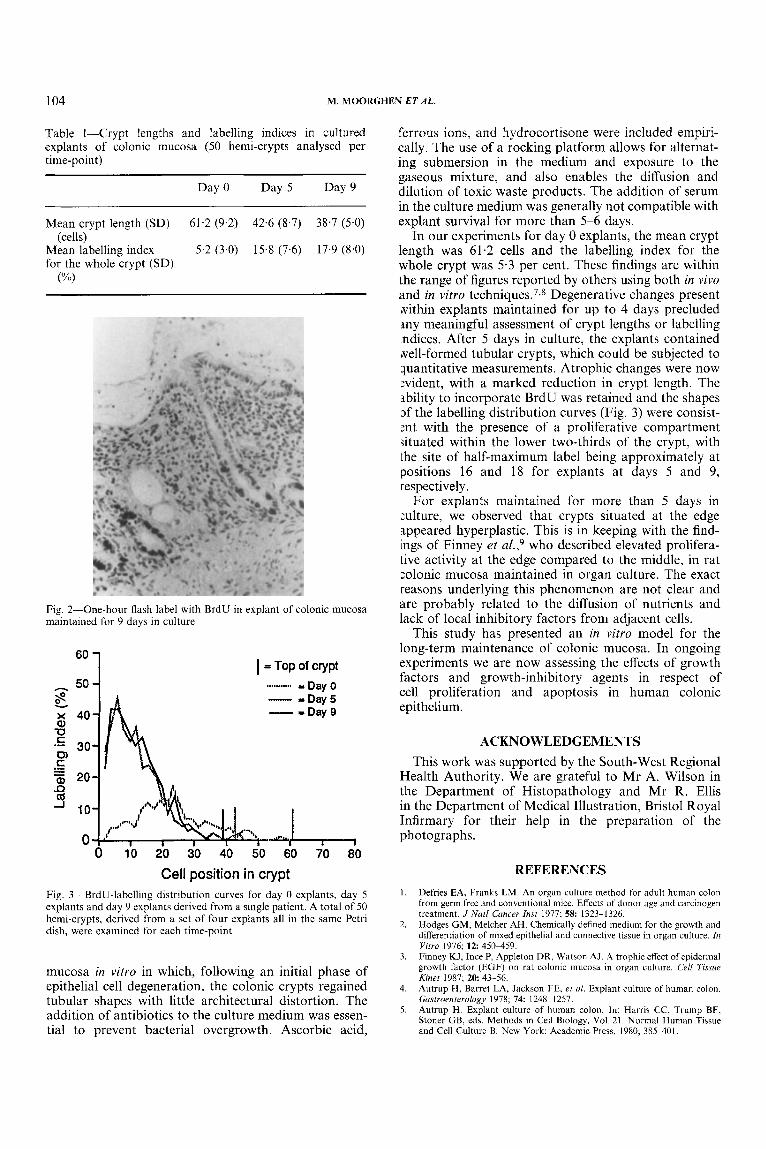
there was a significant reduction in crypt lengths with a corresponding increase in labelling indices (Table I). In day 0 explants, peak labelling of 16-1 7 per cent occurred at positions 2425, whereas in the shorter crypts of explants maintained for 5 and 9 days, peak labelling indices of 24-25 per cent were obtained at positions 8-10 (Figs 2 and 3).
DISCUSSION
In this study we have presented an experimental model for the long-term maintenance of human colonic
104 M. MOORGHEN ET AL.
Table I-Crypt lengths and labelling indices in cultured explants of colonic mucosa (50 hemi-crypts analysed per time-point)
Day 0 Day 5 Day 9
Mean crypt length (SD) 61.2 (9.2) 42.6 (8.7) 38.7 (5.0) (cells)
PA,)
Mean labelling index 5.2 (3.0) 15.8 (7.6) 17.9 (8.0) for the whole crypt (SD)
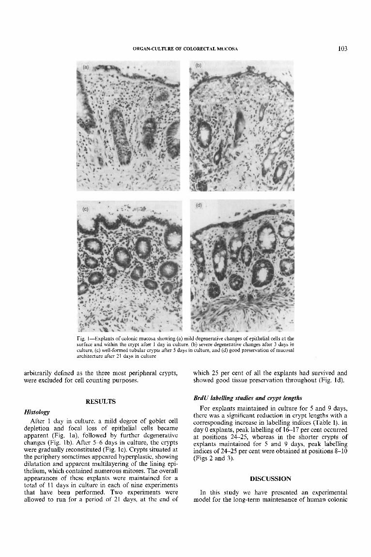
Fig. 2-One-hour flash label with BrdU in explant of colonic mucosa maintained for 9 days in culture
I = TOP Of Cvpt ........... = Day 0 - = D a y 5 I n - I Day 9
.~,....(I , , 0 10 20 30 40 50 60 70 80
Cell position in crypt Fig. 3-BrdU-labelling distribution curves for day 0 explants, day 5 explants and day 9 explants derived from a single patient. A total of 50 hemi-crypts, derived from a set of four explants all in the same Petri dish, were examined for each time-point
mucosa in vitro in which, following an initial phase of epithelial cell degeneration, the colonic crypts regained tubular shapes with little architectural distortion. The addition of antibiotics to the culture medium was essen- tial to prevent bacterial overgrowth. Ascorbic acid,
ferrous ions, and hydrocortisone were included empiri- cally. The use of a rocking platform allows for alternat- ing submersion in the medium and exposure to the gaseous mixture, and also enables the diffusion and dilution of toxic waste products. The addition of serum in the culture medium was generally not compatible with explant survival for more than 5-6 days.
In our experiments for day 0 explants, the mean crypt length was 61.2 cells and the labelling index for the whole crypt was 5.3 per cent. These findings are within the range of figures reported by others using both in vivo and in vitro t e c h n i q ~ e s . ~ , ~ Degenerative changes present vvithin explants maintained for up to 4 days precluded m y meaningful assessment of crypt lengths or labelling indices. After 5 days in culture, the explants contained well-formed tubular crypts, which could be subjected to quantitative measurements. Atrophic changes were now :vident, with a marked reduction in crypt length. The ability to incorporate BrdU was retained and the shapes Df the labelling distribution curves (Fig. 3 ) were consist- :nt with the presence of a proliferative compartment situated within the lower two-thirds of the crypt, with the site of half-maximum label being approximately at positions 16 and 18 for explants at days 5 and 9, respectively.
For explants maintained for more than 5 days in xlture, we observed that crypts situated at the edge appeared hyperplastic. This is in keeping with the find- ings of Finney et u Z . , ~ who described elevated prolifera- tive activity at the edge compared to the middle, in rat zolonic mucosa maintained in organ culture. The exact reasons underlying this phenomenon are not clear and are probably related to the diffusion of nutrients and lack of local inhibitory factors from adjacent cells.
This study has presented an in vitro model for the long-term maintenance of colonic mucosa. In ongoing experiments we are now assessing the effects of growth factors and growth-inhibitory agents in respect of cell proliferation and apoptosis in human colonic epithelium.
ACKNOWLEDGEMENTS This work was supported by the South-West Regional
Health Authority. We are grateful to Mr A. Wilson in the Department of Histopathology and Mr R. Ellis in the Department of Medical Illustration, Bristol Royal Infirmary for their help in the preparation of the photographs.
REFERENCES 1. Defries EA, Franks LM. An organ culture method for adult human colon
from germ free and conventional mice. Effects of donor age and carcinogen treatment. J Natl Cancer Inst 1977; 5 8 1323-1326. Hodges GM, Melcher AH. Chemically defined medium for the growth and differentiation o f rnixed epithelial and connective tissue in organ culture. In Vitro 1976; 1 2 4513459. Finney KJ, Ince P., Appleton DR, Watson AJ. A trophic etfect of epidermal growth factor (EGF) on rat colonic mucosa in organ culture. Cell Tissue Kinet 1987; 2 0 43-56. Autrup H, Barret LA, Jackson FE, et rrl. Explant culture of human colon. Gastroenterology 1978; 74: 1248-1257. Autrop H. Explant culture of human colon. In: Harris CC, Trump BF, Stoner GB, eds. Methods in Cell Biology, Val. 21. Normal Human Tissue and Cell Culture 13. New York: Academic Press, 1980; 385401.
2.
3.
4.
5.

ORGAN-CULTURE OF COLORECTAL MUCOSA 105
6. Senior PV, Pritchett CJ, Sunter JP, Appleton DR, Watson AJ. Crypt regeneration in adult human colonic mucosa during prolonged organ culture. J Anat 1982; 134: 459469. Potten CS, Kellett M, Roberts SA, Rew DA, Wilson GD. Measurement of in-vivo proliferation in human colorectal mucosa using bromodeoxyuridine. Gur 1992; 3 3 71-78.
8. Maskens AP, Deschner EE. Tritiated thymidine incorporation into epi- thelial cells of normal-appearing colorectal mucosa of cancer patients. J Nut/ Cancer Inst 1977; 58: 1221-1224. Finney KJ, Appleton DR, Ince P, Sunter JP, Watson AJ. Proliferative status of colonic mucosa in organ culture: 3H-thymidine-labelling studies and computer modelling. Virchoiw Arch B (Gem Patho/] 1989; 5 6 397-405.
7. 9.