Embed Size (px)
Citation preview
An Ultra-Conservative Approach of Treating Coronal Tooth Fragment: Re-attachment Case ReportsM Jayadev, Karunakar P, Raji Viola Solomon, Raji Viola Solomon, G Sushma ShravaniDepartment of Conservative Dentistry & Endodontics, Panineeya Institute of Dental Sciences & Research Institute , Hyderabad,India
AbstractDental traumatic fractures are the common reason for seeking dental care and coronal fractures being the frequent type of dentaltrauma encountered in the permanent dentition. The acid etch adhesive technique may be used to restore function and esthetics offractured anterior teeth. But whenever the fractured fragment is available, re-attachment will be a viable option as it offers certainadvantages over the composite restorations Reported here are the two case reports describing the treatment modalities of patientswho sustained fractures of maxillary teeth due to trauma. At one year follow up, no marginal discoloration, no fracture segment de-bonding or recurrent decay were found and the resultant appearance was acceptable to the patient.
Key words: Re-attachment, Fiber-reinforced post
IntroductionThe majority of dental traumatic injuries involve the anteriorteeth, especially the maxillary incisors nearly up to 80% of alltraumatic injuries and this high incidence is due to theiranatomical position and protrusion caused by eruptive process[1]. A dental trauma with the resulting fracture of the anteriorteeth is an agonizing experience for a young individual whorequires immediate attention, not only because of the physicalimpairment but also because of the psychological impact onthe patient [2].
When the fractured fragment is available, with advances inadhesive dentistry and growing interest towards minimalinvasive procedures, reattachment of the tooth fragment is thechoice of treatment [3].
Depending on different clinical situations, varioustechniques and materials were advocated to restore fractureteeth [4]. Tennery was the first person to report the re-attachment of a fractured fragment using the concept ofadhesive dentistry [1].
The advantages of this procedure over conventionalcomposite restoration are: esthetics, economical approach,psychological comfort to the patient, exact reconstruction oftooth morphology and increased wear steadiness [5-6].
The present case reports discuss the reattachment of thecoronal tooth fragment with the fracture involving enamel,dentin and pulp using fiber post with a follow up period ofone year.
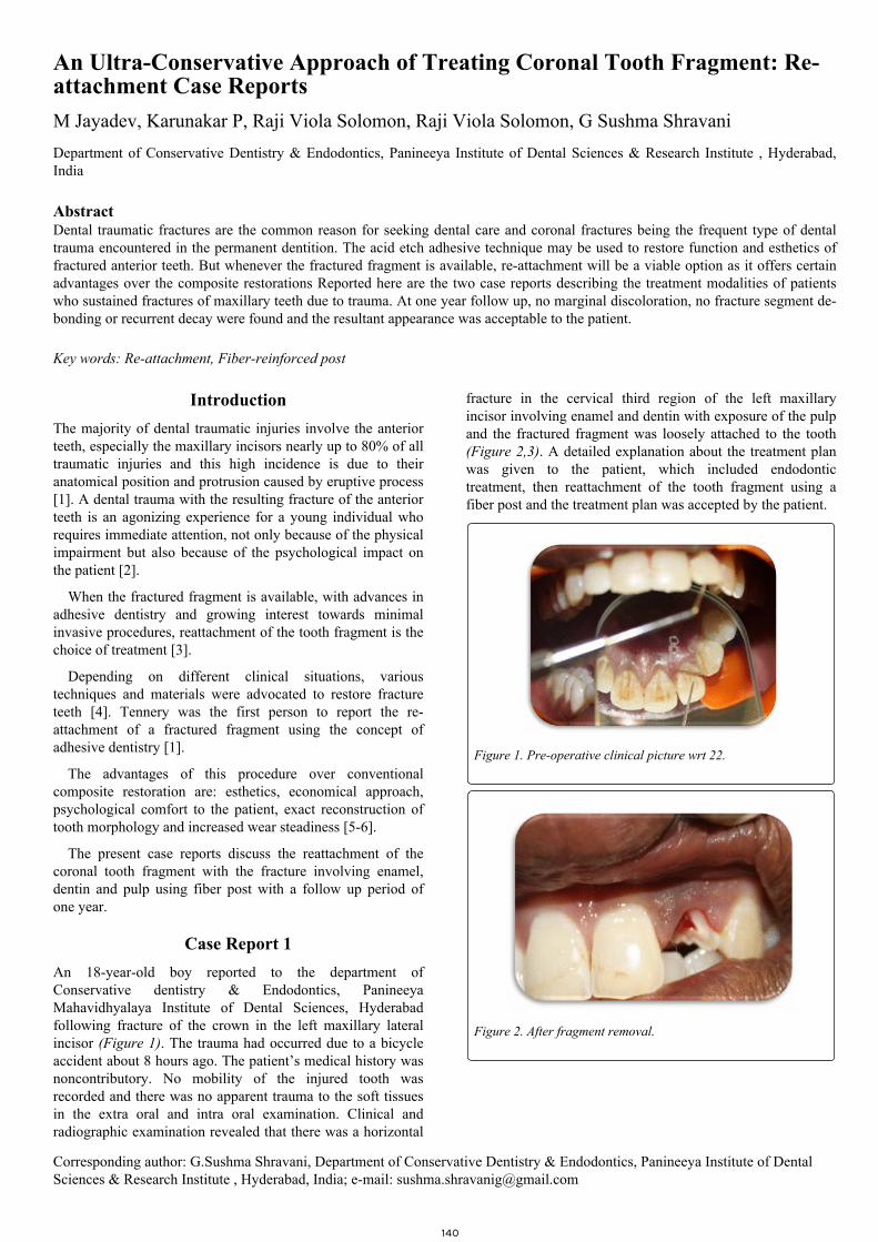
Case Report 1An 18-year-old boy reported to the department ofConservative dentistry & Endodontics, PanineeyaMahavidhyalaya Institute of Dental Sciences, Hyderabadfollowing fracture of the crown in the left maxillary lateralincisor (Figure 1). The trauma had occurred due to a bicycleaccident about 8 hours ago. The patient’s medical history wasnoncontributory. No mobility of the injured tooth wasrecorded and there was no apparent trauma to the soft tissuesin the extra oral and intra oral examination. Clinical andradiographic examination revealed that there was a horizontal
fracture in the cervical third region of the left maxillaryincisor involving enamel and dentin with exposure of the pulpand the fractured fragment was loosely attached to the tooth(Figure 2,3). A detailed explanation about the treatment planwas given to the patient, which included endodontictreatment, then reattachment of the tooth fragment using afiber post and the treatment plan was accepted by the patient.
Figure 1. Pre-operative clinical picture wrt 22.
Figure 2. After fragment removal.
Corresponding author: G.Sushma Shravani, Department of Conservative Dentistry & Endodontics, Panineeya Institute of DentalSciences & Research Institute , Hyderabad, India; e-mail: [email protected]
140
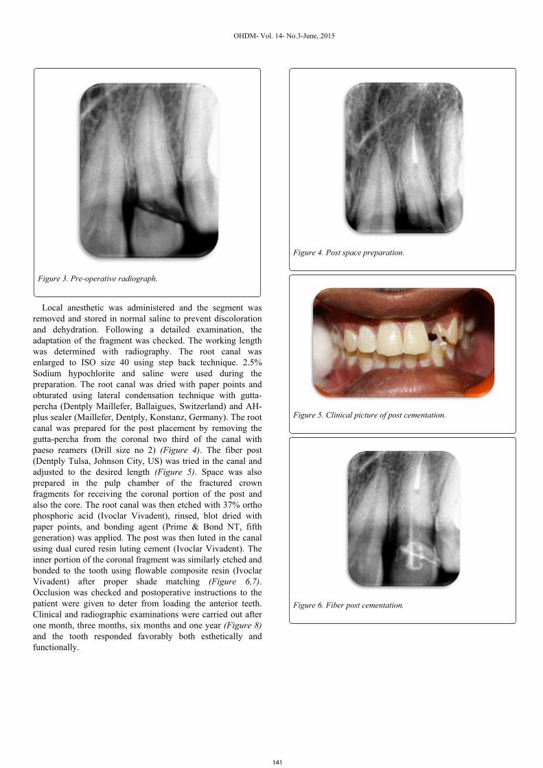
Figure 3. Pre-operative radiograph.
Local anesthetic was administered and the segment wasremoved and stored in normal saline to prevent discolorationand dehydration. Following a detailed examination, theadaptation of the fragment was checked. The working lengthwas determined with radiography. The root canal wasenlarged to ISO size 40 using step back technique. 2.5%Sodium hypochlorite and saline were used during thepreparation. The root canal was dried with paper points andobturated using lateral condensation technique with gutta-percha (Dentply Maillefer, Ballaigues, Switzerland) and AH-plus sealer (Maillefer, Dentply, Konstanz, Germany). The rootcanal was prepared for the post placement by removing thegutta-percha from the coronal two third of the canal withpaeso reamers (Drill size no 2) (Figure 4). The fiber post(Dentply Tulsa, Johnson City, US) was tried in the canal andadjusted to the desired length (Figure 5). Space was alsoprepared in the pulp chamber of the fractured crownfragments for receiving the coronal portion of the post andalso the core. The root canal was then etched with 37% orthophosphoric acid (Ivoclar Vivadent), rinsed, blot dried withpaper points, and bonding agent (Prime & Bond NT, fifthgeneration) was applied. The post was then luted in the canalusing dual cured resin luting cement (Ivoclar Vivadent). Theinner portion of the coronal fragment was similarly etched andbonded to the tooth using flowable composite resin (IvoclarVivadent) after proper shade matching (Figure 6,7).Occlusion was checked and postoperative instructions to thepatient were given to deter from loading the anterior teeth.Clinical and radiographic examinations were carried out afterone month, three months, six months and one year (Figure 8)and the tooth responded favorably both esthetically andfunctionally.
Figure 4. Post space preparation.
Figure 5. Clinical picture of post cementation.
Figure 6. Fiber post cementation.
OHDM- Vol. 14- No.3-June, 2015
141
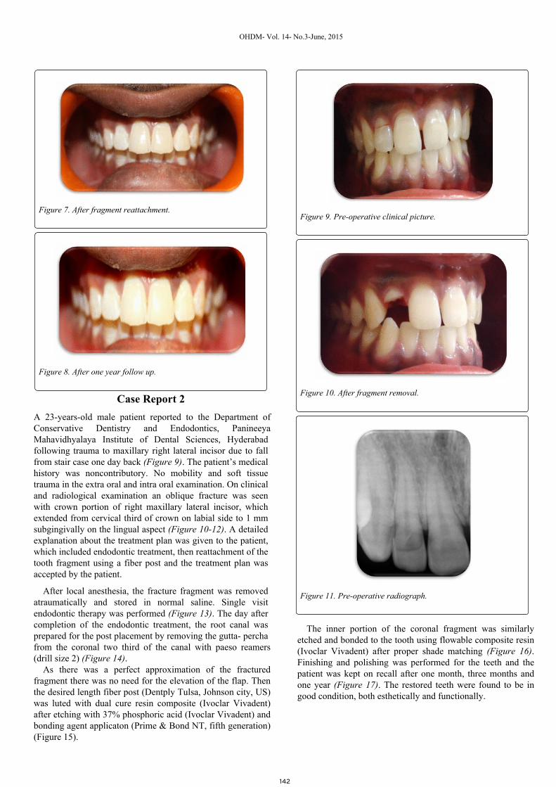
Figure 7. After fragment reattachment.
Figure 8. After one year follow up.
Case Report 2A 23-years-old male patient reported to the Department of Conservative Dentistry and Endodontics, Panineeya Mahavidhyalaya Institute of Dental Sciences, Hyderabad following trauma to maxillary right lateral incisor due to fall from stair case one day back (Figure 9). The patient’s medical history was noncontributory. No mobility and soft tissue trauma in the extra oral and intra oral examination. On clinical and radiological examination an oblique fracture was seen with crown portion of right maxillary lateral incisor, which extended from cervical third of crown on labial side to 1 mm subgingivally on the lingual aspect (Figure 10-12). A detailed explanation about the treatment plan was given to the patient, which included endodontic treatment, then reattachment of the tooth fragment using a fiber post and the treatment plan was accepted by the patient.
After local anesthesia, the fracture fragment was removed atraumatically and stored in normal saline. Single visit endodontic therapy was performed (Figure 13). The day after completion of the endodontic treatment, the root canal was prepared for the post placement by removing the gutta- percha from the coronal two third of the canal with paeso reamers (drill size 2) (Figure 14).
As there was a perfect approximation of the fractured fragment there was no need for the elevation of the flap. Then the desired length fiber post (Dentply Tulsa, Johnson city, US) was luted with dual cure resin composite (Ivoclar Vivadent) after etching with 37% phosphoric acid (Ivoclar Vivadent) and bonding agent applicaton (Prime & Bond NT, fifth generation) (Figure 15).
Figure 9. Pre-operative clinical picture.
Figure 10. After fragment removal.
Figure 11. Pre-operative radiograph.
OHDM- Vol. 14- No.3-June, 2015
142
The inner portion of the coronal fragment was similarly etched and bonded to the tooth using flowable composite resin (Ivoclar Vivadent) after proper shade matching (Figure 16). Finishing and polishing was performed for the teeth and the patient was kept on recall after one month, three months and one year (Figure 17). The restored teeth were found to be in good condition, both esthetically and functionally.
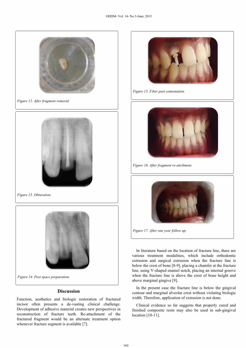
Figure 12. After fragment removal.
Figure 13. Obturation.
Figure 14. Post space preparation.
Figure 15. Fiber post cementation.
Figure 16. After fragment re-attchment.
Figure 17. After one year follow up.
OHDM- Vol. 14- No.3-June, 2015
143
DiscussionFunction, aesthetics and biologic restoration of fracturedincisor often presents a de-vasting clinical challenge.Development of adhesive material creates new perspectives inreconstruction of fracture teeth. Re-attachment of thefractured fragment would be an alternate treatment optionwhenever fracture segment is available [7].
In literature based on the location of fracture line, there arevarious treatment modalities, which include orthodonticextrusion and surgical extrusion when the fracture line isbelow the crest of bone [8-9], placing a chamfer at the fractureline, using V-shaped enamel notch, placing an internal groovewhen the fracture line is above the crest of bone height andabove marginal gingiva [9].
In the present case the fracture line is below the gingivalcontour and marginal alveolar crest without violating biologicwidth. Therefore, application of extrusion is not done.
Clinical evidence so far suggests that properly cured andfinished composite resin may also be used in sub-gingivallocation [10-11].
In the present case reports fractured segment is reinforcedusing fiber post. The post interlocks two separated fragmentsand minimizes the stress on the remaining tooth structure. Thefiber posts have a modulus of elasticity similar to dentin, thatallows more even distribution of occlusal stress in the rootdentin and they provide significantly less resistance to failurethan cemented custom cast posts and core [12].
A quote by liew is very appropriate in describing theprognosis for this procedure. He believes this restoration toact as “a short to medium term temporary restoration whichhas a potential for indefinite success” [13].
The longevity of a tooth fragment reattachment is notforeseeable, but the real merit of reattachment lies in the factthat all other restorative options, such as direct adhesivetechniques, veneers, and crowns will always be open. Withthe advancement in dental bonding technology, now it ispossible to achieve excellent results with reattachment ofdislocated tooth fragments, provided that the biologic factorsand selection of materials are logically assessed and managed.
The success of reattachment procedure depends on thecumulative effects of, proper use of bonding protocols,bonding materials, preparation techniques and patienteducation.
ConclusionNow in the era of concern with minimal invasive techniquesand esthetic considerations, re-attachment is one suchprocedure which offers with an ultraconservative, safe, fast,esthetically and functionally pleasing results whenever thefractured fragment is available.
References1. Tennery TN. The fractured tooth reunified using the acidetch
bonding technique. Texas Dental Journal. 1978; 96:16-17.2. Ece Eden, Saniye Çiçek Yanar, Şule Sönmez. Reattachment of
subgingivally fractured central incisor with open apex. DentalTraumatology. 2007; 23: 184-189.
3. RJ Simonsen. Traumatic fracture restoration: an alternativeuse of the acid etch technique. Quintessence International. 1979; 10:15–22.
4. Baklava P, Anup N. Risk factors for traumatic dental injuriesin an adolescent male population in India. The Journal ofContemporary Dental Practice. 2007; 8: 35-42
5. RD Trushkowsky. Esthetic, biologic, and restorativeconsiderations in coronal segment reattachment for a fractured tooth:a clinical report. The Journal of Prosthetic Dentistry. 1998; 79: 115–119.
6. K Arapostathis, A Arhakis, and S Kalfas. A modifiedtechnique on the reattachment of permanent tooth fragmentsfollowing dental trauma. Case report. Journal of Clinical PediatricDentistry. 2005; 30: 29–34, 2005.
7. Yucel Yilmaz, Cigdem Zehir, Ozge Eyuboglu, Nihal Belduz.Evaluation of success in reattachment of coronal fracture. DentalTraumatology. 2008; 24:151-158.
8. Holan G, Shmueli Y. Knowledge of physicians in hospitalemergency rooms in Israel on their role in cases of avulsion ofpermanent incisors. International Journal of Paediatric Dentistry.2003; 13: 13-19
9. Villat C, Machtou P, Naulin-lfi C. Multidisciplinary approachto the immediate esthetic repair and long term treatment of anoblique crown – root fracture . Dental Traumatology. 2004; 20:56-60.
10. Maia EA, Baratieri LN, de Andrada MA, Monteiro S Jr, deAraújo EM Jr. Tooth fragment reattachment: fundamentals of thetechnique and two case reports. Quintessence International. 2003;34: 99-107
11. LN Baratieri, SM J´unior, AC Cardoso, JCD Filho. Coronalfracture with invasion of the biologic width: a case report.Quintessence International. 1993; 24: 85–91.
12. Greenfeld RS, Roydhouse RH, Marshall FJ, Schoner BA.Comparison of two post systems under applied compressive-shearloads. Journal of Prosthetic Dentistry. 1989; 61:17-24.
13. Liew VP . Reattachment of original tooth fragment to afractured crown: Case report. Australian Dental Journal. 1988; 33:47-50.
OHDM- Vol. 14- No.3-June, 2015
144