Embed Size (px)
Citation preview
Pediatric Anesthesia and Critical Care Journal 2015; 3(1):61-65 doi:10.14587/paccj.2015.12
Sawant et al. Anaesthesia and Moya Moya disease 61
Key points
Moyamoya syndrome is a progressive occlusive cerebrovascular disorder that usually presents as recurrent strokes in
children. The risk factors for perioperative complications, predominantly the cerebral ischemic events aree: history of
transient ischemic attacks, severity of disease, type of revascularistion procedure, significant reduction in hematocrit,
intraoperative hypotension, intraoperative hypercapnia and reduction in circulating blood volume. We describe the
anaesthetic management of a case undergoing surgery for moya moya disease.
Anaesthetic management of children with Moya Moya Disease P. Sawant, S. Sharma, U. Rangwala
Department of Paediatric Anaesthesia, BJ Wadia Hospital for Children, Mumbai, India
Corresponding author: S. Sharma, Department of Paediatric Anaesthesia, BJ Wadia Hospital for Children, Mumbai, India. Email: [email protected]
Abstract
Moyamoya syndrome (MMD) is a progressive occlusive
cerebrovascular disorder that usually presents as recur-
rent strokes in children. Distal internal carotid and basi-
lar arteries gradually narrow, leading to proliferation of
penetrating arteries, primarily at the base of the brain.
Moya moya in japanese, means "something hazy, like a
puff of cigarette smoke drifting in the air." Because of
precarious cerebral circulation these patients represent
an anaesthetic challenge. The risk factors for periopera-
tive complications, predominantly the cerebral ischemic
events in patients with MMD are: history of transient
ischemic attacks, severity of disease, type of revascula-
ristion procedure, significant reduction in hematocrit,
intraoperative hypotension, intraoperative hypercapnia
and reduction in circulating blood volume.1-3
We describe the anaesthetic management of a case un-
dergoing surgery for moya moya disease.
Keywords: ischaemia, stroke, moya moya disease, hy-
pocapnia, seizures
Introduction
Moyamoya Disease is a rare cerebrovascular disease
seen both in children and adults with variable progres-
sion and presentation. It could be congenital or acquired,
the former linked to chromosome 17, and latter associa-
ted with head trauma, down’s syndrome, neurofibroma-
tosis, etc. It is characterized by angiographic evidence
of progressive stenosis or occlusion of terminal portions
of the internal carotid arteries and the proximal portion
of the anterior and middle cerebral arteries. The poste-
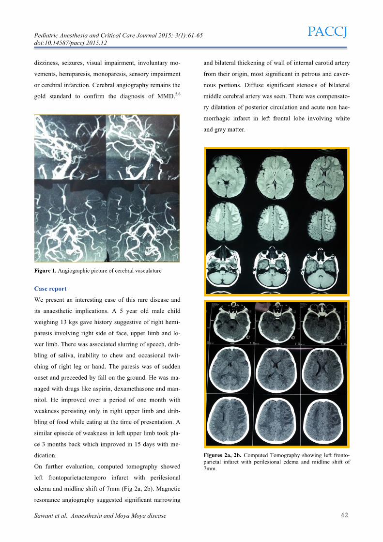
rior cerebral arteries may also be involved (Fig. 1).4
This disease is more common in Asian populations, but
even in Japan the overall incidence remains below 1 per
100,000. The male-to-female ratio has been shown to be
1:1.65 in one large series. The peak age of onset of
moyamoya disease in the Asian population is bimodal,
with an early peak occurring in the first decade of life,
and a second peak in the fourth decade of life.
The compensatory collateral circulation that develops is
weak and small, hence prone to haemorrhage, aneurysm
and thrombosis. Clinical picture includes transient
ischemic attacks, slow cognitive decline, headaches,
Pediatric Anesthesia and Critical Care Journal 2015; 3(1):61-65 doi:10.14587/paccj.2015.12
Sawant et al. Anaesthesia and Moya Moya disease 62
dizziness, seizures, visual impairment, involuntary mo-
vements, hemiparesis, monoparesis, sensory impairment
or cerebral infarction. Cerebral angiography remains the
gold standard to confirm the diagnosis of MMD.5,6
Figure 1. Angiographic picture of cerebral vasculature
Case report
We present an interesting case of this rare disease and
its anaesthetic implications. A 5 year old male child
weighing 13 kgs gave history suggestive of right hemi-
paresis involving right side of face, upper limb and lo-
wer limb. There was associated slurring of speech, drib-
bling of saliva, inability to chew and occasional twit-
ching of right leg or hand. The paresis was of sudden
onset and preceeded by fall on the ground. He was ma-
naged with drugs like aspirin, dexamethasone and man-
nitol. He improved over a period of one month with
weakness persisting only in right upper limb and drib-
bling of food while eating at the time of presentation. A
similar episode of weakness in left upper limb took pla-
ce 3 months back which improved in 15 days with me-
dication.
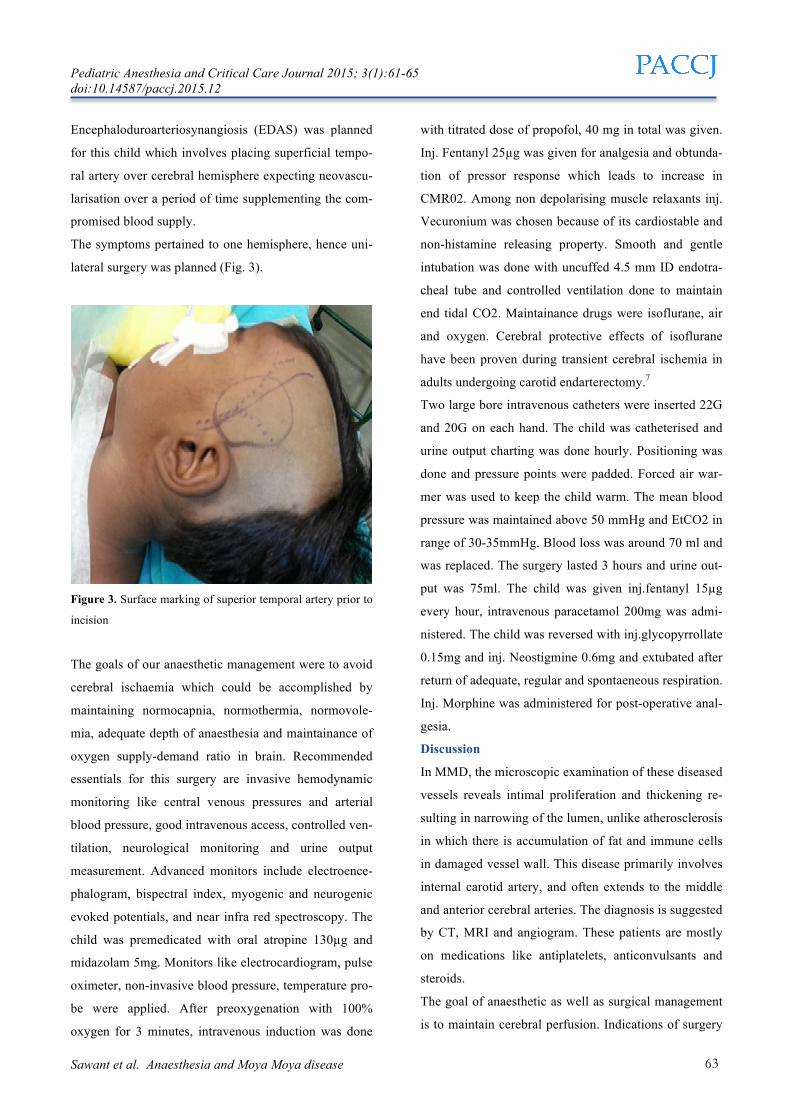
On further evaluation, computed tomography showed
left frontoparietaotemporo infarct with perilesional
edema and midline shift of 7mm (Fig 2a, 2b). Magnetic
resonance angiography suggested significant narrowing
and bilateral thickening of wall of internal carotid artery
from their origin, most significant in petrous and caver-
nous portions. Diffuse significant stenosis of bilateral
middle cerebral artery was seen. There was compensato-
ry dilatation of posterior circulation and acute non hae-
morrhagic infarct in left frontal lobe involving white
and gray matter.
Figures 2a, 2b. Computed Tomography showing left fronto-parietal infarct with perilesional edema and midline shift of 7mm.
Pediatric Anesthesia and Critical Care Journal 2015; 3(1):61-65 doi:10.14587/paccj.2015.12
Sawant et al. Anaesthesia and Moya Moya disease 63
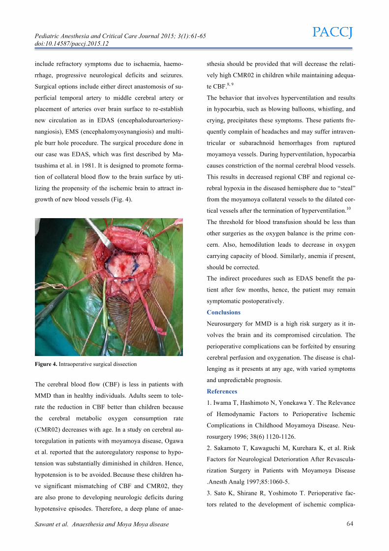
Encephaloduroarteriosynangiosis (EDAS) was planned
for this child which involves placing superficial tempo-
ral artery over cerebral hemisphere expecting neovascu-
larisation over a period of time supplementing the com-
promised blood supply.
The symptoms pertained to one hemisphere, hence uni-
lateral surgery was planned (Fig. 3).
Figure 3. Surface marking of superior temporal artery prior to
incision
The goals of our anaesthetic management were to avoid
cerebral ischaemia which could be accomplished by
maintaining normocapnia, normothermia, normovole-
mia, adequate depth of anaesthesia and maintainance of
oxygen supply-demand ratio in brain. Recommended
essentials for this surgery are invasive hemodynamic
monitoring like central venous pressures and arterial
blood pressure, good intravenous access, controlled ven-
tilation, neurological monitoring and urine output
measurement. Advanced monitors include electroence-
phalogram, bispectral index, myogenic and neurogenic
evoked potentials, and near infra red spectroscopy. The
child was premedicated with oral atropine 130µg and
midazolam 5mg. Monitors like electrocardiogram, pulse
oximeter, non-invasive blood pressure, temperature pro-
be were applied. After preoxygenation with 100%
oxygen for 3 minutes, intravenous induction was done
with titrated dose of propofol, 40 mg in total was given.
Inj. Fentanyl 25µg was given for analgesia and obtunda-
tion of pressor response which leads to increase in
CMR02. Among non depolarising muscle relaxants inj.
Vecuronium was chosen because of its cardiostable and
non-histamine releasing property. Smooth and gentle
intubation was done with uncuffed 4.5 mm ID endotra-
cheal tube and controlled ventilation done to maintain
end tidal CO2. Maintainance drugs were isoflurane, air
and oxygen. Cerebral protective effects of isoflurane
have been proven during transient cerebral ischemia in
adults undergoing carotid endarterectomy.7
Two large bore intravenous catheters were inserted 22G
and 20G on each hand. The child was catheterised and
urine output charting was done hourly. Positioning was
done and pressure points were padded. Forced air war-
mer was used to keep the child warm. The mean blood
pressure was maintained above 50 mmHg and EtCO2 in
range of 30-35mmHg. Blood loss was around 70 ml and
was replaced. The surgery lasted 3 hours and urine out-
put was 75ml. The child was given inj.fentanyl 15µg
every hour, intravenous paracetamol 200mg was admi-
nistered. The child was reversed with inj.glycopyrrollate
0.15mg and inj. Neostigmine 0.6mg and extubated after
return of adequate, regular and spontaeneous respiration.
Inj. Morphine was administered for post-operative anal-
gesia.
Discussion
In MMD, the microscopic examination of these diseased
vessels reveals intimal proliferation and thickening re-
sulting in narrowing of the lumen, unlike atherosclerosis
in which there is accumulation of fat and immune cells
in damaged vessel wall. This disease primarily involves
internal carotid artery, and often extends to the middle
and anterior cerebral arteries. The diagnosis is suggested
by CT, MRI and angiogram. These patients are mostly
on medications like antiplatelets, anticonvulsants and
steroids.
The goal of anaesthetic as well as surgical management
is to maintain cerebral perfusion. Indications of surgery
Pediatric Anesthesia and Critical Care Journal 2015; 3(1):61-65 doi:10.14587/paccj.2015.12
Sawant et al. Anaesthesia and Moya Moya disease 64
include refractory symptoms due to ischaemia, haemo-
rrhage, progressive neurological deficits and seizures.
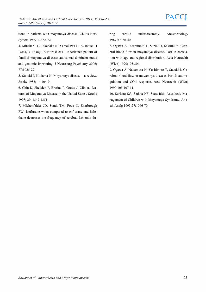
Surgical options include either direct anastomosis of su-
perficial temporal artery to middle cerebral artery or
placement of arteries over brain surface to re-establish
new circulation as in EDAS (encephaloduroarteriosy-
nangiosis), EMS (encephalomyosynangiosis) and multi-
ple burr hole procedure. The surgical procedure done in
our case was EDAS, which was first described by Ma-
tsushima et al. in 1981. It is designed to promote forma-
tion of collateral blood flow to the brain surface by uti-
lizing the propensity of the ischemic brain to attract in-
growth of new blood vessels (Fig. 4).
Figure 4. Intraoperative surgical dissection
The cerebral blood flow (CBF) is less in patients with
MMD than in healthy individuals. Adults seem to tole-
rate the reduction in CBF better than children because
the cerebral metabolic oxygen consumption rate
(CMR02) decreases with age. In a study on cerebral au-
toregulation in patients with moyamoya disease, Ogawa
et al. reported that the autoregulatory response to hypo-
tension was substantially diminished in children. Hence,
hypotension is to be avoided. Because these children ha-
ve significant mismatching of CBF and CMR02, they
are also prone to developing neurologic deficits during
hypotensive episodes. Therefore, a deep plane of anae-
sthesia should be provided that will decrease the relati-
vely high CMR02 in children while maintaining adequa-
te CBF.8, 9
The behavior that involves hyperventilation and results
in hypocarbia, such as blowing balloons, whistling, and
crying, precipitates these symptoms. These patients fre-
quently complain of headaches and may suffer intraven-
tricular or subarachnoid hemorrhages from ruptured
moyamoya vessels. During hyperventilation, hypocarbia
causes constriction of the normal cerebral blood vessels.
This results in decreased regional CBF and regional ce-
rebral hypoxia in the diseased hemisphere due to “steal”
from the moyamoya collateral vessels to the dilated cor-
tical vessels after the termination of hyperventilation.10
The threshold for blood transfusion should be less than
other surgeries as the oxygen balance is the prime con-
cern. Also, hemodilution leads to decrease in oxygen
carrying capacity of blood. Similarly, anemia if present,
should be corrected.
The indirect procedures such as EDAS benefit the pa-
tient after few months, hence, the patient may remain
symptomatic postoperatively.
Conclusions
Neurosurgery for MMD is a high risk surgery as it in-
volves the brain and its compromised circulation. The
perioperative complications can be forfeited by ensuring
cerebral perfusion and oxygenation. The disease is chal-
lenging as it presents at any age, with varied symptoms
and unpredictable prognosis.
References
1. Iwama T, Hashimoto N, Yonekawa Y. The Relevance
of Hemodynamic Factors to Perioperative Ischemic
Complications in Childhood Moyamoya Disease. Neu-
rosurgery 1996; 38(6) 1120-1126.
2. Sakamoto T, Kawaguchi M, Kurehara K, et al. Risk
Factors for Neurological Deterioration After Revascula-
rization Surgery in Patients with Moyamoya Disease
.Anesth Analg 1997;85:1060-5.
3. Sato K, Shirane R, Yoshimoto T. Perioperative fac-
tors related to the development of ischemic complica-
Pediatric Anesthesia and Critical Care Journal 2015; 3(1):61-65 doi:10.14587/paccj.2015.12
Sawant et al. Anaesthesia and Moya Moya disease 65
tions in patients with moyamoya disease. Childs Nerv
System 1997:13; 68-72.
4. Mineharu Y, Takenaka K, Yamakawa H, K. Inoue, H
Ikeda, Y Takagi, K Nozaki et al. Inheritance pattern of
familial moyamoya disease: autosomal dominant mode
and genomic imprinting. J Neurosurg Psychiatry 2006;
77:1025-29.
5. Sukuki J, Kodama N. Moyamoya disease – a review.
Stroke 1983; 14:104-9.
6. Chiu D, Shedden P, Bratina P, Grotta J. Clinical fea-
tures of Moyamoya Disease in the United States. Stroke
1998; 29; 1347-1351.
7. Michenfelder JD, Sundt TM, Fode N, Sharbrough
FW. Isoflurane when compared to enflurane and halo-
thane decreases the frequency of cerebral ischemia du-
ring carotid endarterectomy. Anesthesiology
1987;67336-40.
8. Ogawa A, Yoshimoto T, Suzuki J, Sakurai Y. Cere-
bral blood flow in moyamoya disease. Part 1: correla-
tion with age and regional distribution. Acta Neurochir
(Wien) 1990;105:304.
9. Ogawa A, Nakamura N, Yoshimoto T, Suzuki J. Ce-
rebral blood flow in moyamoya disease. Part 2: autore-
gulation and CO:! response. Acta Neurochir (Wien)
1990;105:107-11.
10. Soriano SG, Sethna NF, Scott RM. Anesthetic Ma-
nagement of Children with Moyamoya Syndrome. Ane-
sth Analg 1993;77:1066-70.