Embed Size (px)
Citation preview

NASA Technical Memorandum 87195
Analysis of a Thioether Lubricant byInfrared Fourier MicroemissionSpectrophotometry
N86-16379(H&SA-TH-87195) flNALYSIS OF ft THIOETHEBL U B R I C A N T BY IHFBABED FOUBIEB BICBOEHISSIOHSPECTBOPHOTOHETBY ( N A S S ) 16 p HC A02/HJ? &01
CSCL 11H DBClas63/27 05211
William R. Jones, Jr. and Wilfredo MoralesLewis Research CenterCleveland, Ohio
and
James L. LauerRensselaer Polytechnic InstituteTroy, New York
January 1986
NASA
https://ntrs.nasa.gov/search.jsp?R=19860006909 2018-09-07T09:09:50+00:00Z

ANALYSIS.OF A THIOETHER LUBRICANT BY INFRARED FOURIER
MICROEHISSION SPECTROPHOTOMETRY
William R. Jones, Jr. and Wllfredo MoralesNational Aeronautics and Space Administration
Lewis Research CenterCleveland, Ohio 44135
and
James L. LauerRensselaer Polytechnic Institute
Troy, New York 12181
SUMMARY
An Infrared Fourier m1croem1ss1on spectrophotometer was used to obtainspectra (wavenumber range, 630 to 1230 cm-1) from mlcrogram quantities ofthloether lubricant samples deposited on aluminum foil. Infrared bands 1n thespectra were reproducible and could be Identified as originating from aromaticspecies (1,3-d1subst1tuted benzenes). Spectra from all samples (neat andformulated, used and unused) were very similar. Additives (an add and aphosphlnate) present 1n low concentration (0.10 percent) 1n the formulatedfluid were not detected. This Instrument appears to be a viable tool 1n help-Ing to Identify lubricant components separated by liquid chromatography.
INTRODUCTION
One of the first achievements of modern Infrared Interferometry was therecording of Infrared emission spectra from moderately heated surfaces. Thespectra of organic adds on flat metal surfaces were reported by Low (ref. 1).Griffith (ref. 2) showed spectra of thin (1 ym) coatings of grease onmetallic mirrors. Bates (ref. 3), 1n a review paper, provides many otherexamples.
In concentrated contact lubrication, metal surfaces are exposed to liquidlubricants at high temperatures. These lubricants contain reactive species(additives) which form protective layers on the metal surfaces. The chemicalcomposition of these surface films will provide Important Information aboutthe mechanisms of additive action.
.Most of the above studies Involved unlimited radiating areas. However,1n concentrated contact lubrication, contact diameters are always less than500 ym and usually less than 100 ym. Therefore, a mlcrospectrophotometerwas developed (ref. 4) for dealing with the analysis of areas as small as10 ym 1n diameter.
Another area of analytical Interest 1s the determination of the chemicalstructure of eluted fractions of degraded lubricants separated by high perform-ance liquid chromatography (ref. 5). Here, the very low concentrations ofdegraded lubricants 1n a large amount of liquid mobile phase makes conventionalInfrared analysis difficult.

Therefore, the objective of this Investigation was to determine theability of the Fourier emission Infrared mlcrospectrophotometer to elucidatethe chemical structure of eluted fractions of a degraded high temperatureliquid lubricant (thloether).
APPARATUS
The apparatus for Infrared Emission Fourier Mlcrospectrophotometry 1sshown schematically 1n figure 1. The sample 1s shown below the heated metalblock to which 1t 1s attached. The sample was a strip of aluminum foil, onwhich a drop of solution was allowed to evaporate, leaving a solid organicresidue. The area analyzed In these experiments was 100 \tm 1n diameter. It1s exceedingly Important to maintain the sample at constant temperaturethroughout to avoid a vertical temperature gradient within the residue. Atemperature gradient could cause partial reabsorptlon of emitted radiation,making spectral analysis difficult, 1f not Impossible.
The lens below the sample (fig. 1) 1s of Cassegra1n1an design. A smallportion of the central section 1s blocked by a convex mirror which 1s facingdownward, reflecting the radiation gathered by the larger concave mirrorthrough a hole 1n Its center Into the rest of the Instrumentation. A smallplanar mirror at 45° with the vertical, bends the radiation Into the horizontalplane_pf the Interferometer. However, Immediately adjacent to the 45° mirror1s the tuning fork chopper, whose reflecting tines are at a 45° degree anglewith respect to the optic axis. In the open position of the chopper, sampleradiation 1s passed through, while 1n the closed position, reference blackbodyradiation 1s reflected and transferred downstream. The amplifier will detectonly the difference between the sample signal and the blackbody signal afterboth have passed through the Interferometer. After the Fourier transformation,a spectrum representing the difference between the sample and blackbody spectra1s plotted by the recorder.
Figure 2 1s a photograph of the Inlet system and the sample holder. Thelens looks at the bottom side of the sample held on a heatable mechanical stageallowing for x,y,z and rotational motion. Micrometer screws permit reproduc-ible positioning of the sample. The rotating filter holder below the lens 1sconnected to the Targe geared wheel 1n the foreground by a belt. This wheelhas sectors painted white and black for the reflecting optical photocellpickup.
The blackbody 1s shown 1n figure 3. Radiation emerges from the hole 1nfront. The flux can be controlled by the spring-loaded, adjustable wedge 1nfront. The blackbody 1s shown schematically 1n figure 4. It can be bothheated and cooled to a desired temperature and maintained there by a thermo-statlc control. The outer shell 1s always at ambient temperature. This black-body differs from commercial ones of similar size because Its temperature andflux Intensity are separately variable at any time over a wide and continuousrange.
TEST FLUIDS
Four liquid lubricant samples were studied 1n this Investigation. Theyare described 1n Table I. Sample 1 was an unused thloether base fluid

containing no additives. It 1s a mixture of polyphenyl ether and thloethercomponents as shown 1n figure 5. Sample 2 was a formulated version of thethloether base fluid containing 0.1 wt % of l-methylethyl-phenylphosph1nateand 0.05 wt % trlchloroacetlc add. Structures of these additives appear 1nfigure 6. Sample 3 was a used sample of the formulated thloether from a hightemperature bearing test which ran for 13 hr. Details of this test appear 1nreference 6. The final sample (4) 1s a concentration of the degraded productsfrom sample 3 (the bearing test). Sample 4 was prepared by dissolving thebearing test sample 1n a mixture of acetonltrlle and heptane. Two phasesresulted with the nondegraded thloether concentrating 1n the heptane phasewhile the degraded thloether concentrated 1n the acetonltrlle phase. Theacetonltrlle phase was then extracted and evaporated to dryness and recon-stituted 1n 3 ml of chloroform. Samples 1, 2, and 3 were prepared by dissolv-ing 40 vl of the lubricant 1n 3 ml of chloroform to simulate the concentra-tion 1n an eluted fraction from a liquid chromatograph. One drop of thesesamples was allowed to evaporate on an aluminum foil surface (cleaned with -.carbon tetrachlorlde) for the Infrared emission analysis. Evaporation tookplace at room temperature 1n a vacuum oven.
RESULTS
Infrared spectra obtained with the mlcrospectrophotometer over a wave-number range of 630 to 1230 cnr1 appear 1n figure 7. Figure 7(a) shows theemission spectra for the unused thloether base fluid (Sample 1). Figure 7(b)contains three different spectra from sample 2 (the unused formulated thloetherbase fluid). These spectra differ because of different sample and referencetemperatures. Similar data for sample 3 (used formulated thloether from the•bearing test) appear 1n figure 7(c). Figure 7(d) contains spectra for sample4 (the concentrated degradation products from the bearing sample).
Spectra were also obtained over the spectral range of 1274 to 1851 crrr1.However, the data 1n this range was not reproducible and therefore not Included1n this paper.
DISCUSSION
Comparison of Spectra
In order to compare bands from different spectra the following procedurewas used. An approximate baseline was drawn for each spectrum. The areas ofthe prominent emission bands were approximated by (1) drawing a vertical line(height, h) from the peak to the baseline, marking the midpoint between peakand baseline and drawing another line (width at half height, w) parallel to thebaseline through the midpoint until 1t Intersects with the band contour and (2)by multiplying h and w. The band areas were thus approximated by triangles.
Since the spectra had been "normalized" (I.e., the highest band having themaximum possible ordlnate), the band areas were corrected by multiplicationwith the greatest unnormallzed amplitude (QUA). With this treatment, bandsfrom different spectra can be compared. A summary of corrected band Intensi-ties for most of the spectra appears 1n figure 8.

Infrared Spectral Analysis
Before discussing these results, a few things should be pointed out. It1s necessary to pay attention to these remarks 1n the context of the extremelyweak signals recorded. The area concentration of sample on the foils 1s muchless than a mlcrogram/square centimeter. Ideally, the same spectral band(arising from the same vibration) should have the same energy, regardless oftemperature. This 1s because the vlbratlonal energy of a simple harmonicoscillator should not vary with temperature. However, deviations from thisrule do occur and can be either experimental or physical.
Experimentally, the most Important factors are the blackbody flux andtemperature. Most of the spectra of this study represent differences betweensample radiant energy and reference radiant energy. If the reference energy1s higher than the sample energy, Inversion of spectral bands may occur.Apparent absorption bands will be seen, rather than emission bands. If thereference temperature 1s different from the sample temperature, the baselineslope will not be horizontal. It may be positive 1n one spectral region andnegative 1n another, as governed by the Planck's distribution law. This mayresults 1n the appearance of spurious satellite bands. This 1s because thelevel of the reference radiation 1s sometimes between the peak and the bottomof an emission band (fig. 9). Since only absolute values of the amplitude ofthe difference between sample and reference are calculated by the Fouriertransformation, distorted spectra can result, as shown 1n figure 9. This pre-viously described Inversion can be difficult to recognize since correct emis-sion bands may appear 1n one frequency range, but Inverted bands 1n another.This problem can be minimized by (1) optically limiting the frequency range asmuch as possible and thereby reducing the slopes and (2) by obtaining duplicatespectra w.1th slightly different reference temperatures. An example 1s shown 1nfigure 10. Here, an emission spectrum of a polyphenyl ether was obtained withtoo high a reference temperature. Figure 10 shows an apparent absorption bandat 720 cm"1. Figure 11 shows an emission spectrum obtained under Identicalconditions of figure 10, except at a lower reference temperature. Here thecorrect emission band occurs at 690 cnr1.
Physically, Infrared emission bands are broadened as the temperature 1sIncreased (1) because of the excitation of additional rotational states and (2)because of a change of equilibrium between rotational Isomers. This 1s becausebands for two rotational states may be so close to each other that they are notresolvable.
In addition, the frequency of the band peak can also shift with tempera-ture for both experimental and physical reasons. Experimentally, the primaryreason 1s due to a change in baseline slope. Physically, frequency shifts mayoccur because of the lower spring constant at higher temperatures which wouldresult 1n a lowering of the peak frequency. Hydrogen bonding and other Inter-molecular Interactions may result 1n Increases 1n frequency. These frequencyshifts may be as great as ^5 cnr1.
With the above comments in mind, the spectra obtained for the four differ-ent samples are now discussed.
Thloether base fluid (Sample 1). - The single beam spectrum (I.e., notreferenced) for this fluid appears 1n figure 7(a). Strong emission bands occurat about 750 and 690 cnr1. Moderate bands are seen at approximately 660,

790, 835, 880, 905, 965, 1005, and 1015 cm-1. Weaker bands occur at 1025,1135, and 1180 cm-'. Most of these bands are representative of 1,3-d1substituted benzenes as compiled by Socrates (ref. 7). For example, bands at690, 790, 880, and 965 cm-1 are due to aromatic (=C-H) out of plane deformationvibrations: Aromatic (=C-H) 1n plane deformations occur at 1075 and .1180-cm'1.Other bands are associated with phenoxy (C-H) out of plane deformations(750 cm"1) and C-S-C stretch (660 cnr1). The remaining bands are not readilyassigned! Aromatic (-S) stretch also occurs at 1075 cm"1.
Formulated thloether fluid (sample 2). - Three spectra (800, 802; and 804)for this fluid appear 1n figure 7(b). All three spectra were taken at a sampletemperature of 38 °C. Spectrum 800 was not referenced. Spectra 802 and 804were referenced at temperatures of 28 and 31 °C, respectively. Taking Intoaccount variations among these spectra, bands appear at 670 to 690, 750 to 770,800, 850 to 870, 920, 940, 990, 1050, 1130, and 1170 cm-"1. Most of thesebands match those of the base fluid (sample 1) as would be expected. It doesnot appear that any new bands due to the additive content are detectable 1nthis frequency range.
Degraded bearing test fluid (sample 3). - Spectra (810, 812, and 814) forthis fluid appear 1n figure 7(c). Bands are observed at about 640, 665, 740,795,' 855, 910; 940, 980, 1025, 1055, 1125, and 1180 cm'1. This was adegraded sample of lubricant taken at the conclusion of a 13 hr high tempera-ture bearing test which was to have run for 100 hr. The test was terminatedat 13 hr because of severe cage wear and high production of solids. The fluidwas darkened and showed a viscosity Increase of about 10 percent. Neverthe-less, the spectra of this sample are very similar to those of the unused fluid.Previously, Infrared spectra of high molecular weight products from polyphenylethers (ref. 8) have also been almost Identical to the nondegraded fluid.Other results, using size exclusion chromatography (ref. 5), Indicate that thedegradation products of thloethers are essentially higher molecular weighthomologs of the base fluid. Since these materials differ only 1n the numberof aromatic rings, 1t 1s not surprising that their spectra are quite similar.
Concentrated degraded bearing test fluid (sample 4). - Since only a smallpercentage of a used lubricant 1s degraded, an extraction technique was used toconcentrate these products. Although this concentration technique (described1n the test fluids section) appeared to be visually successful, the spectra(fig. 7(d)) are the same as the nonconcentrated fluid (sample 3). In light ofthe discussion about the spectra from the nonconcentrated fluid, this 1s notsurprising.
In conclusion, Infrared emission mlcrospectrophotometry appears to be aviable technique for helping to Identify eluted components separated by liquidchromatography.
SUMMARY OF RESULTS
Four samples of a thloether lubricant were analyzed by Infrared Fourierm1croem1ss1on spectrophotometry. The following results were obtained:

1; Infrared emission spectra (from 630 to 1230 cm-1) were successfullyobtained from less than mlcrogram quantities of thioether lubricants depositedon surfaces of aluminum foil.
2. Infrared spectra from unused and degraded thloe.thers were essentiallythe same 1n the frequency range from 630 to 1230 cnr1.
3. The technique was not able to detect two additives (an add and aphosphlnate) which were present at low concentration (0.1 wt %) 1n thebase fluid.
REFERENCES
1. Low, M.J.D.; and Coleman, I.: The Measurement of Infrared EmissionSpectra Using Multlple-Scane Interferometry. Spectrochlm. Acta, vol. 22,no. 3, 1966, pp. 369-376. ;
2. Griffith, P.R.: Infrared Emission Spectroscopy. I. Basic Considerations.Appl. Spectrosc., vol. 26, no. 1, Jan.-Feb. 1972, pp. 73-76.
3. Bates, J.B.: Infrared Emission Spectroscopy. Fourier Transform InfraredSpectroscopy, vol. l-App!1cat1ons to Chemical Systems, J.B. Ferraro, andL.J. Baslle, eds., Academic Press, New York, 1978, pp. 99-142.
4. Lauer, J.L;; and King, V.W.: Fourier Emission Infrared Microspectrophoto-meter for Surface Analysis - I. Application to Lubrication Problems.Infrared Phys., vol. 19, no. 3-4, Aug. 1979, pp. 395-412.
5. Jones, U.R., Jr.; and Morales, W.: Thermal and Ox1dat1ve DegradationStudies of Formulated C-ethers by Gel-Permeation Chromatography. NASATP-1994, 1982.
6. Clark, F.S.; and Miller, D.R.: Formulation and Evaluation of C-EtherFluids as Lubricants Useful to 260 °C. (MRC-SL-1007, Monsanto ResearchCorp.; NASA Contract NAS3-1974-6) NASA CR-159794, 1980.
7. Socrates, G.: Infrared Characteristic Group Frequencies. John Wiley &Sons, 1980.
8. Jones, W.R., Jr.: Ferrographlc Analysis of Wear Debris from BoundaryLubrication Experiments with a Five Ring Polyphenyl Ether. ASLE Trans.,vol. 18, no. 3, July 1975, pp. 153-162.

TABLE I. - DESCRIPTION OF TEST SAMPLES
Sample Fluid Additive content
1
2
Unused thloetherbase fluid
Unused formulatedthloether
Used formulated thloetherfrom 13 hr high tem-perature bearing test3
Concentrated sample ofdegradation productsfrom sample 3
None
0.1 wt % 1-methylethylphenyl-phosphlnate
0.05 wt % trlchloroacetlc add
Same as sample 2
Reference 6.

Sample -
KCI window
Figure 1. - Source unit for Fourier microspectrophotometry (operation with chopper andblackbody reference giving spectra representing the difference between source and black-body signals).

ORIGINAL PAGE ISOF POOR QUALITY
Figure 2. - View of apparatus to Record Emission Spectra. The heated sample holder is above the lens.The polarizer is rotated below the lens. Black and white sectors on the large gear in the foregroundlocate the angular polarizer postion for the photoelectric pickup. The opening in the interferometercover below the polarizer is the entrance for the emitted radiation to be analayzed.
Figure 3. - Blackbody Reference Source. The radiation exits through the round hole and wedge infront.

Aperture Thermocouple
Cartridge heater
j^- Cool ing systems
Figure 4. - Schematic of Blackbody reference source.
1,3-bis (phenylthio) benzene
1, l'-thiobis [3-phenoxybenzene]
l-phenoxy-3-[[3-(phenylthio)phenyOthip] benzene
1, l'-thiobis [3-(phenylthio)benzene]
Figure 5.- Chemical structures of Thioethercomponents.
0 CH3M iP-O-CHI 1
H CH3
(a) 1-Methylethyl Phenylphosphinate
CI3C-C$)H
(b) Trichloroacetic acid
Figure 6. - Structures of additives informulated Thioether.

Spectrum #1518
Spectrum #804TS-38°C. Tref-31°C
Spectrum #802TS.-38°C, T r e f-28°C
750 870 990Wave number, I/cm
1110 1230
(b) Unused formulated Thioether fluid.
Figure 7. - Infrared emission spectra from 630 to 1230 cm"1.

ORIGINAL PAGE JS
OF POOR QUALITY
100
75
50
25
Spectrum #810T s -38°CT r e f -28°C
Spectrum #812T s -49°CT r e f -28°C
Spectrum #814Ts-50°CT r e f32°C
I
.£ (c) Used formulated Thioether fluid from a 13 hr high temperature bearing test100
Spectrum #816Ts - 38 °C Tref • 29 °C
75
50
25
-25.
--- Spectrum #81850°CT r e f-29°C
/\ Spectrum #820
refT s -50°CT r o f -3L5°C
830 750 870 990
Wave number, I/cm1110 1230
(d) Concentrated sample of degradation products from formulated Thioether fluid froma 13 hr high temperature bearing test
Figure 7. - Concluded.

40
20
0
40
20
t 40
20 h-
0
40
20
0
40
20
(b) Sample 3.
Spectrum804
I ll
802
40 ,—
20
n
- n
II 1 1
800
•
ll III
(a) Sample 2.60 i—
40
20
n
814
—
—
1 | 1 ll 1
- | ||
812
1 ll 1
810
820
818
_LL750 870 990 1110 1230
Frequency, cm"'
(c) Sample 4.
Figure 8.- Normalized infrared emission bands for each sample.

I ' .
I"
ReferenceSample
Figure 9. - Distortion of calculated emission bands asa result of a maladjusted reference level.
.87
.76
.•= .66c
£- -55ro
• .45
S -34
.13
.02
.08
.19629.9 689.9 780.0 810.1 870.1 930.2 990.2 106a9 1110.4 1170.4 1230.5
Wavenumber, cm'l
Figure 10. - Emission spectrum of a polyphenylether obtained with too high a refer-ence temperature.
.87
.78
.69
§ .60>»
1 50B «JUL_
(U
a,- .41
£ .32
E .22•g
* .13
.04
.06629.9 689.9 750.0 81011 870.1 950.2 990.2 1050.3 1110.4 1170.4 1230.6
Wavenumber, cm~l
Figure 11. - Emission spectrum for the same conditions on those of figure 10, but obtainedwith a lower reference temperature.
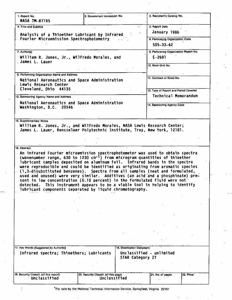
1. Report No.
NASA TH-871952. Government Accession No. 3. Recipient's Catalog No.
4. Title and Subtitle
Analysis of a Thloether Lubricant by InfraredFourier M1croem1ss1on Spectrophotometry
5. Report Date
January 1986
6. Performing Organization Code
505-33-62
7. Authors)
William R. Jones, Jr., Wllfredo Morales, and. James L. Lauer
8. Performing Organization Report No.
E-2691
10. Work Unit No.
9. Performing Organization Name and Address
National Aeronautics and Space AdministrationLewis Research CenterCleveland, Ohio 44135
11. Contract or Grant No.
12. Sponsoring Agency Name and Address
National Aeronautics and Space AdministrationWashington, D.C. 20546
13. Type of Report and Period Covered
Technical Memorandum
14. Sponsoring Agency Code
15. Supplementary Notes
William R. Jones, Jr., and Wllfredo Morales, NASA Lewis Research Center;James L. Lauer, Rensselaer Polytechnic Institute, Troy, New York, 12181.
16. Abstract
An Infrared Fourier m1croem1ss1on spectrophotometer was used to obtain spectra(wavenumber range, 630 to 1230 cm-1) from mlcrogram quantities of thloetherlubricant samples deposited on aluminum foil. Infrared bands 1n the spectrawere reproducible and could be Identified as originating from aromatic species(1,3-d1subst1tuted benzenes). Spectra from all samples (neat and formulated,used and unused) were very similar. Additives (an add and a phosphlnate) pre-sent 1n low concentration (0.10 percent) 1n the formulated fluid were notdetected. This Instrument appears to be a viable tool In helping to Identifylubricant components separated by liquid chromatography.
7. Key Words (Suggested by Authors))
Infrared spectra; Thloethers; Lubricants18. Distribution Statement
Unclassified - unlimitedSTAR Category 27
9. Security Classif. (of this report)Unclassified
20. Security Classif. (of this page)Unclassified
21. No. of pages 22. Price'
*For sale by the National Technical Information Service, Springfield, Virginia 22161

National Aeronautics andSpace Administration
Lewis Research CenterCleveland. Ohio 44135
Official BusinessPenalty for Private Use $300
SECOND CLASS MAIL
ADDRESS CORRECTION REQUESTED
Postage and Fees PaidNational Aeronautics andSpace AdministrationNASA-451
NASA