Embed Size (px)
Citation preview
ipHeterologous gene expression in Saccharomyces cerevisiae:
Analysis of Bacillus subtilis /?-glucanase and Escherichia col/?-glucuronidase.
ThesisPresented for the Degree of
DOCTOR OF PHILOSOPHY by Sharon Hunter, B.A. Mod. (Genetics)
under the supervision of Thecla Ryan, Ph.D.
School of Biological Sciences Dublin City University
August 1991
I hereby declare that the research described within this thesis is based entirely upon my own work.
QUsXii&iÆ M l
SHARON HUNTER
Sof V-

ABSTRACT
The production of the Bacillus subtilis /3-glucanase enzyme was examined in the yeast Saccharomyces cerevisiae. The ability of the bacterial signal peptide to translocate and secrete the enzyme was analysed and compared to the yeast a-factor signal peptide. A truncated version of the signal was also examined as was the effect of complete removal of the signal sequence. Glycosylation of the /3-glucanase enzyme in the yeast secretory pathway was examined using both Western blots and activity gels and the effects of glycosylation on /3-glucanase production was assessed. A second bacterial enzyme, /3-glucuronidase, was also examined for its potential use as a reporter enzyme in yeast expression and secretion systems. This work included attempts to secrete the enzyme, the definition of its active site, investigation of its potential for use in translational fusion experiments and the generation of a polyclonal antibody to the E.coli /3-glucuronidase enzyme.
ACKNOWLEDGEMENTS
I wish to thank my supervisor, Dr. Thecla Ryan, for her help, guidance, encouragement and most of all her enthusiasm, maintained throughout this research project.
I would also like to thank Dr. John Dalton, Susan and Carolyn for their help with raising the antibody and advice on immunological aspects of this work.
To all at D.C.U.: staff, postgrads and especiallythe technicians, thanks for all your help.
Thanks also to all in the John-(Good Time)-Barry Building for helping to make my time here so memorable!! and enjoyable. Special thanks to my two lab colleagues Big Maggie and Hugh for everything, to our honorary lab-member Peter, to Ger for having printing parameters etched deeply in her brain not to mention her photographic skills, to John for his much needed help with computers, and to everyone who helped in proof reading and printing this thesis.
To Domo for lending me the computer and to Anne for her help with the typing - I'm eternally grateful!
I would also like to thank my parents for the constant support, interest and encouragement they have shown throughout my years at college (finished at last!).
This thesis is dedicated to my family for their support and encouragement over the years and to Alan who helped more than he thinks.
TABLE OF CONTENTS
CHAPTER 1. Introduction.................,.................... 11.1.0 Heterologous protein production in yeast......... 21.1.1 Yeast expression systems.......................... 21.1.2 Heterologous protein stability................... 61.1.3 Improving production by secretion................ 81.2.0 Introduction to the secretory process............ 101.2.1 The secretory pathway of eukaryotic cells........ 111.2.2 The signal hypothesis............................. 141.3.0 Characterizing eukaryotic and prokaryotic
signal peptides.................................. 171.3.1 Physical disection of signal peptides............ 171.3.2 Disruption of the hydrophobic domain............. 191.3.3 The hydrophillic domain........................... 221.3.4 The signal peptide cleavage site.................. 261.4.0 Proteins that interact with signal peptides...... 271.4.1 The signal recognition particle (Srp) and
Srp receptor...................................... 271.4.2 Proteins involved in the yeast translocation
event............................................. 291.4.3 The importance of conformation for translocation
competence........................................ 311.4.4 Unfolding in yeast: the role of heat shock
proteins.......................................... 321.4.5 Characterization of signal peptidases............ 331.5.0 Sorting signals in the secretory pathway......... 351.5.1 Retention in the ER................................ 351.5.2 Vacuolar sorting signals.......................... 371.6.0 N-linked glycosylation of eukaryotic proteins.... 391.6.1 Core oligosaccharide assembly..................... 411.6.2 Transfer and trimming of the core
oligosaccharides................................. 431.6.3 Elongation in the Golgi............................ 441.6.4 Glycosylation of yeast secretory proteins........ 471.7.0 The secretion of heterologous proteins

CHAPTER 2 Materials and Methods............................. 522.1 Materials.......................................... 532 . 2 Table of strains............................... 532 . 3 Table of plasmids.................................. 542.4 Media.............................................. 572.5 Antibiotics........................................ 582.6 Growth conditions and strain storage............... 592.7 Buffers and solutions............................. 592.8 Transformation of E.coli.......................... 642.9 Transformation of S .cerevisiae.................... 652.10 Generation of yeast spheroplasts.................. 652.11 Small scale isolation of plasmid DNA from E.coli. 652.12 Large scale isolation of plasmid DNA from E.coli. 672.13 Agarose gel electrophoresis....................... 682.14 General DNA manipulations......................... 682.15 Bal31 digestion.................................... 692.16 Sepharose CL6B columns............................. 702.17 Isolation of DNA fragments from agarose.......... 702.18 Plate assay to detect /3-glucanase activity....... 712.19 DNS assay for measuring ¿3-glucanase activity 712.20 Preparation of yeast extracts for DNS assays 722.21 Polyacrylamide gel electrophoresis (PAGE)........ 722.22 Preparation of yeast extracts and supernatants
for PAGE.......................................... 722.23 Western blotting procedure........................ 732.24 /3-glucanase activity gels................... . 742.25 Isolation of RNA from S.cerevisiae................ 742.26 Formaldehyde gel electrophoresis....... 752.27 Transfer of RNA to nitrocellulose................. 752.28 Random primer labeling of DNA. ................ 762.29 Hybridization of probe to Northern blots........ 762.30 glucuronidase assays............................. 772.31 glucuronidase activity gels..................... 782.32 Incorporation of RC631 into MYGP agar............. 78
in S .cerevisiae.................................... 49
CHAPTER 3. SECTION 3.1:Analysis of /3-glucanase production and secretion in
S .cerevisiae
3.1 Introduction ,................ 803.2 The /3-glucanase plate assay....................... 8 53.3 The DNS assay for measuring /3-glucanase activity. 873.4 /3-glucanase production in pJG317, pJG205
and pJG314........................................ 893.5 Construction of pVT314............................ 933.6 Removal of the /3-glucanase signal peptide........ 1003.7 Construction of pJBC55A........................... 1033.8 Northern analysis of pJB/34 and pJBC55A........... 1053.9 Assay of pJB/34 and pJBC55A in a pep4-3
mutant of S . cerevisiae........................... 1033.10 Analysis of /3-glucanase secretion in
secretion defective mutants................ 1103.11 The effect of tunicamycin on /J-glucanase
production.......... ............................. 1193.12 Western blot analysis of the Bacillus
/9-glucanase enzyme............................... 1213.13 Activity gel analysis of the /9-glucanase enzyme.. 126
CHAPTER 3 SECTION 3.2Preliminary studies on the suitability of an E.coli
¡3-glucuronidase as a reporter gene in S . cerevisiae.
3.14 glucuronidase as a reporter enzymefor S . cerevisiae,,,,,,, *..... .... 134
3.15 Expressing /3-glucuronidase in S .cerevisiae....... 1343.16 Fusion of /9-GUS to the a-factor signal peptide... 1383.17 Adapting /3-glucuronidase as a useful reporter
enzyme in yeast................... ............... 1413.18 Truncating /9-GUS from the 3' end.................. 1423.19 Truncating /9-GUS from the 5' end.............. 1443.20 /3-glucuronidase as a fusion protein........... 1493.21 Raising a polyclonal antibody to the E.coli
/3-glucuronidase enzyme 151
CHAPTER 4 Discussion................................... . 15154.1 The /3-glucanase signal peptide................... 1564.2 Deletion of the /3-glucanase signal peptide....... 1574.3 Truncating the /3-glucanase signal peptide....... 1604.4 Comparison of the /3-glucanase and a-factor
signal peptdes in pJG317, pJG314 and pVT314 1624.5 The effect of glycosylation on the Bacillus
/3-glucanase enzyme............................... 1644.6 Processing of the a-factor signal peptide in
pJG314 and pVT314................................ 1684.7 Conclusions........................................ 170
CHAPTER 5 References......................................... 172Appendices....... 202
LIST OF FIGURESFigure
1.1 The secretory pathway of yeast.................... 131.2 The early signal hypothesis................... 151.3 The signal hypothesis............................. 161.4 Selection of translocation mutants in yeast...... 301.5 Assembly of core oligosaccharides................ 401.6 Extension of the core oligosaccharide........... 451.7 Outer chain additions in an mnn9 glycoproteins... 461.8 Processing of prepro-a-factor..................... 43
Figure3.1 Features of the /9-glucanase signal peptide....... 813.2 pJG314.............................................. 833.3 pJG317 and pJG205 .................................. 843.4 The /3-glucanase plate assay....................... 863.5 pH and temperature profiles of /3-glucanase
produced in S . cerevisiae......................... 883.6a Growth curve pJG317 in DBY746..................... 903.6b Growth curve pJG205 in DBY746....... 903.6c Growth curve pJG314 in DBY746..................... 9.L
3.7 Secreted /3-glucanase in pJG317 , pJG205and pJG314....................................... 91
3.8a pUGS4.............................................. 933.8b Construction of pJGS4............................. 953.8c pVT314............................................. 963.9 Growth curve pVT314 in DBY746..................... 973.10 Secreted /3-glucanase in pJG317 , pJG314
and pVT314........................................ 983.11 Construction of pJB/34............................. 1013.12 Restriction analysis of pJB/34..................... 1023.13 Construction of pJBC55A........................... 1043.14a Northern gel showing RNA preparations
from S .cerevisiae strains.................... . 1063.14b Autoradiograph of /3-glucanase transcripts........ 1073.15 /3-glucanase production in MD50 transformed
with pJBC55A, pJB/34 and controls................ 1093.16 Calibration curve for /3-glucanase plate assay.... 1123.17a Block in secretion of /3-glucanase in RSY11
(pJG317) at 37°C................................. 1133.17b DBY746 control..................................... 1133.18 Increase in /3-glucanase activity seen at
37°C in RSY12 (pJG317)........................... 1143.19 The a-factor plate assay......................... , 1153.20 Block in a-factor secretion at 37°C in
RSY45 transformed with p69A..................... 1663.21 Block in a-factor secretion at 37°C in
supernatants of RSY12 (pJG317 and pJG314)...... 1173.22 /3-glucanase activity in pJG317, pJG314 and
and pJG205 after tunicamycin treatment.......... 1203.23 Western blot showing extracts and supernatants
prepared from pJG317 and pJG314................. 1223.24 Western blot showing extracts and supernatants
prepared from pJG205, pAAH5 and p69B............ 1233.25 Activity gel showing intracellular /3-glucanase
activity in pJG317, pJG314 and pJG205........... 12/3.26 Activity gel of intracellular and secreted
forms of /3-glucanase in pJG317 and pJG314...... 129
3.27 Activity gel of supernatants with and withouttunicamycin treatment......................... 131
3.28 Activity gel showing pVT314 intracellularand extracellular fractions..................... 132
3.29 Construction of pJBU7.............................. 13153.30 /3-glucuronidase production in pJBU7............... 1363.31 The pGUS series of plasmids....................... 1383.32 Construction of pGUSa6 ............. 1393.33 Growth curve pGUSa6 in DBY746........ ,.......... 1403.34 3' Bal31 deletions of the /3-GUS gene.............. 1433.35 Generation of 5' BaI31 deletions.................. 1443.36 Bal31 deletions cut with Clal and EcoRl........... 1463.37 Construction of pGUS2c............................ 1473.38 MUG assay of Bal31 deletions...................... 1483.39 Construction of /3-glucanase / /3-glucuronidase
fusion plasmids................. 1503.30 Titring the /3-glucuronidase antibody.............. 1523.31 Reactivity of the /3-glucuronidase antibody
with the enzyme produced in yeast............... 154
Figure4.1 Physical disection of the /3-glucanase signal...... 1564.2 5' untranslated regions of pJB/34 and pJBC55A 1594.3 Sequence around the sigal cleavage site
in pJG314 and pVT314............................. 169
LIST OF TABLESTable3.1 Summary of /3-glucanase construct and levels of
enzyme production at one point in growth cycle.. 993.2 Relative intensities of transcripts calculated
by densiometric scanning........................ , 1073 . 3 Summary of sec mutants.............................. 110
Abbreviations used in diagrams:B : BaraHl E : EcoRlP : Pstl Sm : Sma 1H : H indili D : DdelS : Sail X : Xbal
C : Clal
'P' : promoter.! # : terminator.Sp : signal peptide.
Other abbreviationsS .cerevisiae : Saccharomyces cerevisiae.
E .coli
B .subcilis
/3-GUS
Escherichia coli. Bacillus subcilis. ß-glucuronidase gene.
Amino acid codes:A = Alanine.D •= Aspartic acid. E = Glutamic acid. L = Leucine.K = Lysine.H = Histidine.
CHAPTER 1 INTRODUCTION
1
(1.1) Heterologous protein production in yeast: an overview.S . cerevisiae has many advantages as a host for the production of proteins of commercial value. The high cell densities reached in fermentations, using simple media, has made it an ideal organism for applications at an industrial level. In addition, the rapid progress being made in characterizing S . cerevisiae at biochemical and molecular levels and the ease with which the organism can be manipulated genetically, has greatly enhanced its potential in this respect.There are a wide range of systems available for introducing, expressing and maintaining foreign genes in yeast, some of which are outlined below. The production and isolation of a particular protein however, goes beyond the successful maintenance and expression of the gene. A major problem encountered is insolubility of the gene product in the yeast cytoplasm. This problem is also associated with intracellular production in the bacterial host, E.coli and can result in the recovery of malfolded, biologically inactive material. Other factors such as protein instability and degradation can also affect recovery levels. Some of these problems and possible solutions are discussed in the remainder of this initial section. The potential of improving both recovery levels and product quality, by directing heterologous proteins through the secretory pathway is also examined.
(1.1.1) Yeast expression systems.A number of vector systems are available for the introduction and maintenance of exogenous genes in S .cerevisiae. These include autonomously replicating plasmids which are maintained at high, medium or low copy numbers and integrating vectors whose copy number can also be controlled to a certain extent. The most widely used autonomously replicating vectors are based on the native 2/j.m circle plasmid (Hartley and Donelson, 1980 ) . These vectors include the 2\j> origin and REP3 regions required for maintaining copy number and partitioning fidelity (Jayaram et al 1985). Copy number is controlled by the REP1, REP2 and FLP functions which are provided by endogenous 2n circles in
2
cir+ hosts (Wu et al 1987) . 2/i based vectors are maintained at relatively high copy numbers (20-40 copies/cell) and stability is high under selective growth conditions. Autonomously replicating sequences (ARS), derived from chromosomal elements have also been included into vectors and although higher copy numbers are achieved, this is at the expense of stability and partitioning fidelity, even with selective growth conditions (Murray and Szostak, 1983).Methods of increasing copy number and maintaining stability of 2p, based vectors have been developed. The introduction of the leucine defective (leu-d) gene as a selectable plasmid marker maintains a higher than average number of plasmid copies per cell. This is due to the inefficiency of the leu2 defective promoter which reduces transcription levels of the gene. High copy number is required to allow cell growth in leu2 auxotrophic strains (Erhart and Hollenberg, 1983). Along a similar line, the TPI1 (triose phosphate isomerase) gene from S z .pombe, which is not expressed well in S .cerevisiae, has been introduced into vectors for propagation in tpi 1 mutants. High copy number is maintained to ensure that adequate levels of TPI are produced to allow cell growth (Thim et al, 1986)One copy of a plasmid can be stably maintained by the introduction of a yeast centromeric sequence (CEN) into an ARS or 2/x based plasmid. CEN plasmids are maintained at 1-2 copies per haploid genome and confer stability onto unstable vector systems such as ARS based plasmids (Tschumper and Carton,1983). The advantages of maintaining low copy numbers come into play when for example, the protein of interest is toxic to the host.More rigid control of copy number can be achieved by directing an expression cassette (ie. promoter and heterologous gene) into the yeast genome using integrating vectors (Smith et al,
1985). Integrating vectors lack replication origins and can be directed to any part of the genome by carrying at least one portion of a yeast gene to provide homology. In this way the heterologous sequences are maintained in the genome without the need for selective growth conditions. Multiple integration
3

events can be achieved by directing the vector to Ty element«,S sequences or rDNA genes which are present in the genome at >1 copy (Melnick et al, 1990; Fuj ii et al, 1990). It has been shown that some secreted proteins can be produced at higher levels and secreted with greater efficiencies when the expression/secretion cassette is integrated into the genome. Prochymosin and urinary plasminogen activator are secreted almost 4 times as efficiently under these conditions compared to levels secreted when expressed on autonomously replicating multicopy vectors (Smith et al, 1985; Melnick et al, 1990).This suggests; that very high expression levels are not necessarily ideal for heterologous protein secretion.The expression level of a gene can also be controlled by the choice of promoter. Constitutive promoters such as ADH1 (Ammerer, 1983), PGK1 (Hitzeman et al 1982) and GAPDH1 (Bitter and Egan, 1984) have been cloned, characterised and used for the expression of heterologous genes. These promoters transcribe their natural products at levels of >1% of total cell mRNA and protein. However, transcription of heterologous DNA in autonomously replicating vectors is not as efficient using these promoters as transcription of the natural gene. PGK1 for example is highly expressed in yeast, accounting for up to 5% of total cell protein and mRNA. Proteins produced from fusions to the cloned promoter sequence however result in much lower yields (Mellor et al, 1985). Even PGK produced in this manner is transcribed at lower than normal levels.Inducible promoters are particularly useful in yeast expression systems in that high cell densities can be reached before the expression of the product is switched on. Many inducible promoters depend on the carbon source. The GAL1 and GAL.LO promoters are tightly repressed in glucose medium but rapidly induced when galactose is introduced as a carbon source (Johnson and Davis, 1984). Changing the growth medium however, is not necessarily ideal in many industrial situations and the ADH2 promoter is more useful in this respect (Yu et al, 1989). ADH2 is turned off in the presence of glucose. The gradual depletion of glucose from the medium during cell growth can be
4
exploited to allow expression from this promoter to occur naturally, late in the growth cycle.Transcriptional initiation from yeast promoter sequences usually occurs between 11 and 160 nucleotides from thetranslational initiation codon, (AUG). The average length of the 5' untranslated region is 52 nucleotides (Cigan andDonahue, 1987). Fusions to foreign genes however, usuallyresults in lengthening the 5' non-coding region with theintroduction of foreign sequences, often derived from restriction sites used in manipulating the various components of the expression cassette. Within the above range, the length of the leader region does not appear to affect translation efficiency of heterologous transcripts in yeast. However, the sequence content of this non-coding region can be of majorimportance.Examination of leader sequences of highly expressed genes shows them to be largely void of secondary structure (Cigan andDonahue, 1987). The introduction of regions of dyad symmetryinto the 5' region of HIS4 was shown to cause a reduction in translation to <5% of wild type levels (Cigan et al, 1988).Yeast appears to be sensitive even to single G and C nucleotides in this region and in the absence of secondary structure, the presence of Gs and Cs can have a detrimental effect on translation. This is thought to be due to intra and intermolecular interactions in this region of the transcript. The marked absence of Gs and abundance of As in the 5' untranslated leaders of yeast genes is a reflection of this effect (Cigan and Donahue, 1988).Another feature affecting translation efficiency in yeast is the importance of the first AUG initiation codon. Studies examining preferred leader regions have shown that translation is initiated preferentially at the first AUG encountered by the ribosome. The introduction of out of frame AUG codons 5' of the initiator in HIS4 was found to reduce translation to less than 20% of normal levels (Donahue and Cigan, 1987). With the definition of ideal sequences arising out of the extensive analyses of Cigan and Donahue outlined briefly above, there is
5
definite potential for optimizing leader regions of foreign genes in order to maximize translation efficiency. Increased production levels of various foreign proteins in yeast has been observed when the 5' non-coding sequences are replaced either with yeast sequences or optimized leaders (Kniskern et al, 1986; Bitter and Egan, 1988).
(1.1.2) Heterologous protein stability.A protein that can be produced in yeast cytoplasm in a soluble active form, can be recovered in quantities exceeding the levels produced in many secretion systems (Sabin et al, 1989). However, there are additional problems associated with intracellular expression and production of proteins apart from those already described. A major problem is instability of the foreign gene product in the yeast cytoplasm and this can depend on the nature of the N-terminus of the protein. The N-terminal formylated methionine residue of cytoplasmic proteins in E.co.li is always removed by the action of MAP (methionine aminopeptidase) (Ben-Bassat et al. 1987). In eukaryotes,including S .cerevisiae, this methionine is not always removed and when it is retained, it may or may not be modified by acylation. Removal of the initiator methionine is dependant on the identity of the adjacent amino acid residue. Once exposed, this amino acid in turn may be acylated (Bradshaw, 1989). These modifications are often essential for biological activity of proteins. For proteins that are normally cytoplasmic residents, intracellular production in yeast will usually result in processing according to the general rules. However, secretory proteins rarely have N-terminal methionine residues in their mature sequences and may be processed incorrectly when produced in the cytoplasm.For the production of proteins for pharmaceutic and therapeutic use, an authentic gene product identical to the naturally occurring counterpart is usually required. Removal of an unwanted initiator methionine can occur naturally in vivo. This can also be achieved by chemical treatment with cyanogen bromide or enzymatically using the cloned E.coli MAP protein.
6
In yeast, the identity of the penultimate residue then exposed can be vital in determining the stability of proteins in the cytoplasm and can be responsible for targeting a protein for degradation by the ubiquitin degradation pathway.Ubiquitin is a 76 amino acid protein involved in the recognition system of protein degradation pathways in eukaryotic cells (Ozykaynak et al. 1987). Although the exactmechanism of its action is poorly understood, ubiquitin is known to form conjugates with a variety of intracellular proteins and targets abnormal or damaged proteins for proteolysis. Ubiquitin is processed by a specific protease that recognizes the carboxyl terminal Arg-Gly-Gly sequence. The specific processing of ubiquitin has been exploited to examine the effects of various N-terminal amino acids on protein stability in the yeast cytoplasm.Ubiquitin was fused to the N-terminal of /3-galactosidase with the Arg-Gly-Gly cleavage site separating the two proteins (Bachmair et al, 1986). Fusions were arranged so that different residues were present at the N-terminus of the /3-galactosidase protein after processing by ubiquitinase. These fusions were efficiently processed by this enzyme and the processed ^-galactosidase examined for stability. Amino acids Met, Ser, Gly, Val, Pro, Cys, Ala, Thr were identified as stabilizing residues, giving /9-galactosidase a half-life of up to 30 hours. Amino acids Glu, Gin, Asp, Asn, He, Leu, Phe, Trp, Tyr, His, Lys, Arg were shown to be destabilizing when present at the N-terminal of ¡3-galactosidase, exhibiting half-lives of 30 minutes or less.The definition of this N-end rule has allowed the choice of appropriate fusions to maximize stability of heterologous proteins in yeast. The ubiquitin fusion system can be used to produce active stable proteins with authentic N-termini. Human 7IFN (interferon) and a^PI (proteinase inhibitor) have been produced in yeast using this system (Sabin et al, 1989). Insome cases, increased levels of a heterologous protein have been reported when produced as a ubiquitin fusion (Ecker et al, 1989) .
7
An additional problem associated with the recovery of heterologous proteins from the yeast cytoplasm, is degradation by the action of cellular proteases. This can present a problem, not only for cytoplasmically produced proteins, but also in assaying the intracellular pool of proteins directed to the secretory pathway. The complement of vacuolar proteases, particularly proteases A and B (PrA and PrB) and carboxypeptidases Y and S (CpY and CpS) are the major source of the proteolysis problem in yeast. These proteases complex with cytosolic inhibitors when released from the vacuole during cell lysis procedures. In many cases however, the nature of the lysis solutions and assaying procedures promote release of the proteases from their inhibitors, rendering them fully active (Jones, 1984). The presence of SDS, increased temperatures and pH can have this effect (Ulane and Cabib, 1976; Zubenko et al, 1979). Certain precautions, such as the use of protease inhibitors like PMSF and EDTA can alleviate the problem to some extent. However, a genetic approach to the protease problem has proved more successful.PEP4 is the structural gene encoding the PrA precursor. This protein activates the other vacuolar proteases PrB and CpY on transfer into the vacuole (Ammerer et al, 1986). Disruption of PEP4 by the introduction of nonsense codon (pep4-3 mutants) has a pleiotropic effect on vacuolar hydrolases. PrA production is eliminated and the levels of PrB and CpY are significantly reduced. However, PrB can still cause problems even in a pep4 mutant. The prbl mutation eliminating the PrB protein altogether, has also been isolated and the use of a double mutant (pep4-3 prbl) is recommended where proteolysis is a major concern.
(1.1.3) Improving production by secretion.Many of the problems associated with intracellular production of heterologous proteins in yeast can be avoided by directing the protein through the secretory pathway. Producing proteins in the yeast culture medium, relatively free of contaminating host proteins, greatly eases purification procedures and is a
8
major advantage over the popular bacterial host, E.coli. In the reducing environment of the ER, disulphide bond formation occurs more readily than in the cytoplasm and this is of major importance for maintaining solubility and in some cases, biological activity. Protein glycosylation in the secretory pathway can contribute to product solubility and biological activity of mammalian proteins in particular. In addition, by generating the correct fusions, secretion can allow the generation of authentic N-termini and the secretory pathway can afford foreign proteins protection from the action of cellular proteases.Two proteins, prochymosin and human serum albumin (HSA), are examples of success (Smith et al, 1985; Etcheverry et al, 1986). In both cases, attempts to produce the protein intracellularly in yeast resulted in low yields of largely insoluble and inactive material. Secretion however allowed soluble, correctly folded, fully active protein to be recovered. Directing the human growth hormone (hGH) through the secretory pathway has also led to increased recovery levels by up to 300 fold (Brake et al, 1984).Although the secretion of foreign proteins in yeast is not without its o'm particular problems, many of which will become apparent, detailed characterization of the yeast secretory process has gone a long way in helping to overcome many of these. A better understanding of secretion has also been invaluable in working towards optimizing heterologous protein production in yeast.Protein translocation across membrane structures is a process common to prokaryotic and eukaryotic cells. All cells produce proteins that must be transported from their site of synthesis to various subcellular organelles and compartments, or out into the cell medium, in order to perform their specific functions. Uncovering the molecular mechanisms involved in selective protein transport and sorting has been the subject of intensive research over the last 20 to 25 years. The rest of this review will attempt to cover the substantial progress that has been made in the area of protein transport through eukaryotic
9
secretory systems. Much of the information that has allowed characterization of the yeast secretory process has come from analysis of secretion in bacterial and mammalian cells. Emphasis will be placed on the developments made in eukaryotes and relevant aspects of the bacterial secretory process will also be discussed. Finally, a short summary of the initial successes in the area of heterologous protein secretion in S .cerevisiae is also included.
(1.2.0) Introduction to the secretory process.Although bacterial cells do not have distinct organelles they are compartmentalised to a certain extent. In the gram negative bacterium E.coli, for example, the cytoplasm is surrounded by an inner and an outer membrane, separated by the periplasmic space. Proteins are transported from the cytoplasm to both membranes and to the periplasm by what is believed to be a common export pathway (Ito et al. 1981). Eukaryotic cells on the other hand, are compartmentalised to a much higher degree. To understand the complexities of the mechanisms that control eukaryotic protein transport, one must consider that apart from directing soluble proteins through the secretory pathway, the secretory process is also involved in cell surface assembly and in the assembly of subcellular organelles (Erikson and Blobel, 1985; Rome et al. 1979; Shore et al. 1979). Inaddition, proteins produced in the cytoplasm must be transported to the nucleus, the mitochondrion and chloroplasts, while others are inserted into cellular and subcellular membranes. The molecular mechanisms by which cells sort and traffic proteins to their various destinations are being gradually uncovered. The process however is far from fully understood.Most of the initial information about protein translocation into and transport across membranes came from genetic evidence in prokaryotes (Benson et a l . 1975) and from biochemicalstudies in eukaryotic cell free systems (Walter et a l . 1984).Studies on protein localisation in these systems has allowed the identification of a large number of components involved in
1 0
the secretory process. A notable outcome of this work was the realisation that the mechanisms involved have been conserved to some extent between these groups. Bacteria have been shown to export eukaryotic secretory proteins (Talmadge et al. 1980) and in turn, eukaryotic cells can secrete certain bacterial proteins (Weidmann et al. 1984).With eukaryotic cell free translation and translocation systems, extrapolation to in vivo situations is difficult. The lower eukaryote yeast, has a secretory system very similar to that of higher eukaryotic cells (Schekman et al. 1982). In contrast to mammalian cells it is easily manipulated genetically and therefore has the additional advantage of allowing in vitro assays to be backed up at a molecular level by the creation of mutants. For these reasons the potential of yeast as an organism to examine and identify components and steps involved in eukaryotic protein secretion has been realised. Several yeast genes have been cloned covering various steps and components involved in the secretory process and yeast has become a useful test system for translocation and transport analysis.
(1.2.1) The secretory pathway of eukaryotic cells.The eukaryotic secretory pathway was defined by following the intracellular route taken through cells by pulse labeled proteins using electron microscope autoradiography and cell fractionation techniques (Jamieson and Palade, 1967a; 1967b;1968). This pioneering work was carried out in pancreatic exocrine cells which are specialised for secretion. Secretory proteins were shown to be first sequestered into the lumen of the endoplasmic reticulum (ER). From there they enter the cis face of the Golgi apparatus via ER-membrane derived vesicles. The proteins are concentrated in secretory vesicles which are released from the trans side of the Golgi. These vesicles fuse with appropriate membranes, releasing the proteins into their correct subcellular location or out into the cell medium (Palade, 1975).In yeast the secretory pathway was defined by the isolation of
11
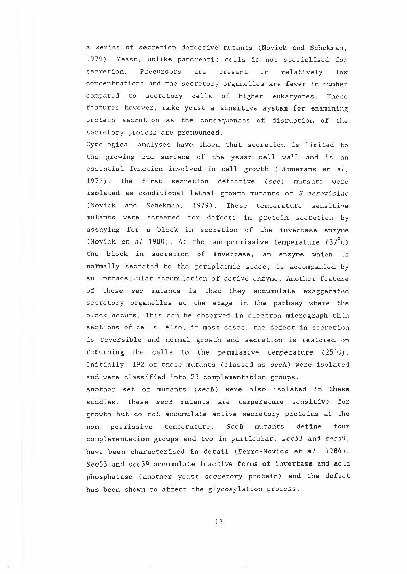
a series of secretion defective mutants (Novick and Schekman,1979). Yeast, unlike pancreatic cells is not specialised for secretion. Precursors are present in relatively low concentrations and the secretory organelles are fewer in number compared to secretory cells of higher eukaryotes. These features however, make yeast a sensitive system for examining protein secretion as the consequences of disruption of the secretory process are pronounced.Cytological analyses have shown that secretion is limited to the growing bud surface of the yeast cell wall and is an essential function involved in cell growth (Linnemans et al, 1977). The first secretion defective (sec) mutants were isolated as conditional lethal growth mutants of S .cerevisiae (Novick and Schekman, 1979). These temperature sensitive mutants were screened for defects in protein secretion by assaying for a block in secretion of the invertase enzyme (Novick et al 1980). At the non-permissive temperature (37°C) the block in secretion of invertase, an enzyme which is normally secreted to the periplasmic space, is accompanied by an intracellular accumulation of active enzyme. Another feature of these sec mutants is that they accumulate exaggerated secretory organelles at the stage in the pathway where the block occurs. This can be observed in electron micrograph thin sections of cells. Also, in most cases, the defect in secretion is reversible and normal growth and secretion is restored on returning the cells to the permissive temperature (25°C). Initially, 192 of these mutants (classed as secA) were isolated and were classified into 23 complementation groups.Another set of mutants (secB) were also isolated in these studies. These secB mutants are temperature sensitive for growth but do not accumulate active secretory proteins at the non permissive temperature. SecB mutants define four complementation groups and two in particular, sec53 and sec59, have been characterised in detail (Ferro-Novick et al. 1984). Sec53 and sec59 accumulate inactive forms of invertase and acid phosphatase (another yeast secretory protein) and the defect has been shown to affect the glycosylation process.
12
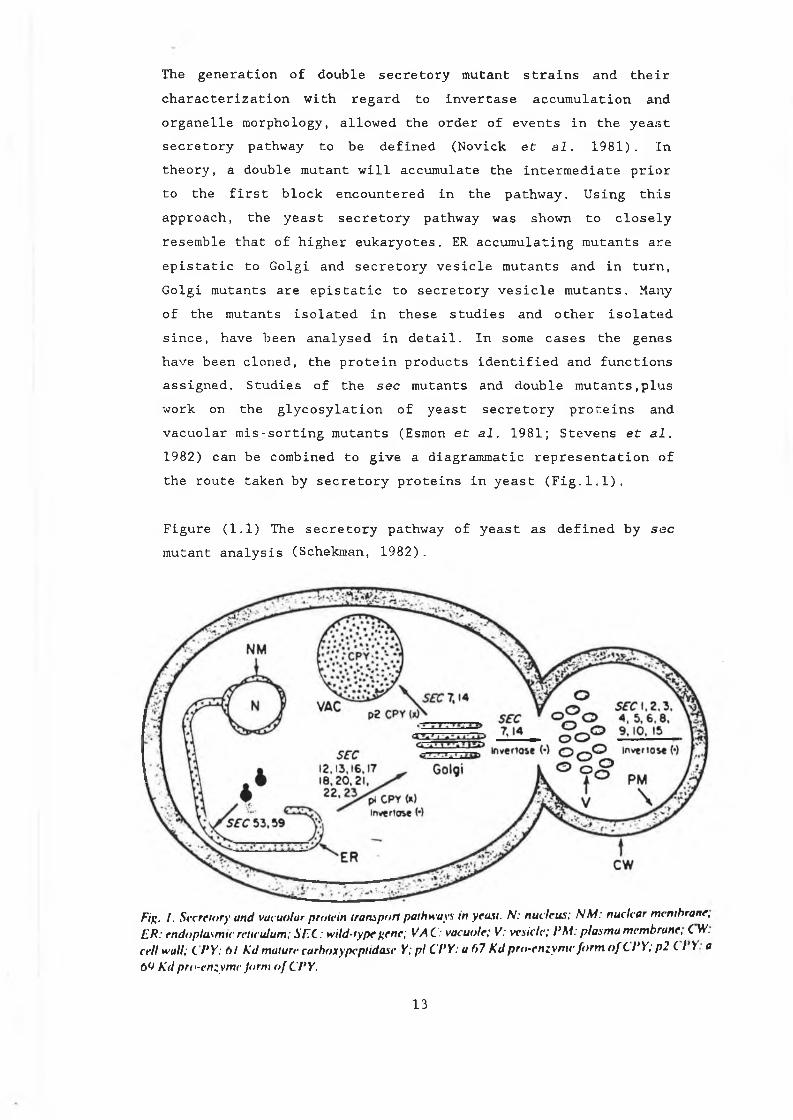
The generation of double secretory mutant strains and their characterization with regard to invertase accumulation and organelle morphology, allowed the order of events in the yeast secretory pathway to be defined (Novick et al. 1981). Intheory, a double mutant will accumulate the intermediate prior to the first block encountered in the pathway. Using this approach, the yeast secretory pathway was shown to closely resemble that of higher eukaryotes. ER accumulating mutants are epistatic to Golgi and secretory vesicle mutants and in turn, Golgi mutants are epistatic to secretory vesicle mutants. Many of the mutants isolated in these studies and other isolated since, have been analysed in detail. In some cases the genes have been cloned, the protein products identified and functions assigned. Studies of the sec mutants and double mutants,plus work on the glycosylation of yeast secretory proteins and vacuolar mis-sorting mutants (Esmon et al. 1981; Stevens et al. 1982) can be combined to give a diagrammatic representation of the route taken by secretory proteins in yeast (Fig.1.1).
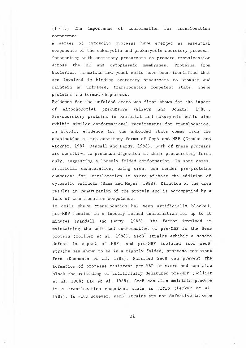
Figure (1.1) The secretory pathway of yeast as defined by sec mutant analysis (Schekman, 1982).
Fig. / . Secretory and vacuolar protein transport pathways in yeast. N: nucleus; NM: nuclear membrane, ER: endoplasmic reticulum; SFC: wild-type f enc; VA C: vacuole; V: vesicle; PM: plasma membrane, C W. cell wall; C P Y: A/ Kd mature carboxypeptidase Y; pi CPY: a 67 Kd pro-enzyme form oftPY; p2 C I V a b'J Kd pre-enzyme Jorni of CPY.
1 3
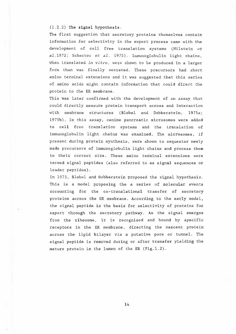
(1.2.2) The signal hypothesis.The first suggestion that secretory proteins themselves contain information for selectivity in the export process came with the development of cell free translation systems (Milstein et 3.1.1972; Schecter et al. 1975). Immunoglobulin light chains,when translated In vitro, were shown to be produced in a larger form than was finally secreted. These precursors had short amino terminal extensions and it was suggested that this series of amino acids might contain information that could direct the protein to the ER membrane.This was later confirmed with the development of an assay that could directly measure protein transport across and interaction with membrane structures (Blobel and Dobberstein, 1975a;1975b). In this assay, canine pancreatic microsomes were added to cell free translation systems and the translation ofimmunoglobulin light chains was examined.. The microsomes, if present during protein synthesis, were shown to sequester newly made precursors of immunoglobulin light chains and process them to their correct size. These amino terminal extensions were termed signal peptides (also referred to as signal sequences or leader peptides).In 1975, Blobel and Dobberstein proposed the signal hypothesis. This is a model proposing the a series of molecular eventsaccounting for the co-translational transfer of secretory proteins across the ER membrane. According to the early model, the signal peptide is the basis for selectivity of proteins for export through the secretory pathway. As the signal emerges from the ribosome, it is recognised and bound by specific receptors in the ER membrane, directing the nascent proteinacross the lipid bilayer via a putative pore or tunnel. The signal peptide is removed during or after transfer yielding the mature protein in the lumen of the ER (Fig.1.2).
1 4
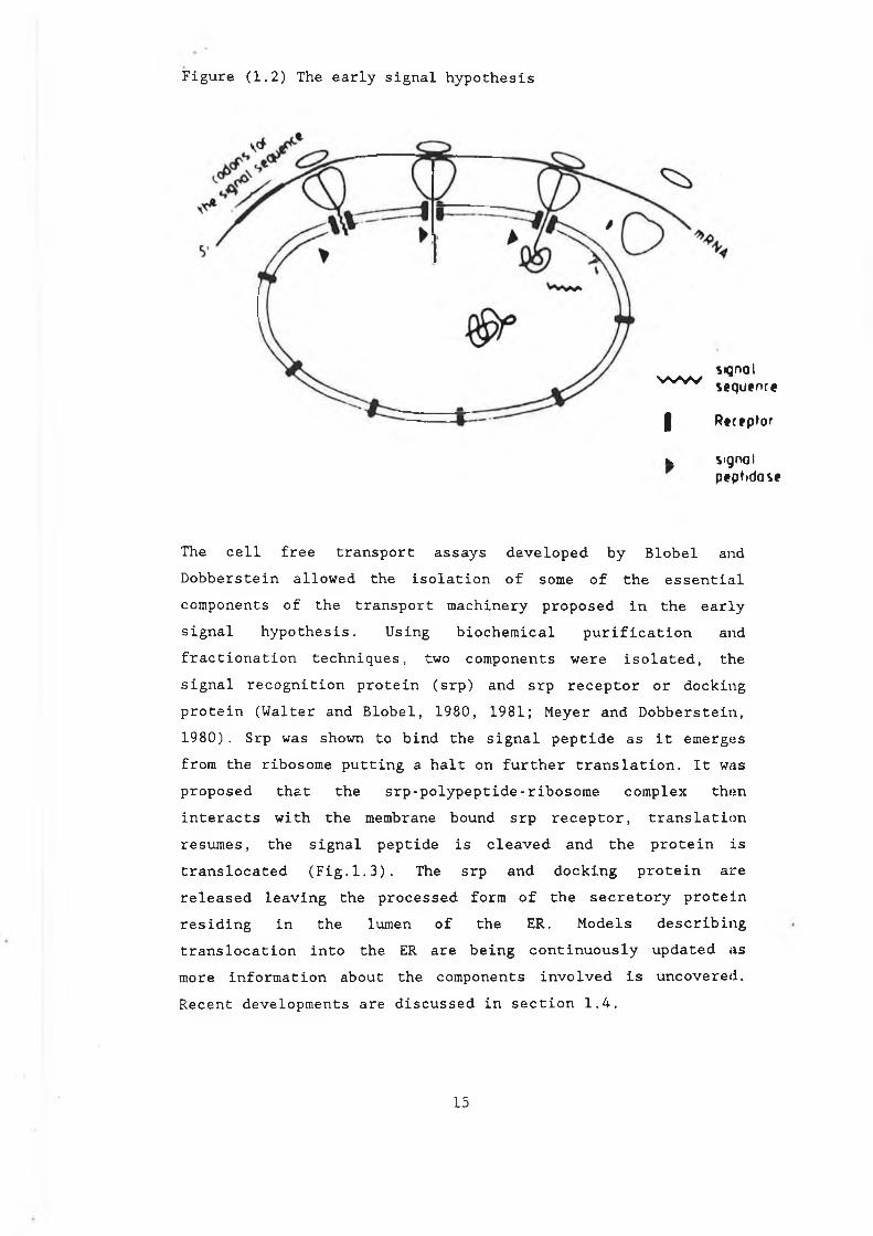
Figure (1.2) The early signal hypothesis
signalw w sequence
I Receptor
v signalpeptidase
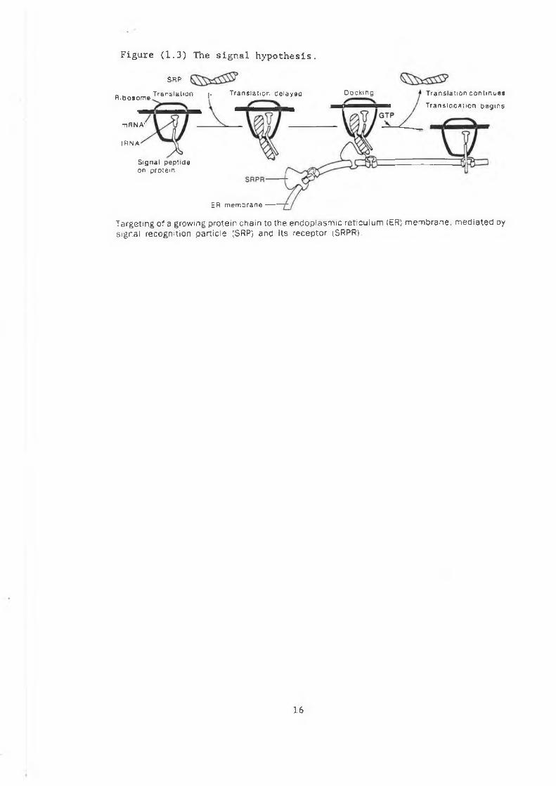
The cell free transport assays developed by Blobel and Dobberstein allowed the isolation of some of the essential components of the transport machinery proposed in the early signal hypothesis. Using biochemical purification and fractionation techniques, two components were isolated, the signal recognition protein (srp) and srp receptor or docking protein (Walter and Blobel, 1980, 1981; Meyer and Dobberstein,1980) . Srp was shown to bind the signal peptide as it emerges from the ribosome putting a halt on further translation. It was proposed that the srp-polypeptide-ribosome complex then interacts with the membrane bound srp receptor, translation resumes, the signal peptide is cleaved and the protein is translocated (Fig.1.3). The srp and docking protein are released leaving the processed form of the secretory protein residing in the lumen of the ER. Models describing translocation into the ER are being continuously updated as more information about the components involved is uncovered. Recent developments are discussed in section 1.4.
15
ER m em b rane
Figure (1.3) The signal hypothesis.
SRP
„ w Translat ion i. T ranslat ion ce layeoR ibosom e 1
mRNA'
IRN A
Signal peptide on protein
Docking T ranslat ion c o ntinues
Tran s lo ca t io n begins
Targeting of a growing protein chain to the en dop lasm ic re ticu lum (ER) m em b ran e , m e d ia te d by signal recognition p artic le (SRP) and its receptor (SRPR).
16
(1.3) Characterizing eukaryotic and prokaryotic signal peptides.After the isolation of the first eukaryotic signal peptide, it soon emerged that prokaryotic secretory proteins are also produced in precursor forms with signal sequences similar toeukaryotes (Inouye and Beckwith, 1977). The similarities between the two systems, despite the differences incomplexities, soon became apparent. Eukaryotic and prokaryotic signals are functionally interchangeable. The E.coli secretory protein, /3-lactamase, can be synthesised in a eukaryotic cellfree translation system and is efficiently translocated intodog pancreatic microsomes (Muller et al. 1982). Srp has beenshown to interact directly with this bacterial signal peptide effecting its translocation and when microinjected into Xenopus oocytes, it is efficiently processed and secreted into the cell medium (Weidmann et al. 1984).This functional interchangeability is successful in vivo also. The Bacillus a-amylase enzyme, for example, is secreted in yeast using its own signal peptide (Ruohonen et al. 1987).Similarly in bacterial cells, some eukaryotic proteins can be secreted (Talmadge et al. 1980). Also, signal peptides candirect cytosolic proteins to the secretory pathway. Theeukaryotic globin protein for example, when fused to the /3-lactamase signal peptide is translocated into microsomalvesicles (Lingappa et al. 1984) and in E.coli, the LamB(receptor for phage lambda) signal peptide targets /3-galactosidase to the cell membrane (Emr et al. 1980; Emr and Silhavy, 1980). Signal sequences therefore play an central role in protein export. They are necessary for directing proteins to the bacterial or ER membrane, a role that appears to be conserved to some extent between two evolutionarily diverse groups.
(1.3.1) Physical disection of signal peptides.Although signal peptides perform conserved functions, systematic comparisons of amino acid sequences from prokaryotes and eukaryotes reveal no overall consensus sequences or
1 7
absolute amino acid requirements (von Heijne 1984a, 1984b;Perlman and Halvorsen, 1983). Furthermore, signal peptides differ in length as well as in amino acid composition. These comparisons however, reveal that all signal peptides conform to an overall tripartite structure of three structurally distinct domains : a positively charged amino terminal domain(N-domain), a central hydrophobic core (H-domain) and a polar carboxyl terminus (C-domain) which contains the cleavage site.In a study comparing 118 eukaryotic and 32 prokaryotic signal peptides, von Heijne (1985) attempted to ascertain by statistical analysis, if there were limitations to the variations that could occur within these domains. The study reemphasises the variations that do occur but also suggests that minimal requirements for the distinct regions may exist. Briefly, comparison of the sequences showed that the length of the N-region can vary considerably but that the overall net charge does not. On average, in prokaryotes anyway, the mean charge is approximately +1.7. In the hydrophobic core there are no sequence restraints and the choice of hydrophobic amino acid in this region is random. The minimum length of the H-domain is 7 - 8 residues and this region spans from positions -6 to -13 and -7 to - L4 in eukaryotes and prokaryotes respectively. Extremes and exceptions do exist. Human pancreatic polypeptide and C .diptheriae toxin for example, have H-domains of 15 and 16 amino acids, and, the H-region of human choriogonadotrophin /3-subunit signal peptide has 9 residues, 7 of which are leucine.The cleavage site at the C-terminus is the least variable of the three. The length seldom varies from 5 amino acids in eukaryotes and 6 in prokaryotes. Also, certain sequence restraints are adhered to in that particular amino acids are preferred at particular positions in the cleavage site (section 1.3.4). These observations highlight the degeneracy of signal peptides at an amino acid level. However, they also underline the physical organisational pattern that signal sequences adhere to. The potential importance of these structural domains in maintaining signal function and the possible role of each in
18
the translocation process has been investigated.
(1.3.2) Disruption of the hydrophobic domain.Extensive studies of signal peptide mutants have shown that disruption of the structural domains can affect leader peptide function with varying degrees of severity. In vitro mutagenesis of the H-domain of prokaryotic and eukaryotic signal peptides has generated mutants that significantly affect protein secretion. Hydrophobic regions of polypeptides have the potential to form cc-helices, a thermodynamically preferred conformation in the non-polar environment of the lipid bilayer. It has been suggested that this region of the signal peptide interacts directly with the membrane during translocation (Wickner et al. 1980; Engleman, 1981). For this reason, a great deal of attention has focused on disruption mutants in the H-core of signal sequences.Analysis of E.coli secretory protein mutants has shown that disruptions to the H-domain can severely affect secretion. Mutations of the lamB signal peptide, were isolated by constructing a fusion between it and /?-galactosidase, a protein not normally secreted by this organism (Emr and Silhavy, 1980; Emr et al. 1980). Overexpression of this hybrid gene in E.co.li results in ¡3-galactosidase being directed to the cytoplasmic membrane, effectively jamming the normal export system. Because secretion is blocked, the cell cannot secrete its own enzymes and is prevented from using maltose as a carbon source (maltose sensitive). Mutations that alter secretion of the hybrid protein and therefore relieve jamming of the membrane result in normal secretion patterns and a maltose resistant phenotype. The mutations isolated in this manner, disrupting the function of the lamB signal peptide, all map to a group of 4 amino acids in the hydrophobic core. Some introduce a charged residue in the region while others yield various deletions. Similar results are obtained when /3-galactosidase is fused to the signal peptide of another secretory protein, the maltose binding protein (MBP), in E.coli (Bedoulle et al. 1980).In eukaryotes, the yeast secretory protein invertase has been
1 9
used for similar analyses. S .cerevisiae produces two types of invertase enzyme. Both are encoded by separate mRNA transcripts synthesized from a common gene, SUC2 (Perlman et al. 1982). The cytoplasmic form is constitutively produced and transcription of the secreted form is glucose repressed. When grown on sucrose as a carbon source, the secreted form is transcribed and the enzyme is produced in the precursor form with a signal peptide that directs the enzyme to the secretory pathway where it is transported to the periplasmic space.Deletion of 4 amino acids in the H-region of the signal peptide, reducing it from 8 to 4 residues, resulted in accumulation of active enzyme in the cytoplasm (Perlman et al.1986). Western blot analysis revealed that the mutant enzyme, now located in the cytoplasm, was not glycosylated and retained its signal peptide. This indicates that the enzyme had no interaction with the ER membrane. (Disruption of the invertase signal peptide cleavage site alone, inhibiting signal processing does not interfere with translocation (Schauer et al.1985).Along a similar line, human lysozyme is efficiently secreted and processed in yeast when fused to a synthetic signal peptide constructed to optimise the H-region (Yamamoto and Kikuchi1989). In this signal, 8 contiguous leucine residues make up the hydrophobic core. Leucine was chosen for this purpose because of its hydrophobic nature and its disposition toward forming a-helical structures (Chou and Fasman, 1978). Reduction of this core to just 6 leucine residues resulted in a decrease in secretion efficiency to 40% compared to the 8 leucine sequence.Overall, mutant leader peptides seem to have varying effects on translocation efficiency. In bacteria, point mutations and substitutions, sometimes affecting only one amino acid can have drastic consequences blocking secretion completely. In eukaryotes however, mutations that intefere with signal function and block secretion tend to be large deletions or rearrangements (Perlman et al. 1986; Gething and Sambrook,1982). In some cases, as outlined above, partial signal
20
function is retained. In bacteria also, deletions in the H-region of the outer membrane lipoprotein (lpp) of E.coli disrupt secretion to different extents, some deletions having more pronounced effects than others (Inouye et al. 1984).Certain mutations therefore, even in this highly conserved core retain the ability to translocate proteins, albeit with lower efficiencies.To investigate the extent that alternate sequences can function as signal peptides, Kaiser (1987) adapted the secretion of invertase in yeast as a model test system. The invertase signal peptide was removed and replaced with random peptide sequences derived from human genomic DNA. 20% of these random sequences were shown to contain some signal function, allowing the secretion of invertase, reflected by the ability of these cells to grow on sucrose. Although the sequences secreted invertase with different efficiencies (only a small amount of secreted enzyme is required for growth on sucrose), all allowed a proportion of the enzyme to be translocated into the lumen of the ER where core glycosylation took place. Sequence comparisons to non-functional isolates showed the functional ones to be enriched in hydrophobic residues and depleted in charged amino acids.Revertants of the non-functional, secretion defective isolates generated in this study were also examined (Preuss and Botstein, 1989). The revertants were selected by their ability to grow on sucrose as a sole carbon source after treatment with the mutagen ethylmethylsulphate (EMS), or with ultra violet (UV) irradiation. Spontaneous revertants were also isolated. Sequencing the revertant leaders showed that the mutations allowing secretion were all point mutations that either introduced hydrophobic residues or deleted charged residues in the N-terminal random sequences.Kaisers experiments demonstrate the broad range of sequences that can function, however inefficiently, as signal peptides allowing targeting and translocation to the ER membrane. The hydrophobic domain is clearly important and the functional sequences isolated in the above studies resemble known signal
21
peptides in this respect. This region has a potential role in membrane targeting as well as interacting with the ER membrane itself. However, the specificity of the export process is unlikely to depend on the presence of a hydrophobic domain alone, especially considering that most proteins contain hydrophobic sequences. The inefficiency of the sequences isolated in Kaisers experiments underline this.The conservation of overall signal peptide structure, as already mentioned, goes further than the presence of this hydrophobic domain. A positively charged amino terminus and the cleavage site region are equally well conserved (von Heijne, 1985). The roles of these domains in the translocation process and overall signal function and their interaction with each other have also been considered.
(1.3.3) The hydrophillic domain: the role of positively charged amino acids.The genetic selections used to generate signal peptide mutants in vivo, some of which have been described, all yielded disruptions mapping to the hydrophobic domain. In no case were alterations to the hydrophillic N-domain of the signal peptide obtained that resulted in a secretion defective phenotype. The hydrophillic domain however is a conserved feature in all signal peptides and in prokaryotes, secretory proteins carry a net positive charge in this region (von Heijne 1984a).The potential importance of these charged residues and their possible role in prokaryotic signal function, was first proposed in the "loop model" for protein translocation (Inouye and Halegoua, 1982). According to this model, the N-terminal of the signal peptide does not leave the cytoplasm. The basic amino acids in this region are proposed to interact with the acidic groups in the phospholipids of the bacterial cytoplasmic membrane, anchoring it in the cytoplasm and allowing the hydrophobic core to loop into the membrane. The electrochemical potential that exists across the bacterial cytoplasmic membrane, which is negatively charged on the cytoplasmic side, would promote this type of interaction (Li et al. 1988).
22
Oligonucleotide site-directed mutagenesis has generated mutations mapping to the hydrophillic N-domain of prokaryotic and eukaryotic signal peptides. A lot of interest has focussedon positively charged amino acids in this region. Although theN-termini of all prokaryotic signal peptides contain at least one basic amino acid, evidence suggests that this feature isnot absolutely required for signal function. Removal of the 3 basic residues in the maltose binding protein (MBP) of E.coll, for example, has no effect on export of the protein (Puziss et al. 1989). Similarly, when the 2 lysine residues are removedfrom the prolipoprotein signal peptide, no discernable effect on translocation is observed (Vlasuk et al. 1983). However, anet charge of at least +1 is required to obtain correctprocessing when this signal peptide is fused to /3-lactamase (Lunn et al. 1987). The introduction of net negative charges in the N-domain of prolipoprotein and MBP does affecttranslocation, decreasing the kinetics of the processconsiderably (Vlasuk et al. 1983; Pusiss et al. 1989).Translocation takes place, but at a slower rate compared to the wild type sequences.These observations have led to the suggestion that the N-domain of the signal peptide plays a facilitative role in translocation. The presence of positive charges, while not absolutely necessary, may act to improve the efficiency of the whole process. The exact function of these charged residueshowever remains unclear. Although it has not been proved conclusively, cross-linking studies suggest that the SecA protein, a cytoplasmic component of the E.coll secretory pathway that is essential for translocation, interacts with the positive charges in the OmpA signal peptide (Akita et al.
1990). The extent of the interaction is dependant on the number of positive charges present in the hydrophillic domain. The SecA protein also interacts with other known secretory pathway components and could therefore mediate interactions between the signal peptide and other components involved (Fandl et a.l.
1988). PrlD2 supressor strains can partially rescue the phenotype of MBP N-domain mutants (Puziss et al. 1989). The
2 3
prlD2 supressor strain, which is allelic to SecA (Fikes and Bassford, 1989), has no effect on hydrophobic core mutations and it is possible that SecA interacts more strongly with the hydrophillic section of the signal peptide.In yeast, a potential role for the hydrophillic domain of signal peptides is less easy to define. Not all yeast signal peptides exhibit a net positive charge in the N-domain (Kaiser and Botstein, 1986) and the ER membrane does not contain a general electrochemical potential. None of the random sequences generated by Kaiser (1987) that promoted translocation of invertase, contained hydrophillic segments or positive charges. In contrast to this, with a similar experiment carried out in E.coli, most of the functional leader peptides derived from random sequences did contain a net positive charge preceding the hydrophobic domain (Zhang and Broome-Smith, 1989).The signal peptide of the yeast secretory protein alpha-factor, has an arginine residue at position 3 in the pre-region. The position of this positively charged residue rather than it's presence appears to be an important feature in the functioning of this leader peptide (Green et al. 1989). Replacement of this amino acid with a neutral phenylalanine residue, has no effect on secretion. Misplacement of the positive charge by one position however, has a significant effect, reducing translocation efficiency by up to 70%.Green (1989) proposes that misplacement of the positive charge in the a-factor pre-region may adversely affect interactions of the protein precursor with molecular chaperones such as heat shock proteins (hsps) (section 1.4.4), or a similar interaction with other components of the secretory pathway. It has also been suggested that this charged residue may be important in intramolecular interactions with a series of 3 contiguous acidic amino acids in the mature region of the a-factor protein (at positions 7-9). Transposition of the charged residue could promote or destroy an electrostatic interaction in this region, resulting in a reduction in translocation efficiency.The interaction of positive charges in the mature sequence of prokaryotic secretory proteins and the N-domain of signal
24
peptides has been investigated. A net positive charge at the extreme N-terminus of chicken triosephosphate isomerase, when fused to the E.coll lamB signal peptide, has a detrimental effect on translocation of the protein (Andrews e C al. 1989; Summers and Knowles, 1989). As the positive charges are moved further away from the N-terminus, by the insertion of lamB sequences, the blocking effect weakened. The introduction of positive charges into this region of the mature alkalinephosphatase sequence results in a similar defect intranslocation efficiency (Li et al. 1988). A study ofprokaryotic signal peptides (von Heijne, 1986) shows that in most cases there is a net negative or neutral charge in thearea including the C-domain and the extreme N-terminus of themature sequences of secretory proteins. This characteristic may have evolved to avoid the detrimental effects positive charges can have on translocation of certain proteins. No such pattern is observed however, in the majority of eukaryotic secretory proteins.The potential importance of the N-domain in the folding ofsecretory precursors has been mentioned. Positive charges inthis region of yeast secretory proteins appears to be important in some cases only. Signal peptides, having evolved with a specific protein, will presumably be optimised for the translocation needs of that particular molecule, taking into account its charged regions, folding characteristics and conformational requirements. In many cases, the particularproperties of some signal peptides or the requirements of certain proteins to achieve translocation only become apparent when specific characteristics are modified, or in heterologous fusion experiments.It seems likely that the N-domain is important in improving the efficiency of the translocation process. With the exception of hsps, specific components that interact with this region have yet to be identified in yeast. The recent identification of proteins involved in early tranlocation events in the yeast secretory pathway (section 1.4.2) may reveal more information about protein interactions in this region.
25
(1.3.4) The signal peptide cleavage site.The C-terminal of signal peptides, containing the cleavage site, is the only region where the conservation of amino acid sequences is observed. A statistical analysis of sequences around the cleavage site of prokaryotic and eukaryotic leader peptides shows that certain patterns of amino acids are preferred in this region. (von Heijne, 1983, Perlman and Halvorsen, 1983). These studies have revealed that small neutral amino acids predominate at positions -1 and -3 (position -1 being defined as one amino acid upstream of the actual cleavage site), and that aromatic residues are strictly avoided at these positions but are present at position -2. More extensive analyses show that alanine is the preferred residue at -1 and -3 and, that an order of preference exists for other acceptable amino acids at these positions (von Heijne, 1983).A general model has been proposed for how signal peptides maintain their cleavage specificity (von Heijne, 1984b). This model incorporates the position of the last residue of the H-domain (occurring at position -6 in eukaryotes and -7 in prokaryotes). The end of the H-domain defines a window of potential cleavage sites that compete for access to the signal peptidase. Cleavage occurs at the most probable site defined by the observed amino acid preferences revealed in the study outlined above. Although this model is based on statistical analysis of known signal sequences, a study of approximately 300 eukaryotic, prokaryotic and viral leader peptides (Watson1984), showed that for 74% of viral and eukaryotic proteins, cleavage occurred at the site with the highest processing probability. 11% of these proteins were cleaved at the second most likely site. For prokaryotic signal sequences, 39% of actual cleavage sites were as predicted with 15% at the second most likely site.Analysis of signal sequence termini shows that in many cases greater than one potential cleavage site exists and in vivo cleavage heterogeneity does occur. Bovine growth hormone for example, is processed at an alanine residue in 65% of case/3, the remaining 35% of cleavage events occurring one position
26
upstream (Lingappa et al. 1977). Human interferon when produced in yeast is processed at more than one site (Hitzeman et al.
1983) , and cleavage of rat prolactin can be forced to take place at a less favourable site by replacing one amino acid at the ideal cleavage site (Hortin and Boime, 1981).
(1.4) Proteins that interact with signal peptides.
(1.4.1) The signal recognition particle (Srp) and Srp receptor. The first components of the eukaryotic secretory pathway identified to be involved in sequestering secretory proteins, were the srp and srp receptor (section 1.2.2). Disassembly, fractionation and reconstitution experiments (summarized by Seigel and Walter, 1988a), have allowed the detailed characterization of mammalian srp and its functions. Srp is made up of 6 polypeptides and a 7SL RNA molecule. The complex, which is held together by protein/protein and protein/RNA interactions, is arranged into 4 units consisting of 2 dimers (p9/14 and p68/72) and 2 monomers (pl9 and p54).Srp recognizes the signal peptide of a nascent secretory protein as it emerges from the ribosome (Walter and Blobel,1981). The specificity of this recognition and binding is dependant on nascent chain length. The efficiency of srp directed targeting drops dramatically as polypeptides emerging from the ribosome exceed approximately 140 amino acids in length (Siegel and Walter, 1988b). Translation arrest, which occurs after binding to the signal peptide and ribosome and which is a characteristic feature of membrane targeting in higher eukaryotes, is associated with the p9/14 dimer subunit of srp (Siegel and Walter, 1988a). Photocrosslinking experiments show that the 54kDa polypeptide of srp specifically binds signal peptides of various secretory proteins (Kurzchalia et al. 1986; Krieg et al. 1986). Sequence analysis of thispolypeptide reveals that it is made up of 2 domains, a G-domain which has the potential to bind GTP, and an M-domain, rich in methionine residues (Romisch et al. 1989; Bernstein et al.
1989). This protein also shares homology with the srp receptor
27
which is present on the ER membrane.Proteins sharing homology with parts of the mammalian srp havebeen identified in E.coli and yeast (S.cerevisiae andSz.pombe). In yeast, the genes were isolated on the basis of their sequence homology to the p54 subunit of mammalian srp and predict protein sequences that are 50% identical to themammalian counterpart (Hann et al. 1989). The yeast proteinscontain a G-domain and an M-domain and although no functions have been designated as of yet, they represent the firstevidence of srp related proteins in yeast. In E.coli, a 4.I5S RNA molecule which specifically binds a 48kDa (p48) protein has been identified (Ribes et al. 1990). The mammalian srpcomponent, p54, can also bind this RNA in a complex that can functionally replace the 4.5S RNA/p48 complex to promote translocation of /3-lactamase in E.coli. Also the mammalian 7S RNA species can functionally replace the E.coli 4.5S RNA. This E.coli 4.5S RNA and p48 are thought to be structural and functional homologues of the mammalian species and are involved in the transloocation of at least some of E.coli's secretory proteins.Srp has a close affinity for its receptor in the ER membrane. Interaction with the srp receptor results in displacement of srp from the nascent chain/ribosome complex, a process which involves GTP hydrolysis (Connolly and Gilmore 1989). Thisinteraction results in transfer of the signal peptide to the ER membrane where it is found in close proximity to a 34kDaintegral membrane protein known as the signal sequence receptor protein (ssr) (Weidmann et al. 1989). The ssr protein is aconstituent of the protein environment where secretory proteins are transferred through the membrane. Antibodies raised against the protein inhibit translocation across microsomal membranes in vitro (Hartmann et al. 1989) and it seems likely that thisprotein forms part of a putative translocation complex in theER membrane. Further investigations should reveal moreinformation about the nature of this complex and the mechanisms by which translocation, after the targetting event, isachieved.
28
(1.4.2) Proteins involved in the yeast translocation event.Genetic selections have been used to isolate components of the yeast secretory process (section 1.2). None of the temperature sensitive, secretion defective (sec) mutants isolated in these initial studies however, uncovered steps in the early translocation stage of secretion. The earliest acting mutants isolated in this screen were the glycosylation mutants affecting proteins after translocation into the ER (Ferro-Novick et al. 1984).A more refined assay, developed to specifically target translocation events (described in detail in Fig.1.4), resulted in the the identification of three genes SEC61, SEC62 and SEC(53 (Deshaies and Schekman, 1987; Rothblatt et al. 1989). In vivo and in vitro studies show that at the non-permissive temperature, these temperature sensitive mutants block transport of secretory and vacuolar proteins into the ER (Deshaies and Schekman, 1987 and 1989) and accumulation of unprocessed precursors occurs in the cytoplasm. Interestingly, all three mutants isolated in this screen encode products that are membrane associated, no yeast equivalents of srp-like components were identified, although Sec61 is a possible candidate.Initially Sec62 was shown to be an integral membrane protein (Deshaies and Schekman, 1989 and 1990) and sequence analysis of SEC63 reveals that the gene product has a potential membrane spanning domain (Sadler et al. 1989). Antibodies to the Sec<52 protein were used to investigate the nature of the protein's association with the ER membrane (Deshaies et a l . 1991'). Immunoprécipitations using this antibody on solubilised membranes, showed that Sec62 is associated with Sec61 and Sec(33 in the ER membrane, comprising a multisubunit membrane associated complex. Furthermore, Sec61 appears to have a labile association with this putative complex, suggesting a potential intermediary role, possibly involved in targetting. Two other proteins of molecular weights 31.5 and 28kDa were precipitated along with this complex. Based on molecular weight comparisons, these proteins were tentatively proposed to be comparable to 2 subunits of the mammalian ssr protein. It is likely that this
2 9
complex is involved in the actual translocation of secretory proteins at this site in the ER membrane. Further manipulations, including reconstitution experiments, may uncover the mechanism by which proteins are translocated across membrane structures in yeast, the details of which have proved elusive in the less easily manipulated mammalian systems.
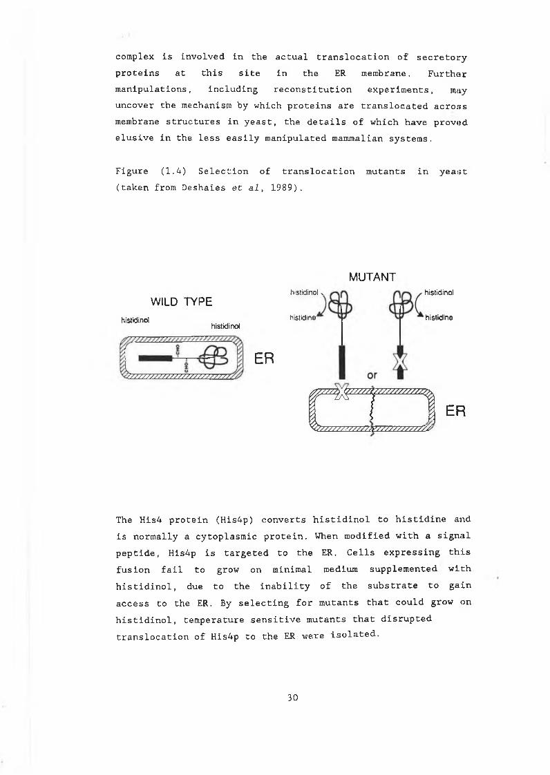
Figure (1.4) Selection of translocation mutants in yeast (taken from Deshaies et al, 1989).
WILD TYPE
histidinolhistidinol
ER?/ // / / / / / / / / / / / / / / / / / / / / / / .
MUTANThistidinol »
h istid ine '
histidinol
h istidine
m2Z22222222ZreZZZZZZ22ZZZZy
ER
The His4 protein (His4p) converts histidinol to histidine and is normally a cytoplasmic protein. When modified with a signal peptide, His4p is targeted to the ER. Cells expressing this fusion fail to grow on minimal medium supplemented with histidinol, due to the inability of the substrate to gain access to the ER. By selecting for mutants that could grow on histidinol, temperature sensitive mutants that disrupted translocation of His4p to the ER:were isolated.
30
(1.4.3) The importance of conformation for translocationcompetence.A series of cytosolic proteins have emerged as essential components of the eukaryotic and prokaryotic secretory process, interacting with secretory precursors to promote translocation across the ER and cytoplasmic membranes. Proteins from bacterial, mammalian and yeast cells have been identified that are involved in binding secretory precursors to promote and maintain an unfolded, translocation competent state. These proteins are termed chaperones.Evidence for the unfolded state was first shown for the import of mitochondrial precursors (Eliers and Schatz, 1986).Pre-secretory proteins in bacterial and eukaryotic cells also exhibit similar conformational requirements for translocation. In E.coli, evidence for the unfolded state comes from the examination of pre-secretory forms of OmpA and MBP (Crooke and Wickner, 1987; Randall and Hardy, 1986). Both of these proteins are sensitive to protease digestion in their presecretory forms only, suggesting a loosely folded conformation. In some cases, artificial denaturation, using urea, can render pre-proteins competent for translocation in vitro without the addition of cytosolic extracts (Sanz and Meyer, 1988). Dilution of the urea results in renaturation of the protein and is accompanied by a loss of translocation competence.In cells where translocation has been artificially blocked, pre-MBP remains in a loosely formed conformation for up to 10 minutes (Randall and Hardy, 1986). The factor involved in maintaining the unfolded conformation of pre-MBP is the SecB protein (Collier et al. 1988). SecB strains exhibit a severe defect in export of MBP, and pre-MBP isolated from secB strains was shown to be in a tightly folded, protease resistant form (Kumamoto et al. 1988). Purified SecB can prevent the formation of protease resistant pre-MBP in vitro and can also block the refolding of artificially denatured pre-MBP (Collier et al. 1988; Liu et al. 1988). SecB can also maintain proOmpA in a translocation competent state in vitro (Lecker et al. 1989). In vivo however, secB strains are not defective in OmpA
3 1
export and sec B cell extracts can still promote OmpA translocation in vitro (Kumamoto et al. 1989). Another chaperone protein, trigger factor, has been shown to specifically interact with proOmpA to promote translocation in much the same: way as SecB (Crooke and Wickner 1987) . Thisevidence suggests that there are several chaperone proteins involved in protein export and that different ones are specific for different proteins.Eukaryotic cells also appear to require the presence of molecular chaperones and it has been suggested that srp may act as such in mammalian cells. Srp can replace trigger factor to stabilise proOmpA for translocation across bacterial, yeast and mammalian membrane structures (Sanz and Meyer, 1988). It is also functional in promoting prepro a-factor translocation into yeast microsomes (Crooke et al. 1988). Dénaturation of these proteins prior to the addition of srp was required to allow translocation. This correlates with the co-translationaltargeting role of srp in vivo and the general idea thatchaperones are likely to bind precursors as they are synthesized rather than actively unfolding the completed proteins.
(1.4.4) Unfolding in yeast: the role of heat shock proteins.In yeast, genetic and biochemical evidence suggests that members of the heat shock family of proteins (hsps) play a role in promoting translocation competence in pre-secretory proteins (Chirico et al. 1988). When prepro-a-factor is produced in wheat germ lysate, it can only be translocated into yeastmicrosomes if yeast cytosolic extract is added. Fractionation of the extract showed that this activity is associated with 2 yeast hsps, SSAl and SSA2 (members of the 70kDa hsp family). The effect can also be achieved if the protein is denaturedbefore addition of the yeast membranes. This reinforces the idea that proteins must be unfolded in order to be translocated in yeast. The involvement of these proteins is backed up at a genetic level in that ssal mutants accumulate a-factor, carboxypeptidase-Y and mitachondrial precursors in unprocessed,
32
untranslocated forms (Werner-Washburner et al. 1987).The in vitro evidence involving fully formed a-factor, suggests that hsps can act in a post-translational capacity. It has been proposed that these proteins may act as "unfoldases", possibly using energy derived from ATP hydrolysis to actively unfold protein precursors before translocation (Rothman and Kornberg,1986). Hsps have also been implicated in post-translational translocation in mammalian cells (Zimmerman et al. 1988) and in E.coli where the GroEL protein has been shown to interact with /3-lactamase to promote its translocation (Kusukawa et al. 1989).
(1.4.5) Characterization of signal peptidases.Two E.coli signal cleavage enzymes have been identified, leader peptidase or SP1 (Zwizinski and Wickner 1980) and lipoprotein peptidase or SP11 (Tokunaga et al. 1982). Both signalpeptidases are integral membrane proteins with molecular masses of 36 and 18kDa respectively. Repression of leader peptidase function results in accumulation of unprocessed precursors in the E.coli periplasm (Dalbey and Wickner, 1985) This suggests that in E.coli, while signal peptidase is not essential for translocation, retention of the signal peptide inhibits the release of secretory proteins into the periplasm.Signal peptidases of higher eukaryotic cells appear to be more complex. Canine and avian signal peptidases have been isolated as multisubunit complexes (Evans et al. 1986; Baker and Lively,1987). Canine microsomal signal peptidase consists of 6 polypeptides and hen oviduct signal peptidase exists as a heterodimer. Two of the canine and one of the avian subunits are glycosylated and share sequence homologies (Shelness et al.1988). Despite the differences in complexities, the substrate specificities of prokaryotic and eukaryotic signal peptidases are remarkably similar and both enzymes cleave eukaryotic and prokaryotic signal peptides at the correct cleavage sites (Watts et al. 1983; Muller et al. 1982). The extra subunits of the vertebrate enzyme complex may provide additional functions associated with increasing the efficiency or cleavage
33
specificity of the signal cleavage process in vertebrates (Randall and Hardy, 1989).Disruption of signal peptide cleavage sites in yeast secretory proteins results in retention of the signal peptide. Translocation of the mutant protein is not affected. Replacement of an alanine residue with valine at position -1 in the secreted form of invertase inhibits cleavage and the unprocessed form of the enzyme is translocated into the lumen of the ER (Schauer et al. 1985). Although translocation is achieved, the mutant invertase accumulates in the ER and further secretion is delayed. This has been shown to be a result of insolubility of this form of invertase in the lumen of the organelle, combined with a possible association between the mutant protein and the ER membrane via the signal peptide (Schauer et al. 1985). Another yeast secretory protein, acidphosphatase, is also transiently retained in the ER when signal cleavage is inhibited (Haguenauer-Tsapis et al. 1986).A mutation to the yeast signal peptidase would presumably mimic this phenotype of delayed secretion patterns. One of the yeast temperature sensitive sec mutants, secll, was shown to transport invertase and acid phosphatase at severely reduced rates (Bohni et al. 1988). At the restrictive temperature,secll cells accumulate core glycosylated acid phosphatase and invertase with their signal peptides still intact. The SEC11 gene was cloned and shown to encode an 18.8kDa integral membrane protein with one potential glycosylation site. The mass of the protein is very similar to the avian and canine glycosylated subunits described and although it does not share exact sequence homologies with the canine subunit, the two proteins have similar arrangements of hydrophillic and hydrophobic domains (Shelness et al. 1988). The SEC11 geneproduct is essential for signal peptidase activity in yeast. At this stage it is not clear if the yeast signal peptidase, as it is now referred to, acts as part of a complex. Also it is not known if the Secll protein is responsible for the actual cleavage event. A method has been developed for the solubilisation of microsomal membranes retaining signal
3 4
peptidase activity and should allow further characterisation of the Secll protein (Ya Deau and Blobel, 1989).
(1.5.0) Sorting signals in the secretory pathway.In addition to secretory proteins, the ER contains proteins bound for membrane structures, proteins en route to the Golgi and proteins destined to remain in the ER itself. A large number of these ER proteins interact with incoming proteins allowing them to fold and mature before being passed on (Pelham1989) . An intriguing question which arises out of studying this transport process, is how the cell sorts these proteins. What distinguishes ER resident proteins from those that are to be passed on and how are proteins targeted to their final destinations?. Also, in the Golgi, how are proteins destined for vacuolar compartments diverted from the bulk flow of protein traffic into vacuole specific vesicles? While the question of protein sorting in the secretory pathway ofeukaryotic cells is far from fully understood, the investigation of two sorting events has allowed some insights into the mechanisms involved and the types of signals used.
(1.5.1) Retention In the ER.Proteins enter the ER in an unfolded state (section 1.4). In order to be transported further, refolding, assembly and ERmodifications must first take place. In mammalian cells, an ER protein known as BiP (binding protein) was found associated with malfolded secretory proteins that were blocked from further transport in the ER of mammalian cells (Gething et al, 1986). Enhanced expression of the protein was observed under conditions that promote the accumulation of unfolded or mutant proteins (Kozutsumi et al, 1986). BiP was also found to betransiently associated with wild type forms proteins in the ER (Bole et al, 1986). It was proposed that BiP, and possiblyother ER proteins, could be involved in monitoring the folding of secretory proteins, allowing correct assembly, folding and modifications to take place before further transport
35
(Normington et al 1989).The yeast homologue of BiP acts in a similar fashion to its mammalian counterpart (Rose et al 1989; Normington et al 1989). Expression of yeast BiP is induced when secretory proteins accumulate in the ER, such as in seec'. 53 mutants or after treatment with tunicamycin (section 1.6).The identification of BiP and other ER resident proteins such as PDI (protein disulphide isomerase), has allowed some insight into ER sorting mechanisms. Comparison of BiP and PDI sequences reveals a common carboxyl terminus of four amino acids, KDEL (Munro and Pelham, 1987). Removal of this KDEL sequence results in secretion of the proteins (Mazzarella et al 1990). The addition of the tetrapeptide to the C-terminal of secretory proteins, results in their accumulation in the ER (Pelham,1988). Experiments with S.cerevisiae BiP and PDI proteins have yielded similar results, although in this organism the retention signal differs to the mammalian by one amino acid ie.HDEL instead of KDEL (Hardwick et al 1990). In Sz.pombe, thesequence is ADEL while K.lactls recognizes both DDEL and HDEL (Inhohara et al, 1989; Lewis et al, 1990).In S.cerevisiae, secretory proteins tagged with HDEL accumulatein the ER, having acquired modifications known to occur in early Golgi compartments (Dean and Pelham, 1990) (see section 1.6 for glycosylation of secretory proteins). This observation led to a model for retention based on retrieval. It was proposed that ER proteins can leave the ER in secretory vesicles but are retrieved by interaction with a putative membrane bound receptor. This receptor would bind the HDEL sequence and return the proteins to the ER via a special set of transport vesicles.This receptor (the ERD2 gene product) was identified in yeast by the isolation of mutants that failed to retain secretory proteins tagged with the HDEL retention sequence (Semenza et al1990). The gene was shown to encode a membrane spanning protein, the expression levels of which control the capacity of the retention system. This gene was also cloned from K.lactis. (Lewis et al 1990). S .cerevisiae, carrying the K.lactis ERD2 gene, recognises both HDEL and DDEL sequences, demonstrating
36
that this receptor specifically recognizes the retentionsignal. The human homologue of ERD2 has also been identified and the gene product localised to a post-ER Golgi-lilce compartment (Lewis and Pelham, 1990). While the availableevidence supports a model of retrieval for ER retention in higher and lower eukaryotes, the exact nature of theinteractions between signal and receptor and the process of retrieval are not fully understood.
(1.5.2) Vacuolar sorting signals.The second major sorting step that has attracted muchinvestigation is the diversion of proteins from the Golgi into secretory vesicles bound for the lysosome. In mammalian cells, tagging of acid hydrolyses with mannose-6-phosphate (Man-6-P) has been shown to signal targeting of these proteins (Sly and Fischer,1982).The yeast equivalent of the lysosome is the vacuole, an acidic compartment containing numerous hydrolytic enzymes. Studies on proteins directed to the vacuole has led to an understanding of some of the requirements for targeting in S .cerevislae. Carboxypeptidase-Y (CpY) is the best studied of these vacuolar hydrolases. It is produced as an inactive precursor (proCpY) and undergoes glycosylation in the ER before sorting occurs at the level of the Golgi (Hasilik and Tanner, 1978; Stevens et al 1982). In the vacuole, CpY is converted to its mature active form by the action of another vacuolar protease proteinase A (PrA) , the product of the PEP4 gene (Ammerer et al 1986) . Glycosylation is not involved in protein sorting to the vacuole in yeast and inhibition of glycosylation has no effect on localisation of CpY (Schwaiger et al 1982).Fusion experiments involving portions of the proCpY N-terminal and yeast invertase, showed that the propeptide of CpY can direct invertase to the vacuole (Bankaitis et al 1986). This fusion protein, when expressed on a low copy number plasmid, targeted invertase to the vacuole with < 2% being secreted to the cell surface. Increasing the copy number resulted in up to 20% of the invertase being secreted, suggesting that the
3 7
targeting system is saturable. This is also the case when wild type CpY is overexpressed in S.cerevisiae (Stevens et al 1986). Deletion studies and site directed mutagenesis of CpY identified four contiguous amino acids (QPRL), at positions 24 -27 in the pro-region of the signal sequence, to be responsible for targeting of CpY to the vacuole (Vails et al 1987) . Similar analyses with the other vacuolar hydrolase proteinase A (PrA), demonstrated that the pro region of this peptide can also target CpY to the vacuole (Klionski et al 1988). Sequence analysis of PrA, however, did not reveal a QPRL motif or any other similarities to CpY in this region, suggesting that greater than one recognition signal may exist.The CpY-invertase fusion was used to isolate potential receptors involved in the targeting process. In a sue strain, mutants missorting the CpY-invertase fusion to the cell surface were isolated on the basis of their ability to grow on sucrose as a sole carbon source. A large number of mutants, defining 47 complementation groups, were identified using this screen (Bankaitis et al 1986; Banta et al 1988). One mutant in particular, vpsl5 (producing a mutant protein kinase) has been analysed in detail. Study of this enzyme, which exhibits phosphorylating and autophosphorylating activities, has led to the suggestion that phosphorylation may play a role in the regulation of vacuolar recognition and sorting events in S.cerevisiae (Herman et al, 1991).
3 8
(1.6.0) N-linked glycosylation of eukaryotic proteins.Secretory proteins passing through the ER membrane, acquire asparagine-linked (N-linked) core oligisaccharide chains which are subsequently modified and extended in the Golgi. Studies in yeast using mutants defective in secretion and glycosylation have proved invaluable in characterising the individual steps involved in assembly, transfer and modification of these oligosaccharides.The assembly of oligosaccharide chains onto yeast and mammalian glycoproteins involves a large number of transfer reactions. This core oligosaccharide is assembled in both cases on a phosphorylated dolichol glycolipid (P-Dol), in the ER membrane. The core unit is made up of 14 sugars (3 glucose, 9 mannose and 2 N-acetylglucosamine residues), Glc^MangGlcNAc^, which are transferred to the polypeptide chain via an N-glycosidic bond to asparagine (Asn) residues. In both _yeast and mammalian cells, this Asn residue is a substrate when it occurs within the sequence Asn-X-Ser/Thr, where X can be any amino acid except proline (Lehle and Bause, 1984). The pathway for assembly and transfer of this core oligosaccharide is similar in both systems. The differences lie in the trimming and addition reactions that are initiated in the ER and completed in the Golgi.Much of the initial information about core glycosylation has come from investigations in mammalian cells. Yeast however, has confirmed this model with the isolation of alg (asparagine linked glycosylation) mutants that have allowed further characterisation of the individual steps. The assembly, transfer and trimming of the yeast oligosaccharide core is summarized below in sections (1.6.1) and (1.6.2) and in Figure(1.5) (Deshaies et al 1989).
3 9
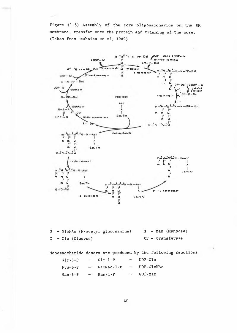
Figure (1.5) Assembly of the core oligosaccharide on the ER membrane, transfer onto the protein and trimming of the core. (Taken from Deshaies et al, 1989)
4 G O P— MM— M — N - N - P P - D o l ^ 4 P — Dol + 4 G D P — M
|3 f M-P-Dol aynthaaa4 M —P — Dol
M ^ —-4N — N — P P — Dol a-mannoayltr ^ translocate
4 mannoayltra- mannoayltr m—*M — M ^ N — N— P P —Dol
P — OolU D P — N \ ^ P P - O o l p h o s p h a t a s e
P P — Dol
ollgoaaccharyltr
I* Is l ’m m M
|> |>
m MIs
G - G - G — M
Xt
Ser/Thr
a - 0 iuco»iäia« I
|2 |J I«m m M
I2 |2 m M
IXI
Ser/Thr
a-alucoaidne II
m— M--"w*-4I2 Is Ism m M—* |> I2
m MI2
U
m m MI s I2
m MI2
M
a-glucoayltr
3 P- D o l + 3U D P - G3 - P - D o l}>\ Q-P f ayntlaynthaaaDol
m - M —* M — * N — N — P P — DolI la I3 m m M
I2 I2m M
G — G — M
m— M — M — N - N -A snI2 I3 I3 I
m m M
XI
Ser/Th r
MI2
M
a 1 — 2 m a n n o s id t s e
XI
Ser/Th r
N — GlcNAc (N-acetyl glucosamine) M - Man (Mannose)G - Glc (Glucose) tr - transferase
Monosaccharide donors are produced by the following reactions: Glc-6-P - Glc-l-P -*• UDP-GlcFru-6 - P -+ GlcNAc-l-P -» UDP-GlcNAcMan-6-P -*■ Man-l-P -*■ GDP-Man
40
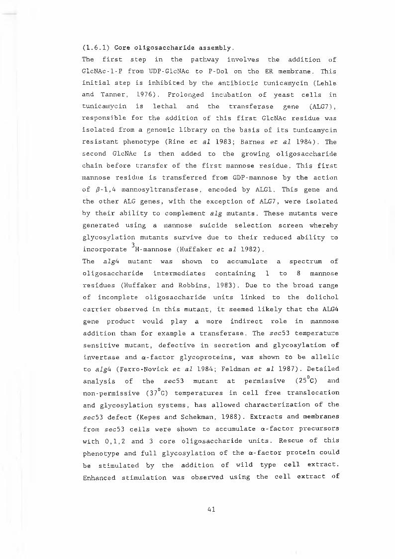
(1.6.1) Core oligosaccharide assembly.The first step in the pathway involves the addition of GlcNAc-l-P from UDP-GlcNAc to P-Dol on the ER membrane. This initial step is inhibited by the antibiotic tunicamycin (Lehle and Tanner, 1976). Prolonged incubation of yeast cells in tunicamycin is lethal and the transferase gene (ALG7), responsible for the addition of this first GlcNAc residue was isolated from a genomic library on the basis of its tunicamycin resistant phenotype (Rine et al 1983; Barnes et al 1984). The second GlcNAc is then added to the growing oligosaccharide chain before transfer of the first mannose residue. This first mannose residue is transferred from GDP-mannose by the action of /3-l,4 mannosyltransferase, encoded by ALG1. This gene and the other ALG genes, with the exception of ALG7, were isolated by their ability to complement alg mutants. These mutants were generated using a mannose suicide selection screen wherebyglycosylation mutants survive due to their reduced ability to
3incorporate H-mannose (Huffaker et al 1982) .The algU mutant was shown to accumulate a spectrum of oligosaccharide intermediates containing 1 to 8 mannose residues (Huffaker and Robbins, 1983). Due to the broad range of incomplete oligosaccharide units linked to the dolichol carrier observed in this mutant, it seemed likely that the ALG4 gene product would play a more indirect role in mannose addition than for example a transferase. The sec53 temperature sensitive mutant, defective in secretion and glycosylation of invertase and a-factor glycoproteins, was shown to be allelic to algh (Ferro-Novick et al 1984; Feldman et al 1987). Detailed analysis of the sec53 mutant at permissive (25°C) and non-permissive (37°C) temperatures in cell free translocation and glycosylation systems, has allowed characterization of the sec53 defect (Kepes and Schekman, 1988). Extracts and membranes from sec53 cells were shown to accumulate a-factor precursors with 0,1,2 and 3 core oligosaccharide units. Rescue of this phenotype and full glycosylation of the a-factor protein cou.Ld be stimulated by the addition of wild type cell extract. Enhanced stimulation was observed using the cell extract of
4 1
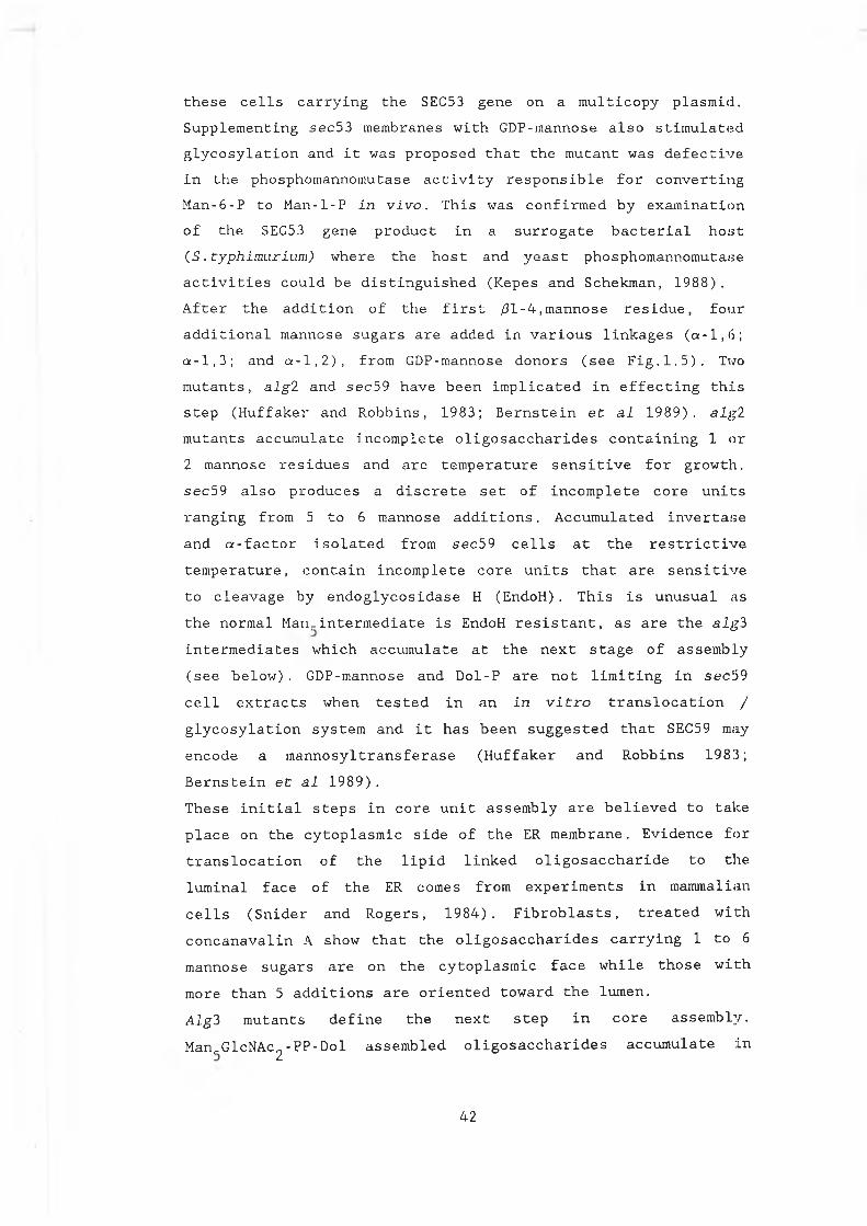
these cells carrying the SEC53 gene on a multicopy plasmid. Supplementing sec53 membranes with GDP-mannose also stimulated glycosylation and it was proposed that the mutant was defective in the phosphomannomutase activity responsible for converting Man-6-P to Man-l-P in vivo. This was confirmed by examination of the SEC53 gene product in a surrogate bacterial host (S. typhimurium) where the host and yeast phosphomannomutase activities could be distinguished (Kepes and Schekman, 1988). After the addition of the first 01-4,mannose residue, four additional mannose sugars are added in various linkages (a-1,6; a-1,3; and a-1,2), from GDP-mannose donors (see Fig.1.5). Two mutants, alg2 and sec59 have been implicated in effecting this step (Huffaker and Robbins, 1983; Bernstein et al 1989). algl mutants accumulate incomplete oligosaccharides containing 1 or 2 mannose residues and are temperature sensitive for growth. sec59 also produces a discrete set of incomplete core units ranging from 5 to 6 mannose additions. Accumulated invertase and a-factor isolated from sec 59 cells at the restrictive temperature, contain incomplete core units that are sensitive to cleavage by endoglycosidase H (EndoH). This is unusual as the normal Man,.intermediate is EndoH resistant, as are the alg3 intermediates which accumulate at the next stage of assembly (see below). GDP-mannose and Dol-P are not limiting in sec59cell extracts when tested in an in vitro translocation /glycosylation system and it has been suggested that SEC59 may encode a mannosyltransferase (Huffaker and Robbins 1983; Bernstein et al 1989) .These initial steps in core unit assembly are believed to take place on the cytoplasmic side of the ER membrane. Evidence for translocation of the lipid linked oligosaccharide to the luminal face of the ER comes from experiments in mammalian cells (Snider and Rogers, 1984). Fibroblasts, treated withconcanavalin A show that the oligosaccharides carrying 1 to 6 mannose sugars are on the cytoplasmic face while those with more than 5 additions are oriented toward the lumen.Algl mutants define the next step in core assembly.Man,_GlcNAc2-PP-Dol assembled oligosaccharides accumulate in
4 2
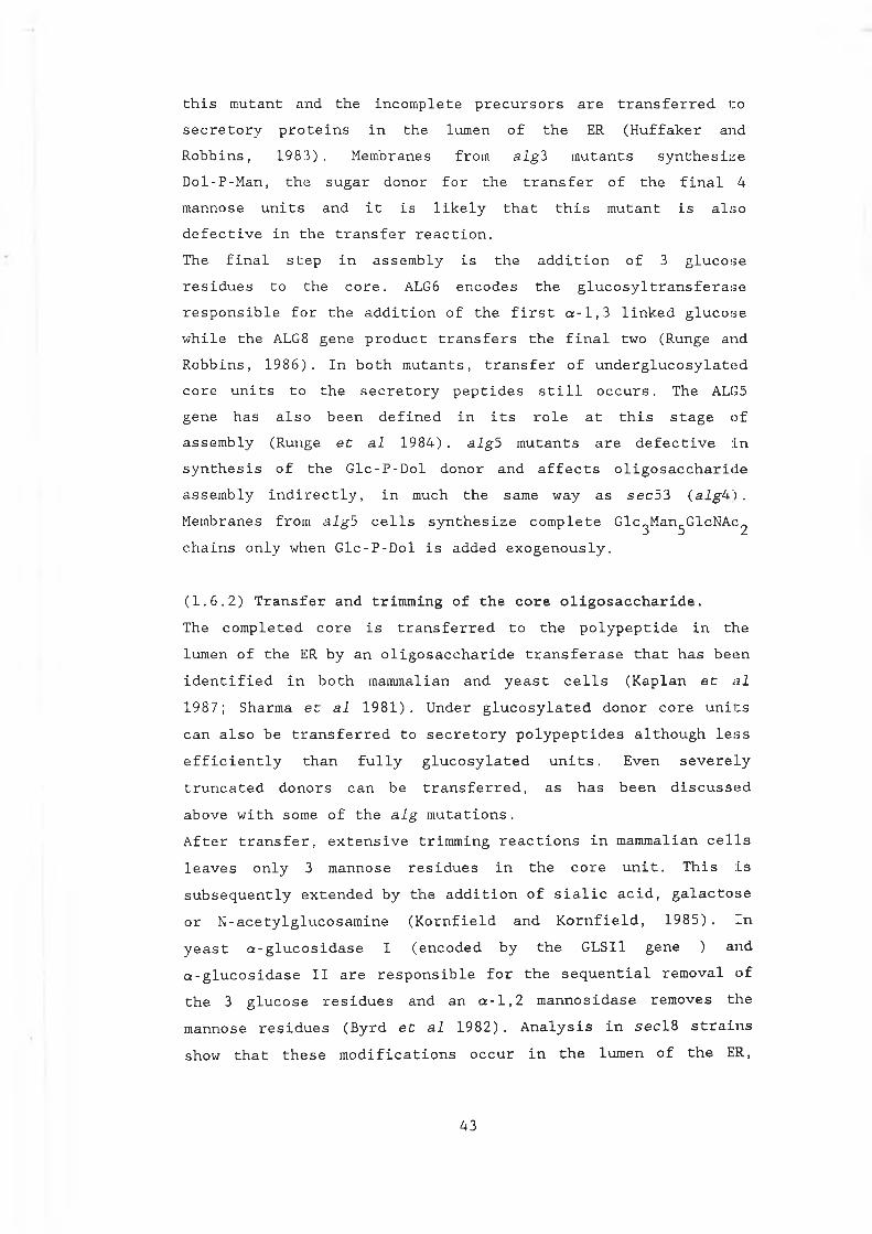
this mutant and the incomplete precursors are transferred to secretory proteins in the lumen of the ER (Huffaker and Robbins, 1983). Membranes from alg3 mutants synthesize Dol-P-Man, the sugar donor for the transfer of the final 4 mannose units and it is likely that this mutant is also defective in the transfer reaction.The final step in assembly is the addition of 3 glucose residues to the core. ALG6 encodes the glucosyltransferase responsible for the addition of the first a-1,3 linked glucosewhile the ALG8 gene product transfers the final two (Runge andRobbins, 1986). In both mutants, transfer of underglucosylated core units to the secretory peptides still occurs. The ALG5 gene has also been defined in its role at this stage of assembly (Runge et al 1984) . alg5 mutants are defective in synthesis of the Glc-P-Dol donor and affects oligosaccharide assembly indirectly, in much the same way as sec53 (algW) .
Membranes from alg5 cells synthesize complete Glc^Man^GlcNAc^ chains only when Glc-P-Dol is added exogenously.
(1.6.2) Transfer and trimming of the core oligosaccharide.The completed core is transferred to the polypeptide in the lumen of the ER by an oligosaccharide transferase that has been identified in both mammalian and yeast cells (Kaplan et al
1987; Sharma et al 1981). Under glucosylated donor core units can also be transferred to secretory polypeptides although less efficiently than fully glucosylated units. Even severely truncated donors can be transferred, as has been discussed above with some of the alg mutations.After transfer, extensive trimming reactions in mammalian cells leaves only 3 mannose residues in the core unit. This is subsequently extended by the addition of sialic acid, galactose or N-acetylglucosamine (Kornfield and Kornfield, 1985). In yeast a-glucosidase I (encoded by the GLSI1 gene ) and a-glucosidase II are responsible for the sequential removal of the 3 glucose residues and an a-1,2 mannosidase removes the mannose residues (Byrd et al 1982). Analysis in secl8 strains show that these modifications occur in the lumen of the ER,
4 3
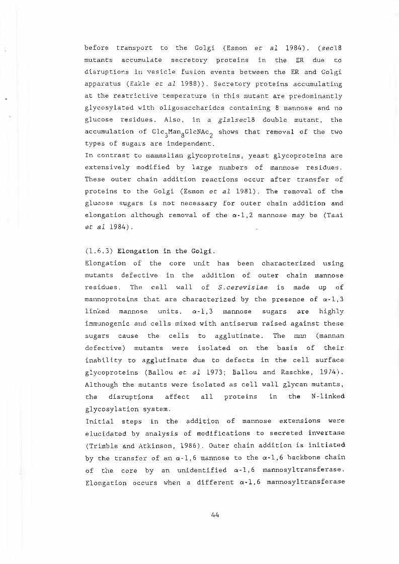
before transport to the Golgi (Esmon et al 1984) . (sec'.L8 mutants accumulate secretory proteins in the ER due to disruptions in vesicle fusion events between the ER and Golgi apparatus (Eakle et al 1988)). Secretory proteins accumulating at the restrictive temperature in this mutant are predominantly glycosylated with oligosaccharides containing 8 mannose and no glucose residues. Also, in a glslsecl& double mutant, the accumulation of Glc_Man0GlcNAc0 shows that removal of the two
J O /types of sugars are independent.In contrast to mammalian glycoproteins, yeast glycoproteins are extensively modified by large numbers of mannose residues. These outer chain addition reactions occur after transfer of proteins to the Golgi (Esmon et al 1981) . The removal of the glucose sugars is not necessary for outer chain addition and elongation although removal of the a-1,2 mannose may be (Tsai et al 1984).
(1.6.3) Elongation in the Golgi.Elongation of the core unit has been characterized using mutants defective in the addition of outer chain mannose residues. The cell wall of S .cerevisiae is made up of mannoproteins that are characterized by the presence of a-1,3 linked mannose units. a-1,3 mannose sugars are highly immunogenic and cells mixed with antiserum raised against these sugars cause the cells to agglutinate. The mnn (mannan defective) mutants were isolated on the basis of their inability to agglutinate due to defects in the cell surface glycoproteins (Ballou et al 1973; Ballou and Raschke, 1974). Although the mutants were isolated as cell wall glycan mutants, the disruptions affect all proteins in the N-linked glycosylation system.Initial steps in the addition of mannose extensions were elucidated by analysis of modifications to secreted invertase (Trimble and Atkinson, 1986). Outer chain addition is initiated by the transfer of an a-1,6 mannose to the a-1,6 backbone chain of the core by an unidentified a-1,6 mannosyltransferase. Elongation occurs when a different a-1,6 mannosyltransferase
4 4
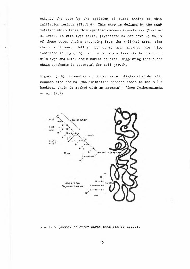
extends the core by the addition of outer chains to this initiation residue (Fig. 1.6). This step is defined by the mwi9 mutation which lacks this specific mannosyltransferase (Tsai et al 1984). In wild type cells, glycoproteins can have up to 15 of these outer chains extending from the N-linked core. Side chain additions, defined by other mnn mutants are also indicated in Fig.(1.6). wnn9 mutants are less viable than both wild type and outer chain mutant strains, suggesting that outer chain synthesis is essential for cell growth.
Figure (1.6) Extension of inner core oligisaccharide with mannose side chains (the initiation mannose added to the a,1-6 backbone chain is marked with an asterix). (from Kurkuruzinslca et al, 1987)
x = 1-15 (number of outer cores that can be added).
45
More recent analysis of the structure of mannose additions in wnn9 mutants, suggests that the elongation reaction may stem from a different point in the core than previously suggested for invertase (Hernandez et al 1989) . The discovery that the a-1,6 mannose residue previously proposed to be the initiation point for elongation is phosphorylated in mnn9 glycoproteins, precludes it as the point of the first mannose addition and therefore for elongation. An alternative structure is proposed in Fig.(1.7), suggesting that elongation extends from the a-1,3 mannose as indicated. While the possibility remains that different structures may occur in different glycoproteins, this proposal is more conducive with the observation that initiation and elongation are two separate events and are promoted by the action of two distinct mannosyltransferases with different specificities.
Figure (1.7). Outer chain addition extends from the a,1-3 mannose in mnn9 glycoproteins (the invertase a,1-6 mannoseresidue now phosphorylated is indicated with asterix).(Hernandez et al. 1989).
M
m nn- ■
mnn5
mnn 1
M
•■I"M
mnn9
M- f ; ------- t— -M-4
ß M — iG N A c) -
/ >
I-’ V ' I
jr I!5 1p ........ IT“ 'r \
M M
OUTER CHAIN
mnn3 : Reduces average chain lenth m nn? . mnn8 S im ila r lo m nnlQ
m nnlQ x = 1- 4
V> ild T>pe * = -10 * Additional phosphorylation sues
MM
rM *
M
MCOR£
- M — M— - M— -
BASE-LABILE
OLIGOSACCHARIDES
S irT h r
4 6
(1.6.4) Glycosylation of yeast secretory proteins.Much of the information on the yeast glycosylation process was obtained by studying glycosylation patterns of proteins naturally secreted by S.cerevlsie. These studies have allowed some insight Into the role of glycosylation in the secretion and maturation of proteins through the secretory pathway. Invertase for example, accumulates as an inactive, ER membrane bound precursor at the restrictive temperature in sec53 and sec59 cells (Ferro-Novick et al 1984). On return to thepermissive temperature, the protein appears to refold, becomes partially active again and is secreted. The majority of secreted invertase exists as an octamer (Esmon et al 1987) . Assembly of this octamer and its localization in the yeast periplasmic space is related to glycosylation of the enzyme. Octamers are not formed when invertase is synthesized in the presence of tunicamycin and secretion of the enzyme isinhibited under these conditions. Small amounts of invertase dimers are released from the cell wall when cells are washed with buffer or /3-mercaptoethanol. Release of the octameric form of the enzyme is only achieved with cell wall disruption. Thishas also been shown for other periplasmic proteins such as acidphosphatase and a-galactosidase.Studies on a-factor production in yeast has also been characterized in glycosylation defective mutants. a-factor production involves a series of complex processing reactions which are summarized in Figure (1.8) (Bussey, 1988). Three copies of the a-factor protein are synthesized as a larger precursor of 165 amino acids, 85 of which comprise the signal sequence (Julius et al 1983) . The first 20 residues (the pre-region) are removed by signal peptidase on translocation into the ER, leaving the three copies of a-factor attached to the 65 amino acid pro-region. This pro-region has 3 glycosylation sites at positions 23, 57 and 67. Studies insecl8 mutants show that this region is core glycosylated in the ER. Removal of the pro-peptide occurs in the Golgi, by the action of a specific peptidase, KEX2 (Julius et al 1984a). Further processing requires the action of two additional
4 7
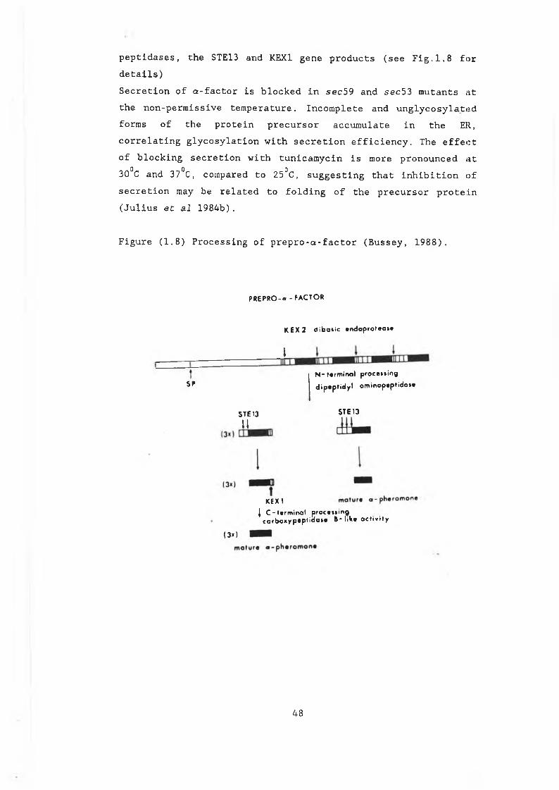
peptidases, the STE13 and KEX1 gene products (see Fig.1,8 for details)Secretion of a-factor is blocked in sec59 and sec53 mutants at the non-permissive temperature. Incomplete and unglycosylated forms of the protein precursor accumulate in the ER, correlating glycosylation with secretion efficiency. The effect of blocking secretion with tunicamycin is more pronounced at 30°C and 37°C, compared to 25°C, suggesting that inhibition of secretion may be related to folding of the precursor protein (Julius et al 1984b).
Figure (1.8) Processing of prepro-a-factor (Bussey, 1988).
P R E P R O - a - F A C T O R
K E X 2 d i b o i i c * r * d o p r o t e o » »
I
SPN - t e r m i n a l p r o c e » » in g
d i p a p t i d y l a m i n o p a p t i d o « *
STE13 STE 13
11
KEX 1| C - t # r m i n o l p r o c » » » in g
CQfbo*yp*p*idai« B - l i t ( * a c t i v i t y
(3*1
4 8
(1.7.0) The secretion of heterologous proteins in S.cerevisiae. The first proteins to be secreted in yeast, human interferon q-1 and interferon a-2, were directed by their natural signal peptides (Hitzeman et al, 1983). These heterologous signalpeptides however, were processed by signal peptidase at greater than one site and heterogeneous products were recovered from the medium. Human serum albumin (HSA) on the other hand, also secreted using its own signal peptide, is correctly processed by the KEX2 cleavage enzyme when produced in yeast (Etcheverry et al,1986; Bathurst et al, 1987). This human signal peptide is surprisingly efficient, more efficient than many homologous secretion signals (Sleep et al, 1990) and has also been used to secrete other heterologous proteins.As well as its use as a signal peptide in yeast, the HSA protein, with its signal sequence still intact, has been used in fusion studies. Secretion and glycosylation of invertase was achieved with the HSA protein and signal peptide fused to its 3’ end. Stomatostatin has also been produced in yeast when fused to HSA (Hitzeman et al, 1990). These experimentsdemonstrate the potential of using heterologous signals, not only to secrete heterologous proteins, but also (by fusing to the heterologous protein itself) for stabilizing and secreting smaller proteins in yeast. Other heterologous signal peptides have been shown to direct their natural products through the yeast secretion system with varied efficiencies. Human and wheat a-amylases are secreted using their native signal sequences, although at a lower efficiency compared to HSA (Nakamura et al, 1986). al antitrypsin on the other hand,cannot be secreted using its native signal peptide (Rothstein et al, 1987).Homologous yeast signal sequences from yeast secretory proteins, fused to the mature portions of heterologous genes are widely used to direct secretion. The invertase signal sequence efficiently directs secretion of HSA, prochymosin and interferon a2, among others (Etcheverry et al, 1986; Smith et al, 1985; Chang et al, 1986). The a-factor signal peptide isalso widely used in the production of heterologous proteins.
4 9
The a-factor prepro-region was shown to secrete IFNal more efficiently than the IFN signal peptide (Singh et al, 1984).However, in this case the Glu-Ala repeats that occur after the Lys-Arg KEX2 cleavage site were retained in a large proportion of the secreted material. Attempts to produce this protein by direct fusion to the Lys-Arg site, resulted in inefficient cleavage by KEX2 (Zsebo et al, 1986) showing that heterologous sequences in the fusions can affect cleavage specificity in homologous signal peptides in some cases. Direct fusion to the Lys-Arg site of the a-factor prepro-region however, has resulted in efficient processing and secretion in most other cases (Ernst et al, 1986; Guisez et al,1990 Loison et al, 1988; Shaw et al, 1988 are examples).A comparison of a number of signal sequences and their ability to secrete HSA has been carried out (Sleep et al, 1990). Inthis study the HSA signal, the a-factor signal and the killer toxin signal from K.lactis (Stark and Boyd, 1986) were compared along with two fusion signal peptides. These fusions comprised the HSA signal with 5 amino acids around the cleavage site replaced by the KEX2 cleavage site of a-factor and the killer toxin signal with its last 13 amino acids replaced by the same a-factor sequence (Sleep et al, 1990). Results show that bothfusion peptides were more efficient at secreting HSA than any of the other 3. The natural HSA signal was more efficient than both a-factor and killer toxin, which was the least efficient of the series.These observations show that it is difficult to predict how well a particular protein will be secreted by a homologous or heterologous signal peptide. The heterologous protein in this case, was most efficiently secreted using its natural signal peptide. However, the HSA signal was improved by inserting a-factor signal sequences, as was the killer toxin signal peptide. Other heterologous sequences have not been efficient in directing secretion of their native products and the introduction of a yeast signal has proved more successful. Different signals therefore, appear to work with varied efficiencies when fused to different proteins. The efficiency
50
of a particular signal peptide is likely to be related to its structural interaction with the heterologous protein itself and the overall interaction of the new combination with the secretory apparatus as a whole.To summarize, in attempting to improve secretion levels of a heterologous protein in yeast, there are several parameters which can be altered and improved. Different signal peptides can be assessed to give the most efficient and correctly processed products. Further improvements in recovery levels can be sought by optimizing expression either by the choice of promoter or type of expression vector and translational efficiency can be optimized by choosing appropriate leader regions. Host strains can be manipulated to minimize proteolysis and supersecreting strains, which exhibit increased secretion efficiencies, have also been generated (Smith et al, 1985). These strains, including the sscl (or pmr1) mutation, appear to enhance secretion by overcoming in some way what is thought to be the rate limiting step in protein transport, ie. the ER to Golgi transfer step.Other mutant strains can manipulate the glycosylation levels of heterologous proteins, eg. mnn9 mutants (section 1.6). Mammalian proteins, when produced in yeast, are often glycosylated to a far greater extent than in their normal hosts. This arises out of the fundamental differences in the glycosylation processes of higher and lower eukaryotes. Hyperglycosylation can cause problems for mammalian proteins, particularly those of potential therapeutic use in humans. This is due to problems that arise in the immuno genic ity of the characteristic extended mannose chains of proteins produced in yeast (Melnick et al, 1990; Kurkuruzinski et al, 1987). Attempts to minimize this problem have included the alteration of the acceptor sequences by site directed mutagenesis to completely inhibit glycosylation and the production of the protein in glycosylation mutant strains such as mnn9 (Myaj iina et al, 1986; Ernst et al, 1987; Melnick et al, 1990). This approach has proved successful in the secretion of human a-antitrypsin in yeast (Moir and Dumais, 1987).
5 1
CHAPTER 2 MATERIALS AND METHODS
5 2
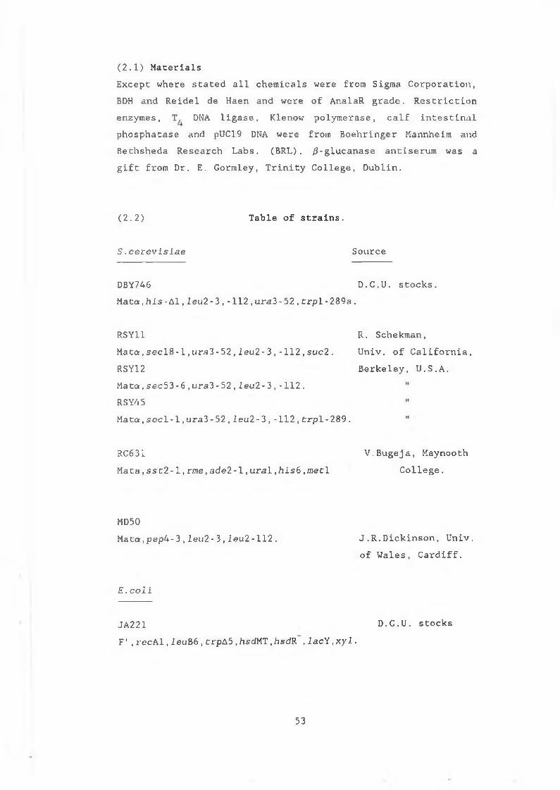
(2.1) MaterialsExcept where stated all chemicals were from Sigma Corporation, BDH and Reidel de Haen and were of AnalaR grade. Restriction enzymes, DNA ligase, Klenow polymerase, calf intestinalphosphatase and pUC19 DNA were from Boehringer Mannheim and Bethsheda Research Labs. (BRL). /?-glucanase antiserum was a gift from Dr. E. Gormley, Trinity College, Dublin.
(2.2) Table of strains.
S.eerevisiae Source
DBY746 D.C.U. stocks.
Mata,secl8-1,ura3-52,leu2-3,-112,suc2. Univ. of California,
Mata,his-Al,l e u2-3,-112,ura3-52,trpl-289a.
RSY11 R. Schekman,
RSY12Mata.sec53 - 6,ura3-52,leu2-3,-112.RSY45Mata,seel-1,ura3- 52,Ieu2- 3,-112,trpl-289.
Berkeley, U.S.A.
Mata,sst2-1,rme,ade2-1,ural,his6.metlRC631 V.Bugeja, Maynooth
College.
MD50Mata,pep4-3,leu2- 3,leu2-112. J.R.Dickinson, Univ.
of Wales, Cardiff.
E .coli
JA221F',recAl,l e uB6,crpA5,hsdMT,hsdR ,lacY,x y l .
D.C.U. stocks
5 3
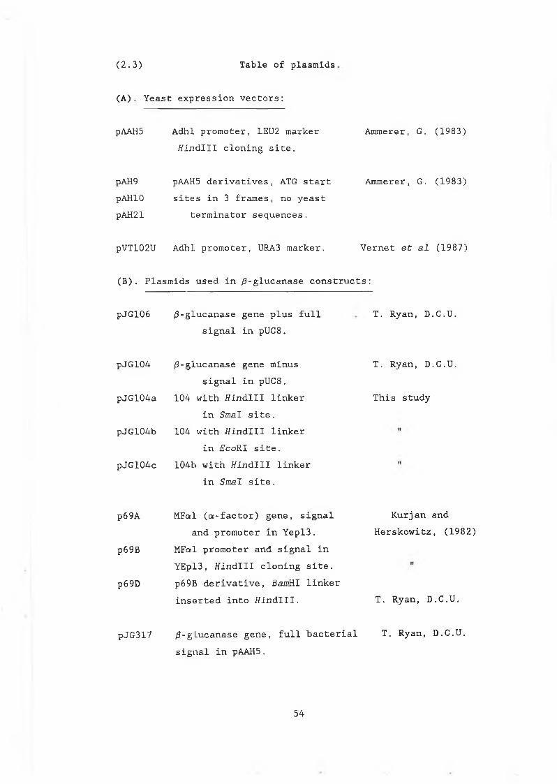
( 2 . 3 ) Table of plasmids
(A). Yeast expression vectors:
pAAH5 Adhl promoter, LEU2 marker /{indili cloning site.
Ammerer, G. (1983)
pAH9 pAAH5 derivatives, ATG startpAHIO sites in 3 frames, no yeastpAH21 terminator sequences.
Ammerer, G. (1983)
pVT102U Adhl promoter, URA3 marker. Vernet et al (1987)
(B) . Plasmids used in /3-glucanase constructs:
pJG106 /3-glucanase gene plus full signal in pUC8.
T . Ryan, D .C .U .
pJG104 /3-glucanase gene minus signal in pUC8.
pJG104a 104 with //indili linker in Smal site.
pJG104b 104 with //indili linker in EcoRI site.
pJGl04c 104b with //indili linker in Smal site.
T . Ryan, D .C .U .
This study
p69A
p69B
p69D
pJG317
MFal (a:-factor) gene, signal and promoter in Yepl3.
MFccl promoter and signal in YEpl3, //indlll cloning site. p69B derivative, BamHI linker inserted into Hindlll.
Kurjan and Herskowitz, (1982)
T . Ryan, D .C .U .
/3-gLucanase gene, full bacterial T. Ryan, D.C.U.signal in pAAH5.
54
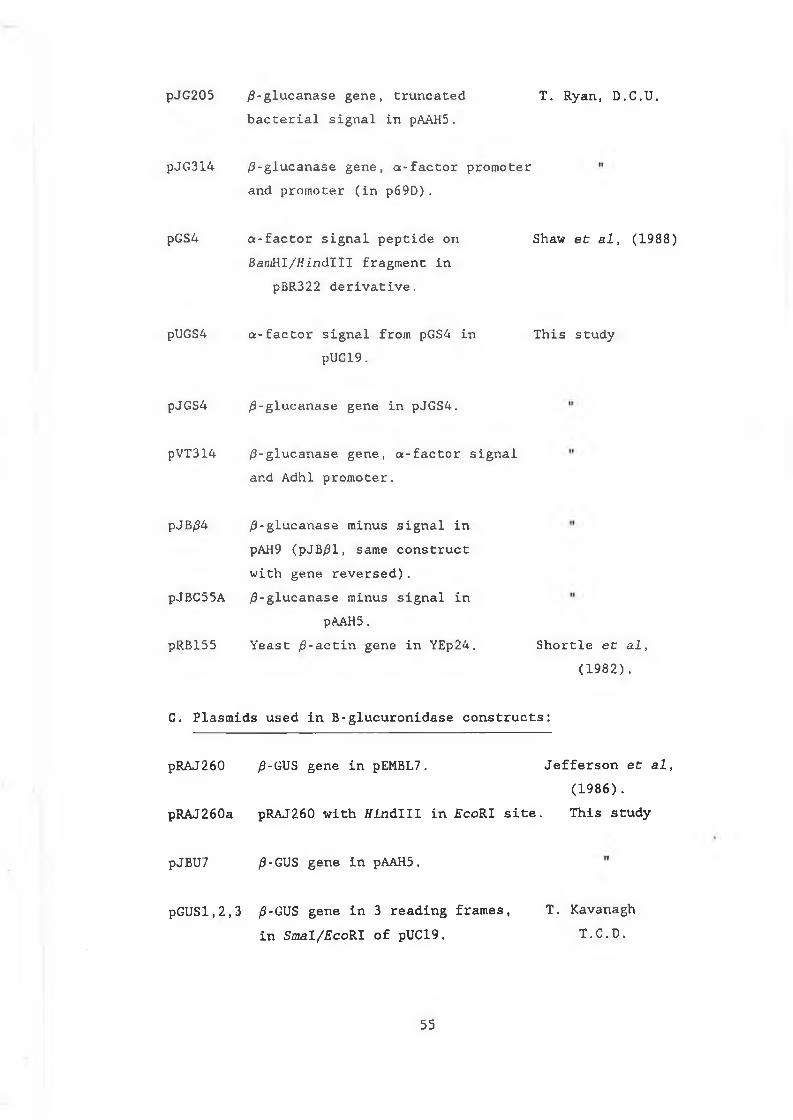
pJG205 /3-glucanase gene, truncated T. Ryan, D.C.U.bacterial signal in pAAH5.
pJG314 ß-glucanase gene, a-factor promoter and promoter (in p69D).
pGS4 a-factor signal peptide onSamHI/tfindlH fragment in
pBR322 derivative.
Shaw et al, (1988)
pUGS4 a-factor signal from pGS4 inpUC19.
This study
pJGS4 /3-glucanase gene in pJGS4.
pVT314 /3-glucanase gene, a-factor signal and Adhl promoter.
pJB/34 /3-glucanase minus signal inpAH9 (pJB/31, same construct with gene reversed).
pJBC55A /3-glucanase minus signal inpAAH5.
pRB155 Yeast /3-actin gene in YEp24, Shortle et al, (1982).
C. Plasmids used in B-glucuronidase constructs:
pRAJ260 /J-GUS gene in pEMBL7. Jefferson et al,(1986).
pRAJ260a pRAJ260 with ffindlll in £coRI site. This study
pJBU7 /3-GUS gene in pAAH5.
pGUSl,2,3 /3-GUS gene in 3 reading frames, T. Kavanagh in Smal/EcoRI of pUC19. T.C.D.
5 5
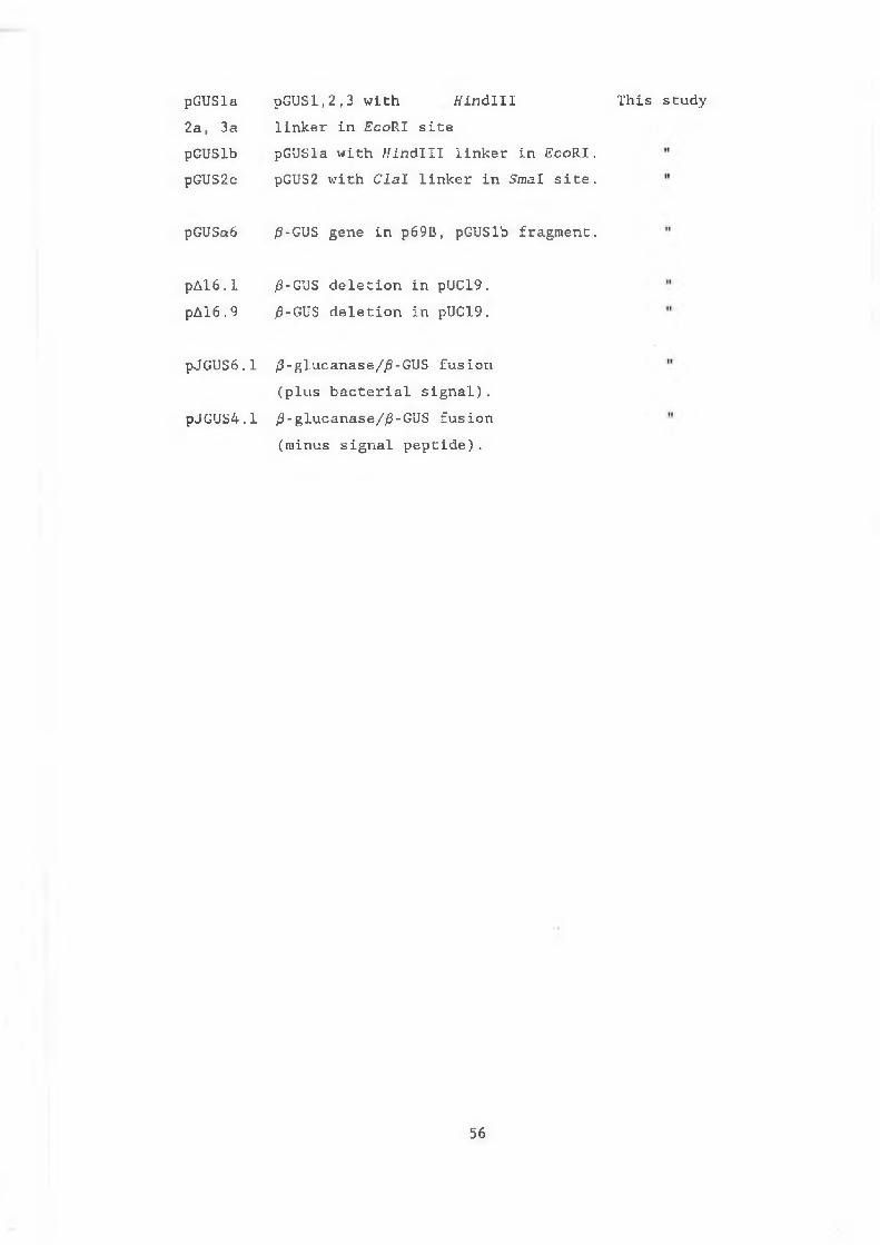
pGUSla pGUSl,2,3 with Hindlll2a, 3a linker in £coRI sitepGUSlb pGUSla with //indill linker in EcoRI.pGUS2c pGUS2 with Clal linker in Smal site.
This study
pGUSa6 /3-GUS gene in p69B, pGUSlb fragment.
pAl6.1 /3-GUS deletion in pUC19.pAl6.9 /9-GUS deletion in pUC19.
pJGUS6.1 /3-glucanase//3-GUS fusion (plus bacterial signal).
pJGUS4.1 /3-glucanase/)3-GUS fusion (minus signal peptide).
56
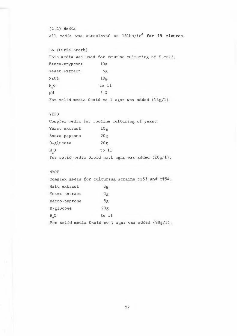
All media was autoclaved at 151bs/in for 15 minutes.( 2 . 4 ) Media
2
LB (Luria Broth)This media was used for routine culturing of E.coli Bacto-tryptone lOgYeast extract 5gNaCl lOgH O to 112pH 7.5For solid media Oxoid no.l agar was added (13g/l).
YEPDComplex media for routine culturing of yeast.Yeast extract lOgBacto-peptone 20gD-glucose 20gH O to 11
2For solid media Oxoid no.l agar was added (20g/l).
MYGPComplex media for culturing strains YT53 and YT54.Malt extract 3gYeast extract 3gBacto-peptone 5gD-glucose 20gH O to 11
2For solid media Oxoid no.l agar was added (20g/l).
57
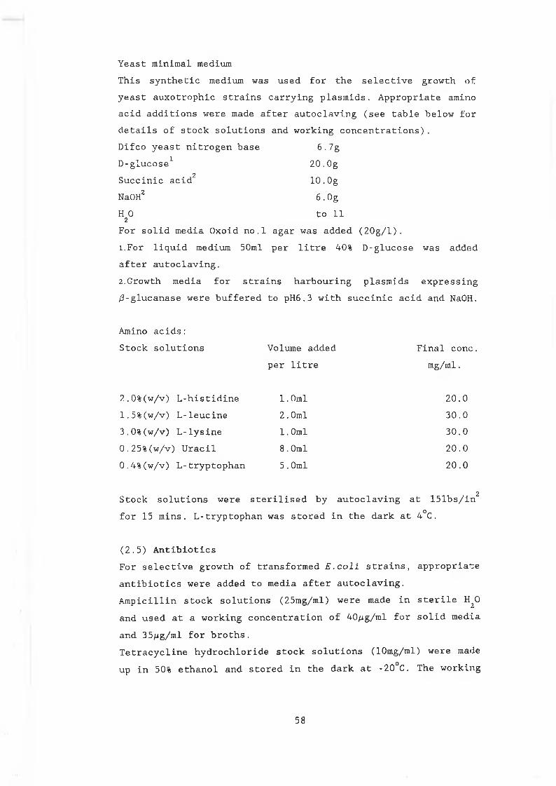
Yeast minimal mediumThis synthetic medium was used for the selective growth of yeast auxotrophic strains carrying plasmids. Appropriate amino acid additions were made after autoclaving (see table below for details of stock solutions and working concentrations).Difco yeast nitrogen base 6.7gD-glucose1 20.Og
2Succinic acid 10.OgNaOH2 6.OgH O to 11
2
For solid media Oxoid no.1 agar was added (20g/l).1.For liquid medium 50ml per litre 40% D-glucose was added after autoclaving.2 .Growth media for strains harbouring plasmids expressing /9-glucanase were buffered to pH6.3 with succinic acid and NaOH.
Amino acids :Stock solutions Volume added Final conc.
per litre mg/ml.
2.0%(w/v) L-histidine 1.0ml 20.01.5%(w/v) L-leucine 2.0ml 30.03.0%(w/v) L-lysine 1.0ml 30.00.25%(w/v) Uracil 8.0ml 20.00.4%(w/v) L-tryptophan 5.0ml 20.0
2Stock solutions were sterilised by autoclaving at 151bs/in for 15 mins. L-tryptophan was stored in the dark at 4°C.
(2.5) AntibioticsFor selective growth of transformed E.coli strains, appropriate antibiotics were added to media after autoclaving.Ampicillin stock solutions (25mg/ml) were made in sterile H^O and used at a working concentration of 40/jg/ml for solid media and 35/ig/ml for broths.Tetracycline hydrochloride stock solutions (10mg/ml) were made up in 50% ethanol and stored in the dark at -20°C. The working
58
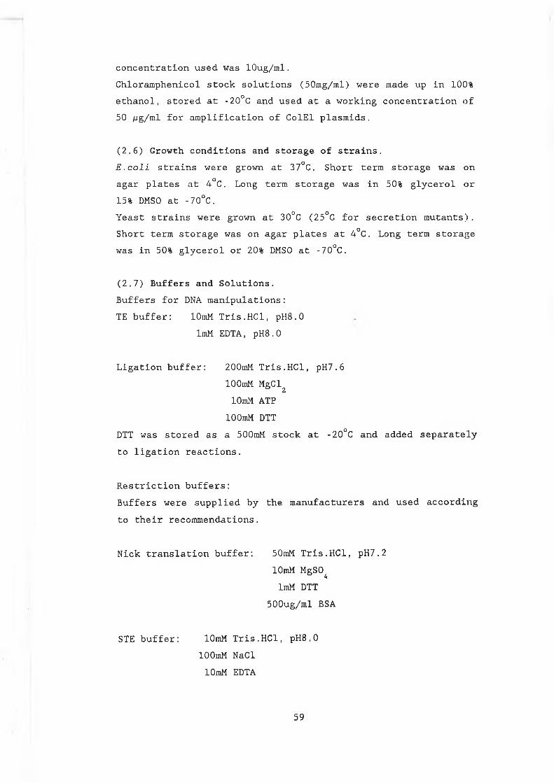
concentration used was lOug/ml.Chloramphenicol stock solutions (50mg/ml) were made up in 100% ethanol, stored at -20°C and used at a working concentration of 50 /¿g/ml for amplification of ColEl plasmids.
(2.6) Growth conditions and storage of strains.E.colL strains were grown at 37°C. Short term storage was on agar plates at 4°C. Long term storage was in 50% glycerol or 15% DMSO at -70°C.Yeast strains were grown at 30°C (25°C for secretion mutants). Short term storage was on agar plates at 4°C. Long term storage was in 50% glycerol or 20% DMSO at -70°C.
(2.7) Buffers and Solutions.Buffers for DNA manipulations:TE buffer: lOmM Tris.HCl, pH8.0
ImM EDTA, pH8.0
Ligation buffer: 200mM Tris.HCl, pH7.6lOOmM MgClz lOmM ATP
lOOmM DTTDTT was stored as a 500mM stock at -20°C and added separately to ligation reactions.
Restriction buffers:Buffers were supplied by the manufacturers and used according to their recommendations.
Nick translation buffer: 50mM Tris.HCl, pH7.2lOmM MgSO^ImM DTT
500ug/ml BSA
STE buffer: lOmM Tris.HCl, pH8.0lOOmM NaCl lOmM EDTA
5 9
2X Bal31 buffer: 40mM Tris.HCl, pH7.225inM MgCl2 25mM CaCl
2
2mM EDTA 1.2M NaCl
RNase (DNase free):RNaseA (lOrag/ml) was dissolved in lOmMTris.HCl (pH7.5), 15mM NaCl; heated to 100°C for 15 minutes .allowed to cool slowly to room temperature and stored at -20°C.
Phenol/chloroform mix:lOOg of phenol (AnalaR grade) was disolved in 100ml chloroform with 4.0ml isoamylalcohol and 0.8g 8-hydroxyquinoline. The phenol mix was stored under lOOmM Tris.HCl, pH7.5, at 4°C in the dark.
Buffers for agarose gel electrophoresis: lOx TBE: 0.89M Tris. borate
0.89M Boric acid 0.02M EDTA pH8.3
50x TAE: 2.0M Tris. borate242g Glacial acetic acid
100ml 0.5M EDTA pH8.0
5x Bromophenol blue: 0.25%(w/v) Bromophenol blue25% Ficoll (type 400)
60
Solutions for isolation of DNA from agarose:Sodium iodide solution:90.8g Nal was disolved in 100ml H20 . The solution was filtered through Whatman no. 1 filter paper, 15g of Na^O^ was added and the solution stored at 4°C in the dark.
Ethanol wash solution: 50% EthanollOOmM NaCllOmM Tris. HC1, pH7.5 ImM EDTA
This solution was stored at -20°C.
Solutions for preparation of RNA:All solutions for preparation of RNA were made up in DEPC (diethyl polycarbonate) treated H20 .
DEPC treatment:DEPC (0.1%) was added to distilled ^0, allowed to stand for 20 mins. at room temperature and autoclaved for 20 mins. at 151b/in2 to remove the DEPC.
Letts buffer: 0.1M LiCl0.1M EDTA0.01M Tris.HCl pH7.4 0.02% SDS
5x Formamide running buffer: 0.1M MOPS (pH7.0)40mM Sodium acetate 5mM EDTA
Solutions for Northern blotting and hybridisation:20x SSC: 175.3gNaCl
88.2g Tri-sodium acetateH 0 to 11 2pH 7.0
6 1
M
Prehybridisation buffer: 6X SSC2x Denhardts reagent 0.1% SDS100/ig/ml Salmon sperm DNA
50x Denhardts reagent: 5.0g Ficoll5.0g Polyvinylpyrrolidone 5.Og BSAH O to 11
2
Buffers for dialysing yeast extracts:5x PBS: 5.45g KH P0° 2 *
10.7g Na HPO° 2 h
45.Og NaClH 0 to 11 2
Preparation of dialysis tubing:Appropriate lengths of tubing were boiled for 15 mins. in lOmM EDTA, rinsed and boiled for 15 mins. in distilled H O.
Solutions for preparation of yeast extracts: lOx Lysis buffer: 50mM NaPO , pH7.0* k
lOmM EDTA0.1% Triton X-1000.1% SarkosyllOmM /?-mercaptoethanol
PMSF:PMSF was made up freshly as a lOmM stock in 2-propanol and used as required.
6 2
DNS (3,5-dinitrosalicylic acid) solution for measuring /3-glucanase activity: 5 . Og DNS
l.Og Phenol 0.25g Na S03lOOg Sodium potassium tartrate 2% NaOH11 total volume
Buffers for polyacrylamide gel electrophoresis (PAGE):SDS electrophoresis buffer: 25mM Tris. HC1, pH8.3
192mM Glycine 0.1% SDS
Solubilisation buffer: lOOmM Dithiothreitiol (DTT)80mM Tris. HC1, pH6.810% (w/v) Glycerol2.0% (w/v) SDS0.2% (w/v) Bromophenol blue
Coomassie blue stain:0.5% Coomassie brilliant blue in acetic acid : water : methanol ( 1 : 1 0 : 8 ) .
Destain solution: Acetic acid : water : methanol (1 : 10 : 8)
Solutions for Western blotting:Transfer buffer: (per litre)
3.03g Tris. base 14.4g glycine 200ml methanol
TBST buffer: 150mM NaCllOmM Tris. HC1, pH8.00.05% Tween-20
Blocking solution: TBST with 1% (w/v) BSA
63
AP (alkaline phosphatase) buffer: lOOmM Tris. HC1, pH9 . 5lOOmM NaCl
5mM MgClz
NBT stock: NBT (nitroblue tetrazolium), 50mg/ml in 70%N ,N-dimethylformamide.
BCIP stock: BCIP (5-bromo-4-chloro-3-indolyl phosphate),50mg/ml in 100% N,N-dimethylformamide.
AP colour development solution: 10ml AP buffer66ul NBT stock 33ul BCIP stock
NBT was added to the AP buffer and mixed. BCIP was then added and mixed. The solution was made up freshly from stocks before use.
(2.8) Transformation of E.coli Preparation of competent cells:100ml of LB was innoculated with 1.0ml of an overnight cultureof JA221 and grown at 37°C with aeration to an OD run of 0.3
600
to 0.4. The culture was chilled on ice for 30 mins and cellswere harvested by centrifugation at 10,000rpm for 10 mins at4°C. The cells were washed in half the original volume of icecold 50mM MgCl2 and resuspended in half the original volume of50mM CaCl . After incubation on ice for 30 mins, the cells were
2
harvested as before and resuspended in one tenth the original culture volume of 50mM CaCl .
7.
Transformation of competent cells:Plasmid DNA (up to 250ng) was added to 200ul aliquots of competent cells in an eppendorf and incubated on ice for 60 mins. The cells were heat-shocked by placing them at 420C for exactly 2 mins and returned to ice immediatly. 0.8ml of LB was added and the samples incubated at 37°C to allow expression of the plasmid encoded antibiotic resistant marker gene. The cells were plated in 200ul aliquots on LB agar containing the
6 4
appropriate selective antibiotic and incubated overnight at 37 °C.
(2.9) Transformation of S .cerevisiae.Preparation of yeast competent cells :100ml of YEPD was innoculated with 50^1 of an overnight culture of S . cerevisiae '"'and grown at 30°C with agitation to an OD600nm of 0.7. The cells were harvested at room temperature by centrifugation at 5,000 rpm. After 2 washes in half the original culture volume of TE, pH 7.5, the cells were resuspended in one tenth the original culture volume of lOOmM LiAc and incubated at 30°C with agitation for 60 mins. The cells were harvested as before and resuspended in the same volume of lOOmM LiAc. Routinely, 10ml samples of culture were used yielding 1.0ml competent cells.l. When preparing and transforming competent cells of of S .cerevisiae secretion defective mutants, growth and incubation temperatures were 25°C.Transformation of yeast competent cells:Plasmid DNA (0. l/ig-10/ig) was added to 300^1 competent cells. 0.7ml of 50% P E G aooo was added, mixed gently by inversion and incubated at 30°C for 60 mins. The cells were then placed at 42°C for 5 mins and plated directly onto selective media.
(2.10) Generation of yeast spheroplasts.Cells were grown in YEPD to OD =0.7, harvested and washed b 600twice in 1.2M sorbitol. One tenth volume of zymolyase (3mg/ml) was added and the cells shaken (150 rpm) at 30°C for 60 mini;. Spheroplast formation was monitored by adding a sample to H^0 and following the drop in ODgoQ readings as the cells lyse.
(2.11) Small scale isolation of DNA from E.coli.Two methods for isolating small amounts of plasmid DNA were generally employed:Method 1 (Birnboim and Doly 1979):Cells were grown overnight at 37°C in LB with appropriate antibiotics. 1.5ml was pelleted by centrifugation and the
6 5
supernatant removed completely. The pellet was resuspended in100 /¿I of Solution 1 and left on ice for 5 mins. 200^1 ofSolution 2 was added mixed, by inversion and incubated on icefor 10 mins. 150/il of Solution 3 was added, mixed by inversionand the sample left on ice for 5 mins. After centrifugation at10,000 rpm for 5 mins, 400/il of the supernatant was removed toa fresh tube. An equal volume of phenol/chloroform was added,vortexed and spun . The aqueous layer was removed to a freshtube and 0.8ml of ice cold ethanol was added. The sample wasallowed to stand at room temperature for 10 mins. The DNA waspelleted by centrifugation at 10,000 rpm for 10 mins and thepellet washed in 70% ethanol. The DNA was dried under vacumnand resuspended in 30-50/il TE, pH 8.0. 5/il RNaseA (lOmg/ml) wasadded and the DNA stored at 4°C.Solution 1:1.0ml 0. 5M Glucose1.0ml 0.1M EDTA0.25ml 1M Tris.HC1,pH8.07.75ml H 0
2
Solution 2: (made freshly every month and stored at RT)2.0ml IN NaOH1.0ml 10% SDS7 .0ml HO
2
Solution 3: (3M Potassium acetate)To 60ml of 5M potassium acetate, 11.5ml of glacial acetic acidand 25ml of H 0 were added.
2
Method 2 (Holmes and Quigley 1981) :Cells were grown overnight at 37°C on LB agar containing the appropriate antibiotics. A patch of growth was transferred to an eppendorf using a sterile cocktail stick. The cells were resuspended by vortexing in 300/il STET buffer. 20pl lysozyme (lOmg/ml) was added and the samples were allowed to stand at RT for 10 mins. The samples were placed in a boiling water bathfor 60 seconds, centrifuged at 10,000 rpm for 5 mins. and thesupernatant removed to a fresh tube. An equal volume of
66
isopropanol was added and the samples incubated at -20°C for 30 mins. The DNA pellet was washed with ether, dried andresuspended in 30-50/il TE buffer, pH 8.0. 5/xl of RNaseA (lOmg/ml) was added and the DNA stored at 4°C .STET buffer: 8% w/v Sucrose
5% w/v Triton X-10050mM EDTA50mM Tris.HCl, pH 8.0
(2.12) Large scale preparation of plasmid DNA from E . c o l i .
250ml of LB (with the appropriate antibiotic addition) was innoculated with 1ml of an overnight culture of E . c o l i , and grown at 37°C with shaking to an OD600nm of 0.8.Chloramphenicol (1.0ml of 50/ig/ml stock) was added to allow plasmid amplification and the cells were returned to 37°C for a further 12 to 16 hours.Cells were harvested by centrifugation (10,000 rpm, 10 mins at 4°C) and resuspended in 2.0 ml sucrose solution. The cellsuspension was transferred to a screw cap Beckman polycarbonate 50iTi tube and 0.4ml of lysozyme (20mg/ml in 0.25M Tris.HCl, pH 8.0) was added. After incubation on ice for 5 mins, 0.8ml of 0.25M EDTA was added, the cells mixed by inversion and incubated on ice for a further 10 mins. Triton-mix (3.2ml) was added, mixed and left on ice for 15 mins. When cell lysis was evident (indicated by an increase in viscosity of the mixture) the samples were centrifuged at 40,000 rpm for 20 mins at 4°C. The cleared lysate was removed to a 10ml tube and 6.7g of caesium chloride was added and mixed gently at roomtemperature. The solution was transferred to a 20ml Beckman quickseal polyallomer tube and 180/il of ethidium bromide (lOmg/ml) was added. The total solution weight was brought to 14. lg using lOmM EDTA, pH 8.0. The tube was filled to the top with mineral oil heat sealed and centrifuged at 50,000 rpm for 24hrs at 18°C. The plasmid band was visualised by ultra violet transillumination and extracted from the tube using a sterile 18 guage needle and 1ml syringe.
67
Ethidium bromide was removed from the sample by extracting with isopropanol saturated with 20X SSC. The DNA was dialysed against TE pH 8.0 for at least 12 hrs with 3 changes of buffer. DNA was concentrated by ethanol precipitation.Sucrose solution: 25% Sucrose
50mM Tris.HCl, pH 8.0 Triton Mix: 5.0ml 20% Triton X-100
12.5ml 0.25M EDTA2.5ml 1.0M Tris.HCl, pH 8.0
(2.13) Separation of DNA molecules by agarose gel electrophoresis:Agarose gels were made up and run at constant voltage in IX TAE or TBE buffer. 5X bromophenol blue dye was added to the samples before loading. Gels were stained in ethidium bromide (5/ug/ml)for 20 mins, destained in H O for 10 mins and DNA was
2
visualised by UV transillumination.Gels were photographed using Kodak TriX-pan 35mm film. The film was developed with Kodak Universal developer (1/8 v/v in distilled H20) for 10 mins at 20°C and fixed in Kodafix (1/4 v/v in distilled ^0) for 10 mins. Printing was on Kodak photographic paper using the above developer and fixer mixes.
(2.14) General DNA manipulations.Restriction digestions:Restriction buffers were supplied by the manufacturers and used under the recommended conditions. When restricted DNA was required for further manipulations it was cleaned up by extraction with phenol/chloroform and ethanol precipation or was passed through a sepharose column.Ethanol precipitations:For the routine precipitation of DNA l/10th volume of 3M sodium acetate and 2 volumes of ethanol were added. After incubation at -20°C for at least 60 mins, the DNA was recovered by centrifugation at 10,000 rpm for 20 mins, the pellet washed once in 70% ethanol, dried under vacumn and resuspended in TE at the required pH.
68
Filling in 5' overhangs on DNA restricted DNA fragments: Restricted DNA was incubated in IX Klenow buffer, 2mM dNTPs and 1 unit of Klenow polymerase (lunit/^1) for 1 hour at 22°C. The reaction was stopped by heating to 70°C for 5 mins. Conditions were then adjusted for further manipulations.Dephosphorylation of DNA:1 unit of calf intestinal phosphatase (CIP) was added at the end of a restriction digest and the sample incubated at 370C for 20 mins. The reaction was stopped by heating to 65°C for 5 mins after the addition of l/10th volume 10X STE, 10mm EDTA and l/20th volume 10% SDS (added to denature the enzyme). The DNA was then extracted with phenol/chloroform and ethanol precipated.Addition of linkers:Phosphorylated DNA linkers were ligated to blunt ended DNA in IX LKB. Spermidine (final concentration of lOmM) and T ligase (lUnit) were added and the reaction carried out at 22°C for (SO mins. lUnit T ligase was added and the reaction left at 12°C 0/N .
Ligations:Ligations were carried out at 12-14°C 0/N in IX LKB using 1U T DNA ligase. Vector and insert DNAs were mixed in a ratio of approx. 1:10.
(2.15) Bal31 digestions.Linearised DNA (30/ig) disolved in TE pH 7.2, was mixed in anequal volume of 2x Bal31 buffer (total volume was usually200/j1) . Bal31 enzyme (2.5 units) was added and the reactionincubated at 30°C. At appropriate time points, 20/il sampleswere removed into 2/il 0.25M EGTA on ice to stop the reaction.
2+Because EGTA is a strong chelator of Ca ions it does not inhibit restriction enzymes. Time points could therefore be analysed by restriction digestion directly after Bal31 deletion. For further manipulations,the DNA was cleaned up using sepharose CL6B columns.
6 9
(2.16) Sepharose CL6B columns (pers.comm. T. Kavannagh,TCD).For cleaning up DNA preparations and changing buffer conditions samples were spun through mini-sepharose columns. The columns were prepared as follows : A hole was pierced in the base of a 0.75ml eppendorf. A small amount of glass beads (40 mesh) was placed in the tube to cover the hole and 0.5ml of 70% sepharose was added. The tube was placed in a 1.5ml eppendorf and spun at 1,500 rpm for exactly 2 mins. The liquid was removed from the large eppendorf and 20-50/il TE buffer added to the top of thecolumn. The column was spun as before. This washing procedurewas repeated once more. The column was then placed in a fresh, sterile 1.5ml eppendorf and the DNA added to the top of thecolumn, the column spun and the DNA collected in the sterile1.5ml eppendorf. In order to avoid increasing or decreasing the volume of the DNA the timing and speed of each spin was kept identical in each case.
(2.17) Isolation of DNA fragments from agarose.The glass beads were prepared by T. Ryan (D.C.U.).The DNA was separated by agarose gel (TAE) electrophoresis. The fragment of interest was cut out of the gel using a clean blade. The gel slice was chopped into small pieces, weighed and sodium iodide solution was added (2ml/g agarose). The sample was vortexed and incubated at 55°C for 5 mins or untill the agarose had disolved. 1-2/j1 of glass bead slurry was added, the sample vortexed and left on ice for 5 mins. The beads were spun at high speed for 10 secs and the supernatant removed. The glass bead pellet was washed 3 times in 50^1 ice cold ethanol wash solution, spinning for 10 secs between each wash. The pellet was resuspended in 10-20/il TE, pH 8.0, and the DNA allowed to elute from the beads at 55°C for 3 mins. The beads were then spun and the eluate removed into a sterile eppendorf. This elution step was repeated to ensure maximum recovery of DNA.
70
(2.18) Plate assay to detect /3-glucanase activity.Yeast strains producing /3-glucanase were grown on selective minimal media containing 0.05% lichenan. The medium was buffered using succinic acid and NaOH (section 2.7). Extracellular /3-glucanase activity was detected by direct staining with congo red dye (0.1%). Intracellular activity was released by lysing the cells with a hot agar overlay containing 0.05% lichenan. The overlay was allowed to solidify and the plates incubated 0/N at 37-45°C. /3-glucanase activity was detected by staining with congo red dye (0.1%).To assay cell supernatants and extracts, 6mm wells were cut in buffered agar plates containing 0.05% lichenan. The plates were incubated 0/N at 37-45°C. To obtain clearer halos, plates were destained by rinsing in 1M NaCl.
(2.19) DNS assay for measuring /3-glucanase activity.The DNS assay (Hiller et al. 1960) was used to accuratelyquantify /3-glucanase activity. This assay measures the levels of reducing sugars produced by the enzyme from the /i-glucan substrate. In order to ensure accurate quantitation of activity in cell supernatants, glucose present in the growth medium was removed before assaying, by dialysis. 2.0-5.0ml samples of supernatant were dialysed against PBS at 4°C for 12-16 hrs. The buffer was changed at least 3 times during that period. The substrate /3-glucan was prepared by boiling /3-glucan (1% w/v) in 0.1M sodium phosphate buffer, pH6.5 (section 2.7).Assay: 1.0ml of substrate was prewarmed to 45°C for 10 minsbefore the addition of 1.0ml enzyme sample. Each sample was mixed by vortexing and incubated at 45°C for 20 mins exactly. The reaction was stopped by cooling under running water for 15 secs, and the addition of 3.0ml DNS solution. Blanks were prepared by mixing 1.0ml substrate, 1.0ml enzyme sample and 3.0ml DNS solution. All samples were boiled for 15 mins exactly and cooled under running water. 5.0ml H^O was added and the absorbance at 540nm measured against the stopped blanks. The amount of reducing sugars present was read from a standard curve prepared using glucose standards.
7 1
(2.20) Preparation of yeast cell extracts for DNS assays.5 X 107 cells were harvested by centrifugation (5,000 rpm, 5 mins) and resuspended in IX lysis buffer (section 2.7), 10 rnM PMSF. Lysis was typically carried out in a volume of 5.0ml. l.Og glass beads was added and the cells were vortexed for 3 mins (6 X 30 second bursts with 30 seconds on ice between each burst) . The cell debris was spun out and the cleared lysate removed to a fresh tube. For smaller preparations, volumes were adjusted appropriately. (In this procedure, 40 mesh glass beads were used for lysing the cells).* see end of '"eth°ds s e c t i o n CP 9 78)
(2.21) Separation of proteins by polyacrylamide gel electrophoresis (PAGE).Preparation of running gel:
8% 10% 15%Acrylamide:bis-acrylamide (29:1) 5.30ml 7 .00ml 10.30ml1.87M Tris.HCl.pH 8.8 4.20ml 4.20ml 4.20ml10% SDS 0.12ml 0.12ml 0.12ml10% Ammonium persulphate 0.10ml 0.10ml 0.10mlTEMED 0.02ml 0.02ml 0.02mlH 0 2 11.10ml 9.40ml 6.10ml
Preparation of stacking gel (3%) :Acrylamide:bis -acrylamide 2.50ml0.5M Tris.HCl, pH 6.8 1.20ml10% SDS 0.10ml10% Ammonium persulphate 0.10mlTEMED 0.10mlH 0 2 7.50ml
(2.22) Preparation of yeast cell extracts and supernatants for PAGE.Yeast cells were lysed as per section 2.20. 1.0ml samples ofextract and supernatant were dialysed 0/N against ImM NaPO^, pH 7.0, freeze dried and resuspended in IX solubilisation buffer (20-100/il). Samples were boiled for 3 mins before loading.
72
(2.23) Western blotting procedure.Transfer of proteins to nitrocellulose:Following electrophoresis, proteins were transferred to nitrocellulose using a Bio-Rad transfer cell. Gels were equilibrated in transfer buffer (section 2.7) for 20 mins before transfer. The transfer unit was assembled as follows: the fibre pads, Whatmann paper and nitrocellulose were soaked in transfer buffer . A fibre pad was placed on the cathode panel of the gel unit. A piece of Whatmann no. 2 paper, cut to the size of the gel was placed on top. This was followed by the gel, a sheet of nitrocellulose, another piece of Whatmann paper and the second fibre pad. The unit was closed and placed in the transfer tank. Transfer was carried out in transfer buffer at30V,0.1A 0/N or at 60V, 0.21A for 4 hrs. A small stirring barwas used to circulate the buffer throughout transfer in order to improve heat transfer and ensure a homogenous pH throughout the tank. Efficiency of transfer was monitored by the use of pre-stained markers (Sigma). After transfer was complete, themembrane was used directly for binding to antibodies orwas sealed in plastic and stored at 4°C. At no stage was the membrane allowed to dry out.Binding of primary antibody and secondary enzyme-conjugated antibody to blotted proteins:The membrane was submerged and rinsed in TBST. The TBST was replaced with blocking solution and incubated at room temperature with gentle agitation for 30-60 mins. The blocking solution was decanted and replaced with TBST (O.lml/cnO containing the primary antibody (20/xl for /3-glucanase) . After inbcubation at room temperature for 60 mins, the membrane was washed 3 times in TBST (5-10 mins per wash). The membrane was transferred to TBST containing a 1:7,500 dilution of anti-rabbit IgG alkaline phosphatase (AP) conjugate for /3-glucanase, or anti-rat IgG AP conjugate for /3-glucuronidase. This was incubated for 60 mins at room temperature and washed 3 times in TBST (5-10 mins per wash).Development of AP colour reaction:The membrane was blotted dry on filter paper and transferred to
7 3
10ml of AP colour development solution (prepared as described section 2.7). Colour development was continued until bands of the desired intensity appeared. The reaction was stopped by rinsing for several minutes in distilled H^O. The membrane was air dried and stored, sealed in plastic.
(2.24) /3-glucanase activity gel.Yeast cell extracts and supernatants were prepared and subjected to SDS PAGE as described in sections 2.21 and 2.22. After electrophoresis, the gel was renatured in 50mM NaPO , 1% SDS for 60 mins at room temperature. The gels were agitated gently and the renaturation buffer changed at least 3 times during incubation. The gel was then placed on top of a second acrylamide gel containing 0.15% lichenan (buffered with Succinic acid and NaOH) , and left at 25°C for 24hrs. The lichenan gel was stained in congo red dye. Destaining was in 1M NaCl. Bands of /?-glucanase activity appeared as white on a red background. To obtain a clearer background, the lichenan gel was made from a stock solution of lichenan (0.3%), that had been boiled in ^ 0 for lOmins. The insoluble lichenan particles were spun out of the solution before adding to the polyacrylamide gel.
(2.25) Isolation of RNA from S .cerevisiae.Total RNA was isolated from yeast using a method adapted from Sherman et al. (1986).In order to minimize RNase activity during the isolation of RNA the following precautions were taken. All solutions (section2.7) used in this procedure were made up in DEPC treated H^O
2and autoclaved at 151b/in for 15 mins. Glassware and glass beads were washed in 3M HC1 and baked at 180°C for 8 hrs before use. Plasticware was rinsed in chloroform and autoclaved before use.Cells were grown in appropriate media to an OD600nm of approximately 1.0. 100ml of cells were harvested bycentrifugation at 5,000 rpm for lOmins at 4°C and resuspended in 2.0ml Letts buffer. The cell suspension was transferred to a
7 4
15ml corex tube containing 8g glass beads (40 mesh) and 2inl phenol/chloroform that had been equilibrated with Letts buffer. The cells were lysed by vortexing for 3 mins (6 x 30 sec.bursts with 30 secs intervals on ice between each burst). 2. Oinl Letts buffer was added, the sample mixed by vortexing and spun for 5 mins at 8,000 rpm at 4°C. The aqueous layer was removed and the sample extracted 3 times with phenol/chloroform The RNA was precipitated in 0.1% 5M LiCl and 2 volumes ice cold absolute ethanol and incubated at -20°G for at least 3 hrs. Total RNA was harvested by centrifugation at 10,000 rpm for 10 mins at 4°C. The pellet was washed in 80% ethanol, air dried and resuspended in 1.0-1.5ml DEPC treated H^O. RNA concentrations were estimated at 0D260nm.
(2.26) Formaldehyde gel electrophoresis.RNA molecules were separated on formaldehyde gels before blotting onto nitrocellulose.(a) Preparation of formaldehyde gel: For a 200ml gel (1.2%),2.4g agarose was boiled in 124ml DEPC treated H^O and allowed to cool to 60°C. 40ml 5X formaldehyde buffer (section 2.7) and 36ml 37% formaldehyde was added and the gel allowed to set at room temperature.(b) Preparation of RNA samples for loading: For a total samplevolume of 2 0 f j . l, 4/il 5X formaldehyde buffer, 5/tl f ormamide, 2)j , l37% formaldehyde and 1/il ethidium bromide (lmg/ml) were mixed in an eppendorf. Up to 8/jg of RNA was added. A separate sample containing 4/il 5X bromophenol blue and 4^1 H^O and no RNA was also prepared as a marker for running the gel. The samples were heated to 65°C for 10 mins before loading.The gel was run at 3V/cm in IX formaldehyde buffer for 4-6hrs until the dye had reached half way down the gel.
(2.27) Transfer of RNA to nitrocellulose.After staining, the gel was soaked in 0.05N NaOH to partially hydrolyse the RNA, rinsed in DEPC treated H^0 and soaked in 20X SSC for 45 mins.Bi-directional transfer of RNA to nitrocellulose: An 8cm high
75
stack of paper towels was placed on a glass plate. Two pieces of Whatmann no. 2 paper, soaked in 2X SSC and cut to the dimensions of the gel, were placed on top. A sheet of nitrocellulose, soaked in 20X SSC was placed on top of the filter paper. This was followed by the gel, a second presoaked sheet of nitrocellulose, 2 pieces of Whatmann paper (soaked in 2X SSC) , an 8cm stack of paper towels and finally a glass plate. To ensure even transfer the stack was inverted 4 times in the first 30mins and transfer allowed to continue for 12hrs. After transfer, the towels and Whatmann paper were removed and the positions of the wells marked on the nitrocellulose. The nitrocellulose filters were washed in 6X SSC at room temperature and allowed to air dry for approximately 60 mins. The gel was stained again in ethidium bromide to assess efficiency of transfer. The filters were placed between 2 sheets of Whatmann No.l paper, baked for 2hrs. at 80°C and stored at room temperature sealed in plastic.
(2.28) Random primer labelling of DNA.Random primer labelling of probe DNA was carried out using a Promega "Prime a gene" kit.25ng of DNA was denatured by heating to 100°C for 2 mins andtransferred to ice. The DNA was mixed with 10/il 5X Labelling buffer, 2/il 500/iM dNTPs and 2/il lmg/ml acetylated BSA. 5/il
32[a P] dATP and 5/il Klenow polymerase were added and the volume made up to 50/il with ^0. The reaction was mixed gently andincubated at room temperature for 60 mins. The reaction was terminated by heating to 100°C for 2 mins and chilled on ice. 2/il 0. 5M EDTA was added and the labelled, denatured probe DNA was used dire'ctly for hybridisation to RNA filters.
(2.29) Hybridisation of probe to RNA blots.The baked filter was floated on the surface of 6X SSC until wet, submerged and allowed to soak for 2 mins. The filter was then placed in a plastic bag and pre-hybridisation mix (section2.7) added to volume of 0.2ml/cm2 nitrocellulose. The bag was resealed, eliminating as much air as possible and placed at
7 6
68°C for 2 hrs with shaking. After pre-hybridisation, the probe was added to the filters, the bag resealed and hybridisation allowed to take place at 68°C for 16-24 hrs. After hybridisation, the filters were washed for 20 mins at room temperature for 20 mins in IX SSC, 0.1% SDS. This was followed by 3 washes of 20 mins each in 0. 2X SSC, 0.1% SDS at 68°C. After washing, the filter was air dried for 60 mins, sealed in plastic and exposed to X-ray film (Kodak 0M84) with intensifying screens at -70°C for up to 1 week. The films were developed in Kodak LX24 developer for 5 mins at room temperature. Fixing was in Kodak FX40 fixer, 2 X 2 mins at room temperature. The films were rinsed in water and allowed to dry.
(2.30) /3-glucuronidase assays.Spectrophotometric assay:Samples of yeast cell extracts and supernatants were incubated in IX reaction buffer (section 2.28) and ImM p-nitrophenyl glucuronide (PNPG) in a total volume of 1.0ml at 37°C. The reaction was stopped by the addition of 0.4ml 2. !5M2 - amino,2-methyl propanediol at intervals of 30, 60 and 90 mins for any one assay (samples were prepared in triplicate) . The absorbance at 450nm was measured against a substrate blank for each time point and a standard curve for each sample was constructed (OD450nm vs. time). The amount of /3-glucuronidase in a sample was calculated. One unit of ¡3-glucuronidase enzyme is defined as the amount that will produce 1.0 nmole of p-nitrophenol per minute at 37°C. The molar extinction coefficient of p-nitrophenol is 14,000. In a total volume of 1.4ml, an absorbance of 0.01 is equivalent to 1.0 nmole of product. 1.0 nmole of product is equivalent to 5.Ong of ¡3- glucuronidase enzyme.Flourimetric assay:E.colL strains expressing /3-glucuronidase from multicopy plasmids were identified by spraying colonies growing on agar plates with linM MUG (4-methyl umbelliferyl glucuronide). Plates were then incubated for 30 mins. at 37 C and viewed by UV transillumination.
77
(2.31) /3-glucuronidase activity gel.SDS polyacrylamide gels were renatured in IX reaction buffer (50mM NaPO pH 7.0, 0.1% Triton X-100, lOmM EDTA, lOinM/3-mercaptoethanol) for 60 mins at room temperature with gentle agitation. The buffer was changed at least 3 times during this period. The renatured gel was transferred to IX reaction buffer containing 0.5mM MUG (4-methyl umbelliferyl /3-D-glucuronide) and incubated at 37°C for 30 mins. The gel was washed in 0. 2M NaCO^ to stop the reaction and to enhance fluorescence. /3-glucuronidase activity was viewed by UV transillumination and appeared as fluorescent bands.
(2.32) Incorporation of Mata cells into MYGP agar.-2RC631 cells were grown to an OD =1.0 units. 1.0ml of a 10600
dilution of the cells was added to 10ml of molten MYPG agar (at 45°C), mixed and poured into a petri dish. The agar was allowed to set at room temperature before use.
* Cell lysis was monitored microscopically and was standardised using the Bradford assay for measuring total cell protein. Based on the total protein / dried weight ratios obtained, efficient cell lysis was achieved routinely.
7 8
CHAPTER 3 RESULTS
Section 3.1Analysis of /3-glucanase production and secretion in
S .cerevisiae.
79

(3.1) Introduction.Endo (1-3), (1-4) -/3 -D -glucahase, from B.subtilis (from nowon referred to as simply /3-glucanase) , hydrolyses ¿3-1,4 glucosidic bonds provided they are adjacent to a >3-1,3 glucosidic bond. These type of bonds comprise the major type of linkages in the glucans of malt and barley cell wall endosperm. In the brewing process, these glucans are normally degraded by endogenous barley and malt /3-glucanase enzymes. However, the high temperatures associated with mashing and kilning procedures destroys these enzymes and this can result in theglucans being extracted in the wort. The presence of highmolecular weight glucans in the wort can ultimately cause areduction in beer quality. The B.subtilis /3-glucanase enzyiae cleaves the same /3-l,4 linkages and produces a similar range of products from malt and barley /3-glucans as the cereal enzymes. The B.subtilis gene was cloned with the intention of producing a yeast strain that could hydrolyse these excess /3-glucans during the fermentation process (Cantwell and McConnell 1983). The cloned gene has a coding sequence of 726bp (app.l),producing a protein of 242 amino acids (Murphy et al. 1984). This sequence includes a signal peptide of 26 -28 amino acids. The signal sequence is characteristic of most signal peptides in that it includes a short hydrophillic domain of six amino acids, followed by a hydrophobic sequence. The signal ends with two potential cleavage sites as shown below (Fig.3.1). A useful feature of the cleavage site is the presence of a DdeI site which allows the convenient separation of signal and mature sequences during manipulations.
80
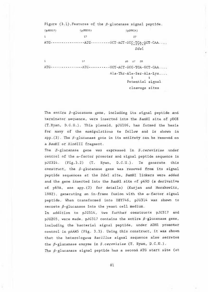
(pJG317) (pJG205) (pJG314)
1 17 27
ATG-.............. ATG.........GCT - ACT - GCC - TCA- GCT - CAA. .* * * * * * *
Ddel
1 17 26 27 28
ATG.............. ATG..........GCT - ACT - GCC - TCA - GCT - CAA. .Ala-Thr-Ala-Ser-Ala-Lys..
4- -!■Potential signalcleavage sites
Figure (3 .1) . Features of the /3-glucanase signal peptide.
The entire /3-glucanase gene, including its signal peptide and terminator sequence, were inserted into the BamHI site of pUC8 (T.Ryan, D.C.U.). This plasmid, pJG106, has formed the basis for many of the manipulations to follow and is shown in app.(3). The /3-glucanase gene in its entirety can be removed on a BamHI or tfindlll fragment.The /3-glucanase gene was expressed in S .cerevisiae under control of the a-factor promoter and signal peptide sequence in pJG314. (Fig.3.2) (T. Ryan, D.C.U.). To generate thisconstruct, the /3-glucanase gene was removed from its signal peptide sequences at the DdeI site, BamHI linkers were added and the gene inserted into the BamRI site of p69D (a derivative of p69A, see app.(2) for details) (Kurjan and Herskowitz, 1982), generating an in-frame fusion with the a-factor signal peptide. When transformed into DBY746, pJG314 was shown tosecrete /3-glucanase into the yeast cell medium.In addition to pJG314, two further constructs pJG317 and pJG205, were made. pJG317 contains the entire /9-glucanase gene, including the bacterial signal peptide, under AD H1 promotercontrol in pAAH5 (Fig. 3.3). Using this construct, it was shown that the heterologous Bacillus signal sequence also secretesthe /3-glucanase enzyme in S.cerevisiae (T. Ryan, D.C.U.).The /3-glucanase signal peptide has a second ATG start site (at
81
amino acid position 17) , in frame with the initiation codon. The gene including the signal peptide from this second ATG, was introduced into S.cerevisiae under control of the ADH1 promoter in pAAH5 (Cantwell et al. 1986). This plasmid, pJG205(Fig.3.3), was also shown to produce and secrete the enzyme despite its truncated signal sequence. Initial investigations suggested, that in terms of secretion of the enzyme in S .cerevisiae, pJG317 and pJG205 were more efficient than pJG314 (T.Ryan, pers. comm.). In fermentation trials, an ale and lager brewing strain transformed with pJG205(l) (pJG205 modified bythe insertion of the CUP1 dominant selectable marker), also secreted the enzyme in sufficient quantities to significantly reduce the levels of /3-glucan during the fermentation process (Cantwell et al. 1985).The work presented here was undertaken to examine in more detail the production and secretion of the B.subtilis /3-glucanase enzyme in S.cerevisiae using these three plasmids. Of particular interest was to determine whether the extracellular levels of activity observed in pJG317 and pJG205, were due to translocation into and transport through the conventional yeast secretory pathway, rather than simply due to cell lysis or leakage. Also, it was hoped to examine the efficiency of the bacterial signal and assess its potential for use as a general secretion sequence for heterologous protein production in yeast. Examination of the Bacillus protein product, with respect to processing and modification, was also carried out. The difficulties associated with rapid quantitation of /3-glucanase levels prompted the investigation of another heterologous protein, /3-glucuronidase (an E.coli enzyme), and its potential for development as a reporter enzyme for S.cerevisiae.
82
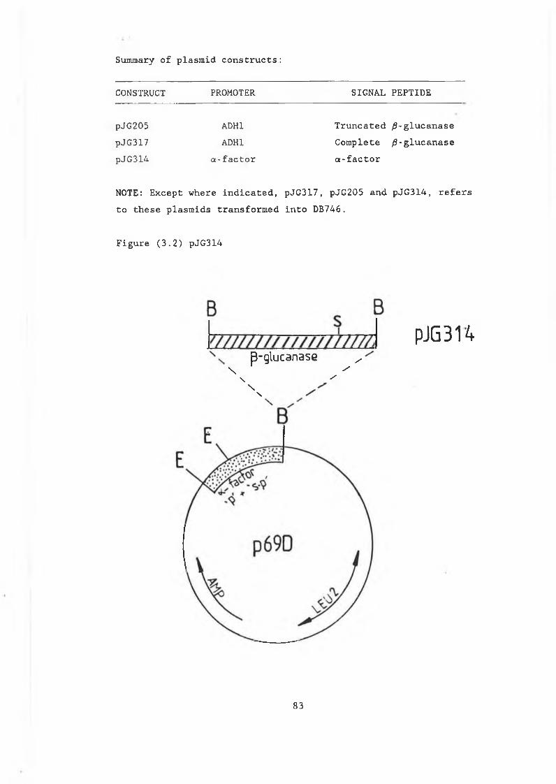
Summary of plasmid constructs:
CONSTRUCT PROMOTER SIGNAL PEPTIDE
pJG205 ADH1 Truncated ^-glucanasepJG317 ADH1 Complete /3-glucanasepJG314 a-factor a-factor
NOTE: Except where indicated, pJG317, pJG205 and pJG314, refers to these plasmids transformed into DB746.
Figure (3.2) pJG314
pJG 314p-glucanase ^
\ ^\ /
n
83
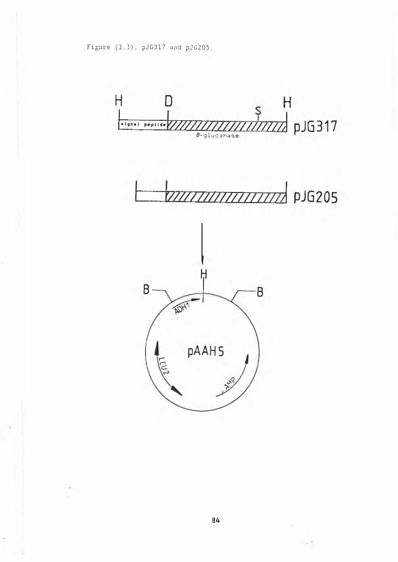
Figure (3.3). pJG317 and pJG205
84
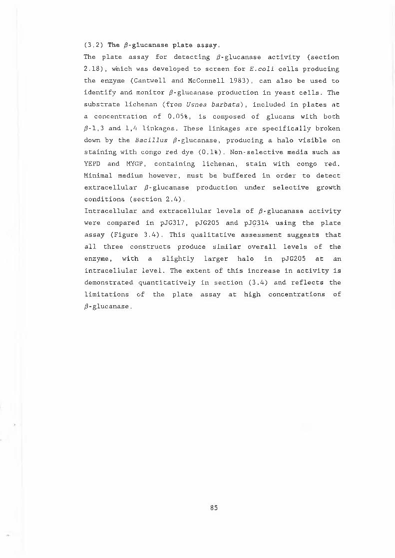
(3.2) The /3-glucanase plate assay.The plate assay for detecting /3-glucanase activity (section 2.18), which was developed to screen for E.coli cells producing the enzyme (Cantwell and McConnell 1983), can also be used to identify and monitor ¿3-glucanase production in yeast cells. The substrate lichenan (from Usnea barbata), included in plates at a concentration of 0.05%, is composed of glucans with both ¿3-1,3 and 1,4 linkages. These linkages are specifically broken down by the Bacillus /3-glucanase, producing a halo visible on staining with congo red dye (0.1%). Non-selective media such as YEPD and MYGP, containing lichenan, stain with congo red. Minimal medium however, must be buffered in order to detect extracellular /3-glucanase production under selective growth conditions (section 2.4).Intracellular and extracellular levels of ¿3-glucanase activity were compared in pJG317, pJG205 and pJG314 using the plate assay (Figure 3.4). This qualitative assessment suggests that all three constructs produce similar overall levels of the enzyme, with a slightly larger halo in pJG205 at an intracellular level. The extent of this increase in activity is demonstrated quantitatively in section (3.4) and reflects the limitations of the plate assay at high concentrations of /3-glucanase.
85
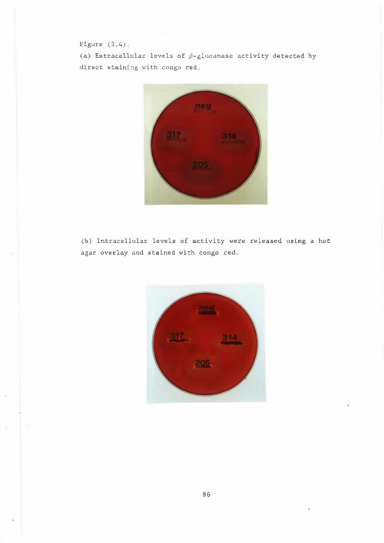
Figure (3.4).(a) Extracellular levels of /3-glucanase activity detected by direct staining with congo red.
(b) Intracellular levels of activity were released using a hot agar overlay and stained with congo red.
86
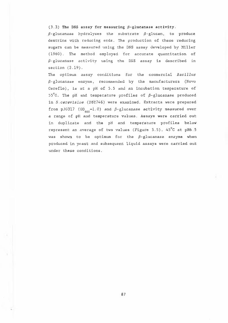
(3.3) The DNS assay for measuring /3-glucanase activity. /9-glucanase hydrolyses the substrate /3-glucan, to produce dextrins with reducing ends. The production of these reducing sugars can be measured using the DNS assay developed by Miller (1960). The method employed for accurate quantitation of ¿3-glucanase activity using the DNS assay is described in section (2.19).The optimum assay conditions for the commercial Bacillus ^-glucanase enzyme, recommended by the manufacturers (Novo Cereflo) , is at a pH of 5.5 and an incubation temperature of 55°C. The pH and temperature profiles of /9-glucanase produced in S .cerevisiiie (DBY746) were examined. Extracts were prepared from pJG317 (0D =1.0) and /3-glucanase activity measured overa range of pH and temperature values. Assays were carried out in duplicate and the pH and temperature profiles below represent an average of two values (Figure 3.5). 45°C at pH6.5 was shown to be optimum for the /3-glucanase enzyme when produced in yeast and subsequent liquid assays were carried out under these conditions.
87
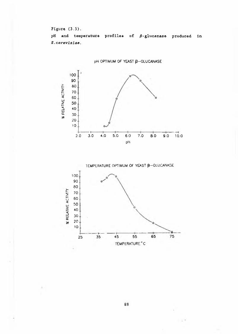
F ig u re ( 3 . 5 ) .
pH and temperature profiles of ^-glucanase produced in S .c e r e v l s l a e .
pH OPTIMUM OF YEAST p-GLUCANASE
100 .
90 . .
8 0 . .>h~ 7 0 . .o< 60 ..U J> 50 .1- <• — 1 40UJc n 30&
2 0 .
1 0
2.0— i—3.0 4.0 5.0 6.0
pH
7.0 8.0 9.0 10.0
TEMPERATURE OPTIMUM OF YEAST p -G LU C A N A S E
2b~o<
5ii jcz
100.
90..80 . .70 ..60.. 50 .. 4 0
3 0 1 2 0 .
1 0 .
25
TEMPERATURE °C
8 8
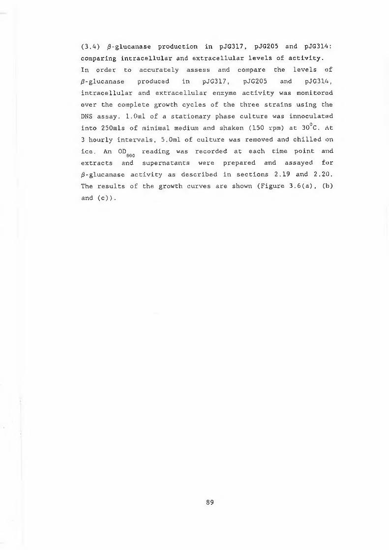
(3.4) $-glucanase production in pJG317, pJG205 and pJG314:comparing intracellular and extracellular levels of activity.In order to accurately assess and compare the levels of/0-glucanase produced in pJG317, pJG205 and pJG314,intracellular and extracellular enzyme activity was monitoredover the complete growth cycles of the three strains using theDNS assay. 1.0ml of a stationary phase culture was innoculatedinto 250mls of minimal medium and shaken (150 rpm) at 30°C. At3 hourly intervals, 5.0ml of culture was removed and chilled onice. An OD reading was recorded at each time point and
600 b
extracts and supernatants were prepared and assayed for /3-glucanase activity as described in sections 2.19 and 2.20. The results of the growth curves are shown (Figure 3.6(a), (b) and (c)).
89
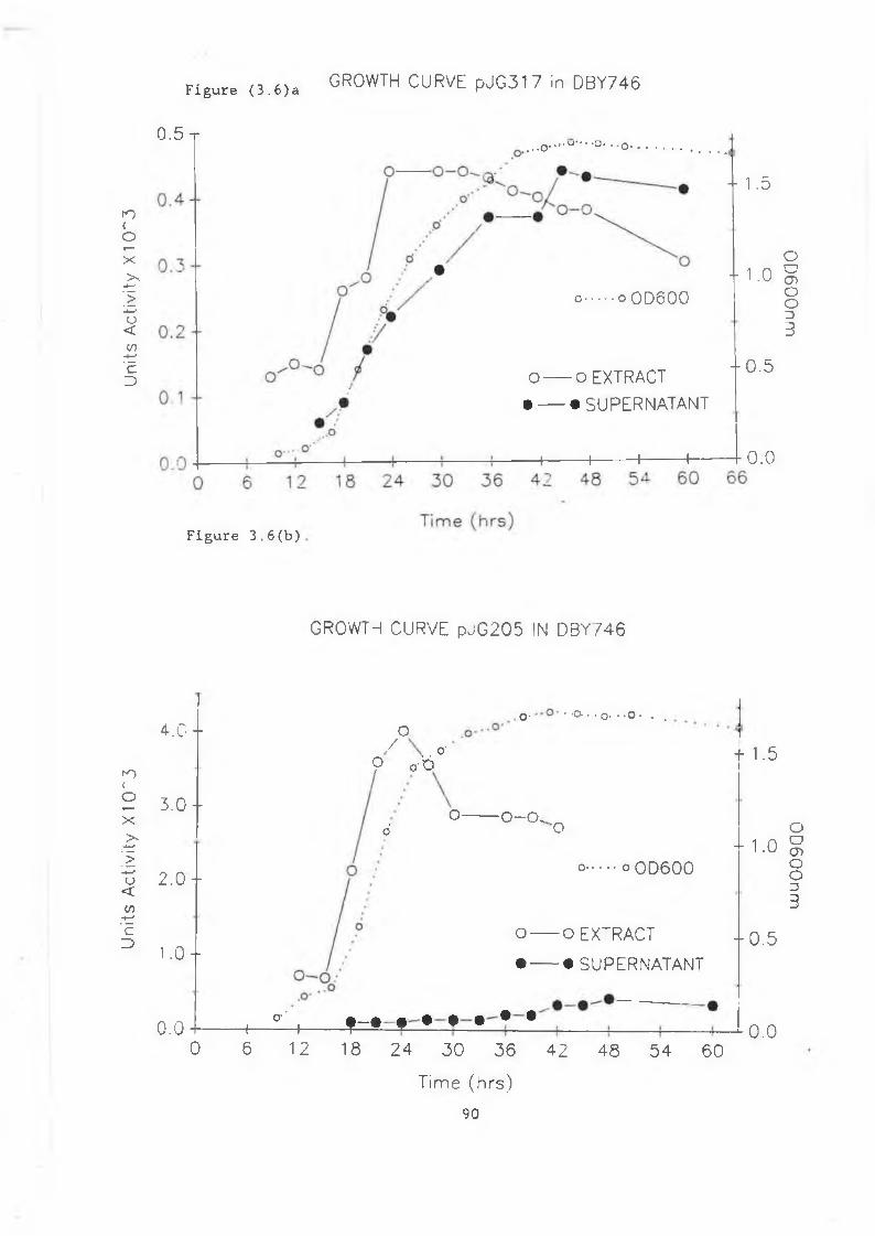
Units
Ac
tivity
X1
CT3
Units
Ac
tivity
X
10~
3
F ig u re ( 3 . 6)a GROWTH CURVE pJG317 in DBY746
0.5 t• o.
- 1.5
- 1.0o o OD600
O o EXTRACT• — • SUPERNATANT
- 0 . 5
H-------------1-------------1-------------1-------------1- 0 . 0
Figure 3.6(b)
4.0 -f O
3 . 0 -
2.0 -
1 . 0 -
0 . 0
GROWTH CURVE pJG205 IN DBY746
O- • -Q- • * O- • 'O - . .
/ O'? o b
o O - O .o'
- 1.5
•o
° ........° OD6OO± 1 . 0
o o EXTRACT I 0.5
• ----- • SUPERNATANT
4 - ^ 4 f « « « « • •
0 6 12 18 2 4 3 0 36 42 4 8 5 4 60
Time (hrs)
90
0 . 0
0D600nm
0
D6
00
nm
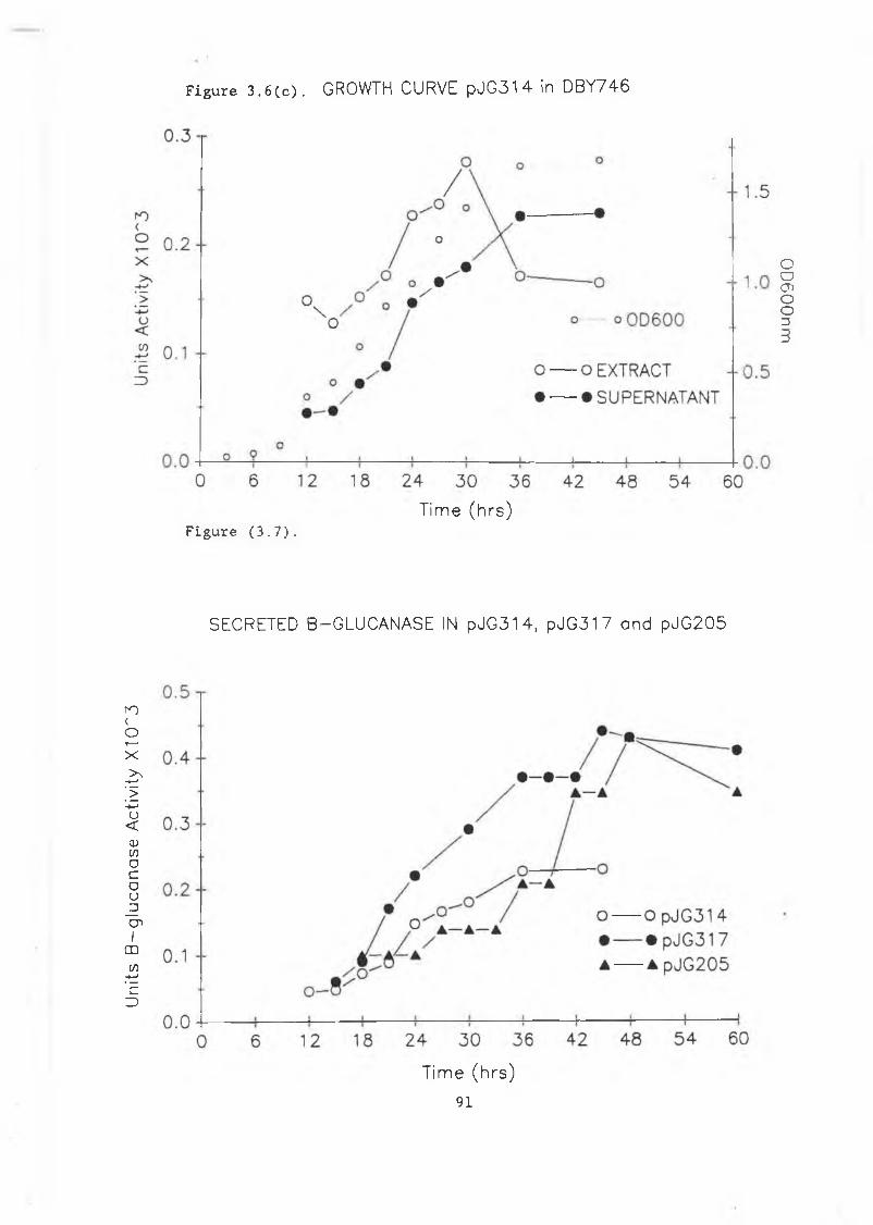
Units
B
-glu
cana
se
Activ
ity
X1C
P3
F i g u r e 3 . 6 ( c ) . GROWTH CURVE pJG31 4 in DBY746
Time (hrs)F i g u r e ( 3 . 7 ) .
SECRETED B-GLUCANASE IN pJG314, pJG317 and pJG205
Time (hrs)
9 1
OD
GO
Onm
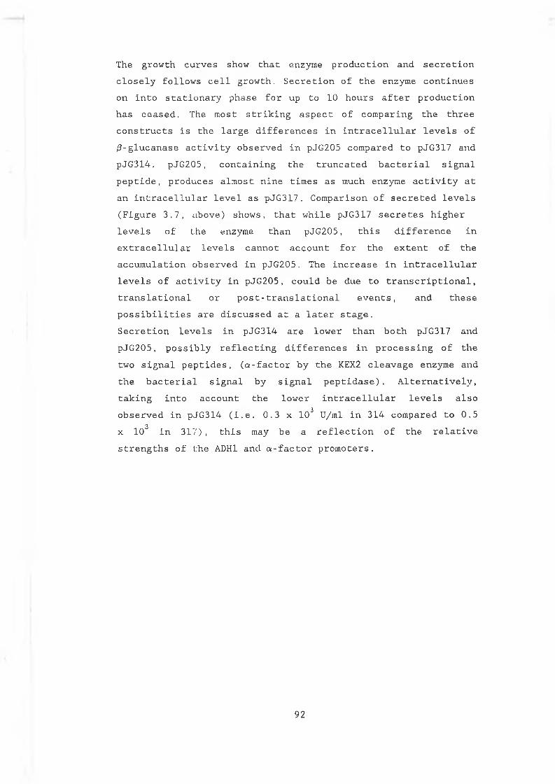
The g r o w t h c u r v e s s h o w t h a t e n z y m e p r o d u c t i o n a n d s e c r e t i o n c l o s e l y f o l l o w s c e l l g r o w t h . S e c r e t i o n o f t h e e n z y m e c o n t i n u e s o n i n t o s t a t i o n a r y p h a s e f o r up t o 1 0 h o u r s a f t e r p r o d u c t i o n h a s c e a s e d . The m o s t s t r i k i n g a s p e c t o f c o m p a r i n g t h e t h r e e c o n s t r u c t s i s t h e l a r g e d i f f e r e n c e s i n i n t r a c e l l u l a r l e v e l s o f / 3 - g l u c a n a s e a c t i v i t y o b s e r v e d i n pJ G 2 0 5 c o m p a r e d t o pJ G 3 1 7 a n d p J G 3 1 4 . p J G 2 0 5 , c o n t a i n i n g t h e t r u n c a t e d b a c t e r i a l s i g n a lp e p t i d e , p r o d u c e s a l m o s t n i n e t i m e s a s much e n z y m e a c t i v i t y a t a n i n t r a c e l l u l a r l e v e l a s p J G 3 1 7 . C o m p a r i s o n o f s e c r e t e d l e v e l s ( F i g u r e 3 . 7 , ¿ ib o v e) s h o w s , t h a t w h i l e p J G 3 1 7 s e c r e t e s h i g h e r l e v e l s o f t h e e n z y m e t h a n p J G 2 0 5 , t h i s d i f f e r e n c e i n e x t r a c e l l u l a r l e v e l s c a n n o t a c c o u n t f o r t h e e x t e n t o f t h e a c c u m u l a t i o n o b s e r v e d i n p J G 2 0 5 . T h e i n c r e a s e i n i n t r a c e l l u l a r l e v e l s o f a c t i v i t y i n p J G 2 0 5 , c o u l d b e d u e t o t r a n s c r i p t i o n a l , t r a n s l a t i o n a l o r p o s t - t r a n s l a t i o n a l e v e n t s , a n d t h e s e p o s s i b i l i t i e s a r e d i s c u s s e d a t a l a t e r s t a g e .S e c r e t i o n l e v e l s i n p J G 3 1 4 a r e l o w e r t h a n b o t h p J G 3 1 7 a n d p J G 2 0 5 , p o s s i b l y r e f l e c t i n g d i f f e r e n c e s i n p r o c e s s i n g o f t h e t w o s i g n a l p e p t i d e s , ( a - f a c t o r b y t h e KEX2 c l e a v a g e e n z y m e a n d t h e b a c t e r i a l s i g n a l b y s i g n a l p e p t i d a s e ) . A l t e r n a t i v e l y , t a k i n g i n t o a c c o u n t t h e l o w e r i n t r a c e l l u l a r l e v e l s a l s o o b s e r v e d i n p J G 3 1 4 ( i . e , 0 . 3 x 1 0 3 U / m l i n 3 1 4 c o m p a r e d t o 0 . 5
3x 1 0 i n 3 1 7 ) , t h i s may b e a r e f l e c t i o n o f t h e r e l a t i v e s t r e n g t h s o f t h e ADH1 a n d a - f a c t o r p r o m o t e r s .
92
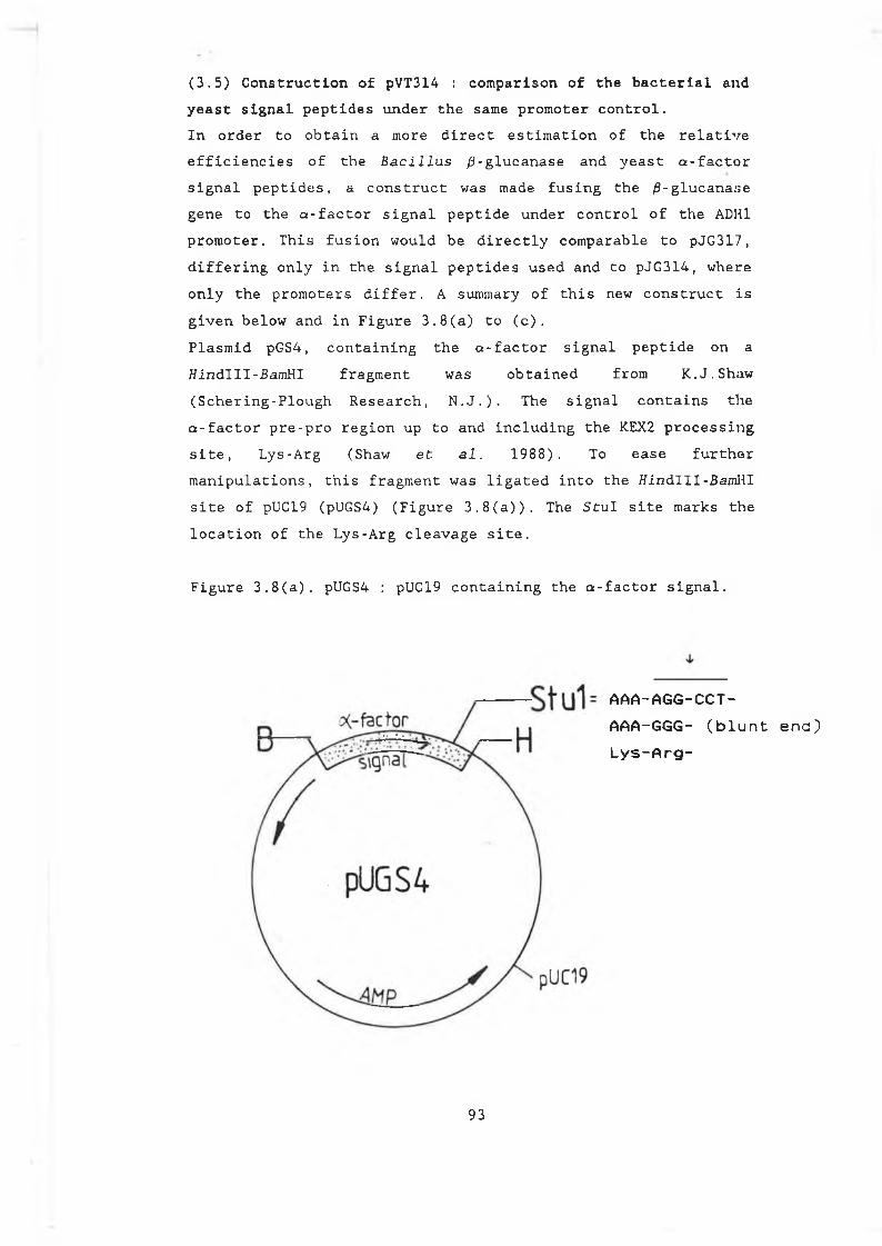
(3.5) Construction of pVT314 : comparison of the bacterial and yeast signal peptides under the same promoter control.I n o r d e r t o o b t a i n a m o r e d i r e c t e s t i m a t i o n o f t h e r e l a t i v e e f f i c i e n c i e s o f t h e B a c i l l u s / 3 - g l u c a n a s e a n d y e a s t a - f a c t o r s i g n a l p e p t i d e s , a c o n s t r u c t wa s made f u s i n g t h e / 3 - g l u c a n a s e g e n e t o t h e a - f a c t o r s i g n a l p e p t i d e u n d e r c o n t r o l o f t h e ADH1 p r o m o t e r . T h i s f u s i o n w o u l d b e d i r e c t l y c o m p a r a b l e t o p J G 3 1 7 , d i f f e r i n g o n l y i n t h e s i g n a l p e p t i d e s u s e d a n d t o p J G 3 1 4 , w h e r e o n l y t h e p r o m o t e r s d i f f e r . A summary o f t h i s new c o n s t r u c t i s g i v e n b e l o w a n d i n F i g u r e 3 . 8 ( a ) t o ( c ) .P l a s m i d p G S 4 , c o n t a i n i n g t h e a - f a c t o r s i g n a l p e p t i d e o n a H i n d l l l - B a m H I f r a g m e n t w a s o b t a i n e d f r o m K . J . S h a w ( S c h e r i n g - P l o u g h R e s e a r c h , N . J . ) . The s i g n a l c o n t a i n s t h e a - f a c t o r p r e - p r o r e g i o n up t o a n d i n c l u d i n g t h e KEX2 p r o c e s s i n g s i t e , L y s - A r g (S h a w e t a.1. 1 9 8 8 ) . To e a s e f u r t h e rm a n i p u l a t i o n s , t h i s f r a g m e n t w a s l i g a t e d i n t o t h e t f i n d l l l - B a m H I s i t e o f pUC19 (pUGS4) ( F i g u r e 3 . 8 ( a ) ) . The S t u I s i t e m a r k s t h e l o c a t i o n o f t h e L y s - A r g c l e a v a g e s i t e .
F i g u r e 3 . 8 ( a ) . pUGS4 : pUC19 c o n t a i n i n g t h e a - f a c t o r s i g n a l .
AAA-AGG-CCT- AAA-GGG- (blunt ena) Lys-Arg-
9 3
T he / 3 - g l u c a n a s e g e n e , i n c l u d i n g i C s c o m p l e t e s i g n a l p e p t i d e i s p r e s e n t b e t w e e n tw o / / i n d l l l s i t e s i n p J G 1 06 ( F i g u r e 3 . 8 ( b ) ) . Th e D d e l s i t e i n d i c a t e d w i t h a n a s t e r i x , m a r k s t h e j u n c t i o n b e t w e e n t h e e n d o f t h e B a c i l l u s s i g n a l p e p t i d e a n d t h e b e g i n n i n g o f t h e m a t u r e s e q u e n c e o f t h e g e n e (M u rp h y e t a l . 1 9 8 4 ) . The g e n e , m i n u s i t s s i g n a l p e p t i d e w a s i s o l a t e d f r o m pJ G 1 0 6 o n a D d e l f r a g m e n t a n d t h e 5 ' o v e r h a n g s w e r e f i l l e d i n t o g i v e a b l u n t e n d e d f r a g m e n t . T h i s f i l l e d i n f r a g m e n t w a s t h e n c u t w i t h H i n d l l l a n d t h e / 3 - g l u c a n a s e g e n e , m i n u s i t s s i g n a l p e p t i d e a n d pUC19 s e q u e n c e s , w a s p u r i f i e d f r o m a g e l . T h i s b l u n t / s t i c k y e n d e d f r a g m e n t w a s l i g a t e d ( i n f r a m e ) i n t o S t u I / / / i n d I I I - c u t pUGS4 t o g i v e p J G S 4 ( F i g u r e 3 . 8 ( b ) ) . The / 3 - g l u c a n a s e g e n e a n d a - f a c t o r s i g n a l p e p t i d e w a s r e m o v e d f r o m pJGS4 u s i n g Ba/nHI a n d l i g a t e d i n t o pVT102U ( V e r n e t e t a l , 1 9 8 7 ) , p l a c i n g i t u n d e r c o n t r o l o f t h e ADHI p r o m o t e r ( F i g u r e 3 . 8 ( c ) ) . O r i e n t a t i o n w a s c o n f i r m e d b y r e s t r i c t i o n d i g e s t a n d t h e p l a s m i d na m ed p V T 3 1 4 .
9 4
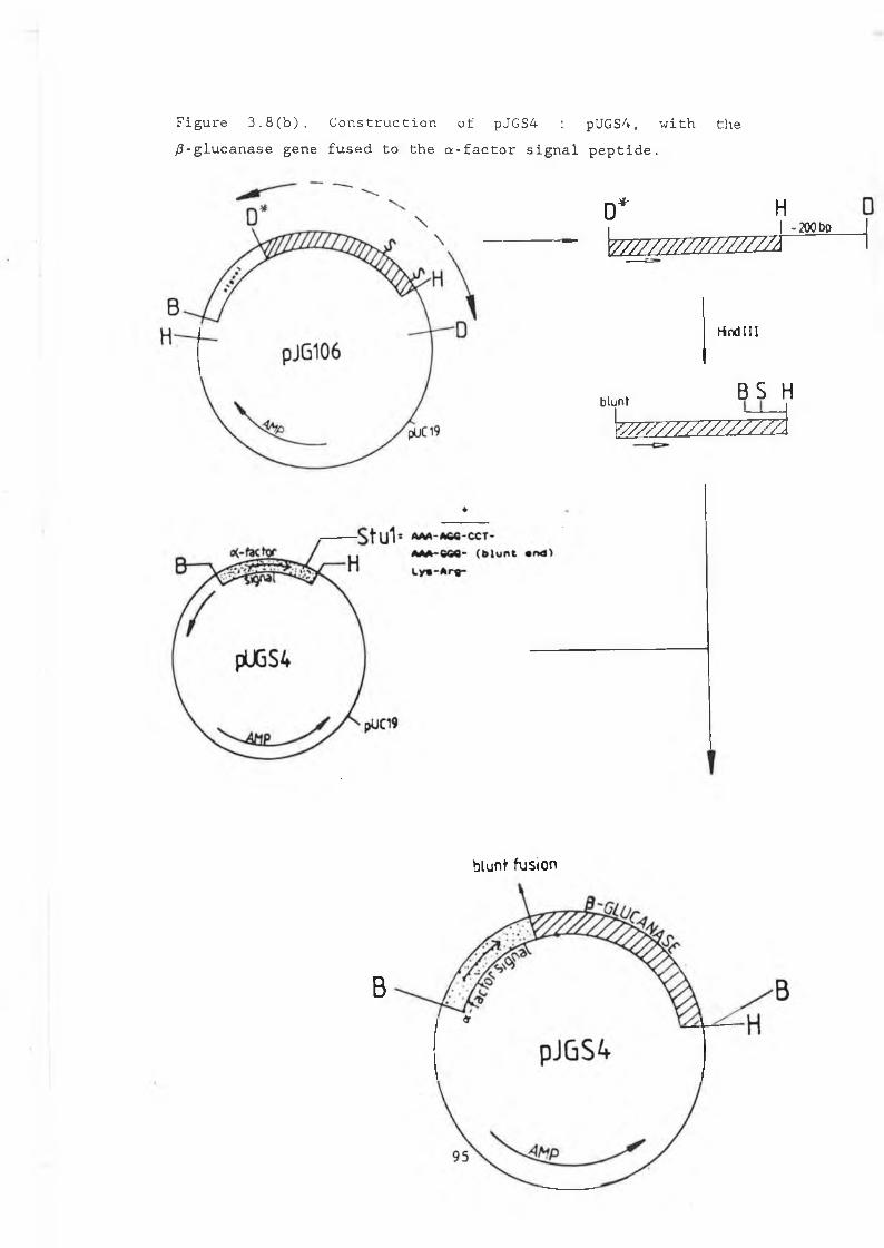
F i g u r e 3 . 8 ( b ) . C o n s t r u c t i o n o f pJGS4 : p U G S 4 , w i t h t h e / 3 - g l u c a n a s e g e n e f u s e d t o t h e a - f a c t o r s i g n a l p e p t i d e .
D * HI I -200 boW 7T/77/Z7/7777Zm
hind I I I
blunt 8 S H^ 7ZZZZZZZZZZZZ1
blunt fusion
B
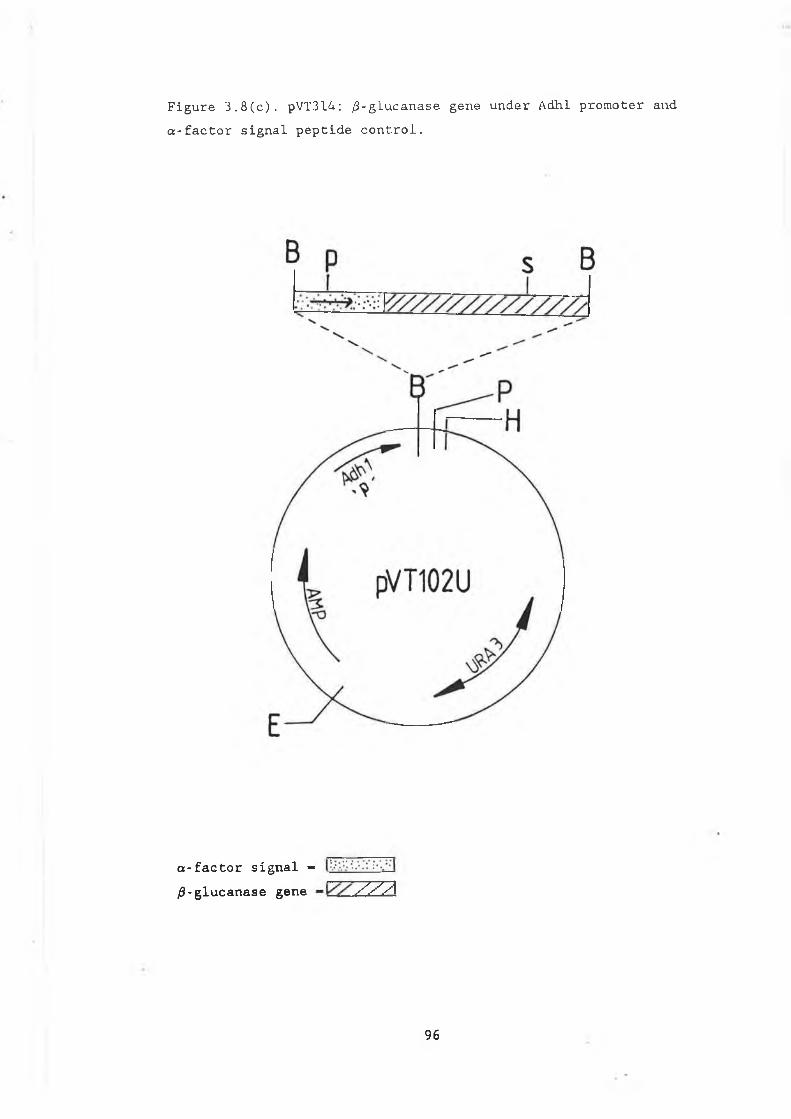
F i g u r e 3 . 8 ( c ) . pV T 3 14 : / 3 - g L u c a n a s e g e n e u n d e r A d h l p r o m o t e r a n d a - f a c t o r s i g n a l p e p t i d e c o n t r o l .
a-factor signal - /3-glucanase gene Z 2 2
96
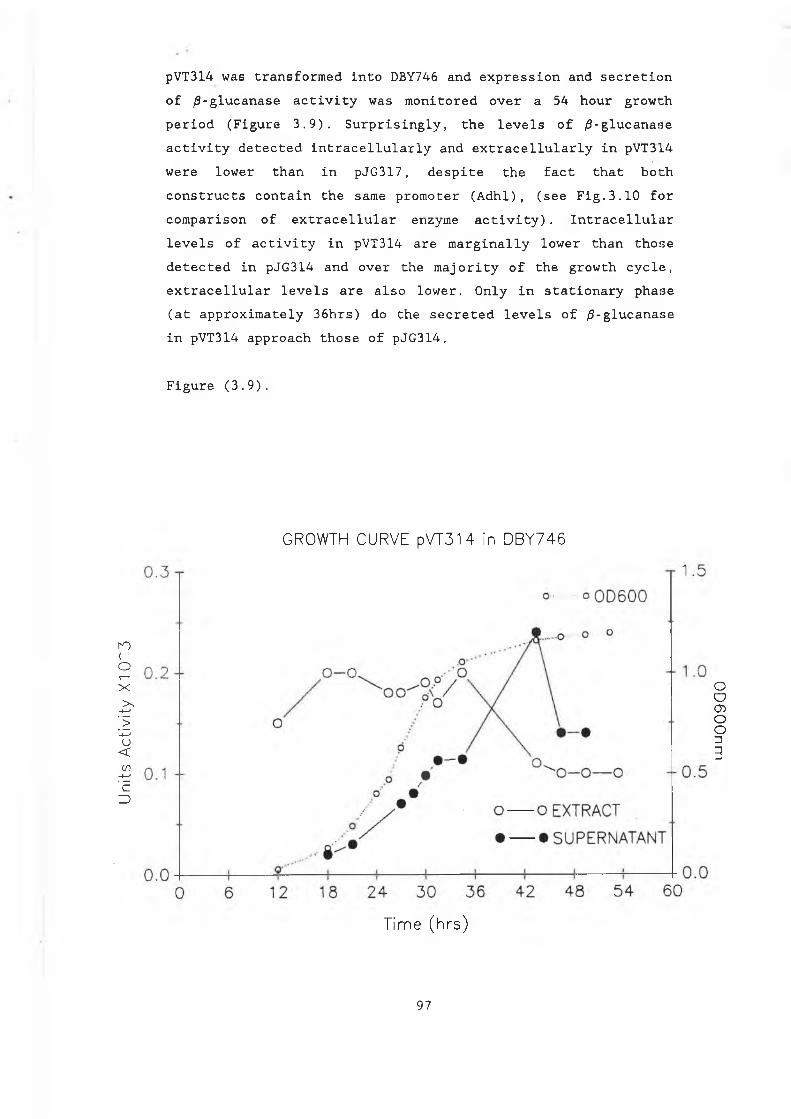
Units
Ac
tivity
X1
O'"
3
pVT3 14 w a s t r a n s f o r m e d i n t o DBY746 a n d e x p r e s s i o n a n d s e c r e t i o n o f / 9 - g l u c a n a s e a c t i v i t y w a s m o n i t o r e d o v e r a 5 4 h o u r g r o w t h p e r i o d ( F i g u r e 3 . 9 ) . S u r p r i s i n g l y , t h e l e v e l s o f / S - g l u c a n a s e a c t i v i t y d e t e c t e d i n t r a c e l l u l a r l y a n d e x t r a c e l l u l a r l y i n pVT 314 w e r e l o w e r t h a n i n p J G 3 1 7 , d e s p i t e t h e f a c t t h a t b o t h c o n s t r u c t s c o n t a i n t h e sa m e p r o m o t e r ( A d h l ) , ( s e e F i g . 3 . 1 0 f o r c o m p a r i s o n o f e x t r a c e l l u l a r e n z y m e a c t i v i t y ) . I n t r a c e l l u l a r l e v e l s o f a c t i v i t y i n pVT3 14 a r e m a r g i n a l l y l o w e r t h a n t h o s e d e t e c t e d i n p J G 3 1 4 a n d o v e r t h e m a j o r i t y o f t h e g r o w t h c y c l e , e x t r a c e l l u l a r l e v e l s a r e a l s o l o w e r . O n l y i n s t a t i o n a r y p h a s e ( a t a p p r o x i m a t e l y 3 6 h r s ) d o t h e s e c r e t e d l e v e l s o f / 3 - g l u c a n a s e i n pV T 314 a p p r o a c h t h o s e o f p J G 3 1 4 .
F i g u r e ( 3 . 9 ) .
GROWTH CURVE pVT314 in DBY746
Time (hrs)
9 7
0D600nm
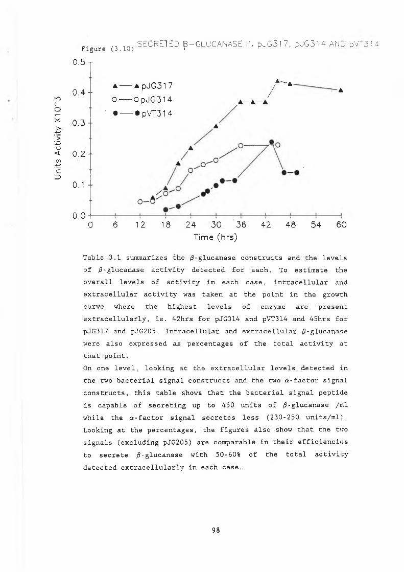
Units
Ac
tivity
X
1 0~3
i n . SECRETEO B - G L U C A N A S E ¡N p^‘G 3 1 7 , p J G 3 l 4 A N ü oV ,3 :4 -F i g u r e ( 3 . 1 0 ) r0.5 r
a — a PJG31 7A ' A
0 . 4 ” / AO o pJG314 A— A —A
0 . 3 -• — • pVT31 414
A
0 . 2 -
0.1 ~
0.00 6 12 18 24 30 36 42 48 54 60
T a b l e 3 . 1 s u m m a r i z e s t h e / 3 - g l u c a n a s e c o n s t r u c t s a n d t h e l e v e l s o f / 3 - g l u c a n a s e a c t i v i t y d e t e c t e d f o r e a c h . To e s t i m a t e t h e o v e r a l l l e v e l s o f a c t i v i t y i n e a c h c a s e , i n t r a c e l l u l a r a n d e x t r a c e l l u l a r a c t i v i t y w a s t a k e n a t t h e p o i n t i n t h e g r o w t h c u r v e w h e r e t h e h i g h e s t l e v e l s o f e n z y m e a r e p r e s e n t e x t r a c e l l u l a r l y , i e . 4 2 h r s f o r p J G 3 1 4 a n d p V T 3 1 4 a n d 4 5 h r s f o r p J G 3 1 7 a n d p J G 2 0 5 . I n t r a c e l l u l a r a n d e x t r a c e l l u l a r / 3 - g l u c a n a s e w e r e a l s o e x p r e s s e d a s p e r c e n t a g e s o f t h e t o t a l a c t i v i t y a t t h a t p o i n t .On o n e l e v e l , l o o k i n g a t t h e e x t r a c e l l u l a r l e v e l s d e t e c t e d i n t h e t w o b a c t e r i a l s i g n a l c o n s t r u c t s a n d t h e tw o a - f a c t o r s i g n a l c o n s t r u c t s , t h i s t a b l e s h o w s t h a t t h e b a c t e r i a l s i g n a l p e p t i d e i s c a p a b l e o f s e c r e t i n g up t o 4 5 0 u n i t s o f / 3 - g l u c a n a s e / i n i w h i l e t h e a - f a c t o r s i g n a l s e c r e t e s l e s s ( 2 3 0 - 2 5 0 u n i t s / m l ) . L o o k i n g a t t h e p e r c e n t a g e s , t h e f i g u r e s a l s o s h o w t h a t t h e tw o s i g n a l s ( e x c l u d i n g p J G 2 0 5 ) a r e c o m p a r a b l e i n t h e i r e f f i c i e n c i e s t o s e c r e t e / 3 - g l u c a n a s e w i t h 5 0 -6 0 % o f t h e t o t a l a c t i v i t y d e t e c t e d e x t r a c e l l u l a r l y i n e a c h c a s e .
Time (h rs )
98
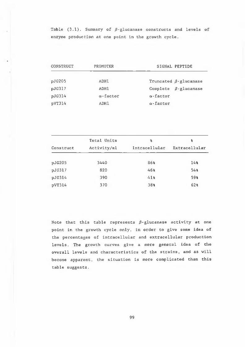
T a b l e ( 3 . 1 ) . Summary o f / 3 - g l u c a n a s e c o n s t r u c t s a n d l e v e l s o f e n z y m e p r o d u c t i o n a t o n e p o i n t i n t h e g r o w t h c y c l e .
CONSTRUCT PROMOTER SIGNAL PEPTIDE
pJ G 2 0 5 ADH1 T r u n c a t e d / 3 - g l u c a n a s ep J G 317 ADH1 C o m p l e t e / 3 - g l u c a n a s ep J G 3 1 4 a - f a c t o r a - f a c t o rpVT314 ADH1 a - f a c t o r
C o n s t r u c tT o t a l U n i t s A c t i v i t y / m l
%I n t r a c e l l u l a r
%E x t r a c e l l u l a r
p J G 2 0 5 3 4 4 0 86% 14%pJ G 3 17 8 2 0 46% 54%pJ G 3 1 4 3 9 0 41% 59%pVT314 3 7 0 38% 62%
N o t e t h a t t h i s t a b l e r e p r e s e n t s / 3 - g l u c a n a s e a c t i v i t y a t o n e p o i n t i n t h e g r o w t h c y c l e o n l y , i n o r d e r t o g i v e s o m e i d e a o f t h e p e r c e n t a g e s o f i n t r a c e l l u l a r a n d e x t r a c e l l u l a r p r o d u c t i o n l e v e l s . Th e g r o w t h c u r v e s g i v e a m o re g e n e r a l i d e a o f t h e o v e r a l l l e v e l s a n d c h a r a c t e r i s t i c s o f t h e s t r a i n s , a n d a s w i l l b e c o m e a p p a r e n t , t h e s i t u a t i o n i s m o re c o m p l i c a t e d t h a n t h i s t a b l e s u g g e s t s .
99
(3.6) Removal of the /9-glucanase signal peptide.The e x t r a c e l l u l a r l e v e l s o f / 3 - g l u c a n a s e a c t i v i t y o b s e r v e d i n p J G 3 1 7 a n d p J G 2 0 5 w o u l d s u g g e s t t h a t i n y e a s t , t h i s h e t e r o l o g o u s s i g n a l p e p t i d e i s c a p a b l e o f d i r e c t i n g / 3 - g l u c a n a s e o u t i n t o t h e m e d iu m . I f t h i s b a c t e r i a l s i g n a l p e p t i d e i s t o b e u s e f u l a s a g e n e r a l s e c r e t i o n s i g n a l f o r h e t e r o l o g o u s p r o t e i n p r o d u c t i o n i n y e a s t , i t i s n e c e s s a r y t o e n s u r e t h a t t h e e x t r a c e l l u l a r / 3 - g l u c a n a s e l e v e l s o b s e r v e d i n pJ G 3 1 7 a n d p J G 2 05 a r e d u e t o t r a n s p o r t o f t h e e n z y m e t h r o u g h t h e c o n v e n t i o n a l s e c r e t o r y p a t h w a y a n d n o t d u e t o c e l l l e a k a g e o r l y s i s . I n o r d e r t o i n v e s t i g a t e t h i s a n d t o e x a m i n e t h e o v e r a l l p r o d u c t i o n l e v e l s o f t h e e n z y m e i n t h e s e v e c t o r s y s t e m s , i t w a s d e c i d e d t o r e m o v e t h e / 3 - g l u c a n a s e s i g n a l p e p t i d e a n d e x p r e s s t h e g e n e d i r e c t l y u n d e r ADH1 p r o m o t e r c o n t r o l . I n t h i s c o n s t r u c t , t h e / 3 - g l u c a n a s e a c t i v i t y w a s e x p e c t e d t o b e l i m i t e d t o t h e y e a s t c y t o p l a s m .I n r e m o v i n g t h e / 3 - g l u c a n a s e s i g n a l p e p t i d e , t h e ATG t r a n s l a t i o n a l s t a r t s i t e i s a l s o d e l e t e d . Th e pAH s e r i e s o f v e c t o r s ( p A H 9 , pAHIO a n d pAH21) p r o v i d e t h e ADH1 p r o m o t e r o n t h r e e d i f f e r e n t p l a s m i d s e a c h w i t h a n ATG s t a r t s i t e i n o n e o f t h e t h r e e r e a d i n g f r a m e s a n d a f f i n d l l l c l o n i n g s i t e (A m m er er , 1 9 8 3 ) . Th e / 3 - g l u c a n a s e g e n e , m i n u s i t s s i g n a l p e p t i d e ( s t a r t i n g f r o m t h e Dde I s i t e ) w a s c l o n e d i n t o t h e BamHI s i t e o f pUC8 a f t e r t h e a d d i t i o n o f BamHI l i n k e r s t o t h e f i l l e d i n e n d s t o g i v e p J G 1 0 4 , a p p . ( 4 ) (T . R y a n , D . C . U . ) . A H i n d l l l l i n k e r( 8 ' m e r ) w a s i n t r o d u c e d i n t o t h e S m a l s i t e o f p J G 1 0 4 ( p J G l 0 4 a ) , ( F i g u r e 3 . 1 1 ) . The t f i n d l l l f r a g m e n t f r o m p J G 1 0 4 a w a s t r a n s f e r r e d i n t o t h e c l o n i n g s i t e o f pAH9 t o g e n e r a t e a n i n - f r a m e f u s i o n b e t w e e n t h e ATG s t a r t a n d t h e / 3 - g l u c a n a s e g e n e . T h i s c o n s t r u c t w a s n a m ed pJB/?4 . O r i e n t a t i o n o f t h e / 3 - g l u c a n a s e g e n e w a s c o n f i r m e d b y S a i l d i g e s t s ( F i g u r e 3 . 1 2 ) a n d t h e p l a s m i d (p JB/34) t r a n s f o r m e d i n t o DBY 746 . H o w e v e r , no / 3 - g l u c a n a s e a c t i v i t y w a s d e t e c t a b l e i n t r a c e l l u l a r l y o r e x t r a c e l l u l a r l y u s i n g t h e p l a t e o r DNS a s s a y .
100
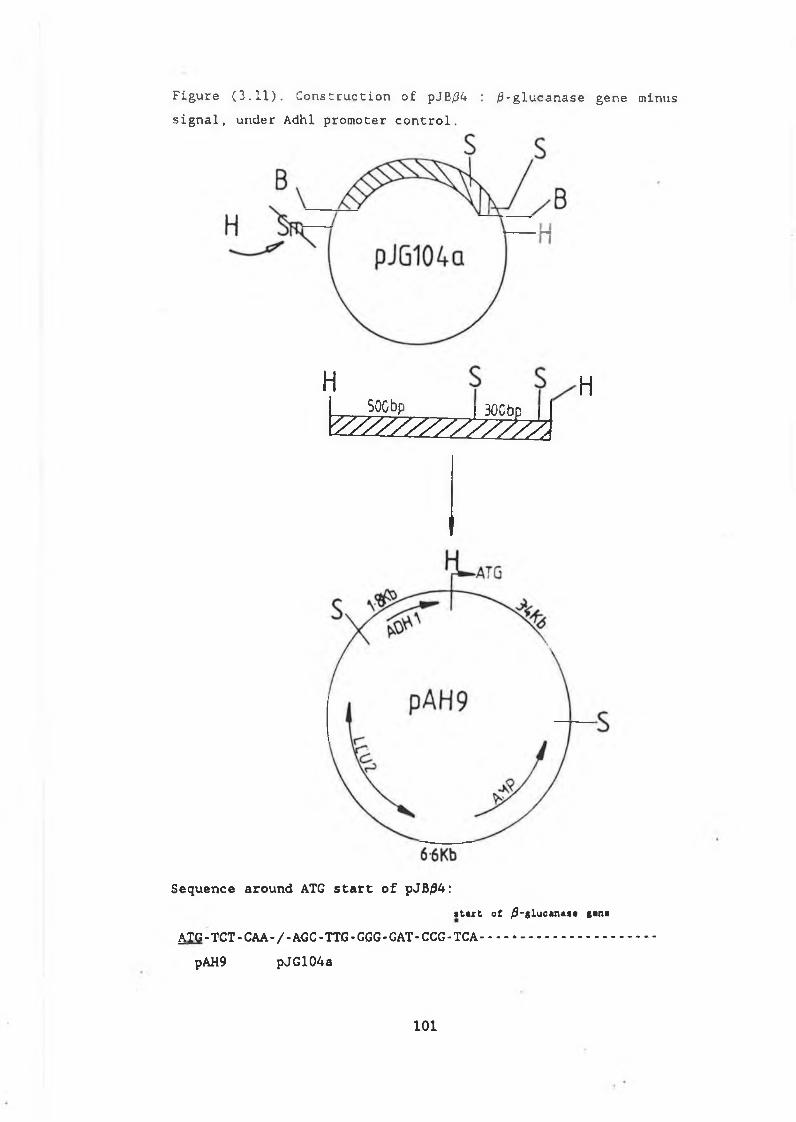
s i g n a lFigure
HSOCbp | JUODp | |
V //////////777 ,130CbcH
Sequence around ATG start of pJB04:i t a r t o f / 9 - g l u c « n a s a g a n a
&TG-TCT-CAA-/-AGC-TTG-GGG-GAT-CCG-TCA.......................pAH9 pJG104a
( 3 . 1 1 ) . C o n s t r u c t i o n o f pJB/34 : / 5 - g l u c a n a s e g e n e m i n u s , u n d e r A d h l p r o m o t e r c o n t r o l .
101
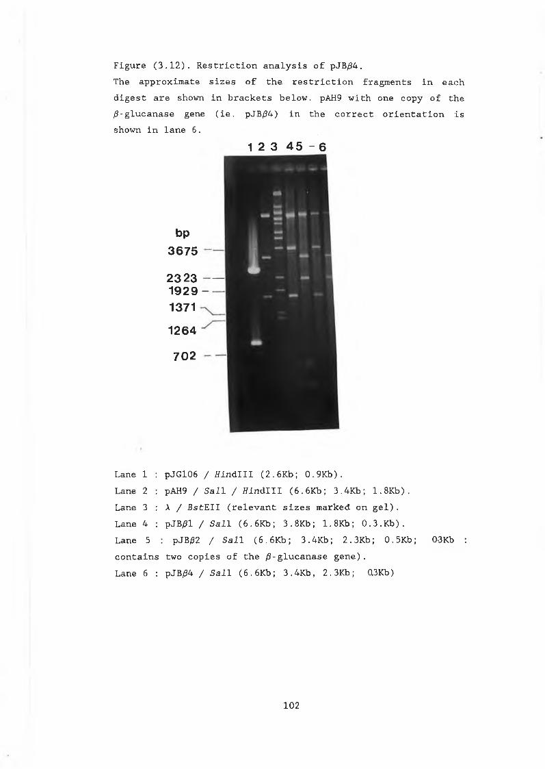
F i g u r e ( 3 . 1 2 ) . R e s t r i c t i o n a n a l y s i s o f pJB/34.The a p p r o x i m a t e s i z e s o f t h e r e s t r i c t i o n f r a g m e n t s i n e a c h d i g e s t a r e sh o w n i n b r a c k e t s b e l o w . pAH9 w i t h o n e c o p y o f t h e / 3 - g l u c a n a s e g e n e ( i e . pJB/34) i n t h e c o r r e c t o r i e n t a t i o n i s s h o w n i n l a n e 6 .
1 2 3 4 5 - 6
bp3675 2323 1929 1371 1264 702
L a n e 1 : p J G 1 0 6 / H i n d l l l ( 2 . 6 K b ; 0 . 9 K b ) .La n e 2 : pAH9 / S a i l / f f i n d l l l ( 6 . 6 K b ; 3 . 4 K b ; 1 . 8 K b ) .L a n e 3 : A / B s t E I I ( r e l e v a n t s i z e s m a r k e d o n g e l ) .L a n e 4 : p J B £ l / S a i l ( 6 . 6 K b ; 3 . 8 K b ; 1 . 8 K b ; 0 . 3 . K b ) .L a n e 5 : p J B £ 2 / S a i l ( 6 . 6 K b ; 3 . 4 K b ; 2 . 3 K b ; 0 . 5 K b ; 0-3Kb :c o n t a i n s t w o c o p i e s o f t h e / 9 - g l u c a n a s e g e n e ) .L a n e 6 : pJB/34 / S a i l ( 6 . 6 K b ; 3 . 4 K b , 2 . 3 K b ; 0.3Kb)
102
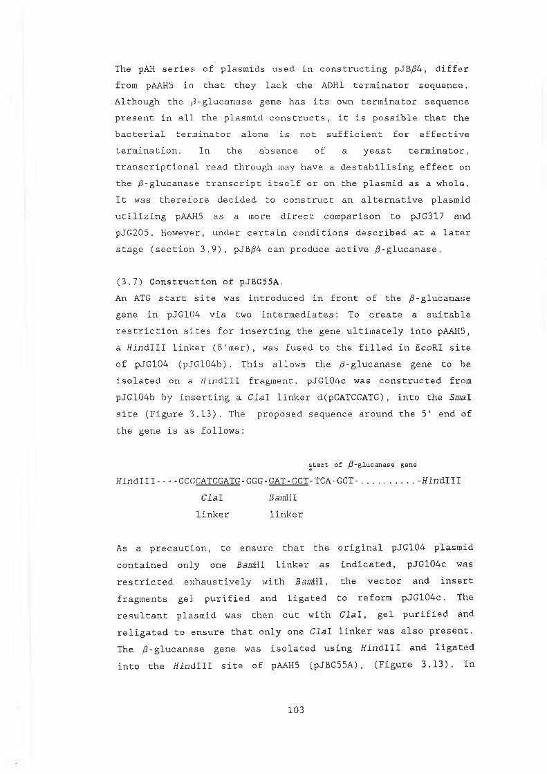
The pAH s e r i e s ! o f p l a s m i d s u s e d i n c o n s t r u c t i n g pJB/34 , d i f f e r f r o m pAAH5 i n t h a t t h e y l a c k t h e ADH1 t e r m i n a t o r s e q u e n c e . A l t h o u g h t h e / 3 - g l u c a n a s e g e n e h a s i t s own t e r m i n a t o r s e q u e n c e p r e s e n t i n a l l t h e p l a s m i d c o n s t r u c t s , i t i s p o s s i b l e t h a t t h e b a c t e r i a l t e r m i n a t o r a l o n e i s n o t s u f f i c i e n t f o r e f f e c t i v e t e r m i n a t i o n . I n t h e a b s e n c e o f a y e a s t t e r m i n a t o r , t r a n s c r i p t i o n a l r e a d t h r o u g h may h a v e a d e s t a b i l i s i n g e f f e c t o n t h e / 3 - g l u c a n a s e t r a n s c r i p t i t s e l f o r o n t h e p l a s m i d a s a w h o l e . I t w a s t h e r e f o r e d e c i d e d t o c o n s t r u c t a n a l t e r n a t i v e p l a s m i d u t i l i z i n g pAAH5 a s a m o r e d i r e c t c o m p a r i s o n t o p J G 3 1 7 a n d p J G 2 0 5 . H o w e v e r , u n d e r c e r t a i n c o n d i t i o n s d e s c r i b e d a t a l a t e r s t a g e ( s e c t i o n 3 . 9 ) , pJB/34 c a n p r o d u c e a c t i v e /3 - g l u c a n a s e .
( 3 . 7 ) C o n s t r u c t i o n o f pJB C55A.An ATG s t a r t s i t e w a s i n t r o d u c e d i n f r o n t o f t h e / 3 - g l u c a n a s e g e n e i n p J G 1 0 4 v i a t w o i n t e r m e d i a t e s : To c r e a t e a s u i t a b l er e s t r i c t i o n s i t e s f o r i n s e r t i n g t h e g e n e u l t i m a t e l y i n t o pAAH5, a f f i n d l l l l i n k e r ( 8 ' m e r ) , w a s f u s e d t o t h e f i l l e d i n £ c o R I s i t e o f p J G 1 0 4 ( p J G 1 0 4 b ) . T h i s a l l o w s t h e / 3 - g l u c a n a s e g e n e t o b e i s o l a t e d o n a H i n d l l l f r a g m e n t . p J G l 0 4 c w a s c o n s t r u c t e d f r o m p J G 1 0 4 b b y i n s e r t i n g a C l a l l i n k e r d(pCATCGATG), i n t o t h e SmaI s i t e ( F i g u r e 3 . 1 3 ) . T h e p r o p o s e d s e q u e n c e a r o u n d t h e 5 ' e n d o f t h e g e n e i s a s f o l l o w s :
s ta r t o f /3-glucanase genef f i n d l l l ------- CCGCATCGATG - GGG - GAT - CCT - TCA-GCT - ............................- f f i n d l l l
C l a l BamHIl i n k e r l i n k e r
As a p r e c a u t i o n , t o e n s u r e t h a t t h e o r i g i n a l p J G 1 0 4 p l a s m i d c o n t a i n e d o n l y o n e BamHI l i n k e r a s i n d i c a t e d , p J G 1 0 4 c w a s r e s t r i c t e d e x h a u s t i v e l y w i t h BamHI , t h e v e c t o r a n d i n s e r t f r a g m e n t s g e l p u r i f i e d a n d l i g a t e d t o r e f o r m p J G 1 0 4 c . The r e s u l t a n t p l a s m i d w a s t h e n c u t w i t h C l a l , g e l p u r i f i e d a n d r e l i g a t e d t o e n s u r e t h a t o n l y o n e C l a l l i n k e r w a s a l s o p r e s e n t . Th e / 3 - g l u c a n a s e g e n e w a s i s o l a t e d u s i n g f f i n d l l l a n d l i g a t e d i n t o t h e t f i n d l l l s i t e o f pAAH5 ( p J B C 5 5 A ) , ( F i g u r e 3 . 1 3 ) . I n
103
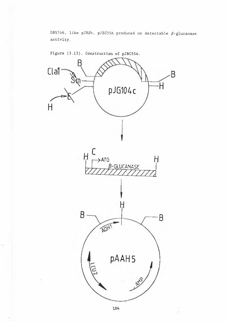
DB Y7 46 , l i k e pJB/34, pJBC55A p r o d u c e d no d e t e c t a b l e ^ f l - g l u c a n a s e a c t i v i t y .
F i g u r e ( 3 . 1 3 ) . C o n s t r u c t i o n o f pJB C55A.
-> A T G HJ-G L U C A N A S EVzzzizt7ZZZZZZL
104
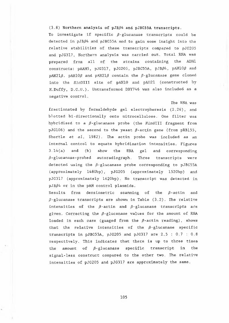
(3.8) Northern analysis of pJB/94 and pJBC55A transcripts.To i n v e s t i g a t e i f s p e c i f i c / 3 - g l u c a n a s e t r a n s c r i p t s c o u l d b e d e t e c t e d i n pJB/34 a n d pJB C55A a n d t o g a i n s o m e i n s i g h t i n t o t h e r e l a t i v e s t a b i l i t i e s o f t h e s e t r a n s c r i p t s c o m p a r e d t o pJ G 205a n d p J G 3 1 7 , N o r t h e r n a n a l y s i s w a s c a r r i e d o u t . T o t a l RNA w a sp r e p a r e d f r o m a l l o f t h e s t r a i n s c o n t a i n i n g t h e ADH1c o n s t r u c t s : pAAH5, p J G 3 1 7 , p J G 2 0 5 , pJ B C 55 A , pJB/34 , pAH10/3 a n d pAH21/3. pAH10/3 a n d pAH21/3 c o n t a i n t h e / 3 - g l u c a n a s e g e n e c l o n e d i n t o t h e T i i n D I I I s i t e o f pAHIO a n d pAH21 ( c o n s t r u c t e d b y M . D u f f y , D . C . U . ) . U n t r a n s f o r m e d DBY746 w a s a l s o i n c l u d e d a s a n e g a t i v e c o n t r o l .
The RNA wa sf r a c t i o n a t e d b y f o r m a l d e h y d e g e l e l e c t r o p h o r e s i s ( 2 . 2 6 ) , a n d b l o t t e d b i - d i r e c t i o n a l l y o n t o n i t r o c e l l u l o s e . One f i l t e r w a sh y b r i d i s e d t o a / 3 - g l u c a n a s e p r o b e ( t h e t f i n d l l l f r a g m e n t f r o m p J G 1 0 6 ) a n d t h e s e c o n d t o t h e y e a s t / 9 - a c t i n g e n e ( f r o m p R B 1 5 5 , S h o r t l e e t a l , 1 9 8 2 ) . The a c t i n p r o b e w a s i n c l u d e d a s a ni n t e r n a l c o n t r o l t o e q u a t e h y b r i d i s a t i o n i n t e n s i t i e s . F i g u r e s 3 . 1 4 ( a ) a n d ( b ) s h o w t h e RNA g e l a n d c o r r e s p o n d i n g / 3 - g l u c a n a s e - p r o b e d a u t o r a d i o g r a p h . T h r e e t r a n s c r i p t s w e r e d e t e c t e d u s i n g t h e / 3 - g l u c a n a s e p r o b e c o r r e s p o n d i n g t o pJBC5!5A ( a p p r o x i m a t e l y 1 4 8 0 b p ) , p J G 2 0 5 ( a p p r o x i m a t e l y 1 5 2 0 b p ) a n d p J G 3 1 7 ( a p p r o x i m a t e l y 1 6 2 0 b p ) . No t r a n s c r i p t w a s d e t e c t e d i n pJB/34 o r i n t h e pAH c o n t r o l p l a s m i d s .R e s u l t s f r o m d e n s i o m e t r i c s c a n n i n g o f t h e / 3 - a c t i n a n d / 3 - g l u c a n a s e t r a n s c r i p t s a r e s h o w n i n T a b l e ( 3 . 2 ) . Th e r e l a t i v e i n t e n s i t i e s o f t h e / 3 - a c t i n a n d / 3 - g l u c a n a s e t r a n s c r i p t s a r e g i v e n . C o r r e c t i n g t h e / 3 - g l u c a n a s e v a l u e s f o r t h e a m o u n t o f RNA l o a d e d i n e a c h c a s e ( g u a g e d f r o m t h e / 3 - a c t i n r e a d i n g ) , s h o w s t h a t t h e r e l a t i v e i n t e n s i t i e s o f t h e / 9 - g l u c a n a s e s p e c i f i c t r a n s c r i p t s i n pJ B C 55 A , p J G 2 0 5 a n d p J G 3 1 7 a r e 2 . 5 : 0 . 7 : 0 . 8 r e s p e c t i v e l y . T h i s i n d i c a t e s t h a t t h e r e i s up t o t h r e e t i m e s t h e a m o u n t o f / 9 - g l u c a n a s e s p e c i f i c t r a n s c r i p t i n t h e s i g n a l - l e s s c o n s t r u c t c o m p a r e d t o t h e o t h e r t w o . Th e r e l a t i v e i n t e n s i t i e s o f p J G 2 0 5 a n d p J G 3 1 7 a r e a p p r o x i m a t e l y t h e s a m e .
105
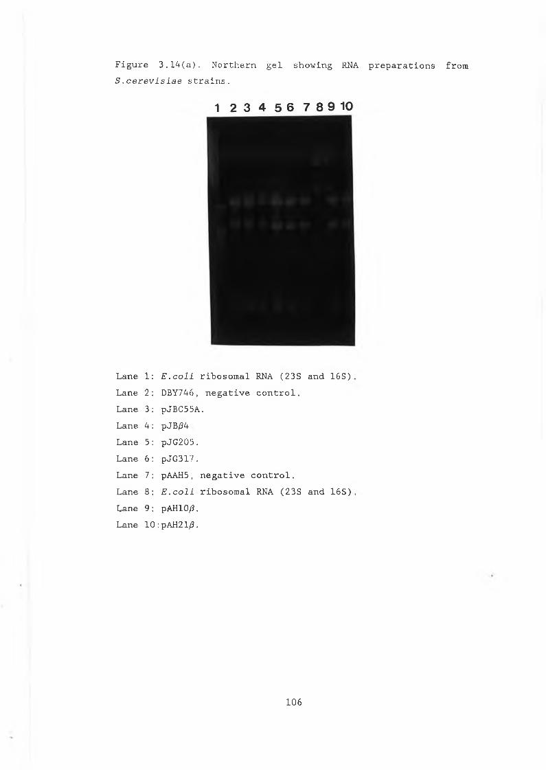
F i g u r e 3 . 1 4 ( a ) . N o r t h e r n g e l s h o w i n g RNA p r e p a r a t i o n s f r o m S . c e r e v i s i a e s t r a i n s .
1 2 3 4 5 6 7 8 9 10
L a n e 1: E . c o l i r i b o s o m a l RNA ( 2 3 S a n d 1 6 S )L a n e 2: DBY746 , n e g a t i v e c o n t r o l .L a n e 3: p J B C 5 5 A .L a n e 4 : pJB/34L a n e 5: p J G 2 0 5 .L a n e 6: p J G 3 1 7 .La n e 7 : pAAH5, n e g a t i v e c o n t r o l .L a n e 8: E . c o l i r i b o s o m a l RNA ( 2 3 S a n d 1 6 S )L a n e 9: pAH10/3.L a n e 10 : pAH21/3.
106
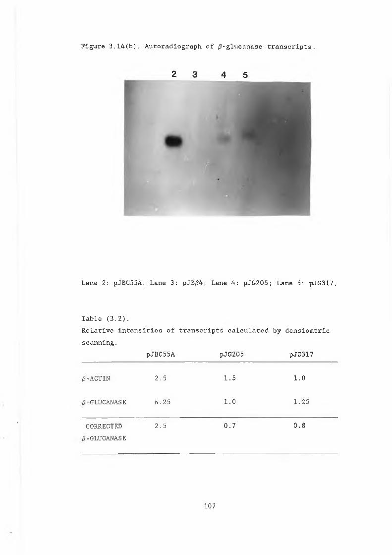
2 3 4 5
Figure 3.14(b). Autoradiograph of /3-glucanase transcripts.
L a n e 2 : pJB C55A; L a n e 3 : pJB/94; L a n e 4 : p J G 2 0 5 ; L a n e 5 : p J G 3 1 7 .
T a b l e ( 3 . 2 ) .R e l a t i v e i n t e n s i t i e s o f t r a n s c r i p t s c a l c u l a t e d b y d e n s i o m t r i c s c a n n i n g .
pJ BC 55A p J G 2 0 5 p J G 3 1 7
0 -A CT IN 2 . 5 1 . 5 1 . 0
/3-GLUCANASE 6 . 2 5 1 . 0 1 . 2 5
CORRECTED/3-GLUCANASE
2 . 5 0 . 7 0 . 8
107
(3.9) Assay of pJB/34 and pJBC55A in a pep4-3 (protease deficient) mutant of S . c e r e v i s i a e .W i t h t h e p r e s e n c e o f a s t a b l e t r a n s c r i p t i n p J B 5 5 A , t h e p o s s i b i l i t y r e m a i n e d o p e n t h a t t h e l a c k o f d e t e c t a b l e en zy ine a c t i v i t y a s s o c i a t e d w i t h t h i s c o n s t r u c t , may b e a r e s u l t o f i n s t a b i l i t y o f t h e h e t e r o l o g o u s p r o t e i n i n t h e y e a s t c y t o p l a s m . I n t h e c y t o p l a s m , / 3 - g l u c a n a s e c o u l d b e a t a r g e t o f c e l l u l a r p r o t e a s e s i n v i v o , p o s s i b l y e v e n a s u b s t r a t e o f t h e u b i q u i t i n d i r e c t e d d e g r a d a t i o n s y s t e m ( s e c t i o n 1 . 1 . 2 ) . F o l d i n g d i f f e r e n c e s , d u e t o t h e a b s e n c e o f t h e s i g n a l p e p t i d e s e q u e n c e may a l s o c o n t r i b u t e t o a n y i n s t a b i l i t y o f t h e / 3 - g l u c a n a s e e n z y m e i n t h e c y t o p l a s m . I n a d d i t i o n , t h e p r o t e i n may b e s u s c e p t i b l e t o t h e a c t i o n o f v a c u o l a r a n d o t h e r p r o t e a s e s , w h i c h c a n b e r e l e a s e d d u r i n g a s s a y i n g p r o c e d u r e s ( s e e s e c t i o n 1 . 1 . 2 ) . To i n v e s t i g a t e t h i s , t h e s i g n a l p e p t i d e - m i n u s c o n s t r u c t s w e r e t r a n s f o r m e d i n t o t h e p r o t e a s e d e f i c i e n t m u t a n t MD50. T h i s m u t a n t i s d e f i c i e n t i n t h e f o u r m a j o r v a c u o l a r p r o t e i n a s e s P r A l , P r B l , P r C l a n d C p S l a n d c o n t a i n s t h e p e p 4 - 3 m u t a t i o n (Ammerer e t a l 1 9 8 6 ) .pJBC55A a n d pJB/34 w e r e t r a n s f o r m e d i n t o MD50 a n d p l a t e d o n m i n i m a l m ed iu m . Th e c e l l s w e r e i n c u b a t e d a t 25°C f o r 4 8 h r s , l y s e d u s i n g a h o t a g a r o v e r l a y ( s e c t i o n 2 . 1 8 ) a n d i n c u b a t e d a t
25°C f o r a f u r t h e r 4 8 h r s b e f o r e s t a i n i n g w i t h c o n g or e d d y e . Th e l o w e r t e m p e r a t u r e w a s c h o s e n t o m i n i m i z e p r o t e o l y s i s , e n z y m e i n s t a b i l i t y a n d t e m p e r a t u r e a c t i v a t i o n o f p r o t e a s e s a f t e r l y s i s . S t a i n i n g w i t h c o n g o r e d r e v e a l e d h a l o s a r o u n d pJB/34 ( p l a t e d i n d u p l i c a t e ) , i n d i c a t i n g i n t r a c e l l u l a r / 3 - g l u c a n a s e a c t i v i t y ( F i g u r e 3 . 1 5 ) . T h e h a l o o b s e r v e d f o r pJB/34 w a s c o n s i d e r a b l y s m a l l e r t h a n t h o s e g e n e r a t e d a r o u n d pJG317 a n d p J G 2 0 5 ( a l s o t r a n s f o r m e d i n t o t h e MD50 m u t a n t ) , p r e s u m a b l y r e f l e c t i n g d i f f e r i n g l e v e l s o f a c t i v e e n z y m e . Th e a p p a r e n t i n s t a b i l i t y o f t h e pJB/34 RNA t r a n s c r i p t ( s e c t i o n 3 . 8 ) , m u s t c o m p o u n d t h i s p r o t e a s e p r o b l e m a n d t h e c o m b i n a t i o n o f t h e s e two f a c t o r s a r e l i k e l y t o b e t h e r e a s o n f o r t h e l a c k o f d e t e c t a b l e a c t i v i t y f o r pJB/34 i n DBY746 .I n c o n t r a s t t o pJB/34, n o , 5 - g l u c a n a s e a c t i v i t y w a s d e t e c t e d i n pJBC 55A t r a n s f o r m e d i n t o MD50, d e s p i t e t h e o b v i o u s s t a b i l i t y o f
108
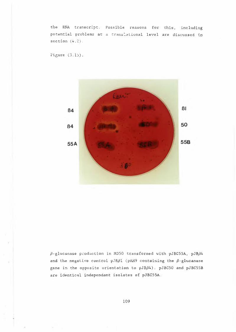
t h e RNA t r a n s c r i p t . P o s s i b l e r e a s o n s f o r t h i s , i n c l u d i n g p o t e n t i a l p r o b l e m s a t a t r a n s l a t i o n a l l e v e l a r e d i s c u s s e d i n s e c t i o n ( 4 . 2 ) .
F i g u r e ( 3 . 1 5 ) .
/ 3 - g l u c a n a s e p r o d u c t i o n i n MD50 t r a n s f o r m e d w i t h pJ B C5 5 A, pJB/34 a n d t h e n e g a t i v e c o n t r o l pJB/31 (pAH9 c o n t a i n i n g t h e / 3 - g l u c a n a s e g e n e i n t h e o p p o s i t e o r i e n t a t i o n t o pJB/34) . pJ B C 50 a n d pJBC5!5B a r e i d e n t i c a l i n d e p e n d a n t i s o l a t e s o f p J B C 5 5 A .
109
( 3 . 1 0 ) A n a l y s i s o f / 3 - g l u c a n a s e s e c r e t i o n i n s e c r e t i o n d e f e c t i v e m u t a n t s .A t t h e n o n - p e r m i s s i v e t e m p e r a t u r e ( 3 7 ° C ) , s e c r e t i o n d e f e c t i v e ( s e c ) m u t a n t s o f S . c e r e v i s i a e b l o c k s e c r e t i o n a t v a r i o u s s t a g e s a l o n g t h e s e c r e t o r y p a t h w a y . Th e a b i l i t y o f t h e b a c t e r i a l s i g n a l p e p t i d e t o t r a n s p o r t t h e / 3 - g l u c a n a s e e n z y m e t h r o u g h t h e c o n v e n t i o n a l y e a s t s e c r e t o r y p a t h w a y w a s e x a m i n e d i n a s e r i e s o f t h e s e m u t a n t s . T a b l e ( 3 . 3 ) s u m m a r i z e s t h e s e c s t r a i n s u s e d a n d t h e c h a r a c t e r i s t i c s o f e a c h m u t a n t .
T a b l e ( 3 . 3 ) . Summary o f s e c m u t a n t s .S t r a i n M u t a t i o n B l o c k I n P a t h w a yRSY11 s e c l 8 ER t o G o l g i , ( l a c k s p r o t e i n i n v o l v e d i n
v e s i c l e f u s i o n ) .RSY12 s e c 5 3 ER, ( g l y c o s y l a t i o n m u t a n t , s e c t i o n 1 . 6 )
RSY45 s e e l S e c r e t o r y v e s i c l e s b e t w e e n G o l g i a n d c e l l s u r f a c e .
pJ G 3 1 7 w a s t r a n s f o r m e d i n t o e a c h o f t h e m u t a n t s t r a i n s a n d / 3 - g l u c a n a s e s e c r e t i o n e x a m i n e d a t p e r m i s s i v e a n d n o n - p e r m i s s i v e t e m p e r a t u r e s , u s i n g t h e f o l l o w i n g p r o c e d u r e :M u t a n t s , t r a n s f o r m e d w i t h p J G 3 1 7 , w e r e g r o w n t o 0 0 ^ = 1 . 0 , a t 25°C. Th e c e l l s w e r e w a s h e d t w i c e i n 1M s o r b i t o l t o r e m o v e / 3 - g l u c a n a s e p r e s e n t i n t h e m ed iu m a n d d i v i d e d i n t o t w o e q u a l s a m p l e s . The s a m p l e s w e r e r e s u s p e n d e d i n f r e s h m ed iu m p r e w a r m e d t o ( a ) 25°C a n d ( b ) 37°C. G r o w t h w a s a l l o w e d t o c o n t i n u e a tt h e s e r e s p e c t i v e t e m p e r a t u r e s f o r 3 - 6 h o u r s . The c e l l s w e r e t h e n h a r v e s t e d a n d t h e e x t r a c t s a n d s u p e r n a t a n t s a s s a y e d f o r / 3 - g l u c a n a s e a c t i v i t y u s i n g t h e p l a t e a s s a y . 6mm w e l l s w e r e c u t i n t o b u f f e r e d a g a r c o n t a i n i n g 0 . 0 5 % l i c h e n a n . S u p e r n a t a n t s w e r e a d d e d t o t h e w e l l s a n d t h e p l a t e s i n c u b a t e d a t 37°C o v e r n i g h t . H a l o s i n d i c a t i n g / 3 - g l u c a n a s e a c t i v i t y w e r e v i s u a l i s e d b y s t a i n i n g w i t h c o n g o r e d d y e ( s e c t i o n 2 . 1 8 ) a n d F i g u r e ( 3 . 1 6 ) . As a c o n t r o l , t h i s p r o c e d u r e w a s a l s o c a r r i e d o u t w i t h DBY746 t r a n s f o r m e d w i t h p J G 3 1 7 .A b l o c k i n s e c r e t i o n o f / 3 - g l u c a n a s e w a s o b s e r v e d i n b o t h RSY11
110
a n d RSY45 a t t h e r e s t r i c t i v e t e m p e r a t u r e ( F i g . 3 . 1 7 ) . T h i s i n d i c a t e s t h a t t h e b a c t e r i a l s i g n a l p e p t i d e i s a c t i n g a s a s e c r e t i o n s i g n a l i n i t s h e t e r o l o g o u s h o s t .I n RSY12 ( s e c 5 3 ) , a n u n e x p e c t e d r e s u l t w a s o b s e r v e d . I n t h i s m u t a n t , no b l o c k i n s e c r e t i o n o f / 3 - g l u c a n a s e w a s s e e n a t t h e n o n - p e r m i s s i v e t e m p e r a t u r e . I n s t e a d , a s i g n i f i c a n t i n c r e a s e i n t h e l e v e l o f e n z y m e a c t i v i t y w a s d e t e c t e d a t 37°C c o m p a r e d t o 25°C ( F i g . 3 . 1 8 ) . More a c c u r a t e q u a n t i t a t o n o f t h i s i n c r e a s e , u s i n g t h e DNS a s s a y , i n d i c a t e s t h a t a t t h e r e s t r i c t i v e t e m p e r a t u r e , up t o 3 t i m e s a s much a c t i v i t y i s p r o d u c e d b o t h i n t r a c e l l u l a r l y a n d e x t r a c e l l u l a r l y .p J G 2 0 5 w a s a l s o t e s t e d f o r / 3 - g l u c a n a s e a c t i v i t y i n t h e s e s e c m u t a n t s t r a i n s . A b l o c k i n s e c r e t i o n o f t h e e n z y m e w a s o b s e r v e d i n RSY11 a n d RSY45 a t t h e n o n - p e r m i s s i v e t e m p e r a t u r e . S i m i l a r t o p J G 3 1 7 , a n i n c r e a s e i n / 3 - g l u c a n a s e a c t i v i t y w a s d e t e c t e d b o t h i n t r a c e l l u l a r l y a n d e x t r a c e l l u l a r l y , a t t h e r e s t r i c t i v e t e m p e r a t u r e i n R S Y 1 2 .
Ill
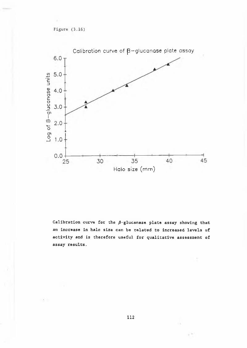
Log
of
B—
gluc
angs
e u
nits
F ig u re ( 3 . 1 6 )
Calibration curve of j3 -g lu ca n a se plate assay
H alo s ize (m m )
Calibration curve for the ^-glucanase plate assay showing that an increase in halo size can be related to increased levels of activity and is therefore useful for qualitative assessment of assay results.
112
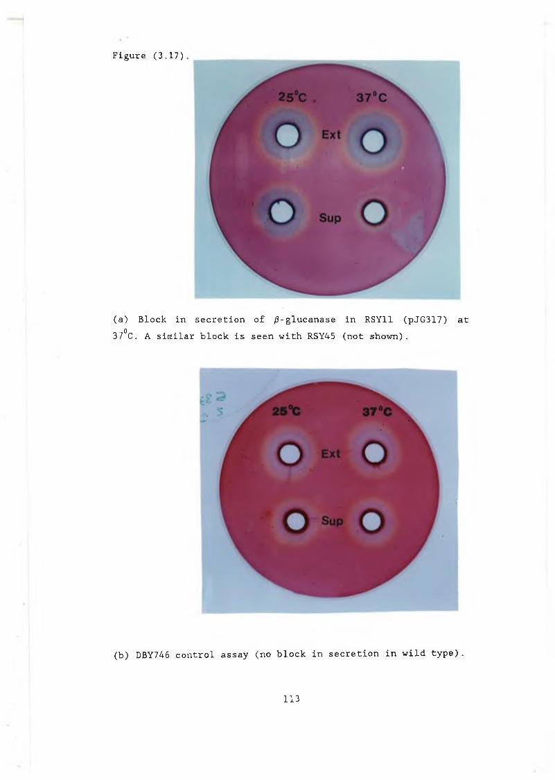
Figure (3.17)
( a ) B l o c k i n s e c r e t i o n o f / 3 - g l u c a n a s e i n RSY11 ( p J G 3 1 7 ) a t 37°C. A s i m i l a r b l o c k i s s e e n w i t h RSY45 ( n o t s h o w n ) .
( b ) DBY746 c o n t r o l a s s a y ( n o b l o c k i n s e c r e t i o n i n w i l d t y p e ) .
113
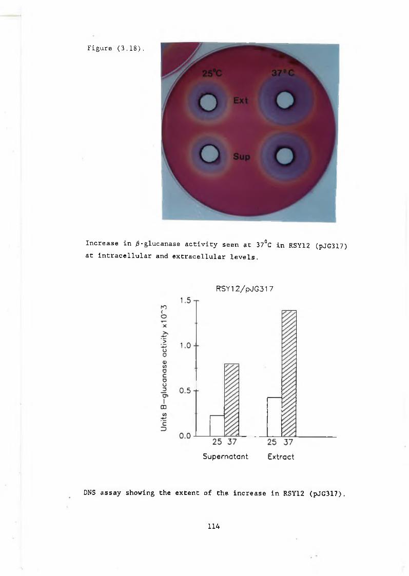
Figure (3 .18).
Increase in ^-glucanase activity seen at 37°C in RSY12 (pJG317) at intracellular and extracellular levels.
RSY12 / p J G 3 1 7
Supernatant Extract
DNS assay showing the extent of the increase in RSY12 (pJG317).
1 1 4
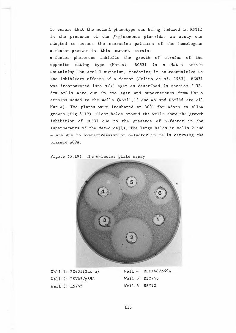
To e n s u r e t h a t t h e m u t a n t p h e n o t y p e w a s b e i n g i n d u c e d i n RSY12 i n t h e p r e s e n c e o f t h e / 9 - g l u c a n a s e p l a s m i d s , a n a s s a y was a d a p t e d t o a s s e s s t h e s e c r e t i o n p a t t e r n s o f t h e h o m o l o g o u s a - f a c t o r p r o t e i n i n t h i s m u t a n t s t r a i n :a - f a c t o r p h e r o m o n e i n h i b i t s t h e g r o w t h o f s t r a i n s o f t h e o p p o s i t e m a t i n g t y p e ( M a t - a ) . RC631 i s a M a t - a s t r a i nc o n t a i n i n g t h e s s t 2 - l m u t a t i o n , r e n d e r i n g i t e x t r a s e n s i t i v e t o t h e i n h i b i t o r y e f f e c t s o f a - f a c t o r ( J u l i u s e t a.1. 1 9 8 3 ) . RC631 w a s i n c o r p o r a t e d i n t o MYGP a g a r a s d e s c r i b e d i n s e c t i o n 2 . 3 2 . 6mm w e l l s w e r e c u t i n t h e a g a r a n d s u p e r n a t a n t s f r o m M a t - a s t r a i n s a d d e d t o t h e w e l l s ( R S Y 1 1 , 1 2 a n d 4 5 a n d DBY746 a r e a l l M a t - a ) . Th e p l a t e s w e r e i n c u b a t e d a t 30°C f o r 4 8 h r s t o a l l o w g r o w t h ( F i g . 3 . 1 9 ) . C l e a r h a l o s a r o u n d t h e w e l l s s h o w t h e g r o w t h i n h i b i t i o n o f RC631 d u e t o t h e p r e s e n c e o f a - f a c t o r i n t h e s u p e r n a t a n t s o f t h e M a t - a c e l l s . Th e l a r g e h a l o s i n w e l l s 2 a n d 4 a r e d u e t o o v e r e x p r e s s i o n o f a - f a c t o r - i n c e l l s c a r r y i n g t h e p l a s m i d p 6 9 A .
F i g u r e ( 3 .
W e l l 1: R C 6 3 1 ( M a t a ) W e l l 4 : D B Y 7 4 6 / p 6 9 AW e l l 2: R S Y 4 5 / p 6 9 A W e l l 5 : DBY746W e l l 3: RSY45 W e l l 6 : RSY12
1 9 ) . T h e a - f a c t o r p l a t e a s s a y
1 1 5

p 6 9 A ( a p p . 2 ) , i s a m u l t i c o p y p l a s m i d c o n t a i n i n g t h e a - f a c t o r s t r u c t u r a l g e n e s u n d e r c o n t r o l o f t h e a - f a c t o r p r o m o t e r a n d s i g n a l p e p t i d e ( K u r j a n a n d H e r s k o w i t z 1 9 8 2 ) . When t h e m u t a n t p h e n o t y p e i s i n d u c e d i n m u t a n t s R S Y 1 1 . 1 2 a n d 4 5 , c a r r y i n g t h i s p l a s m i d , a b l o c k i n s e c r e t i o n o f a - f a c t o r i s o b s e r v e d a t 37°C ( F i g . 3 . 2 0 ) . T h i s b l o c k i s r e f l e c t e d i n a d e c r e a s e i n t h e z o n e o f i n h i b i t i o n o f R C6 31 .
F i g u r e ( 3 . 2 0 ) .
B l o c k i n a - f a c t o r s e c r e t i o n a t 37°C i n RSY45 t r a n s f o r m e d w i t h p 6 9 A . W i l d t y p e DBY746 s h o w s n o b l o c k a t t h a t t e m p e r a t u r e .
116
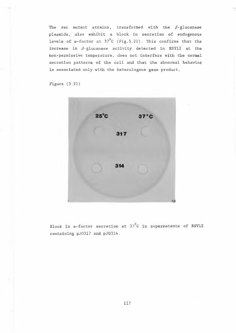
T h e s e c m u t a n t s t r a i n s , t r a n s f o r m e d w i t h t h e / 3 - g l u c a n a s e p l a s m i d s , a l s o e x h i b i t a b l o c k i n s e c r e t i o n o f e n d o g e n o u s l e v e l s o f a - f a c t o r a t 37°C ( F i g . 3 . 2 1 ) . T h i s c o n f i r m s t h a t t h e i n c r e a s e i n / 5 - g l u c a n a s e a c t i v i t y d e t e c t e d i n RSY12 a t t h e n o n - p e r m i s s i v e t e m p e r a t u r e , d o e s n o t i n t e r f e r e w i t h t h e n o r m a l s e c r e t i o n p a t t e r n s o f t h e c e l l a n d t h a t t h e a b n o r m a l b e h a v i o r i s a s s o c i a t e d o n l y w i t h t h e h e t e r o l o g o u s g e n e p r o d u c t .
F i g u r e ( 3 . 2 1 )
B l o c k i n a - f a c t o r s e c r e t i o n a t 37°C i n s u p e r n a t a n t s o f RSY12 c o n t a i n i n g pJ G 3 1 7 a n d p J G 3 1 4 .
117
To i n v e s t i g a t e : i f t h e i n c r e a s e o b s e r v e d i n p J G 3 1 7 a n d pJ G 2 05 w a s a s s o c i a t e d w i t h t h e B a c i l l u s s i g n a l p e p t i d e , p J G 3 1 4 , w h i c h c o n t a i n s t h e / 3 - g l u c a n a s e g e n e u n d e r c o n t r o l o f t h e a - f a c t o r s i g n a l p e p t i d e , w a s a l s o t e s t e d f o r s e c r e t i o n o f / 3 - g l u c a n a s e a t t h e r e s t r i c t i v e t e m p e r a t u r e i n RSY1 2. An i n c r e a s e w a s o b s e r v e d b o t h i n t r a c e l l u l a r l y a n d e x t r a c e l l u l a r l y i n t h i s c a s e a l s o . B e c a u s e pJ G 3 1 4 c o n t a i n s t h e y e a s t a - f a c t o r s i g n a l p e p t i d e , t h e i n c r e a s e i n a c t i v i t y c a n n o t b e s i g n a l s p e c i f i c b u t m u s t b e a s s o c i a t e d w i t h t h e / 3 - g l u c a n a s e e n z y m e i t s e l f . To i n v e s t i g a t e w h e t h e r t h i s e f f e c t i s p e c u l i a r t o / 3 - g l u c a n a s e , a n o t h e r h e t e r o l o g o u s p r o t e i n , a B a c i l l u s a - a m y l a s e , w a s a l s o t e s t e d i n RSY12 f o r s e r e t i o n a t t h e n o n - p e r m i s s i v e t e m p e r a t u r e . S e c r e t i o n o f t h i s h e t e r o l o g o u s e n z y m e w a s b l o c k e d a t 37°C ( d a t a n o t s h o w n ) .
1 1 8
( 3 . 1 1 ) The e f f e c t o f t u n i c a r a y c i n o n / 3 - g l u c a n a s e p r o d u c t i o n .RSY12 ( s e c 5 3 ) i s d e f i c i e n t t h e p h o s p h o m a n n o m u t a s e a c t i v i t yr e s p o n s i b l e f o r t h e c o n v e r s i o n o f i n a n n o s e - 6 - p h o s p h a t e t om a n n o s e - l - p h o s p h a t e ( s e c t i o n 1 . 5 . 1 ) . The g e n e r a t i o n o f t h e s em a n n o s e r e s i d u e s i s r e q u i r e d f o r t h e a s s e m b l y o f c o r eo l i g o s a c c h a r i d e u n i t s i n t h e N - g l y c o s y l a t i o n g r o u p s o fs e c r e t o r y p r o t e i n s . A t 37 °C , s e c 5 3 m u t a n t s a c c u m u l a t e s e c r e t o r yp r o t e i n s w i t h i n c o m p l e t e c o r e o l i g o s a c c h a r i d e u n i t s i n t h e ER( s e c t i o n 1 . 5 . 1 ) . T u n i c a m y c i n a l s o i n h i b i t s t h e a s s e m b l y o ft h e s e c o r e u n i t s , a l t h o u g h a t a n e a r l i e r s t a g e i n t h e i rs y n t h e s i s i e . t h e a d d i t i o n o f t h e f i r s t G l c N A c r e s i d u e .S e q u e n c e a n a l y s i s o f t h e / 3 - g l u c a n a s e g e n e s h o w s t h a t t h e e n z y m eh a s f o u r p o t e n t i a l g l y c o s y l a t i o n s i t e s , a t p o s i t i o n s 3 1 , 3 8 , 6 4a n d 1 8 5 ( a p p . l ) . Th e r e s u l t o b t a i n e d w i t h RSY12 s u g g e s t t h a tt h e i n c r e a s e i n a c t i v i t y m i g h t b e r e l a t e d t o t h e g l y c o s y l a t i o np a t t e r n o f t h e h e t e r o l o g o u s p r o t e i n i n y e a s t . I f t h i s i s s o ,t u n i c a m y c i n t r e a t m e n t o f w i l d t y p e c e l l s c a r r y i n g t h e/ 3 - g l u c a n a s e p l a s m i d s s h o u l d y i e l d s i m i l a r r e s u l t s . T h i sp o s s i b i l i t y w a s t e s t e d f o r p J G 3 1 7 , p J G 2 0 5 a n d p J G 3 1 4 i n DBY746:C e l l s w e r e g r o w n t o OD = 1 . 0 a t 30°C, h a r v e s t e d , w a s h e d t w i c e
° 6 00i n 1M s o r b i t o L a n d d i v i d e d i n t o tw o e q u a l s a m p l e s . T u n i c a m y c i n ( 1 0 / ^ g / m l ) w a s a d d e d t o o n e s a m p l e i n e a c h c a s e a n d b o t h s a m p l e s r e t u r n e d t o 30°C f o r 6 h r s . S u p e r n a t a n t s w e r e a s s a y e d f o r / 3 - g l u c a n a s e a c t i v i t y u s i n g b o t h t h e w e l l p l a t e a n d DNS a s s a y s . T r e a t m e n t w i t h t u n i c a m y c i n r e s u l t e d i n a n i n c r e a s e i n / 3 - g l u c a n a s e a c t i v i t y i n t h e s u p e r n a t a n t s o f a l l t h r e e s t r a i n s ( F i g . 3 . 2 2 , f o r r e s u l t s o f t h e DNS a s s a y ) . I n p J G 3 1 7 , t h e e f f e c t w a s m o re s t r i k i n g t h a n o b s e r v e d i n t h e s e c 5 3 m u t a n t , w i t h a l m o s t 9 t i m e s t h e a c t i v i t y b e i n g r e l e a s e d i n t h i s i n s t a n c e w i t h t h e a d d i t i o n o f t u n i c a m y c i n . I n p J G 2 0 5 a n d p J G 3 1 4 a n i n c r e a s e o f 3 - 4 f o l d w a s o b s e r v e d .I n t r a c e l l u l a r L y , p J G 3 1 7 a l s o e x h i b i t s a n i n c r e a s e i n a c t i v i t y a f t e r t r e a t m e n t w i t h t u n i c a m y c i n . T h i s i s i n c o n t r a s t t o pJ G 2 0 5 a n d p J G 3 1 4 w h e r e l o w e r l e v e l o f a c t i v i t y a r e d e t e c t e d i n s i d e t h e c e l l .
119
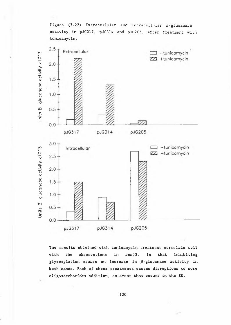
Uni
ts
B—
gluc
anas
e ac
tivity
x1
(P3
Units
B
—gl
ucan
ase
activ
ity
x10
~3
F i g u r e ( 3 . 2 2 ) E x t r a c e l l u l a r a n d i n t r a c e l l u l a r ^ - g l u c a n a s e a c t i v i t y i n p J G 3 1 7 , p J G 3 1 4 a n d p J G 2 0 5 , a f t e r t r e a t m e n t w i t h t u n i c a r a y c i n .
The results obtained with tunicamycin treatment correlate well with the observations in sec53, in that inhibiting glycosylation causes an increase in /9-glucanase activity in both cases. Each of these treatments causes disruptions to core oligosaccharides addition, an event that occurs in the E R .
120
( 3 . 1 2 ) W e s t e r n b l o t a n a l y s i s o f t h e B a c i l l u s / 3 - g l u c a n a s e e n z y ia e To e x a m i n e t h e g l y c o s y l a t i o n p a t t e r n o f t h e B a c i l l u s e n z y m e i n D B Y 7 4 6 , W e s t e r n b l o c a n a l y s i s w a s c a r r i e d o u t . T h e / 3 - g l u c a n a s e a n t i b o d y u s e d i n C h i s a n a l y s i s w a s o b t a i n e d f r o m E . G o r m l e y , T . C . D . T h i s p o l y c l o n a l a n t i b o d y w a s r a i s e d a g a i n s t a c o m m e r c i a l p r e p a r a t i o n o f t h e m a t u r e B . s u b t i l i s / 3 - g l u c a n a s e e n z y m e ( E . G o r m l e y , P h . D . t h e s i s , T . C . D . ) .E x t r a c t s a n d s u p e r n a t a n t s w e r e p r e p a r e d ( s e c t i o n 3 . 1 1 ) f r o m p J G 3 1 7 , pJ G 2 0 5 a n d pJ G 3 1 4 a f t e r g r o w t h i n t h e p r e s e n c e a nd a b s e n c e o f t u n i c a m y c i n . T h e s a m p l e s w e r e p r e p a r e d f o r SDS PAGE a s d e s c r i b e d s e c t i o n ( 2 . 2 2 ) . The g e l s (10%) w e r e b l o t t e d o n t o n i t r o c e l l u l o s e a n d h y b r i d i s e d t o t h e / 3 - g l u c a n a s e a n t i b o d y a n d e n z y m e - c o n j u g a t e d s e c o n d a r y a n t i b o d y ( s e c t i o n 2 . 2 3 ) . F i g u r e s( 3 . 2 3 ) a n d ( 3 . 2 4 ) s h o w t h e W e s t e r n b l o t s o f e x t r a c t s a n d s u p e r n a t a n t s f r o m p J G 3 1 7 p J G 3 1 4 a n d p J G 2 0 5 .
121
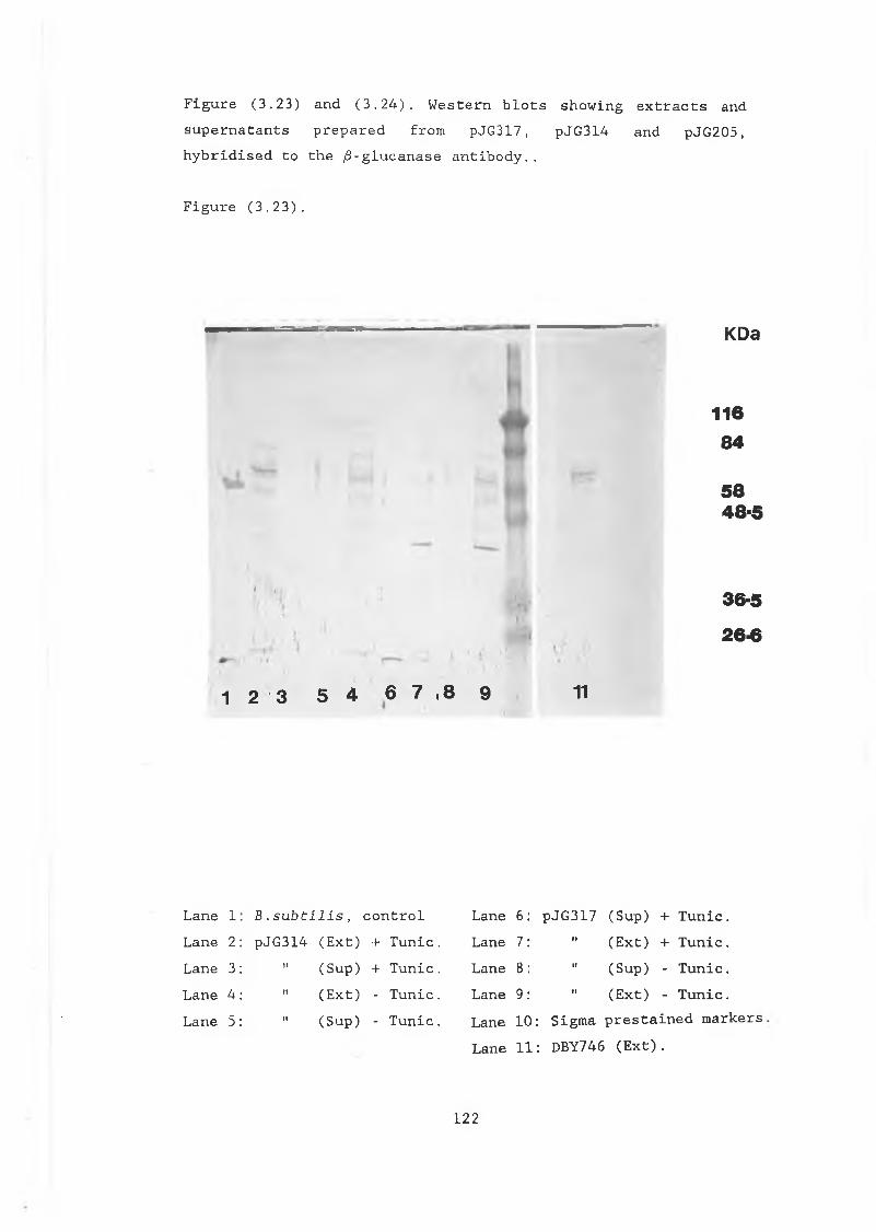
F i g u r e ( 3 . 2 3 ) a n d ( 3 . 2 4 ) . W e s t e r n b l o t s s h o w i n g e x t r a c t s a nd s u p e r n a t a n t s p r e p a r e d f r o m p J G 3 1 7 , p J G 3 1 4 a n d p J G 2 0 5 , h y b r i d i s e d t o t h e / 3 - g l u c a n a s e a n t i b o d y . .
F i g u r e ( 3 . 2 3 ) .
KDa
1 2 3 5 4 6 7 , 8 9 11
11684
5848-5
i it36-5
26-6
L a n e 1 : B . s u b t i l i s , c o n t r o l L a n e 2 : p J G 3 1 4 ( E x t ) + T u n i c . L a n e 3 : " ( S u p ) + T u n i c .L a n e 4 : " ( E x t ) - T u n i c .L a n e 5 : " ( S u p ) - T u n i c .
L a n e 6 : p J G 3 1 7 ( S u p ) + T u n i c .La n e 7: " ( E x t ) + T u n i c .L a n e 8: " ( S u p ) - T u n i c .L a n e 9 : " ( E x t ) - T u n i c .L a n e 1 0 : S i g m a p r e s t a i n e d m a r k e r sL a n e 1 1 : DBY746 ( E x t ) .
122
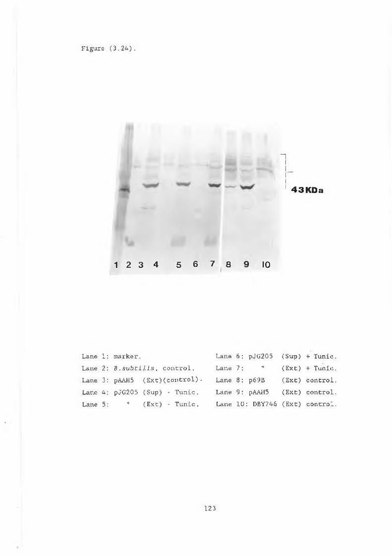
Figure (3.24)
1 2 3 4 5 6 7 8 IO
"I
ri-A 43KDa
Lane 1: marker.Lane 2 Lane 3 Lane 4 Lane 5
B .subcilis, control. pAAHS (E x t )(control). pJG205 (Sup) - Tunic.
" (Ext) - Tunic.
Lane 6: pJG205 (Sup) + Tunic.Lane 7: " (Ext) + Tunic.Lane 8: p69B (Ext) control.Lane 9: pAAH5 (Ext) control.Lane 10: DBY746 (Ext) control.
123
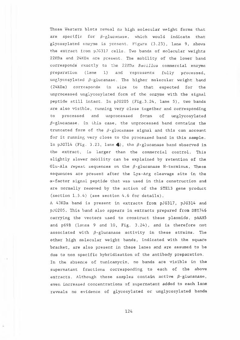
T h e s e W e s t e r n b l o t s r e v e a l no h i g h m o l e c u l a r w e i g h t f o r m s t h a t a r e s p e c i f i c f o r / 3 - g l u c a n a s e , w h i c h w o u l d i n d i c a t e t h a t g l y c o s y l a t e d « n z y m e i s p r e s e n t . F i g u r e ( 3 . 2 3 ) , l a n e 9 , s h o w s t h e e x t r a c t f r o m pJ G 3 17 c e l l s . Two b a n d s o f m o l e c u l a r w e i g h t s 22KDa a n d 24KDa a r e p r e s e n t . Th e m o b i l i t y o f t h e l o w e r b a n d c o r r e s p o n d s e x a c t l y t o t h e 22KDa B a c i l l u s c o m m e r c i a l e n z y m e p r e p a r a t i o n ( l a n e 1 ) a n d r e p r e s e n t s f u l l y p r o c e s s e d , u n g l y c o s y l a t e d / 3 - g l u c a n a s e . Th e h i g h e r m o l e c u l a r w e i g h t b a n d (2 4 K Da ) c o r r e s p o n d s i n s i z e t o t h a t e x p e c t e d f o r t h e u n p r o c e s s e d u n g l y c o s y l a t e d f o r m o f t h e e n z y m e w i t h t h e s i g n a l p e p t i d e s t i l l i n t a c t . I n p J G 2 0 5 ( F i g . 3 . 2 4 , l a n e 5 ) , t w o b a n d s a r e a l s o v i s i b l e , r u n n i n g v e r y c l o s e t o g e t h e r a n d c o r r e s p o n d i n g t o p r o c e s s e d a n d u n p r o c e s s e d f o r m s o f u n g l y c o s y l a t e d / 3 - g l u c a n a s e . In t h i s c a s e , t h e u n p r o c e s s e d b a n d c o n t a i n s t h e t r u n c a t e d f o r m o f t h e / 3 - g l u c a n a s e s i g n a l a n d t h i s c a n a c c o u n t f o r i t r u n n i n g v e r y c l o s e t o t h e p r o c e s s e d b a n d i n t h i s s a m p l e . I n p J G 3 1 4 ( F i g . 3 . 2 3 , l a n e 4). t h e / 3 - g l u c a n a s e b a n d o b s e r v e d i n t h e e x t r a c t , i s l a r g e r t h a n t h e c o m m e r c i a l c o n t r o l . T h i s s l i g h t l y s l o w e r m o b i l i t y c a n b e e x p l a i n e d b y r e t e n t i o n o f t h e G l u - A l a r e p e a t s e q u e n c e s o n t h e /3- g l u c a n a s e N - t e r m i n u s . T h e s e s e q u e n c e s a r e p r e s e n t a f t e r t h e L y s - A r g c l e a v a g e s i t e i n t h ea - f a c t o r s i g n a l p e p t i d e t h a t w a s u s e d i n t h i s c o n s t r u c t i o n a n da r e n o r m a l l y r e m o v e d b y t h e a c t i o n o f t h e STE13 g e n e p r o d u c t ( s e c t i o n 1 . 5 . 4 ) ( s e e s e c t i o n 4 . 6 f o r d e t a i l s ) .
A 43KDa b a n d i s p r e s e n t i n e x t r a c t s f r o m p J G 3 1 7 , p J G 3 1 4 a n dp J G 2 0 5 . T h i s b a n d a l s o a p p e a r s i n e x t r a c t s p r e p a r e d f r o m DBY746 c a r r y i n g t h e v e c t o r s u s e d t o c o n s t r u c t t h e s e p l a s m i d s , pAAH5 a n d p 6 9 B ( l a n e s 9 a n d 1 0 , F i g . 3 . 2 4 ) , a n d i s t h e r e f o r e n o ta s s o c i a t e d w i t h / 3 - g l u c a n a s e a c t i v i t y i n t h e s e s t r a i n s . Th e o t h e r h i g h m o l e c u l a r w e i g h t b a n d s , i n d i c a t e d w i t h t h e s q u a r e b r a c k e t , a r e a l s o p r e s e n t i n t h e s e l a n e s a n d a r e a s s u m e d t o b e d u e t o n o n s p e c i f i c h y b r i d i s a t i o n o f t h e a n t i b o d y p r e p a r a t i o n . I n t h e a b s e n c e o f t u n i c a m y c i n , n o b a n d s a r e v i s i b l e i n t h e s u p e r n a t a n t f r a c t i o n s c o r r e s p o n d i n g t o e a c h o f t h e a b o v e e x t r a c t s . A l t h o u g h t h e s e s a m p l e s c o n t a i n a c t i v e / 3 - g l u c a n a s e , e v e n i n c r e a s e d c o n c e n t r a t i o n s o f s u p e r n a t a n t a d d e d t o e a c h l a n e r e v e a l s no e v i d e n c e o f g l y c o s y l a t e d o r u n g l y c o s y l a t e d b a n d s
124
( d a t a n o t s h o w n ) .The i n c r e a s e i n / 3 - g l u c a n a s e a c t i v i t y , s e e n p r e v i o u s l y a f t e r t u n i c a m y c i n t r e a t m e n t ( s e c t i o n 3 . 1 1 ) , i s r e f l e c t e d i n t h eW e s t e r n b l o t w i t h t h e a p p e a r a n c e o f a s t r o n g b a n d a t 22KDa i n t h e s u p e r n a t a n t f r a c t i o n o f p J G 3 1 7 ( F i g . 3 . 2 3 , l a n e 6 ) . Th e b a n d c o r r e s p o n d s t o f u l l y p r o c e s s e d , u n g l y c o s y l a t e d / 3 - g l u c a n a s e . T h i s f i n d i n g s u p p o r t s t h e i d e a t h a t s o m e f o r m o f g l y c o s y l a t i o n o f t h e e n z y m e m u s t b e t a k i n g p l a c e . I t i s p o s s i b l e t h a t t h eg l y c o s y l a t e d f o r m i s s e c r e t e d b u t i s o n l y r e c o g n i s e d b y t h ea n t i b o d y whe n t h e a d d i t i o n o f t h e s u g a r m o i e t i e s i s i n h i b i t e d b y t h e a c t i o n o f t u n i c a m y c i n . G l y c o s y l a t e d f o r m s o f / 3 - g l u c a n a s e a r e n o t d e t e c t e d i n c e l l e x t r a c t s f r o m p J G 3 1 7 u s i n g t h e/ 3 - g l u c a n a s e a n t i b o d y . H o w e v e r , t h e i n c r e a s e i n i n t e n s i t y o f t h e tw o / 3 - g l u c a n a s e s p e c i f i c b a n d s , a f t e r t u n i c a m y c i n t r e a t m e n t , s u g g e s t s t h a t g l y c o s y l a t e d f o r m s a r e a l s o p r e s e n t i n t r a c e l l u l a r l y a t t h i s s t a g e . I n . p J G 3 1 4 a n d p J G 2 0 5 e x t r a c e l l u l a r f r a c t i o n s , n o b a n d s a p p e a r i n t h e s u p e r n a t a n t s o f e i t h e r s t r a i n e v e n a f t e r t u n i c a m y c i n t r e a t m e n t .
125
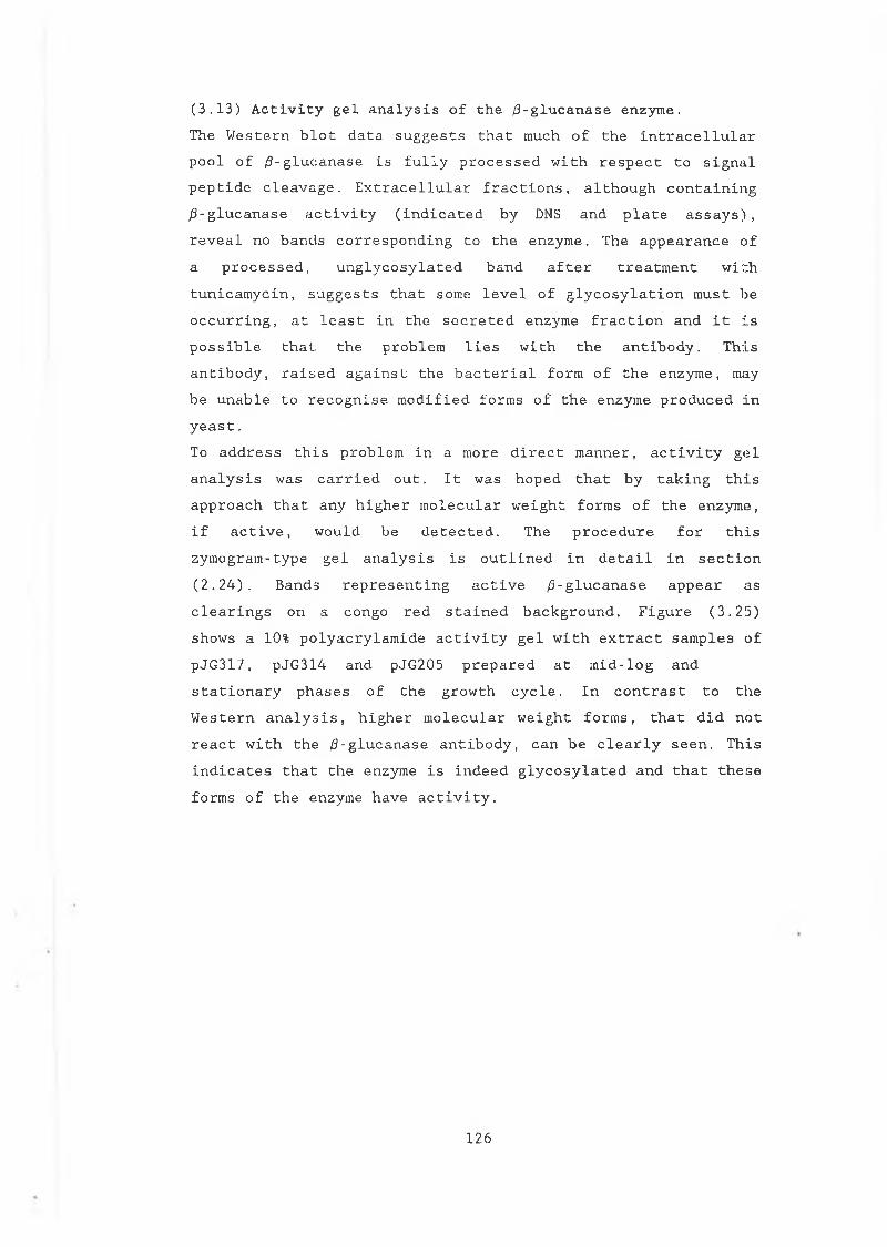
( 3 . 1 3 ) A c t i v i t y g e l a n a l y s i s o f t h e / 3 - g l u c a n a s e e n z y m e .Th e W e s t e r n b l o t d a t a s u g g e s t s t h a t much o f t h e i n t r a c e l l u l a r p o o l o f / 3 - g l u c a n a s e i s f u l l y p r o c e s s e d w i t h r e s p e c t t o s i g n a l p e p t i d e c l e a v a g e . E x t r a c e l l u l a r f r a c t i o n s , a l t h o u g h c o n t a i n i n g / 3 - g l u c a n a s e a c t i v i t y ( i n d i c a t e d b y DNS a n d p l a t e a s s a y s ) , r e v e a l n o b a n d s c o r r e s p o n d i n g t o t h e e n z y m e . The a p p e a r a n c e o f a p r o c e s s e d , u n g l y c o s y l a t e d b a n d a f t e r t r e a t m e n t w i t h t u n i c a m y c i n , s u g g e s t s t h a t s o m e l e v e l o f g l y c o s y l a t i o n m u s t b e o c c u r r i n g , a t l e a s t i n t h e s e c r e t e d e n z y m e f r a c t i o n a n d i t i s p o s s i b l e t h a t t h e p r o b l e m l i e s w i t h t h e a n t i b o d y . T h i s a n t i b o d y , r a i s e d a g a i n s t t h e b a c t e r i a l f o r m o f t h e e n z y m e , may b e u n a b l e t o r e c o g n i s e m o d i f i e d f o r m s o f t h e e n z y m e p r o d u c e d i n y e a s t .To a d d r e s s t h i s p r o b l e m i n a m o r e d i r e c t m a n n e r , a c t i v i t y g e l a n a l y s i s w a s c a r r i e d o u t . I t w a s h o p e d t h a t b y t a k i n g t h i s a p p r o a c h t h a t a n y h i g h e r m o l e c u l a r w e i g h t f o r m s o f t h e e n z y m e , i f a c t i v e , w o u l d b e d e t e c t e d . T h e p r o c e d u r e f o r t h i s z y m o g r a m - t y p e g e l a n a l y s i s i s o u t l i n e d i n d e t a i l i n s e c t i o n( 2 . 2 4 ) . B a n d s r e p r e s e n t i n g a c t i v e / 3 - g l u c a n a s e a p p e a r a s c l e a r i n g s o n a c o n g o r e d s t a i n e d b a c k g r o u n d . F i g u r e ( 3 . 2 5 ) s h o w s a 10% p o l y a c r y l a m i d e a c t i v i t y g e l w i t h e x t r a c t s a m p l e s o f p J G 3 1 7 , p J G 3 1 4 a n d p J G 2 05 p r e p a r e d a t m i d - l o g a n d s t a t i o n a r y p h a s e s o f t h e g r o w t h c y c l e . I n c o n t r a s t t o t h e W e s t e r n a n a l y s i s , h i g h e r m o l e c u l a r w e i g h t f o r m s , t h a t d i d n o t r e a c t w i t h t h e / 3 - g l u c a n a s e a n t i b o d y , c a n b e c l e a r l y s e e n . T h i s i n d i c a t e s t h a t t h e e n z y m e i s i n d e e d g l y c o s y l a t e d a n d t h a t t h e s e f o r m s o f t h e e n z y m e h a v e a c t i v i t y .
126
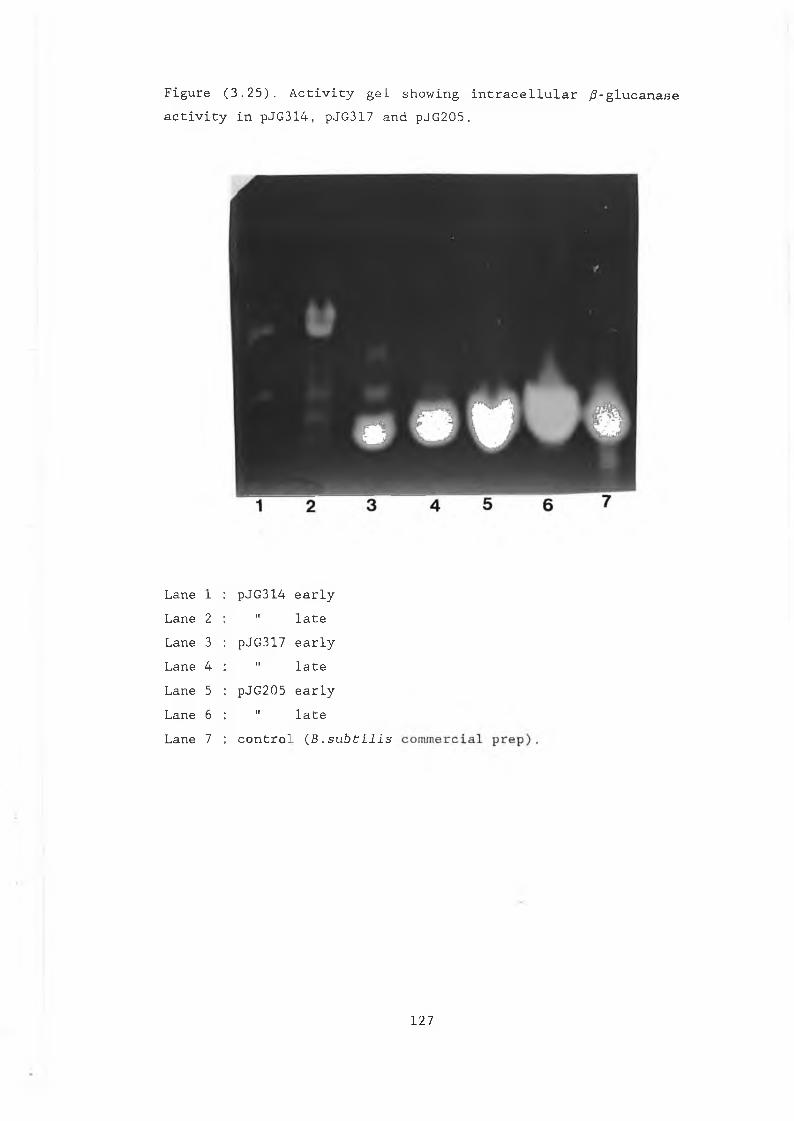
F i g u r e ( 3 . 2 5 ) . A c t i v i t y g e l s h o w i n g i n t r a c e l l u l a r / 3 - g l u c a n a s e a c t i v i t y i n p J G 3 1 4 , pJ G 3 1 7 a n d p J G 2 0 5 .
L a n e 1 p J G 3 1 4 e a r l yL a n e 2 II l a t eLan e 3 p J G 3 17 e a r l yL a n e 4 II l a t eLa ne 5 p J G 2 05 e a r l yL a n e 6 II l a t eL a n e 7 c o n t r o l (B . s u b t i l i s
127
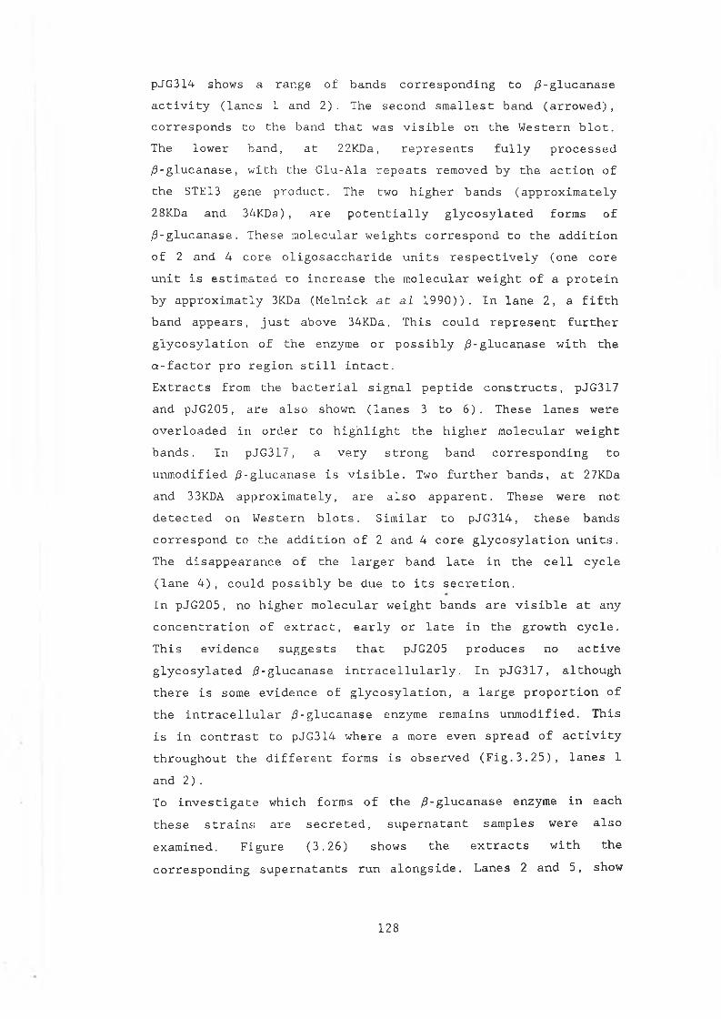
p J G 3 1 4 s h o w s a r a n g e o f b a n d s c o r r e s p o n d i n g t o / 3 - g l u c a n a s e a c t i v i t y ( l a n e s 1 a n d 2 ) . Th e s e c o n d s m a l l e s t b a n d ( a r r o w e d ) , c o r r e s p o n d s t o t h e b a n d t h a t w a s v i s i b l e o n t h e W e s t e r n b l o t . Th e l o w e r b a n d , a t 22KDa, r e p r e s e n t s f u l l y p r o c e s s e d / 3 - g l u c a n a s e , w i t h t h e G l u - A l a r e p e a t s r e m o v e d b y t h e a c t i o n o f t h e STE13 g e n e p r o d u c t . The tw o h i g h e r b a n d s ( a p p r o x i m a t e l y 28KDa a n d 3 4 K D a ) , a r e p o t e n t i a l l y g l y c o s y l a t e d f o r m s o f / 3 - g l u c a n a s e . T h e s e m o l e c u l a r w e i g h t s c o r r e s p o n d t o t h e a d d i t i o n o f 2 a n d 4 c o r e o l i g o s a c c h a r i d e u n i t s r e s p e c t i v e l y ( o n e c o r e u n i t i s e s t i m a t e d t o i n c r e a s e t h e m o l e c u l a r w e i g h t o f a p r o t e i n b y a p p r o x i m a t l y 3KDa ( M e l n i c k a t al 1 9 9 0 ) ) . I n l a n e 2 , a f i f t h b a n d a p p e a r s , j u s t a b o v e 34KDa. T h i s c o u l d r e p r e s e n t f u r t h e r g l y c o s y l a t i o n o f t h e e n z y m e o r p o s s i b l y / 3 - g l u c a n a s e w i t h t h e a - f a c t o r p r o r e g i o n s t i l l i n t a c t .E x t r a c t s f r o m t h e b a c t e r i a l s i g n a l p e p t i d e c o n s t r u c t s , pJ G 3 1 7 a n d p J G 2 0 5 , a r e a l s o s h o w n ( l a n e s 3 t o 6 ) . T h e s e l a n e s w e r e o v e r l o a d e d i n o r d e r t o h i g h l i g h t t h e h i g h e r m o l e c u l a r w e i g h t b a n d s . I n p J G 3 1 7 , a v e r y s t r o n g b a n d c o r r e s p o n d i n g t o u n m o d i f i e d / 3 - g l u c a n a s e i s v i s i b l e . Two f u r t h e r b a n d s , a t 27KDa a n d 33KDA a p p r o x i m a t e l y , a r e a l s o a p p a r e n t . T h e s e w e r e n o t d e t e c t e d o n W e s t e r n b l o t s . S i m i l a r t o p J G 3 1 4 , t h e s e b a n d s c o r r e s p o n d t o t h e a d d i t i o n o f 2 a n d 4 c o r e g l y c o s y l a t i o n u n i t s . Th e d i s a p p e a r a n c e o f t h e l a r g e r b a n d l a t e i n t h e c e l l c y c l e ( l a n e 4 ) , c o u l d p o s s i b l y b e d u e t o i t s s e c r e t i o n .I n p J G 2 0 5 , n o h i g h e r m o l e c u l a r w e i g h t b a n d s a r e v i s i b l e a t a n y c o n c e n t r a t i o n o f e x t r a c t , e a r l y o r l a t e i n t h e g r o w t h c y c l e . T h i s e v i d e n c e s u g g e s t s t h a t p J G 2 0 5 p r o d u c e s n o a c t i v e g l y c o s y l a t e d / 3 - g l u c a n a s e i n t r a c e l l u l a r l y . I n p J G 3 1 7 , a l t h o u g h t h e r e i s s o m e e v i d e n c e o f g l y c o s y l a t i o n , a l a r g e p r o p o r t i o n o f t h e i n t r a c e l l u l a r / 3 - g l u c a n a s e e n z y m e r e m a i n s u n m o d i f i e d . T h i s i s i n c o n t r a s t t o p J G 3 1 4 w h e r e a m o re e v e n s p r e a d o f a c t i v i t yt h r o u g h o u t t h e d i f f e r e n t f o r m s i s o b s e r v e d ( F i g . 3 . 2 5 ) , l a n e s 1a n d 2 ) .To i n v e s t i g a t e w h i c h f o r m s o f t h e / 3 - g l u c a n a s e e n z y m e i n e a c ht h e s e s t r a i n s a r e s e c r e t e d , s u p e r n a t a n t s a m p l e s w e r e a l s oe x a m i n e d . F i g u r e ( 3 . 2 6 ) s h o w s t h e e x t r a c t s w i t h t h e c o r r e s p o n d i n g s u p e r n a t a n t s r u n a l o n g s i d e . L a n e s 2 a n d 5 , s h o w
128
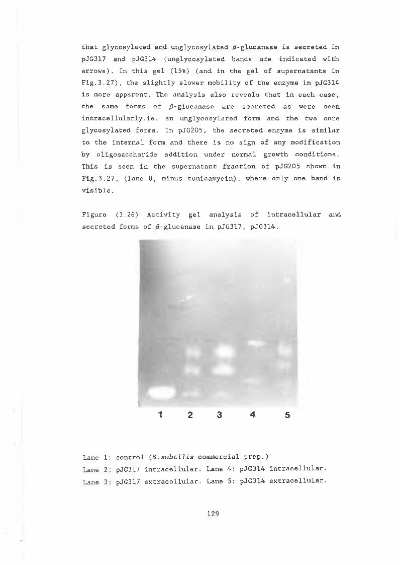
t h a t g l y c o s y l a t e d a n d u n g l y c o s y l a t e d / 3 - g l u c a n a s e i s s e c r e t e d i n p J G 3 1 7 a n d p J G 3 1 4 ( u n g l y c o s y l a t e d b a n d s a r e i n d i c a t e d w i t h a r r o w s ) . I n t h i s g e l (15%) ( a n d i n t h e g e l o f s u p e r n a t a n t s i n F i g . 3 . 2 7 ) , t h e s l i g h t l y s l o w e r m o b i l i t y o f t h e e n z y m e i n p J G 3 1 4 i s m o r e a p p a r e n t . Th e a n a l y s i s a l s o r e v e a l s t h a t i n e a c h c a s e , t h e s a m e f o r m s o f / 3 - g l u c a n a s e a r e s e c r e t e d a s w e r e s e e n i n t r a c e l l u l a r l y . i e . a n u n g l y c o s y l a t e d f o r m a n d t h e t w o c o r e g l y c o s y l a t e d f o r m s . I n p J G 2 0 5 , t h e s e c r e t e d e n z y m e i s s i m i l a r t o t h e i n t e r n a l f o r m a n d t h e r e i s n o s i g n o f a n y m o d i f i c a t i o n b y o l i g o s a c c h a r i d e a d d i t i o n u n d e r n o r m a l g r o w t h c o n d i t i o n s . T h i s i s s e e n i n t h e s u p e r n a t a n t f r a c t i o n o f p J G 2 0 5 s h o w n i n F i g . 3 . 2 7 , ( l a n e 8 , m i n u s t u n i c a m y c i n ) , w h e r e o n l y o n e b a n d i s v i s i b l e .
F i g u r e ( 3 . 2 6 ) A c t i v i t y g e l a n a l y s i s o f i n t r a c e l l u l a r a n d s e c r e t e d f o r m s o f / 3 - g l u c a n a s e i n p J G 3 1 7 , p J G 3 1 4 .
*1 2 3 4 5
L a n e 1 : c o n t r o l ( B . s u b t i l i s c o m m e r c i a l p r e p . )L a n e 2 : pJ G 317 i n t r a c e l l u l a r . L a n e 4 : p J G 3 1 4 i n t r a c e l l u l a r . L a n e 3 : pJ G 317 e x t r a c e l l u l a r . L a n e 5 : p J G 3 1 4 e x t r a c e l l u l a r .
129
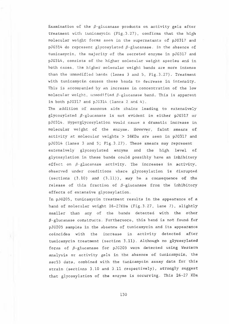
E x a m i n a t i o n o f t h e / 3 - g l u c a n a s e p r o d u c t s o n a c t i v i t y g e l s a f t e r t r e a t m e n t w i t h t u n i c a m y c i n ( F i g . 3 . 2 7 ) , c o n f i r m s t h a t t h e h i g h m o l e c u l a r w e i g h t f o r m s s e e n i n t h e s u p e r n a t a n t s o f pJ G 3 1 7 an d p J G 3 1 4 do r e p r e s e n t g l y c o s y l a t e d / 3 - g l u c a n a s e . I n t h e a b s e n c e o f t u n i c a m y c i n , t h e m a j o r i t y o f t h e s e c r e t e d e n z y m e i n p J G 3 1 7 a n d p J G 3 1 4 , c o n s i s t s o f t h e h i g h e r m o l e c u l a r w e i g h t s p e c i e s a n d i n b o t h c a s e s , t h e h i g h e r m o l e c u l a r w e i g h t b a n d s a r e m o re i n t e n s e t h a n t h e u n m o d i f i e d b a n d s ( l a n e s 3 a n d 5 , F i g . 3 . 2 7 ) . T r e a t m e n t w i t h t u n i c a m y c i n c a u s e s t h e s e b a n d s t o d e c r e a s e i n i n t e n s i t y . T h i s i s a c c o m p a n i e d b y a n i n c r e a s e i n c o n c e n t r a t i o n o f t h e l o w m o l e c u l a r w e i g h t , u n m o d i f i e d / 3 - g l u c a n a s e b a n d . T h i s i s a p p a r e n t i n b o t h p J G 3 17 a n d p J G 3 1 4 ( l a n e s 2 a n d 4 ) .T he a d d i t i o n o f m a n n o s e s i d e c h a i n s l e a d i n g t o e x t e n s i v e l y g l y c o s y l a t e d / 9 - g l u c a n a s e i s n o t e v i d e n t i n e i t h e r p J G 3 1 7 o r p J G 3 1 4 . H y p e r g l y c o s y l a t i o n w o u l d c a u s e a d r a m a t i c i n c r e a s e i n m o l e c u l a r w e i g h t o f t h e e n z y m e . H o w e v e r , f a i n t s m e a r s o f a c t i v i t y a t m o l e c u l a r w e i g h t s > 58KDa a r e s e e n i n p J G 3 1 7 a n d p J G 3 1 4 ( l a n e s 3 a n d 5 ; F i g . 3 . 2 7 ) . T h e s e s m e a r s may r e p r e s e n t e x t e n s i v e l y g l y c o s y l a t e d e n z y m e a n d t h e h i g h l e v e l o f g l y c o s y l a t i o n i n t h e s e b a n d s c o u l d p o s s i b l y h a v e a n i n h i b i t o r y e f f e c t o n / 3 - g l u c a n a s e a c t i v i t y . The i n c r e a s e s i n a c t i v i t y , o b s e r v e d u n d e r c o n d i t i o n s w h e r e g l y c o s y l a t i o n i s d i s r u p t e d ( s e c t i o n s ( 3 . 1 0 ) a n d ( 3 . 1 1 ) ) , may b e a c o n s e q u e n c e o f t h e r e l e a s e o f t h i s f r a c t i o n o f / 3 - g l u c a n a s e f r o m t h e i n h i b i t o r y e f f e c t s o f e x t e n s i v e g l y c o s y l a t i o n .I n p J G 2 0 5 , t u n i c a m y c i n t r e a t m e n t r e s u l t s i n t h e a p p e a r a n c e o f a b a n d o f m o l e c u l a r w e i g h t 2 6 - 2 7 K D a ( F i g . 3 . 2 7 , l a n e 7 ) , s l i g h t l y s m a l l e r t h a n a n y o f t h e b a n d s d e t e c t e d w i t h t h e o t h e r / 3 - g l u c a n a s e c o n s t r u c t s . F u r t h e r m o r e , t h i s b a n d i s n o t f o u n d f o r p J G 2 0 5 s a m p l e s i n t h e a b s e n c e o f t u n i c a m y c i n a n d i t s a p p e a r a n c e c o i n c i d e s w i t h t h e i n c r e a s e i n a c t i v i t y d e t e c t e d a f t e r t u n i c a m y c i n t r e a t m e n t ( s e c t i o n 3 . 1 1 ) . A l t h o u g h no g l y c o s y l a t e d f o r m s o f / 3 - g l u c a n a s e f o r p J G 2 0 5 w e r e d e t e c t e d u s i n g W e s t e r n a n a l y s i s o r a c t i v i t y g e l s i n t h e a b s e n c e o f t u n i c a m y c i n , t h e s e c 5 3 d a t a , c o m b i n e d w i t h t h e t u n i c a m y c i n a s s a y d a t a f o r t h i s s t r a i n ( s e c t i o n s 3 . 1 0 a n d 3 . 1 1 r e s p e c t i v e l y ) , s t r o n g l y s u g g e s t t h a t g l y c o s y l a t i o n o f t h e e n z y m e i s o c c u r r i n g . T h i s 2 6 - 2 7 KDa
130
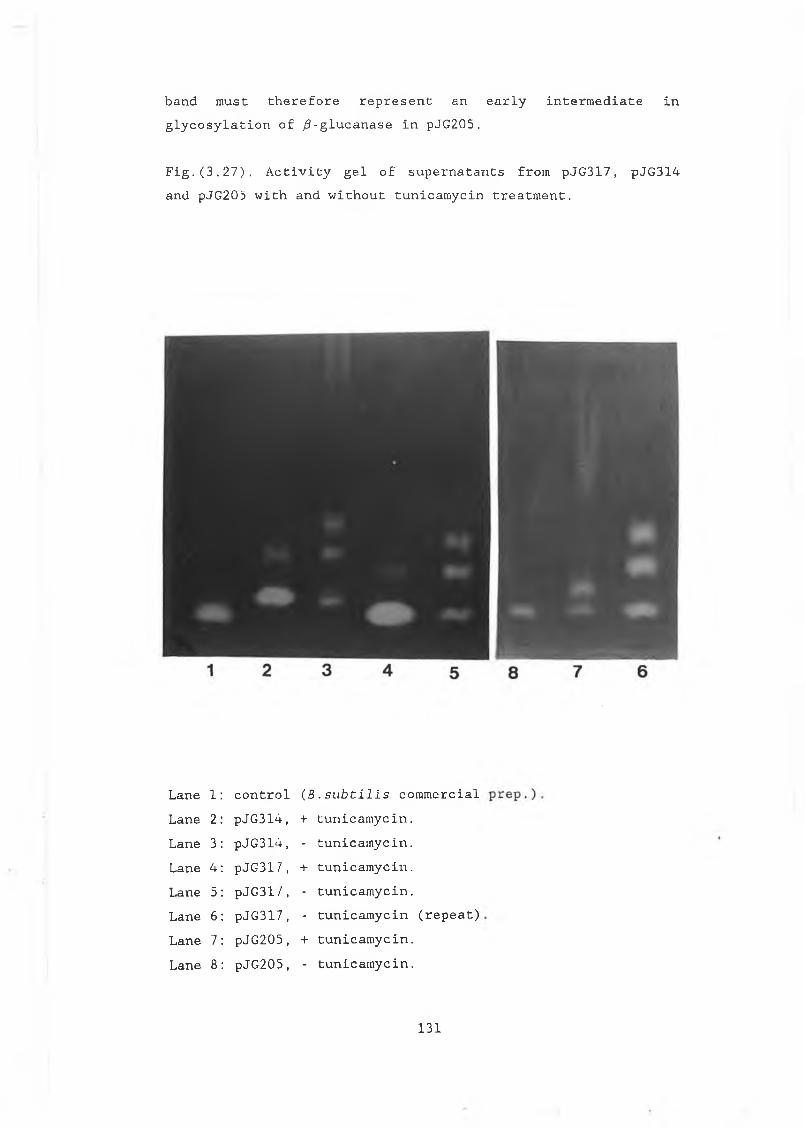
b a n d m u s t t h e r e f o r e r e p r e s e n t a n e a r l y i n t e r m e d i a t e i n g l y c o s y l a t i o n o f / 3 - g l u c a n a s e i n p J G 2 0 5 .
F i g . ( 3 . 2 7 ) . A c t i v i t y g e l o f s u p e r n a t a n t s f r o m p J G 3 1 7 , p J G 3 1 4 a n d p J G 2 0 5 w i t h a n d w i t h o u t t u n i c a m y c i n t r e a t m e n t .
L a n e 1: c o n t r o l ( B . s u b t i l i s c o m m e r c i a lL a n e 2: p J G 3 1 4 , + t u n i c a m y c i n .L a n e 3: p J G 3 1 4 , - t u n i c a m y c i n .L a n e 4 : p J G 3 1 7, + t u n i c a m y c i n .L a n e 5: p J G 3 1 7, - t u n i c a m y c i n .L a n e 6: p J G 3 1 7 , - t u n i c a m y c i n ( r e p e a t )L a n e 7: p J G 2 0 5 , + t u n i c a m y c i n .L a n e 8: p J G 2 0 5 , - t u n i c a m y c i n .
131
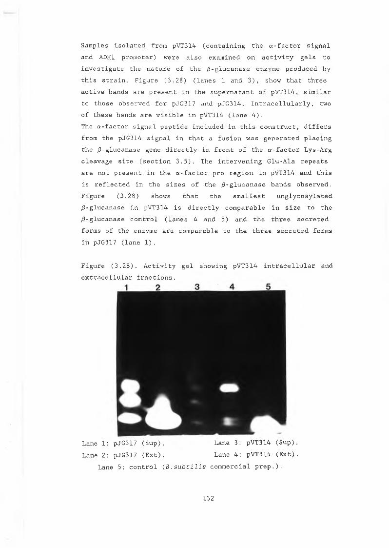
S a m p l e s i s o l a t e d f r o m pVT3 14 ( c o n t a i n i n g t h e a - f a c t o r s i g n a l a n d ADH1 p r o m o t e r ) w e r e a l s o e x a m i n e d o n a c t i v i t y g e l s t o i n v e s t i g a t e t h e n a t u r e o f t h e / 3 - g l u c a n a s e e n z y m e p r o d u c e d b y t h i s s t r a i n . F i g u r e ( 3 . 2 8 ) ( l a n e s 1 a n d 3 ) , s h o w t h a t t h r e e a c t i v e b a n d s a r e p r e s e n t i n t h e s u p e r n a t a n t o f p V T 3 1 4 , s i m i l a r t o t h o s e o b s e r v e d f o r p J G 3 1 7 a n d p J G 3 1 4 . I n t r a c e l l u l a r l y , tw o o f t h e s e b a n d s a r e v i s i b l e i n pVT3 14 ( l a n e 4 ) .Th e a - f a c t o r s i g n a l p e p t i d e i n c l u d e d i n t h i s c o n s t r u c t , d i f f e r s f r o m t h e p J G 3 1 4 s i g n a l i n t h a t a f u s i o n w a s g e n e r a t e d p l a c i n g t h e / 3 - g l u c a n a s e g e n e d i r e c t l y i n f r o n t o f t h e a - f a c t o r L y s - A r g c l e a v a g e s i t e ( s e c t i o n 3 . 5 ) . The i n t e r v e n i n g G l u - A l a r e p e a t s a r e n o t p r e s e n t i n t h e a - f a c t o r p r o r e g i o n i n pV T 3 14 a n d t h i s i s r e f l e c t e d i n t h e s i z e s o f t h e / 3 - g l u c a n a s e b a n d s o b s e r v e d . F i g u r e ( 3 . 2 8 ) s h o w s t h a t t h e s m a l l e s t u n g l y c o s y l a t e d / 3 - g l u c a n a s e i n pV T 3 1 4 i s d i r e c t l y c o m p a r a b l e i n s i z e t o t h e / 3 - g l u c a n a s e c o n t r o l ( l a n e s 4 a n d 5 ) a n d t h e t h r e e s e c r e t e d f o r m s o f t h e e n z y m e a r e c o m p a r a b l e t o t h e t h r e e s e c r e t e d f o r m s i n pJG3 17 ( l a n e 1 ) .
F i g u r e ( 3 . 2 8 ) . A c t i v i t y g e l s h o w i n g p V T 3 1 4 i n t r a c e l l u l a r a n d e x t r a c e l l u l a r f r a c t i o n s .
L a n e 1: pJ G 3 17 ( S u p ) . L a n e 3: p V T 3 1 4 ( S u p ) .L a n e 2: pJ G 3 1 7 ( E x t ) . L a n e 4 : p V T 3 1 4 ( E x t ) .
La n e 5: c o n t r o l ( B . s u b t i l i s c o m m e r c i a l p r e p . ) .
132
Section (3.2)P r e l i m i n a r y s t u d i e s o n t h e s u i t a b i l i t y o f a n ¡3 - g l u c u r o n i d a s e a s a r e p o r t e r g e n e f o r S . c e r e v i s l a e .
E . c o l i
133
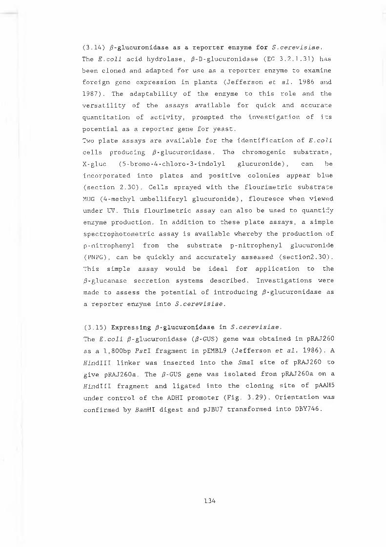
( 3 . 1 4 ) / 3 - g l u c u r o n i d a s e a s a r e p o r t e r e n z y m e f o r S . c e r e v i s i a e .The E . c o l i a c i d h y d r o l a s e , / 3 - D - g l u c u r o n i d a s e (EC 3 . 2 . 1 . 3 1 ) h a s b e e n c l o n e d a n d a d a p t e d f o r u s e a s a r e p o r t e r e n z y m e t o e x a m i n e f o r e i g n g e n e e x p r e s s i o n i n p l a n t s ( J e f f e r s o n e t a l . 1 9 8 6 a n d1 9 8 7 ) . The a d a p t a b i l i t y o f t h e e n z y m e t o t h i s r o l e a n d t h e v e r s a t i l i t y o f t h e a s s a y s a v a i l a b l e f o r q u i c k a n d a c c u r a t e q u a n t i t a t i o n o f a c t i v i t y , p r o m p t e d t h e i n v e s t i g a t i o n o f i t s p o t e n t i a l a s a r e p o r t e r g e n e f o r y e a s t .Two p l a t e a s s a y s a r e a v a i l a b l e f o r t h e i d e n t i f i c a t i o n o f E . c o l i c e l l s p r o d u c i n g /3 - g l u c u r o n i d a s e . Th e c h r o m o g e n i c s u b s t r a t e , X - g l u c ( 5 - b r o m o - 4 - c h l o r o - 3 - i n d o l y l g l u c u r o n i d e ) , c a n b e i n c o r p o r a t e d i n t o p l a t e s a n d p o s i t i v e c o l o n i e s a p p e a r b l u e ( s e c t i o n 2 . 3 0 ) . C e l l s s p r a y e d w i t h t h e f l o u r i m e t r i c s u b s t r a t e MUG ( 4 - m e t h y l u m b e l l i f e r y l g l u c u r o n i d e ) , f l o u r e s c e w h e n v i e w e d u n d e r UV. T h i s f l o u r i m e t r i c a s s a y c a n a l s o b e u s e d t o q u a n t i f y e n z y m e p r o d u c t i o n . I n a d d i t i o n t o t h e s e p l a t e a s s a y s , a s i m p l e s p e c t r o p h o t o m e t r i c a s s a y i s a v a i l a b l e w h e r e b y t h e p r o d u c t i o n o f p - n i t r o p h e n y l f r o m t h e s u b s t r a t e p - n i t r o p h e n y l g l u c u r o n i d e ( P N P G ) , c a n b e q u i c k l y a n d a c c u r a t e l y a s s e s s e d ( s e c t i o n 2 . 3 0 ) . T h i s s i m p l e a s s a y w o u l d b e i d e a l f o r a p p l i c a t i o n t o t h e / 3 - g l u c a n a s e s e c r e t i o n s y s t e m s d e s c r i b e d . I n v e s t i g a t i o n s w e r e made t o a s s e s s t h e p o t e n t i a l o f i n t r o d u c i n g / 3 - g l u c u r o n i d a s e a s a r e p o r t e r e n z y m e i n t o S . c e r e v i s i a e .
( 3 . 1 5 ) E x p r e s s i n g / 3 - g l u c u r o n i d a s e i n S . c e r e v i s i a e .The E . c o l i /3 - g l u c u r o n i d a s e ( /3-GUS) g e n e w a s o b t a i n e d i n pR AJ 26 0 a s a l , 8 0 0 b p P s t I f r a g m e n t i n pEMBL9 ( J e f f e r s o n e t a l . 1 9 8 6 ) . A H i n d l l l l i n k e r w a s i n s e r t e d i n t o t h e S m a l s i t e o f pR A J 2 6 0 t og i v e p R A J 2 6 0 a . Th e /3-GUS g e n e w a s i s o l a t e d f r o m p R A J 2 6 0 a o n at f i n d l l l f r a g m e n t a n d l i g a t e d i n t o t h e c l o n i n g s i t e o f pAAH5 u n d e r c o n t r o l o f t h e ADHI p r o m o t e r ( F i g . 3 . 2 9 ) . O r i e n t a t i o n w a s c o n f i r m e d b y Bamt t l d i g e s t a n d pJBU7 t r a n s f o r m e d i n t o DBY 746 .
134
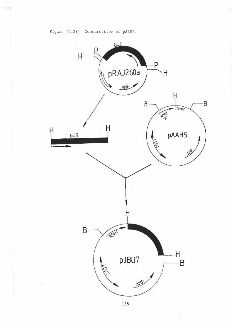
Figure (3.29). Construction of pJBU7.
r-vllS
135
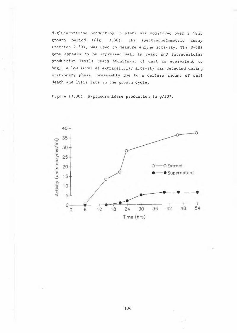
(9 -g l u c u r o n i d a s e p r o d u c t i o n i n pJBU7 w a s m o n i t o r e d o v e r a 4 8 h r g r o w t h p e r i o d ( F i g . 3 . 3 0 ) . Th e s p e c t r o p h o t o m e t r i c a s s a y ( s e c t i o n 2 . 3 0 ) , w a s u s e d t o m e a s u r e e n z y m e a c t i v i t y . T h e 0 -GUS g e n e a p p e a r s t o b e e x p r e s s e d w e l l i n y e a s t a n d i n t r a c e l l u l a r p r o d u c t i o n l e v e l s r e a c h 4 0 u n i t s / m l ( 1 u n i t i s e q u i v a l e n t t o 5 n g ) . A l o w l e v e l o f e x t r a c e l l u l a r a c t i v i t y wa s d e t e c t e d d u r i n g s t a t i o n a r y p h a s e , p r e s u m a b l y d u e t o a c e r t a i n a m o u n t o f c e l l d e a t h a n d l y s i s l a t e i n t h e g r o w t h c y c l e .
F i g u r e ( 3 . 3 0 ) . g l u c u r o n i d a s e p r o d u c t i o n i n pJB U7.
1 3 6
A d a p t a t i o n o f t h e p l a t e a s s a y s t o ¡3- g l u c u r o n i d a s e p r o d u c t i o n i n DBY746 h o w e v e r , p r o v e d l e s s s u c c e s s f u l . pJ B U 7, w h e n g r o w n i n t h e p r e s e n c e o f X - G l u c , d o e s n o t a p p e a r b l u e i n c o l o u r . A l s o , s p r a y i n g c o l o n i e s w i t h t h e f l o u r i m e t r i c s u b s t r a t e MUG, d o e s n o t p r o d u c e f l o u r e s c e n c e u n d e r UV i l l u m i n a t i o n . I t i s l i k e l y t h a t t h e y e a s t c e l l w a l l i s n o t p e r m e a b l e t o t h e s u b s t r a t e s X - G l u c a n d MUG. H o w e v e r , pJBU7 i n l i q u i d c u l t u r e d o e s f l o u r e s c e u n d e r UV a f t e r i n c u b a t i o n i n ImM MUG. T h i s i s d u e p r e s u m a b l y t o d i e t h e l o w l e v e l o f e x t r a c e l l u l a r a c t i v i t y t h a t c a n b e d e t e c t e d i n t h e m e d iu m , l a t e i n t h e g r o w t h c y c l e . T h i s s u g g e s t s t h a t t h e p l a t e a s s a y s c o u l d b e u s e f u l , i f t h e /3- g l u c u r o n i d a s e e n z y m e w a s b e i n g s e c r e t e d f r o m t h e c e l l s . I n o r d e r t o a s s e s s t h e p o t e n t i a l o f / 3 - g l u c u r o n i d a s e t o m o n i t o r s e c r e t i o n i n S . c e r e v i s i a e , t h e /3-GUS g e n e w a s p l a c e d u n d e r c o n t r o l o f t h e a - f a c t o r s i g n a l p e p t i d e i n p 6 9 B .
137
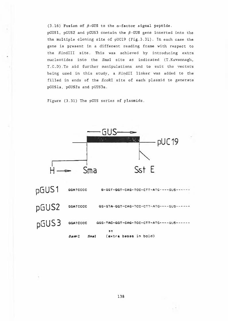
(3.16) Fusion of fi-GVS to the a-factor signal peptide.p G U S l , pGUS2 a n d pGUS3 c o n t a i n t h e /3-GUS g e n e i n s e r t e d i n t o t h e t h e m u l t i p l e c l o n i n g s i t e o f pUC19 ( F i g . 3 . 3 1 ) . I n e a c h c a s e t h e g e n e i s p r e s e n t i n a d i f f e r e n t r e a d i n g f r a m e w i t h r e s p e c t t o t h e / i i n d l l l s i t e . T h i s wa s a c h i e v e d b y i n t r o d u c i n g e x t r a n u c l e o t i d e s i n t o t h e Sm a l s i t e a s i n d i c a t e d ( T . K a v a n n a g h , T . C . D ) . T o a i d f u r t h e r m a n i p u l a t i o n s a n d t o s u i t t h e v e c t o r s b e i n g u s e d i n t h i s s t u d y , a H i n d l l l i n k e r w a s a d d e d t o t h e f i l l e d i n e n d s o f t h e F c o R I s i t e o f e a c h p l a s m i d t o g e n e r a t e p G U S l a , pGUS2a a n d pGUS3a.
F i g u r e ( 3 . 3 1 ) The pGUS s e r i e s o f p l a s m i d s .
GUS-
H
pGUSl
pGUS2
pGUSB
\i H
pUC19
Sma Sst E
GGATCCCC G - G G T - G G T - C A G - T C C - C T T - A T G --------- GUS --------------
GGATCCCC G G - G T A - G G T - C A G - T C C - C T T - A T G --------- GUS---------------
GGATCCCC G G G -T A C “ G G T - C A G - T C C - C T T - A T G --------- GUS -------------
ttBsMH I Smal (extra bases in bold)
1 3 8
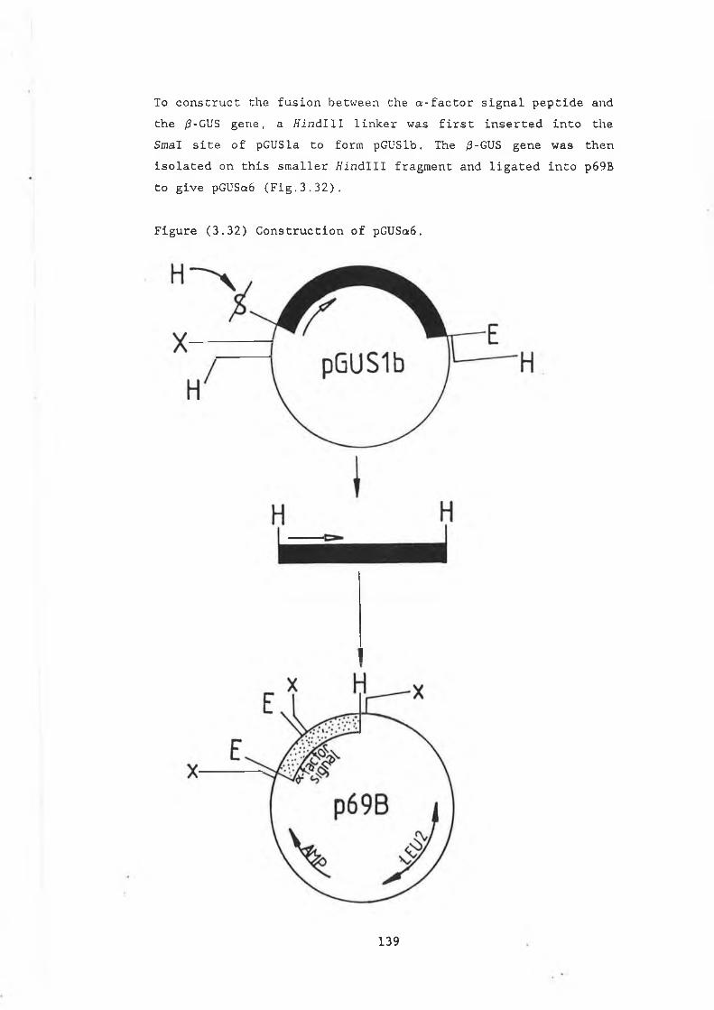
To c o n s t r u c t t h e f u s i o n b e t w e e n t h e a - f a c t o r s i g n a l p e p t i d e a n d t h e /3-GUS g e n e , a t f i n d l l l l i n k e r w a s f i r s t i n s e r t e d i n t o t h e S m a l s i t e o f p G U S l a t o f o r m p G U S l b . The /3-GUS g e n e w a s t h e n i s o l a t e d o n t h i s s m a l l e r f f i n d l l l f r a g m e n t a n d l i g a t e d i n t o p 6 9 B t o g i v e pGUSa6 ( F i g . 3 . 3 2 ) .
F i g u r e ( 3 . 3 2 ) C o n s t r u c t i o n o f pGUSa6.
139
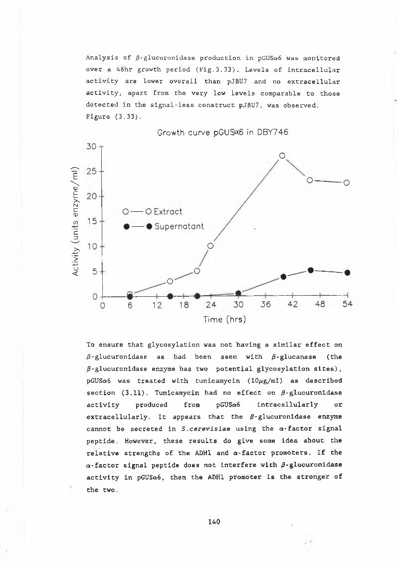
Act
ivity
(u
nits
e
nzy
me
/ml)
A n a l y s i s o f g l u c u r o n i d a s e p r o d u c t i o n i n pGUSa6 w a s m o n i t o r e d o v e r a 4 8 h r g r o w t h p e r i o d ( F i g . 3 . 3 3 ) . L e v e l s o f i n t r a c e l l u l a r a c t i v i t y a r e l o w e r o v e r a l l t h a n pJBU7 a n d n o e x t r a c e l l u l a r a c t i v i t y , a p a r t f r o m t h e v e r y l o w l e v e l s c o m p a r a b l e t o t h o s e d e t e c t e d i n t h e s i g n a l - l e s s c o n s t r u c t pJ B U 7, w a s o b s e r v e d . F i g u r e ( 3 . 3 3 ) .
Growth curve pGUS°(6 in DBY746
Time (hrs)
To ensure that glycosylation was not having a similar effect on /3-glucuronidase as had been seen with /?-glucanase (the
glucuronidase enzyme has two potential glycosylation sites), pGUSa6 was treated with tunicamycin (10/ig/ml) as described section (3.11). Tunicamycin had no effect on /3-glucuronidase activity produced from pGUSa6 intracellularly or extracellularly. It appears that the /9-glucuronidase enzyme cannot be secreted in S.cerevisiae using the a-factor signal peptide. However, these results do give some idea about the relative strengths of the ADH1 and a-factor promoters. If the a-factor signal peptide does not interfere with ^-glucuronidase activity in pGUSa6, then the ADH1 promoter is the stronger of the two.
140
(3.17) Adapting /3-glucuronidase as a useful reporter in yeast./ 3 - g l u c u r o n i d a s e d o e s n o t a p p e a r t o b e a m e n a b l e t o m o n i t o r i n g s e c r e t i o n i n S . cerevisiae . I n t h e y e a s t c y t o p l a s m h o w e v e r , t h e e n z y m e s h o w s n o s i g n s o f i n s t a b i l i t y a n d c a n b e u s e d t o m o n i t o r e x p r e s s i o n l e v e l s . /3- g l u c u r o n i d a s e may a l s o b e u s e f u l i n o t h e r r e s p e c t s f o r h e t e r o l o g o u s p r o t e i n p r o d u c t i o n i n S .cerevisiae. F o r e x a m p l e , t h e e n z y m e c o u l d p o s s i b l y b e u s e d a s a f u s i o np r o t e i n t o a c t a s a c a r r i e r f o r o t h e r f o r e i g n p r o t e i n s i n y e a s t . I t may a l s o b e p o s s i b l e t o s t a b i l i z e p r o t e i n s , e g . / 3 - g l u c a n a s e , b y f u s i o n t o t h e / 3 - g l u c u r o n i d a s e e n z y m e ./ 3 - g l u c u r o n i d a s e i s a 73KDa p r o t e i n , a n d a s m a l l e r p r o t e i n w o u l d b e p o t e n t i a l l y m o r e u s e f u l f o r f u s i o n s t u d i e s . I f a t r u n c a t e d , b u t s t i l l a c t i v e f o r m o f t h e e n z y m e c o u l d b e g e n e r a t e d , t h i s w o u l d b e u s e f u l b o t h a s a f u s i o n t o s t a b i l i z e o t h e r p r o t e i n s i n t h e y e a s t c y t o p l a s m a n d t o i n v e s t i g a t e t h e y e a s t s e c e t o r yp a t h w a y f u r t h e r . I f t h e l i m i t i n g f a c t o r f o r s e c r e t i n g / 9 - g l u c u r o n i d a s e i n y e a s t i s i t s s i z e , t r u n c a t e d f o r m s o f t h e e n z y m e w o u l d b e p a r t i c u l a r l y u s e f u l . A l t e r n a t i v e l y , i f t h e r e a r e s e q u e n c e s i n t h e e n z y m e t h a t d i r e c t t h e p r o t e i n e l s e w h e r ei n t h e y e a s t c e l l , t r u n c a t e d f o r m s w o u l d a l s o a i d t h ei n v e s t i g a t i o n o f t h i s p o s s i b i l i t y .An e a r l i e r o b s e r v a t i o n i n v o l v i n g / 9 - g l u c u r o n i d a s e s u g g e s t e d t h a t i t s h o u l d b e p o s s i b l e t o p r o d u c e a s h o r t e n e d f o r m o f t h e e n z y m e t h a t w a s s t i l l a c t i v e ( T . K a v a n a g h , T . C . D . ) . On a c t i v i t y g e l s , a c t i v e e n z y m e b a n d s w e r e o b s e r v e d b e l o w t h e m a i n a c t i v e b a n d (7 2 K D a ) i n p a r t l y d e g r a d e d p r e p a r a t i o n s o f t h e e n z y m e . T h i s i n d i c a t e d t h a t t h e a c t i v e d o m a i n o f t h e e n z y m e w a s c o n f i n e d t o a s h o r t e r d e f i n e d r e g i o n o f t h e m o l e c u l e . I n i t i a l s t e p s w e r e t a k e n t o l o c a t e t h e a c t i v e s i t e o f /9 - g l u c u r o n i d a s e u s i n g B a l 31 d e l e t i o n a n a l y s i s f r o m b o t h t h e N - t e r m i n a l a n d c a r b o x y t e r m i n a l o f t h e e n z y m e . A n t i b o d y t o t h e E . c o l i e n z y m e w a s a l s o r a i s e d t o a i d f u t u r e i n v e s t i g a t i o n s i n t h i s a r e a a n d a f u s i o n p r o t e i n i n v o l v i n g t h e / 9 - g l u c a n a s e a n d ^ - g l u c u r o n i d a s e g e n e s wa s c o n s t r u c t e d .
141
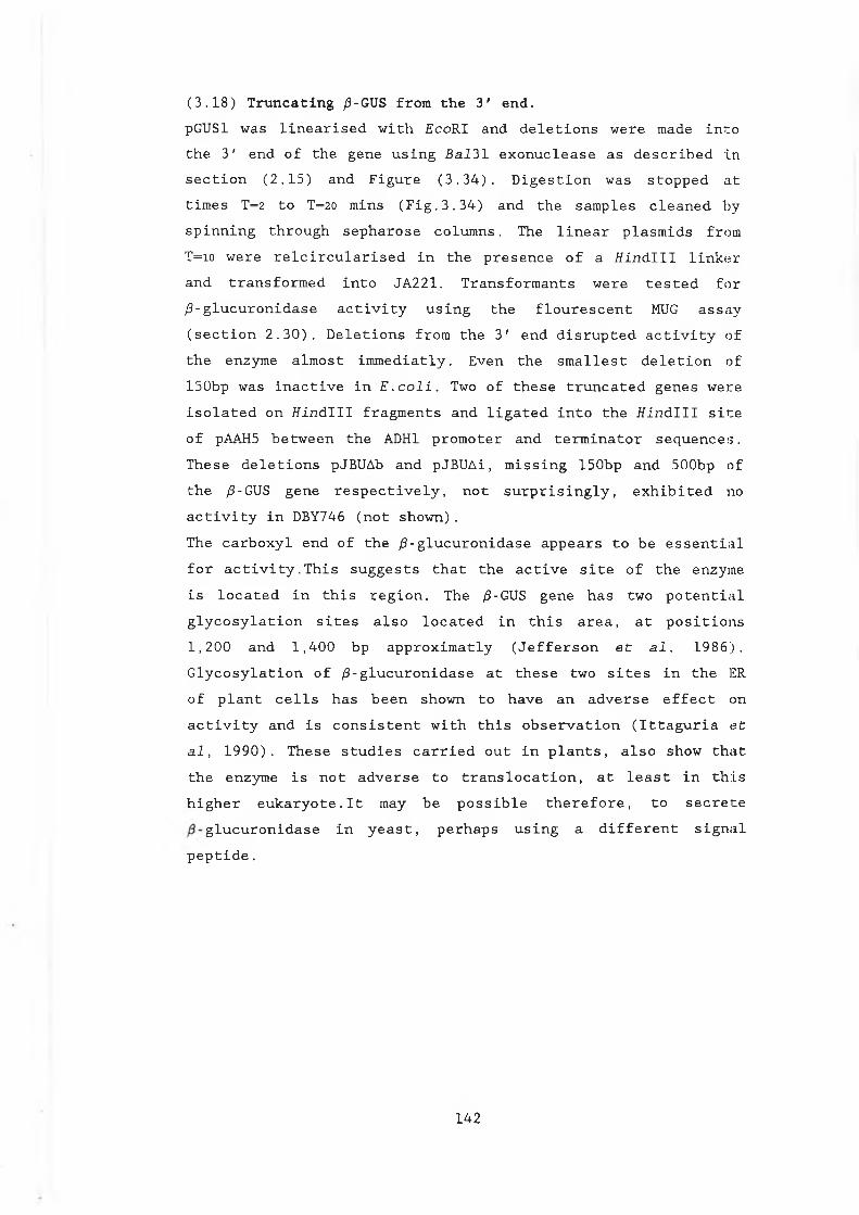
( 3 . 1 8 ) T r u n c a t i n g /9-GUS f r o m t h e 3 ' e n d .pGUSl w a s l i n e a r i s e d w i t h E c o R I a n d d e l e t i o n s w e r e made i n t o t h e 3 ' e n d o f t h e g e n e u s i n g B a l 3 1 e x o n u c l e a s e a s d e s c r i b e d i n s e c t i o n ( 2 . 1 5 ) a n d F i g u r e ( 3 . 3 4 ) . D i g e s t i o n w a s s t o p p e d a t t i m e s T= 2 t o T= 2 0 m i n s ( F i g . 3 . 3 4 ) a n d t h e s a m p l e s c l e a n e d b y s p i n n i n g t h r o u g h s e p h a r o s e c o l u m n s . The l i n e a r p l a s m i d s f r o m T=io w e r e r e l c i r c u l a r i s e d i n t h e p r e s e n c e o f a t f i n d l l l l i n k e r a n d t r a n s f o r m e d i n t o J A 2 2 1 . T r a n s f o r m a n t s w e r e t e s t e d f o r / 3 - g l u c u r o n i d a s e a c t i v i t y u s i n g t h e f l o u r e s c e n t MUG a s s a y ( s e c t i o n 2 . 3 0 ) . D e l e t i o n s f r o m t h e 3 ' e n d d i s r u p t e d a c t i v i t y o f t h e e n z y m e a l m o s t i m m e d i a t l y . E v e n t h e s m a l l e s t d e l e t i o n o f 1 5 0 b p w a s i n a c t i v e i n E . c o l i . Two o f t h e s e t r u n c a t e d g e n e s w e r e i s o l a t e d o n f f i n d l l l f r a g m e n t s a n d l i g a t e d i n t o t h e t f i n d l l l s i t e o f pAAH5 b e t w e e n t h e ADH1 p r o m o t e r a n d t e r m i n a t o r s e q u e n c e s . T h e s e d e l e t i o n s pJBUAb a n d p J B U A i , m i s s i n g 1 5 0 b p a n d 5 0 0 b p o ft h e /3-GUS g e n e r e s p e c t i v e l y , n o t s u r p r i s i n g l y , e x h i b i t e d noa c t i v i t y i n DBY746 ( n o t s h o w n ) .T h e c a r b o x y l e n d o f t h e / 3 - g l u c u r o n i d a s e a p p e a r s t o b e e s s e n t i a l f o r a c t i v i t y . T h i s s u g g e s t s t h a t t h e a c t i v e s i t e o f t h e e n z y m e i s l o c a t e d i n t h i s r e g i o n . T h e /9-GUS g e n e h a s tw o p o t e n t i a l g l y c o s y l a t i o n s i t e s a l s o l o c a t e d i n t h i s a r e a , a t p o s i t i o n s 1 , 2 0 0 a n d 1 , 4 0 0 b p a p p r o x i m a t l y ( J e f f e r s o n e t a l . 1 9 8 6 ) .G l y c o s y l a t i o n o f / 3 - g l u c u r o n i d a s e a t t h e s e t w o s i t e s i n t h e ER o f p l a n t c e l l s h a s b e e n s h o w n t o h a v e a n a d v e r s e e f f e c t o na c t i v i t y a n d i s c o n s i s t e n t w i t h t h i s o b s e r v a t i o n ( I t t a g u r i a e ta l , 1 9 9 0 ) . T h e s e s t u d i e s c a r r i e d o u t i n p l a n t s , a l s o s h o w t h a t t h e e n z y m e i s n o t a d v e r s e t o t r a n s l o c a t i o n , a t l e a s t i n t h i s h i g h e r e u k a r y o t e . l t may b e p o s s i b l e t h e r e f o r e , t o s e c r e t e
g l u c u r o n i d a s e i n y e a s t , p e r h a p s u s i n g a d i f f e r e n t s i g n a l p e p t i d e .
142
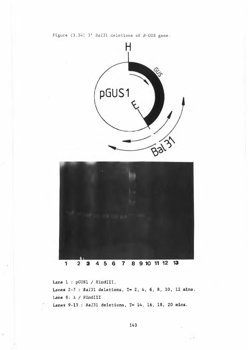
Figure (3.34) 3' Bal31 deletions of /3-GUS gene.
H
1 2 3 4 5 6 7 8 9 10 11 12 13
Lane 1 : pGUSl / tfindlll.Lanes 2-7 : BaI31 deletions, T- 2, 4, 6, 8, 10, 12 mins. Lane 8: X / HindiIILanes 9-13 : Bal31 deletions, T- 14, 16, 18, 20 rains.
1 4 3
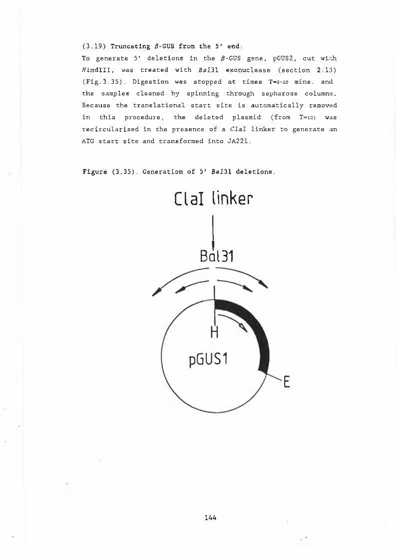
(3.19) Truncating /3-GUS from the 5' end.To g e n e r a t e 5' d e l e t i o n s i n t h e ^-GUS g e n e , pGUS2, c u t w i t h / / i n d l l l , wa s t r e a t e d w i t h B a l 31 e x o n u c l e a s e ( s e c t i o n 2 .115) ( F i g . 3 . 3 5 ) . D i g e s t i o n w a s s t o p p e d a t t i m e s T= 2-20 m i n s . a n d t h e s a m p l e s c l e a n e d b y s p i n n i n g t h r o u g h s e p h a r o s e c o l u m n s . B e c a u s e t h e t r a n s l a t i o n a l s t a r t s i t e i s a u t o m a t i c a l l y r e m o v e d i n t h i s p r o c e d u r e , t h e d e l e t e d p l a s m i d ( f r o m T = 1 0 ) w as r e c i r c u l a r i s e d i n t h e p r e s e n c e o f a C l a l l i n k e r t o g e n e r a t e an ATG s t a r t s i t e a n d t r a n s f o r m e d i n t o JA221.
Figure (3.35). Generation of 5' Bal31 deletions,
C la l linker
Bq I31
144
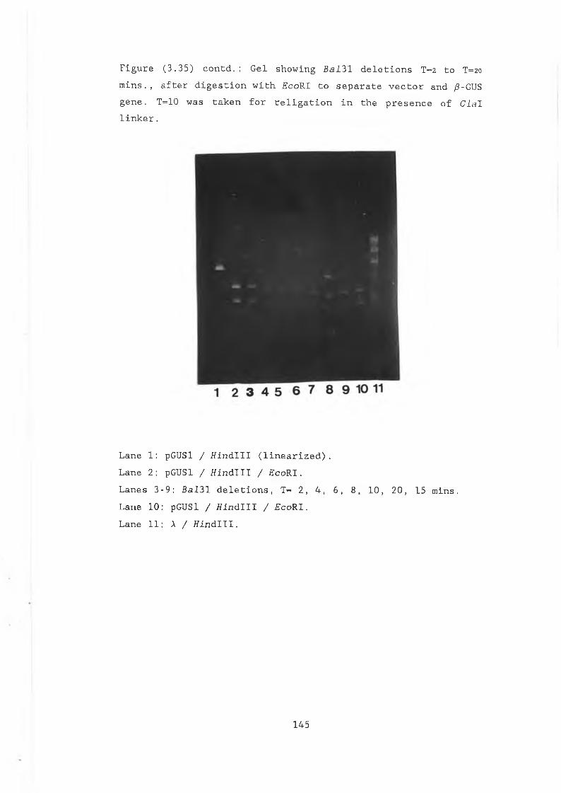
F i g u r e ( 3 . 3 5 ) c o n t d . : G e l s h o w i n g Balll d e l e t i o n s T= 2 t o T=20
m i n s . , a f t e r d i g e s t i o n w i t h E c o R I t o s e p a r a t e v e c t o r a n d /3-GUS g e n e . T=10 w a s t a k e n f o r r e l i g a t i o n i n t h e p r e s e n c e o f C l a l l i n k e r .
L a n e 1 : pGUSl / H i n d l l l ( l i n e a r i z e d ) .L a n e 2 : pGUSl / t f i n d l l l / E c o R I .L a n e s 3 - 9 : B a l 3 1 d e l e t i o n s , T= 2 , 4 , 6 , 8 , 1 0 , 2 0 , 15 m i n s . L a n e 1 0 : pGUSl / f f i n d l l l / E c o R I .L a n e 1 1 : X / H i n d l l l .
145
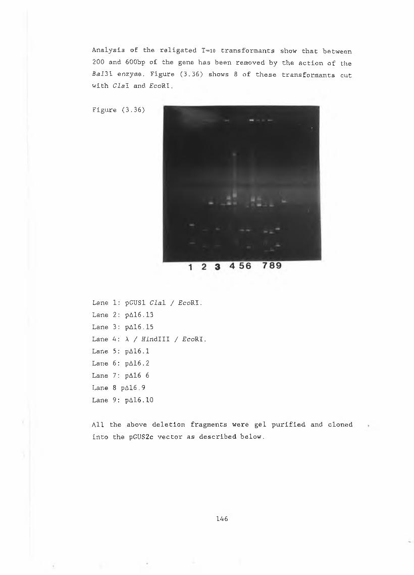
A n a l y s i s o f t h e r e l i g a t e d T=io t r a n s f o r m a n t s s h o w t h a t b e t w e e n 2 0 0 a n d 6 0 0 b p o f t h e g e n e h a s b e e n r e m o v e d b y t h e a c t i o n o f t h e B a l 3 1 e n z y m e . F i g u r e ( 3 . 3 6 ) s h o w s 8 o f t h e s e t r a n s f o r m a n t s c u t w i t h C l a l a n d £ c o R I .
F i g u r e ( 3 . 3 6 )
L a n e 1 : pG USl C l a l / F c o R I .L a n e 2 : p A l 6 . 1 3 L a n e 3 : p A l 6 . 1 5 L a n e 4 : A / f f i n d l l l / £ c o R I .L a n e 5 : p A l 6 . 1 L a n e 6 : p A 1 6 .2 L a n e 7 : p A l 6 6 L a n e 8 p A l 6 .9 L a n e 9 : p A l 6 . 1 0
A l l t h e a b o v e d e l e t i o n f r a g m e n t s w e r e g e l p u r i f i e d a n d c l o n e d i n t o t h e pGUS2c v e c t o r a s d e s c r i b e d b e l o w .
146
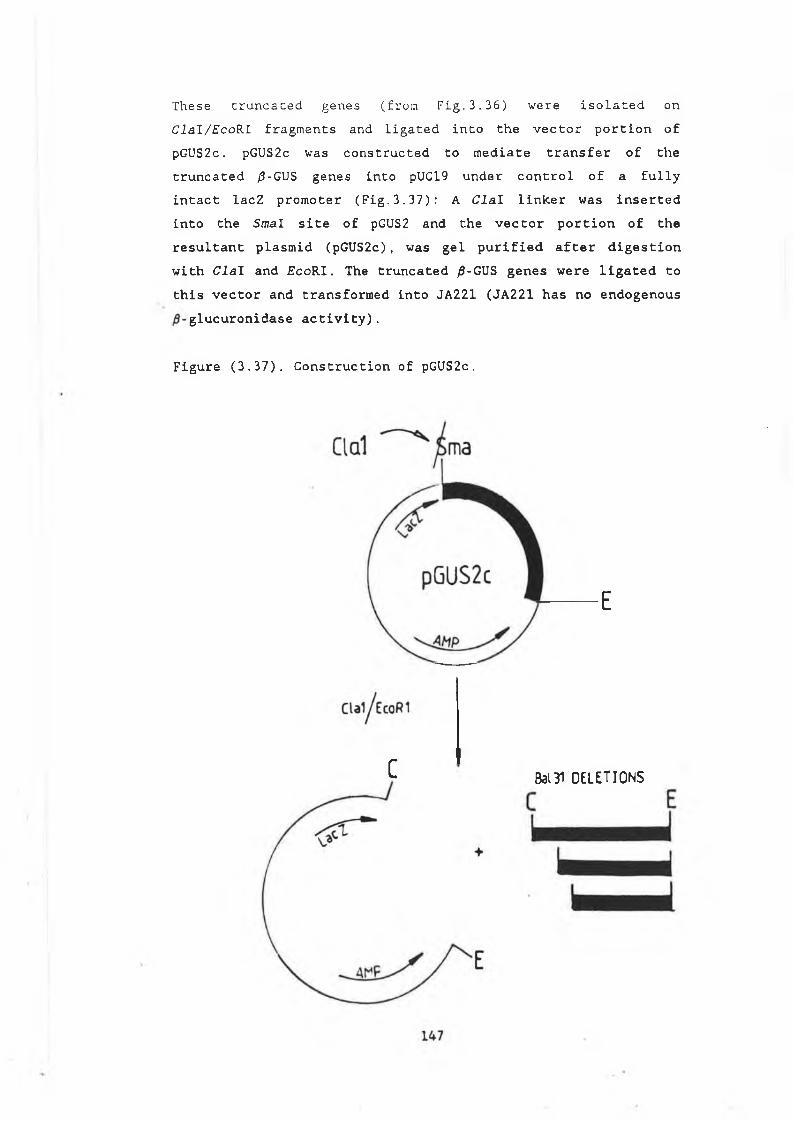
These t r u n c a t e d genes ( f r o m Fig.3.36) were i s o l a t e d o n Clal/EcoRl fragments and ligated into the vector portion of pGUS2c. pGUS2c was constructed to mediate transfer of the truncated /3-GUS genes into pUC19 under control of a fully intact lacZ promoter (Fig.3.37): A Clal linker was insertedinto the Smal site of pGUS2 and the vector portion of the resultant plasmid (pGUS2c), was gel purified after digestion with Clal and £coRI. The truncated /3-GUS genes were ligated to this vector and transformed into JA221 (JA221 has no endogenous
glucuronidase activity).
Figure (3.37). Construction of pGUS2c.
E
C Bal31 OELETIONS
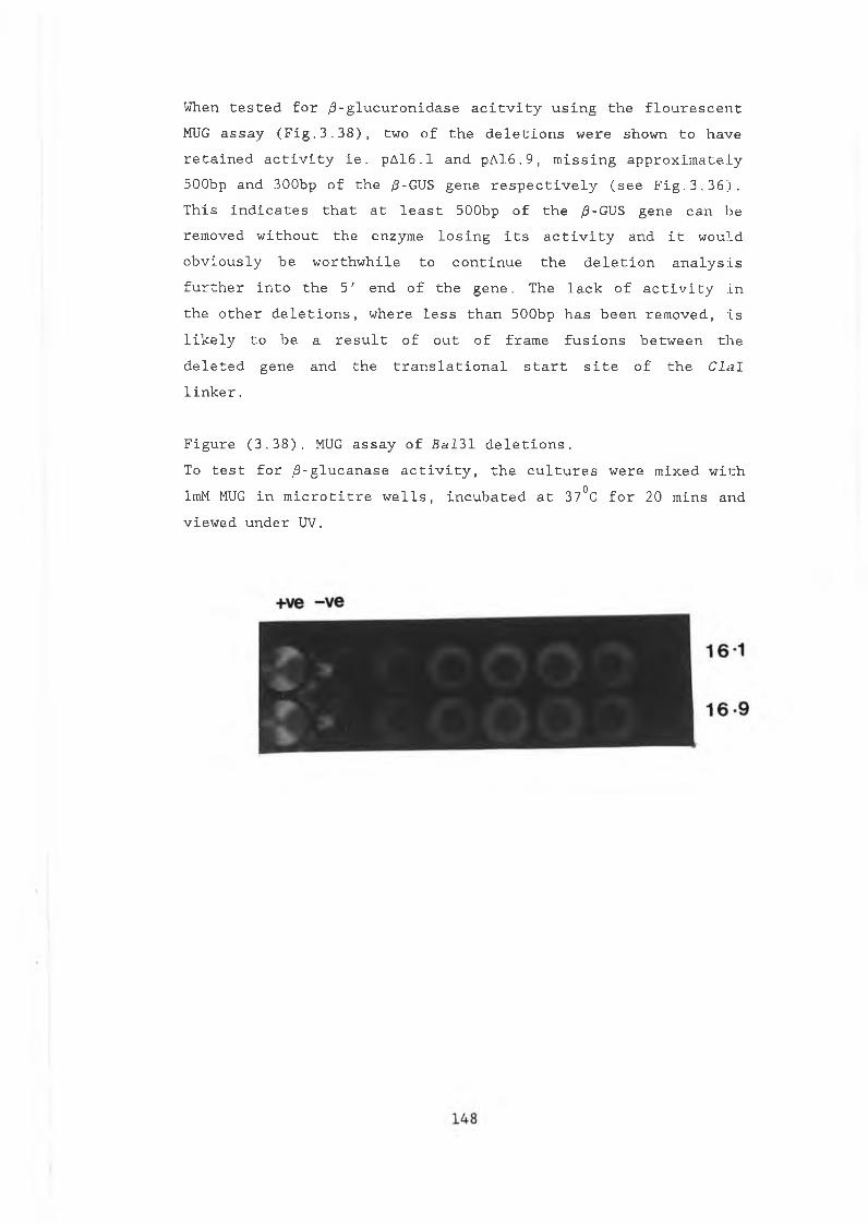
When t e s t e d f o r / 3 - g l u c u r o n i d a s e a c i t v i t y u s i n g t h e f l o u r e s c e n t MUG a s s a y ( F i g . 3 . 3 8 ) , tw o o f t h e d e l e t i o n s w e r e s h o w n t o h a v e r e t a i n e d a c t i v i t y i e . p A l 6 . 1 a n d p A l 6 . 9 , m i s s i n g a p p r o x i m a t e l y 5 0 0 b p a n d 3 0 0 b p o f t h e /3-GUS g e n e r e s p e c t i v e l y ( s e e F i g . 3 . 3 6 ) . T h i s i n d i c a t e s t h a t a t l e a s t 5 0 0 b p o f t h e /3-GUS g e n e c a n b e r e m o v e d w i t h o u t t h e e n z y m e l o s i n g i t s a c t i v i t y a n d i t w o u l d o b v i o u s l y b e w o r t h w h i l e t o c o n t i n u e t h e d e l e t i o n a n a l y s i s f u r t h e r i n t o t h e 5 ' e n d o f t h e g e n e , Th e l a c k o f a c t i v i t y i n t h e o t h e r d e l e t i o n s , w h e r e l e s s t h a n 5 0 0 b p h a s b e e n r e m o v e d , i s l i k e l y t o b e a r e s u l t o f o u t o f f r a m e f u s i o n s b e t w e e n t h e d e l e t e d g e n e a n d t h e t r a n s l a t i o n a l s t a r t s i t e o f t h e C l n I l i n k e r .
F i g u r e ( 3 . 3 8 ) . MUG a s s a y o f B a I 3 1 d e l e t i o n s .To t e s t f o r / 3 - g l u c a n a s e a c t i v i t y , t h e c u l t u r e s w e r e m i x e d w i t h ImM MUG i n m i c r o t i t r e w e l l s , i n c u b a t e d a t 37°C f o r 2 0 m i n s a n d v i e w e d u n d e r UV.
( 3 . 2 0 ) . / 3 - g l u c u r o n i d a s e a s a f u s i o n p r o t e i n .To i n v e s t i g a t e i f / 3 - g l u c u r o n i d a s e c a n t o l e r a t e a 5 ' f u s i o n , t h e / 3 - g l u c a n a s e g e n e w a s l i g a t e d i n - f r a m e t o t h e 5 ' e n d o f t h e /3-GUS g e n e i n t h e p l a s m i d p G U S l . I n g e n e r a t i n g t h i s f u s i o n , i t w a s n e c e s s a r y t o l i g a t e t h e tw o g e n e s i n - f r a m e , e l i m i n a t i n g a l l t r a n s c r i p t i o n a l a n d t r a n s l a t i o n a l t e r m i n a t i o n s e q u e n c e s b e t w e e n t h e t w o . T h i s w a s a c h i v e d b y i s o l a t i n g t h e / 3 - g l u c a n a s e g e n e o n a f f i n d l l l - S a l l f r a g m e n t f r o m b o t h p J G 1 0 4 a n d p J G 1 0 6 , a s o u t l i n e d i n F i g u r e ( 3 . 3 9 ) . B o t h p l a s m i d s w e r e u s e d i n o r d e r t o g e n e r a t e f u s i o n s b e t w e e n / 3 - g l u c u r o n i d a s e a n d t h e / 3 - g l u c a n a s e g e n e , w i t h a n d w i t h o u t i t s s i g n a l p e p t i d e . T r u n c a t i n g t h e / 3 - g l u c a n a s e g e n e a t t h e S a l I s i t e ( p o s i t i o n 1 1 1 5 , a p p . 1 f o r d e t a i l s ) r e s u l t s i n t h e r e m o v a l o f 5 4 a m i n o a c i d s o f t h e / 3 - g l u c a n a s e c a r b o x y l t e r m i n u s . W h e t h e r o r n o t t h i s d e s t r o y e d t h e a c t i v e s i t e o f t h e / 3 - g l u c a n a s e e n z y m e r e m a i n e d t o b ed e t e r m i n e d . H o w e v e r , t h e c o m p l e t e / 3 - g l u c u r o n i d a s e g e n e w o u l d b e p r e s e n t i n t h e s e f u s i o n s a n d i t w a s h o p e d t h a t t h i s e n z y m ew o u l d r e m a i n a c t i v e . T h e t r u n c a t e d / 3 - g l u c a n a s e g e n e s w e r e e a c h l i g a t e d i n t o t f i n d l l l - S a l l c u t p G U S l , 5 ' t o t h e /3-GUS g e n e ,g e n e r a t i n g a n i n - f r a m e f u s i o n b e t w e e n t h e t w o e n z y m e s i n e a c h c a s e ( F i g . 3 . 3 9 ) . Th e r e s u l t a n t p l a s m i d s p J G U S 4 . 1 ( c o n t a i n i n g t h e s i g n a l - l e s s / 3 - g l u c a n a s e ) a n d p J G U S 6 , l ( w i t h t h e / 3 - g l u c a n a s e s i g n a l i n c l u d e d ) , w e r e t r a n s f o r m e d i n t o J A 2 2 1 , r e p l i c a p l a t e d a n d t e s t e d f o r / 3 - g l u c a n a s e a c t i v i t y u s i n g t h e p l a t e a s s a y( s e c t i o n 2 . 1 8 ) a n d f o r / 3 - g l u c u r o n i d a s e u s i n g t h e f l o u r e s c e n t MUG a s s a y ( s e c t i o n 2 . 3 0 ) . I n b o t h c a s e s , t h e f u s i o n s w e r e p o s i t i v e f o r / 3 - g l u c u r o n i d a s e a n d n e g a t i v e f o r / 3 - g l u c a n a s e a c t i v i t y . T h i s i n d i c a t e s t h a t / 3 - g l u c u r o n i d a s e c a n b e made a s a n a c t i v e f u s i o n t o o t h e r p e p t i d e s . T h e s e f u s i o n s h a v e y e t t o b e t e s t e d i n y e a s t .
149
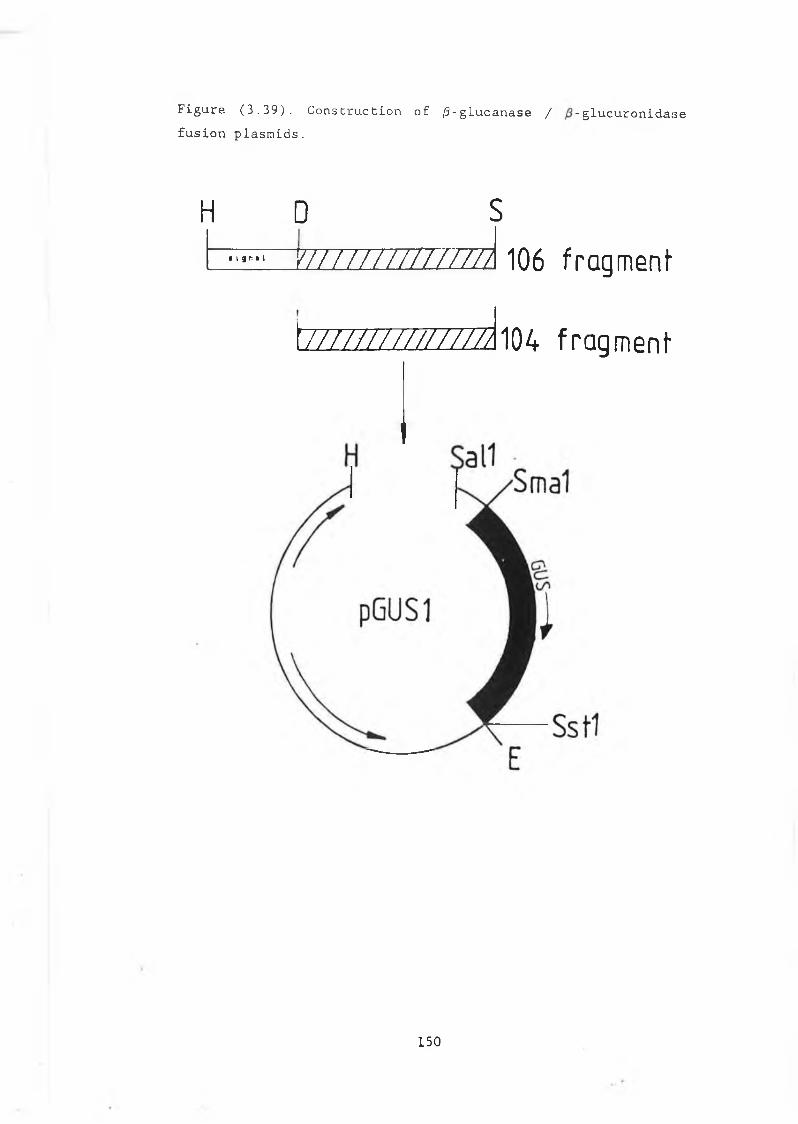
F i g u r e ( 3 . 3 9 ) . C o n s t r u c t i o n o f / 3 - g l u c a n a s e / - g l u c u r o n i d a i s ef u s i o n p l a s m i d s .
H D S^ EZZZZZZZZZZZZZZ3 106 fragment
tZ Z Z Z Z Z Z Z Z Z Z Z Z a i04 fragment
150
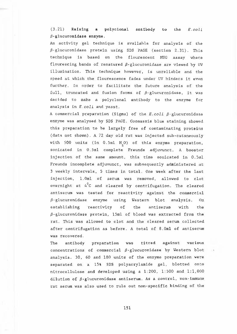
(3.21) Raising a polyclonal antibody to the E . c o l i ¡3-glucuronidase enzyme.An a c t i v i t y g e l t e c h n i q u e i s a v a i l a b l e f o r a n a l y s i s o f t h e /3- g l u c u r o n i d a s e p r o t e i n u s i n g SDS PAGE ( s e c t i o n 2 . 3 1 ) . T h i s t e c h n i q u e i s b a s e d o n t h e f l o u r e s c e n t MUG a s s a y w h e r e f l o u r e s c i n g b a n d s o f r e n a t u r e d ¿3-g l u c u r o n i d a s e a r e v i e w e d b y UV i l l u m i n a t i o n . T h i s t e c h n i q u e h o w e v e r , i s u n r e l i a b l e a n d t h e s p e e d a t w h i c h t h e f l o u r e s c e n c e f a d e s u n d e r UV h i n d e r s i t e v e n f u r t h e r . I n o r d e r t o f a c i l i t a t e t h e f u t u r e a n a l y s i s o f t h e f u l l , t r u n c a t e d a n d f u s i o n f o r m s o f /3-g l u c u r o n i d a s e , i t w a s d e c i d e d t o make a p o l y c l o n a l a n t i b o d y t o t h e e n z y m e f o r a n a l y s i s i n E . c o l i a n d y e a s t .A c o m m e r c i a l p r e p a r a t i o n ( S i g m a ) o f t h e E . c o l i / 3 - g l u c u r o n i d a s e e n z y m e w a s a n a l y s e d b y SDS PAGE. C o o m a s s i e b l u e s t a i n i n g s h o w e d t h i s p r e p a r a t i o n t o b e l a r g e l y f r e e o f c o n t a m i n a t i n g p r o t e i i i s ( d a t a n o t s h o w n ) . A 72 d a y o l d r a t w a s i n j e c t e d s u b - c u t a n e o u s l y w i t h 5 0 0 u n i t s ( i n 0 . 5 m l H^O) o f t h i s e n z y m e p r e p a r a t i o n , s o n i c a t e d i n 0 . 5 m l c o m p l e t e F r e u n d s a d j u v u n c t . A b o o s t e r i n j e c t i o n o f t h e sa me a m o u n t , t h i s t i m e s o n i c a t e d i n 0 . 5rnl F r e u n d s i n c o m p l e t e a d j u v u n c t , w a s s u b s e q u e n t l y a d m i n i s t e r e d a t 3 w e e k l y i n t e r v a l s , 5 t i m e s i n t o t a l . One w e e k a f t e r t h e l a s t i n j e c t i o n , 1 . 0 m l o f s e r u m w a s r e m o v e d , a l l o w e d t o c l o t o v e r n i g h t a t 4°C a n d c l e a r e d b y c e n t r i f u g a t i o n . T h e c l e a r e d a n t i s e r u m w a s t e s t e d f o r r e a c t i v i t y a g a i n s t t h e c o m m e r c i a l /3 - g l u c u r o n i d a s e e n z y m e u s i n g W e s t e r n b l o t a n a l y s i s . On e s t a b l i s h i n g r e a c t i v i t y o f t h e a n t i s e r u m w i t h t h e / 3 - g l u c u r o n i d a s e p r o t e i n , 1 5 m l o f b l o o d w a s e x t r a c t e d f r o m t h e r a t . T h i s w a s a l l o w e d t o c l o t a n d t h e c l e a r e d s e r u m c o l l e c t e d a f t e r c e n t r i f u g a t i o n a s b e f o r e . A t o t a l o f 8 . 0 m l o f a n t i s e r u m w a s r e c o v e r e d .T h e a n t i b o d y p r e p a r a t i o n w a s t i t r e d a g a i n s t varioAis c o n c e n t r a t i o n s o f c o m m e r c i a l / 3 - g l u c u r o n i d a s e b y W e s t e r n b l o t a n a l y s i s . 3 0 , 6 0 a n d 1 8 0 u n i t s o f t h e e n z y m e p r e p a r a t i o n w e r es e p a r a t e d o n a 15% SDS p o l y a c r y l a m i d e g e l , b l o t t e d o n t o n i t r o c e l l u l o s e a n d d e v e l o p e d u s i n g a 1 : 2 0 0 , 1 : 5 0 0 a n d 1 : 1 , 0 0 0d i l u t i o n o f / 3 - g l u c u r o n i d a s e a n t i s e r u m . As a c o n t r o l , n o n - i m m u n e r a t s e r u m w a s a l s o u s e d t o r u l e o u t n o n - s p e c i f i c b i n d i n g o f t h e
1 5 1
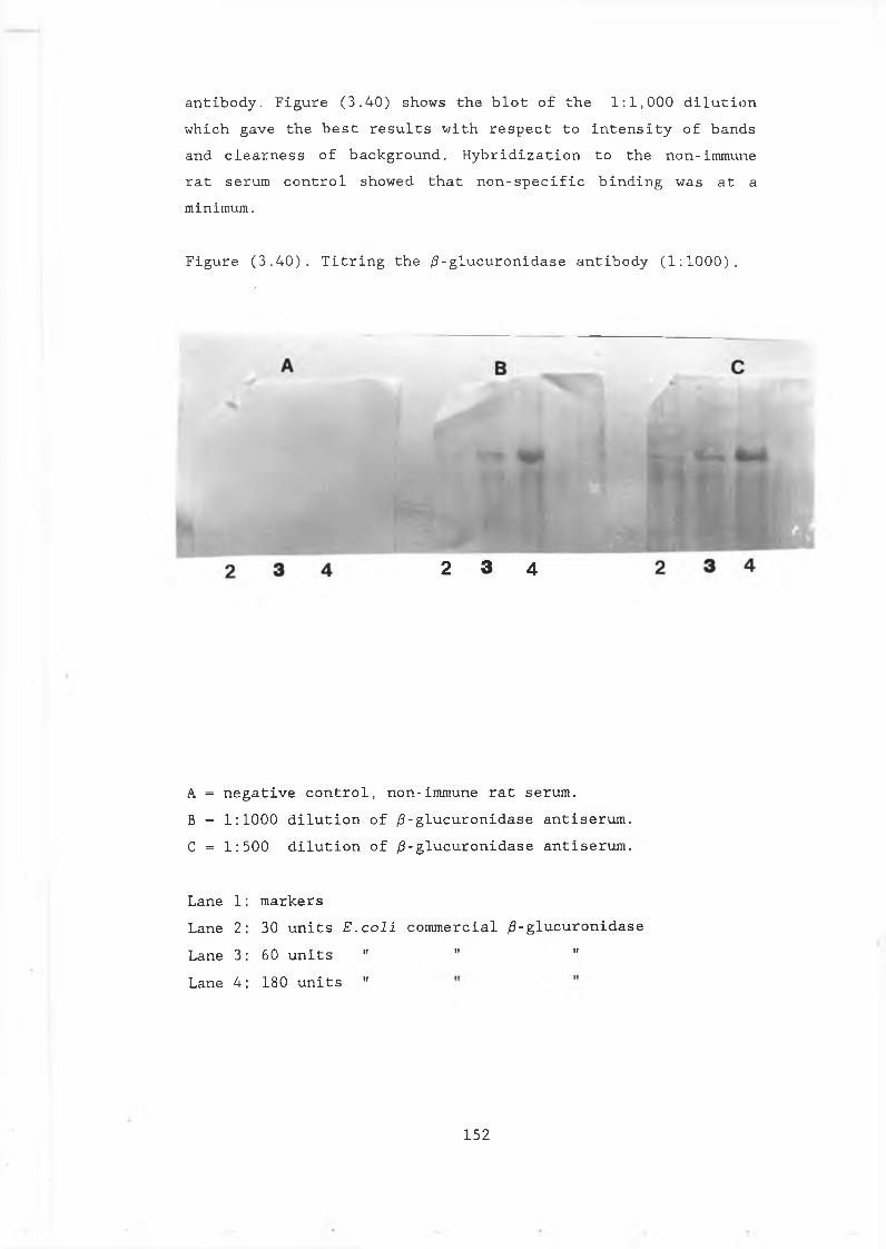
a n t i b o d y . F i g u r e ( 3 . 4 0 ) s h o w s t h e b l o t o f t h e 1 : 1 , 0 0 0 d i l u t i o n w h i c h g a v e t h e b e s t r e s u l t s w i t h r e s p e c t t o i n t e n s i t y o f b a n d s a n d c l e a r n e s s o f b a c k g r o u n d . H y b r i d i z a t i o n t o t h e n o n - i m m u n e r a t s e r u m c o n t r o l s h o w e d t h a t n o n - s p e c i f i c b i n d i n g w a s a t a m i n i m u m .
F i g u r e ( 3 . 4 0 ) . T i t r i n g t h e ¡3- g l u c u r o n i d a s e a n t i b o d y ( 1 : 1 0 0 0 ) ,
2 3 4
A = n e g a t i v e c o n t r o l , n o n - i m m u n e r a t s e r u m .B = 1 : 1 0 0 0 d i l u t i o n o f ¡3- g l u c u r o n i d a s e a n t i s e r u m . C = 1 : 5 0 0 d i l u t i o n o f / 3 - g l u c u r o n i d a s e a n t i s e r u m .
L a n e 1 L a n e 2 L a n e 3 L a n e 4
m a r k e r s3 0 u n i t s E . c o l i c o m m e r c i a l 0 - g l u c u r o n i d a s e 6 0 u n i t s " " 111 8 0 u n i t s "
152
To ensure that the antibody also recognises ¡3-glucuronidase produced in the yeast cytoplasm and to compare the sizes of the E.coli and S.cerevisiae enzyme, the antibody was tested against a protein extract prepared from pJBU7. The Western blot, sho^m in Figure (3.41) reveals a band at the same mobility in E.coli and pJBU7 (72KDa). pAAH5 was also run to ensure against non-specific binding and the same blot was tested against non-immune rat serum. The faintness of the band in pJBU7 is likely to be a result of the low amount of extract run on the gel (approximately equivalent to 200^1 of a stationary phase culture). This shows however, that the antibody does recognize the enzyme when produced in yeast, even at low concentrations and should prove useful in future investigations of /3-glucuronidase production S .cerevisiae. Contaminating bands that appear in the commercial preparation of the enzyme, running at a faster mobility than /3 - glucuronidase, also appear in the yeast extract samples. These bands are most likely to be a result of non-specific binding of the antibody preparation and are not associated with /3-glucuronidase activity.
153
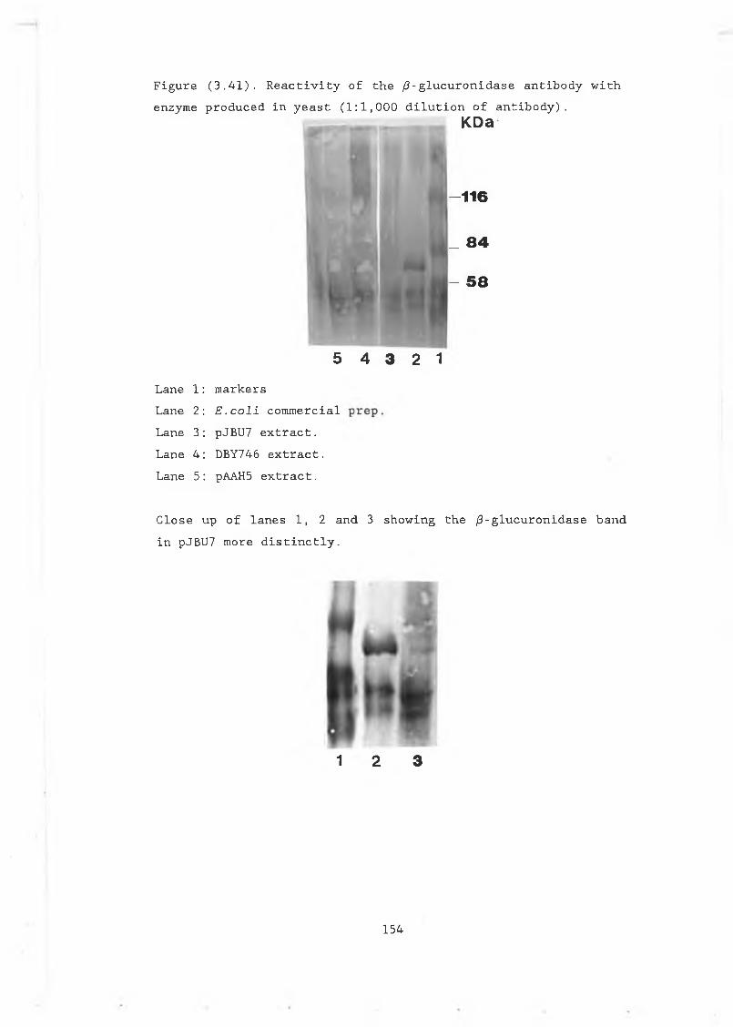
Figure (3.41). Reactivity of the /?-glucuronidase antibody with enzyme produced in yeast (1:1,000 dilution of antibody).
KDa
-116
_ 84
- 58
Lane 1: markersLane 2: E .coli commercialLane 3: pJBU7 extract.Lane 4: DBY746 extract.Lane 5: pAAH5 extract.
Close up of lanes 1, 2 and 3 showing the /3-glucuronidase band in pJBU7 more distinctly.
1 2 3
5 4 3 2 1
154
CHAPTER 4 Discussion
155
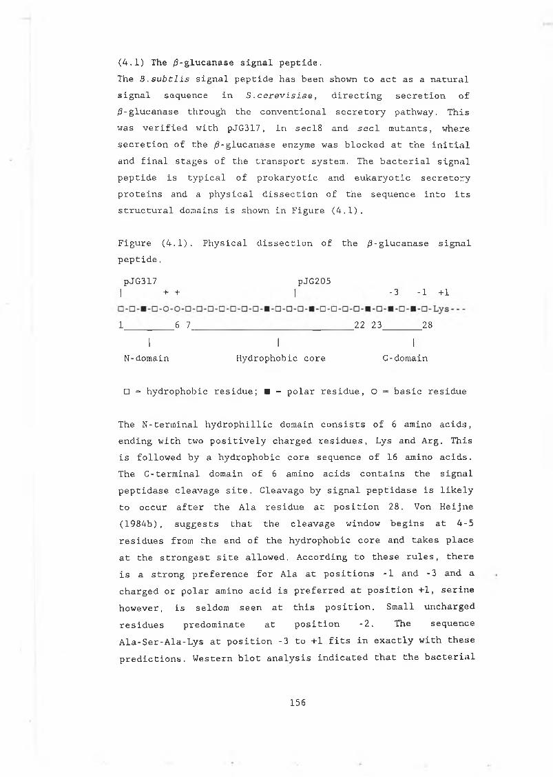
(4.1) The ¿3-glucanase signal peptide.The B.subtlis signal peptide has been shown to act as a natural signal sequence in S .cerevisiae, directing secretion of /3-glucanase through the conventional secretory pathway. This was verified with pJG317, in secl8 and seel mutants, where secretion of the /?-glucanase enzyme was blocked at the initial and final stages of the transport system. The bacterial signal peptide is typical of prokaryotic and eukaryotic secretory proteins and a physical dissection of the sequence into its structural domains is shown in Figure (4.1).
Figure (4.1). Physical dissection of the /3-glucanase signal peptide.
pJG317 pJG205| + + | - 3 - 1 + 1
1 _ 6 7_____________________________ _22 23_______ 28
I I IN-domain Hydrophobic core C-domain
□ = hydrophobic residue; ■ = polar residue, O = basic residue
The N-terminal hydrophillic domain consists of 6 amino acids, ending with two positively charged residues, Lys and Arg. This is followed by a hydrophobic core sequence of 16 amino acids. The C-terminal domain of 6 amino acids contains the signal peptidase cleavage site. Cleavage by signal peptidase is likely to occur after the Ala residue at position 28. Von Heijne (1984b), suggests that the cleavage window begins at 4-5 residues from the end of the hydrophobic core and takes place at the strongest site allowed. According to these rules, there is a strong preference for Ala at positions -1 and -3 and a charged or polar amino acid is preferred at position +1, serine however, is seldom seen at this position. Small unchargedresidues predominate at position -2. The sequenceAla-Ser-Ala-Lys at position -3 to +1 fits in exactly with thesepredictions. Western blot analysis indicated that the bacterial
156
signal peptide is processed by signal peptidase during translocation of /3-glucanase across the ER membrane This is demonstrated in the appearance of a single, processed band in the supernatant of pJG317 after tunicamycin treatment (Fig. 3.24). Although protein sequence analysis would be required to establish the exact cleavage site, according to the general rules, cleavage is most likely to occur at this site.
(4.2) Deletion of the /3-glucanase signal peptide.Complete removal of the /3-glucanase signal peptide in JB/34 and pJBC55A, resulted in a loss of detectable enzyme activity when expressed on a multicopy plasmid in the yeast cytoplasm. In the protease deficient mutant MD50 however, partial recovery of low levels of activity was achieved with pJB/34.
Recovery of low levels of /3-glucanase activity in the cytoplasm in the absence of these proteases demonstrates that without its signal peptide, the /3-glucanase enzyme is unstable and is a substrate for proteolysis. In vivo, this form of the enzyme may also be a substrate for cellular proteases in the yeast cytoplasm.Degradation in the cytoplasm is a common problem encountered with the production of foreign proteins in yeast and in higher eukaryotic cells (Townsend et al 1986). This has been shown for the yeast secretory protein acid phospatase, where up to 80% of the protein is subject to degradation with the signal peptide removed (Silve et al 1987). Removal of the signal peptide in /3-glucanase is likely to result in conformational changes in the /3-glucanase enzyme, possibly rendering it more sensitive to the action of cellular proteases. The identity of the N-terminal amino acid residue has also been shown to affect protein stability in yeast (section 1.1.2). In pJB/34 and PJBC55A however, the respective N-terminal Met-Ser-Lys and Met-Gly-Asp sequences, fall into the stabilizing category. Even if the N-terminal Met was removed in vivo, the second residue in each case is also stabilizing.
157
In pJB/34, a contributory factor to the lack of detectable activity in DBY746 and the low levels of activity produced in MD50, is likely to be the instability of the /3-glucanase transcript. This instability is reflected in Northern blots where no /3-glucanase specific transcript is detected for pJBjS4. Efficient termination has been shown to be important for RITA stability in yeast and expression levels can increase dramatically when this is achieved (Ammerer, 1983; Zaret and Sherman, 1982). In pJB/34, the bacterial terminator alone may not terminate transcription efficiently.pJBC55A on the other hand, produces a remarkably strong /3-glucanase transcript, but reveals no sign of any enzyme activity when expressed in DBY746, or in MD50. Examination of the 5' untranslated leader regions of the signal-less gene in pJB55A, suggests that the absence of active enzyme may be associated with translation efficiency. Highly expressed yeast genes tend to contain untranslated 5' leader regions that are short (approximatly 52 nucleotides) and void of secondary structure (Cigan and Donaghue, 1987). A lack of secondarystructure is promoted by a bias toward A and a strict avoidance of G nucleotides in this region. These features promote conditions ideal for attachment and movement of the ribosome towards the initiator codon. In addition, a consensus sequence has been identified around the AUG start that has been shown to promote efficient translation, presumably optimizing conditions for recognition of the initiator. This sequence, compiled by examining nucleotide bias around the AUG of highly expressed genes, is also rich in A and void of G nucleotides and takes the form A/Y-A-A/U-A-AUG, where Y is a pyrimidine (U or C) . Optimization of the 5' leaders of foreign genes in accordance with these guidelines, has been shown to have dramatic effects, resulting in increased expression levels in yeast (Bitter and Egan, 1988; Fieschko et al 1987).Comparison of the 5' leaders of pJB/34 and pJBC55A, reveals differences both in their lengths and composition. Assuming that transcription is initiated at the same site as that defined for the ADH1 promoter (between positions -37 and -27,
158
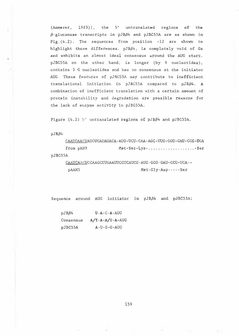
(Ammerer, 1983)), the 5' untranslated regions of the /?-glucanase transcripts in pJB/34 and pJBC55A are as shown in Fig.(4.2). The sequences from position -12 are shown to highlight these differences. pJB/34, is completely void of Gs and exhibits an almost ideal consensus around the AUG start. pJBC55A on the other hand, is longer (by 9 nucleotides), contains 3 G nucleotides and has no consensus at the initiator AUG. These features of pJBC55A may contribute to inefficient translational initiation in pJBC55A compared to pJB/34. A combination of inefficient translation with a certain amount of protein instability and degradation are possible reasons for the lack of enzyme activity in pJBC55A.
Figure (4.2) 5' untranslated regions of pJB/34 and pJBC55A.
pJB/34CAAUCAACUAUCUCAUAUACA-AUG-UCU-CAA-AGC-UUG-GGG-GAU-CCG-UCAfrom pAH9 Met-Ser-Lys-.................... -Ser
pJBC55ACAAUCAACUCCAAGCUUGAAUUCCCCAUCG-AUG -GGG-GAU-CCU-UCA- - pAAH5 Met-Gly-Asp.....Ser
Sequence around AUG initiator in pJB/34 and pJBC55A:
pJB/34 U-A-C-A-AUGConsensus A/Y-A-A/U-A-AUG pJBC55A A-U-C-G-AUG
159
(4.3) Truncating the /3-glucanase signal peptide.pJG205 contains a truncated version of the /3-glucanase signal peptide, starting with the Met residue at position 17 (see Fig. 4.1). This signal peptide, although missing the complete hydrophillic and the bulk of the hydrophobic domains, directs secretion of /3-glucanase into the yeast cell medium. Sec mutant analysis confirms that the extracellular /3-glucanase activity detected in pJG205, is due to transport through the conventional secretory pathway. This suggests that the severely truncated bacterial signal sequence can direct translocation and secretion of heterologous proteins in yeast.Kaiser et al (1987), has proposed that translocation of proteins into the ER only requires the presence of a short stretch of hydrophobic amino acids (section 1.3.2). pJG205 fulfills this obligation with a hydrophobic core of 4 residues. However, Kaisers experiments also showed that the levels of translocation achieved with limited hydrophobicity are extremely low. Initial observations with pJG205, comparing the levels of secreted enzyme to pJG317, suggested that the truncated version of the signal peptide secretes /3-glucanase in amounts comparable to the complete sequence, with extracellular levels of enzyme activity being almost the same in each case. This comparison however, is complicated by the very high levels of /3-glucanase activity detected intracellularly in pJG205. Taking into account the total amount of /3-glucanase activity produced by the cells, the secretion efficiency in pJG205 is only 14%, whereas in pJG317, approximately 50% of the overall levels are secreted. The total amount of activity produced in pJG317 however, is significantly lower than in pJG205 (80-90% lower) (section 3.4).Examination of the steady state levels of the /3-glucanase RNA transcripts by Northern analysis, revealed no significant differences between pJG317 and pJG205. This indicates that the lower levels of activity observed in pJG317 are not due to RNA instability. Also, differences in translation efficiencies are not likely to be the cause of the discrepancies in overall levels of enzyme produced. The two plasmid constructions are
160
identical except for a region of 10 and 11 base pairs, derived from the /3-glucanase gene sequence preceding the initiator methionines. These 5' untranslated regions, while they do not constitute ideal 5' leader sequences according to the accepted guidelines outlined in section (4.2) above, are similar in length and contain equal numbers of G nucleotides. Of the two, the pJG317 sequence is more conducive to efficient initiation of translation than pJG205. (The pJG205 sequences 5' to the initiator Met are: TGGATTGTTTATG ; the pJG317 sequences 5' to the initiator Met are: GGAATGCCAATATG).Glycosylation of the /3-gluucanase enzyme, in the secretory pathway of DBY746, appears to inhibit activity of the enzyme (see section 4.5). Activity gel analysis suggests that glycosylation of the enzyme produced in pJG205 does not occur to the extent seen in pJG317 or pJG314. Reduced glycosylation may therefore contribute to the high levels of activity observed intracellularly in pJG205.This lack of glycosylation in pJG205 could reflect inefficient translocation of the /3-glucanase enzyme into the secretory pathway, caused by the presence of the truncated signal peptide. Western analysis of pJG205 extracts suggests that there is a substantial amount of unprocessed enzyme, with the signal peptide still intact, present intracellularly in this strain. The wide smear of activity observed on activity gels is also consistent with the presence of processed and unprocessed bands (Fig. 3.25). The fraction that is secreted in pJG205 however, is processed with respect to signal cleavage. This is evident by comparing the size of the extracellular fraction to the B.subtilis control enzyme (Fig. 3.26).
161
(4.4) Comparison of the /3-glucanase and a-factor signal peptides in pJG317, pJG314 and pVT314.Comparison of the full bacterial signal peptide with the yeast a-factor signal, in relation to their abilities to secrete /3-glucanase, is also complicated by the problem of glycosylation. The growth curves presented in section (3.4), suggest that the Bacillus signal is at least as efficient as the a-factor signal. In pJG317 and pJG314, approximately half of the overall levels produced in each case is secreted. In pJG314, the total amount of /3-glucanase produced is approximately 50% of that produced in pJG317. These lower overall levels were assumed to reflect the differences in the relative strengths of the Adhl and a-factor promoters. However, pJG317 demonstrates that DBY746 is capable of secreting almost 450 units of /3-glucanase activity per ml of culture under the conditions used. The lower levels secreted in pJG314 may simply reflect reduced efficiency of the a-factor signal peptide to secrete and process the enzyme.In an attempt to clarify this and to obtain a more direct comparison of the secretion efficiencies of the bacterial and yeast signal peptides, pVT314 was made. This construct places the /3-glucanase gene, fused to the a-factor signal sequence, under control of the Adhl promoter and was intended to act as a direct control for comparison to pJG317. It was expected that the same promoter in pVT314 and pJG317 would result in similar overall levels of /3-glucanase production with the levels secreted reflecting the relative strengths of the two signal peptides. The levels detected in pVT314 however, intracellularly and extracellularly, are significantly reduced. Surprisingly, these overall levels are also lower than those observed in pJG314. Over much of the growth curve the secreted enzyme levels of pVT314 are also lower than those observed in pJG314 despite the fact that in both constructs the /3-glucanase gene is fused to the a-factor signal peptide.Other studies have shown that overexpression of heterologous genes, in comoination with certain signal peptides, can result in reduced secretion efficiencies (Smith eC al, 1985). This is
162
not the case with pVT314, as interference in secretion efficiencies alone would presumably be accompanied by accumulation inside the cell. Instead, as with the other constructs, approximately 50-60% of what is produced in pVT314 is secreted, although lower levels are reached overall.The vector used in constructing pVT314 is similar to pAAH5 and p69D, except for the auxotrophic marker used for selection (uracil in pVT314 and leucine in the pJG series). According to the growth curves shown, higher cell densities are reached in both pJG317 and pJG314 compared to pVT314, and this feature of pVT314 may contribute to the overall lower levels. However, other factors such as glycosylation must also be considered. Glycosylation appears to interfere with activity of the /3-glucanase enzyme and would therefore interfere with the true approximation of /3-glucanase production in each strain. This effect would also mask the relative efficiencies of the two signal peptides, since the extent of glycosylation must also be taken into fccount. If the enzyme is more likely to be glycosylated when attached to the a-factor signal, then the lower level observed in these constructs would be expected. This is also the case if the a-factor signal is more efficient than the bacterial signal at translocating the enzyme. These a-factor signal constructs however, also pose additional problems for /3-glucanase when one considers that the signal itself is also glycosylated in vivo in yeast. Furthermore, in contrast to the bacterial signal which is cleaved as the enzyme is translocated through the ER membrane, the a-factor signal is retained until the it reaches Golgi.The sharp increase in extracellular activity seen in the growth curves of pJG314 and pVT314 at approximately 36 hours interesting feature which may reflect processing characteristics of the a-factor signal peptide. This increase is accompanied by a corresponding decrease in activity levels intracellularly and is not apparent in the bacterial signal constructs, a-factor is not processed until the Golgi stage of the pathway and ongoing processing of the intracellular pool of /3-glucanase may manifest in this slight delay in secretion of
163
the enzyme.
(4.5) The effect of glycosylation on the Bacillus /3-glucana:>e enzyme.It is not entirely unexpected that glycosylation could interfere with the activity of a bacterial protein when produced in yeast. Glycosylation of secretory proteins is not a feature of the bacterial secretory process and modifications to areas of a protein that may constitute active sites, can easily be envisaged as interfering with enzyme activty. The evidence from the sec mutants, combined with the tunicamycin effect, shows that this is the case for the B.subtilis /3-glucanase enzyme. The enzyme appears to be less active when modified by glycosylation than in its natural state.Tunicamycin has a more profound effect on recovery of enzyme activity than production of the enzyme at the restrictive temperature in the glycosylation mutant, sec53. As described earlier, tunicamycin inhibits all glycosylation by interfering at the earliest stage in core assembly. The sec53 phenotype reduces glycosylation by limiting the number of mannose residues available for incorporation into glycoproteins. This difference could explain the larger increase seen with the inhibition of all sugar additions in the presence of tunicamycin.Glycosylation has also been shown to interfere with the activity of ^-glucanase produced in another Bacillus species (B.macerans). A reduced specific activity was observed for the glycosylated form when compared to the native form (Oleson and Thomsen, 1991).The glycosylated form of the B.macerans enzyme was shown to be more stable than the unglycosylated form. Preliminary analysis of the thermostability of the B.subtilis enzyme (not shown) however, suggests the opposite. In this case the unglycosylated form (prepared by treatment with tunicamycin) exhibits a greater tolerance for incubation at high temperatures.Glycosylation of the E.coli /5-glucuronidase enzyme in the ER of the tobacco plant has also been shown to interfere with the
164
activity of this bacterial enzyme (Ittaguria et al 1989). Tunicamycin treatment of tobacco cell suspension cultures, carrying the /3-GUS gene fused to various lengths of the plant patatin signal sequence, resulted in up to a 100 fold increase in /?-glucuronidase activity. Western analysis showed that the enzyme was present at a higher molecular weight than expected in the plant cultures and treatment with tunicamycin or EndoH, reduced it to its normal mature size. In this case, the glycosylation sites occur at amino acid positions 358 and 423. The disruption of activity by glycosylation at these sites, suggests that the active site of the enzyme is toward the carboxy terminus. This is futher substantiated by the Bal 31 deletion analysis carried out in this study.Fusion of the /3-GUS gene to the a-factor signal peptide in pGUSa6, did not allow secretion of the enzyme out into the yeast culture medium. Also, tunicamycin had no effect on the activity of the enzyme in this strain. Taken in conjunction with Ittaguria's observations, this suggests that the enzyme is not translocated in pGUSa6 . Why translocation is prevented may be associated with the large size of the enzyme, in combination with the relatively large signal peptide, presenting steric problems for translocation.The potential glycosylation sites in the /3-glucanase enzyme are more widely spread, occurring at positions 31,38,64 and 184. Activity gel analysis shows that in both pJG317 and pJG314, all of these sites can become modified with core oligosaccharides. However, complete modification does not always occur and a proportion of the enzyme in the secretory pathway is only partly modified at two sites, while a further fraction contains no glycosylation additions. Although most of the secreted fraction in both pJG317 and pJG314 is partially glycosylated to some extent, unglycosylated enzyme is also secreted. This shows that glycosylation is not a prerequisite for secretion of this enzyme in yeast. The extent of the modifications to the higher molecular weight forms of the enzyme, present as a smear in the extracellular samples in pJG317 and pJG314, cannot be examined in this type of zymogram analysis.
165
Different levels of glycosylation appears to have different effects on the activity of the enzyme. It is obvious that core glycosylation does not completely inhibit /3-glucanase activity and it is impossible to tell if the bands visible on activity gels, have reduced enzymatic activity compared to the unmodified bands. It is likely that the core additions do affect activity of the enzyme, but that the inhibition observed is also contributed to by extensive chain additions which have a more severe effect.Examination of tunicamycin treated /3-glucanase in pJG317 and pJG314 confirms that these higher molecular weight bands represent glycosylated forms of the enzyme. In the gel shown in figure (3.27), the higher molecular weight forms are reduced to the size of the unglycosylated band, which can be clearly seen to have increased in intensity. In these supernatant samples, the loadings represent equal levels of /3-glucanase activity as determined by DNS assay (ie. a 1/10 dilution of the +tunicamycin sample in pJG317 and a 1/3 dilution of the +tunicamycin sample in pJG314). This dilution was necessary as the large increases in activity caused by tunicamycin treatment resulted in indistinguishable smears of activity running the length of the +tunicamycin lane when equal volumes were added. This underlines the extent of the increase seen and suggests that it is contributed to by previously unseen material, possibly representing highly modified /3-glucanase. When cyclohexamide is added to RSY12 cells at the same time as the mutant phenotype is induced, the increases in /3-glucanase activity seen at intracellular and extracellular levels are no longer observed (data not shown). Extracellular levels of /3-glucanase activity are equal at both 25°C and 37 C. Although cyclohexamide will have halted protein synthesis, the enzyme activity detected extracellularly is presumably derived from the intracellular levels of /3-glucanase that was synthesized before induction of the mutant phenotype. This further substantiates the idea that the increases seen at the non-permissive temperature, in the absence of cyclohexamide, is due to the inhibition of oligosaccharide additions to newly
166
formed /3-glucanase.The smallest glycosylated band representing core oligosaccharide additions at 2 sites, is still visible in the the supernatants after tunicamycin treatment. Because of the nature of this analysis a certain amount of glycosylated enzyme, modified before the addition of tunicamycin and subsequently secreted, is likely to be present and this band may reflect this fraction. In retrospect, this could be eliminated by a short preincubation period in the presence of the drug. A similar band appears in the supernatant of pJG205 cells after tunicamycin treatment. Its slightly faster mobility suggests that it could represent /3-glucanase with one core oligosaccharide addition or possibly the enzyme with its signal peptide still intact. Whatever its identity, the band is only evident after glycosylation is inhibited and therefore is derived from /3-glucanase material not seen in pJG205 under normal growth conditions.Why the /3-glucanase enzyme produced in yeast is present in a range of different glycosylated forms may be due to the folding properties of the protein as it passes through the ER. Rapid folding may preclude glycosylation, while modification at one site may promote further additions. In pJG317 and pJG205 the proportion of glycosylated material seems to be affected by the amount that is translocated and subsequently secreted. How the majority of the enzyme in pJG205 avoids glycosylation is not clear. Also, the proportion that appears to be glycosylated in this strain seems to have undergone extensive modifications and is only assayable after glycosylation is disrupted. These differences may be associated with the nature of the translocation event and the orientation of the protein passing through the ER membrane when fused to the truncated signal. This limited amount of glycosylation observed in pJG205 can explain the lower increases in activity observed in tunicamycin treated pJG203 compared to tunicamycin treated pJG317 (section 3.11). Where less glycosylation has taken place the inhibitory effect on ¿3-glucanase activity would be expected to be less pronounced.
167
When compared to pJG317, the effect of tunicamycin in pJG314 is also less dramatic. Inhibition of glycosylation of the a-factor pro-region results in a reduction of a-factor secretion. This is thought to be a result of interference with the folding patterns of the region causing a certain amount of accumulation in the ER (Julius et al, 1984b). Reduced secretion of /3-glucanase, when fused to the a-factor signal peptide in pJG314, would dilute the extent of the increase expected after tunicamycin treatment in the supernatant of this strain. The resultant accumulation of unprocessed and malfolded enzyme in the lumen of the ER, may also account for the lack of an increase seen at intracellular levels.
(4.6) Processing of the a-factor signal peptide in pJG314 and pJG317.As outlined in section (1.6.4), processing of the a-factor pheromone protein involves a series of enzymatic cleavage events. The main section of the pro-region is removed after the Lys Arg dibasic cleavage site by the action of the KEX2 protease. The mature portion of the a-factor protein is further processed by the action of STE13 which removes the remaining N-terminal Glu-Ala sequences (Bussey, 1988).In pJG314, the sequence around the cleavage site of the a-factor pro-region includes these Glu-Ala repeats. A short segment of pUC8 DNA is also included, derived from the BamHl site used to isolate the signal-less /3-glucanase gene from pJG104 (Fig. 4.3). Cleavage at Lys-Arg results in the /9-glucanase enzyme retaining 10 extra amino acids at its N-terminus. This is reflected in the slower mobility of the unglycosylated, processed band in the extract of pJG314, seen on the Western blots. The action of the STE13 gene product in a-factor maturation is thought to be the rate limiting factor in secretion of the pheromone (Bussey, 1988). In conjunction with heterologous sequences, cleavage at this site is often inefficient (section 1.7.0) and this seems to be the case in pJG314. The activity gel in Figure (3.25), shows that a small amount of processing by STE13 has occurred with the appearance
168
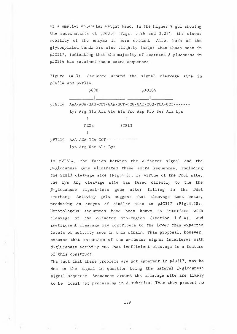
of a smaller molecular weight band. In the higher % gel showing the supernatants of pJG314 (Figs. 3.26 and 3.27), the slower mobility of the enzyme is more evident. Also, both of the glycosylated bands are also slightly larger than those seen in pJG317, indicating that the majority of secreted /3-glucanase in pJG314 has retained these extra sequences.
Figure (4.3). Sequence around the signal cleavage site in pJG314 and pVT314.
p69D pJG104
pJG314 AAA-AGA-GAG-GCT-GAA-GCT-CCG-GAT-CCG -TCA-GCT------Lys Arg Glu Ala Glu Ala Pro Asp Pro Ser Ala Lys
t tKEX2 STE134-
pVT314 AAA-AGA-TCA-GCT --------Lys Arg Ser Ala Lys
In pVT314, the fusion between the a-factor signal and the /3-glucanase gene eliminated these extra sequences, includingthe STE13 cleavage site (Fig.4.3). By virtue of the Stul site,the Lys Arg cleavage site was fused directly to the the/3-glucanase ,signal-less gene after filling in the Dde 1 overhang. Activity gels suggest that cleavage does occur,producing an enzyme of similar size to pJG317 (Fig.3.28).Heterologous sequences have been known to interfere with cleavage of the a-factor pro-region (section 1.6.4), andinefficient cleavage may contribute to the lower than expected levels of activity seen in this strain. This proposal, however, assumes that retention of the a-factor signal interferes with /3-glucanase activity and that inefficient cleavage is a feature of this construct.The fact that these problems are not apparent in pJG317, may be due to the signal in question being the natural /3-glucanase signal sequence. Sequences around the cleavage site are likely to be ideal for processing in B.subtilis. That they present no
169
obvious problem for the yeast signal peptidase, combined with the secretion levels observed, suggests that this signal may be useful for the production of other foreign proteins in yeast. How the signal fares in conjunction with other sequences, however, remains to be seen.
(4.7) Conclusion.The B.subtilis /3-glucanase enzyme is secreted in yeast using its own bacterial signal peptide. Western analysis showed that the bacterial signal is recognized and cleaved by the yeast signal peptidase. Comparisons to the yeast a-factor signal sequence suggests that the bacterial signal is at least ¡is efficient as the yeast signal peptide. These studies show that the B.subtilis signal has definite potential for use as a general secretion signal in yeast. Removal of the bacterial signal peptide resulted in a loss of /3-glucanase activity. This was shown to be associated with stability of the enzyme in the yeast cytoplasm and demonstrated the importance of optimizing sequences around the translational initiation site in the generation of fusion constructs. Analysis of a mutant form of the signal peptide showed that a truncated version of the bacterial signal sequence can still translocate /3-glucanase, although with reduced efficiency.Analysis of /9-glucanase secretion was obstructed somewhat by the inhibitory effects of glycosylation on activity of the enzyme. On one level this caused problems in assessing the relative efficiencies of the signal peptides used. Glycosylation also interfered with Western analysis in that the antibody raised against the B.subtilis /3-glucanase enzyme did not recognize glycosylated forms of the enzyme produced in yeast. This is likely to be a result of the widely spread glycosylation sites preventing recognition of the antigenic sites in this relatively small (22KDa) protein. Activity gel analysis circumvented this problem to some extent, but examination of extensively glycosylated forms of the enzyme was still difficult.Glycosylation also affects the activity of /3-glucuronidase, the
170
other bacterial enzyme examined in this work. This enzyme was chosen for investigation due to its attractive assaying procedures. Although translocation of /?-glucuronidase was not achieved in this study, it may be possible to secrete the protein, or a truncated version of the protein, using another signal sequence. The antibody raised against E.coli /3-glucuronidase is less likely to have problems in recognizing modified forms of the enzyme in yeast than the 0 -glucana.se antibody . The larger size of the protein (72KDa) , and the presence of only two glycosylation sites in close proximity to each other, should not prevent recognition of the antibody at other antigenic sites in the enzyme. These glycosylation sites could possibly be removed by the introduction of conservative amino acid substitutions. If this did not interfere with activity of the enzyme, the problem of reduced activity due to glycosylation would also be removed.
171
CHAPTER 5 REFERENCES
172
Akita, M., Sasaki, S., Matsuyama, S., Mizushimi, S.(1990) J. Biol. Chem. 265, 8164-8169.SecA interacts with secretory proteins by recognizing the +ve charge at the amino terminus of the signal peptide.
Ammerer, G. (1983) Meths. Enzymol. 101, 192-201.Expression of genes in yeast using the ADC1 promoter.
Ammerer, G., Hunter, C.P., Rothman, J.H., Saari, G.C., Vails, L.A., Stevens, T.H. (1986) Mol. Cell. Biol. 6 , 2490-2499.PEP4 gene of S.cerevisiae encodes proteinase A, a vacuolar enzyme required for processing of vacuolar precursors.
Andrews, D.W., Perera, E., Lesser, C., Lingappa, V.R.(1989) J. Biol. Chem. 263, 15791-15798.Sequences beyond the cleavage site influence signal function.
Bachmair, A., Finley, D., Varshavsky, A.(1986) Science 234, 179-186.The in vivo half life of a protein is a function of its amino terminal residue.
Baker, R.K., Lively, M.D. (1987) Biochemistry 26, 8561-8567. Purification and characterization of hen oviduct microsomal signal peptidase.
Ballou, C.E., Kern, K.A., Raschke, W.C.(1973) J. Biol. Chem. 248, 4667-4673.Genetic control of yeast mannan structure.
Bankaitis, V.A., Johnson, L.M., Emr, S.D.(1986) Proc. Nat. Acad. Sci. USA 83, 9075-9079.Isolation of yeast mutants defective in protein targeting to the vacuole.
173
Banta, L.M., Robinson, J.S., Klionski, D.J., Emr, S.D.(1988) J. Cell Biol. 107, 1369-1383.Organelle assembly in yeast : characterization of yeast mutants defective in vacuolar biogenesis and protein sorting.
Barnes, G., Hansen, W. , Holcomb, C.L., Rine, J.,(1984) Mol. Cell. Biol. 4, 2381-2388.
Bathurst, I.C., Brennan, S.O., Carrel, R.W., Cousens, L.S.,Brake, A.J., Barr, P.J. (1987) Science 235, 348-351.Yeast KEX2 protease has the properties of a human proalbuminconverting enzyme.
Bedoulle, H., Bassford, P.J., Fowler, A.V., Zabin, I., et al. (1980) Nature 285, 78-81.Mutations that alter the function of the signal sequence of the maltose binding protein of E.coli.
Ben-Bassat, A., Bauer, K. , Chang, S.Y., Myambo, K. , Boosman,A., Chang, S. (1987) J. Bact. 169, 751-757.Processing of the initiation methionine from proteins: properties of the E.coli methionine aminopeptidase.
Benson, S.A., Hall, M.N., Silhavy, T.J.(1985) Ann. Rev. Biochem. 54, 101-134.Genetic analysis of protein export in E.coli.
Bernstein, H.D., Poritz, M.A., Strub, K., Hoben, P.J., Brenner, S., Walter, P. (1989) Nature 340, 482-486.Model for signal sequence recognition from amino acid sequence of the 54K subunit of Srp.
Bernstein, M . , Kepes, F., Schekman, R.(1989) Mol. Cell. Biol. 9, 1191-1199.SEC59 encodes a membrane protein required for core glycosylation in S .cerevisiae.
174
Birnboim, H.C., Doly, J., (1979) Nuc. Acid Res.7, 1513-1523.A rapid alkaline extraction procedure for screening recombinant plasmid DNA.
Bitter, G.A., Egan, K.M. (1984) Gene 32, 263-274.Expression of heterologous genes in S .cerevisiae from vectors utilizing the GAPDH promoter.
Bitter, G.A., Egan, K.M., (1988) Gene 69, 193-207.Expression of human interferon-7 from hybrid yeast GPD promoters containing upstream regulatory sequences from the GAL1-GAL10 intergenic region.
Blobel, G., Dobberstein, B., (1975a) J. Cell Biol. 67, 83 Transfer of proteins across membranes I: Presence ofproteolytically processed and unprocessed nascent immunoglobulin light chains on membrane bound ribosomes of murine myeloma.
Blobel, G., Dobberstein, B., (1975b) J.Cell Biol. 67, 852 Transfer of proteins across membranes II: Recognition offunctional rough microsomes from heterologous components.
Bohni, P.C., Deshaies, R.J., Schekman, R.W.(1988) J. Cell Biol. 106, 1035-1042.SECll is required for signal peptide processing and yeast cell growth.
Bole, D.G., Hendershot, L.M., Kearny, J.F.,(1986) J. Cell Biol. 102, 1558-1566.Post-translational association of immunoglobulin heavy chain binding protein with nascent heavy chains in nonsecreting and secreting hybridomas.
Bradshaw, R.A., (1989) TIBS 14, 276-279.Protein translocation and turnover in eukaryotic cells.
175
Brake, A.J., Merryweather, J.P., Coit, D.G., Heberlein, U.A., Masiartz, F.R., Mullenbach, G.T., Urdea, M.S., et al.
(1984) Proc. Nat. Acad. Sei. USA. 81,4642-4646.a-factor directed synthesis and secretion of mature foreign proteins in S.cerevisiae.
Bussey, M., (1988) Yeast 4, 17-26.Proteases and the processing of precursors to secreted proteins in yeast.
Byrd, J.C., Tarentino, A.L., Maley, F., Atkinson, P.H., Trimble, R.B., (1982) J. Biol. Chem. 257, 14657-14666. Glycoprotein synthesis in yeast.
Cantwell, B., Brazil, G., Hurley, J., McConnell, D.J.(1985) Procceedings of the European Brewery Convention Congress (Helsinki), pp259-166.Expression of the gene for endo-ß-1,3,-1,4-glucanase fromB .subtilis in S .cerevisiae.
Cantwell, B., Brazil, G., Murphy, N,, McConnell. D.J.(1986) Curr. Genet. 11, 65-70.Comparison of expression of the endo-/3-l, 3-1,4-glucanase gene from B. subtilis in S.cerevisiae from the CYC1 and ADH1 promoters.
Cantwell, B., McConnell, D.J., (1983) Gene 23, 211-219. Molecular cloning and expression of a B .subtilis ß -glucanase in E .coli .
Chang, C.N., Matteuci, M., Perry, J., Wulf, J.J., Chen, C.Y., Hitzeman, R., (1986) Mol. Cell. Biol. 6, 1812-1819.S.cerevisiae secretes and correctly processes human interferon hybrid proteins containing yeast invertase signal peptide.
Chou, P.Y., Fasman, G.D.(1978) Ann. Rev. Biochem. 47, 251-276.
176
Chirico, W.J., Waters, M.G., Blobel, G.(1988) Nature 332, 805-810.70K heat shock related proteins stimulate protein translocation into microsomes.
Cigan, A.M., Donahue, T.F., (1987) Gene 59, 1-10.Sequence and structural features associated with translational initiator regions in yeast - a review.Cigan, A.M., Donahue, T.F.(1988) Mol. Cell. Biol. 8, 2964-2975.Mutational analysis of the HIS4 translational initiator region in S .cerevisiae.
Collier, D.N., Bankaitis, V.A., Weiss, J.B., Bassford, P.J.(1988) Cell 53, 273-283.The antifolding activity of SecB promotes the export of the E.coli MBP.
Connolly, T., Gilmore, R., (1989) Cell 57, 599-610.The SRP receptor mediates the GTP dependant displacement of SRP from the signal sequence of the nascent polypeptide.
Crooke, E., Guthrie, B., Lecker, S., Lill, R., Wickner, W.(1988) Cell 54, 1003-1011.ProOmpA is stabilized for membrane translocation by either purified E.coli trigger factor or canine Srp.
Crooke, E., Wickner, W.(1987) Proc. Nat. Acad. Sci. 84, 5216-5220.Trigger factor: a soluble protein that folds proOmpA into amembrane assembly competent form.
Dalbey, R.E., Wickner, W.(1985) J. Biol. Chem. 260, 15925-15931.Leader peptidase catalyzes the release of exported proteins from the outer surface of the E.coli plasma membrane.
177
Dean, N., Pelham, H.R.B.,(1990) J. Cell. Biol. Ill, 369-377.
Deshaies, R.J., Kepes, F., Bohni, P.C. (1989) TIGS 5, 87-93. Genetic disection of the early stages of protein secretion in yeast.
Deshaies, R.J., Sanders, S.L., Feidheim, D.A., Schekman, R.(1991) Nature 349, 806-808.Assembly of yeast secretory proteins involved in translocation into the ER, into a membrane bound multisubunit complex.
Deshaies, R.J., Schekman, R. (1987) J. Cell Biol. 105, 633-645.A yeast mutant defective at an early stage in import of secretory protein precursors into the ER.
Deshaies, R.J., Schekman, R.(1989) J. Cell Biol. 109, 2653-2644.SEC62 encodes a putative membrane protein required for protein translocation into the yeast ER,
Deshaies, R.J., Schekman, R.(1990) Mol. Cell. Biol. 10, 6024-6035.Structural and functional disection of Sec62p, a membrane bound component of the yeast ER protein import machinery.
Donahue, T.F., Cigan, A.M.,(1988) Mol. Cell. Biol. 8, 2955-2963.Genetic selection for mutations that reduce or abolish ribosomal recognition of the HIS4 translational initiator region.
Eakle, K., Bernstein, M . , Emr, S.D.(1988) Mol. Cell. Biol. 8, 4098-4109.Cloning of SEC18 gene product necessary for transport from the ER to Golgi.
178
Ecker, D.J., Stadel, J.M., Butt, T.R., Marsh, J.A., Moira, B.P. (1989) J. Biol. Chem. 264, 7715-7719.Increasing gene expression in yeast by fusion to ubiquitin.
Eliers, M., Schatz, G. (1986) Nature 322, 228-232.Binding of a specific ligand inhibits import of a purified precursor protein into mitochondria.
Emr, S.D., Silhavy, T.J., (1980) J. Mol. Biol. 141, 63-90. Mutations affecting localization of an E.coli outer membrane protein, the bacteriophage A receptor.
Emr, S.D., Hedgpeth, J., Clement, J .M.,Silhavy, T.J.,Hofnung,M.(1980) Nature 285, 82-85.Sequence analysis of mutations that prevent export of A receptor, an E.coli outer membrane protein.
Englemen, D.M., Steitz, T .A. (1981) Cell 23, 411-422.The spontaneous insertion of proteins into and acrossmembranes: the helical hypothesis.
Erhart, E., Hollenberg, C.P. (1983) J. Bact. 156, 625-635.The presence of defective LEU2 gene on 2 p DNA recombinnant plasmids of S .cerevisiae is responsible for curing and high copy number.
Erikson, A.H., Blobel, G., (1985) J. Biol. Chem. 254, 11771-74. Early events in the biosynthesis of the lysosomal enzyme cathepsin D.
Ernst, J.F. (1986) DNA 5, 483-491.Improved secretion of heterologous proteins by S .cerevisiae: effects of promoter substitutions in a-factor fusions.
Ernst, J.F., Mermod, J.J., mattaliano, R., Moonen, P.,(1987) Bio/technology 5, 831-834.0-glycosylation and novel processing events during secretion of
179
a-factor / GM-CSF fusions by S.cerevisiae.
Esmon, B., Novick, P., Schekman, R. (1981) Cell 25, 451-460. Compartmentalized assembly of oligosaccharides on exported glycoproteins in yeast.
Esmon, B., Esmon, P.C., Schekman, R.(1984) J.Biol. Chem. 259, 10322-10327.Early steps in processing of glycoproteins.
Esmon, P.C.,Esmon, B.E., Schauer, I.E., Taylor, A.,Schekman, R.(1987) J. Biol. Chem. 262, 4387-4394.Structure, assembly and secretion of octameric invertase.
Etcheverry, T., Forrester, W., Hitzeman, R.A.(1986) Bio/technology 4, 726-730.Regulation of the chelatin promoter during expression of human serum albumin or yeast phosphoglycerate kinase.
Evans, E.A., Gilmore, R., Blobel, G.(1986) Proc. Natl. Acad. Sci. USA. (1986) 83, 581-585. Purification of microsomal signal peptidase as a complex.
Fandle, J.P., Canbelli, R. , Oliver, D., Tai, P.C.(1988) Proc. Natl. Acad. Sci. USA. 85, 8953-8957.SecA suppresses the temperature sensitive SecY24 defect in protein translocation in E.coli membrane vesicles.
Feldman, R.I., Bernstein, M., Schekman, R.(1987) J. Biol. Chem. 262, 9332-9339.Product of SEC53 is required for folding and glycosylation of secretory proteins in the lumen of the ER.
Ferro-Novick, S., Novick, P., Fields, C., Schekman, R.(1984) J. Cell. Biol. 98, 35-43.Yeast secretory mutants that block the formation of active cell surface enzymes.
180
Fieschko, J.L., Egan, K.M. , Ritch, T., Koski, R.A. , Jones, M.D., Bitter, G.A. (1987) Biotech. Bioeng. 29, 1113-1121. Controlled expression and purification of human immune interferon from high cell density fermentations of S .cerevisiae.
Fikes, J.D., Bassford, P.J.(Jr). (1989) J.Bact. 171, 402-409. Novel alleles of SecA improve export of MBP synthesized with a defective signal peptide.
Fujii, T., Kondo, K., Shimizu, F., Sone, H., Tanaka, J .,Inoue,T. (1990) Appl. Env. Micro. 56, 997-1003.Application of a ribosomal DNA integration vector in the construction of a brewers yeast having a-acetolactate decarboxylase activity.
Gething, M.J., Sambrook, J., (1982) Nature 300, 598-603. Construction of influenza hemagglutinin genes that code for intracellular and secreted forms of the protein.
Gething, M-J., McCammon, K., Sambrook, J.(1986) Cell 46, 939-950.Expression of wild type and mutant forms of influenza virus hemagglutinin: the role of folding in intracellular transport.
Green, R., Kramer, R.A., Shields, D.(1989) J. Biol. Chem. 264, 2963-2968.Misplacement of the amino-terminal positive charge in the prepro a-factor signal peptide disrupts membrane translocation in vivo.
Guisez, Y., Tison, B., Vanderkockhove, J., Demolder, J., Bauw,G., Haegeman, G., Fiers, W., Contreras, R.(1991) Eur. J. Biochem. 198, 217-222.Production and purification of recombinant human interleukin-6 secreted by the yeast S .cerevisiae.
181
Haguenauer-Tsapis, R. , Nagy, M. , Ryter, A.(1986) Mol, Cell. Biol. 6, 723-729.A deletion that includes the segment coding for the signal peptidase cleavage site delays release of S.cerevisiae : acidphosphatase from the ER.
Hann, B.C., Poritz, M.A., Walter, P.,(1989) J. Cell Biol. 109, 3228-3230.S.cerevisiae and Sz.pombe contain a homologue to the 54KDa subunit of the Srp that in S.cerevisiae is essential for growth.
Hartley, J.L., Donelson, J.E. (1980) Nature 286, 860-864. Nucleotide sequence of the yeast plasmid.
Hartmann, E., Weidmann, M., Rapoport, T.(1989) EMBO, J. 8, 2225-2229.A membrane component of the ER that may be essential for protein translocation.
Hasilik, A., Tanner, W. (1978) Eur. J. Biochem. 85, 599-608. Biosynthesis of the vacuolar glycoprotein carboxypeptidase Y. Conversion of the precursor into the enzyme.
Hardwick, K.G., Lewis, L.M., Semenza, J., Dean, N. , Pelham, H.R.B., (1990) EMBO J. 9, 623-630.ERD1, a yeast gene required for the retention of luminal ER proteins, affects glycoprotein processing in the ER.
Herman, P.K., Stack, J.H., DeModena, J.A., Emr, S.D.(1991) Cell 64, 425-437.A novel protein kinase homologue essential for protein sorting to the yeast lysosome-like vacuole.
182
Hernandez, L.M., Ballou, L., Alvardo, E., Gilece-Castro, B.L., Burlingame, A.L., Ballou, C.E.(1989) J. Biol. Chem. 264, 11849-11856.A new S .cerevisiae mnn mutant N-linked oligosaccharide structure.
Hitzeman, R., Leung, D.W., Perry, L.J., Kohr, W.J.(1983) Science 219, 620-625.Secretion of human interferon by yeast.
Hitzeman, R.A., Hagie, F.E., Hayflick, J.S., Chen, C . Y . , Seeburg, P.H., Derynck, R. (1982) Nuc. Acid Res. 10, 7791-1808. The primary structure of the S .cerevisiae gene for 3-phosphoglycerate kinase.
Hitzeman, R.A., Leung, D.W., Perry, L.J., Kohr, W.J., Levine,H.L., Goeddel, P.V., (1983) Science 219, 620-625.Secretion of human interferons by yeast.
Hitzeman, R.A., Chen, C.Y., Dowbenko, D.J., Renz, M.E., et al.(1990) Meths. in Enzymol. 185, 421-440.Use of heterologous and homologous signal sequences for secretion of heterologous proteins from yeast.
Holmes, D.S., Quigley, M. (1981) Anal. Biochem. 114, 193-197.A rapid boiling method for the preparation of bacterial plasmids.
Hortin, G., Boime, I., (1981) Cell 24, 453-461.Miscleavage at the presequence of rat preprolactin synthesized in pituitary cells incubated with a threonine analog.
Huffaker, T.C., Robbins, P.W.(1982) J. Biol. Chem. 257, 3203-3210.Temperature-sensitive yeast mutants deficient in asparagine linked glycosylation.
183
Huffaker, T.C., Robbins, P.W.(1983) Proc. Natl. Acad. Sci. USA. 80, 7466-7470.Yeast mutants deficient in protein glycosylation.
Inhohara, T., Shimomura, S., Fukiji, T., Futai, M.(1989) Proc. Natl. Acad. Sci. USA. 86, 3564-3568.Auxin binding protein located in the ER of maize shoots, molecular cloning and complete primary structure.
Inouye, H., Beckwith, J.(1977) Proc. Natl. Acad. Sci. USA. 74, 1440-1444.Synthesis and processing of an E.coli alkaline phosphatase precursor in vitro.
Inouye, S., Soberon, X. , Franceschini, K., Nakamura, K,, Itakura, K., Inouye, M.(1982) Proc. Natl. Acad. Sci. USA. 79, 3438.Role of positive charge at the amino terminal region of the signal peptide for protein secretion across the membrane.
Inouye, S., Vlasuk, G., Hsuing, H., Inouye, M.(1984) J. Biol. Chem. 259, 3729Effects of mutations at glycine residues in the hydrophobic region of E.coli prolipoprotein signal peptide on secretion across the membrane.
Ito, K., Bassford, P.J.Jr., Beckwith, J.(1981) Cell 24, 707-717.Protein localization in E.coli: is there a common step in the secretion of periplasmic and outer membrane proteins.
Ittaguria, G., Jefferson, R.A., Bevan, M.W.(1989) The Plant Cell 1, 381-390.ER targeting and glycosylation of hybrid proteins in transgenic tobacco.
184
Jamieson, J., Palade, G. (1967a) J. Cell Biol. 34, 577-596. Intracellular transport of secretory proteins in the pancreatic exocrine cell 1: role of peripheral elements of the Golgiapparatus.
Jamieson, J., Palade, G. (1967b) J. Cell Biol. 34, 597-615. Intracellular transport of secretory proteins in the pancreatic exocrine cell 2: transport of condensing vacuoles and zymogengranules.
Jamieson, J., Palade, G. (1968) J. Cell Biol. 39, 580-588. Intracellular transport of secretory proteins in the pancreatic exocrine cell 3: dissociation of intracellular transport fromprotein synthesis.
Jayaram, M., Sutton, A., Broach, J.R.,(1985) Mol. Cell. Biol. 5, 2466-2472.
Jefferson, R.A., Burgess, S.M., Hirsch, D.(1986) Proc. Natl. Acad. Sci. USA. 83, 8447-8451./3-glucuronidase from E.coli as a gene fusion marker.
Jefferson, R.A., Kavanagh, T., Bevan, M.W.(1987) EMBO, J. 6, 3901-3907.GUS fusions: glucuronidase as a sensitive and versatile genefusion marker.
Johnston, M. , Dans, R.W., (1984) Mol. Cell. Biol. 4, 1440-1448. Sequences that regulate the divergent GAL1-GA110 promoter in S .cerevis Lae.
Jones, E.W., (1984) Ann. Rev. Genet. 18, 223-240.The synthesis and function of proteases in Saccharomyces : genetic approaches,
185
Julius, D., Blair, L., Brake, A., Sprague, G., Thorner, J.(1983) Cell 32, 839-852.Yeast a-factor is processed from a larger precursor polypeptide: the essential role of a membrane bound dipeptidvl aminopeptidase.
Julius, D., Brake, A., Blair, L., Kunisawa, R., Thorner, J. (1984a) Cell 37, 1075-1098.Isolation of the putative structural gene for the lysine-arginine cleaving endopeptidase required for the processing of yeast prepro-a-factor.
Julius, D., Schekman, R., Thorner, J.(1984b) Cell 36, 309-318.Glycosylation and processing of prepro-a-factor through the yeast secretory pathway.
Kaplan, H.A., Welphy, J.K., Lennarz, W.J.(1987) Biochem. Biophys. Acta. 906, 161-173.Oligosaccharyl transferase: the central enzyme in the pathwayof glycoprotein assembly.
Kaiser, C., Botstein, D.,(1986) Mol. Cell. Biol. 6, 2382-2391.
Kaiser, C.A., Preuss, D., Grisafi, P., Botstein, D.(1987) Science 235, 321-317Many random sequences functionally replace the secretion signal sequence of yeast invertase.
Kepes, F. , Schekman, R., (1988) J. Biol. Chem. 263, 9155-9161. The yeast SEC53 gene encodes a phosphomannomutase.
Klionski, D.J., Banta, L.M., Emr, S,D.(1988) Mol. Cell. Biol. 8, 2105-2166.Intracellular sorting and processing of a yeast vacuolar hydrolase: proteinase A propeptide contains vacuolar targeting
186
information.
Kniskern, P.J., Hagopian, A., Montgomery, D.L., Burke, P., Dunn, N.R., Hofman, K.J., Miller, W.J., Ellis, R.W.(1986) Gene 46, 135-141.Unusually high expression of a foreign (hepatitis B core antigen) in S .cerevisiae.
Kozutsumi, Y., Segal, M., Normington, K., Gething, M-J.,Sambrook, J., (1988) Nature 332, 462-464.The presence of malfolded proteins in the ER signals theinduction of glucose regulated proteins.
Kornfield, R., Kornfield, S.(1985) Ann. Rev. Biochem. 54, 631-664.Assembly of asparagine linked oligosaccharides.
Krieg, U.C., Walter, P., Johnston, A.E.(1986) Proc. Natl. Acad. Sci. 83, 6804-6808.Photocrosslinking of the signal sequence of nascentpreprolactin to the 54KDa polypeptide of the Srp.
Kumamoto, C.A., Gannon, P.M.(1988) J. Biol. Chem. 263, 11554-11558.Effects of E.coli secB mutations on pre-maltose binding protein conformation and export kinetics.
Kumamoto, C.A., Chen, L., Fandel, J., Tai, P.C.(1989) J. Biol. Chem. 264, 2242-2249.Purification of the E.coli SECB gene product and demonstrationof its activity in an in vitro protein translocation system.
Kurzchalia, T.V., Weidmann, M. , Girshovich, A.S., Bochkareva, E.S., Bielka, H., Rapoport, T.A. (1986) Nature 320, 634-636.The signal sequence of nascent preprolactin interacts with the 54K polypeptide of the SRP.
187
Kurkurzinska, M.A., Bergh, M.L.E., Jackson, B.J.,(1987) Ann. Rev. Biochem. 56, 915-944.Protein glycosylation in yeast.
Kurjan, J., Hrskowitz, I., (1982) Cell 30, 933-943,Structure of a yeast pheromone gene (MFa): A putative a-factor precursor contains four tandem copies of mature a-factor.
Lecker, S., Lill, R., Ziegelhoffer, T., Georgopoulos, C., Basford, P.J.Jr., eCal. (1989) EMBO J . 8, 2703-2709.Three pure chaperone proteins of E.coli SecB, trigger factor and GroEL - form soluble complexes with precursor proteins in vitro.
Lehle, L., Tanner, W., (1976) FEBS Lett. 71, 167-170.The specific site of tunicamycin inhibition in the formation of dolichol-bound-N-acetylglucosamine derivatives.
Lehle, L., Bause, E.,(1984) Biochim. Biophys. Acta. 799, 246-251.Primary structural requirements for N and 0- glycosylation of yeast mannoproteins.
Lewis, M.J., Sweet, D.J., Pelham, H.R.B.,(1990) Cell 61, 1359-1363.The ERD2 gene determines the specificity the luminal ER protein retention system.
Lewis, M.J., Pelham, H.R.B., (1990) Nature 348, 162-163.A human horaologue of the yeast HDEL receptor.
Li, P., Beckwith, J., Inouye, H.,(1988) Proc. Natl. Acad. Sci. USA. 85, 7685-7689.Alteration of the amino terminus of the mature sequence of a periplasmic protein can severely affect protein export in E .coli.
188
Lingappa, V.R., Chaidez, J., Yost, S.C., Hedgpeth, J.,(1984) Proc. Natl. Acad. Sci. USA. 81, 456-460.Deteminants for protein localization : /3-lactamasesignal-sequence directs globin across microsomal membranes.
Linnemans, W.A.M., Boer, P., Elbers, P.F.,(1977) J. Bact. 131, 638-644.Localization of acid phosphatase in S.cerevisiae : a clue tocell wall formation.
Lin, G., Topping, T.B., Randall, L.L.,(1989) Proc.. Natl. Acad. Sci. USA. 86, 9213-9217.Physiological role during export for the retardation of folding by the leader peptide of MBP.
Loison, G., Findeli, A., Bernard, S., Nguyen-Juilleret, M. , et al. (1988) Bio/technology 6, 72-77.Expression and secretion in S .cerevisiae of biologically active leech hirudin.
Lunn, C.A., Inouye, M., (1987) J.Biol. Chem. 262, 8318-8324. Effects of prolipoprotein signal peptide mutations of secretion of hybrid prolipo-/3-lactamase in E.coli.
Mazzerella, R.A., Srinivasan, M., Haugejorden, S.M., Green, M.,(1990) J. Biol. Chem. 265, 1094-1101.ERp72, an abundant luminal ER protein, contains three copies of the active site sequences of protein disulphide isomerase.
Mellor, J., Dobson, M.J., Roberts, N. , Kingsman, A.J., Kingsman, S.M., (1985) Gene 33, 215-226.Factors affecting heterologous gene expression in S .cerevisiae.
Melnick, L.M., Turner, B.G., Puma, P., Price-Tillotson, B., et al. (1990) J. Biol. Chem. 265, 801-807.Characterization of a non-, glycosylated single chain urinary plasminogen activator secreted from yeast.
189
Meyer, D.I., Dobberstein, B., (1980) J. Cell. Biol. 87, 503-507 Identification and characterization of a membrane component essential for the translocation of nascent proteins across the membrane of the ER.
Miller, G.L., (1959) Anal. Chem. 31, 426-428.Use of dinitrosalicyclic acid reagent for determination of reducing sugars.
Milstein, C., Brownlee, G.G., Harrison, T.M., Mathews, M.B., (1972) Nature New Biol. 239, 117.A possible precursor of immunoglobulin light chains.
Moir, D.T., Dumais, D.R., (1987) Gene 56, 209-217.Glycosylation and secretion of human a-l-antitrypsin by yeast.
Muller, M., Ibrahimi, I., Chang, C.N., Walter, P., Blobel, G.,(1982) J. Biol. Chem. 257, 11860-11863.A bacterial secretory protein requires SRP for translocation across mammalian ER.
Munro, S., Pelham, H.R.B., (1987) Cell 48, 499-507.A C-terminal signal prevents secretion of luminal ER proteins.
Murphy, N., McConnell, D. J., Cantwell, B.,(1984) Nuc. Acid Res. 12, 5355-5367.The DNA sequence of the gene and genetic control sites for the exreted B . subtilis enzyme ¿9-glucanase .
Murray, A.W., Szostak, J.W., (1983) Cell 34, 961-970.Pedigree analysis of plasmid segregation in yeast.
Myajima, A., Otsu, K., Schreurs, J., Bond, M.W., Abrams, J.S., Arai, K., (1986) EMBO J . 5, 1193-1197.Expression of murine and human GM-CSF in S .cerevlsiae : mutagenesis of the potential glycosylation sites.
190
Nakamura, Y. , Sato, T., Emi, M. , Miyanohara, A., Nishide, T,, Matsubara, K., (1986) Gene 50, 239-245.Expression of human salivary amylase gene in S .cerevisiae.
Normington, K., Kohno, K., Kozutsumi, Y . , Gething, M-J . , Sambrook, J., (1989) Cell 57, 1223-1236.S .cerevisiae encodes an essential protein homologous in sequence and function to mammalian BiP.
Novick, P., Schekman, R.,(1979) Proc. Natl. Acad. Sci. USA. 76, 1858-1862.Secretion and cell surface growth are blocked in a temperature sensitive mutant of S .cerevisiae.
Novick, P., Field, C., Schekman, R., (1980) Cell 21, 205-215. Identification of 23 complementation groups required for post-translational events in the yeast secretory pathway.
Novick, P., Ferro, S., Schekman, R., (1981) Cell 25, 461-469. Order of events in the yeast secretory pathway.
Oleson, 0., Thomsen, T.K., (1991) J. Gen. Micro. 137, 579-585. Improvement of bacterial /9-glucanase thermostability by glycosylation.
Ozkaynak, E., Finlay, D., Varshavsky, A.,(1987) EMBO J. 6, 1429-1439.The yeast ubiquitin genes : a family of natural fusions.
Palade, G.E., (1975) Science 189, 347-358.Intracellular aspects of the process of protein synthesis.
Pelham, H.R.B., (1988) EMBO J . 7, 913-918.Evidence that luminal ER proteins are sorted from secreted proteins in a post-ER compartment.
191
Pelham, H.R.B.(1989) Ann. Rev. Cell Biol. 61, 723-733.
Perlman, D., Halvorson, H.D., Cannon, L.E.,(1982) Proc. Natl. Acad. Sci. USA. 79, 781-785.Presecretory and cytoplasmic invertase ploypeptides encoded by distinct mRNAs derived from the same structural gene differ by a signal sequence.
Perlman, D., Halvorson, H.D.,(1983) J. Mol. Biol. 167, 391-409.
Perlman, D., Raney, P., Halvorson, H.D.,(1986) Proc. Natl. Acad. Sci. USA. 83, 5033-5037.Mutations affecting the signal sequence alter synthesis and secretion of yeast invertase.
Preuss, D., Botstein, D., (1989) Mol. Cell Biol. 9, 1452-1464. Intragenic revertants of yeast invertase variants with secretion defective leader sequences.
Puziss, J., Fides, J.D., Bassford, P.J.,(1989) J. Bact. 171, 2303-2311.Asalysis of mutational alterations in the hydrophillic segment of the maltose-binding protein signal peptide.
Randall, L.L., Hardy, S.J.S., (1986) Cell 46, 921-928. Correlation of competence for export with lack of tertiary structure of the mature species : a study in vivo of MBP.
Randall, L.L., Hardy, S.J.S., (1989) Science 243, 1156-1159. Unity of function in the absence of consensus in sequence : role of leader peptides in export.
192
Ribes, V., Römisch, K., Giner, A., Dobberstein, B., Tellervey,D. , (1990) Cell 63, 591-600.E. coli 4.5 SRNA is part of a ribonucleoprotein particle that has properties related to SRP.
Rine, J., Hansen, W., Hardeman, E., Davis, R.W.,(1983) Proc. Natl. Acad. Sei. USA. 80, 6750-6754.Targeted selection of recombinant clones through gene dosage effects.
Rome, L.H., Garvin, A.J., Allietta, M.M., Neufeld, E.T.,(1979) Cell 17, 143-153.Two species of lysosomal organelles in cultured human fibroblasts.
Römisch, K., Webb, J., Herz, J., Prehn, S., Frank, R., Vingion, M., Dobberstein, B., (1989) Nature 340, 478-482.Homology of 54K protein of Srp, docking protein and two E.coli proteins with putative GTP-binding domains.
Rose, M.D., Misra, L.M., Vogel, J.P., (1989) Cell 57, 1211-1221 KAR2, a karyogamy gene, is the yeast homologue of the mammalian BiP/GRP 78 gene.
Rothblatt, J.A., Deshaies, R. J., Sanders, S.L., Daum, G., Schekman, R., (1989) J. Cell Biol. 109, 2641-2652.Multiple genes are required for proper insertion of secretory proteins into the ER in yeast.
Rothman, J.E., Kornberg, R.D., (1986) Nature 322, 209-210.An unfolding story of protein translocation.
Rothstein, S.J., Lahners, K.N., Lazarus, C.M., Baulcombe, D.C., Gatenby, A.A., (1987) Gene 55, 353-356.Synthesis and secretion of wheat a-amylase in S .cervisiae.
193
Runge, K.W., Huffaker, T.C., Robbins, P.W.,(1984) J. Biol. Chem. 259, 412-417.Two yeast mutations in glucosylation steps of the N-linkedglycosylation pathway.
Runge, K., Robbins, P.W., (1986) J. Biol. Chem. 261, 15582-90.A new yeast mutation in the glucosylation steps of N-linked glycosylation pathway.
Ruohonen, L. , Hackman, P., Lehtovaara, P., Knowles, J.K.C.,Keranen, S., (1987) Gene 59, 161-170.Efficient secretion of B .amyloliquefaciens a-amylase by its o \m signal peptide from S .cerevisiae host.
Sabin, E., Lee-Ng, C.T., Shuster, J.R., Barr, P.J.,(1989) Bio/technology 7, 705-709.High levels of expression and in vivo processing of chimeric ubiquitin fusion proteins in S .cervisiae.
Sadler, I., Chaing, A., Kurihara, T., Rothblatt, J., Way, J., et al, (1989) J. Cell Biol. 109, 2665-2675.A yeast gene important for protein assembly into the ER and nucleus has homology to DnaJ, an E.colL heat shock protein.
Sanz, P., Meyer, D.I., (1988) EMB0 J. 7, 3553-3557.Signal recognition particle (SRP) stabilizes the translocation competent conformation of pre-secretory proteins.
Schauer, I., Emr, S., Gross, C., Schekman, R . ,(1985) J. Cell Biol. 100, 1664-1675.Invertase signal and mature sequence substitutions that delat intercompartmental transport of active enzyme.
Schechter, I., McKean, D.J., Gryer, R., Terry, W.,(1975) Science 188, 160.Partial amino acid sequence of the precursor of immunoglobulin light chain programmed by mRNA in vitro.
194
Schekman, R., (1982) Trends in Biochemical Sei. 7, 243.The secretory pathway in yeast.
Schwaiger, H., Hasilik, A., Von Figura, K., Wiemken, A., Tanner, W., (1982) Bioch. Biophys. Res. Comm. 104, 950-956. Carbohydrate free CpY is transferred into the lysosome-like yeast vacuole.
Semenza, J.C., Harrwick, K.G., Dean, N., Pelham, H.R.B.,(1990) Cell 61, 1349-1357.ERD2, a yeast gene required for the receptor mediated retrieval of luminal ER proteins from the secretory pathway.
Sharma, C.B., Lehle, L., Tanner, W.,(1987) Biochim. Biophys. Acta. 906, 161-173.
Shaw, K.J., Frommer, B.R., Anagnost, J.A., Narula, S.,Leibowitz, P.J., (1988) DNA 7, 117-126.Regulated secretion of MuGM-CSF in S .cerevis iae via GALl : MFul prepro sequences.
Shelness, G., Kanwar, Y., Blobel, G.,(1988) J. Biol. Chem. 263, 17063-17070.cDNA-derived primary structure of the glycoprotein component of canine microsomal signal peptidase complex.
Sherman, F., Fink, G.R., Hicks, J.B.,(1986) Cold Spring Harbour, Laboratory Course Manual forMethods in Yeast Genetics. ppl43.
Shore, G.C., (1979) J. Cell Science 38, 137-153.Synthesis of intracellular membrane proteins in vitro relationbetween rough ER and mitochondrial outer membrane.
Shortle, D., Haber, J.E., Botstein, D.,(1982) Science 217, 371-373.Lethal disruption of the yeast actin gene by integrative DNA
195
Siegel, V., Walter, P., (1988a) TIBS 13, 314-316.Functional dissection of the signal recognition particle.
Siegel, V., Walter, P., (1988b) EMBO J. 7, 1769-1775.The affinity of SRP for presecretory proteins is dependant nascent chain length.
Silve, S., Monod, M., Hinnen, A., Haguenauer-Tsapis, R.,(1987) Mol. Cell. Biol. 7, 3306-3314.The yeast acid phosphatase can enter the secretory pathway without its N-terminal signal sequence.
Singh, A., Lugowoy, J.M., Hohr, W, J., Perry, L.J.,(1984) Nuc. Acid Res. 12, 8927-8938.Synthesis, secretion and processing of a-factor interferon fusion proteins from yeast.
Sleep, D., Belfield, G.P., Goodey, A.R.,(1990) Bio/technology 8, 42-46.The secretion of human serum albumin from the yeast S .cerevisiae using five different leader sequences.
Sly, W.S., Fischer, H.D., (1982) J. Cell. Biochem. 18, 67-85. The phosphomannlsyl recognition system for intracellular and intercellular transport of lysosomal enzymes.
Smith, R.A., Duncan, M.J., Moir, D.T.,(1985) Science 229, 1219-1224.Heterologous protein secretion from yeast.
Snider, M.D., Rogers, O.C., (1984) Cell 36, 753-761. Transmembrane movement of oligosaccharide-lipids during glycoprotein synthesis.
Start, M.J.R., Boyd, A., (1988) EMBO J . 5, 1995-2002.The killer toxin of K.lactis : characterization of the toxinsubunits and identification of the genes which encode them.
196
Stevens, T., Esmon, B. , Schekman, R., (1982) Cell 30, 439-448. Early stages in the yeast secretory pathway are required for transport of carboxypeptidase Y to the vacuole.
Stevens, T.H., Rothman, J.H., Payne, G.S., Schekman, R.,(1986) J. Cell Biol. 102, 1551-1557.Gene dosage dependant secretion of yeast vacuolor CpY.
Summers, R.G., Knowles, J.R.,(1989) J. Biol. Chem. 264, 20074-20081.Illicit secretion of a cytoplasmic protein into the periplasm of E.coli requires a signal-peptide plus a portion of the cognate secreted protein.
Talmadge, K., Stahl, S., Gilbert, W. ,(1980) Proc. Natl. Acad. Sci. USA. 77, 3369-3373.Eukaryotic signal sequence transports insulin antigen in E.coli
Thim, L., Hansen, M.T., Norris, K., Hoegh, I., Bell, E., Forstrom, J., Ammerer, G., Filil, N.P.,(1986) Proc. Natl. Acad. Sci. USA. 83, 6766-6770.Secretion and processing of insulin precursors in yeast.
Tokunaga, M., Tokunaga, H., Wu, H.C.,(1982) Proc. Natl. Acad. Sci. USA. 79, 2255-2259.Post translational modification and processing of E.coli prolipoprotein in vitro.
Townsend, A.R., Bastin, J., Gould, K., Brownlee, G.G.,(1986) Nature 324, 575-577.Cytotoxic T-lymphocytes recognize influenza hemagglutinin that lacks a signal sequence.
Trimble, R.B., Atkinson, P.H.,(1986) J.Biol. Chem. 261, 9815-9824.Structure of yeast external invertase Man0 GlcNAC processing intermediates by 500 MHz Hi NMR spectroscopy.
197
Tsai, P.K., Ballou, L., Esmon, B., Schekman, R., Ballou, C.E.,(1984) Proc. Natl. Acad. Sci. USA. 81, 6340-6343.Isolation of glucose-containing high-mannose glycoprotein core oligosaccharides.
Tschumper, G., Carbon, J., (1983) Gene 23, 221-232.Copy number control by a yeast centromere.
Ulane, R.E., Cabib, E., (1976) J. Biol. Chem. 251, 3367-3372.The activating system of chitin synthetase from S.cerivisiae.
Vails, L.A., Hunter, C.P., Rothman, J.H., Stevens, T.H.,(1987) Cell 48, 887-897.Protein sorting in yeast : the localization determinant ofyeast vacuolar CpY resides in the propeptide.
Vernet, T., Dignard, d., Thomas, D.Y., (1987) Gene 52, 225-233. A family of yeast expression vectors containing the phage fl intergenic region.
Vlasuk, G.P., Inouye, S., Ito, H., Itakura, K., Inouye M. ,(1983) J. Biol. Chem. 258, 7141-7148.Effects of the complete removal of basic amino acid residues from the signal peptide on secretion of lipoprotein in E.coli.
Von Heijne, G., (1983) Eur. J. Biochem. 133, 17-21.Patterns of amino acids near signal-sequence cleavage sites.
Von Heijne, G., (1984a) EMBO J . 3, 2315-2318.Analysis of the distribution of charge residues in the N-terminal region of signal sequences : implications forprotein export in prokaryotic and eukaryotic cells.
Von Heijne, G., (1984b) J. Mol. Biol. 173, 243-281.How signal sequences maintain their cleavage specificity.
198
Von Heijne, G., (1985) J. Mol. Biol. 184, 99-105.Signal sequences : the limits of variation.
Von Heijne, G., (1986) J. Mol. Biol. 192, 287-290.Net N-C charge imbalance may be important for signal sequence function in bacteria.
Walter, P., Blobel, G.,(1980) Proc. Natl. Acad. Sci. USA. 77, 7112.Purification of a membrane associated protein complex required for protein translocation across the ER.
Walter, P., Blobel, G., (1981) J. Cell. Biol. 91, 557-561. Translocation of proteins across the ER III : signalrecognition protein (SRP) causes signal sequence dependant and site specific arrest of chain elongation that is released by microsomal membranes.
Walter, P., Gilmore, R., Blobel, G., (1984) Cell 38, 5-8. Protein translocation across the ER.
Watson, M.E.E., (1984) Nuc. Acid Res. 12, 5145-5152.Computation of published signal sequences.
Watts, C., Wickner, W., Zimmerman, R.,(1983) 80, 2809-2813.M13 procoat and preimmunoglobulin share processing secificity but use different membrane receptor mechanisms.
Werner-Washburner, M., Stone, D.E., Craig, E.A.,(1987) Mol. Cell. Biol. 7, 2568-2577.
Wickner, W. (1980) Science 210, 861-868.Assembly of proteins into membranes.
199
Wiedmann, M., Huth, A., Rapoport, T.A.,(1984) Nature 309, 637-639.Xenopus eocytes can secrete bacterial ß-lactamase.
Wiedmann, M., Kurzchalia, T.V., Hartman, N.E., Rapoport, T.,(1987) Nature 328, 830-833.A Signal sequence receptor in the ER membrane.
Wu, L.C.C., Fisher, P.A., Broach, I.R.,(1987) J. Biol. Chem. 262, 883-891.A yeast plasmid partitioning protein is a karyoskeletal component.
Ya Deau, J., Blobel, G., (1989) J. Biol. Chem. 264, 2928-2934. Solubilization and characterization of yeast signal peptidase.
Yamamoto, Y., Kikuchi, M., (1989) Eur. J. Biochem. 184, 233-236 Synthesis processing and degradation in yeast of precursor human lysozyme with newly designed signal sequences.
Yu, M., Donoviel, S., Young, T.,(1989) Mol. Cell. Biol. 9, 34-42.Adjacent upstream activation sequence elements synergistically regulate transcription of ADH2 in S .cerevisiae.
Zaret, K.S., Sherman, F., (1982) J. Mol. Biol. 177, 107-135. Mutationally altered 31 ends of yeast CYC1 MRNA affect transcript stability and translation efficiency.
Zhang, Y., Broome-Smith, J.K.,(1989) Mol. Microbiol. 3, 1361-1391.Identification of amino acid sequences that can function as translocators of /3-lactamase in E.coli.
200
Zimmerman, R., Sagstetter, M., Lewis, M.J., Pelham, H.R.B.,(1988) EMBO J. 7, 2875-2880.70KDa heat shock proteins and an additional component from reticulocyte lysate stimulate import of M13 procoat protein into microsomes.
Zsebo, K.M., Lu, H.S., Fieschko, J.C., Goldstein, L., Davis, J., Duker, K., Suggs, S.V., Lai, P.H., Bitter, G.A.,(1986) J. Biol. Chem. 261, 5858-5865.Protein secretion from S.cerevisiae directed by the prepro-afactor leader region,
Zubenko, G.S., Mitchell, A. P., Jones, E.W.,(1979) Proc. Natl. Acad. Sei. USA. 76, 2395-2399.Septum formation, cell division and sporulation in mutants of yeast deficient in proteinase B,
Zwizinski, C., Wickner, W., (1980) J. Biol. Chem. 255, 7973-77. Purification and characterization of leader peptidase from E.coli.
201
APPENDICES
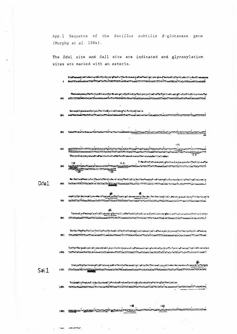
App. 1 Sequence of Che Bacillus subtilis /3-glucanase gene (Murphy et al. 1984).
Ddel
Sail
The D d e l site and S a l 1 site are indicated and glycosylation sites are marked wich an asterix,
i f l C l m f a r L t u U « T y r f ty r A r |F to V a 1 T V l i iL * iL / t f f a a P h * A la C ia A r |U u P f c « A « a C 1 jrT k r l i «Mi eC l u J * r C l * A « ^ A 4 f
I GAAnCAACCAACAATCGCTTCACTATTArCGCTTCCTCACCCACT?AAA£T7TTTC6CSCACCCTCTATTTAACCCCACTCA£ATCCAAA£CCAAC4CC
M w L < w L » » A iF tV ¥ e lL y « C l t iL y e T y i t i * A r § * la T y r C lu C y e T k r L y e L y s i l e C l e f V T y T I le C 1 WUrgC 1 w T y rC lu M i i L f • L # n T h r
101 1 ! TTTOCTBCATACJhCTCAAifiAJ L AACTATCATCCCCCCTATCAATCCACCAACAAAAfCCAAACCTACATTCACCCOCACTATCACCAf AACCTCAC
f t t r A*pC l a L a u L a t f j r L a u r b r I I • l i « IL «C lo A rg V tV ? • IL y a C \« A 1 a
SOI A iC T Q C C ifiC T SC T C T A T ffA A C C A ffC A CAIACAAACCCTACTfAAACAACCATAATCACACCCCTCACATTT'CTCTTTCC TTCTCTTCAC n T T T C T
SOI IACAITCACAT ATCAAAATCCTAGGATTCTT a c t c a t a a a c c a c c c a a a a c c t a a a t t c c a a t c a c t c c c c a t c ATCTCTCTCTC TCCTCATOCT AAlTT
Ob_______ I________ t • I I I ' • • •
« 0 1 A C C T T m A l 1 1 r T n C A fiAC C C A A jG A T C A T C A T A C T T A C A flC A T T C A A C T T A G T A A G A T T C C A T A T T A T C A T T A T T T T C A C C C A T C T T C C C T T T T C A A A
Tr X JU U A A T AAAAAAAC T C r a X T T C T A C T A C r A T C A A T C T tC T A A C T T C A A T C A T T C T A A C C T A T A A T A C T A A T A A A A C T G C C T A C A A C C G A A A A C T T r
T V l y i I l » i . y i L 7 » L * \ i ^ a / W I l * U « l l # T \ r Y * L i r o A * » L * u A » n T Y T '_ # u A j » S « r I l i I \ * M 4 1 ~ T T ~
| D f - * « c f r o T y T L « u I . 7 » A r i V « l L « u L « u l < u L * u V t i r h r C l y L « \ i / h «
1 0 1 C A A tC A T C t A i C A T C A A C A I X : t > t > C r.C T T T C A A I C A A A C C C C A A T C C C A A T A T C C C T T A T C T r jU > A C C A C T C T T C C T & C I I L I 1 L I C A C T G C A T T G T T
c m c t A C A n c t A c n g t A i c i m c c c A A A c r r “m3“ “TT"
» • t S « r U r f W A U f » l ,n « A U r h r A L » J * t A l t l 7 « ' n i r C l j C l j S « t f h « P k « A j p f r o f h » A i » C l y T y c A * n i « r C l j P h « T c p C l r i L j » A l »» % I I • I I f I •
* 0 1 T A tC A l , 1 I I t l 1 N iC A C T C A C T C d A C T C C C ^ ^ ^ C T C A A A C A C C T G liA T C C T T n T C G A C C C T C T T A A C G C C T A T A A C T C C C C T n T T G C C A A A A A C c A
* *A i » C l f T T T j « A « « 6 l y A i « M « l M 1« A » « C y « r b r T r * A r | A l » A * o A i n V t l S « r A « c T h r S « r U > l C l y C l . i M * c A r » L » u A l * L » u T h r S « r l . » u A I *
* I * . 1 I ■ ■ | | |» 1 G A T G C T tA T T C G A A T G C A A A T A T C T T C A A C T G C A C C T G C C C G C C T A A T A A C C T A T C A A T C A C C T C A T T C G C T G A A A T C C C rT T T A G C G C T A A C A A C C C C A C
*T y r A j o L y i f W A j p C j a C l y C l u A a n A j | 5 « r V » l G I n T h r T y r C l y T y r C l y L a u T y r C l u V * l A r t l y i P r o A 1 a L y t A i n T h r C 1 y I I t V a 1
I ____ I " * 1 ■ 1 I I I | I• 0 1 C T T A T A ACAAfiT T T C A C T C f C G C C A A A A C C C T T C T C T T C A A A C A T A T C C C T A T G C A C T T T A T C A A C T C A C A A T G A A A C C A G C T A A A A A CA C W X C A T C C T
* t r i € rF W b « T h iT y rT l i rC ly f rQ r t rA « p C ly T tu P c o r rp A « i )C lu U * A ip n « C l liih»L«uClyLyiA«pThrThrLy«v«lCtivfh«A»«
» 1 TTCAIQCnCTtCACTTACACA£CTCCAACACATCGAACTCCTTGGCATCACATTCATATCCAATTTTTA£CJUUkACACACAACCAAGC?TCAATTTAA£
T y r T j r r T f c r A j o C l y A l * C L y A j o k l i « C l u L y I I 1 « V | l A i p L a u C l y P h « A t p A I aA t * A s n A l a T y r H i i f h r T y r A I ¿ i h « A * p T r j > C 1 n P r o A m & r r
1 0 0 1 T A H A T A C A A A T G C T C C A £ £ A A A C C A T C A C A A C A T T C T T G A T C T C C C C T T T L A T C C A C C C A A T G C C T A T C A T A C L T A T C C A rT C C A T T C C C A C C C A A A C T
TrpAinCyTI l t L y T T y T y f V t L A a y C l y C l p L a u L y l M i » T D f A l i T ^ x A j a C t n l 1 t P r o T h r H i r L o a C l y L y I 1 « H a t H « t A a n L a u T r p A i n C l y T h f
1 1 0 1 CTATCAAATa^ATg^^CC<XAAnAAAACAIACT<XAACAAACCAAATTCCCACAACACmCAAACATCATCATCAACTTCTCCAATCCCACCa;
VaLAifCluTTrlMCl jSarTyrAj^lrVaLAanPraLmTyrAltNiaTycAapTrpValAriTrrThrlylLyil » l TCICCAWAATCeCTTCeCICCrACAATeCTCTAAATCCeCTATAiCCTCATTATGACTOCCTCCCCrATACAAAAAAATAATCCCAAATCTGAAACAAC
- B - l f l, I I t • • « »_ ' L
1 )0 1 C T C a ^ Y~»‘ TAy C A C C C TC rtA T C A l^ CTyT C A C .ILACTTCTTCCCATCAAICTTCTATTCACTCATAATA IC irtA T T T e A T C rtC T C C C a a C T A A
I I A I

App,2. Origin of p69A, p69B, p69C, p69D and p69E. p69A contains the entire a-factor coding sequence, including the signal peptide and structural genes cloned into the BamHl site of YEpl3 (Kurjan and Herskowitz, 1983). The structuralgenes were removed using the ffindlll restriction site that immediatly follows the signal sequence, to give p69B. p69C, D and E were made by inserting 8', 10', and 12'meric BamHI linkers into the tfindlll site of d 69B (T. Rvan D.C.U.).
H
? ^ ffl]promoter+siqna[
■ ■ s tru c tu ra l gene
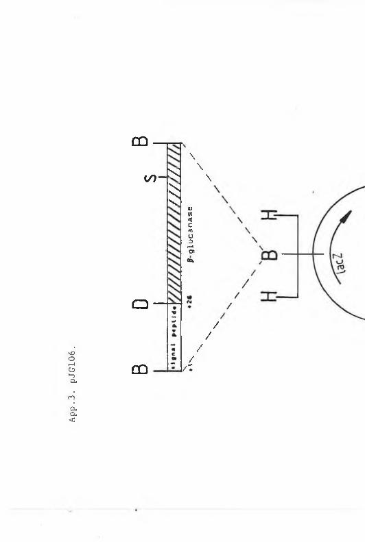
App.
3. pJG106.
OQ
a
CÛ
</> n :
&
\\\
OJV)*0cn;U3•—iC*cet
//
//
/ I
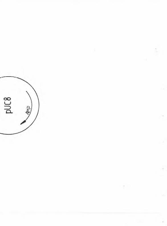
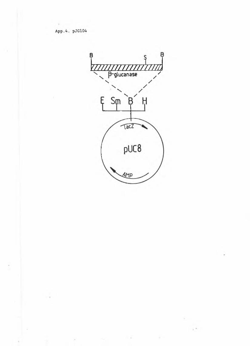
\ tT ///I / / / / / / / / 7 ///7¡