Embed Size (px)
Citation preview

Analysis of Scoliosis From Spinal X-Ray ImagesAbdullah-Al-Zubaer Imran1,2, Chao Huang2, Hui Tang2, Wei Fan2, Kenneth M.C. Cheung3, Michael To3,
Zhen Qian2, and Demetri Terzopoulos1,4
1University of California, Los Angeles, California, USA2Tencent Hippocrates Research Lab, Palo Alto, California, USA
3The University of Hong Kong, Hong Kong, China4VoxelCloud, Inc., Los Angeles, California, USA
Abstract—Scoliosis is a congenital disease in which the spineis deformed from its normal shape. Measurement of scoliosisrequires labeling and identification of vertebrae in the spine.Spine radiographs are the most cost-effective and accessiblemodality for imaging the spine. Reliable and accurate vertebraesegmentation in spine radiographs is crucial in image-guidedspinal assessment, disease diagnosis, and treatment planning.Conventional assessments rely on tedious and time-consumingmanual measurement, which is subject to inter-observer vari-ability. A fully automatic method that can accurately identifyand segment the associated vertebrae is unavailable in theliterature. Leveraging a carefully-adjusted U-Net model withprogressive side outputs, we propose an end-to-end segmentationmodel that provides a fully automatic and reliable segmentationof the vertebrae associated with scoliosis measurement. Ourexperimental results from a set of anterior-posterior spine X-Rayimages indicate that our model, which achieves an average Dicescore of 0.993, promises to be an effective tool in the identificationand labeling of spinal vertebrae, eventually helping doctors inthe reliable estimation of scoliosis. Moreover, estimation of Cobbangles from the segmented vertebrae further demonstrates theeffectiveness of our model.
Index Terms—scoliosis, spine X-Ray, Cobb angle, vertebraesegmentation, progressive U-Net.
I. INTRODUCTION
Scoliosis is an abnormal condition defined by spinal cur-vature towards the left or right. Early detection is key and,when accurate, it can lead to better treatment planning [1].Radiography (X-Ray) is the preferred imaging technique forclinical analysis and measurement of scoliosis as it is highlyavailable, inexpensive, and yields quick results. Conventionalspine image analysis tasks involve tedious manual labor withhand-crafted feature extraction for the measurement of scoliosis.Cobb angle, the standard quantification of scoliosis is estimatedby calculating the angle between the two tangents of the upperand lower end plates of the upper and lower vertebrae. Aperson with a 10◦ or greater Cobb angle is usually consideredfor scoliosis diagnosis [2]. Fig. 1 illustrates the calculation ofthe Cobb angle and the labeling of relevant vertebrae in anX-Ray image.
Conventionally, measurement and assessment, which requiresthe identification and labeling of specific vertebral structures,is manually performed by clinicians. However, the manualmeasurement of scoliosis faces several difficulties. First, largeanatomical variation between patients and low tissue contrast
in spinal X-Ray images make it challenging to accuratelyand reliably assess the severity of scoliosis [3], and effectson the spine and body as a whole, as well as on individualvertebra, pose extra difficulty in the quantification of scoliosis[4]. Second, measurement error is prevalent in the routineclinical assessment of scoliosis due to instrumentation, vertebralrotation, and patient positioning [2], and 5◦–10◦ intra- orgreater inter-observer variation has commonly been reportedin measuring the Cobb angle [5], [6].
Therefore, an automatic technique for the accurate measure-ment of scoliosis is desirable. Our specific contributions in thispaper are the following:
1) A fully automatic and efficient pipeline for the measure-ment and analysis of scoliosis.
2) A novel segmentation network for accurately segmentingvertebrae from spine X-Ray images.
3) Fully automatic and accurate identification and labelingof individual vertebrae merely based on binary segmen-tation.
4) Accurate diagnostic classification of the severity ofscoliosis, which is crucial for treatment planning.
II. RELATED WORK
While several methods for vertebrae segmentation andscoliosis measurement are available, this approach is stillunder-explored in the literature. Existing vertebrae segmentationmethods rely on manual interaction [7], hand-crafted featureengineering limited to customized parameters [8], [9], followpatch-based approaches that lose full spatial context [10], [11],are limited in scope and fail to consider all the requiredvertebrae at a time [12], etc. For Cobb angle estimation, aminimum bounding rectangle was used for the patch-wisesegmented vertebrae [11], an approach that relies on pre-processing steps including spinal region isolation and vertebraedetection. Kusuma et al. [13] proposed a K-means and curve-fitting approach for Cobb angle measurement that requires aset of pre-processing steps [13]. Other Cobb angle estimationmethods have been proposed based on directly finding vertebraecorners as a form of regression task [3], [14]–[16]. Althoughpromising, these supervised methods are less viable for clinicalapplications because of low accuracy, due to the loss of finedetails in the process, and the lack of explainability.
arX
iv:2
004.
0688
7v1
[ee
ss.I
V]
15
Apr
202
0
Normal
Mild
Moderate
Severe
Input X-Ray Vertebrae Segmentation Vertebrae Labelling &Scoliosis Measurement Scoliosis Classification
T11
L4
Scoliosis Analysis Framework
Fig. 1. Overview of our framework for calculating the Cobb angle in a spine X-Ray through segmentation, labeling, and identification of the relevant vertebrae.After determining the most tilted vertebrae above and below the apex, tangents are drawn by extending the upper edge of the upper vertebra and lower edge ofthe lower vertebra. From these tangents, the Cobb angles are calculated and the scoliosis can be classified.
As a departure from prior segmentation-based methods, ourmodel is fully automatic, involving no manual interventionend-to-end, and eschews any kind of pre-processing or post-processing steps.
III. METHODS
A. Vertebrae Segmentation and Labeling
We perform binary segmentation of the spine with a well-distinguishable number n of vertebrae relevant to scoliosisanalysis. To formulate the problem, we assume an unknowndata distribution p(X,Y ) over images X and vertebrae seg-mentation labels Y . The model has access to the labeledtraining set D(x,y) sampled i.i.d. from p(X,Y ). As illustratedin Algorithm 1, the segmentation prediction network Fφ istrained with a set of learnable parameters φ. We specify theobjective as minφF L(y,y), where y is the reference vertebraemask and y is the model prediction in each of the trainingiterations.
Following the progressive dense V-net model [17], [18], wepropose a progressive U-Net with some careful adjustments inthe U-Net [19]. As shown in Fig. 2, our model has an encoderand a decoder with skip connections. In each encoder layer,two 3× 3 convolutions are followed by instance normalization,ReLU activation, and a 2×2 max-pooling. A dropout is appliedin every encoder and decoder stage of the network. We generateside-outputs in every stage of the decoder. Progressively addingone side-output to the next, the segmentation performance isimproved compared to collecting the final output from the finaldecoder stage in a U-Net. However, one key difference with[17] is that our model is trained without side-supervision. Onlythe side-outputs are generated and added progressively, yieldingan improved segmentation at the final output. A convolution
Algorithm 1: Training for vertebrae segmentation fromspine X-Ray images.Input: X-Ray images and reference vertebra masks.Output: Predicted vertebra masks.Require:
Training data x, y ∈ D including spine X-Ray images xand reference vertebra segmentation masks yModel architecture Fφ with learnable parameters φ
for each step over D doSample minibatch M : x(i) ∼ pD(x)
Compute model outputs for the minibatch:y(i) ← F(φ)(x)Calculate loss L(y,y) for the model predictions
Update the model F along its gradient
∇φF
1
|M|∑i∈M
[LF
(y(i),y(i))
]end for
operation is performed to generate the side-output from eachdecoder stage. The progressive side-outputs also ensure thatmicro-structure is not lost from any level of the decoder throughthe convolutional operations. We generate side outputs at x/8,x/4, and x/2 resolutions before the final output at x resolution.Therefore, the side output at resolution x/8 is added to thenext decoder stage, and so on.
B. Measurement of Scoliosis
Our pipeline makes use of the vertebrae segmentation inestimating Cobb angles. Algorithm 2 automatically calculates
Side outputs
Input X-Ray Vertebrae mask
Fig. 2. Architecture of our segmentation network (Progressive U-Net): Side outputs at three different stages of the decoder are generated and progressivelyadded to the next stage side-output. The output from the third side-output is added to the last stage before the final convolution to generate the final segmentationoutput.
Algorithm 2: Cobb angle calculationInput: Vertebra mask y.Output: Cobb angle θ.
From the predicted mask y, get all the contoursfor each contour in contours do
if Number of pixels < a then//to remove any noisy patchesRemove contour
end ifend forThis will give n contours of well-separated vertebrae
Extract four corner points for the contourOrder the corners from bottom to top by comparing thecoordinates of the extracted 4n corner points
Find the two vertebrae (upper and lower) with at least 2vertebrae gap between them
Calculate the Cobb angle, θ =∣∣∣tan−1 ( mu−ml
1+muml
)∣∣∣, wheremu and ml are the upper and lower vertebrae slopes.
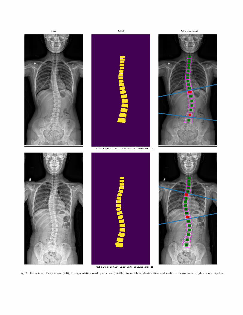
the Cobb angle by analyzing the contours from the segmentedmask. When well-separated from others, each of the contoursrepresents a vertebra relevant to the measurement of scoliosis.To verify if a contour is actually associated to a relevantvertebra, we impose a minimum size on the number of contourpixels (a). After the extraction and ordering of 4n corners, themost tilted upper vertebra and the most tilted lower vertebraare determined from the n relevant vertebrae (Fig. 3). Thenthe Cobb angle is calculated from the slopes of the upper edgeof the upper vertebra and the lower edge of the lower vertebra.
Moreover, the severity of scoliosis can be categorized andappropriate treatment planning is performed depending on thecalculated Cobb angle from the spine X-Ray of a patient. Inour pipeline, we therefore perform an automatic diagnostic
TABLE ICLINICALLY ACCEPTED CLASSIFICATION AND TREATMENT PLANNING FOR
ADOLESCENT SCOLIOSIS BASED ON MEASURED COBB ANGLES
Cobb Angle θ (◦) Severity Treatment Recommendation
θ < 10◦ normal —10◦ < θ < 25◦ mild Check in every 2 years25◦ < θ < 45◦ moderate Wear a brace for 16–23 hours/day45◦ < θ severe Revision surgery in 20–30 years
classification following the clinically recognized scoliosisseverity classes, as shown in Table I. Active treatment istypically not needed when it is mild and rigid braces canstop the progression of scoliosis when it is in moderate stage.Surgery is the last resort for severe cases, but it can be delayedfor the adolescent period [20].
IV. EXPERIMENTAL EVALUATION
A. Implementation Details
Data: We use a dataset of 100 high-resolution spine X-Ray images of children with evidence of scoliosis to variousextents. The dataset contains manual annotation by experts of18 relevant vertebrae (cervical C7, thoracic T1–T12, lumbarL1–L5). We split the dataset into training (80), testing (15),and validation (5) sets. Baselines: As baselines, we use aregular U-Net model with a choice of binary cross-entropy(XE) and Dice as loss function. For simplicity, we denote themodels as UD (UNet with Dice loss), UX (UNet with XE loss),PUD (Progressive UNet with Dice loss), and PUX (ProgressiveUNet with XE loss). Training: The models are trained onthe training set while their performances were evaluated onthe testing set. The validation set is used for hyper-parametertuning and model selection. Inputs: All the images are resizedand normalized to 1024 × 512 × 1 before feeding them tothe network. Hyperparameters: We use the Adam optimizerwith adaptive learning rate starting with an initial rate of 0.01
Raw Mask Measurement
Fig. 3. From input X-ray image (left), to segmentation mask prediction (middle), to vertebrae identification and scoliosis measurement (right) in our pipeline.
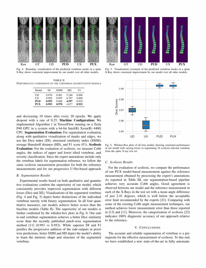
Raw GT UD PUD UX PUXFig. 4. Boundary visualization of the predicted vertebrae masks in a spineX-Ray shows consistent improvement by our model over all other models.
TABLE IIPERFORMANCE COMPARISON OF THE VERTEBRAE SEGMENTATION MODELS
Model DI SSIM HD F1
UD 0.970 0.961 5.246 0.896UX 0.956 0.955 6.767 0.868PUD 0.993 0.966 4.597 0.919PUX 0.993 0.970 4.677 0.922
and decreasing 10 times after every 20 epochs. We applydropout with a rate of 0.25. Machine Configuration: Weimplemented Algorithm 1 in TensorFlow running on a TeslaP40 GPU in a system with a 64-bit Intel(R) Xeon(R) 440GCPU. Segmentation Evaluation: For segmentation evaluation,along with qualitative visualization of masks and edges, weuse the Dice index (DI), structural similarity index (SSIM),average Hausdorff distance (HD), and F1 score (F1). ScoliosisEvaluation: For the evaluation of scoliosis, we measure Cobbangles, the indices of upper and lower tilted vertebrae, andseverity classification. Since the expert annotations include onlythe vertebrae labels for segmentation reference, we follow thesame scoliosis measurement procedure for both the referencemeasurements and for our progressive U-Net-based approach.
B. Segmentation Results
Experimental results based on both qualitative and quantita-tive evaluations confirm the superiority of our model, whichconsistently provides improved segmentation with differentlosses (Dice and XE). Visualizations of the segmented vertebrae(Fig. 4 and Fig. 5) depict better distinctions of the individualvertebrae merely with binary segmentation. In all four quan-titative measures, our models achieve better scores than thebaseline models (Table II). The superiority of our models isfurther confirmed by the whisker-box plots in Fig. 6. Our end-to-end vertebrae segmentation achieves a better Dice similarityscore than the recently published patch-wise segmentationmethod [11] (0.993 vs 0.952). While superior DI and F1justifies the progressive addition of the side-outputs in pixel-wise predictions, better SSIM and HD depict the model’s abilityto learn the intrinsic shape and structure of the segmentedvertebrae.
Raw GT UD PUD UX PUXFig. 5. Visualization (zoomed) of the predicted vertebrae masks in a spineX-Ray shows consistent improvement by our model over all other models.
UD UX PUD PUX PUDA PUXA
0.95
0.96
0.97
0.98
0.99
Dic
e Sc
ores
Fig. 6. Whisker-Box plots of all four models showing consistent performanceof our model with varying losses in segmenting 18 scoliosis-relevant vertebraefrom the spine X-ray test set.
C. Scoliosis Results
For the evaluation of scoliosis, we compare the performanceof our PUX model-based measurement against the referencemeasurement obtained by processing the expert’s annotations.As reported in Table III, our segmentation-based pipelineachieves very accurate Cobb angles. Good agreement isobserved between our model and the reference measurement ineach of the X-Rays in the test set with a mean angle differenceof just 2.41 degrees, which is well below the acceptableerror limit recommended by the experts [21]. Comparing withsome of the existing Cobb angle measurement techniques, ourmethod achieves lower measurement error than those reportedin [13] and [11]. Moreover, the categorization of scoliosis [22]indicates 100% diagnostic accuracy of our approach relativeto the reference.
V. CONCLUSIONS
The accurate and reliable segmentation of vertebrae is a pre-requisite for the effective measurement of scoliosis. To this end,we have established a new state-of-the-art in fully automatic
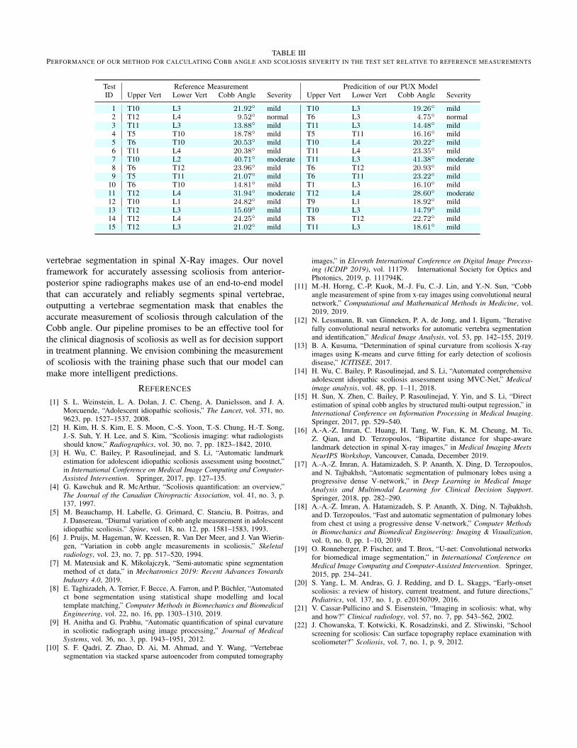
TABLE IIIPERFORMANCE OF OUR METHOD FOR CALCULATING COBB ANGLE AND SCOLIOSIS SEVERITY IN THE TEST SET RELATIVE TO REFERENCE MEASUREMENTS
Test Reference Measurement Predicition of our PUX ModelID Upper Vert Lower Vert Cobb Angle Severity Upper Vert Lower Vert Cobb Angle Severity
1 T10 L3 21.92◦ mild T10 L3 19.26◦ mild2 T12 L4 9.52◦ normal T6 L3 4.75◦ normal3 T11 L3 13.88◦ mild T11 L3 14.48◦ mild4 T5 T10 18.78◦ mild T5 T11 16.16◦ mild5 T6 T10 20.53◦ mild T10 L4 20.22◦ mild6 T11 L4 20.38◦ mild T11 L4 23.35◦ mild7 T10 L2 40.71◦ moderate T11 L3 41.38◦ moderate8 T6 T12 23.96◦ mild T6 T12 20.93◦ mild9 T5 T11 21.07◦ mild T6 T11 23.22◦ mild
10 T6 T10 14.81◦ mild T1 L3 16.10◦ mild11 T12 L4 31.94◦ moderate T12 L4 28.60◦ moderate12 T10 L1 24.82◦ mild T9 L1 18.92◦ mild13 T12 L3 15.69◦ mild T10 L3 14.79◦ mild14 T12 L4 24.25◦ mild T8 T12 22.72◦ mild15 T12 L3 21.02◦ mild T11 L3 18.61◦ mild
vertebrae segmentation in spinal X-Ray images. Our novelframework for accurately assessing scoliosis from anterior-posterior spine radiographs makes use of an end-to-end modelthat can accurately and reliably segments spinal vertebrae,outputting a vertebrae segmentation mask that enables theaccurate measurement of scoliosis through calculation of theCobb angle. Our pipeline promises to be an effective tool forthe clinical diagnosis of scoliosis as well as for decision supportin treatment planning. We envision combining the measurementof scoliosis with the training phase such that our model canmake more intelligent predictions.
REFERENCES
[1] S. L. Weinstein, L. A. Dolan, J. C. Cheng, A. Danielsson, and J. A.Morcuende, “Adolescent idiopathic scoliosis,” The Lancet, vol. 371, no.9623, pp. 1527–1537, 2008.
[2] H. Kim, H. S. Kim, E. S. Moon, C.-S. Yoon, T.-S. Chung, H.-T. Song,J.-S. Suh, Y. H. Lee, and S. Kim, “Scoliosis imaging: what radiologistsshould know,” Radiographics, vol. 30, no. 7, pp. 1823–1842, 2010.
[3] H. Wu, C. Bailey, P. Rasoulinejad, and S. Li, “Automatic landmarkestimation for adolescent idiopathic scoliosis assessment using boostnet,”in International Conference on Medical Image Computing and Computer-Assisted Intervention. Springer, 2017, pp. 127–135.
[4] G. Kawchuk and R. McArthur, “Scoliosis quantification: an overview,”The Journal of the Canadian Chiropractic Association, vol. 41, no. 3, p.137, 1997.
[5] M. Beauchamp, H. Labelle, G. Grimard, C. Stanciu, B. Poitras, andJ. Dansereau, “Diurnal variation of cobb angle measurement in adolescentidiopathic scoliosis.” Spine, vol. 18, no. 12, pp. 1581–1583, 1993.
[6] J. Pruijs, M. Hageman, W. Keessen, R. Van Der Meer, and J. Van Wierin-gen, “Variation in cobb angle measurements in scoliosis,” Skeletalradiology, vol. 23, no. 7, pp. 517–520, 1994.
[7] M. Mateusiak and K. Mikolajczyk, “Semi-automatic spine segmentationmethod of ct data,” in Mechatronics 2019: Recent Advances TowardsIndustry 4.0, 2019.
[8] E. Taghizadeh, A. Terrier, F. Becce, A. Farron, and P. Buchler, “Automatedct bone segmentation using statistical shape modelling and localtemplate matching,” Computer Methods in Biomechanics and BiomedicalEngineering, vol. 22, no. 16, pp. 1303–1310, 2019.
[9] H. Anitha and G. Prabhu, “Automatic quantification of spinal curvaturein scoliotic radiograph using image processing,” Journal of MedicalSystems, vol. 36, no. 3, pp. 1943–1951, 2012.
[10] S. F. Qadri, Z. Zhao, D. Ai, M. Ahmad, and Y. Wang, “Vertebraesegmentation via stacked sparse autoencoder from computed tomography
images,” in Eleventh International Conference on Digital Image Process-ing (ICDIP 2019), vol. 11179. International Society for Optics andPhotonics, 2019, p. 111794K.
[11] M.-H. Horng, C.-P. Kuok, M.-J. Fu, C.-J. Lin, and Y.-N. Sun, “Cobbangle measurement of spine from x-ray images using convolutional neuralnetwork,” Computational and Mathematical Methods in Medicine, vol.2019, 2019.
[12] N. Lessmann, B. van Ginneken, P. A. de Jong, and I. Isgum, “Iterativefully convolutional neural networks for automatic vertebra segmentationand identification,” Medical Image Analysis, vol. 53, pp. 142–155, 2019.
[13] B. A. Kusuma, “Determination of spinal curvature from scoliosis X-rayimages using K-means and curve fitting for early detection of scoliosisdisease,” ICITISEE, 2017.
[14] H. Wu, C. Bailey, P. Rasoulinejad, and S. Li, “Automated comprehensiveadolescent idiopathic scoliosis assessment using MVC-Net,” Medicalimage analysis, vol. 48, pp. 1–11, 2018.
[15] H. Sun, X. Zhen, C. Bailey, P. Rasoulinejad, Y. Yin, and S. Li, “Directestimation of spinal cobb angles by structured multi-output regression,” inInternational Conference on Information Processing in Medical Imaging.Springer, 2017, pp. 529–540.
[16] A.-A.-Z. Imran, C. Huang, H. Tang, W. Fan, K. M. Cheung, M. To,Z. Qian, and D. Terzopoulos, “Bipartite distance for shape-awarelandmark detection in spinal X-ray images,” in Medical Imaging MeetsNeurIPS Workshop, Vancouver, Canada, December 2019.
[17] A.-A.-Z. Imran, A. Hatamizadeh, S. P. Ananth, X. Ding, D. Terzopoulos,and N. Tajbakhsh, “Automatic segmentation of pulmonary lobes using aprogressive dense V-network,” in Deep Learning in Medical ImageAnalysis and Multimodal Learning for Clinical Decision Support.Springer, 2018, pp. 282–290.
[18] A.-A.-Z. Imran, A. Hatamizadeh, S. P. Ananth, X. Ding, N. Tajbakhsh,and D. Terzopoulos, “Fast and automatic segmentation of pulmonary lobesfrom chest ct using a progressive dense V-network,” Computer Methodsin Biomechanics and Biomedical Engineering: Imaging & Visualization,vol. 0, no. 0, pp. 1–10, 2019.
[19] O. Ronneberger, P. Fischer, and T. Brox, “U-net: Convolutional networksfor biomedical image segmentation,” in International Conference onMedical Image Computing and Computer-Assisted Intervention. Springer,2015, pp. 234–241.
[20] S. Yang, L. M. Andras, G. J. Redding, and D. L. Skaggs, “Early-onsetscoliosis: a review of history, current treatment, and future directions,”Pediatrics, vol. 137, no. 1, p. e20150709, 2016.
[21] V. Cassar-Pullicino and S. Eisenstein, “Imaging in scoliosis: what, whyand how?” Clinical radiology, vol. 57, no. 7, pp. 543–562, 2002.
[22] J. Chowanska, T. Kotwicki, K. Rosadzinski, and Z. Sliwinski, “Schoolscreening for scoliosis: Can surface topography replace examination withscoliometer?” Scoliosis, vol. 7, no. 1, p. 9, 2012.
![Ultrasound Imaging of Spinal Vertebrae to Study Scoliosis · Scoliosis is a complex three-dimensional (3D) deformity of the spine associated with vertebral rotation [1]. The curvature](https://img.pdfslide.net/doc/110x75/607c4b7df769fe79aa05317e/ultrasound-imaging-of-spinal-vertebrae-to-study-scoliosis-scoliosis-is-a-complex.jpg)


![Chiropractic treatment of idiopathic scoliosis with the ......Idiopathic scoliosis (IS) is the most common spinal deformity seen in school-age children [1]. According to the National](https://img.pdfslide.net/doc/110x75/6021a0e84b312545bc186f20/chiropractic-treatment-of-idiopathic-scoliosis-with-the-idiopathic-scoliosis.jpg)