Embed Size (px)
Citation preview

Analyzing the Clinical outcomes, and timely interventions for Abdominal compartment syndrome among high risk individuals in a tertiary care hospital in India.
Dissertation submitted to
THE TAMILNADU DR.M.G.R. MEDICAL UNIVERSITY
In partial fulfillment of the degree of
M.S. GENERAL SURGERY
Branch-‐ I
PSG INSTITUTE OF MEDICAL SCIENCES AND RESEARCH
DEPARTMENT OF GENERAL SURGERY

APRIL 2013
CERTIFICATE
This is to certify that Dr.K.Rajavel, postgraduate student (2010-2013) in
the department of General Surgery, PSG Institute of medical sciences and
research, Coimbatore, has done this dissertation titled “Analyzing the
Clinical outcomes, and timely interventions for Abdominal compartment
syndrome among high risk individuals in a tertiary care hospital in India”
under the direct guidance and supervision of guide Dr.S.Premkumar in
partial fulfillment of the regulations laid down by The Tamilnadu Dr.M.G.R.
Medical University, Chennai, for M.S., Branch – I General Surgery degree
examination.
Dr.S.Premkumar, 1st unit Chief, Professor & Head,Dept of Surgery, PSG Institute of Medical Sciences & Research, Coimbatore - 641004
Dr.S.Ramalingam, Principal, PSG Institute of Medical Sciences & Research, Coimbatore - 641004

DECLARATION
I, K.Rajavel, solemnly declare that this dissertation “Analyzing the Clinical
outcomes, and timely interventions for Abdominal compartment syndrome
among high risk individuals in a tertiary care hospital in India” is a
bonafide record of work done by me in the Department of General Surgery,
PSG institute of Medical Sciences & Research, Coimbatore, under the
guidance of Dr.S.Premkumar, Professor and Head of Surgery.
This dissertation is submitted to The Tamilnadu Dr.M.G.R. Medical
University, Chennai, in partial fulfillment of the University regulations for
the award of MS Degree (General Surgery) Branch-I, Examination to be
held in April 2013.
Place: Coimbatore
Date: 26thDecember, 2012 (Dr.K.Rajavel)

ACKNOWLEDGEMENT
I wish to thank our Principal for having permitted me to conduct this study
in this hospital.
I am ever so grateful to the HOD and my Professor Dr.S.Premkumar,
Associate Professors Dr Rajesh Kumar and Dr Krishnakumar and the entire
faculty of the Department of Surgery for the kind guidance, valuable advice,
supervision and encouragement while doing this study.
My thanks are also to my colleagues, Interns and Staff Nurses for the
considerable help extended to me.
I wish to place on record my gratitude to all the patients who have been an
integral part of this study. I am indebted to them for their willing
participation.
Most importantly i would like to thank my wife and family and friends for
their support for completion of this study.

CONTENTS
Page Number
1 INTRODUCTION
: 6
2 AIM AND OBJECTIVES
: 10
3 REVIEW OF LITERATURE
: 12
4 MATERIALS AND METHODS
: 29
5 RESULTS
: 34
6 DISCUSSION
: 58
7 CONCLUSION
: 75

8 References
: 80
Annexure-1: PROFORMA
Annexure -2 : Master Chart

1
1.Introduction
2
INTRODUCTION
Abdominal Compartment Syndrome has very much importance
in surgical practice and in critical care because of its effects on
multiple organ systems and as the patients of this syndrome are
critically ill.
The most common high risk individuals for ACS are post
laprotomy patients.
Laprotomy may be associated with raised intra abdominal
pressure which is defined as intra-abdominal pressure higher
than 12 mm of Hg and one of it’s most dreaded complications
is the Abdominal Compartment Syndrome.
Abdominal Compartment Syndrome (ACS) is defined as the
sudden increase in the Intra-Abdominal pressure (IAP).Base
line is usually ‘0’ resulting in changes in respiration,
haemodynamic stability, renal perfusion and cerebral
perfusion(2).
After laparotomy intra-abdominal pressure increases up to 10
mm of Hg. The physiological changes are observed when IAP
rises above 15 mm of Hg which is also termed as Intra
Abdominal Hypertension (IAH)(3).
Raised intra-abdominal pressure leading to abdominal
compartment syndrome is a highly under-recognised source of
morbidity and mortality associated with laparotomy. Hence

3
intensive monitoring of intra-abdominal pressure as well as
early and aggressive management of abdominal compartment
syndrome is essential to ensure a successful outcome in a
patient undergoing laparotomy.
More recently, laparoscopy has brought-out the consequences
of raised IAP.
This study is being undertaken to evaluate the impact of IAP on
outcome in High risk individuals.

4
DEFINITION
Abdominal Compartment Syndrome is defined as "adverse
physiological consequences that occur as a result of sudden
increase in Intra Abdominal pressure and resolve following
abdominal decompression".
INTRA ABDOMINAL HYPERTENSION
is defined as raised Intra Abdominal pressure above normal.
Normal Intra Abdominal Pressure (IAP) is 0-5 mm Hg. Intra
abdominal pressure varies with position, body habitat and
activity. Intra abdominal Pressure is measured in cm of water
or mm of Hg.
(1 cm of H2O (water) - 0.735 mm of Hg).
Intra abdominal pressure of between 3 to 10 mm Hg is
commonly observed post operatively without adverse effects.

5
2. Aims and Objectives

6
AIMS AND OBJECTIVES
A Prospective analysis to identify the
1) Incidence of
a. Intra Abdominal Hypertension(IAH)
b. Abdominal Compartment Syndrome(ACS)
2) Etiology
3) Effects on morbidity to the patient
4) Timely medical and surgical interventions made among high
risk patients in a tertiary care hospital in Coimbatore, India.

7
3. Review of Literature

8
REVIEW OF LITERATURE
The relationship between laparotomy and rise in intra abdominal
pressure and it’s effects on various organ systems have received
ample attention since the 19th century.
The raised intra-abdominal pressure and its consequences over the
various organ systems has been noted since 1863 when Marey and
Bureau described the relationship between Intra-abdominal
hypertension and respiratory function(4).
In 1870 Paul Bert published a volume on “Physiologie comparée de
la respiration” showed experiments in anesthetized animals,
measuring intra-thoracic and intra-abdominal pressures through
catheters inserted in the trachea and rectum respectively and
described elevation of IAP on inspiration and the descent of the
diaphragm(5).
In 1911 Emerson demonstrated the effects of IAP over morbidity of
cardiovascular system5
Thorington and Schmidt reviewed on urinary output changed with
BP changes (7).
Bellis and Wangensteen demonstrated changes in venous flow in the
abdomen and extremities associated with abdominal distention(8).
Ogilvie, demonstrated the need of laprotomy which was performed
for a patient with burst abdomen, packed with cotton cloth and
sutured over wound edges, and once wound granulated was allowed
for wound to contract 9.

9
Gross showed the benefit of so-called “staged abdominal repair” in
omphalocele, hence stressing the importance of avoiding tension
Early experience with laparoscopy led to recognition of the adverse
effects of pneumoperitoneum associated increase in IAP: Ivankovich
et al described cardiovascular collapse during gynecological
laparoscopy and studied the physiology of the phenomenon(19,20).
Lenz et al, studying cardiovascular changes during laparoscopy,
pointed out the dangers of pneumoperitoneum in patients with
cardiovascular dysfunction, anemia or hypovolemia(21).
Richardson and Trinkle studied hemodynamic and Pulmonary
alterations with raised intra-abdominal pressure(22).
Kashtan et al rediscovered the hemodynamic effects of increased
IAP(23).
Harman et al as well as Richards et al demonstrated how elevated IAP
adversely affects renal function and how abdominal decompression
improves it(24,25).
Le Roith et al studied the effects of increased IAP on plasma
antidiuretic hormone levels(26).
However, the recognition of abdomen as a compartment and the
concept of intra-abdominal hypertension resulting in Abdominal
Compartment Syndrome (ACS) have only recently received attention.
Korn and associates first used the term ACS in 1980s(6).
Smith et al reported reversal of postoperative anuria by
decompressive laparotomy(27).

10
Barnes et al in 1985 studied cardiovascular responses to elevated
IAP(28).
Caldwell and Ricotta measured changes in visceral blood flow(29).
Jacques and Lee reported improvement in renal perfusion after
evacuation of retroperitoneal hematoma which was the cause for
increased IAP(30).
It is only in later 1990s that the patho-physiological consequences of
the increased intra abdominal pressure (IAP) and abdominal
compartment syndrome have been recognized in a wide spectrum of
surgical patients and treated aggressively.
Since then most surgeon’s started believing abdominal hypertension
and the abdominal compartment syndrome as two different
pathologic phenomenon which will have change in normal
physiological process.
Two “collective reviews” of ACS appeared in 1995 and 1996 -
opening the gate to numerous publications, recognizing IAHT/ACS in
a large number of surgical intra abdominal and extra-abdominal,
traumatic and non-traumatic scenarios—and providing an ever
growing list of complications and consequences(33,34).
In 2004 the World Society of the Abdominal Compartment Syndrome
(WSACS) was founded to promote research, foster education and
improve the survival of patients with intra-abdominal hypertension
and/or abdominal compartment syndrome.

11
Important landmarks in the history of IAH through the
years 1850 to 2006 are summarised as follows :
(Chart 3. 1)
The reported incidence of Intra-Abdominal Hypertension and
Abdominal Compartment Syndrome is about 32.1% and 4.2%
respectively in the mixed intensive care unit population. In case of
abdominal procedures it varies from 31.5-40.7%(36).Bruch JM et al(34)
in 1996 graded intra-abdominal pressure using urinary bladder
pressure as an indicator as follows:
Grade: Bladder pressure (mm of Hg): (1 mm of Hg = 1.36 cm of water)
Grade 1 10 – 15

12
Grade 2 16 – 25
Grade 3 26 – 35
Grade 4 >35
The diagnosis of ACS depends on a very high degree of suspicion
and recognition of patients at risk, identification of clinical syndrome
and lastly measurement of IAP.
The various clinical parameters that are considered are abdominal
distension, raised IAP above 20 mm of Hg, elevated peak airway
pressure, massive I.V fluid requirements, oliguria progressing to
anuria not responding to volume repletion, decreased cardiac output,
hypoxemia refractory to increased PEEP, hypercapnia, wide pulse
pressure and acidosis.

13
Acute abdominal hypertension can occur under the
following clinical settings(37):
• Peritonitis
•Severe abdominal traumatic injury
• Fluid overload
• Retroperitoneal hematoma
• Status post operative elective abdominal Surgeries
• Emergency abdominal surgeries
• Ishemic bowel and repercussion injury
•Acute Pancreatitis
• Intestinal Obstruction
•Mass per Abdomen
• Intra abdominal packing for bleeds (liver injury)
• Tension abdominal closure
• Ascites secondary to any cause

14
There are some well known risk factors associated with
development of IAH and ACS(38):
(Table 3.1)
Pathology Aetiology Antecedent cause Aggravating
factor
Fluid resuscitation Increased capillary leak
Massive resuscitation
• Crystalloids
• Colloids
• Transfusions of blood or blood products
Positive fluid balance Oliguria
Burns Trauma Sepsis Severe sepsis Septic shock
Acidosis (pH <
7.2) Coagulopath
y Hypothermia
Increased abdominal contents
Liver disease with ascites Pancreatitis Peritoneal dialysis Peritonitis
Obesity causing increased mesenteric fat Laparoscopy- Pneumoperitoneum
Abdominal
tumor Intraabdom
inal bleeding or
tumor Intraabdom
inal
abscess Retroperit
oneal bleeding or
tumor

15
Increased intraluminal contents
Gastric distension Gastroparesis Ileus Small bowel obstruction
Volvulus Ogilvie syndrome
Enteral
feeding Intralumi
nal tumor
Decreased abdominal wall compliance
Patient-ventilator dyssynchrony Increased work of breathing Extrinsic or intrinsic positive end-expiratory pressure Prone positioning
Burns with abdominal eschars Tight abdominal wall closures Abdominal wall bleeding
Elevated
BMI Central
obesity Pregnancy
Agitation/pain
Michel Chetham et al showed that Majchrzak in 2003 has classified
abdominal compartment syndrome as (3):
“Primary Abdominal cCompartment Syndrome is
essentially organ dysfunction and IAH in the presence of direct
injury to the abdominal contents caused by trauma, peritonitis,
ileus and haemorrhage.
Secondary Abdominal Compartment Syndrome consists of
elevated pressure and organ dysfunction caused by third space
oedema and resuscitation. The examples are resuscitation of
haemorrhagic shock patients and burns.
Recurrent Abdominal Compartment Syndrome in which
the patient has recovered from the ACS once but because of
secondary insults the cycle begins again. This variety is
associated with very high mortality rate”

16
The IAP is usually slightly elevated in the patient on mechanical
ventilator support.
IAP increases in direct relation to body mass index of the patient.
The compliance of the abdominal wall generally limits the raise in
IAP but increases rapidly after a critical IAP.
Critical IAP varies from patient to patient, based on abdominal wall
compliance.
The effects of intra-abdominal hypertension are not limited to the
intra abdominal organs, but rather have an impact either directly or
indirectly on every organ system in the body as shown in Figure 3.1.
Pathophysiologic Implications of Intra-abdominal
Hypertension(39)
(Figure 3.1)

17
ICP – intracranial pressure; CPP – cerebral perfusion pressure; ITP – intrathoracic
pressure; IVC – inferior vena cava; SMA – superior mesenteric artery; pHi – gastric
intramuscosal pH; APP – abdominal perfusion pressure; PIP- peak inspiratory pressure;
Paw – mean airway pressure; PaO2 – oxygen tension; PaCO2 – carbon dioxide tension;
Qs/Qt – intrapulmonary shunt; Vd/Vt – pulmonary dead space ; CO – cardiac output;
SVR – systemic vascular resistance; PVR – pulmonary vascular resistance; PAOP –
pulmonary artery occlusion pressure; CVP – central venous pressure; GFR – glomerular
filtration rate.
There are various systemic manifestations of raised intra-abdominal
pressure as enumerated in the succeeding paragraphs(2):
The CNS manifestations
• increase in the Intra cranial pressure and reduced cerebral perfusion
pressure by reducing the cerebral perfusion pressure secondary to
elevated intra thoracic pressure and elevated central venous pressure
with reduced cerebral venous outflow.

18
The CVS effects
• include hypovolemia
• reduced cardiac output
• reduced venous return, raised Central Venous Pressure and raised Systemic Vascular Resistance
The effects on the Respiratory system
• raised intra-thoracic pressure,
• raised airway pressures,
• reduced compliance,
• reduced PaO2, raised PaCO2 and
• raised Shunt fraction.
The Gastrointestinal system reacts by
• reducing celiac blood flow,
• Superior Mesenteric Artery blood flow and Mucosal blood flow.
The renal effects manifest as reduced urinary output, reduced renal
blood flow and reduced Glomerular Filtration Rate. Oliguria
progressing to anuria and pre renal azotemia unresponsive to volume
expansion is characteristic of renal dysfunction of ACS.
The Hepatic manifestations include
• reduced portal blood flow,
• reduced mitochondrial function and lactate clearance.
Abdominal wall shows reduced compliance and there is reduced
rectus sheath blood flow. Increased pressure reduces the abdominal
wall flow by 60% at an IAP of 10 mm of Hg or more. As collagen

19
deposits and resistance to infection are directly proportional to tissue
perfusion and oxygenation, elevated IAP adversely affects the wound
healing.
It is proved by several clinical studies and experimental studies that
the adverse effects of Abdominal Compartment Syndrome are due to
the mechanical factors influence on intra-abdominal, retroperitoneal
and thoracic components.
Liberal intra-abdominal pressure measurement in the presence of
known risk factors combined with implementation of an evolving and
comprehensive resuscitation strategy have resulted in significant
improvements in both short and long term outcome for patients who
develop Intra-Abdominal Hypertension / Abdominal Compartment
Syndrome(34).
Methods of Intra-Abdominal pressure monitoring:
IAP measured with direct and indirect methods. Though the direct
methods are quite accurate over all ranges of Intra-Abdominal
pressure, it is impractical and not feasible for routine practice.
Indirect pressure measurement is done through Inferior vena cava,
gastric, rectal and urinary bladder pressure measurement.

20
The simplest and the method of choice is the Urinary bladder
pressure measurement. However the measurement may be
inaccurate in cases of neurogenic bladder, small contracted bladder
and bladder trauma cases(40).
Rectal catheterization has the disadvantage of being uncomfortable to
the patient and the need for the catheter to be 10cm above the anal
verge failing which the values are not accurate.
Obeid and colleagues from Detroit found that with a standard 6mm
Hg rise in IAP, as measured by an insufflator, it was best correlated
with the intravesical measurements with a rise of 5.7mm of Hg. The
gastric and rectal pressures were less reliable. He found that the
gastric and rectal pressures were more position dependent and less
reliable than the intravesical approach.
The most widely used method is the trans-urethral measurement of
Urinary bladder pressure using a Foley’s catheter. Kron et al first
described this technique (39).
Sedrak et al had recently come out with a simple fluid column
manometry system via the Foley catheter to measure the intra
abdominal pressure (41). By this simple method the pressure can be
measured on an hourly basis.
A recent study indicated that although both the transducer technique
and the catheter tubing method accurately reflected the intra
abdominal pressure, the catheter method had a slightly stronger
correlation between bladder pressure and the intra-abdominal
pressure(42).

21
IAP measurement can be discontinued when the risk factors for IAH
are resolved or the patient has no signs of acute organ dysfunction,
and IAP values have fallen below 10-12 mmHg for 24-48 hours(43).
IAP monitoring can be resorted to in the under mentioned
conditions(35):
Sepsis / SIRS / Ischemia Reperfusion
•Sepsis and resuscitation with > 6 litres crystalloid/colloid or >
4 units blood in 8
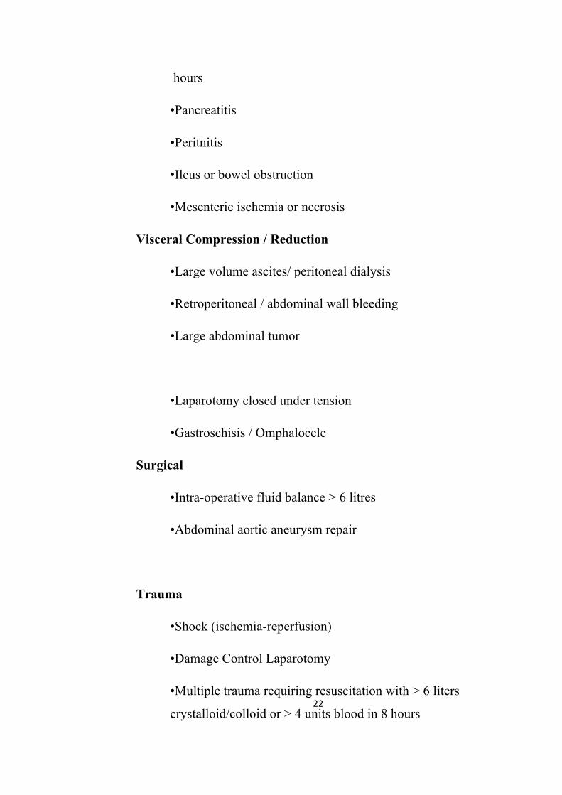
22
hours
•Pancreatitis
•Peritnitis
•Ileus or bowel obstruction
•Mesenteric ischemia or necrosis
Visceral Compression / Reduction
•Large volume ascites/ peritoneal dialysis
•Retroperitoneal / abdominal wall bleeding
•Large abdominal tumor
•Laparotomy closed under tension
•Gastroschisis / Omphalocele
Surgical
•Intra-operative fluid balance > 6 litres
•Abdominal aortic aneurysm repair
Trauma
•Shock (ischemia-reperfusion)
•Damage Control Laparotomy
•Multiple trauma requiring resuscitation with > 6 liters
crystalloid/colloid or > 4 units blood in 8 hours

23
•Major burns (> 25% TBSA)

24

25
MATERIALS AND METHODS
Type of Study: A Prospective Study.
Period of
Study:
Jan 2011
to July
2012.
4. Materials and Methods

26
Place of Study: PSG Hospitals, PSG IMS&R , Coimbatore,
Tamil Nadu. 641004
Sample Size: 100 cases
Group A (Emergency cases) – 66 cases
Group B (Elective cases) – 34 cases
This study had been approved by the Institutional Ethical
committee.
Plan of Study:
The detailed case history of the cases was recorded, clinical
examination and investigations carried out as per the proforma
enclosed.
Patients undergoing elective and emergency laparotomies were
allotted under Group A and Group B respectively. Intra-Abdominal
pressure was monitored daily till the IAP normalized or till post
operative day 7 along with the pulse, blood pressure, respiratory rate,
oxygen saturation, abdominal girth, urine output and arterial blood
gas in patients who underwent a laparotomy.
All these factors were used to monitor the progress and assess the
recovery of the patient.
Criteria for co-morbid conditions:

27
• Obesity: Patients with Body Mass Index (BMI) > 25
kg/m2 were considered as obese
• Hypertension (HTN):
o Patients who were known cases of HTN (on
regular or irregular medication)
o De novo detected cases of HTN (three consecutive
blood pressure readings of 140/90 mm of Hg or
more)
• Diabetes Mellitus (DM):
o Patients who were known cases of DM (on regular
or irregular medication)
o De novo detected cases of DM (deranged BSL
Profile)
Normal values:
BSL (fasting) 70-100 mg/dl
BSL (Post Prandial) <140 mg/dl
Statistical analysis was performed using SPSS software.
Inclusion Criteria:
All patients being planned for emergency and elective
laparotomies.
Exclusion Criteria:
• Cases of Abdominal trauma
• Patients with pre-existing renal and hepatic derangement

28
Method of measurement of Intra-Abdominal pressure:-
A simple fluid column manometry system via the Foley’s catheter
was used to measure the intra abdominal pressure.
The drainage tubing was marked along it’s length and the Foley’s
catheter was marked as ‘0’few mm proximal to the Y-junction, which
served as the zero reference point when it was at the level of the
symphysis pubis.
The drainage tubing was marked at an increment of 1 cm on the tape,
starting from the mark on Foley’s catheter as zero and then 50 ml of
sterile saline was introduced into the bladder.
After reconnecting the Foley’s catheter to the drainage tubing, the
zero reference point was taken at the level of symphysis pubis and the
drainage tubing was raised vertically making sure that the transition
from horizontal to vertical was at ‘0’ mark and was not too abrupt.
The distance the sterile saline raised vertically in the tubing was the
intra-abdominal pressure in cm of H2O. This was converted into
pressure in mm of Hg by dividing the value by the conversion factor
of 1.36.
IAP measurement was discontinued when the risk factors for IAH are
resolved or the IAP values have been below 10-12 mmHg for 24-48
hours.
Measurement of Intra abdominal pressure
(Figure 4.1):
Upper limit of fluid column
Height of fluid column
Measuring tape (cm)
“Y” Junction

29

30
Observations and Results
5. Results

31
In our study, total 213 patients who underwent different surgeries
were included. The study population consisted of 139 emergency
cases and 74 elective cases. The study population was divided into
two groups:
1. Group A: Patients undergoing Emergency Surgery (66 cases).
2. Group B: Patients undergoing Elective Surgery (34 cases).
List of abbreviations Used:
• MAP: Mean arterial pressure
• RR: Respiratory Rate
• A/G: Abdominal Girth
• U/O: Urine Output
• SpO2: Oxygen saturation
• TLC: Total Leucocyte Count
• BUN: Blood Urea Nitrogen
• ALT: Alanine Transaminase
• ALP: Alkaline Phosphatase
Table 5.1: Distribution of cases in study group according to Age
and Gender:
Graph5.1

32
66.2% of the patients were male and majority of the patients
(59.15%) were in the age group of 21-50 years.
Table 5.2: Distribution of cases in study group according to
nature of procedure:
Type of procedure No of cases Percentage
Elective 34 34
Emergency 66 66
Total 100 100
Graph5.2
Sex Age (Yrs)
Male Female
Total
≤ 20 16 (17) 8 (7) 24 (24)
21 – 50 38 (38) 21 (21) 59 (59)
> 50 10(12) 6 (5) 16 (16)
Total 66 (66) 34 (34) 100(100)

33
66% of the cases were emergency procedures.
Table 5.3: Distribution of cases in study group according to Body
Mass Index (BMI)
BMI CASES Percentage
< 18.5 7 7
18.5 – 25 89 89
25 – 30 4 4
Total 100 100
Graph 5.3
34
Majority (88.73%) of the patients were in the BMI group of 18.5 to
25.
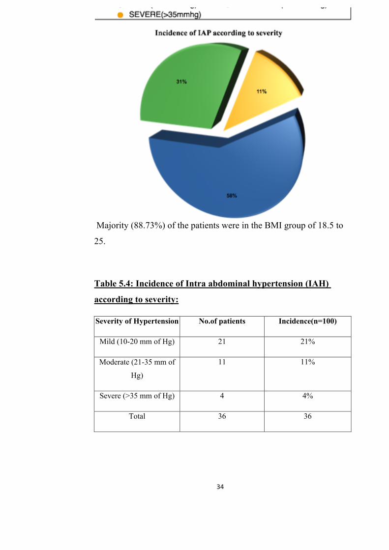
Table 5.4: Incidence of Intra abdominal hypertension (IAH)
according to severity:
Severity of Hypertension No.of patients Incidence(n=100)
Mild (10-20 mm of Hg) 21 21%
Moderate (21-35 mm of
Hg)
11 11%
Severe (>35 mm of Hg) 4 4%
Total 36 36

35
The incidence of IAH in the current study was 36% with 21% having
mild IAH, 11% having moderate IAH and 4% having severe IAH.
Table 5.5: Incidence of Intra abdominal hypertension according
to Grade:
Grade No.of patients
1 17
2 11
3 6
4 2
Total 36

36
Grade 1 IAH was seen in 17%, 11% had Grade 2 IAH, 5% had Grade
3 IAH and 2% had Grade 4 IAH.
Table 5.6: Correlation between IAP and BMI, Pulse, MAP, RR,
A/G, U/O, SpO2 from day 0 to day 7 in study group:
Post operative Correlation
between IAP
and Day 0 Day 1 Day 2 Day 3 Day 4 Day 5 Day 6 Day 7
BMI 0.14* 0.20* 0.17* 0.17* 0.17* 0.14* 0.07 0.02
PR 0.76# 0.88# 0.51# 0.43# 0.37# 0.35# 0.40# 0.34#
MAP -0.76# -0.83# -0.39# -0.34# -0.28# -0.33# -0.35# -0.36#
RR 0.83# 0.88# 0.67# 0.68# 0.55# 0.54# 0.61# 0.68#
U/O -0.86# -0.85# -0.44# -0.34# -0.34# -0.30# -0.33# -0.44#
A/G 0.45# 0.62# 0.30# 0.27# 0.25# 0.22$ 0.19* 0.17*
SpO2 -0.47# -0.56# 0.02 -0.02 -0.05 -0.06 -0.09 -0.37#
*P<0.05 $P<0.005 #P<0.0001

37
There was significant association between PR, MAP, RR, U/O, A/G,
SpO2 and the IAP. However BMI did not show a high degree of
association with IAP.
Table 5.7: Association of IAP with TLC on day 0 and maximum
recorded TLC{TLC(max)}: TLC on day 0 TLC(max) IAP Grade N
Mean ± SD Mean ± SD
0 64 9182 ± 1576 9182 ± 1576
1 17 9273 ± 2399 18676 ± 20494
2 11 12732 ± 1382 12924 ± 1760
3 & 4 8 13447 ± 2506 15513 ± 3369
F Value 48.83 12.96
P Value <0.0001 <0.0001

38
Graph 5.4:
There was statistically significant association between the IAP and TLC
Table 5.8: Association of IAP with BUN on day 0 and maximum
recorded BUN {BUN(max)}:
BUN on day 0 BUN(max) IAP Grade N
Mean ± SD Mean ± SD
0 64 29.79 ± 4.37 29.79 ± 4.37
1 17 31.49 ± 3.31 31.49 ± 3.31
2 11 42.16 ± 5.81 44.88 ± 8.78
3 & 4 8 50.33 ± 10.18 63.33 ± 21.60
F Value 107.63 113.49
P Value <0.0001 <0.0001
Graph 5.5

39
The F test and P value signify a high degree of association between
the IAP and BUN levels.
Table 5.9: Association of IAP with Sr.Creatinine on day 0 and maximum recorded Sr.Creatinine {Sr.Creatinine(max)}:
Sr. Creatinine on day 0 Sr. Creatinine (max) IAP Grade N
Mean ± SD Mean ± SD
0 64 0.69 ± 0.13 0.70 ± 0.13
1 17 0.78 ± 0.13 0.78 ± 0.13
2 11 1.05 ± 0.14 1.10 ± 0.20
3 & 4 8 1.13 ± 0.31 1.44 ± 0.50
F Value 48.83 93.52
P Value <0.0001 <0.0001
Graph5.6

40
The F test and P value signify a high degree of association between the IAP and Serum creatinine levels.
Table 5.10: Association of IAP with T.Bilirubin on day 0 and
maximum recorded T.Bilirubin {T.Bilirubin(max)}:
T. Bilirubin on day 0 T. Bilirubin (max) IAP Grade N
Mean ± SD Mean ± SD
0 64 0.69 ± 0.17 0.69 ± 0.17
1 17 0.72 ± 0.22 0.72 ± 0.22
2 11 0.97 ± 0.16 1.01 ± 0.20
3 & 4 8 1.09 ± 0.24 1.28 ± 0.38
F Value 33.41 51.49
P Value <0.0001 <0.0001
Graph 5.7
The F test and P value signify a high degree of association between the IAP and Total Bilirubin levels.

41
Table 5.11: Association of IAP with D.Bilirubin on day 0 and
maximum recorded D.Bilirubin {D.Bilirubin(max)}:
D. Bilirubin on day 0 D. Bilirubin (max) IAP Grade N
Mean ± SD Mean ± SD
0 64 0.36 ± 0.08 0.36 ± 0.08
1 17 0.33 ± 0.12 0.33 ± 0.12
2 11 0.47 ± 0.08 0.73 ± 1.31
3 & 4 8 0.57 ± 0.19 0.64 ± 0.22
F Value 30.24 6.33
P Value <0.0001 <0.0001
Graph 5.8
The F test and P value signify a high degree of association between the IAP and Direct Bilirubin levels.
Table 5.12: Association of IAP with ALT on day 0 and maximum
recorded ALT {ALT(max)}:
IAP Grade N ALT on day 0 ALT(max)

42
Mean ± SD Mean ± SD
0 64 42.32 ± 7.71 42.67 ± 7.69
1 17 41.62 ± 3.85 41.62 ± 3.85
2 11 49.44 ± 7.91 49.84 ± 8.18
3 & 4 8 74.07 ± 28.70 77.27 ± 27.98
F Value 46.94 57.98
P Value <0.0001 <0.0001
Graph 5.9
The F test and P value signify a high degree of association between the IAP and ALT levels.
Table 5.13: Association of IAP with ALP on day 0 and maximum
recorded ALP {ALP(max)}:
ALP on day 0 ALP(max)/day IAP Grade N
Mean ± SD Mean ± SD
0 64 41.88 ± 8.06 42 ± 7.97
1 17 45.32 ± 6.94 45.32 ± 6.94

43
2 11 53.44 ± 13.41 54.88 ± 12.87
3 & 4 8 76.93 ± 31.60 84.27 ± 33.58
F Value 48.83 59.95
P Value <0.0001 <0.0001
Graph 5.10
The F test and P value signify a high degree of association between
the IAP and ALP levels.
Table 5.14: Comparison of Pre operative IAP and Post operative
IAP in elective and emergency surgery group: Elective Emergency IAP on
Mean ± SD
(n=74)
Mean ± SD
(n=139)
Z Value P Value
Pre operative 6.70 ± 3.66 12.96 ± 8.25 7.64 <0.0001

44
Day 0 6.70 ± 3.66 12.86 ± 8.10 7.62 <0.0001
Day 1 6.59 ± 3.48 10.40 ± 9.80 4.11 <0.0001
Day 2 4.53 ± 3.70 6.42 ± 8.19 2.32 <0.05
Day 3 3.01 ± 3.54 3.71 ± 7.28 0.94 >0.05
Day 4 2.39 ± 4.88 2.32 ± 6.20 0.09 >0.05
Day 5 0.86 ± 3.54 1.83 ± 5.52 1.54 >0.05
Day 6 0.30 ± 1.77 1.46 ± 5.06 2.44 <0.05
Day 7 0.11 ± 0.93 1.02 ± 4.82 2.16 <0.05
Graph 5.11
The mean IAP is significantly higher in the emergency surgery group
as compared to the elective surgery group.

45
Table 5.15: Comparison of Pre Operative IAP and Post
Operative IAP according to age:
Age (≤ 20Yrs) Age (21-50Yrs) Age (>50Yrs) IAP on
Mean ± SD Mean ± SD Mean ± SD
F Value P Value
Pre op 9.33 ± 3.25 11.21 ± 8.26 11.33 ± 9.34 1.23 >0.05
Day 0 9.22 ± 3.31 11.16 ± 8.05 11.33 ± 9.34 1.37 >0.05
Day 1 6.33 ± 3.39 9.70 ± 9.35 10.78 ± 8.95 3.94 <0.05
Day 2 3.59 ± 3.001 5.83 ± 7.22 8.61 ± 9.17 5.66 <0.005
Day 3 1.10 ± 2.09 3.47 ± 5.98 6.83 ± 9.06 9.65 <0.0001
Day 4 0.26 ± 0.79 2.21 ± 4.96 5.81 ± 9.69 10.78 <0.0001
Day 5 0 ± 0 1.37 ± 4.10 4.03 ± 8.82 7.55 <0.001
Day 6 0 ± 0 0.94 ± 3.29 2.97 ± 8.08 5.50 <0.005
Day 7 0 ±0 0.46 ± 2.47 2.56 ± 8.27 5.19 <0.01

46
Graph 5.12
There was no significant association between age and the intra
abdominal pressure.
Patients above the age of 50 years on an average required a longer
time for the normalization of the IAP.

47
Table 5.16: Association between IAP grade and Outcome:
Outcome IAP grade
Survived(%) Death(%)
Total
0 64 0 64
1 17 0 17
2 11 0 11
3 6 0 6
4 1 1 2
Total 99 1 100
Graph 5.13
The single death in the study were associated with IAP of Grade IV
which had a mortality rate of 50%.

48
Table 5.17: Association between IAP grade and complications in study group
Graph 5.14
There was a very high complication rate of 66.67% and 88.89%
associated with Grade III and IV of IAP respectively.
Table 5.18: Comparison of IAP in cases with and without
Diabetes Mellitus (DM):
IAP on DM Present DM Absent Z Value P Value
IAP grade Complications
0 (n=64) 1 (n=17) 2 (n=11) 3 (n=6) 4 (n=1)
Total (n=100)
ARF, Septicemia 0 0 0 0 1 1
Faecal fistula, emphysema
0 0 0 1 0 1
Serous discharge from wound site
0 1 0 0 0 1
Wound dehiscence 0 0 0 1 1 2
Wound gape 2 6 3 2 0 13
Wound gape, oliguria
0 0 1 0 1 2
Total 2 (2) 7 (40.54) 4(24) 4 (66.67) 8 (88.89) 25(17.84)

49
Mean ± SD
(n=17)
Mean ± SD (n=83)
Pre operative 13.9 ± 13.1 10.52 ± 6.91 1.04 >0.05
Day 0 13.4 ± 12.1 10.49 ± 6.93 0.98 >0.05
Day 1 15.5 ± 13.7 8.52 ± 7.54 2.07 >0.05
Day 2 10.9 ± 12.1 5.32 ± 6.25 1.88 >0.05
Day 3 8.9 ± 10.8 3 ± 5.47 2.22 <0.05
Day 4 7.2 ± 10.3 1.93 ± 5.02 2.08 >0.05
Day 5 5.47 ± 9.82 1.15 ± 4.14 1.80 >0.05
Day 6 4.41 ± 9.83 0.77 ± 3.26 1.52 >0.05
Day 7 3.9 ± 10.5 0.43 ± 2.63 1.35 >0.05

50
Graph 5.15
There was no statistically significant association between IAP and
Diabetes Mellitus.
51
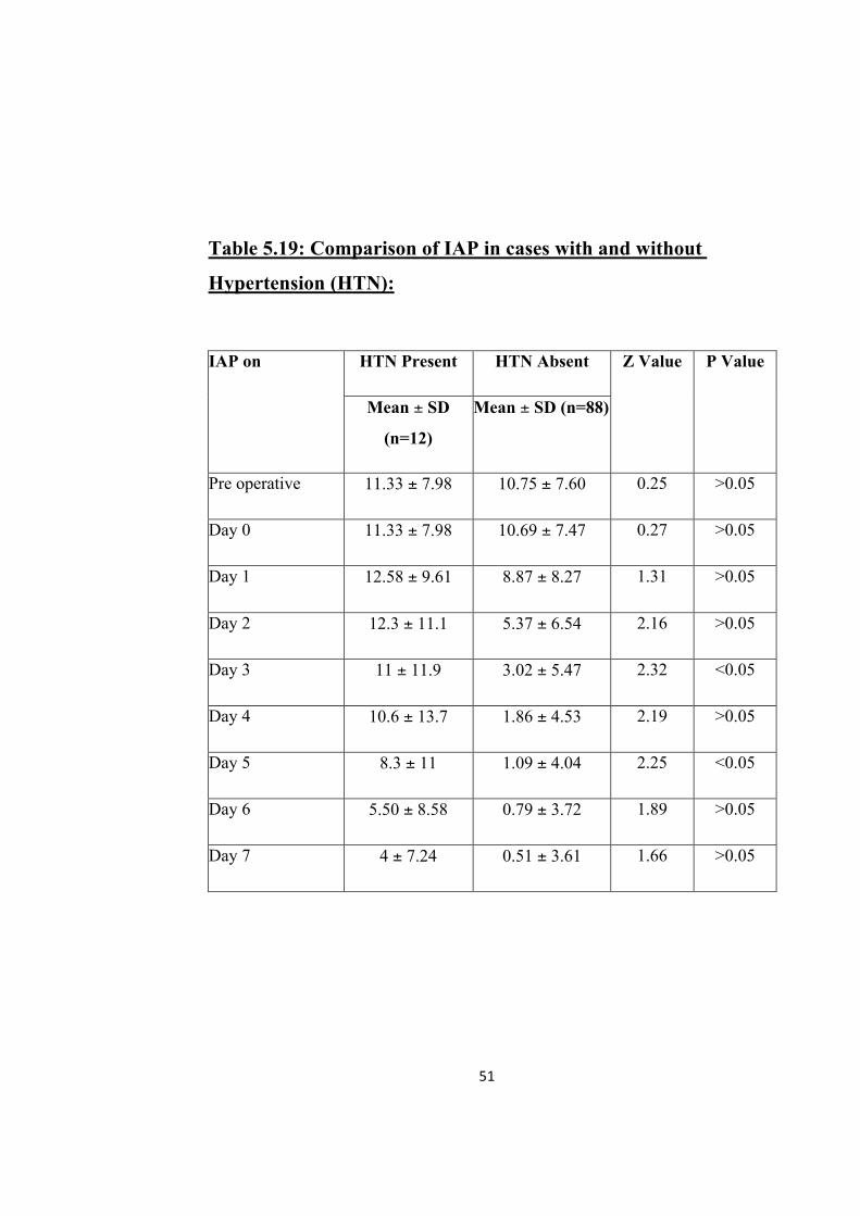
Table 5.19: Comparison of IAP in cases with and without
Hypertension (HTN):
HTN Present HTN Absent IAP on
Mean ± SD
(n=12)
Mean ± SD (n=88)
Z Value P Value
Pre operative 11.33 ± 7.98 10.75 ± 7.60 0.25 >0.05
Day 0 11.33 ± 7.98 10.69 ± 7.47 0.27 >0.05
Day 1 12.58 ± 9.61 8.87 ± 8.27 1.31 >0.05
Day 2 12.3 ± 11.1 5.37 ± 6.54 2.16 >0.05
Day 3 11 ± 11.9 3.02 ± 5.47 2.32 <0.05
Day 4 10.6 ± 13.7 1.86 ± 4.53 2.19 >0.05
Day 5 8.3 ± 11 1.09 ± 4.04 2.25 <0.05
Day 6 5.50 ± 8.58 0.79 ± 3.72 1.89 >0.05
Day 7 4 ± 7.24 0.51 ± 3.61 1.66 >0.05

52
Graph 5.16
There was no statistically significant association between IAP and
systemic hypertension.

53
6.Discussion

54
Discussion
A compartment syndrome is a condition in which increased
pressure in a confined anatomical space adversely affects the
function and viability of the tissues therein. Confined
anatomical spaces mostly associated with compartment
syndromes are the fascial spaces of the extremities, the orbital
globe (glaucoma), the cranial cavity (epidural/subdural
hematoma), the kidney capsule (post-ischemic oliguria) and the
abdominal cavity.
ACS is a condition in which sustained increased pressure
within the abdominal wall, pelvis, diaphragm, and the
retroperitoneum, adversely affects the function of the entire
gastrointestinal tract and connected extraperitoneal organs. It
usually requires operative decompression.
IAH is graded as (33):
• Mild IAH
10-20 mm Hg: Clinically not significant changes occur
which usually doesn’t need surgical intervention
• Moderate IAH
21-35 mm Hg: There may be certain critical changes
occurring and might need interventions
• Severe IAH

55
>35 mm Hg: Abdominal compartment syndrome- needs
definitive surgical intervention.
Five organs of the abdomen were subjective to volume
change and pressure(37).
1) Solid organs - Liver, spleen causes chronic abdominal
hypertension.
2) Hollow viscus leads to inflammation , ileum or bowl
obstruction.
3)Fluid overload in a patient the blood and lymphatics causes
abdominal hypertension.
4) the peritoneum usually absorbs large amount of fluid incase
of inflamation
5) peritoneal cleft also accumulates a large amount of fluid
It is yet to prove in clinical settings which of the above five
causes is the reason for the increase in the volume which is the
imajor cause of IAH.
The peritoneum covers 1.8 m2 of the body surface. It covers the
whole of intestinal organs.On inflammation only 0.5cm
increase in thickness noted and there will be absorption of 1.8
m2 = 18,000 cm2 x 0.5 cm thickening = 9,000 ml of fluid due to
inflammation of peritoneum. Hence fluid shift will be seen in
burns.
Due to its large surface area there will be large amounts of
transudates and exudates formed in a short time due to
irritation or injury.

56
Abdominal wall compliance plays an important role in the
regulation of the intra abdominal pressure:
The dynamic relation between volume and pressure within the
abdomen is important because after a relatively long period of
compensation, deterioration is fast due to limited abdominal
wall compliance. Compliance is structurally dependent on the
stiffness of the peritoneum and its volume-pressure curve (i.e.
compliance is not linear).
Upon IAP increase, abdominal wall fasciae stretch and lose
expandability. Progressively smaller volume increments are
required to further elevate IAP(28). Conversely, high IAP may
be dramatically relieved by decompression.
Due to abdominal hypertension- shallow respiration high
diaphragm on percussion, low output and increased central
venous pressure(44). Multi organ dysfunction progresses unless
IAP is reduced. Hence the requirement of timely intervention
either fluid management or surgical decompression reverses the
effects.
Radiologically computed tomography yields increases in
anteroom-posterior to transverse diameter, renal compression,
bowel thickening and inguinal herniation(45).
Age:
The mean age in our study was 34.48 years. In a study by
Khan S et al. the mean age was 34.78 years(46). Cheatham et al.
have reported a mean age of 51±19 years, Meldrum et al. 39±9
years, and Hong et al. 42 years(47-49).

57
In the current study there was no statistically significant
association between age and IAP (Table 5.1; Graph 5.1).
However the normalization of IAP took a longer time in the
patients above 50 years of age. (Table 5.15; Graph 5.12)
Sex:
There were 141 males and 72 females (66.2% males and
33.8% females) in the current study. A similar ratio was seen in
the studies by Khan S et al. (76% males), Hong et al. (72%
males), Meldrum et al. (70% males), Sugrue et al. and
Cheatham et al. (60% males)(46-50). There was no significant
correlation between sex and IAP in the current study (Table
5.1; Graph 5.1).
Incidence:
The incidence of IAH and ACS reported by various studies
ranges from 2 to 78% and 0.5 to 36%, respectively, and
depends on the population and the values used to define these
entities(49).
In the current study the incidence of IAH was 36.15% and that
of ACS was 4.69% (Table 5.4, 5.5).
The lower incidence observed was because this study includes
low-risk (cholecystectomy, appendectomy etc) as well as high-
risk patients (peritonitis, intestinal obstruction, perforation etc)
whereas most of the previous studies confined data collection
to high-risk patients. While the earlier approach ensures a good
yield of patients with ACS, it may result in a very high
incidence compared with that seen clinically in the general

58
population overall. Furthermore, such an approach potentially
misses those patients who are not at high risk, and yet may
have Multiple Organ Dysfunction Syndrome (MODS) falsely
attributed to sepsis or irreversible shock when in fact they have
unrecognized ACS. By measuring the IAP prospectively in all
patients, this study obtained true overall incidence.
A similar study by Khan S et al. revealed an incidence of 80%
for IAH and 3.05% for ACS(46).
Changes in physiology that are seen with increasing pressure
involve almost all systems and overlap.
Cardiovascular system:
Adverse effects with IAP’s will occur even when the pressure
is as low as 10-15 mm Hg.
Increased IAP’s causes fall in cardiac output blood pressure
remains unaltered. Tachycardia occurs in order to maintain
cardiac output.
Increase in vascular resistance may be due to mechanical
compression of capillary or nitric acid deficiency.
In a study by Chang MC et al, intra abdominal hypertension of
>25 mm of Hg led to a significant increase in heart rate of up to
124±18 /min(51).

59
In an another study by Lazaro Gotloib et al heart rate showed
significant increase with intra abdominal pressures of more
than 15 cm of H2O(52).
The results of the current study were among similar lines
(r=0.34 to 0.88; p<0.0001) (Table 5.6).
In the study by Chang MC et al, intra abdominal hypertension
led to a fall in the mean arterial pressure(51).
In a study of 46 patients by Widergreen et al, the mean arterial
pressure at 40 cm of H2O bladder pressure was 86 mm of Hg
which increased to 92 mm of Hg when the bladder pressure
reduced to 22 cm of H2O(53).
In the study by Lazaro Gotloib et al, mean arterial blood
pressure showed significant increase with intra abdominal
pressures of more than 10 cm of H2O(52).
In the current study there was a significant negative correlation
between MAP and IAP (-0.76 to -0.28; p<0.0001) (Table 5.6).
According to Malbrain et al Intra abdominal hypertension has a
statistically significant association with acidosis(38). In the
current study, out of the 10 patients who underwent Arterial
blood gas analysis(ABG) 8 cases showed acidosis and all of
them were associated with mortality. In the current study ABG
was performed only in those cases where deemed necessary
and as such statistical correlation was not possible. This is one
of the limitations of our study.

60
Total Leucocyte Count (TLC):
In a study of a total of 75 patients by Cem Ibis and Aydin Altan
there was a statistically significant correlation between IAP and
TLC (p=0.002)(40).They concluded that the determination of the
WBC count only is not safe enough to diagnose acute
abdomen. The interpretation of BP level together with WBC
count seems to be more effective because of the statistically
significant difference between the test and the control group
related to the elevated WBC count.
Similar results were obtained in our study which showed a
highly significant association between the IAP and TLC
(p<0.0001) (Table 5.7; Graph5.4).
Respiratory function:
Increase in IAP and decrease thoracic volume pushes the
diaphragm up. Atelectases of pleural cavity and decrease in
alveolar clearance occur due to decreased volume. Early
complication of abdominal hypertension due to peritonitis is
Pneumonia.
However there were no cases of pneumonia in the current
study.
Raise in thoracic pressure, low cardiac output and pulmonary
vascular resistance occur when there is diaphragmatic
protrusion into pleural cavity. Hypoxemia, hypercarbia and
acidosis occur due to ventilation/ perfusion abnormality. When
the pressure is 20mm Hg physiological dysfunction occurs.

61
As noted by David Hopkins and S.W. Gemmell (54) there was a
significant increase in the respiratory rate even in the current
study (r=0.54 to 0.88; p<0.0001) (Table 5.6).
In the current study there was a significant association between
SpO2 and intra abdominal pressure (P<0.0001) (Table 5.6)
which concurs with results of the available literature. However
all the modern day studies measured the association of the IAP
and respiratory function by various scientifically advanced
parameters like PaO2 (partial pressure of oxygen), FiO2
(fraction of inspired oxygen), QI/Qt (intrapulmonary shunt
fraction) , PIP (peak inspiratory pressure), PEEP (positive end-
expiratory pressure) and Cdyn (dynamic compliance) which
were not used in our study and is one of the limitations of our
study.
Renal function:
Complications such as oliguria; anuria usually ensues with
higher pressures.
Mainly due to the decrease in renal blood flow, GFR, urine
output, and various specific tubular functions associated with
raised IAP is of multifactorial etiology.
Improved cardiac output plays a role in diminished renal
perfusion but even when cardiac output is maintained at normal
or supernormal values by blood volume expansion, impairment
of renal function persists. Renal dysfunction is also caused by
compression of the renal vein, which causes partial renal blood

62
outflow obstruction. Compression of the abdominal aorta and
renal arteries contributes to increased renal vascular resistance.
Furthermore, direct compression of the kidneys elevates
cortical pressures, leading to a "renal compartment syndrome".
Elevation of plasma anti diuretic hormone may represent
another etiological factor.
Savino JA et al in a study of 51 patients noted a mean urine
output of 47 ml/hr at a mean abdominal pressure of 33.5 cm of
H2O which increased to 55ml/hr after the mean abdominal
pressure reduced to 19.1 cm of H2O(55).
In a study of 46 patients Widergreen et al, a mean urine output
of 79ml/hr was noted at a bladder pressure of 40 cm of H2O
which increased to 123ml/hr after the mean abdominal pressure
reduced to 22 cm of H2O(53).
In the same study there was also a significant association
between the intra abdominal pressure and Blood urea and
serum creatinine levels(53).
In the current study there was a significant negative correlation
between urine output and IAP (r = -0.3 to -0.86; p<0.0001).
The current study also revealed a significant association of IAP
with Blood urea and serum creatinine levels (Table 5.6, 5.8,
5.9; Graph 5.5, 5.6).
46.67% of patients with ACS developed oliguria and 33.3% of
patients with ACS progressed to ARF and anuria in the current
study (Table 5.17; Graph 5.14).

63
Effects on liver function:
Increase in IAP affects the hepatic arteries and the portal blood
flow. Trauma patients are more susceptible because of shock
induced intestinal vascular resistance.
Synthesis of hepatic acute-phase protein, immunoglobulin and
host defense system were impaired due to decrease in hepatic
blood flow.
Detailed studies addressing the issue of reduced hepatic protein
synthesis have not yet been published.
Transient alterations of hepatic enzymes are frequently
observed after uneventful laparoscopic cholecystectomy,
presumably attributed to the elevated intraabdominal pressure
of the pneumoperitoneum according to Marakis et al(62).
Aspartate aminotransferase, alanine aminotransferase, alkaline
phosphatase, and total bilirubin levels were significantly
elevated following ACS and sepsis in a study performed in rats
by Tolga MA et al(57).
The current study reveals a significant association between IAP
and the four liver function parameters assessed i.e., Total
bilirubin, direct bilirubin, serum ALP and serum ALT (Table
5.10, 5.11, 5.12, 5.13; Graph 5.7, 5.8, 5.9, 5.10).

64
Wound healing:
Increase in IAP impairs the wound healing due to reduced
abdominal wall and fascia blood flow. Abdominal binder must
be avoided as it further reduces the blood flow.
Rectus sheath blood flow was significantly reduced at all
pressure levels when compared to baseline and negatively
correlated (r = -0.82) with increasing IAP in a porcine model
by Diebel L et al(58).
31.17% of patients with IAH developed wound gape and
13.3% of patients with ACS developed wound dehiscence
(Table 5.17; Graph 5.14) (Figure 5.1,5.2).
Gastrointestinal function:
When IAP is 15mmHg, decrease in splanchnic circulation
occurs. As it is documented many of the abdominal arteries,
veins and lymphatics have low perfusion. Alterations of pH,
bacterial growth translocation, motility disorder, harmone
abnormality and exocrine dysfunction were also documented as
secondary effects. Effects on spleen, pancreas, adrenal and
reproductive organs are yet to be documented.

65
Impairment of arterial flow - Abdominal hypertension
impairs intestinal blood flow. Elevation in IAP results in
decreased mesenteric arterial blood flow; intestinal mucosal
blood flow; and arterial perfusion of the stomach, duodenum,
intestine, pancreas, and spleen. As IAP increases, mucosal pH
falls, indicating severe ischemia or necrotizing pancreatitis.
Compartment-induced impaired intestinal perfusion may be a
critical factor in anastomotic healing. Abdominal hypertension
probably plays a role in many of the organ dysfunctions of
currently questionable etiology. Examples may be ischemic
gastritis, acalculous cholecystitis or pancreatitis, colon
ischemia, and some forms of bowel ischemia.
These changes are greater than can be accounted for by the
alterations in cardiac output and also occur when cardiac output
and systemic blood pressure are maintained at normal levels.
Effects on abdominal veins
Even a mild elevation of IAP in cirrhotic patients cause
increase in hepatic venous pressure and azygos blood flow
increases. Vice versa occurs once IAP is lowered. It’s a
controversy whether increase in IAP precipitates variceal
bleed.
Effects on lymph flow
Increase in IAP significantly reduce lymphatic flow of thoracic
duct. the same increases after decompression. Transfer of

66
peritoneal fluid into the thoracic lymphatics decreases due to
stretching of diaphragm.
Translocation:
Due to decreased intestinal perfusion caused by increased IAP,
traslocation of bacteria occurs in high rates. This causes
increase in infection rate and sepsis which may lead to further
septic complications.
According to Diebel L et.al, increased IAP of 25 mm of Hg
leads to decreased mucosal blood flow and to bacterial
translocation, which may contribute to later septic
complications and organ failure(58).
In the current study 33.3% of patients with ACS developed
septicaemia (Table 5.17;Graph 5.14).
Intracranial pressure (ICP):
Idiopathic intracranial hypertension is accentuated due to
chronic abdominal hypertension. During lapraoscopy
abdominal pressure increases which causes increase in
intracranial pressure.
Nature of surgery: (Emergency Vs Elective Surgery):
In a large prospective study from 1999, Sugrue et al. studied
IAH in relation to renal impairment in 263 patients admitted to
the ICU after emergent (n = 174) or elective (n = 89)
abdominal surgery IAP 18 mmHg or greater was found in 41%.

67
IAH after emergency surgery was seen in 46% of patients after
upper and 46 per cent after lower gastrointestinal surgery
compared with 60 per cent after vascular surgery. The
corresponding findings after elective surgery were 29%, 12%,
and 32%, respectively, emphasizing the increased incidence of
IAH/ACS after emergency surgery(59).
They also found the incidence of IAH in postoperative ICU
patients after elective upper gastrointestinal surgery to be 29
per cent. This difference might be attributable to the higher
IAH incidence in ICU patients in general(59).
Scollay et al. studied IAP prospectively in 42 patients
recovering from elective major upper gastrointestinal surgery
and found that 12 per cent had a transient IAH without an
impact on postoperative organ function. No patient developed
ACS and there were no deaths. The only significant finding
was delayed return to oral diet in patients with transient
IAH(60).
In the current study in emergency surgeries (n=139), the pre
operative IAP (Mean ± SD) was 12.96 ± 8.25 cm of H2O and in
elective surgeries (n=74), the pre operative IAP (Mean ± SD)
was 6.70 ± 3.66 cm of H2O and the association was statistically
very significant (p value = <0.0001) (Table 5.14; Graph 5.11).
Mortality:

68
Cheatham et al. had found that elevated IAP alone does not
have sufficient sensitivity or specificity to be useful as a
predictor of mortality(47).
In a study by Hong JJ et al, 50% of the patients with ACS died
as did 22.2% of patients with IAH(56).
A mortality rate of 100% was seen in ACS group and 13.2% in
IAH group in a study by Khan S et al when decompression was
not done, supporting the view that ACS, if left untreated, is
invariably fatal(46).
In the current study a mortality rate of 33.3% was seen in ACS
group and 6.49% in IAH group (Table 5.16;Graph 5.13).
Hence, future studies on this subject should aim at devising a
protocol which may help the healthcare professionals in early
identification of the IAH and ACS patients and thus minimize
the resulting high mortality.
Association with co-morbidities [Diabetes mellitus (DM),
Hypertension (HTN) and Body Mass Index(BMI)]:
Although the association of Chronic IAH with co-morbidities
is well documented, literature correlating IAH and co-
morbidities is scanty.
Varela JE et al performed a study to examine the correlation
between the IAP and obesity-related co-morbidities. Systemic
hypertension was significantly associated with an elevated IAP
but there was no significant association with diabetes
mellitus(61).

69
In the current study there was no statistically significant
association between IAP and DM and HTN (p>0.05) (Table
5.18, 5.19; Graph 5.15,5.16 ).
Varela JE et al described a significant correlation between IAP
and BMI(59).
According to Wilson et.al, elevated BMI does impact IAP, but
the incremental value is small. Markedly increased IAP should
not be attributed solely to elevated BMI and should be
recognized as a pathologic condition(62).
There was a similar statistically significant correlation between
the BMI and IAP even in the current study (Table 5.2, 5.6;
Graph 5.2 ).

70
71
Conclusions:
1. The incidence of IAH in the current study was 36%.
2. There was significant association between Pulse rate,
Mean arterial pressure, Respiratory rate, Urine output,
Body mass index, Abdominal girth, Oxygen saturation
and the Intra abdominal pressure.
3. There was significant association between Total
leucocyte count, Liver function tests, Renal function
tests and intra abdominal pressure.
6.Conclusion

72
4. Patients with Grade III and IV Intra abdominal
hypertension were associated with a higher rate of
mortality and morbidity.
5. There was no significant association between intra
abdominal pressure and co-morbidities like Diabetes
Mellitus and Hypertension.
6. Incidence of intra abdominal hypertension was higher
in emergency laparotomies as compared to elective
laparotomies.
Summary:
Introduction: Abdominal compartment syndrome is one
which pressure increases in a confined anatomical space and
affects its function and viability of the tissue. In abdominal
hypertension there is increase in volume in its contents IAP
impair physiology and organ function, because of the limited
compliance of abdominal wall. This study is being
undertaken to evaluate the impact of IAP on outcome in
patients undergoing laparotomies.

73
Materials and Methods: The detailed case history of two
hundred and thirteen cases was recorded, clinical
examination and investigations carried out. Patients
undergoing elective and emergency laparotomies were
allotted under Group A and Group B respectively. Intra-
Abdominal pressure was monitored daily till the IAP
normalized or till post operative day 7 along with the pulse,
blood pressure, respiratory rate, oxygen saturation,
abdominal girth, urine output and arterial blood gas in
patients who underwent a laparotomy. All these factors were
used to monitor the progress and assess the recovery of the
patient.
Observation and Results: Of the 100 cases in the study,
66.2% of the patients were male and majority of the patients
(59.15%) were in the age group of 21-50 years. 65.26% of
the cases were emergency procedures. The incidence of IAH
in the current study was 36.16% with 20.66% having mild
IAH, 11.27% having moderate IAH and 4.23% having
severe IAH. There was significant association between PR,
MAP, RR, U/O, A/G, SpO2 and the IAP. However BMI did
not show a high degree of association with IAP. There was
also significant association between the IAP and Liver and

74
Renal function tests. The mean IAP is significantly higher in
the emergency surgery group as compared to the elective
surgery group. There was a very high complication rate of
66.67% and 88.89% associated with Grade III and IV of IAP
respectively. The single death in the study were associated
with IAP of Grade IV which had a mortality rate of 50%.
Conclusions: There was significant association between
Pulse rate, Mean arterial pressure, Respiratory rate, Urine
output, Body mass index, Abdominal girth, Oxygen
saturation and the Intra abdominal pressure. There was
significant association between Total leucocyte count, Liver
function tests, Renal function tests and intra abdominal
pressure.
Patients with Grade III and IV Intra abdominal hypertension
were associated with a higher rate of morbidity and
mortality. There was no significant association between intra
abdominal pressure and co-morbidities like Diabetes
Mellitus and Hypertension. Incidence of intra abdominal
hypertension was higher in emergency laparotomies as
compared to elective laparotomies.

75

76
References:
1. Barnes GE, Laine GA, Giam PY, Smith EE, Granger HJ. Cardiovascular
responses to elevation of intra-abdominal hydrostatic pressure. Am J
Physiol. 1985;248:R209-13.
7.References

77
2. Mohapatra B. Abdominal compartment syndrome. Indian J Crit Care
Med2004;8:26-32.
3. Rotondo MF, Cheatham ML, Moore FA, Reilly PM. Abdominal
Compartment Syndrome-Symposium. Contemporary
Surgery2003:59:260-70.
4. Bailey J, Shapiro MJ. Abdominal Compartment Syndrome. Crit
Care2000;11:156-71.
5. Emerson H. Intra-abdominal pressures. Arch Intern Med1911;7:754-84.
6. Moore AFK, Hargest R, Martin M, Delicata RJ. Intra-abdominal
hypertension and the abdominal compartment syndrome. Br. J
Surg2004;91:1102-10.
7. Thorington JM, Schmidt CF. A study of urinary output and blood-
pressure changes resulting in experimental ascites. Am J Med Sci
1923;165:880-90.
8. Bellis CJ, Wangensteen OH. Venous circulatory changes in the abdomen
and lower extremities attending abdominal distention. Proc Soc Exp Biol
Med 1939;4:490-8.
9. Ogilvie WH. The late complications of abdominal war wounds. Lancet
1940;2:253-6.

78
10. Bradely SE, Bradely GP. The effect of increased intra-abdominal pressure
on renal function in man. J Clin Invest 1947;26:1010-15.
11. Gross RE. A new method for surgical treatment of large omphaloceles.
Surgery 1948;24:277-92.
12. Baggot MG. Abdominal blow-out: a concept. Current Research
Anesthesia Analgesia 1951;30:295-8.
13. Gordon ME. The acute effects of abdominal paracentesis in Laennec’s
cirrhosis upon changes of electrolytes and eater, renal function and
hemodynamics. Am J Gastroenterol 1960;33:15-37.
14. Suazzi M, Polese A, Magrini F, et al. Negative influence of ascites in the
cardiac function of cirrhotic patients, Am J Med 1975;59:165-70.
15. Cruikshank DP, Buschsbalm HJ. Effects of rapid paracentesis,
cardiovascular dynamics and body fluid composition. JAMA
1973;225:1361-2.
16. Knauer CM, Love HM. Hemodynamics in cirrhotic patient during
paracentesis. N Engl J Med 1967;276:491-6.
17. Sönderberg G, Westin B. Transmission of rapid pressure increase from
the peritoneal cavity to the bladder. Scan J Urol Nephrol 1970;4:155-65.
18. Shenansky JH, Gillenwater JY. The renal hemodynamic and functional
effects of external counterpressure. Surg Gynecol Obstet 1972;134:253-8.

79
19. Ivankovich AS, Albrecht RF, Zahed B et al. Cardiovascular collapse
during gynecological laparoscopy. Ill Med J 1974;145:58-61.
20. Motev M, Ivankovich AD, Bieniarz J et al. Cardiovascular effects and
acid base and blood gas changes during laparoscopy. Amer J Obstet
Gynecol 1973;116:1002-1012.
21. Lenz RJ, Thomas TA, Wilkins DG. Cardiovascular changes during
laparoscopy. Anaesthesia 1976;31:4-12.
22. Richardson JD, Trinkle JK. Hemodynamic and respiratory alterations
with increased intra-abdominal pressure. J Surg Res 1976;20:401-4.
23. Kashtan J, Green JF, Parson EQ et al. hemodynamic effects of increased
abdominal pressure. J Surg Res 1981;30:249-55.
24. Harman PK, Kron IL, McLachan DH et al. Elevated intra-abdomial
pressure and renal function. Ann Surg 1982;196:594-7.
25. Richards WO, Scovill W, Shin B et al. Acute renal failire associated with
increased intra-abdominal pressure. Ann Surg 1983;197:183-7.
26. Le Roith D, Bark H, Nyska M et al. The effect of abdominal pressure on
plasma antidiuretic hormone levels. J Surg Res 1982;32:65-9.
27. Smith JH, Merrell RC, Raffin TA. Reversal of postoperative anuria by
decompressive celiotomy. Arch Intern Med. 1985;145:553-4.
28. Barnes GE, Laine GA, Giam PY et al. Cardiovascular responses to
elevation of intra-abdominal hydrostatic pressure. Am J Physiol.
1985;248:R208-13.

80
29. Caldwell CB, Ricotta JJ. Changes in visceral blood flow with elevated
intraabdominal pressure. J Surg Res. 1987;43:14-20.
30. Jacques T, Lee R. Improvement of renal function after relief of raised
intra-abdominal pressure due to traumatic retroperitoneal haematoma.
Anaesth Intensive Care. 1988;16:478-82.
31. Cullen DJ, Coyle JP, Teplick R, Long MC. Cardiovascular, pulmonary,
and renal effects of massively increased intra-abdominal pressure in
critically ill patients. Crit Care Med. 1989;17:118-21.
32. Iberti TJ, Lieber CE, Benjamin E. Determination of intra-abdominal
pressure using a transurethral bladder catheter: clinical validation of the
technique. Anesthesiology. 1989;70:47-50.
33. Schein M, Wittmann DH, Aprahamian CC, Condon RE. The abdominal
compartment syndrome: the physiological and clinical consequences of
elevated intra-abdominal pressure. J Am Coll Surg. 1995;180:745-53.
34. Burch JM, Moore EE, Moore FA, Franciose R. The abdominal
compartment syndrome. Surg Clin North Am. 1996;76:833-42.
35. E.J. Kimball, W. Kim, M.L Cheatham, M.L.N.G. Malbrain. Clinical
awareness of intra-abdominal hypertension and abdominal compartment
syndrome in 2007.Acta clinica belgica, 2007; 62-supplement 1.
36. Saggi BH, Sugerman HJ, Ivatury RR, Bloomfield GL. Abdominal
Compartment syndrome. J Trauma1998;45:597-609.

81
37. Wittmann DH. The compartment syndrome of the abdominal cavity. J
Intensive Care Med 2000;15:201-20.
38. Malbrain ML, De laet IE. Intra-abdominal hypertension: evolving
concepts. Clin Chest Med. 2009;30(1):45-70.
39. Cheatham ML.Abdominal compartment syndrome: pathophysiology and
definitions.Scand J Trauma Resusc Emerg Med.2009 Mar 2;17:10.
40. Cem Ibis, Aydin Altan. The value of intra-abdominal pressure
measurement in patients with acute abdomen. Asian J Surg
2009;32(1):33-8.
41. Nathens AB, Brenneman FD, Boulanger BR. The Abdominal
Compartment Syndrome. Can J Surg1997;4:254.
42. Holzheimer RG, Mannick JA, Abdominal Compartment Syndrome.
Surgical Treatment:-Evidence based and problem oriented. Chapter XVI,
http://www.freebooks4doctors.com/fb/spec18.htm#surg.
43. J.J. De waele, I. De laet, M.L.N.G. Malbrain rational intra abdominal
pressure monitoring: how to do it? Acta clinica belgica, 2007; 62-
supplement 1.
44. Fietsam R, Villalba M, Glover JL, Clark K. Intra-abdominal compartment
syndrome as a complication of ruptured abdominal aortic aneurysm
repair. Am Surg. 1989;56:396-402.

82
45. Pickhardt PJ, Shimony JS, Heiken JP, Buchman TG, Fisher AJ. The
abdominal compartment syndrome: CT findings. AJR American Journal
of Roentgenology. 1999;173:575-79.
46. Khan S, Verma AK, Ahmad SM, Ahmad R. Analyzing intra-abdominal
pressures and outcomes in patients undergoing emergency laparotomy. J
Emerg Trauma Shock 2010;3:318-25.
47. Cheatham ML, White MW, Sagraves SG, Johnson JL, Block EF.
Abdominal perfusion pressure: a superior parameter in the assessment of
intra-abdominal hypertension. J Trauma 2000;49:621-7.
48. Meldrum DR, Moore FA, Moore EE, Franciose RJ, Sauaia A, Burch JM.
Prospective characterization and selective management of the abdominal
compartment syndrome. Am J Surg 1997;174:667-73.
49. Hong JJ, Cohn SM, Perez JM, Dolich MO, Brown M, McKenney MG.
Prospective study of the incidence and outcome of intra-abdominal
hypertension and the abdominal compartment syndrome. Br J Surg
2002;89:591-6.
50. Sugrue M, Buist MD, Hourihan F, Deane S, Bauman A, Hillman K.
Prospective study of intra-abdominal hypertension and renal function
after laparotomy. Br J Surg 1995;82:235-8.
51. Chang MC, Miller PR, Meredeth JW. Effects of abdominal
decompression on cardiopulmonary function and visceral perfursion in
patients with intra-abdominal hypertension. J Trauma. 1998;440-5.
52. Lazaro G et al.,Haemodynamic effects of increasing intra abdominal
pressure in peritoneal dialysis. Perit Dial Int.1981;1(4): 41-43.

83
53. Widergreen, J. T. and Batistella, F. D. The open abdomen: Treatment for
intra-abdominal compartment syndrome. J.Trauma. 1994;37(1): 158.
54. David Hopkins, S.W. Gemmell. Intra abdominal hypertension and the
abdominal compartment syndrome. British Journal of Anaesthesia
2001;1(2):56-9.
55. Savino JA, Cerabona TD, Agarwal N, Byrne D. Manipulation of ascitic
fluid pressure in cirrhotics to optimize hemodynamic and renal function.
Ann Surg. 1988;208:504-11.
56. G. Marakis et al.,Alterations In Liver Function Tests Following
Laparoscopic Cholecystectomy. The Internet Journal of Surgery. 2006;
8(1): ISSN: 1528-8242.
57. Tolga MA et al., Liver Injury in Sepsis and Abdominal Compartment
Syndrome in Rats.Surg Today.2006;36(6):519-24.
58. Diebel L et al.,Effect of intra abdominal pressure on abdominal wall
blood flow. Am Surg.1992;58(9):573-6.
59. Sugrue M, Jones F, Deane SA, et al. Intra-abdominal hypertension is an
independent cause of postoperative renal impairment. Arch Surg
1999;134:1082-5.
60. Scollay JM, de Beaux I, Parks RW. Prospective study of intra-abdominal
pressure following major elective abdominal surgery. World J Surg
2009;33:2372-7.

84
61. J. E. Varela et al. Correlations between intra-abdominal pressure and
obesity-related co-morbidities. Surgery for Obesity and Related Diseases
2009;5: 524–8.
62. Wilson A et al. Intra abdominal pressure and the morbidly obese patients:
The effect of body mass index. Journal of Trauma – Injury Infection and
Critical Care 2010;69(1):78-83.

85
Annexure 1: Proforma:
OP/IP No. :
DOA:
DOD:
Name :
Age :
Sex :
Weight(kg):
Occupation:
Address :
HISTORY:
Pain in abdomen:
Duration
Site
Nature
Radiation
Aggravating Factors
Relieving Factors
Distension:
Vomiting:
Nature:

86
Number of episodes:
Quantity:
Fever:
Constipation:
Absolute
Relative
Haematemesis:
Haematochezia:
Malaena:
Trauma:
Others:
Clinical Findings:
Pulse Rate:
Respiratory Rate:
Blood Pressure:
Pallor:
Icterus:
Cyanosis:
Oedema:
Urine Output:
Oxygen Saturation:
Overlying Skin:
Tenderness:
Site:

87
Guarding:
Rigidity:
Rebound Tenderness:
Organomegaly:
Liver
Kidney
Right: Left:
Spleen:
Free Fluid:
Bowel Sounds:
Abdominal Girth:
Cardiovascular System:
Respiratory System:
Bilateral air entry
Provisional Diagnosis:
Investigations:
Haemogram:
Haemoglobin:
Total Leucocyte Count:
Differential Leucocyte Count:
Erythrocyte Sedimentation Rate:
Liver Function Tests:
Serum Bilirubin:
88
Total:
Direct:
S.G.P.T:
Serum Alkaline Phosphatase:
Renal Function Tests:
Blood Urea:
Serum Creatinine:
Serum Electrolytes:
Sodium:
Potassium:
X-Ray Chest:
X-Ray Erect Abdomen:
Ultrasonography of abdomen and pelvis:
Diagnosis:
Nature of surgery: Emergency / Elective
Follow up investigations:
Renal function tests:
Liver Function test:
89
Proforma for daily monitoring:
POD
0
POD
1
POD
2
POD
3
POD
4
POD
5
POD
6
POD
7
Abdominal
Girth
Pulse
Blood
Pressure
Intra-
abdominal
pressure
Urine Output
Oxygen
Saturation
Arterial Blood
Gas
POD: Post operative
day

90
Annexure- 2 - MASTER CHART:

91
S No.
Ip No. Age / Sex
Diagnosis Surgery IAP Grade
1 I10024776 81y/F Ca.Sigmoid Sigmoid resection with Hartman’s Procedure 0
2 I10026779 63y/F Pelvic abscess Laparotomy and proceed 0
3 I10028030 63y/F Small bowel melanoma
Laparotomy and proceed 0
4 I10028323 40y/M Ca Stomach Distal Gastrectomy with GJ 1
5 I10029069 17y/M Polytrauma with blunt injury abdomen
Laparotomy and proceed, Small bowel resection anastamosis.
2
6 I10027832 51y/M Retroperitoneal secondaries
Laparotomy and proceed 1
7 I10031432 20y/F Intestinal obstruction/Abdominal small bowel cocoon
Laparotomy and proceed excision of cocoon sac
0
8 I10027413 45y/M Duodenal Perforation Laparotomy with tranquil vagotomy and pyloroplasty
2
9 I10034276 39y/M RTA with bowel injury Small intestine resection and anastomosis 1
10 I10034719 20y/M Perforated peritonitis Exploratory laparotomy 1
11 I10035621 38y/F Hereditary spherocytosis
Splenectomy 0
12 I10035921 38y/M Colonic perforation Resection anastomosis 0
13 I10036280 52y/M ? Transverse colon diverticular perforation
Resection anastomosis 0
14 I10035437 72y/M Malignant gastric ulcer Partial gastrectomy and anastomosis 1
15 I10037162 61y/M Perforated sigmoid colon with diverticulitis
Resection anastomosis 0
16 I10038523 70y/M Foreign body aspiration
Laparotomy and proceed
0
17 I10039978 57y/M Peritonitis Laparotomy and proceed 0
18 I10040862 67y/F Strangulated umbilical hernia
Resection anastomosis with mesh repair 0
19 I10041460 62y/M Liver injury Laparotomy and proceed 0
20 I10042135 62y/M Bowel Gangrene Resection anastomosis 1
21 I10024776 87y/F Obstructed Inguino femoral hernia
Open mesh repair 0
22 I10042764 41y/M Blunt Injury Abdomen Laparotomy and proceed 0
23 I10042710 21y/F Duodenal Perforation Resection anastomosis 0
24 I10042627 38y/F Carcinoma Transverse Colon
Hartmann’s colostomy 1
25 I10042800 88y/M Bleeding Duodenal ulcer
TVGJ 0
26 I10042688 27y/M GIST Mesenteric cyst excision 0
27 I10043558 77y/M Intestinal Perforation Laparotomy and proceed 1

92
S No.
Ip No. Age / Sex
Diagnosis Surgery IAP Grade
28 I10043584 45y/M ? Hollow viscous perforation
Laparotomy and proceed 0
29 I10042977 62y/M HCC Segmentectomy 0
30 I10043961 45y/F Duodenal Perforation Graham’s Patch 1
31 I10043947 47y/M Intestinal obstruction with peritonitis
Resection anastomosis 0
32 I10043584 45y/M Burst Abdomen Laparotomy and proceed 0
33 I10043292 43y/M Splenomegaly Splenectomy 0
34 I10044372 49y/M Intestinal Perforation Resection anastomosis 0
35 I10043292 43y/M Burst Abdomen Laparotomy and proceed 1
36 I10045024 87y/M Gastric Carcinoma Distal Gastrectomy with GJ 0
37 I10045364 71y/F Incarcerated Hernia Resection anastomosis with mesh repair 0
38 I10045095 77y/M Small bowel stricture Adhesion release 2
39 I10045910 77y/F Subacute Intestinal Obstruction
Laparotomy and proceed 0
40 I10046012 76y/M Small bowel obstruction
Resection anastomosis 0
41 I10045887 52y/M Carcinoma stomach Blllroth II Gastrectomy 0
42 I10046309 49y/F Pelvic abscess Laparotomy and proceed 0
43 I10047003 44y/F Hollow viscous perforation
Laparotomy and proceed 0
44 I10046515 30y/F Intestinal obstruction Resection anastomosis 1
45 I10046012 76y/M Post operative anastomotic leak
Ileostomy 3
46 I10047448 58y/F Carcinoma stomach Palliative GJ 1
47 I10047824 77y/F Duodenal carcinoma Palliative GJ 0
48 I10048444 73y/M Chronic Recurrent Intususception
Right Hemicolectomy 0
49 I10048650 28y/M ?Peritonitis Laparotomy and proceed 0
50 I10048212 52y/M Colonic perforation Ileostomy 1
51 I10048678 20y/M ?Peritonitis Laparotomy and proceed 0
52 I10048920 52y/F Intraabdominal fibromatosis
Laparotomy and proceed 0
53 I10048896 42y/F Carcinoma stomach and pyloric region
Distal Gastrectomy with GJ 0
54 I10049231 82y/F GIST Billroth I Gastrectomy 0
55 I10048531 17y/F Corrosive acid Ingestion - Pyloric Stenosis
GJ 0
56 I10049338 67y/F Carcinoma stomach Blllroth II Gastrectomy 1

93
S No.
Ip No. Age / Sex
Diagnosis Surgery IAP Grade
57 I10049979 72y/M Periampullary Carcinoma
Billroth I Gastrectomy 1
58 I10049745 70y/F Ischemic bowel Resection anastomosis 0
59 I10050142 68y/M Hollow viscous perforation
Laparotomy and proceed 1
60 I10050157 45y/M Hollow viscous perforation
Laparotomy and proceed 0
61 I10050033 22y/F Hereditary spherocytosis
Splenectomy 0
62 I10050753 57y/M Chronic Pancreatitis Frey’s procedure 3
63 I10051625 70y/F Carcinoma Sigmoid with RCC
Sigmoidectomy with radical nephrectomy 0
64 I10052094 21y/F Blunt Injury Abdomen Splenectomy 0
65 I10051544 61y/F Carcinoma stomach Total Gastrectomy 1
66 I10052079 32y/M Splenic abscess Splenectomy 0
67 I10051887 40y/M Gangrenous Bowel Resection anastomosis 0
68 I10052168 35y/M Duodenal Perforation Graham’s Patch 1
69 I10050915 42y/F Carcinoma Sigmoid Left Hemicolectomy 0
70 I10052917 80y/F Intestinal obstruction Right Hemicolectomy 0
71 I1000291 40y/M Perforated Duodenal ulcer
Exploratory laparotomy 2
72 I11001261 44y/F Perforated Appendicitis
Laparotomy and proceed 3
73 I11001355 51y/F Intestinal obstruction Resection anastomosis 1
74 I11003176 36y/M Pseudopancreatic cyst Cystogastrostomy 2
75 I11003982 31y/F Blunt Injury Abdomen Splenectomy 0
76 I11005685 54y/M Hollow viscous perforation
Appendicular abscess drainage 2
77 I11006931 30y/F Blunt Injury Abdomen Laparotomy and proceed 2
78 I11007984 31y/M Small bowel injury Resection anastomosis 1
79 I11009194 40y/M Blunt Injury Abdomen Laparotomy and proceed 2
80 I11009281 23y/M Appendicular perforation
Laparotomy and proceed 3
81 I11009699 71y/M Intestinal obstruction Resection anastomosis 1
82 I11010544 23y/M Splenic Injury Splenectomy 0
83 I11008500 45y/M Acute necrotising pancreatitis
Laparotomy and proceed 4
84 I11011484 24y/F Small bowel obstruction
Resection anastomosis 0
85 I11011828 37y/M Stab injury abdomen Exploratory laparotomy 2

94
S No.
Ip No. Age / Sex
Diagnosis Surgery IAP Grade
86 I11011889 42y/M Duodenal Perforation Graham’s Patch 2
87 I11008500 45y/M Acute necrotising pancreatitis
Laparotomy and proceed 4
88 I11014451 57y/M Internal Hernia Resection anastomosis 1
89 I11014950 69y/F Large Bowel Obstruction
Anterior Resection 1
90 I11016034 33y/M Duodenal Perforation Graham’s Patch 1
91 I11016179 61y/M Intestinal obstruction Resection anastomosis 0
92 I11016463 68y/F Splenic Injury Splenectomy 0
93 I11016287 33y/F Carcinoma stomach Subtotal Gastrectomy 0
94 I110116212 46y/M Carcinoma stomach Blllroth II Gastrectomy 0
95 I11017573 56y/M Perforated Duodenal ulcer
Graham’s Patch 0
96 I11017303 37y/F Mesenteric cyst Cyst Excision 1
97 I11018865 23y/F ITP Splenectomy 0
98 I11019371 68y/M Ischemic bowel Resection anastomosis 0
99 I11020454 61y/M GI Perforation Left Hemicolectomy 0
100 I11019947 49y/F Ca. Oesophagus Ivor lewis procedure 1