Embed Size (px)
Citation preview
S
O
Ap
EG
PS
a
A
R
A
A
K
P
M
N
e
h2a
r e v b r a s o r t o p . 2 0 1 7;5 2(2):169–175
OCIEDADE BRASILEIRA DEORTOPEDIA E TRAUMATOLOGIA
www.rbo.org .br
riginal article
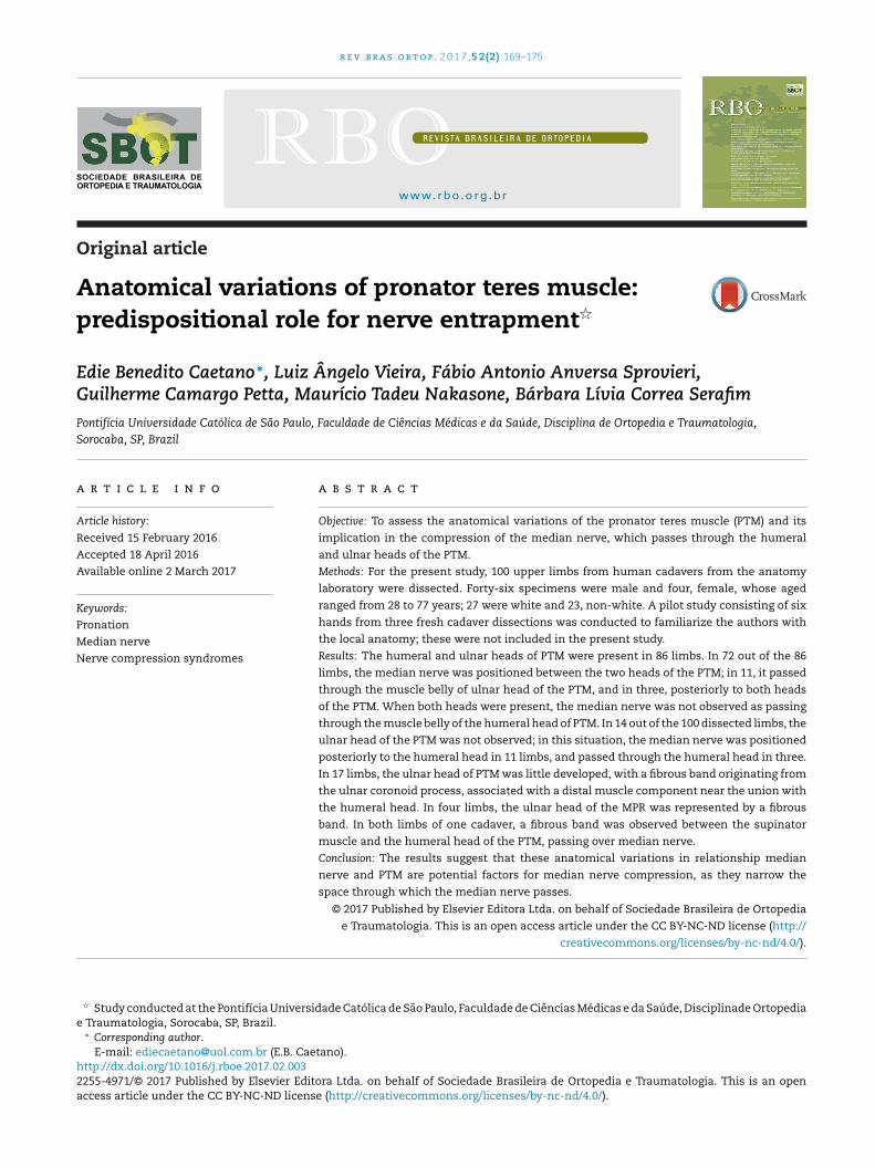
natomical variations of pronator teres muscle:redispositional role for nerve entrapment�
die Benedito Caetano ∗, Luiz Ângelo Vieira, Fábio Antonio Anversa Sprovieri,uilherme Camargo Petta, Maurício Tadeu Nakasone, Bárbara Lívia Correa Serafim
ontifícia Universidade Católica de São Paulo, Faculdade de Ciências Médicas e da Saúde, Disciplina de Ortopedia e Traumatologia,orocaba, SP, Brazil
r t i c l e i n f o
rticle history:
eceived 15 February 2016
ccepted 18 April 2016
vailable online 2 March 2017
eywords:
ronation
edian nerve
erve compression syndromes
a b s t r a c t
Objective: To assess the anatomical variations of the pronator teres muscle (PTM) and its
implication in the compression of the median nerve, which passes through the humeral
and ulnar heads of the PTM.
Methods: For the present study, 100 upper limbs from human cadavers from the anatomy
laboratory were dissected. Forty-six specimens were male and four, female, whose aged
ranged from 28 to 77 years; 27 were white and 23, non-white. A pilot study consisting of six
hands from three fresh cadaver dissections was conducted to familiarize the authors with
the local anatomy; these were not included in the present study.
Results: The humeral and ulnar heads of PTM were present in 86 limbs. In 72 out of the 86
limbs, the median nerve was positioned between the two heads of the PTM; in 11, it passed
through the muscle belly of ulnar head of the PTM, and in three, posteriorly to both heads
of the PTM. When both heads were present, the median nerve was not observed as passing
through the muscle belly of the humeral head of PTM. In 14 out of the 100 dissected limbs, the
ulnar head of the PTM was not observed; in this situation, the median nerve was positioned
posteriorly to the humeral head in 11 limbs, and passed through the humeral head in three.
In 17 limbs, the ulnar head of PTM was little developed, with a fibrous band originating from
the ulnar coronoid process, associated with a distal muscle component near the union with
the humeral head. In four limbs, the ulnar head of the MPR was represented by a fibrous
band. In both limbs of one cadaver, a fibrous band was observed between the supinator
muscle and the humeral head of the PTM, passing over median nerve.
Conclusion: The results suggest that these anatomical variations in relationship median
nerve and PTM are potential factors for median nerve compression, as they narrow the
space through which the median nerve passes.
© 2017 Published by Elsevier Editora Ltda. on behalf of Sociedade Brasileira de Ortopedia
e Traumatologia. This is an open access article under the CC BY-NC-ND license (http://
creativecommons.org/licenses/by-nc-nd/4.0/).
� Study conducted at the Pontifícia Universidade Católica de São Paulo, Faculdade de Ciências Médicas e da Saúde, Disciplinade Ortopedia Traumatologia, Sorocaba, SP, Brazil.∗ Corresponding author.
E-mail: [email protected] (E.B. Caetano).ttp://dx.doi.org/10.1016/j.rboe.2017.02.003255-4971/© 2017 Published by Elsevier Editora Ltda. on behalf of Sociedade Brasileira de Ortopedia e Traumatologia. This is an openccess article under the CC BY-NC-ND license (http://creativecommons.org/licenses/by-nc-nd/4.0/).
170 r e v b r a s o r t o p . 2 0 1 7;5 2(2):169–175
Variacões anatômicas do músculo pronador redondo e sua importâncianas síndromes compressivas
Palavras-chave:
Pronacão
Nervo mediano
Síndromes de compressão
nervosa
r e s u m o
Objetivo: Analisar as variacões anatômicas do músculo pronador redondo (MPR) e suas
implicacões na compressão do nervo mediano, que passa entre as cabecas umeral e ulnar
do MPR.
Método: Foram dissecados 100 membros superiores de cadáveres adultos pertencentes ao
laboratório de anatomia; 46 cadáveres eram do sexo masculino e quatro do feminino. A
idade variou entre 28 e 77 anos; 27 eram da etnia branca e 23, não branca. Um estudo piloto
que incluiu três cadáveres frescos foi feito, para familiarizacão dos autores com a anatomia
regional. Esses não foram incluídos no estudo.
Resultados: Em 86 membros, observou-se a presenca das cabecas umeral e ulnar do MPR. Em
72 dos 86 membros, o nervo mediano estava posicionado entre as cabecas umeral e ulnar
do MPR; em 11, esse encontrava-se através da massa muscular da cabeca ulnar do MPR e em
três, o nervo mediano estava posicionado posteriormente às duas cabecas do MPR. Nos casos
em que as duas cabecas do músculo estavam presentes, não se observou o nervo mediano
passando através da massa muscular da cabeca umeral do MPR. Em 14 dos 100 membros
dissecados, a cabeca ulnar do MPR não estava presente. Nessa situacão, o nervo mediano
posicionava-se posteriormente à cabeca umeral em 11 membros e através da cabeca umeral
em três membros. Em 17 membros, a cabeca ulnar estava muito pouco desenvolvida, com
conformacão fibrosa em sua origem no processo coronoide da ulna, associada a um com-
ponente muscular distal, próximo a sua união com a cabeca umeral. Em quatro membros, a
cabeca ulnar do MPR estava representada apenas por uma banda fibrosa. Nos dois membros
de um cadáver, observou-se uma expansão fibrosa que saía do músculo supinador para a
cabeca umeral do MPR, passando como uma cinta sobre o nervo mediano.
Conclusões: Esses resultados sugerem que as variacões anatômicas na relacão nervo medi-
ano e MPR representam fatores potenciais para compressão nervosa, por estreitar o espaco
no qual passa o nervo mediano.
© 2017 Publicado por Elsevier Editora Ltda. em nome de Sociedade Brasileira de
Ortopedia e Traumatologia. Este e um artigo Open Access sob uma licenca CC BY-NC-ND
across the forearm and lower third of the arm. The skin and
Introduction
There are several anatomical structures that can compress themedian nerve near the elbow joint. From proximal to distal,the compression may be caused by the Struthers’ ligament1,2
with or without the supracondylar process of the humerus,by aponeurotic expansion of the biceps brachii muscle (Lacer-tus fibrosus),3,4 between the humeral and ulnar heads of thepronator teres muscle (PTM),5,6 by the vascular network of theregion,7 and by the arch formed by the two insertions of thesuperficial flexor muscle of the fingers.8
Regardless of these sites where compression occurs, thiscondition is termed pronator teres syndrome, because thecompression occurs most frequently between the two headsof this muscle.9–11 The main causes are the anatomic varia-tions of the PTM. The normal anatomical pattern describedby the classical anatomy studies12–14 is that the PTM is con-stituted by two heads. The humeral head, more extensive,originates in the supracondylar process of the humerus andadjacencies. The ulnar head originates in the coronoid pro-
cess of the ulna. The two portions unite for insertion into thediaphysis of the radius, contouring to it. The median nerve ispositioned between the two heads of the PTM. However, the(http://creativecommons.org/licenses/by-nc-nd/4.0/).
relationship between the median nerve and the humeraland ulnar heads of the PTM is subject to numerousvariations.4,6,15,16 This study aimed to analyze, throughanatomical dissections, the relationship between the PTM andmedian nerve and thus contribute to a better understandingof the causes of the pronator teres syndrome.
Material and methods
One hundred upper limbs of 50 adult cadavers from theanatomy department of this institution were dissected for thisstudy, 46 cadavers were male and four were female. The ageranged from 28 to 77 years; 27 were white and 23, non-white.Cadavers whose forearms were deformed by traumas, malfor-mations, and scars were excluded. A pilot study that includedthree fresh cadavers was conducted so that the authors couldfamiliarize themselves with the local anatomy. These were notincluded in this study.
The dissection was performed through a medial incision
the subcutaneous tissue were folded to the radial and ulnarsides, respectively. The median nerve was identified in themedial margin of the biceps brachii muscle, approximately

0 1 7;5 2(2):169–175 171
1tbitesPtidiItPt(msafiawh
R
Iwpttp(ttibntopa
Aatttaltafimh3a
Median nerve
Median nerve
Ulnar head
Humeral head
Humeral head
Ulnar head
Fig. 1 – In 86 limbs, the humeral and ulnar heads of the PRmuscle were well individualized.
Median nerve
Median nerve
Ulnar head
Ulnar head
Humeral head
Humeral head
r e v b r a s o r t o p . 2
0 cm proximal to the intercondylar line of the humerus; athis location, it was positioned medially in relation to therachial artery. The dissection proceeded distally until reach-
ng the bicipital aponeurosis, which was sectioned, allowinghe visualization of the proximal margin of the PTM. The pres-nce of variations in the surface head and fibrous bands at theite was recorded. Subsequently, the superficial head of theTM was sectioned transversely to allow the visualization ofhe deep head, whose presentation was varied; it was absentn 14 of the 100 dissected limbs. The median nerve was distallyissected until it passed through the arch formed by the prox-
mal insertions of the flexor digitorum superficialis muscles.n all dissected limbs, the first branch of the median nerve inhe forearm was always towards the superficial head of theTM. All forearm muscles were dissected; their innervation,he presence of communication among the forearm nervesMartin-Gruber anastomosis), the relationship between the
edian nerve and the bicipital aponeurosis, and the relation-hip of the median and anterior interosseous nerves with therch of origin of the three heads of the flexor digitorum super-cialis muscle were analyzed. The anatomical variations werennotated and photographed. A Keller 2.5 X magnifying glassas used for magnification. This study was approved by theospital’s ethics committee under the CAAE No. 1.356.351.
esults
n 86 dissected limbs, the humeral and ulnar heads of the PTMere well individualized, consisting of two distinct muscularortions that joined to insert through an enlarged tendon, con-ouring to and inserting in the middle third of the diaphysis ofhe radius (Fig. 1). In 72 of the 86 limbs, the median nerve wasositioned between the humeral and ulnar heads of the PTM
Fig. 1). In 11 limbs (four bilaterally), it was positioned throughhe muscular mass of the ulnar head of the PTM (Fig. 2). Inhree forearms (one bilaterally), the median nerve was pos-tioned posteriorly to the two heads of the PTM (Fig. 3). Whenoth heads of the muscle were present, no cases of the medianerve passing through the muscle mass of the humeral head of
he PTM were observed. In 14 of the 100 limbs, the ulnar headf the PTM was absent. In this situation, the median nerve wasositioned posteriorly to the humeral head in 11 limbs (Fig. 4)nd through the humeral head in three limbs (Fig. 5).
The humeral head was larger than the ulnar head (Fig. 1). poorly developed ulnar head was observed in 17 limbs, with
fibrous conformation in its origin in the coronoid process ofhe ulna, associated with a distal muscular component, closeo its union with the humeral head (Fig. 6). The ulnar head ofhe PTM was represented by a fibrous band not associated with
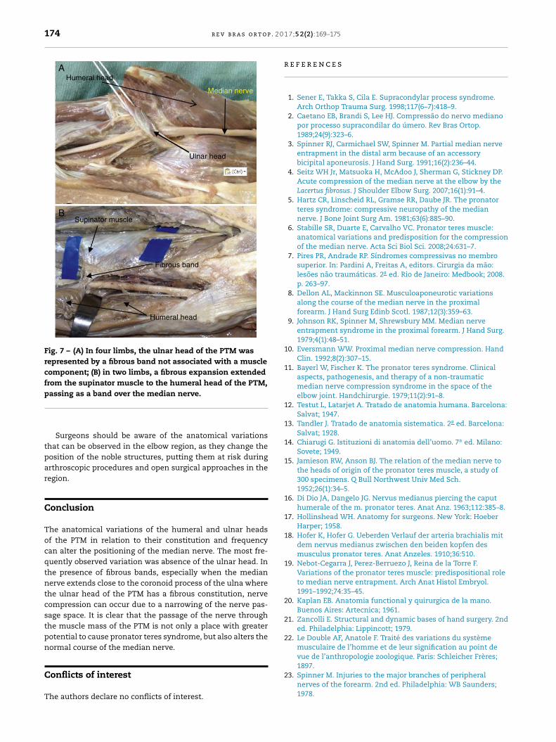
muscular component in only four limbs (Fig. 7A). On bothimbs of a single corpse, a fibrous expansion extending fromhe supinator muscle to the humeral head of the PTM, passings a band over the median nerve, was observed (Fig. 7B). Inve limbs, the ulnar head was inserted alongside the Gantzeruscle, in the coronoid process of the ulna. In eight limbs, a
igh insertion of the humeral head PTM ranging from 2.8 to
.5 cm proximal to the medial epicondyle was observed (Fig. 7And B; Table 1). Fig. 2 – In nine limbs (three bilaterally), the median nervecrossed the muscle mass of the ulnar head of the PTM.
172 r e v b r a s o r t o p . 2 0 1 7;5 2(2):169–175
Table 1 – Evaluation of the humeral and ulnar heads of PTM and their relationships with the median nerve in 100dissected limbs.
Pronator teres Location of the median nerve Percentage % Total
Humeral and ulnarheads present
Between the two heads of the PTM 72 86Posteriorly to the two heads of the PTM 3Through the ulnar head of the PTM 11Through the humeral head of the PTM 0
Absent ulnar head Through the humeral head of the PTM 3 14Posteriorly to the humeral head of the PTM 11
Absent humeral head Not recorded 0 0
a fibrous band in 9% of the cases. In the present study, 17 limbshad a poorly developed ulnar head, with a fibrous component
Total
Discussion
In 86 of the 100 dissected limbs (86%), the humeral andulnar heads of the PTM were present, which is in agreementwith the results reported by Stabille et al.6 (83.5%), Jamiesonand Anson15 (81%), and Hollinshead17 (91%). Other authorsobserved different percentages: Hofer and Hofer,18 56%, andNebot-Cegarra et al.,19 68%. In 74 of these 86 limbs, the mediannerve was positioned between the humeral and ulnar heads ofthe PTM; in 11, the median nerve passed through the muscu-lature of the ulnar head of the PTM. In three limbs, the mediannerve was positioned posteriorly to both PTM heads. In cases
in which both heads of the PTM were present, no cases ofthe median nerve passing through the muscular mass of thehumeral head of the PTM were observed.Median nerve
Median nerve
Humeral head
Humeral head
Ulnar head
Ulnar head
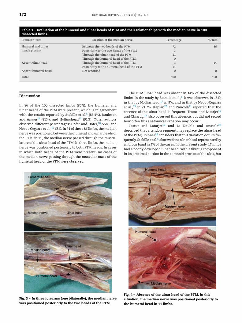
Fig. 3 – In three forearms (one bilaterally), the median nervewas positioned posteriorly to the two heads of the PTM.
100 100
The PTM ulnar head was absent in 14% of the dissectedlimbs. In the study by Stabille et al.,6 it was observed in 15%;in that by Hollinshead,17 in 9%, and in that by Nebot-Cegarraet al.,19 in 21.7%. Kaplan20 and Zancolli21 reported that theabsence of the ulnar head is frequent. Testut and Latarjet12
and Chiarugi14 also observed this absence, but did not recordhow often this anatomical variation may occur.
Testut and Latarjet12 and Le Double and Anatole22
described that a tendon segment may replace the ulnar headof the PTM; Spinner23 considers that this variation occurs fre-quently. Stabille et al.6 observed the ulnar head represented by
in its proximal portion in the coronoid process of the ulna, but
Humeral head
Humeral head
Median nerve
Median nerve
Fig. 4 – Absence of the ulnar head of the PTM. In thissituation, the median nerve was positioned posteriorly tothe humeral head in 11 limbs.
r e v b r a s o r t o p . 2 0 1 7;5 2(2):169–175 173
Median nerve
Median nerve
Humeral head
Humeral head
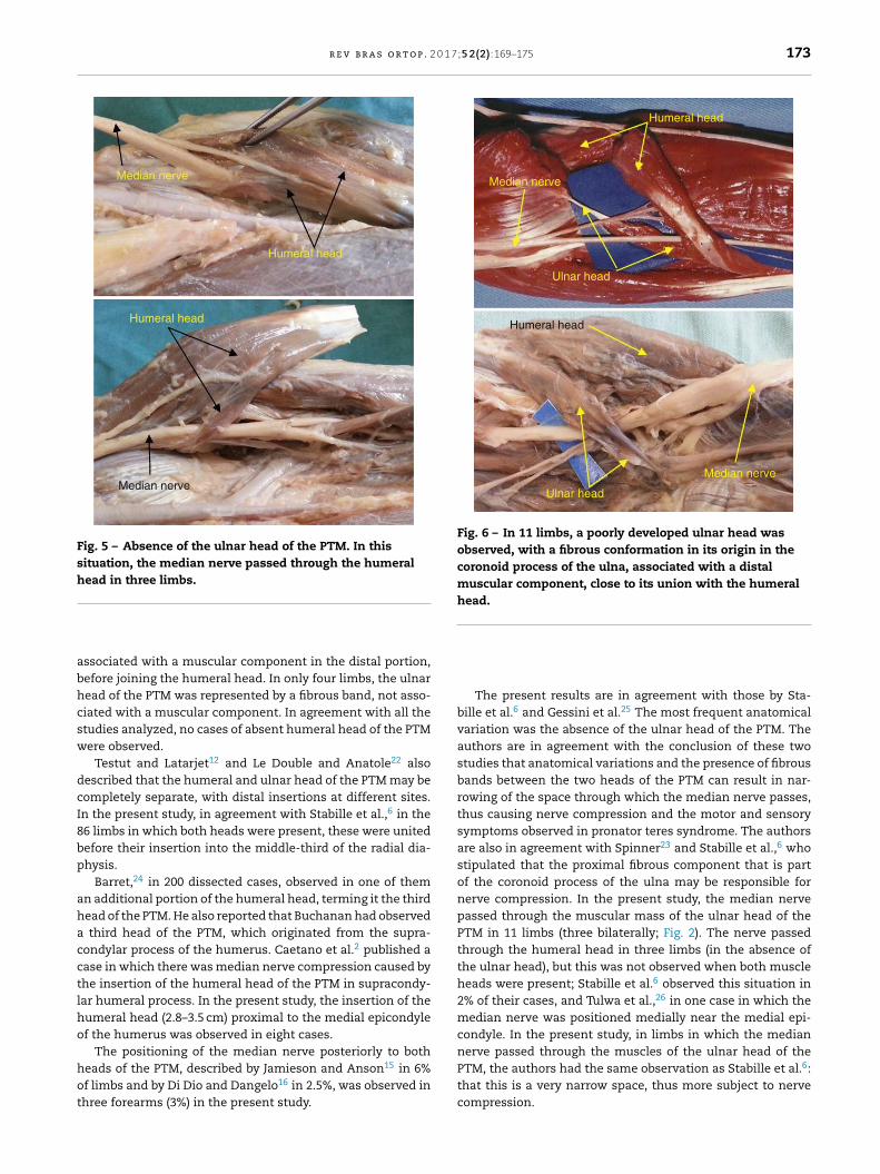
Fig. 5 – Absence of the ulnar head of the PTM. In thissituation, the median nerve passed through the humeralhead in three limbs.
abhcsw
dcI8bp
ahacctlho
hot
Median nerve
Median nerve
Ulnar head
Ulnar head
Humeral head
Humeral head
Fig. 6 – In 11 limbs, a poorly developed ulnar head wasobserved, with a fibrous conformation in its origin in thecoronoid process of the ulna, associated with a distalmuscular component, close to its union with the humeralhead.
nerve passed through the muscles of the ulnar head of thePTM, the authors had the same observation as Stabille et al.6:
ssociated with a muscular component in the distal portion,efore joining the humeral head. In only four limbs, the ulnaread of the PTM was represented by a fibrous band, not asso-iated with a muscular component. In agreement with all thetudies analyzed, no cases of absent humeral head of the PTMere observed.
Testut and Latarjet12 and Le Double and Anatole22 alsoescribed that the humeral and ulnar head of the PTM may beompletely separate, with distal insertions at different sites.n the present study, in agreement with Stabille et al.,6 in the6 limbs in which both heads were present, these were unitedefore their insertion into the middle-third of the radial dia-hysis.
Barret,24 in 200 dissected cases, observed in one of themn additional portion of the humeral head, terming it the thirdead of the PTM. He also reported that Buchanan had observed
third head of the PTM, which originated from the supra-ondylar process of the humerus. Caetano et al.2 published aase in which there was median nerve compression caused byhe insertion of the humeral head of the PTM in supracondy-ar humeral process. In the present study, the insertion of theumeral head (2.8–3.5 cm) proximal to the medial epicondylef the humerus was observed in eight cases.
The positioning of the median nerve posteriorly to botheads of the PTM, described by Jamieson and Anson15 in 6%
f limbs and by Di Dio and Dangelo16 in 2.5%, was observed inhree forearms (3%) in the present study.The present results are in agreement with those by Sta-bille et al.6 and Gessini et al.25 The most frequent anatomicalvariation was the absence of the ulnar head of the PTM. Theauthors are in agreement with the conclusion of these twostudies that anatomical variations and the presence of fibrousbands between the two heads of the PTM can result in nar-rowing of the space through which the median nerve passes,thus causing nerve compression and the motor and sensorysymptoms observed in pronator teres syndrome. The authorsare also in agreement with Spinner23 and Stabille et al.,6 whostipulated that the proximal fibrous component that is partof the coronoid process of the ulna may be responsible fornerve compression. In the present study, the median nervepassed through the muscular mass of the ulnar head of thePTM in 11 limbs (three bilaterally; Fig. 2). The nerve passedthrough the humeral head in three limbs (in the absence ofthe ulnar head), but this was not observed when both muscleheads were present; Stabille et al.6 observed this situation in2% of their cases, and Tulwa et al.,26 in one case in which themedian nerve was positioned medially near the medial epi-condyle. In the present study, in limbs in which the median
that this is a very narrow space, thus more subject to nervecompression.
174 r e v b r a s o r t o p . 2 0
Humeral head
Supinator muscle
Fibrous band
Humeral head
Ulnar head
A
B
Median nerve
Fig. 7 – (A) In four limbs, the ulnar head of the PTM wasrepresented by a fibrous band not associated with a musclecomponent; (B) in two limbs, a fibrous expansion extendedfrom the supinator muscle to the humeral head of the PTM,passing as a band over the median nerve.
r
1
1
1
1
1
1
1
1
1
1
2
2
2
1897.
Surgeons should be aware of the anatomical variationsthat can be observed in the elbow region, as they change theposition of the noble structures, putting them at risk duringarthroscopic procedures and open surgical approaches in theregion.
Conclusion
The anatomical variations of the humeral and ulnar headsof the PTM in relation to their constitution and frequencycan alter the positioning of the median nerve. The most fre-quently observed variation was absence of the ulnar head. Inthe presence of fibrous bands, especially when the mediannerve extends close to the coronoid process of the ulna wherethe ulnar head of the PTM has a fibrous constitution, nervecompression can occur due to a narrowing of the nerve pas-sage space. It is clear that the passage of the nerve throughthe muscle mass of the PTM is not only a place with greaterpotential to cause pronator teres syndrome, but also alters thenormal course of the median nerve.
Conflicts of interest
The authors declare no conflicts of interest.
2
1 7;5 2(2):169–175
e f e r e n c e s
1. Sener E, Takka S, Cila E. Supracondylar process syndrome.Arch Orthop Trauma Surg. 1998;117(6–7):418–9.
2. Caetano EB, Brandi S, Lee HJ. Compressão do nervo medianopor processo supracondilar do úmero. Rev Bras Ortop.1989;24(9):323–6.
3. Spinner RJ, Carmichael SW, Spinner M. Partial median nerveentrapment in the distal arm because of an accessorybicipital aponeurosis. J Hand Surg. 1991;16(2):236–44.
4. Seitz WH Jr, Matsuoka H, McAdoo J, Sherman G, Stickney DP.Acute compression of the median nerve at the elbow by theLacertus fibrosus. J Shoulder Elbow Surg. 2007;16(1):91–4.
5. Hartz CR, Linscheid RL, Gramse RR, Daube JR. The pronatorteres syndrome: compressive neuropathy of the mediannerve. J Bone Joint Surg Am. 1981;63(6):885–90.
6. Stabille SR, Duarte E, Carvalho VC. Pronator teres muscle:anatomical variations and predisposition for the compressionof the median nerve. Acta Sci Biol Sci. 2008;24:631–7.
7. Pires PR, Andrade RP. Síndromes compressivas no membrosuperior. In: Pardini A, Freitas A, editors. Cirurgia da mão:lesões não traumáticas. 2a ed. Rio de Janeiro: Medbook; 2008.p. 263–97.
8. Dellon AL, Mackinnon SE. Musculoaponeurotic variationsalong the course of the median nerve in the proximalforearm. J Hand Surg Edinb Scotl. 1987;12(3):359–63.
9. Johnson RK, Spinner M, Shrewsbury MM. Median nerveentrapment syndrome in the proximal forearm. J Hand Surg.1979;4(1):48–51.
0. Eversmann WW. Proximal median nerve compression. HandClin. 1992;8(2):307–15.
1. Bayerl W, Fischer K. The pronator teres syndrome. Clinicalaspects, pathogenesis, and therapy of a non-traumaticmedian nerve compression syndrome in the space of theelbow joint. Handchirurgie. 1979;11(2):91–8.
2. Testut L, Latarjet A. Tratado de anatomia humana. Barcelona:Salvat; 1947.
3. Tandler J. Tratado de anatomia sistematica. 2a ed. Barcelona:Salvat; 1928.
4. Chiarugi G. Istituzioni di anatomia dell’uomo. 7a ed. Milano:Sovete; 1949.
5. Jamieson RW, Anson BJ. The relation of the median nerve tothe heads of origin of the pronator teres muscle, a study of300 specimens. Q Bull Northwest Univ Med Sch.1952;26(1):34–5.
6. Di Dio JA, Dangelo JG. Nervus medianus piercing the caputhumerale of the m. pronator teres. Anat Anz. 1963;112:385–8.
7. Hollinshead WH. Anatomy for surgeons. New York: HoeberHarper; 1958.
8. Hofer K, Hofer G. Ueberden Verlauf der arteria brachialis mitdem nervus medianus zwischen den beiden kopfen desmusculus pronator teres. Anat Anzeles. 1910;36:510.
9. Nebot-Cegarra J, Perez-Berruezo J, Reina de la Torre F.Variations of the pronator teres muscle: predispositional roleto median nerve entrapment. Arch Anat Histol Embryol.1991–1992;74:35–45.
0. Kaplan EB. Anatomia functional y quirurgica de la mano.Buenos Aires: Artecnica; 1961.
1. Zancolli E. Structural and dynamic bases of hand surgery. 2nded. Philadelphia: Lippincott; 1979.
2. Le Double AF, Anatole F. Traité des variations du systèmemusculaire de l’homme et de leur signification au point devue de l’anthropologie zoologique. Paris: Schleicher Frères;
3. Spinner M. Injuries to the major branches of peripheralnerves of the forearm. 2nd ed. Philadelphia: WB Saunders;1978.

0 1 7
2
2
26. Tulwa N, Limb D, Brown RF. Median nerve compression within
r e v b r a s o r t o p . 2
4. Barrett JH. An additional (third and separate) head of the
pronator teres muscle. J Anat. 1936;70 Pt 4:577–8.5. Gessini L, Jandolo B, Pietrangeli A. Entrapment neuropathiesof the median nerve at and above the elbow. Surg Neurol.1983;19(2):112–6.
;5 2(2):169–175 175
the humeral head of pronator teres. J Hand Surg Br Eur.1994;19(6):709–10.