Embed Size (px)
Citation preview
1
Peripheral Nerve Ultrasound
Jon A. Jacobson, M.D.
Professor of Radiology
Director, Division of Musculoskeletal Radiology
University of Michigan
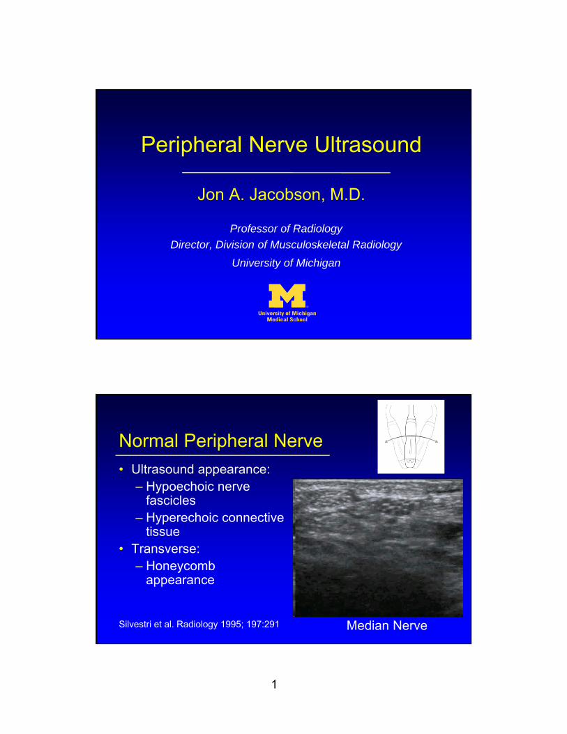
Normal Peripheral Nerve
• Ultrasound appearance:– Hypoechoic nerve
fascicles– Hyperechoic connective
tissue• Transverse:
– Honeycomb appearance
Silvestri et al. Radiology 1995; 197:291 Median Nerve
2
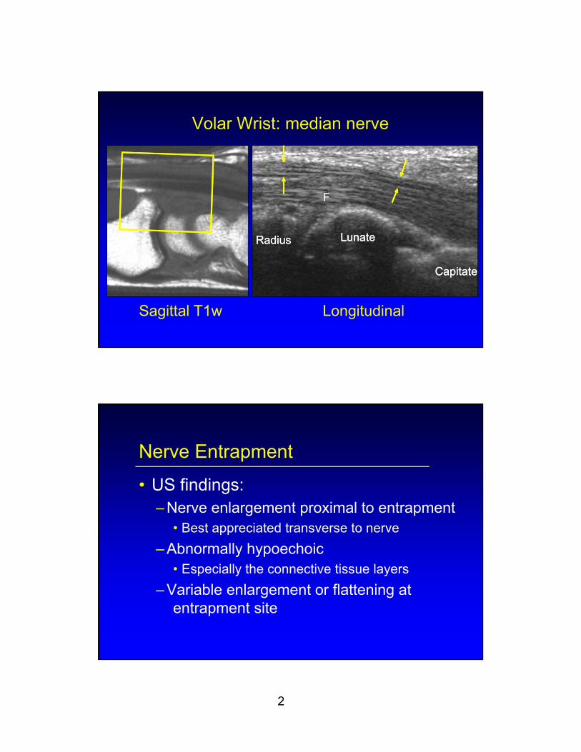
Volar Wrist: median nerve
Sagittal T1w Longitudinal
RadiusRadius LunateLunate
CapitateCapitate
F
Nerve Entrapment
• US findings:– Nerve enlargement proximal to entrapment
• Best appreciated transverse to nerve
– Abnormally hypoechoic• Especially the connective tissue layers
– Variable enlargement or flattening at entrapment site

3
Common Peroneal Nerve: entrapment
Extensor Musculature (Short Axis)
Asymptomatic Atrophy
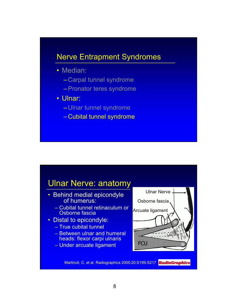
Nerve Entrapment Syndromes
• Median: – Carpal tunnel syndrome
– Pronator teres syndrome
• Ulnar:– Ulnar tunnel syndrome
– Cubital tunnel syndrome

4
Volar Wrist
From: Netter’s Atlas of Human Anatomy
Carpal Tunnel Syndrome:
• Proximal median nerve swelling– Area: circumferential trace– Normal: <9 mm2
– Borderline: 9 – 12 mm2
– Abnormal: > 12 mm2
• 12.8 mm2 = moderate (83% sens, 95% spec)
• 14.0 mm2 = severe (77% sens, 100% spec)
Klauser AS et al. Sem Musculoskel Rad 2010; 14:487Ooi et al. Skeletal Radiol 2014; 43:1387
5
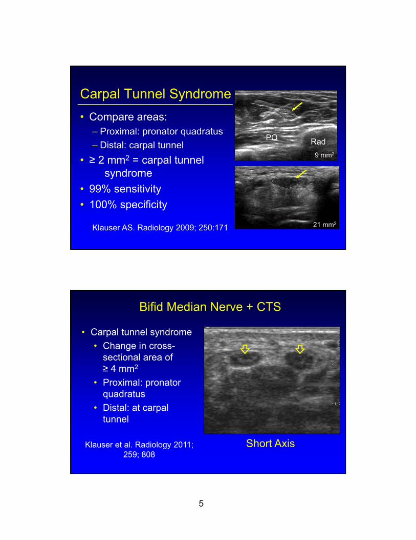
Carpal Tunnel Syndrome
• Compare areas:– Proximal: pronator quadratus
– Distal: carpal tunnel
• ≥ 2 mm2 = carpal tunnel syndrome
• 99% sensitivity
• 100% specificity
Klauser AS. Radiology 2009; 250:171
PQ Rad
21 mm2
9 mm2
Bifid Median Nerve + CTS
Short Axis
• Carpal tunnel syndrome
• Change in cross-sectional area of ≥ 4 mm2
• Proximal: pronator quadratus
• Distal: at carpal tunnel
Klauser et al. Radiology 2011; 259; 808
6
Nerve Entrapment Syndromes
• Median:– Carpal tunnel syndrome
– Pronator teres syndrome
• Ulnar:– Ulnar tunnel syndrome
– Cubital tunnel syndrome
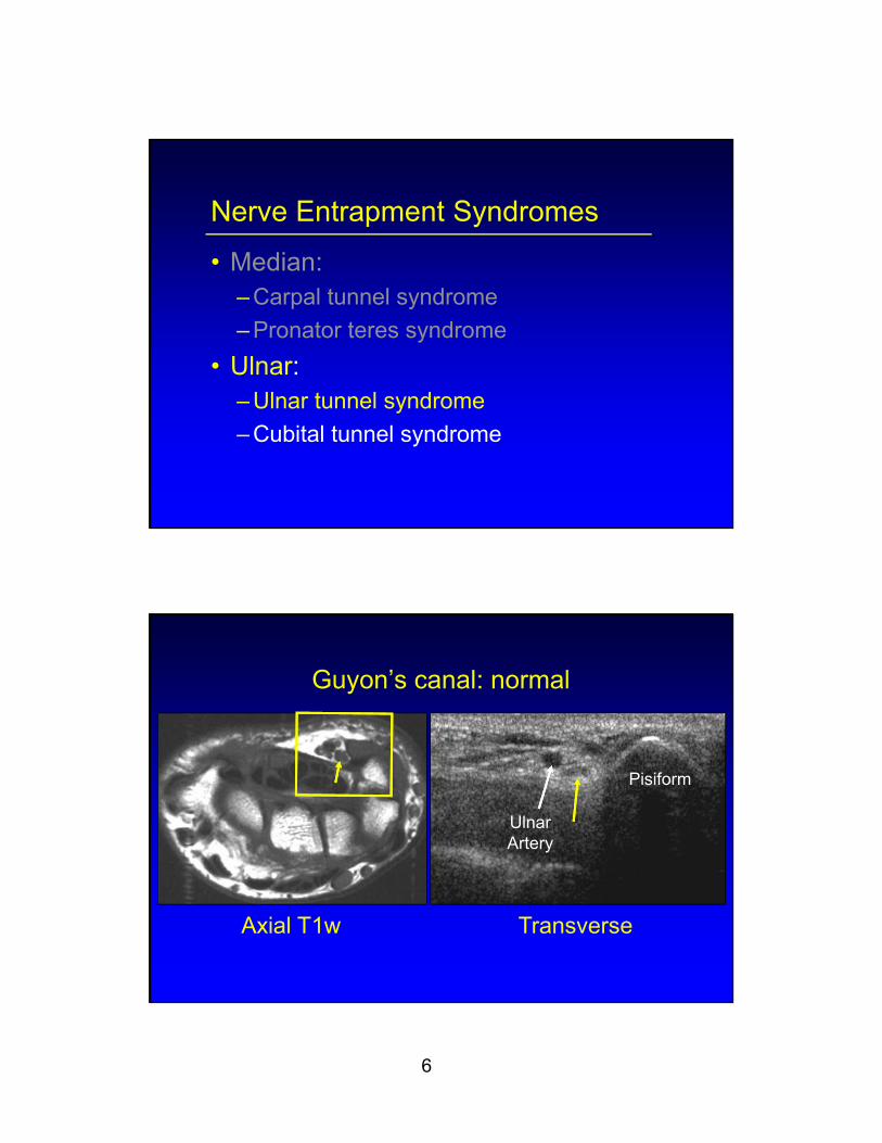
Guyon’s canal: normal
Axial T1w Transverse
UlnarArtery
Pisiform

7
Guyon’s Canal:• Ulnar tunnel syndrome
– Ulnar nerve compression – Accessory Abductor Digiti Minimi1
• Variant: up to 24% of wrists• Hypothenar hammer syndrome2
– Trauma– Ulnar artery thrombosis + distal emboli
1AJR 1999; 172:13972J Vasc Surg 1987; 5:838
Accessory Abductor Digiti Minimi Muscle
Transverse Longitudinal
Ulnar Nerve
Ulnar NerveP
a
Courtesy of V. Flores, MD, Texas
8
Nerve Entrapment Syndromes
• Median: – Carpal tunnel syndrome
– Pronator teres syndrome
• Ulnar:– Ulnar tunnel syndrome
– Cubital tunnel syndrome
Ulnar Nerve: anatomy• Behind medial epicondyle
of humerus:– Cubital tunnel retinaculum or
Osborne fascia• Distal to epicondyle:
– True cubital tunnel– Between ulnar and humeral
heads: flexor carpi ulnaris– Under arcuate ligament
Martinoli, C. et al. Radiographics 2000;20:S199-S217
Ulnar Nerve
Osborne fascia
Arcuate ligament
9
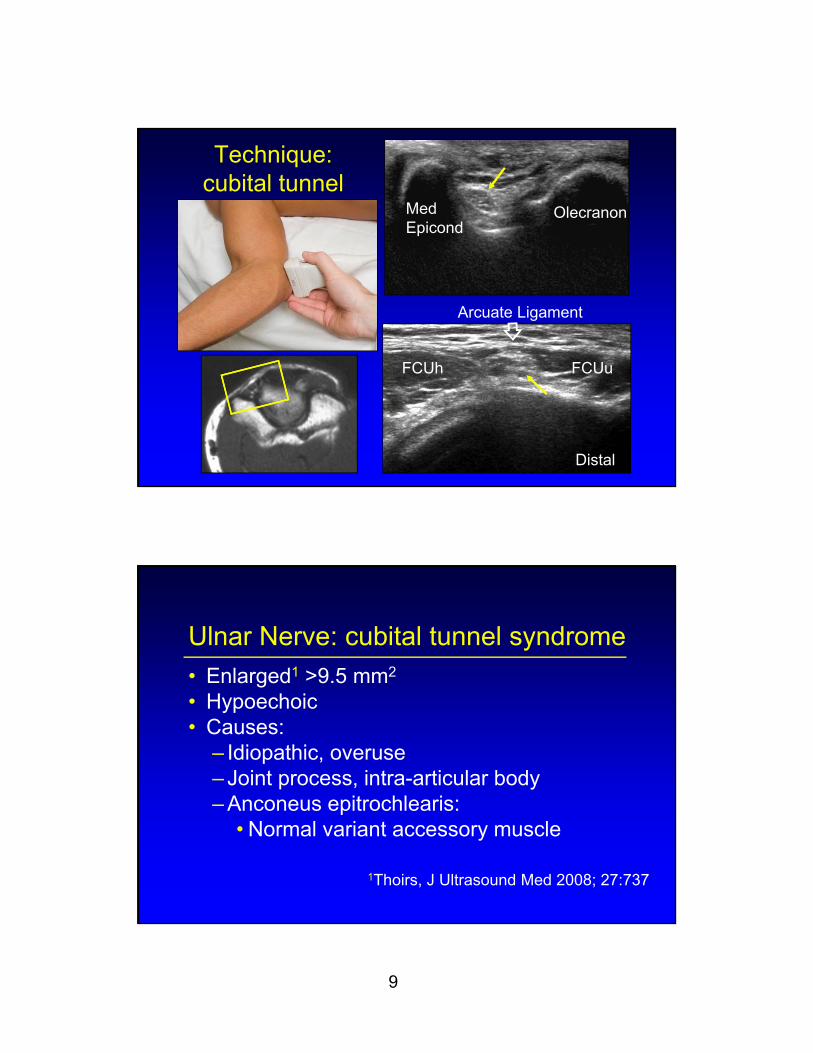
Technique: cubital tunnel
OlecranonMedEpicond
FCUh FCUu
Arcuate Ligament
Distal
Ulnar Nerve: cubital tunnel syndrome
• Enlarged1 >9.5 mm2
• Hypoechoic• Causes:
– Idiopathic, overuse– Joint process, intra-articular body– Anconeus epitrochlearis:
• Normal variant accessory muscle
1Thoirs, J Ultrasound Med 2008; 27:737
10
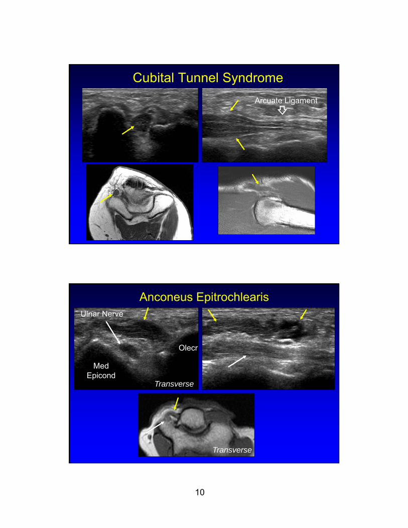
Cubital Tunnel Syndrome
Arcuate Ligament
Anconeus Epitrochlearis
Olecr
Med Epicond
Ulnar Nerve
Transverse
Transverse

11
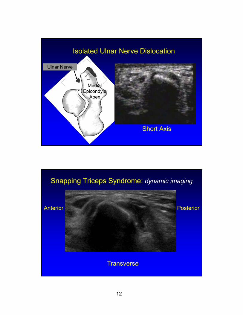
Ulnar Nerve Dislocation
• Occurs in elbow flexion
• Reduces in extension
• Nerve irritation, predisposes to injury
• Found in 20% asymptomatic volunteers
Okamoto, J Hand Surg 2000; 25B:85
Technique: ulnar nerve subluxation
Transverse
OE ET
12
Isolated Ulnar Nerve Dislocation
Short Axis
Medial Epicondyle
Apex
Ulnar Nerve
Snapping Triceps Syndrome: dynamic imaging
Anterior Posterior
Transverse
13
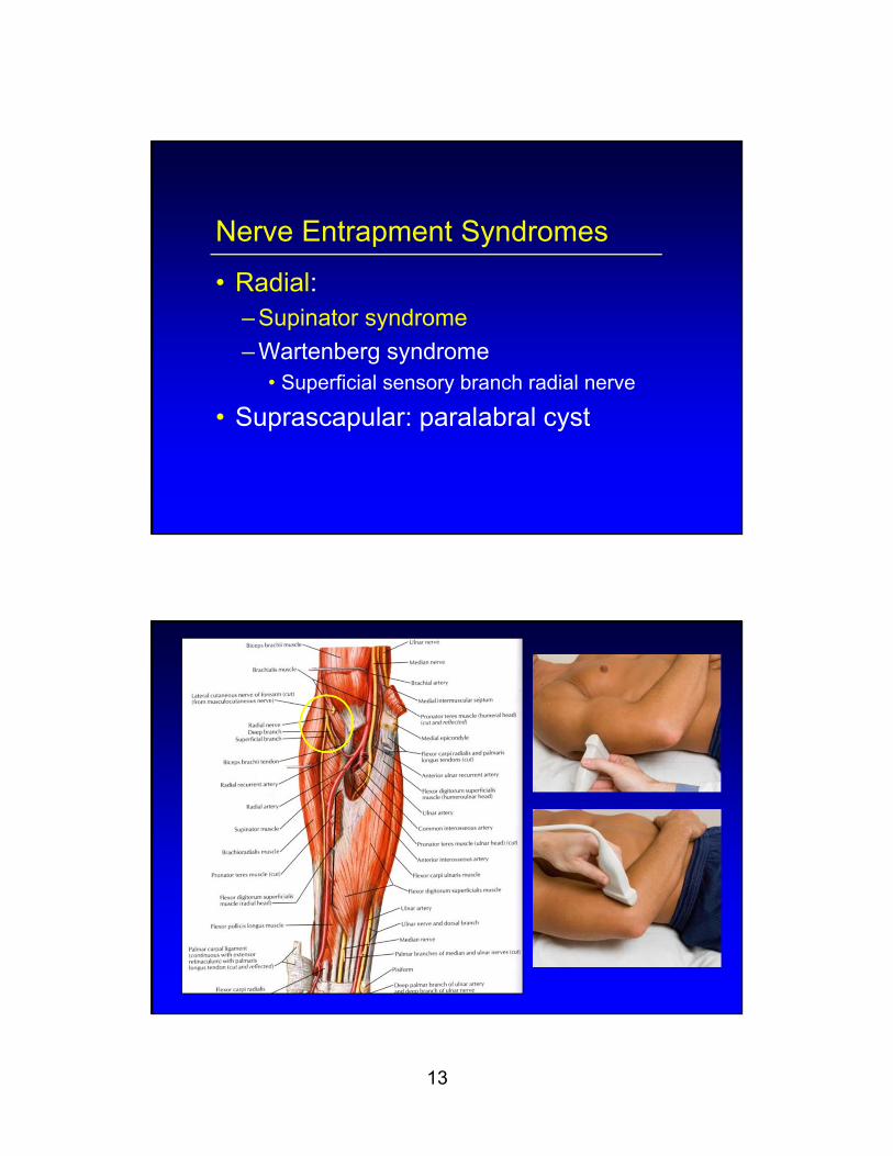
Nerve Entrapment Syndromes
• Radial:– Supinator syndrome
– Wartenberg syndrome• Superficial sensory branch radial nerve
• Suprascapular: paralabral cyst
14
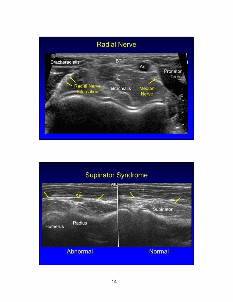
Radial Nerve
Radial Nerve Bifurcation
Brachoradialis BTArt
Brachialis
Pronator Teres
Median Nerve
Supinator Syndrome
Abnormal Normal
HumerusRadius
Supinator
15
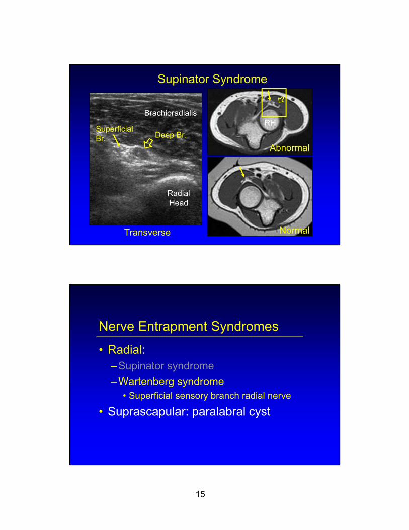
Supinator Syndrome
Transverse
Abnormal
Normal
RadialHead
RHBrachioradialis
Deep Br.Superficial Br.
Nerve Entrapment Syndromes
• Radial:– Supinator syndrome
– Wartenberg syndrome• Superficial sensory branch radial nerve
• Suprascapular: paralabral cyst
16
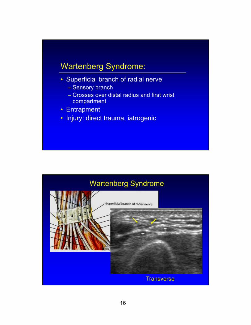
Wartenberg Syndrome:
• Superficial branch of radial nerve– Sensory branch– Crosses over distal radius and first wrist
compartment
• Entrapment• Injury: direct trauma, iatrogenic
Wartenberg Syndrome
Transverse
TT
17
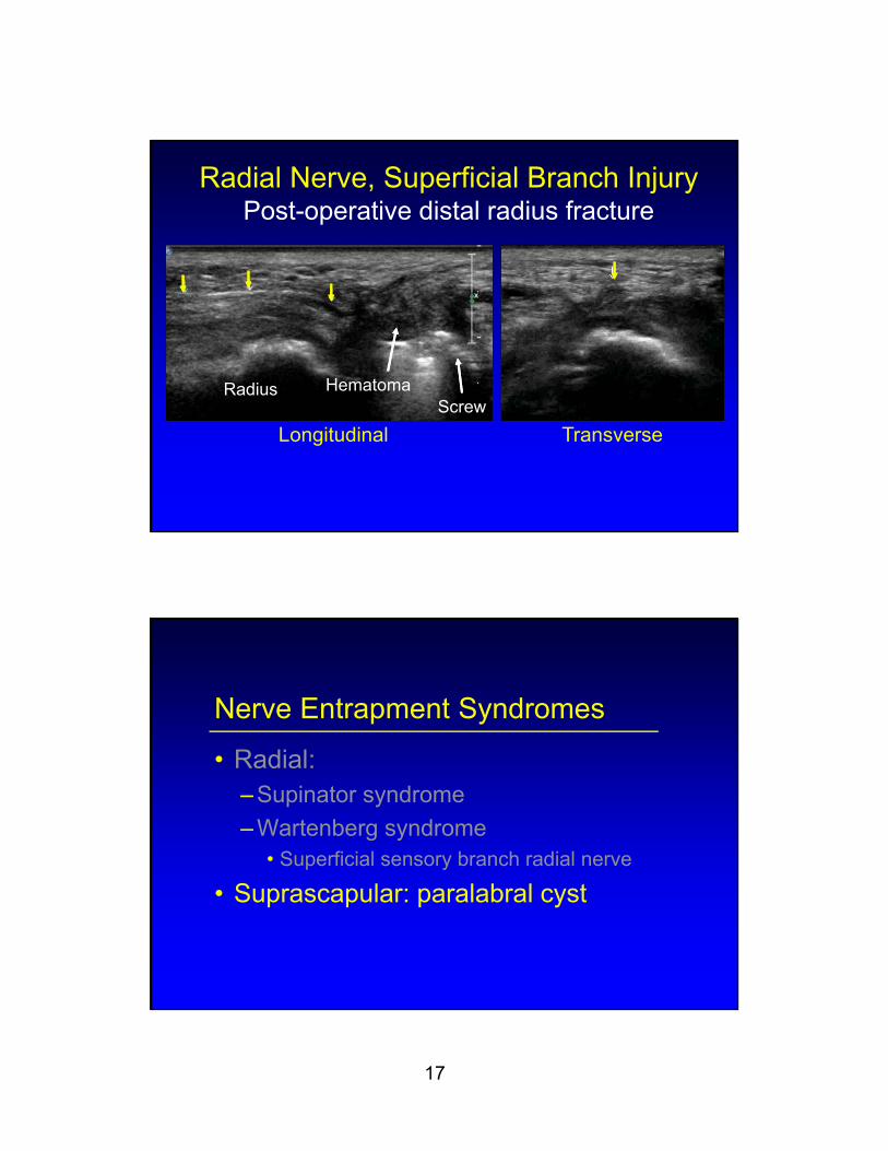
Radial Nerve, Superficial Branch InjuryPost-operative distal radius fracture
Longitudinal Transverse
HematomaRadiusScrew
Nerve Entrapment Syndromes
• Radial:– Supinator syndrome
– Wartenberg syndrome• Superficial sensory branch radial nerve
• Suprascapular: paralabral cyst

18
Labral Cyst:
• Most associated with labral tear• Suprascapular notch:
– Supraspinatus and infraspinatus atrophy
• Spinoglenoid notch:– Infraspinatus atrophy
• US guided aspiration
Labral Cyst: infraspinatus atrophy
19
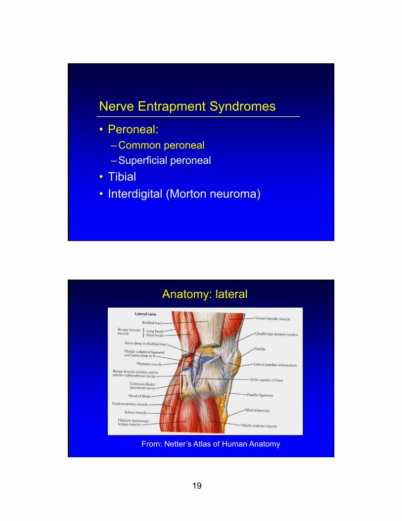
Nerve Entrapment Syndromes
• Peroneal:– Common peroneal
– Superficial peroneal
• Tibial
• Interdigital (Morton neuroma)
Anatomy: lateral
From: Netter’s Atlas of Human Anatomy
20
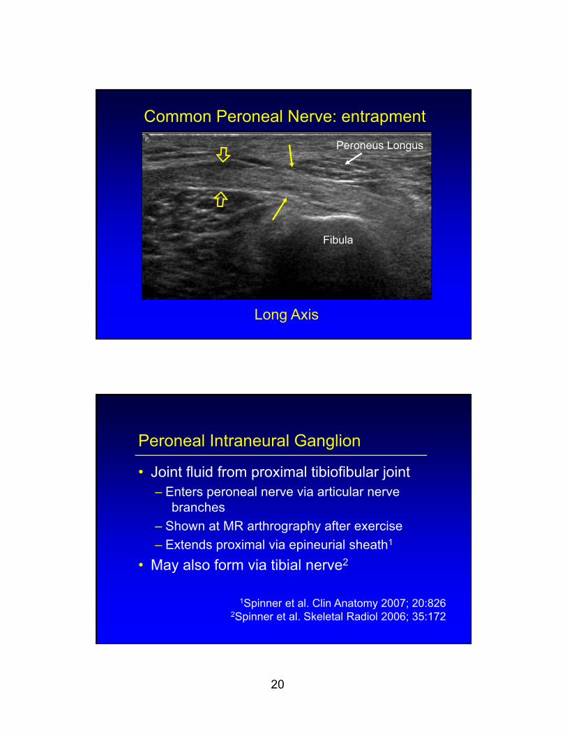
Common Peroneal Nerve: entrapment
Long Axis
Fibula
Peroneus Longus
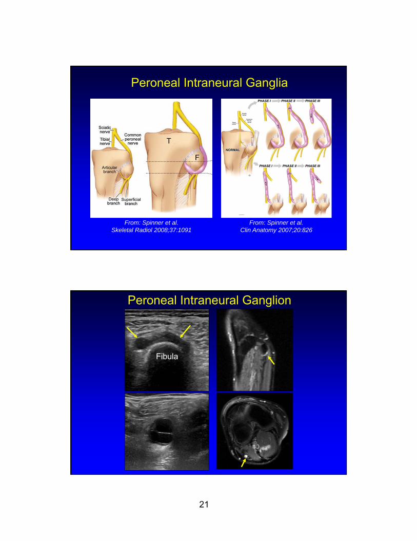
Peroneal Intraneural Ganglion
• Joint fluid from proximal tibiofibular joint– Enters peroneal nerve via articular nerve
branches
– Shown at MR arthrography after exercise
– Extends proximal via epineurial sheath1
• May also form via tibial nerve2
1Spinner et al. Clin Anatomy 2007; 20:8262Spinner et al. Skeletal Radiol 2006; 35:172
21
Peroneal Intraneural Ganglia
From: Spinner et al. Skeletal Radiol 2008;37:1091
From: Spinner et al. Clin Anatomy 2007;20:826
Peroneal Intraneural Ganglion
Fibula

22
Intraneural Ganglion
>15 cm
Atrophy Asymptomatic
Nerve Entrapment Syndromes
• Peroneal:– Common peroneal
– Superficial peroneal
• Tibial
• Interdigital (Morton neuroma)
23
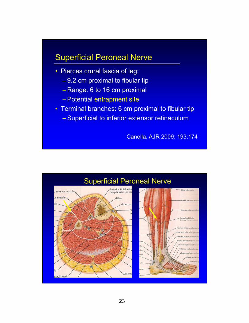
Superficial Peroneal Nerve
• Pierces crural fascia of leg:
– 9.2 cm proximal to fibular tip
– Range: 6 to 16 cm proximal
– Potential entrapment site
• Terminal branches: 6 cm proximal to fibular tip
– Superficial to inferior extensor retinaculum
Canella, AJR 2009; 193:174
Superficial Peroneal Nerve
24
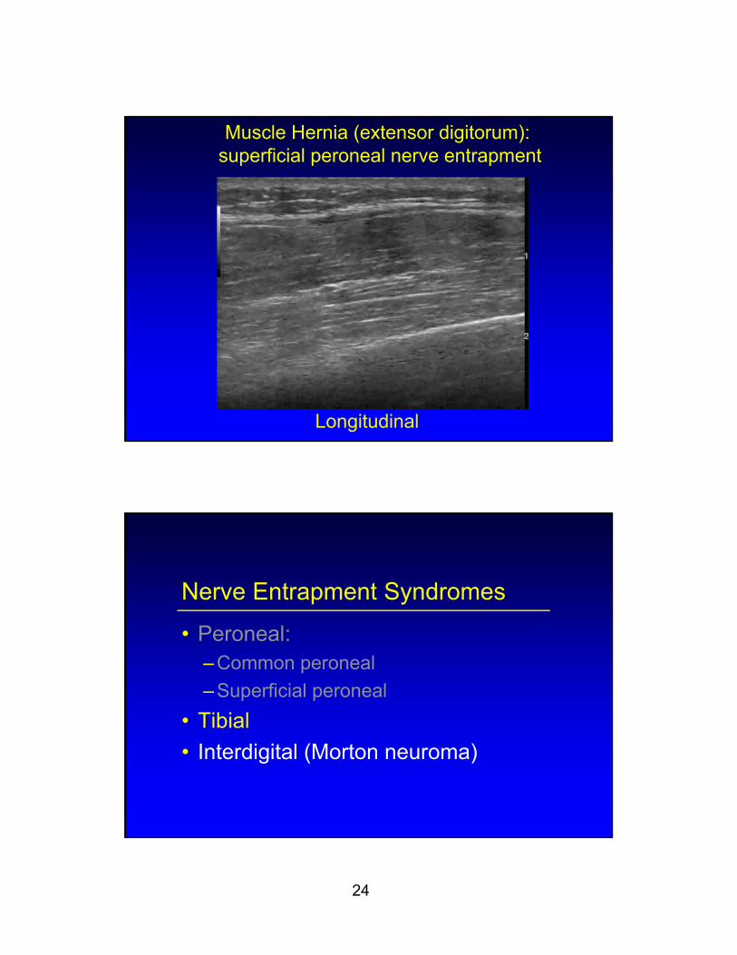
Muscle Hernia (extensor digitorum):superficial peroneal nerve entrapment
Longitudinal
Nerve Entrapment Syndromes
• Peroneal:– Common peroneal
– Superficial peroneal
• Tibial
• Interdigital (Morton neuroma)
25
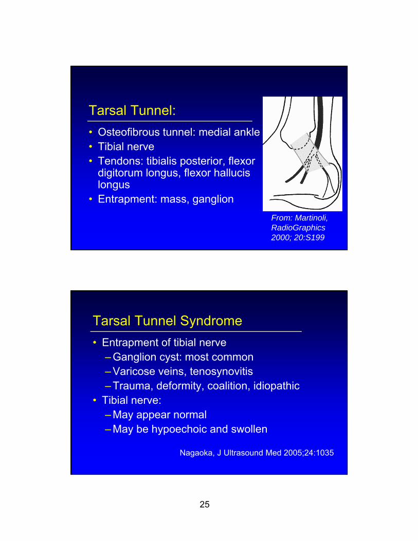
Tarsal Tunnel:
• Osteofibrous tunnel: medial ankle• Tibial nerve• Tendons: tibialis posterior, flexor
digitorum longus, flexor hallucis longus
• Entrapment: mass, ganglion
From: Martinoli, RadioGraphics 2000; 20:S199
Tarsal Tunnel Syndrome
• Entrapment of tibial nerve– Ganglion cyst: most common– Varicose veins, tenosynovitis– Trauma, deformity, coalition, idiopathic
• Tibial nerve:– May appear normal– May be hypoechoic and swollen
Nagaoka, J Ultrasound Med 2005;24:1035
26
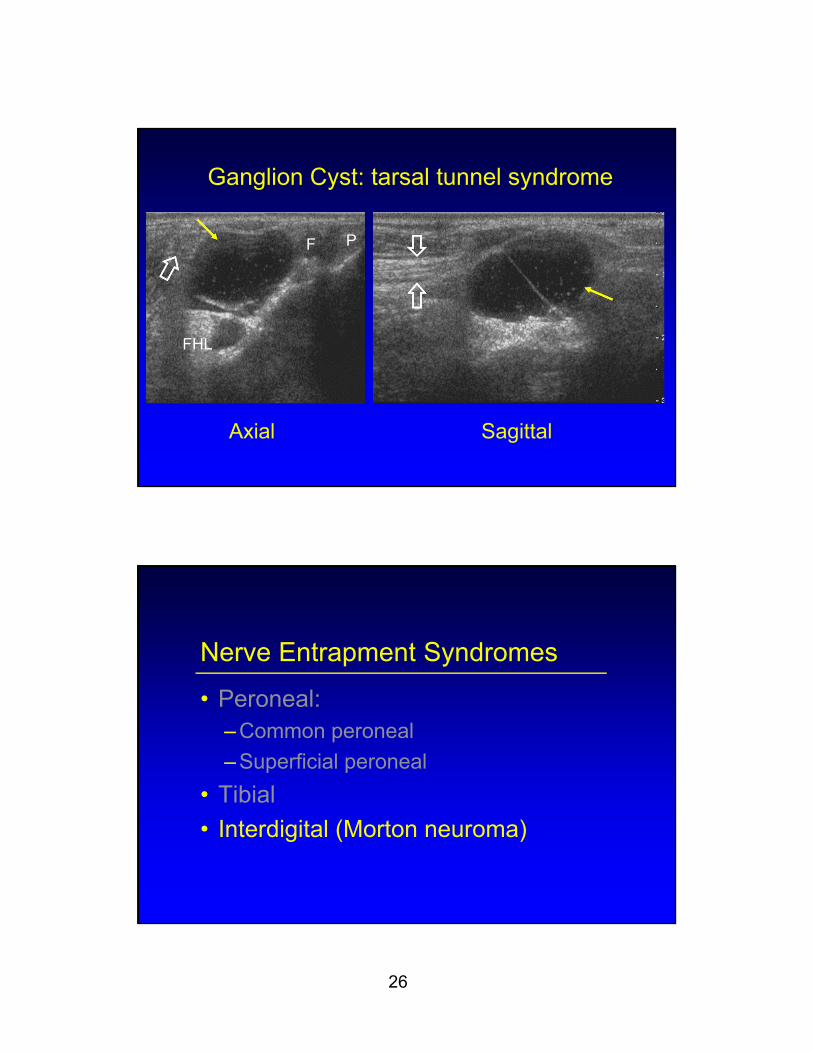
Ganglion Cyst: tarsal tunnel syndrome
Axial Sagittal
F P
FHL
Nerve Entrapment Syndromes
• Peroneal:– Common peroneal
– Superficial peroneal
• Tibial
• Interdigital (Morton neuroma)
27
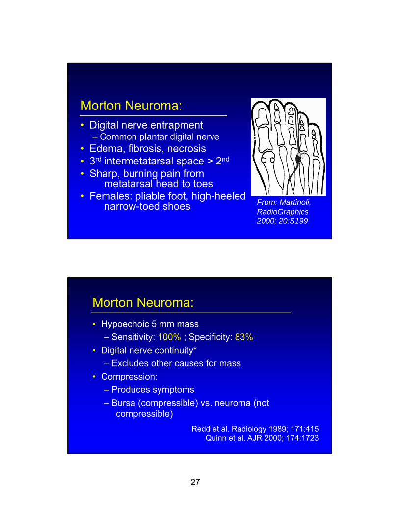
Morton Neuroma:
• Digital nerve entrapment– Common plantar digital nerve
• Edema, fibrosis, necrosis• 3rd intermetatarsal space > 2nd
• Sharp, burning pain from metatarsal head to toes
• Females: pliable foot, high-heeled narrow-toed shoes From: Martinoli,
RadioGraphics 2000; 20:S199
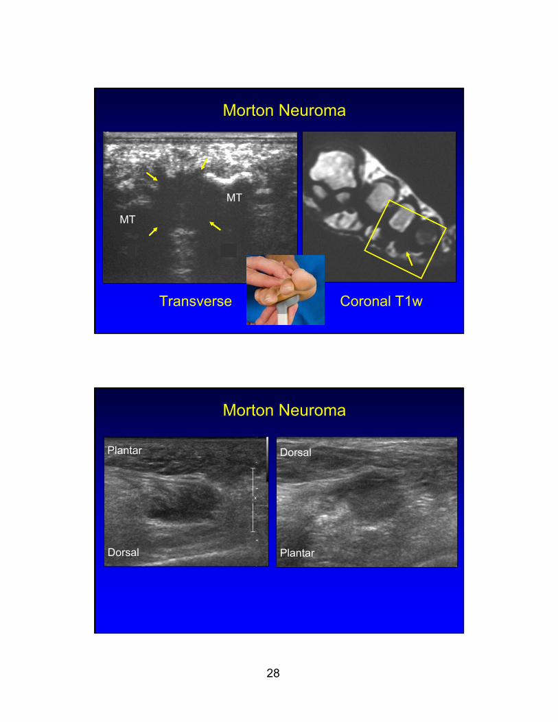
Morton Neuroma:
• Hypoechoic 5 mm mass
– Sensitivity: 100% ; Specificity: 83%
• Digital nerve continuity*
– Excludes other causes for mass
• Compression:
– Produces symptoms
– Bursa (compressible) vs. neuroma (not compressible)
Redd et al. Radiology 1989; 171:415Quinn et al. AJR 2000; 174:1723
28
Morton Neuroma
Transverse Coronal T1w
MT
MT
Morton Neuroma
Plantar
Plantar
Dorsal
Dorsal
29
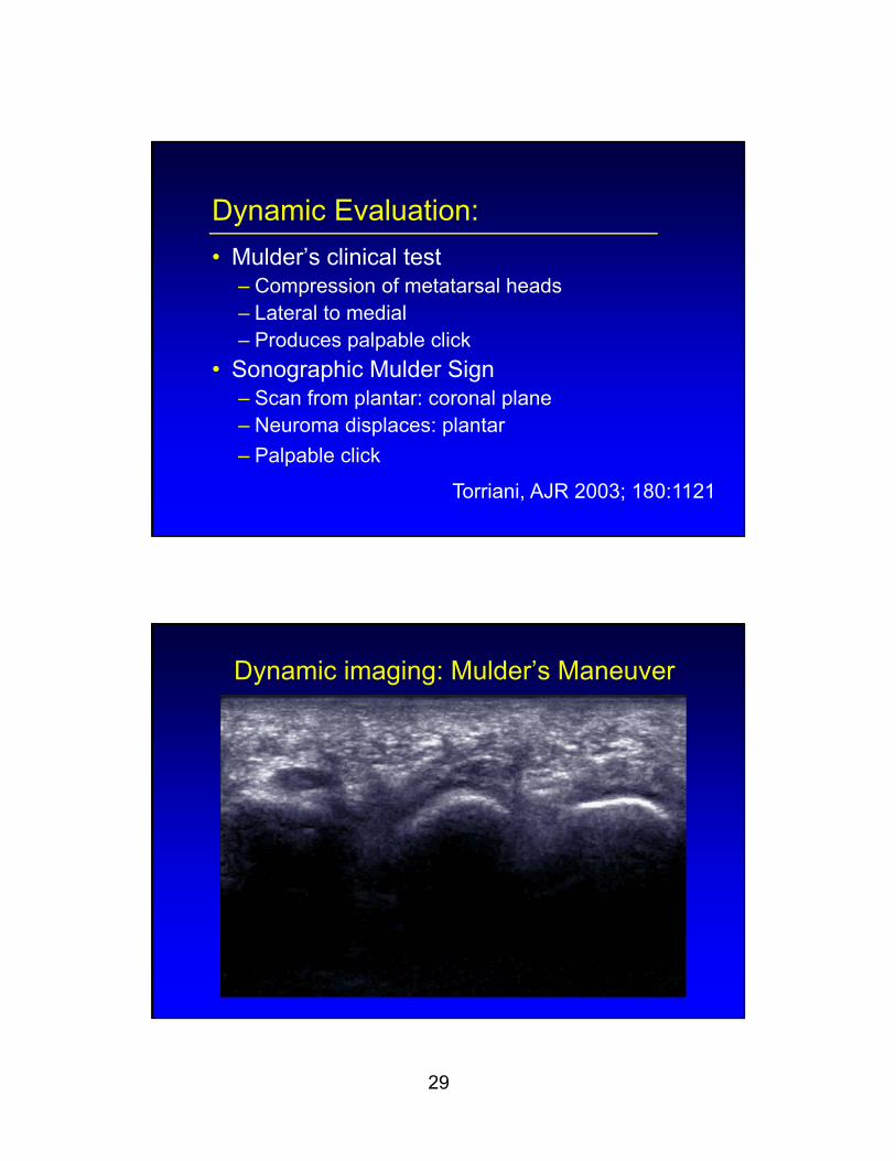
Dynamic Evaluation:
• Mulder’s clinical test– Compression of metatarsal heads– Lateral to medial– Produces palpable click
• Sonographic Mulder Sign– Scan from plantar: coronal plane– Neuroma displaces: plantar
– Palpable click
Torriani, AJR 2003; 180:1121
Dynamic imaging: Mulder’s Maneuver
30
Summary: peripheral nerve US
• Resolution better than MRI
• Evaluate entire limb efficiently
• Easy comparison to contralateral side
• Direct correlation: signs and symptoms
• Dynamic imaging
![Electrophysiological Features of Ulnar Tunnel Syndrome ... · Ulnar tunnel syndrome (UTS) is an uncommon ulnar entrapment neuropathy. Guyon [1] described the anatomy of the area in](https://img.pdfslide.net/doc/110x75/601bca5f935324075a08994b/electrophysiological-features-of-ulnar-tunnel-syndrome-ulnar-tunnel-syndrome.jpg)


![DOI: Journal of Clinical Case Reports...The term “ulnar tunnel syndrome” was coined by DuPont in 1965 to describe the condition of 4 patients with acquired ulnar neuritis [1]](https://img.pdfslide.net/doc/110x75/6085e428a47e3f5d3e52106c/doi-journal-of-clinical-case-reports-the-term-aoeulnar-tunnel-syndromea-was.jpg)


![[Chapter 73] Carpal Tunnel, Ulnar Tunnel, And Stenosing Tenosynovitis](https://img.pdfslide.net/doc/110x75/5451d5deb1af9f83248b4a66/chapter-73-carpal-tunnel-ulnar-tunnel-and-stenosing-tenosynovitis.jpg)


