Embed Size (px)
Citation preview
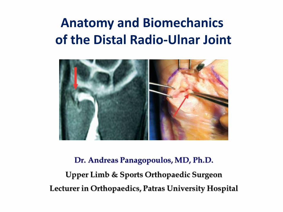
Anatomy and Biomechanics of the Distal Radio-Ulnar Joint
Anatomy and Biomechanics of DRUJ
Extrinsic & intrinsic stabilizers
Clinical considerations
Common problems
- TFCC tears
- ulnar impaction syndrome
- DRJU instability
- DRJU arthritis
Interosseous membrane biomechanics
Contents
DRUJ is a diarthrodial, synovial articulation between the sigmoid notch of the distal radius and ulnar head. It acts as the link between radius and ulna and a pivot for prono-supination. The ulna is the stable unit of the forearm around which the radius rotates
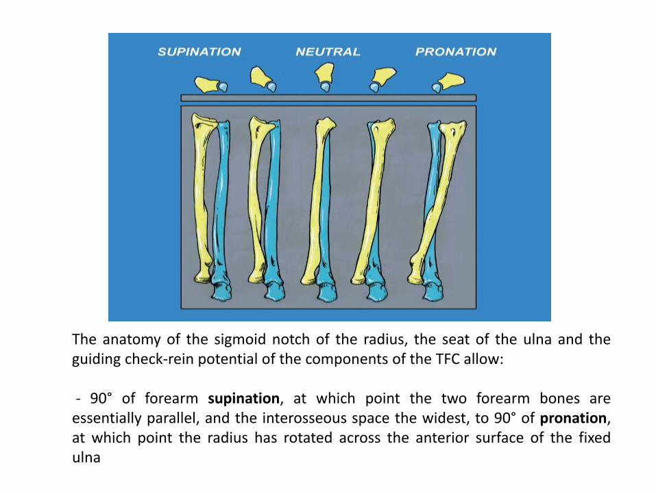
The anatomy of the sigmoid notch of the radius, the seat of the ulna and the guiding check-rein potential of the components of the TFC allow: - 90° of forearm supination, at which point the two forearm bones are essentially parallel, and the interosseous space the widest, to 90° of pronation, at which point the radius has rotated across the anterior surface of the fixed ulna
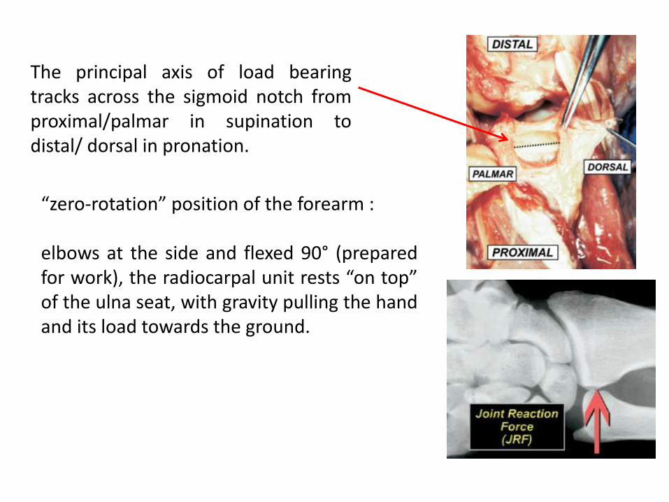
“zero-rotation” position of the forearm : elbows at the side and flexed 90° (prepared for work), the radiocarpal unit rests “on top” of the ulna seat, with gravity pulling the hand and its load towards the ground.
The principal axis of load bearing tracks across the sigmoid notch from proximal/palmar in supination to distal/ dorsal in pronation.
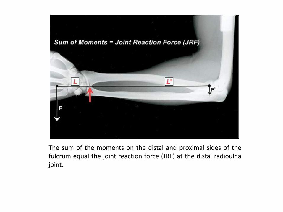
The load in the hand (F) times the distance of the load from the fulcrum (L) must be equal to the length of the forearm from the fulcrum (L’) times the resistance to displacement provided by the annular ligament at the radial head (F’).
The sum of the moments on the distal and proximal sides of the fulcrum equal the joint reaction force (JRF) at the distal radioulna joint.
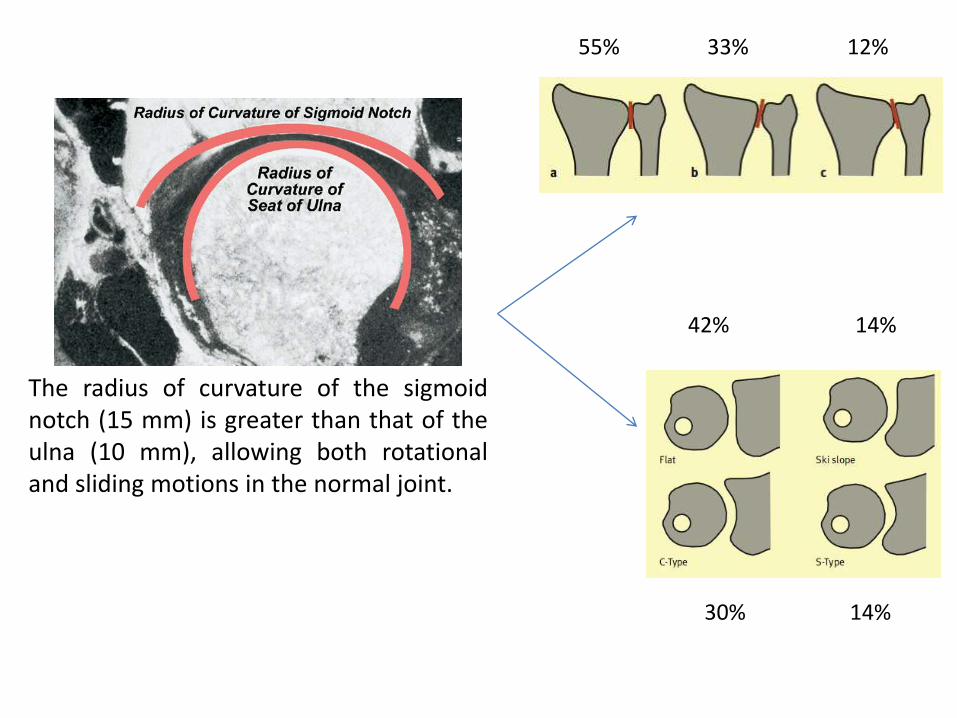
The radius of curvature of the sigmoid notch (15 mm) is greater than that of the ulna (10 mm), allowing both rotational and sliding motions in the normal joint.
55% 33% 12%
42% 14% 30% 14%
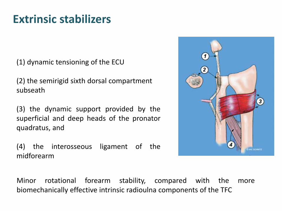
Extrinsic stabilizers
(1) dynamic tensioning of the ECU (2) the semirigid sixth dorsal compartment subseath (3) the dynamic support provided by the superficial and deep heads of the pronator quadratus, and (4) the interosseous ligament of the midforearm
Minor rotational forearm stability, compared with the more biomechanically effective intrinsic radioulna components of the TFC

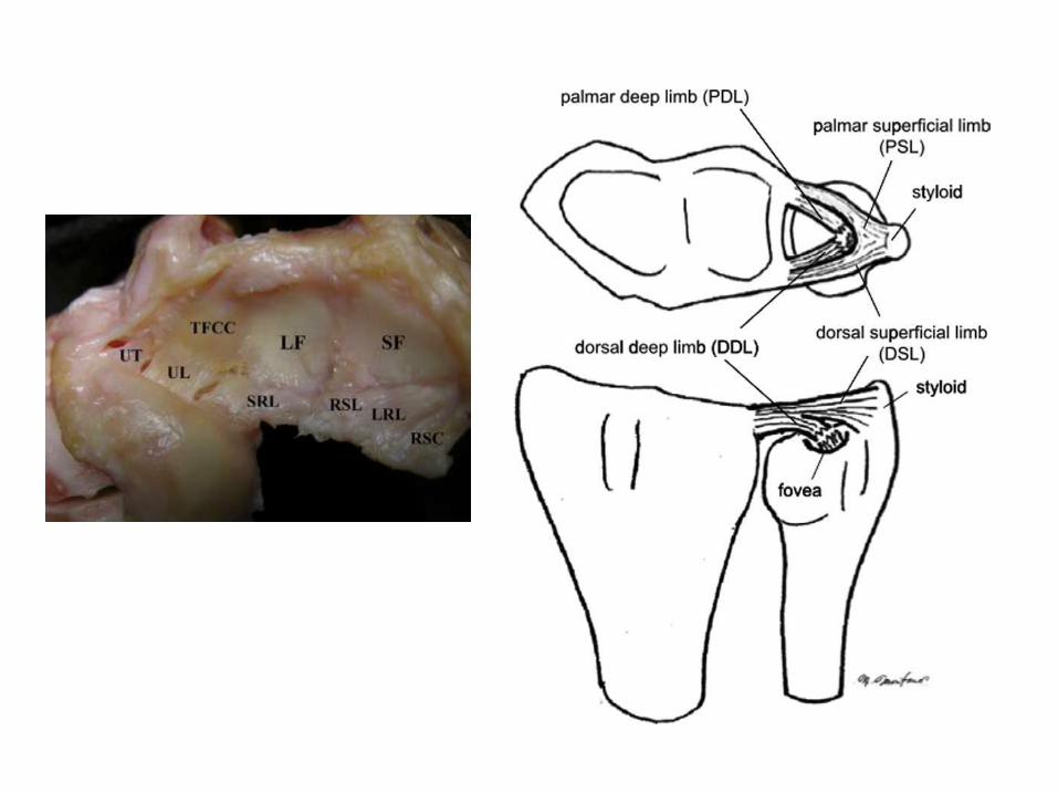
Intrinsic stabilizers
The articular disc is responsible for transferring load from the medial carpus to the pole of the distal ulna.
The TFC complex: superficial (green) and deep (blue) radioulna fibers, the two disc-carpal ligaments (disc-lunate and disc-triquetral), and the central articular disc (white).
fovea styloid
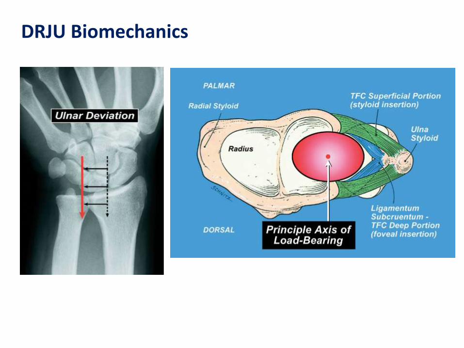
DRJU Biomechanics
84%
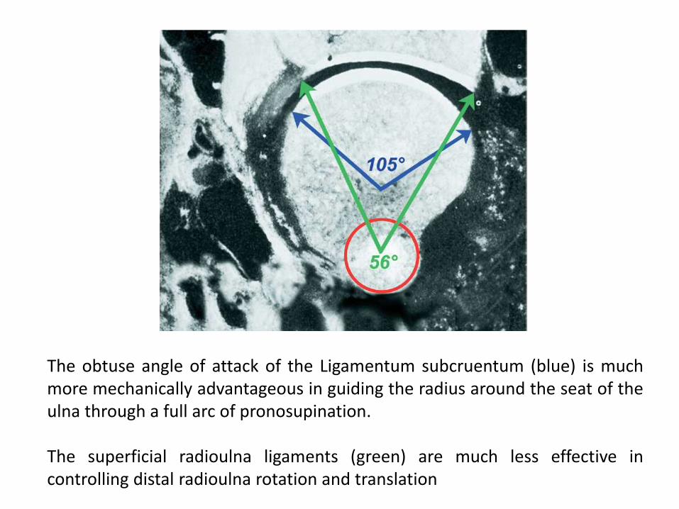
The obtuse angle of attack of the Ligamentum subcruentum (blue) is much more mechanically advantageous in guiding the radius around the seat of the ulna through a full arc of pronosupination. The superficial radioulna ligaments (green) are much less effective in controlling distal radioulna rotation and translation
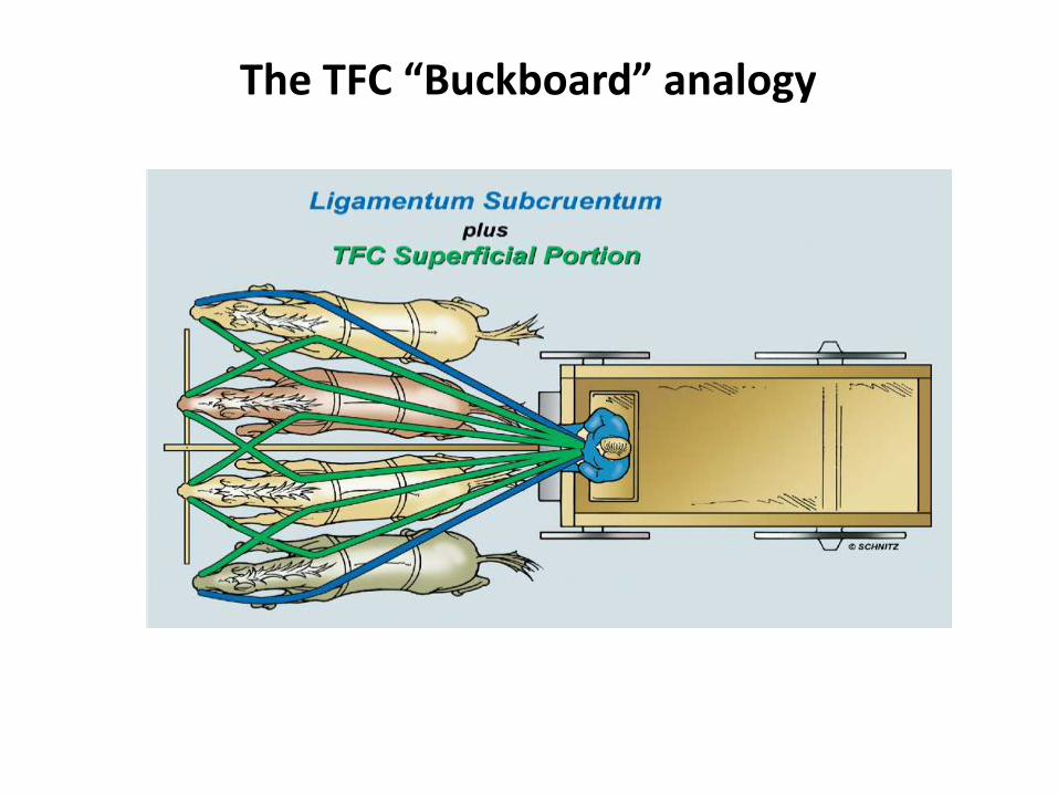
The TFC “Buckboard” analogy

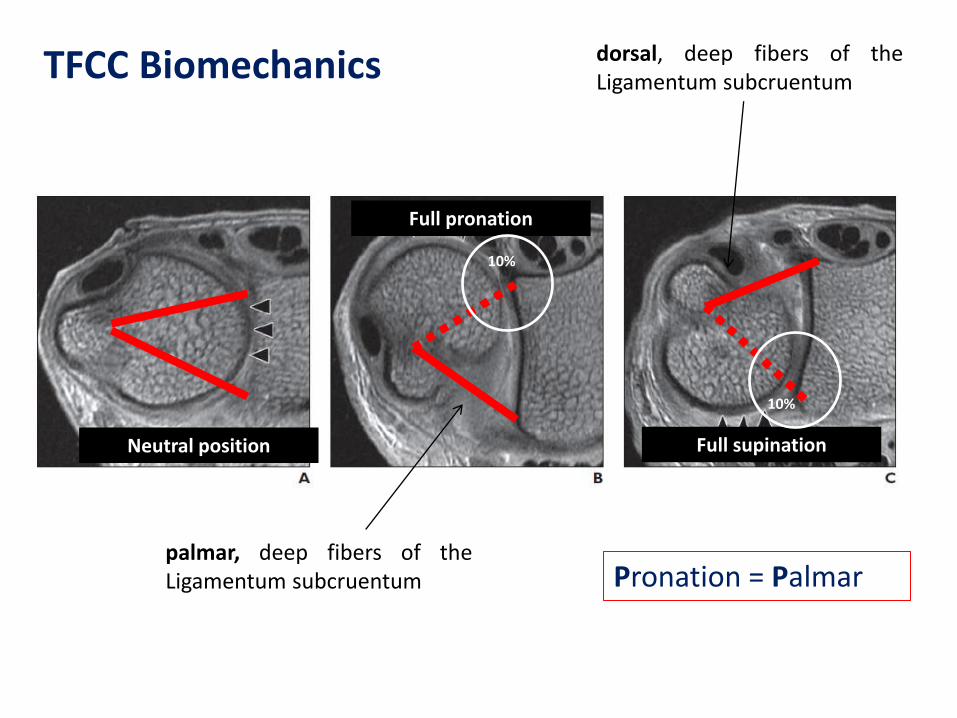
TFCC Biomechanics
In forearm pronation, the dorsal superficial fibers of the TFC must tighten for stability, as do the deep palmar fibers of the Ligamentum subcruentum. In supination, the palmar superficial TFC radioulna fibers (to the ulna styloid) tighten, as do the deep dorsal fibers of the Ligamentum subcruentum
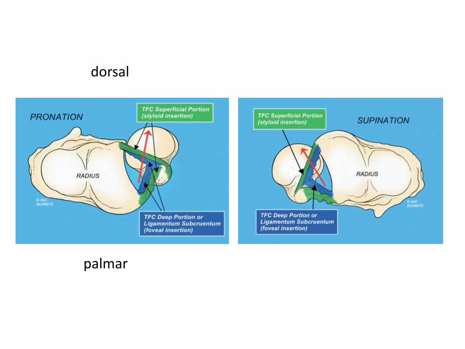
dorsal
palmar
TFCC Biomechanics
palmar, deep fibers of the Ligamentum subcruentum
dorsal, deep fibers of the Ligamentum subcruentum
Full pronation
Full supination Neutral position
10%
10%
Pronation = Palmar
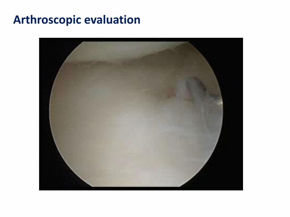
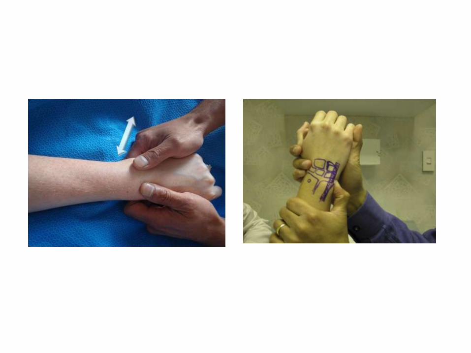
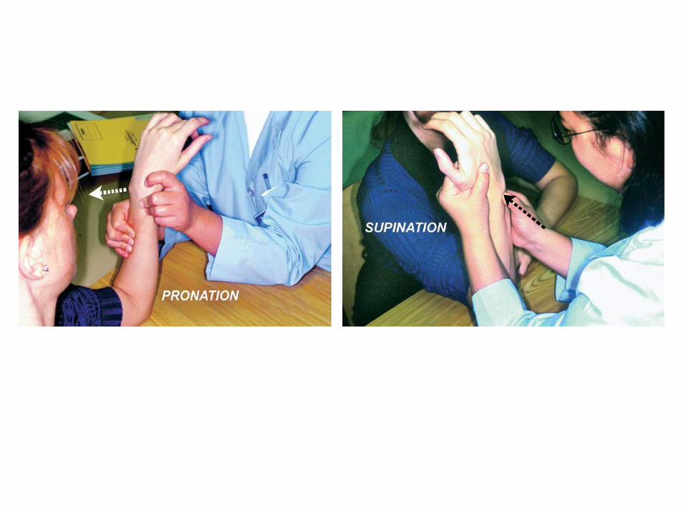
Arthroscopic evaluation

Clinical evaluation
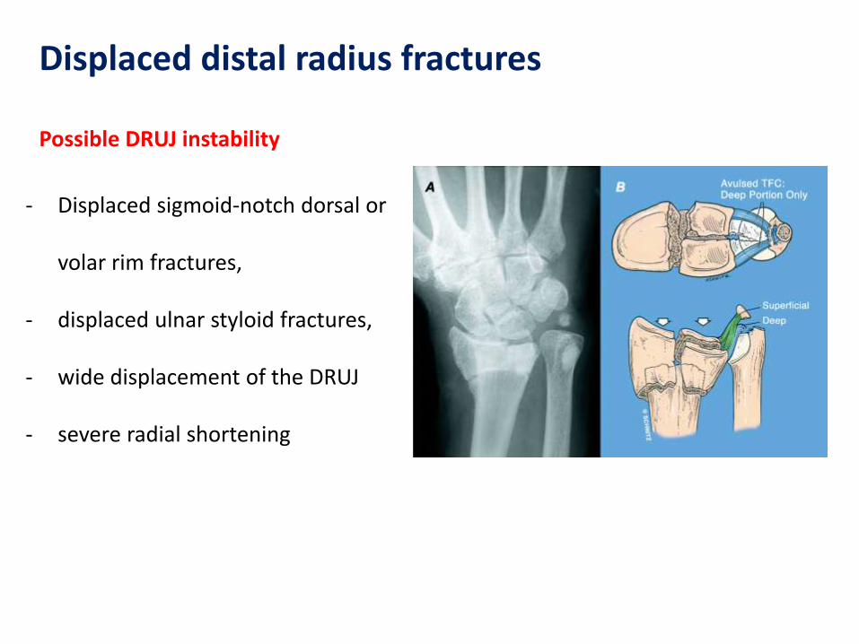
Displaced distal radius fractures
- Displaced sigmoid-notch dorsal or
volar rim fractures,
- displaced ulnar styloid fractures,
- wide displacement of the DRUJ
- severe radial shortening
Possible DRUJ instability

TFCC tears
- the most common source of
ulnar-sided wrist pain,
- traumatic or degenerative
- foveal tenderness
- fall from height or an object
falling against a pronated and
extended wrist

The most common traumatic TFCC injury There is a tear or perforation of the central aspect of the disc of the TFCC Conservative initially (splint), injections Arthroscopic debridement (to create a stable rim of TFCC) Success rates range from 66% to 87% In ulnarpositive wrists an ulnocarpal unloading procedure is recommended
CLASS 1A
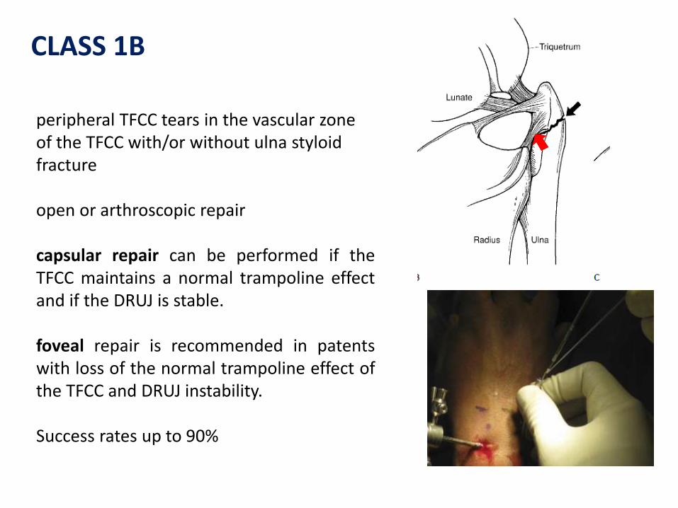
peripheral TFCC tears in the vascular zone of the TFCC with/or without ulna styloid fracture open or arthroscopic repair capsular repair can be performed if the TFCC maintains a normal trampoline effect and if the DRUJ is stable. foveal repair is recommended in patents with loss of the normal trampoline effect of the TFCC and DRUJ instability. Success rates up to 90%
CLASS 1B
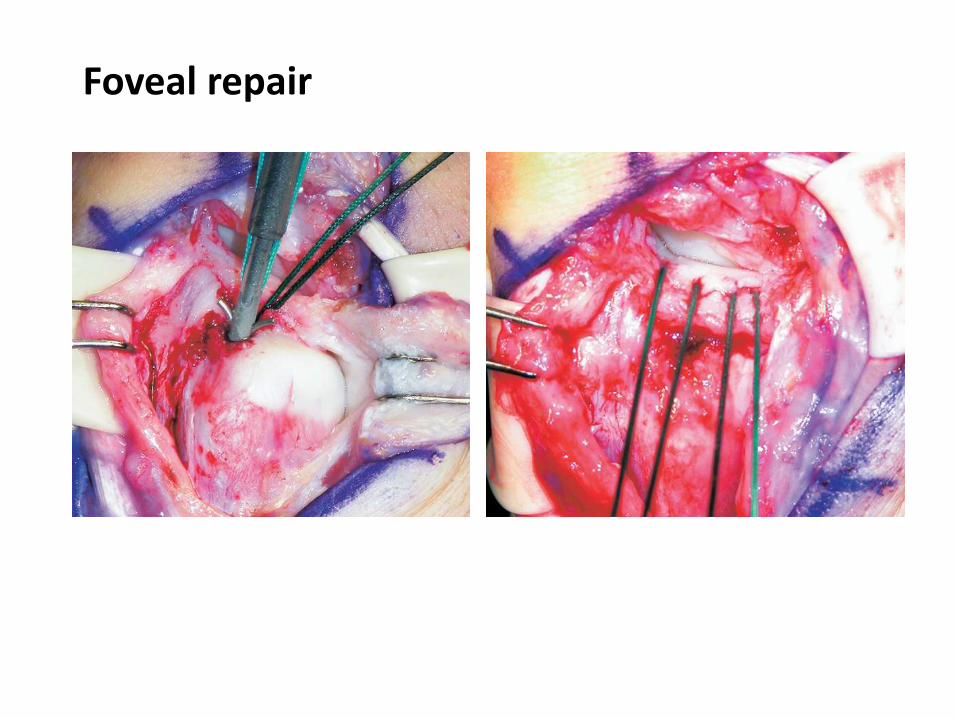
Foveal repair
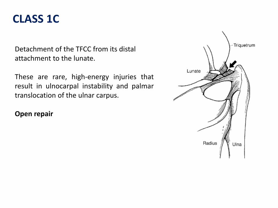
Detachment of the TFCC from its distal attachment to the lunate. These are rare, high-energy injuries that result in ulnocarpal instability and palmar translocation of the ulnar carpus. Open repair
CLASS 1C
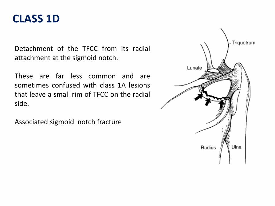
Detachment of the TFCC from its radial attachment at the sigmoid notch. These are far less common and are sometimes confused with class 1A lesions that leave a small rim of TFCC on the radial side. Associated sigmoid notch fracture
CLASS 1D
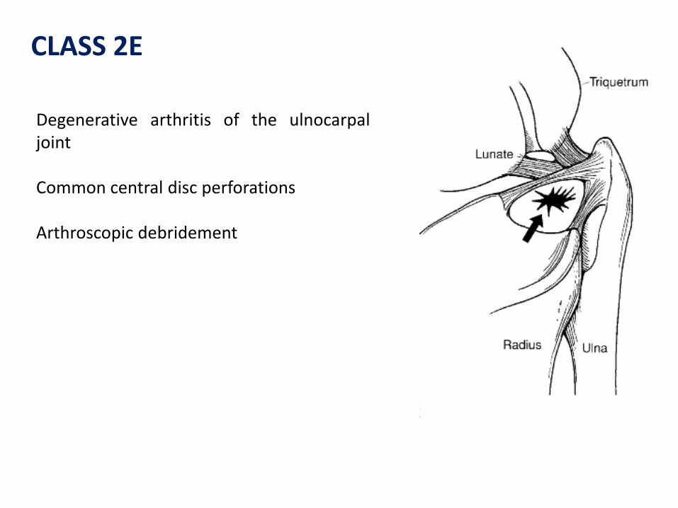
Degenerative arthritis of the ulnocarpal joint Common central disc perforations Arthroscopic debridement
CLASS 2E
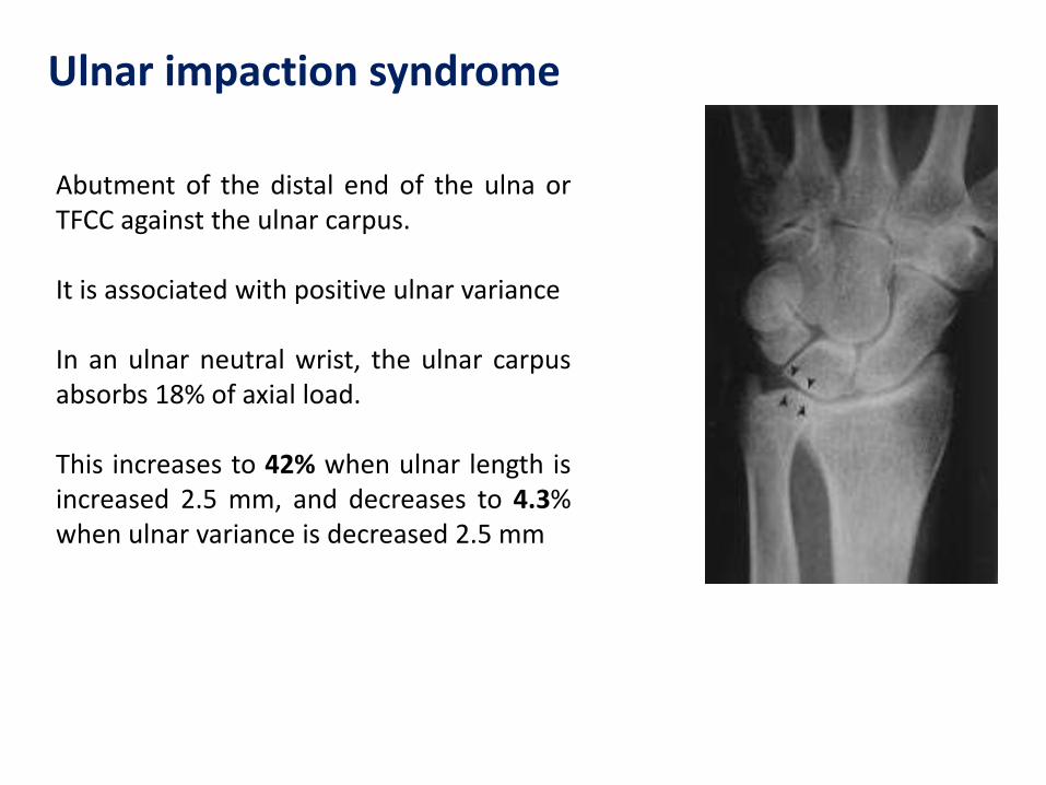
Abutment of the distal end of the ulna or TFCC against the ulnar carpus. It is associated with positive ulnar variance In an ulnar neutral wrist, the ulnar carpus absorbs 18% of axial load. This increases to 42% when ulnar length is increased 2.5 mm, and decreases to 4.3% when ulnar variance is decreased 2.5 mm
Ulnar impaction syndrome
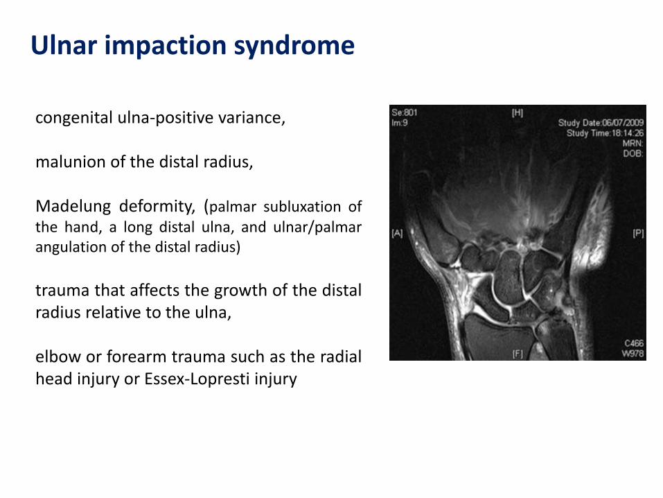
congenital ulna-positive variance, malunion of the distal radius, Madelung deformity, (palmar subluxation of the hand, a long distal ulna, and ulnar/palmar angulation of the distal radius)
trauma that affects the growth of the distal radius relative to the ulna, elbow or forearm trauma such as the radial head injury or Essex-Lopresti injury
Ulnar impaction syndrome
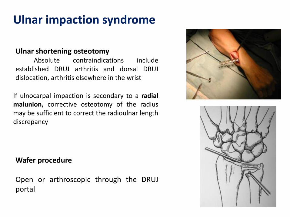
Ulnar shortening osteotomy Absolute contraindications include established DRUJ arthritis and dorsal DRUJ dislocation, arthritis elsewhere in the wrist In patients with a reverse oblique DRUJ shortening can significantly increase joint pressures in the proximal DRUJ.
Wafer procedure Open or arthroscopic through the DRUJ portal
Ulnar impaction syndrome
If ulnocarpal impaction is secondary to a radial malunion, corrective osteotomy of the radius may be sufficient to correct the radioulnar length discrepancy
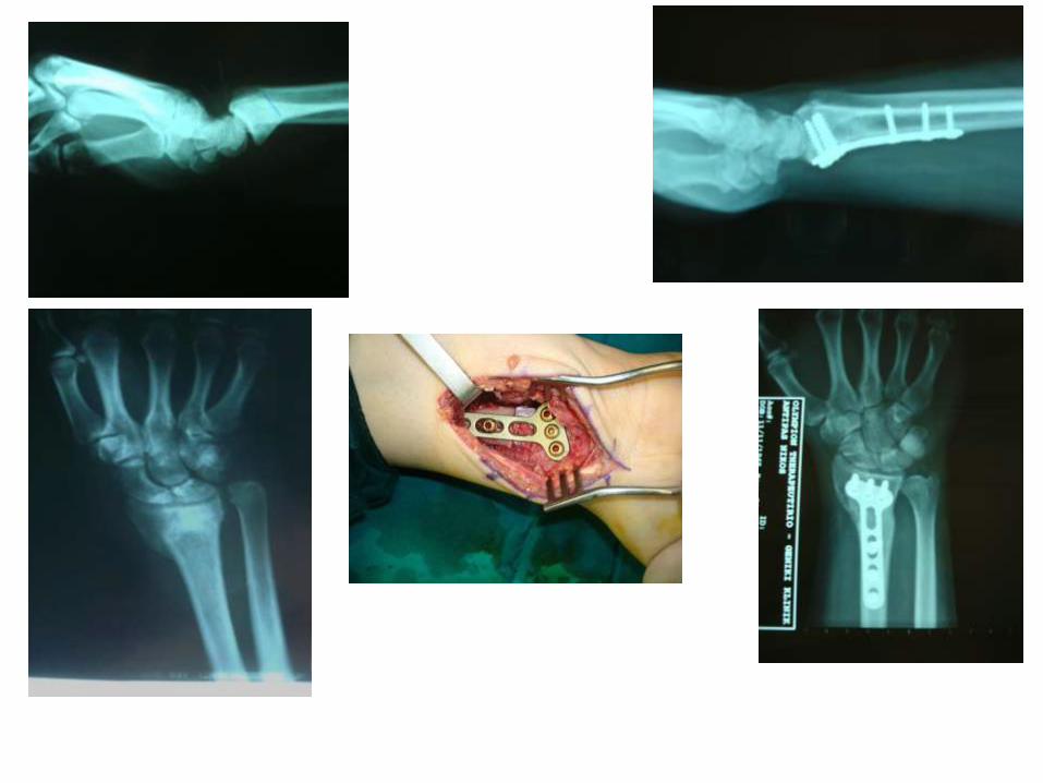
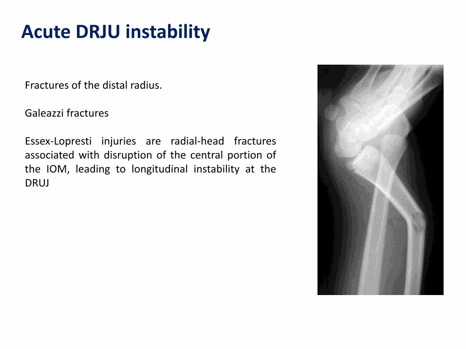
Acute DRJU instability
Fractures of the distal radius. Galeazzi fractures Essex-Lopresti injuries are radial-head fractures associated with disruption of the central portion of the IOM, leading to longitudinal instability at the DRUJ
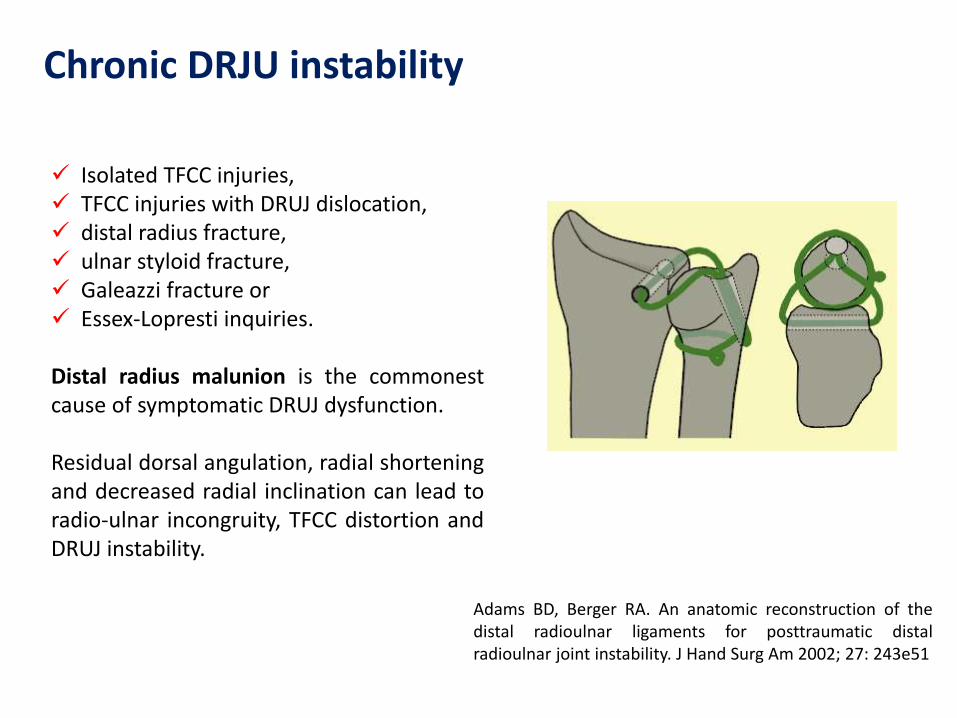
Chronic DRJU instability
Isolated TFCC injuries, TFCC injuries with DRUJ dislocation, distal radius fracture, ulnar styloid fracture, Galeazzi fracture or Essex-Lopresti inquiries. Distal radius malunion is the commonest cause of symptomatic DRUJ dysfunction. Residual dorsal angulation, radial shortening and decreased radial inclination can lead to radio-ulnar incongruity, TFCC distortion and DRUJ instability.
Adams BD, Berger RA. An anatomic reconstruction of the distal radioulnar ligaments for posttraumatic distal radioulnar joint instability. J Hand Surg Am 2002; 27: 243e51
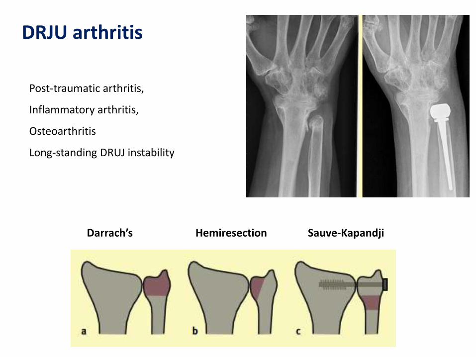
DRJU arthritis
Post-traumatic arthritis,
Inflammatory arthritis,
Osteoarthritis
Long-standing DRUJ instability
Darrach’s Hemiresection Sauve-Kapandji
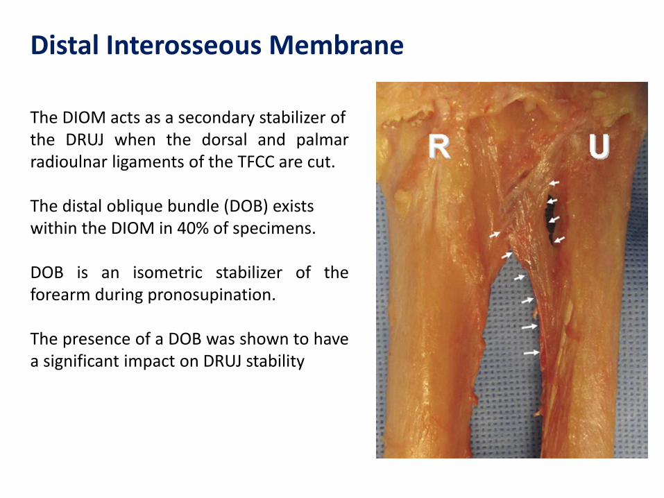
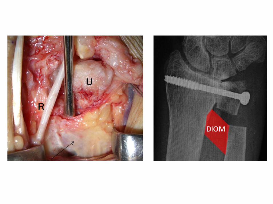
Distal Interosseous Membrane
The DIOM acts as a secondary stabilizer of the DRUJ when the dorsal and palmar radioulnar ligaments of the TFCC are cut. The distal oblique bundle (DOB) exists within the DIOM in 40% of specimens. DOB is an isometric stabilizer of the forearm during pronosupination. The presence of a DOB was shown to have a significant impact on DRUJ stability
Distal Interosseous Membrane
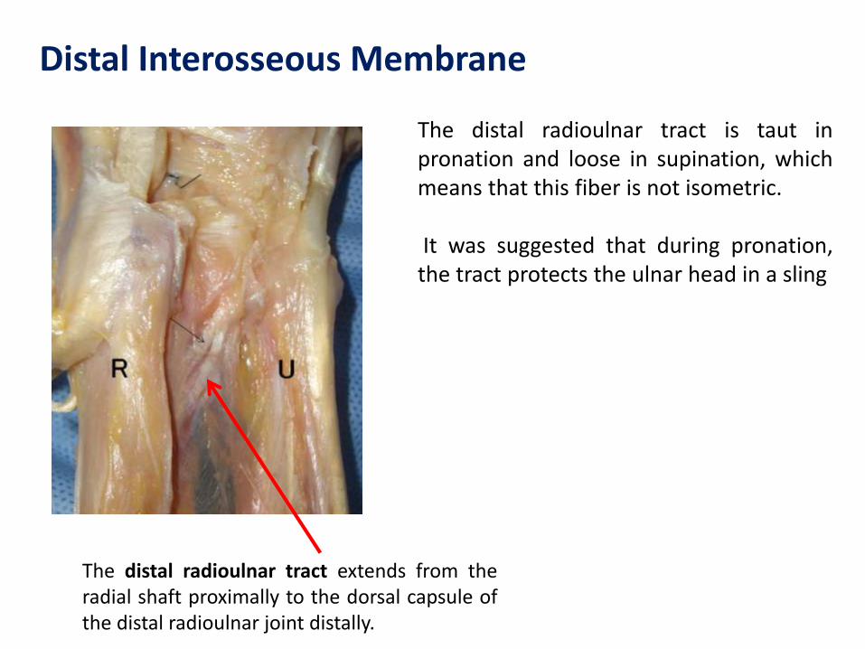
The distal radioulnar tract extends from the radial shaft proximally to the dorsal capsule of the distal radioulnar joint distally.
The distal radioulnar tract is taut in pronation and loose in supination, which means that this fiber is not isometric. It was suggested that during pronation, the tract protects the ulnar head in a sling
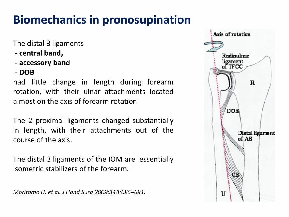
Biomechanics in pronosupination
The distal 3 ligaments - central band, - accessory band - DOB had little change in length during forearm rotation, with their ulnar attachments located almost on the axis of forearm rotation The 2 proximal ligaments changed substantially in length, with their attachments out of the course of the axis. The distal 3 ligaments of the IOM are essentially isometric stabilizers of the forearm.
Moritomo H, et al. J Hand Surg 2009;34A:685–691.
DRUJ laxity and DOB
DRUJ laxity was greater in the group without a DOB than in the group with a DOB. That study hypothesized that DRUJ laxity depends at least in part on the presence and configuration of the DIOM.
Kitamura T, Moritomo H, Arimitsu S, Berglund LJ, Zhao KD, An KN, et al. The biomechanical effect of the distal interosseous membrane on distal radioulnar joint stability. J Hand Surg 2011;36A:1626–1630.
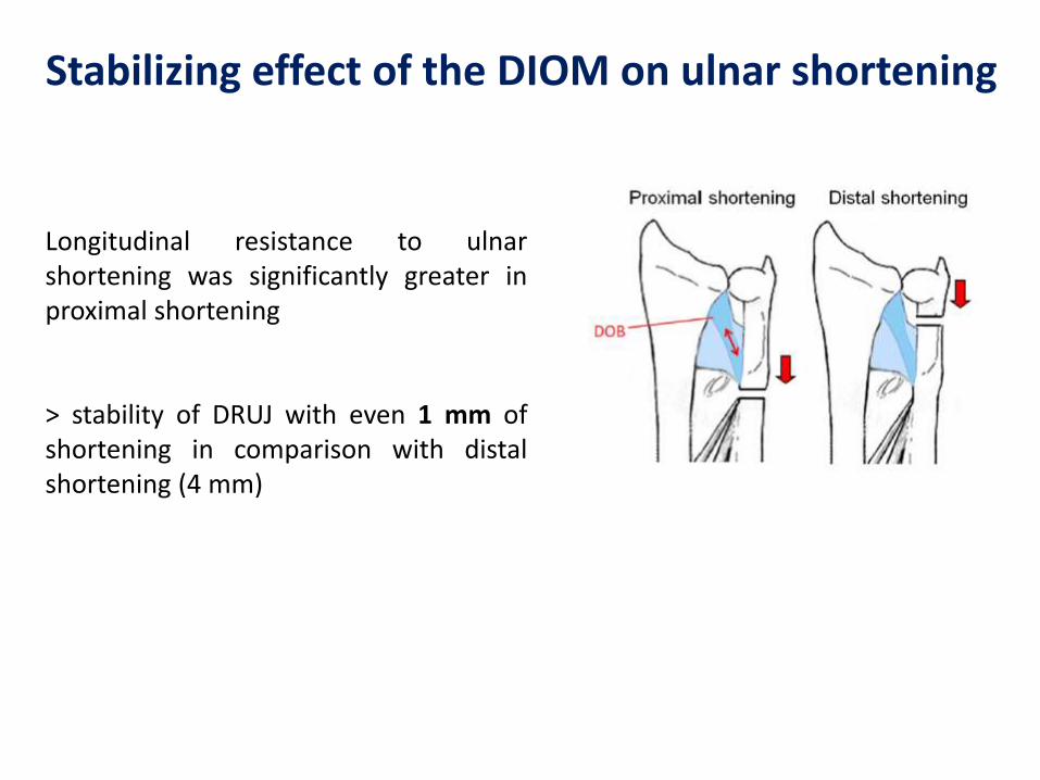
Stabilizing effect of the DIOM on ulnar shortening
Longitudinal resistance to ulnar shortening was significantly greater in proximal shortening > stability of DRUJ with even 1 mm of shortening in comparison with distal shortening (4 mm)


![Electrophysiological Features of Ulnar Tunnel Syndrome ... · Ulnar tunnel syndrome (UTS) is an uncommon ulnar entrapment neuropathy. Guyon [1] described the anatomy of the area in](https://img.pdfslide.net/doc/110x75/601bca5f935324075a08994b/electrophysiological-features-of-ulnar-tunnel-syndrome-ulnar-tunnel-syndrome.jpg)