Embed Size (px)
Citation preview
1
Anatomy of the vertebral column lymphatic network in mice
Laurent Jacob1, Ligia Boisserand2, Juliette Pestel1, Salli Antila3, Jean-Mickael Thomas4,
Marie-Stéphane Aigrot1, Thomas Mathivet5, Seyoung Lee2, Kari Alitalo3, Nicolas Renier1,
Anne Eichmann5,6, Jean-Leon Thomas1,2#
1Université Pierre et Marie Curie Paris 06 UMRS1127, Sorbonne Université, Institut du Cerveau et
de la Moelle Epinière, Paris, France
2Department of Neurology, Yale University School of Medicine, New Haven, CT, 06511, USA
3Wihuri Research Institute and Translational Cancer Biology Program, Faculty of Medicine,
University of Helsinki, Helsinki, Finland
4Ecole Nationale Supérieure d'Art de la Villa Arson, 06100 Nice, France
5INSERM U970, Paris Cardiovascular Research Center, 56 Rue Leblanc, 75015 Paris, France
6Cardiovascular Research Center and the Department of Cellular and Molecular Physiology, Yale
University School of Medicine, New Haven, Connecticut 06510-3221, USA
# corresponding author: [email protected]
2
Abstract (158 words)
Cranial lymphatic vessels (LVs) are involved in transport of fluids, macromolecules and CNS
immune responses. Little information about spinal LVs is available, because these delicate
structures are embedded within vertebral tissues and difficult to visualize using traditional
histology. Here we reveal an extended vertebral column LV network using three-dimensional
imaging of decalcified iDISCO-clarified spine segments. Spinal LVs are metameric circuits exiting
along spinal nerve roots and connecting to lymph nodes and the thoracic duct. They navigate in
the epidural space and the dura mater around the spinal cord, and associate with leukocytes,
peripheral dorsal root and sympathetic ganglia. Spinal LVs are VEGF-C-dependent and remodel
extensively after spinal cord injury. They constitute an extension to cranial circuits for meningeal
fluids, but also a route for perineural fluids and a link with peripheral immune and nervous circuits.
Vertebral column LVs may be potential targets to improve the maintenance and repair of spinal
tissues as well as gatekeepers of CNS immunity.
3
Introduction
The lymphatic vasculature controls fluid homeostasis, macromolecular clearance and immune
responses in peripheral tissues (Alitalo, 2011; Petrova and Koh, 2018). The brain was long
considered to lack lymphatic vasculature, which has raised questions about how interstitial cerebral
fluid (ISF) is cleared of waste products (Louveau et al., 2017; Abbott et al., 2018) and how immune
surveillance of the brain is maintained (Ransohoff and Engelhardt, 2012; Engelhardt et al., 2016;
Filiano, Gadani and Kipnis, 2017). ISF is formed by water and small solutes that are exchanged
through the capillary walls between the blood vessels and the brain. It has a similar composition
to the cerebrospinal fluid (CSF) which drains the brain ventricles and meninges and is mainly
produced in the choroid plexus (Zappaterra and Lehtinen, 2012). The CSF has been described to
dynamically exchange with ISF along glial “lymphatic” (glymphatic) non-vascular periarterial
routes, without crossing the endothelial cell layer, and subsequently to be cleared from the brain
into the subarachnoid space (Iliff, Goldman and Nedergaard, 2015; Engelhardt et al., 2016). The
CSF outflow system involves specific extracranial lymphatic vasculature beds (Pollay, 2010; Proulx
et al., 2017). In addition, cranial meningeal LVs (mLVs) have been recently identified and were
suggested to drain CSF into deep cervical lymph nodes (dcLNs) (Aspelund et al., 2015; Louveau
et al., 2015; Antila et al., 2017). In mice, cranial mLVs are mainly aligned alongside large dural
venous sinuses, meningeal arteries and cranial nerves. Along the sagittal suture, the cranial
lymphatic vasculature is rather discontinuous with valveless and small-diameter LVs, while it forms
a larger network with valves toward the lateral and basal aspects of the skull (Aspelund et al., 2015;
Louveau et al., 2015; Antila et al., 2017). Meningeal lymphatic vasculature also exists in the skull
of primates, including common marmoset monkeys and humans (Absinta et al., 2017; Antila et al.,
2017).
VEGF-C expression in vascular smooth muscle cells and VEGFR3 in lymphatic endothelial cells
(LECs) are essential for the development of cranial mLVs (Antila et al., 2017; Blanchette and
Daneman, 2017). The meningeal lymphatic vasculature develops later than the rest of the
lymphatic network, first appearing at birth in the basal parts of the skull, then expanding during the
neonatal period along dural blood vessels whose vascular smooth muscle cells supply the VEGF-
C (Antila et al., 2017). Classical immuno-histology on whole-mount preparations or cryo-sections
showed that, during the first weeks after birth, LVs also developed a large network closely attached
to the vertebral column (Antila et al., 2017). Vertebral LVs were observed in intervertebral spaces,
ventrally and dorsally to the midline, as well as along spinal nerve roots, laterally and together with
blood vessels. In the cervical spine, vertebral LVs appeared directly connected with cranial mLVs
4
located dorsally around the cisterna magna and ventrally around the foramen magnum. These
observations suggested that cranial mLVs might extend caudally into the spinal lymphatic vascular
system and connect from there to the peripheral lymphatic system. To test this model, we decided
to produce a three-dimensional (3D)-map of the vertebral lymphatic system that respected
structural interactions between the CNS and meninges, the surrounding bone and mesenchymal
environment and the neighboring peripheral nervous system (PNS). This required us to preserve
the overall bone structures around the CNS while simultaneously accessing and labeling the LVs
of meninges contained within the protective layers of muscular and skeletal tissues.
To do so, we used the iDISCO (immunolabeling-enabled three-dimensional imaging of solvent-
cleared organs) technique which enables volume imaging of immunolabeled structures in complex
tissues, without disrupting tissue architecture (Renier et al., 2014, 2016). Imaging of iDISCO
treated vertebral segments with a light sheet fluorescent microscope (LSFM) revealed an extensive
lymphatic vasculature inside the vertebral canal.
Results
Lymphatic vasculature pattern in the thoracic spine
To label vascular, immune and neural cell compartments within the intact vertebral column, we
decalcified segments of 2-4 vertebrae dissected together with the surrounding muscle tissue along
the rostro-caudal axis of the vertebral column. iDISCO tissue clearing and immunolabeling followed
by light-sheet fluorescence microscope (LSFM) was used for 3D-reconstruction of the spinal LV
network.
Because we previously described LVs mainly in the cervical vertebrae (Antila et al., 2017), the
iDISCO protocol was first applied to the thoracic spine, with the goal to characterize the three-
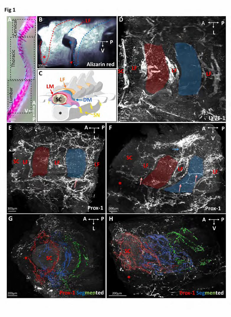
dimensional extension of LVs in the vertebral column (Fig. 1 A). Fig. 1 B illustrates a lateral view
of Alizarin red staining of bones within a cleared spinal column segment to reveal the vertebrae,
intervertebral spaces and ligamentum flavum. Fig. 1 C shows a schematic latero-frontal
perspective view of a thoracic vertebral segment. Lymphatic endothelial cells (LECs) were labeled
using polyclonal antibodies against two well-established LEC markers, the LYVE-1 cell surface
receptor and the nuclear Prox-1 transcription factor (Wigle and Oliver, 1999; Jackson et al., 2001;
Johnson et al., 2008). Prox-1 labeled LYVE-1 positive LECs and LYVE-1 negative cells within the
spinal cord that were previously identified as oligodendroglial cells (Kaser-Eichberger et al., 2016)
(Fig. S1, A and B). LYVE-1 labeled Prox1 positive LECs and Prox1-negative myeloid cells, as

5
previously reported (Kaser-Eichberger et al., 2016) (Fig. S1, A and B). LYVE-1 positive LECs
within the vertebral column were negative for the blood vessel marker Podocalyxin (Testa et al.,
2009) (Fig. S1, C and H).
Despite labeling of some non-LECs, both markers clearly revealed a dense lymphatic network that
was present between vertebrae and appeared confined to the intervertebral spaces (Fig. 1, D and E and movies 1 and 2). A few longitudinal vessels linked adjacent intervertebral lymphatic circuits
together along the spinal cord (Fig. 1, E and F). Each vertebral LV was also connected to the
peripheral lymphatic system surrounding the vertebrae, dorsally through the ligamentum flavum,
dorsolaterally along the dorsal facet joint and ventrolaterally through the intervertebral foramen
along ventral nerve roots and dorsal root ganglia (DRGs) (Fig. 1, E and F). The overall vertebral
lymphatic circuitry was surprisingly dense compared to the previously described lymphatic
vasculature of skull meninges (Aspelund et al., 2015; Louveau et al., 2015; Absinta et al., 2017;
Antila et al., 2017).
We next used Imaris-3D image analysis software to illustrate the anatomy of meningeal lymphatic
circuits within each vertebra and their relationship with the extra-vertebral LVs. Analyses were
performed from global image acquisitions of the thoracic spine, such as the one on display in
movie 2 which shows a succession of vertebral lymphatic units along the rostro-caudal axis.
Images were then segmented to generate a color-coded map of LV circuits. In Fig. 1, G and H and movies 3 and 4 each color defines the Prox-1+ vascular pattern of one vertebra along three
successive thoracic vertebrae (red, blue, green) as well as the peripheral lymphatic vasculature
(white). This identifies a metameric organization of spine LVs.
Vertebral unit lymphatic architecture
We next mapped the LV network in the vertebral canal from the dorsal to the ventral part of a
vertebra. Movie 2 and the corresponding view in Fig. 2, A and B show LVs around one segment
of the thoracic spinal cord, and areas where higher magnification views were taken. Dorsally,
semicircular lymphatic vessels navigate around the spinal cord, at the ventral border of the
ligamentum flavum (Fig. 2 C). At the dorsal midline, these vessels contact lymphatic branches
entering the epidural space from the overlying dense peripheral lymphatic vasculature in the
intervertebral space between two spinous processes (Fig. 2 D). An inner and less complete LV
circle lines the dura mater, hence two layers of LVs border the epidural space (Fig. 2 D). Laterally,
at the level of the transverse facet joints, semicircular vessels including peripheral lymphatic

6
vessels from the dorsal plexus converge toward a lymphatic circle (blue arrow in Fig. 2 C). From
this point, lymphatic vessels redistribute either radially via the periphery, or ventrally toward the
emergence of the ventral spinal nerve root (red double arrow in Fig. 2 C). Movie 2 allows to follow
the route of peripheral lymphatic vessels from their entry at dorsal midline to their lateral exit from
the vertebral canal. At the intervertebral foramen, dorsal root ganglia (DRGs) are covered by LVs
that converge from the ventral and dorso-lateral circuits at their proximal and distal end,
respectively (arrowheads in Fig. 2 E). In addition to these two circuits, a few longitudinal connecting
vessels link vertebral lymphatic units together (Fig. 2 F). Ventrally, a second circuit of semicircular
lymphatic vessels converges toward the ventral spinal nerve root exit, while no lymphatic vessels
are observed at the ventral midline (Fig. 2 G).
We observed a similar LV pattern in other thoracic segments (n=5), allowing us to generate a
schematic representation of these different compartments of the vertebral lymphatic circuitry with
a specific color code for intervertebral circuits (red) and vertebral branches of the peripheral LV
(blue) (Fig. 2 H).
Epidural and dural vertebral lymphatic circuits are surrounded by myeloid cells
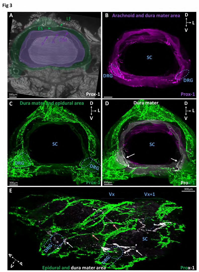
To obtain 3D-resolution of LV localization in the spinal canal and meninges, Prox-1-labeled
vertebral volumetric images were used to generate segmented images of membranes and the
epidural space around the spinal cord. We enhanced the brightness of successive single image
slices of Prox-1 labeled cervical and thoracic vertebrae to identify meninges, the epidural space
and ligamentum flavum (Fig. 3A). We then manually delineated and colored the layers of
meninges, dura mater or epidural space including the Prox1 label. Fig. 3 A shows one image slice
with a color-code for meningeal layers (purple area) and the epidural space (green area). After 3D-
reconstruction of a stack of similarly processed image slices, we generated color-coded layer
masks for the arachnoid and dura mater together (purple area in Fig. 3 B), or the dura mater and
the epidural space together (green area in Fig. 3C). The overlay of both masks revealed that Prox-
1+ LVs mainly localized in the epidural space (green), while the underlying dura mater layer (white)
includes ventral LVs around DRGs and a few LVs on each side of the ventral and dorsal midline
(Fig. 3D and movie 5). As shown on a lateral view (Fig. 3E), dura mater LVs localized most
extensively at bilateral DRGs. Interestingly, connecting vessels between two successive vertebrae
(red arrows in Fig. 3E) navigate in the epidural space and appear to join LVs of the dura mater
close to the DRGs (white arrows in Fig. 3E), suggesting a possible confluence of peripheral lymph
and CSF at this level.
7
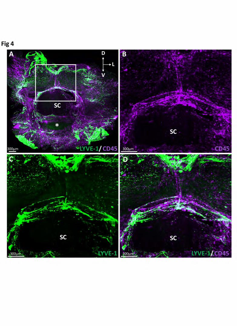
Vertebral LVs moreover appeared to be a privileged environment for immune myeloid cells, as was
previously reported for skull lymphatics (Louveau et al., 2015). Pre-cleared segments of the
vertebral column co-labeled with antibodies against LYVE-1 and the common leucocyte antigen
CD45 show that CD45+ myeloid cells inside the vertebral canal are concentrated around LYVE-1+
vertebral LVs (Fig. 4, A-D).
LV patterning varies between cervical, thoracic and lumbar spine
We next examined the lymphatic vasculature at cervical and lumbar levels of the vertebral column.
A first approach by confocal imaging of wholemount preparations of cervical and lumbar vertebrae
showed differences in the pattern of cervical and lumbar LVs. More specifically, extra-vertebral
LVs on the dorsal aspect of the spine were more abundant in lumbar compared to cervical
vertebrae (Fig. S2, A-D). The variability of LV patterning along the vertebral column was analyzed
in further detail by volume imaging.
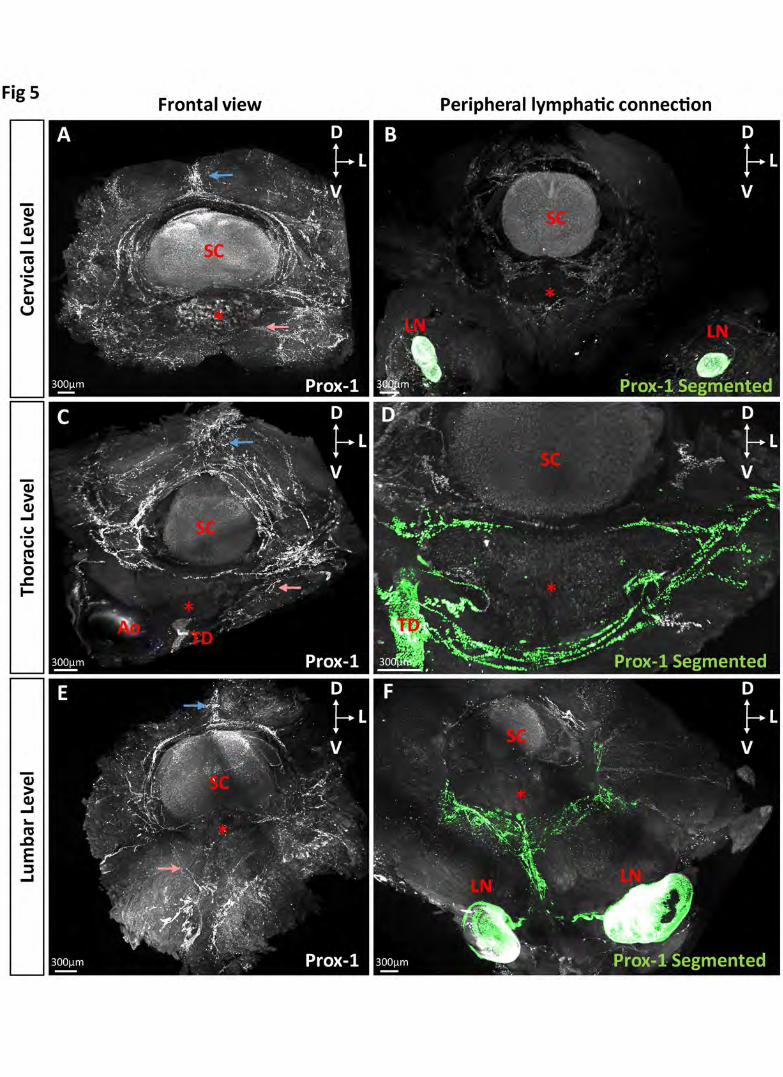
In the cervical region (Fig. 5 A), the dorsal extravertebral lymphatic plexus is reduced in size (green
in Fig. 5 B) and intravertebral LVs exit ventrally and bilaterally through the intervertebral foramen
to connect to cervical lymph nodes (Fig. S2, E and F). The short interspace between cervical
vertebrae is associated with a lack of longitudinal intervertebral vessels and a direct lymphatic
vessel connection from the ventral root to the lateral lymphatic circle of the neighboring vertebra.
Thoracic vertebral LVs, as described above, are defined by a large dorsal extravertebral plexus
(Fig. 5 C) and a direct connection from ventrolateral spinal roots to the thoracic lymphatic duct (Fig. 5 D). The thoracic and lumbar regions display similar extension and pattern of extravertebral
and intravertebral LVs (Fig. 5, C and E). In lumbar vertebrae, the ventro-lateral circuits that exit on
each side of the vertebral canal connect to lymph nodes. As shown in green in Fig. 5 F, lymphatic
vessels circumvent the ventral body of the lumbar vertebra, converge on the ventral midline and
split into two branches running toward the pair of aortic lumbar lymph nodes. Therefore, the
vertebral LV architecture is conserved along the vertebral column, but the extension of
extravertebral and intravertebral vessels around the spinal cord and their connection to the
peripheral lymphatic system differs between the cervical, thoracic and lumbar vertebral levels.

8
Vertebral lymphatic vasculature contacts the sympathetic nervous system
We found that LVs covering the DRG dura mater (Fig. 2F) extend a collateral branch, bilaterally
along the spine, that contacts paravertebral Prox1low ganglia (Fig. 6, A and B). Prox1 is known to
be expressed in the sympathetic neuronal lineage (Holzmann, Hennchen and Rohrer, 2015) and
double-labeling with antibodies against Prox-1 and Tyrosine Hydroxylase (TH), a specific marker
of adrenergic nerves and ganglia, confirmed that specific branches emerging from vertebral LVs
connect to TH+/Prox1low sympathetic ganglia (Fig. 6 C) and that lymphatic vessels double stained
with Prox-1 and LYVE-1 contact one sympathetic ganglion per spinal level (Fig. 6 D-F).
Complementary analyses by high resolution confocal imaging on vertebral column cryosections
indicated that the close spatial relationship between LV and sympathetic ganglia (Fig. 6 G, H)
remained at the surface of the ganglion cortical layer (Fig. 6I). These data reveal a hitherto
unknown anatomical interaction between the autonomous nervous system and lymphatic vessels
derived from spinal meningeal LVs.
Vertebral LVs respond to VEGF-C and spinal cord injury
We next tested if vertebral LVs responded to the major lymphangiogenic growth factor VEGF-C.
Adult mice (2months of age) were injected with AAV-VEGF-C or -empty control (n=4) into the
cisterna magna, followed by analysis one month later (Fig. 7 A). Fig. 7 B-E and movie 8 show
Prox-1 staining of cervical vertebrae. Compared to control injected mice (Fig. 7, B and D), VEGF-
C injected mice showed a strongly expanded LV network, in particular of dorsolateral lymphatic
rings in the intervertebral disk (Fig. 7, C and E and movie 6).
The capacity of adult vertebral LVs to grow in response to growth factors suggested that this
circuitry may respond to pathological conditions affecting the spinal cord and more broadly the
CNS. Based on the prediction that a spinal cord trauma generates tissue destruction, accumulation
of cell debris, and thus requires from the LV a supplement of tissue clearance and immune survey,
we applied a direct injury at the thoraco-lumbar level, using a needle to inject sodium chloride (2µl)
into the spinal cord (Fig. 7F). Within a week after the surgery, a robust lymphangiogenesis was
observed in extravertebral and intravertebral lymphatic vasculature in the injured mice (Fig. 7, G-N and movie 7).

9
Discussion
We here used the i-DISCO protocol combined with LSFM to generate a map of thoracic vertebral
column LVs (Fig. 8). We reveal an extensive and complex lymphatic vasculature in the spinal
column, surprisingly dense in comparison to the one that covers the cranial dura mater. Previous
literature has reported the presence of LVs on whole mount preparations of vertebral dura mater
in monkeys (Miura, Kato and von Lüdinghausen, 1998) and on sections of intact and injured
vertebral tissue in humans (Kashima, Dongre and Athanasou, 2011). These studies identified
elements of lymphatic vasculature in the spine, such as the dura mater and epidural lymphatic
circuits and the absence of LVs in vertebral bones and intervertebral disks in the intact spine. We
here extend these findings by 3D-views of lymphatic vasculature organization and reveal
interactions with surrounding tissues along the spine (Fig. 8).
Each vertebra is drained by semicircular dorsal and ventral vessels, which exit the vertebral column
at intervertebral foramen. Vertebral LVs extend along spinal nerve roots to reach either lymph
nodes in the cervical and lumbar regions, or the thoracic lymphatic duct in the thoracic region. The
vertebral lymphatic network is thus organized as a metameric network of peripheral LV-connected
vertebral lymphatic units that are interconnected by a few thin longitudinal vessels. The absence
of large longitudinal dorsal or ventral LVs suggests that the main flow of vertebral lymph is not
descending as a continuous stream from the cisterna magna along the vertebral column axis, but
is rather drained at the level of each vertebra. In addition to dura mater lymphatic circuits, the
vertebral lymphatic vasculature includes an extensive network of epidural vessels that are located
in the intervertebral tissue and beneath the ligamentum flavum and .appear to drain the non-CNS
peripheral lymph of the vertebral column.
Pioneer studies in the late 19th and early 20th centuries (Schwalbe, 1869); (Quincke H, 1872); (Orr
and Rows, 1907) and later works of Ivanow (Ivanow, G., 1927) and Brierley and Fields (Brierley
and Field, 1948) had investigated the flow of the lymph stream along the spine and in the spinal
roots of the cord as well as lymphatic pathologies and infectious agent propagation in the spine,
providing ‘a body of evidence that lymphatics play a part probably not subordinate to that of the
blood vessels as channels by which infectious agents (toxins, polyomyelitis, tetanus) are conveyed
to the cord and distributed within it (Bruce and Dawson, 1911). These predictions find support in
the present imaging of extended vertebral lymphatic circuits that contact both peripheral lymph and
CSF.
It is striking to note that there is a regional variation in the LV size, which is inversely correlated to
the volume of CSF, with large cerebral ventricular volumes associated with a discrete network of
10
cranial mLVs and a small vertebral ependymal volume correlated with a large vertebral lymphatic
vasculature. One possible explanation is that vertebral LVs strongly contribute to the reuptake of
the CSF that is continuously produced by the ventricular choroid plexus and circulates top-to-
bottom along the spinal cord. This model is supported by presence of dura mater LVs in the spine
and their dense location around spinal nerve root exits. A corollary of this hypothesis is that
glymphatic drainage of interstitial fluids may be faster in the spinal cord than in the brain. A second
and likely possibility is that the largest part of vertebral LV drains peripheral lymph, as suggested
by the large network of epidural vessels that extends into the vertebral canal from the peripheral
lymphatic system.
Vertebral LVs localize mainly at the level of intervertebral ligaments or joints, much like cranial
lymphatics that navigate in skull commissures alongside blood vasculature and spinal nerves
(Antila et al., 2017). They also circulate through the fat tissue filling the epidural space intercalated
between the dural mater and intervertebral ligaments. We and others find that vertebral lymphatic
vessels avoid bone tissues (Edwards et al., 2008). Interestingly, the presence of LVs inside bone
is observed in patients with vanishing bone disease (also called Gorham Stout disease GSD)
(Hominick et al., 2018). GSD is a sporadic disease characterized by the presence of lymphatic
vessels in bone and progressive bone loss. In severe cases, the disease progresses until entire
bones are lost and replaced by fibrous tissue. GSD can affect any bone in the body, but it most
frequently affects the ribs and vertebrae, with poor prognosis (Lala et al., 2013; Dellinger, Garg
and Olsen, 2014). In mice, transgenic VEGF-C overexpression in bone under the control of the
Osx promoter induces lymphatic invasion into bone and osteoclast-mediated bone loss, mimicking
essential aspects of GSD (Hominick et al., 2018). Mechanisms preventing LV formation in bone
are currently unknown. In contrast to skull commissures between skull cap bones, which are few, narrow and fixed, the
vertebral disks, joints and ligaments between vertebral bones are numerous, large and mobile.
They sustain the integrity and flexibility of the spine, which is predicted to require extensive
interstitial fluid drainage. The large network of vertebral epidural LV appears to be exquisitely
adapted to this extensive drainage of non-neural peripheral tissues in the spine and to provide
each vertebra with its proper clearance system. It is predictable that defective spine LVs will alter
vertebral and intervertebral tissue maintenance, leading to spine orthopedic pathologies.
The spine LV circuitry includes epidural vessels in the peripheral lymphatic system that likely drain
intervertebral non-neural tissues, especially the epidural fat tissue of the spinal cord, and dura
mater vessels that may drain CNS tissue fluids at possible hot-spots close and around the DRGs
11
as well as at more restricted positions on each side of the dorsal and ventral midline. These two
circuits appear to be linked, since a part of especially LVs connecting vertebrae together, circulate
through the epidural space before entering the dura mater around the DRGs and exiting the spinal
canal. It remains to be determined whether both circuits converge to the same or different lymph
nodes or lymphatic ducts.
The proximity of two distinct epidural and dura mater LV circuits in the spine raises questions about
the protection of the privileged immune status of the CNS. Like in skull (Louveau et al., 2015), a
specific interaction between LVs and CD45 leucocytes is observed along the spine (Fig. 3). The
spinal cord lymphatic vasculature thus appears as a potentially important immune surveillance
interface between the CNS and peripheral tissues. The cervical and lumbar regions directly drain
into cervical and lumbar lymph nodes, respectively, which suggests that the peri-lymphatic
dendritic immune cells may rapidly transfer to lymph nodes and initiate lymphocyte activation
against specific pathogens or antigens. On the other hand, the promiscuity of epidural and dura
mater LV in the spine may predispose to the propagation of peripheral infections through the
vertebral canal toward neural tissues, especially peripheral nervous ganglia which are in close
contact with the spine LV (Fig. 6). For example, epidural LVs may provide entry for meningitis
infection into spinal meninges. The contact zone between DRGs and LVs connected to the thoracic
duct and lymph nodes appears as another potential gate for entry into the CNS for pathogens
drained by the peripheral lymph. This possibility is in agreeement with the early detection of bovine
scrapie protein in the mesenteric lymph nodes and DRGs of lemurs or cattles infected orally with
the agent of bovine spongiform encephalopathy (BSE) (Bons et al., 1999; Franz et al., 2012).
The spine elicits a variety of diseases including infections caused by either a bacterial or a fungal
infection transfered into the spine through the bloodstream (Darouiche, 2006), acute spinal cord
compressions resulting from trauma, cancer, epidural abscess, or epidural hematoma (Ropper and
Ropper, 2017), and degenerative spine disorders, a common condition especially in the ageing
Western population (Brinjikji et al., 2015). Vertebral LVs are a potential target for these pathologies,
as they are pipes to propagate infections and to drain excessive interstitial fluids. The vertebral
column is also the commonest site for skeletal metastastic tumors, with breast, prostate and lung
cancers being the most common primary sources; as many as 70% of cancer patients have spinal
metastases, and up to 10% of cancer patients develop metastatic cord compression (Choi et al.,
2010). Since lymphatics may serve as conduits for primary tumor cells in metastatic spreading
(Karaman and Detmar, 2014), specific interference in the vertebral lymphatic vasculature could
reduce or prevent the spinal metastasis process. Alternatively, lymphatic vessels are the first

12
barrier for antigen-presenting cells that are transported into the next lymph node to initiate an
adaptive immune response (Card, Yu and Swartz, 2014). Facilitating the entry of immune cells into
vertebral lymphatic vessels could thus also potentially improve the efficiency of immune checkpoint
inhibitor treatments to trigger the adaptive immune response against spinal tumor cells.
We find that adult vertebral LVs rapidly expand in response to VEGF-C or tissue injury. Tissue
injury causes inflammatory processes and calls for repair mechanisms. These processes require
increased LV activity and thus lymphangiogenesis which is likely VEGF-C/VEGFR3 signaling
dependent, as in chronic skin inflammation (Hagura et al., 2014) and macrophage VEGF-C-driven
(Ji, 2012). Spine LVs and VEGF-C may thus be new therapeutic targets to support and improve
these attemps to protect and repair injured spine tissues.
Vertebral LVs never contact the spinal cord tissue, even upon VEGF-C overexpression or acute
spine lesion. Like cranial LVs, vertebral LVs are thus unlikely to reuptake CSF circulating through
perivascular spaces of the spinal cord and pia mater. In contrast, vertebral LVs are closely apposed
around the somatic and autonomous nervous ganglia chains. The targeting of paravertebral
sympathetic chains by specific vertebral LVs is another discovery resulting from the spine volume
imaging. Although no lymphatic vascularization of sympathetic ganglia was observed, lymphatic
vessels may provide molecular signals to the sympathetic neurons that control vascular tone of
lymphatic ducts and cerebral arteries and arterioles. Previous observations also showed that
adrenergic fibers connect to the thoracic lymphatic duct and also innervate the wall of lymph node
arterioles (Villaro, Sesma and Vazquez, 1987; Mignini et al., 2012). The crosstalk between spine
LV and the sympathetic system is thus likely relevant for the regulation of peripheral lymph and
glymphatic drainage and may coordinate them with the activity of brain and spine tissues. We
speculate that a regulatory loop may link meningeal LV, sympathetic chain neurons and both CNS
and peripheral fluid drainage.
To conclude, this study shows that the volume imaging technique allows the description of
neurovascular systems by preserving the anatomy and the 3D-continuity of vascular and neural
structures. In particular, we have revealed a new set of information on the anatomical organization
and plasticity of the lymphatic vasculature along the spine. Our findings identify vertebral LVs as a
potential targets for improving the maintenance and repair of vertebral tissues as well as a
gatekeeper of CNS immunity.
Acknowledgements
13
This work was supported by Institut National de la Sante et de la Recherche Medicale (to J.-L.T.),
Agence Nationale Recherche (ANR-17-CE14-0005-03 to J.-L.T. and A.C.E.), Federation pour la
Recherche sur le Cerveau (FRC 2017 to J.L.T. and A.C.E), Carnot Maturation (to L.J.), Leducq
Foundation Transatlantic Network of Excellence (ATTRACT to A.C.E.), NIH (R01EB016629-01 to
J.-L.T., NHLBI 1R01HLI125811, NEI 1R01EY025979-01, P30 EY026878 to A.C.E.) and the Yale
School of Medicine (J.L.T.). K.A. funding was provided by the Jane and Aatos Erkko Foundation,
the European Research Council (European Union Horizon 2020 research and innovation
program, grant 743155), the Wihuri Foundation, the Academy of Finland (Centre of Excellence
Program 2014-2019, grants 271845 and 307366), and the Finnish Brain Foundation. We
acknowledge the ICM-QUANT cellular imaging, ICM-histomics and PHENO-ICMICE platforms,
Andrey Anisimov and the AAV Gene Transfer and Cell Therapy Core Facility of Biomedicum
Helsinki, University of Helsinki, for providing us AAV-expression vectors.

14
Figure legends
Fig 1. Segmental pattern of the vertebral lymphatic vasculature in the thoracic spine
(A) Alcian blue/Alizarin red staining of the mouse vertebral column with boxes indicating position
of images shown in Figs 1-4 (thoracic vertebrae) and 5 (cervical and lumbar vertebrae), spatial
orientation (A: anterior, D: dorsal, L: lateral, V: ventral). (B, C) Alizarin red staining of a thoracic
vertebral column segment (B) and corresponding latero-frontal perspective schematic view (C).
Two successive vertebrae are delimited by red/blue dots. DM: dura mater, LF: ligamentum flavum,
LM: leptomeninges (pia mater and arachnoid), SC: spinal cord, SN: spinal nerve, blue arrow:
dorso-lateral facet joint, red arrowhead: ventral intervertebral disk, blue star: intervertebral
foramen, red (B) or black (C) star: ventral vertebral body. (D) Dorsal view of LYVE-1 staining. Red
and blue areas correspond to two successive vertebrae. Note LVs lining ligamentum flavum. (E,
F) Dorsal (E) and lateral (F) views of the Prox-1 expression pattern. Red and blue areas correspond
to two successive vertebrae. Salmon arrows: intervertebral LVs, red arrow: dorsal LVs, red star:
vertebral ventral body. (G, H) Segmented images of the Prox-1 LV network (fronto-dorsal (G) and
lateral (H) views) highlighting three successive vertebral LV units (red, blue, green).
Fig 2. Organization of individual thoracic vertebral column lymphatic units
(A) Frontal view of a cleared thoracic vertebra stained with an anti-Prox-1 antibody. Red star:
vertebral ventral body, SC: spinal cord. (B) Magnification of red boxes referring to images in C-G.
(C) Semicircular dorsal LVs (red arrow) surround the spinal cord, exit dorso-laterally (blue arrow)
and also extend a latero-ventral connection (double red arrows) to the ventral nerve root (see
double arrow in F). Note Prox1 positive cells in SC and perivertebral muscles (M), FJ: facet joint.
(D) At the ventral face of the ligamentum flavum (LF) located between two spinous processes,
dorsal LVs (blue arrows) enter the vertebral canal and join semicircular LVs (red arrows). Note
circles of LVs bordering the upper side of the epidural space (ES). (E) Ventro-lateral LV circuitry
around DRG (red arrowhead). Blue dotted-lines: spinal nerve roots, red dotted-lines: DRG. (F)
Lateral view with intervertebral LVs (salmon arrows), red dotted-lines: DRG. (G) Two ventral
branches arising on each side of the ventral midline (VM) and reaching the DRG (see single red
arrows in F). (H). Schematic representation of a frontal view of a thoracic vertebral LV unit.
Longitudinal connecting vessels between vertebral units are not represented. FJ: facet joint; LF:
ligamentum flavum; ES: epidural space; DRG: dorsal root ganglia; VM: ventral midline; SC: spinal
cord. Black letters referring to images in C-G.
15
Fig 3. Epidural and dural lymphatic circuits of the spine
(A) 2D-single frontal image slice (2µm thick) of the cervical vertebral column with enhanced
brightness to reveal Prox-1-expressing nuclei and spinal tissues including spinal cord (SC),
meninges including pia mater (P), arachnoid (A) and dura mater (D), the epidural space (ES) and
the ligamentum flavum (LF). A color-coded segmentation of layers around the spinal cord shows
the meninges in purple and the dura mater plus the epidural space in green. (B-D) 3D-
reconstruction of frontal images of the cervical vertebral column with color-coded layers: the
arachnoid and dura mater in purple (B); the dura mater and epidural space in green (C); combined
layer marks showing the arachnoid in purple, the dura mater in white and the epidural space in
green (D). A noticeable LV network fills the epidural space (green) while dura mater LVs (white)
are mainly restricted to DRGs (white arrows) and few branches on each side of the dorsal and
ventral midline. (E) 3D-reconstruction of lateral images of the thoracic vertebral column with color-
coded layers illustrated in (D). Blue dotted-lines: bilateral DRGs; salmon arrows: intervertebral LVs;
Red star: vertebral ventral body.
Fig 4. Interactions of spinal LVs with immune cells
Double labeling of cleared thoracic vertebral column segments with LYVE-1 (purple in A, C, D) and
CD45 (green in A, B, D) to identify myeloid cells. (B-D) are magnifications of white box in (A).
Merged pictures (D). Note that myeloid cells concentrate along LVs. White star: vertebral ventral
body; SC: Spinal cord.
Fig 5. Variations of LV patterns along the vertebral column
Pattern of Prox-1+ LVs in the cervical (A, B), thoracic (C, D) and lumbar (E, F) vertebral column.
Left panels show frontal views, right panels show connection to peripheral lymph nodes (LN) and
thoracic duct (TD). (A, C, E). Note fewer LVs in the dorsal plexus between intervertebral spinous
processes of cervical and lumbar vertebrae compared to thoracic ones (blue arrows). Also note
differences in ventral root exit circuits between regions (salmon arrows, A ,C, E). (B, D, F). LV
ventral exit circuits (green) and deep cervical LNs (B), thoracic duct (D) or connections to aortic
lumbar LNs (F). Red star: vertebral ventral body; SC: spinal cord; Ao: Aorta.

16
Fig 6. Lymphatic vessel connections with sympathetic ganglia
(A, B). Thoracic Prox-1+ LVs contact paravertebral sympathetic ganglia (SG) (blue arrow),. White
dotted-lines: DRG. (C) Prox-1 (white) and tyrosine hydroxylase (TH, red) double labeling shows a
ventral LV branch contacting a paravertebral TH+ sympathetic ganglion (blue arrow). (D-F). Prox-
1 (white) / LYVE-1 (red) double-labeling of the LV-SG connection (blue arrow). (G-I). 2D-confocal
images of cervical cryosections labeled with LYVE-1 (white), TH (red), and Dapi (Blue). White box:
area magnified in (H) and (I). A LV contacts a TH+ SG (blue arrow). Note a second ventral LV
branch running along the SG, without entering its cortical layer (G-I, salmon arrow). This branch is
also seen in panel (B) (salmon arrow). White dotted-lines: DRG; Red star: vertebral ventral body;
SC: spinal cord.
Fig 7. Spinal LVs expand in response to VEGF-C and injury
(A-E). Response of vertebral LVs to AAV-control or AAV-VEGF-C delivery into the CSF. (A) 2-mo
mice received Intra-cisterna magna injection of AAVs and were analyzed by 3D-iDISCO one month
later. Frontal views (B, C) and lateral views (D, E) show that AAV-VEGF-C (C, E) induced
lymphangiogenesis in the cervical vertebral column as compared to AAV-control (B, D). Red star:
vertebral ventral body; SC: Spinal cord.
(F-J) Spinal cord (Sc) lesion. (F) Experimental procedure: 2ul NaCl were injected into the spinal
cord at the indicated level and mice were analyzed one week later. Frontal views (G, H) and lateral
views (I, J) of Prox-1+ LVs. Note that SC lesion induces robust lymphangiogenesis but LVs do not
enter lesioned SC tissue. Red star: vertebral ventral body; SC: Spinal cord.
Fig 8. Schema of thoracic vertebral column LVs
LVs around spinal cord (gray), in the epidura space (green), in the dura mater (purple), extra-
vertebral LV ventral connexion with SG and TD (blue). Blue dots indicate connexion with extra-
vertebral lymphatic networks. Black star: vertebral ventral body; DRG: dorsal root ganglia, FJ: facet
joint, LF: ligamentum flavum, SC: Spinal cord; SG: sympathetic ganglia.

17
Supplementary data:
Fig. S 1: LVs and blood vasculature in the thoracic vertebral column
(A, B) Prox-1 (white) / LYVE-1 (red) double staining of a clarified thoracic vertebral column
segment. Note overlap of Prox-1 and LYVE-1 labeling in LVs (arrowheads). Also note Prox-1-
labeling of SC oligodendrocytes (A) and LYVE-1 staining of myeloid cells in meninges (arrows, B).
(C-H) Double labeling of a clarified thoracic vertebral column segment with LYVE-1 (green) and
Podocalyxin (purple) to identify blood vessels. F-H: magnifications of boxed areas in C-E. White
star: vertebral ventral body; SC: Spinal cord.
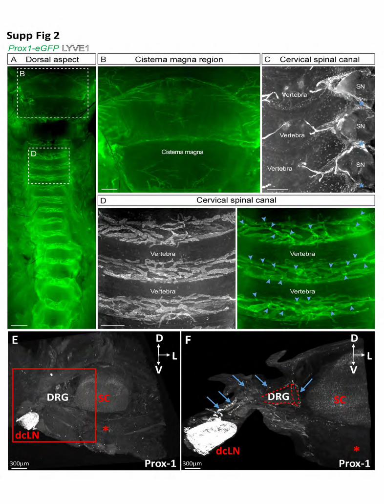
Fig. S 2 : LV patterning varies between cervical, thoracic and lumbar spine
(A-D) Whole-mount immunostaining of Prox1-eGFP reporter mice showing lymphatic vasculature
(green, gray). (A) Dorsal lymphatic vasculature around cisterna magna and vertebrae in cervical
and thoracal regions. (B) Close-up of lymphatic vessels around cisterna magna in close
association with the LVs around first cervical vertebra. (C) Side-view of the vertebral LVs
connecting longitudinally with each other (marked with blue asterisks) and surrounding the spinal
nerves (SN). (D) Close-up of vertebral lymphatic vessels in the dorsal aspect of the spine showing
their localization mainly in intervertebral spaces. Blue arrowheads mark lymphatic valves. Data
shown are representative n=3-6 per one staining. Scale bars: 1 mm (A), 200 µm (B, C), 400 µm
(D).
(E, F) Prox-1-staining of a cervical vertebral segment shows connection between a LV surrounding
the DRG and a dcLN (blue arrows). (F): higher magnification view of boxed area in (E). Red star:
vertebral ventral body; SC: Spinal cord.
Movie 1: Lymphatic vasculature of a thoracic vertebral segment labeled with LYVE-1 antibody
Movie 2: Lymphatic vasculature of a thoracic vertebral segment labeled with Prox-1 antibody

18
Movie 3: Time-lapse z series of the Prox-1-labeled lymphatic vasculature of a thoracic vertebral segment
Movie 4: 3D-image segmentation of Prox-1-labeled LVs of a thoracic vertebral segment
Movie 5: 3D-image segmentation of Prox-1-labeled LVs in meningeal and epidural layers around the cervical spinal cord.
Movie 6: Prox-1-labeled cervical LVs following AAV-VEGF-C administration
Movie 7: Prox-1-labeled lumbar LVs at one week after spinal cord injury
Materials and methods
Study approval
All mouse experiments were approved by the Institutional Animal Care and Use Committee of ICM
Brain and Spine Institute and Yale University.
Mice
C57BL/6 background mice were used for all the experiments. Littermate mice were used as
controls. Mice were considered as adults at 2 to 3-months (mo) of age.
Tissue preparation
Mice were given a lethal dose of Sodium Pentobarbital (Euthasol Vet) and perfusion-fixed through
the left ventricle with 10ml ice-cold PBS then 20ml 4% paraformaldehyde (PFA) in PBS after
puncture of the right atrium. To dissect the spine, the skin was completely removed, all the organs
were eliminated and the ribs were completely cut to keep the vertebra from the cervical part until
the lumbar part with the whole spinal cord inside. All the surrounding tissue as muscles, aorta,
ligaments were maintained around vertebral column. The spine was cut into pieces of about 0,5cm

19
(1 to 3 vertebrae) corresponding to the cervical, thoracic and lumbar regions. The different spinal
segments were immediately immersed in ice-cold 4% PFA, fixed overnight at +4°C, washed in
PBS, and processed for staining.
Samples pretreatment in methanol for iDISCO protocol
We used the classical iDISCO protocol ((Renier et al., 2014) http://www.idisco.info). In detail, fixed
samples were dehydrated progressively in methanol/PBS, 20%, 40%, 60%, 80% and 100% for 1
hr each (all steps were done with agitation). They were then incubated overnight in a solution of
Methanol 33%/dichloromethane 66% (DCM) (Sigma 270997-12X100ML). After 2 x 1h washes with
methanol 100%, samples were then bleached with 5% H2O2 in methanol (1 vol 30% H2O2/ 5 vol
methanol) at 4°C overnight. After bleaching, samples were rehydrated in methanol for 1 hr each,
80%, 60%, 40%, 20% and PBS. To clarify vertebral bone, we here added a decalcification step
using Morse solution (1/1 tri-sodium citrate and 45% formic acid) during 30min at RT. Samples
were then washed rapidly with PBS then incubated 2x 1h in PTx2 (PBS/0.2% Triton X-100). At this
step they were processed for immunostaining.
Immunolabelling iDISCO protocol
Pretreated samples were incubated in PBS/0.2% Triton X-100/20% DMSO/0.3M glycine at 37°C
for 24h, then blocked in PBS/0.2% Triton X-100/10% DMSO/6% Donkey Serum at 37°C for 24h.
Samples were incubated in primary antibody dilutions in PTwH (PBS/0.2% Tween-20 with 10mg/ml
heparin) /5% DMSO/3% Donkey Serum at 37°C for 6 days. Samples were washed five times in
PTwH until next day, and then incubated in secondary antibody dilutions in PTwH/3% Donkey
Serum at 37°C for 4 days. Samples were finally washed in PTwH five times until the next day
before clearing and imaging.
iDISCO tissue Clearing
Immunolabeled samples were dehydrated progressively in methanol in PBS, 20%, 40%, 60%, 80%
and 100% each for 1 hr. They were then incubated overnight in a solution of Methanol 33%/DCM
66% followed by incubation in 100% DCM for 2x 15 minutes to wash the methanol. Finally, samples
were incubated in DiBenzyl Ether (DBE) (without shaking) until cleared (4 hr) and then stored in
DBE at room temperature before imaging.

20
Cryostat immunostaining
For cryosections of the spinal canal, the fixed tissues underwent decalcification with 0.5 M EDTA,
pH 7.4, at 4°C. When the bone was soft, samples were washed thoroughly with PBS and immersed
in PBS containing 20% sucrose and 2% polyvinylpyrrolidone for 24 h at 4°C, embedded in OCT
compound (Tissue-Tek), and frozen for storage at −80°C. 50–100-μm-thick sections were cut using
a cryostat (Microm HM 550/CryoStar NX70; Thermo Fisher Scientific), air-dried, encircled with a
pap pen, permeabilized with 0.3% PBS-TX, washed with PBS, and blocked in 5% donkey serum
in PBS-TX at RT. After overnight primary antibody incubation at 4°C in the same solution, the
sections were washed with PBS and incubated with the appropriate fluorophore-conjugated
secondary antibodies diluted in 0.3% PBS-TX for 1–2 h at RT. After washes with PBS, the sections
were mounted with Vectashield mounting medium (Vector Laboratories), sealed with Cytoseal 60,
and imaged as soon as possible.
Antibodies
The following primary antibodies were used for iDISCO protocol of mouse tissues: Rat anti–mouse
podocalyxin (1:2500 MAB1556; R&D Systems), goat anti–human PROX1 (1:1200, AF2727; R&D
Systems), rabbit anti–mouse LYVE-1 (1:800;11-034, AngioBio), goat anti-mouse CD45 (1:2000,
AF114; R&D Systems), rabbit anti–mouse Tyrosine Hydroxylase (1:1500, T9237-13, United States
Biological).
The primary antibodies were detected with the corresponding Alexa Fluor -555, -568 or -647
conjugated secondary antibodies from Jackson ImmunoResearch at 1/1000 dilution in all different
conditions.
Light-Sheet and Confocal Imaging
Cleared samples were imaged in transverse orientation with a light-sheet microscope
(Ultramicroscope II, LaVision Biotec) equipped with a sCMOS camera (Andor Neo) and a 23/0.5
objective lens (MVPLAPO 23) equipped with a 6 mm working distance dipping cap. Version v144
of the Imspector Microscope controller software was used. The microscope chamber was filled
with DBE. We used one laser configuration. The light sheet was generated by scanning a
supercontinuum white light laser, filtered with the following excitation band pass filters: 560/30nm
21
(Alexa Fluor -568 or- 555) or -617/83nm (Alexa Fluor-647). In this configuration we matched the
xy width of the light sheet to the sample and NA. Samples were generally imaged under 45% laser
power. We used the following emission filters: 595/40 for Alexa Fluor-568 or -555, and -680/30 for
Alexa Fluor-647. Stacks were acquired using 2μm z steps and a 30ms exposure time per step.
Pixel sizes ranged from 5.16 to 0.52 µm, with the zoom factor used for acquisition (8X). Mosaic
acquisitions were done with different number of frame (X, Y) 2x2; 3x2 or 3x3 in function of the
samples size.
Laser scanning confocal micrographs of the fluorescently labeled brain were acquired using a
Leica TCS SP8 confocal microscope (air objectives 10× Plan-Apochromat with NA 0.45 and 25×
Plan-Apochromat with NA 1.1) with multichannel scanning in-frame.
Image Processing and Analysis
For display purposes in the figures, a gamma correction of 1.47 was applied on the raw data
obtained from the light-sheet microscope.
Images acquired with Imspector acquisition software in tif fomat was converted with Imaris File
Converter to have IMS files. Mosaics were reconstructed with Imaris stitcher then Imaris software
(Bitplane, http://www.bitplane.com/imaris/imaris) was used to: generate the orthogonal projections
of data shown in all figures; perform area segmentation on a stack of image slices and apply a
color code to selected lymphatic networks.
AAV injection
Intra cisterna magna (CM) injection was done into adult male C57BL/6J mice of 8–10 wk of age.
A single dose of 2 μl (109 viral particles per μl) of AAV encoding VEGF-C (Anisimov et al., 2009).
Mice were anesthetized with isoflurane (induction 4%, maintenance 2%) and placed in a
stereotactic apparatus (Stoelting). Injection into the CM was done using a Hamilton syringe with a
34-G needle and a flow rate of 0.5 μl/min. The needle tip was retracted 2 min after the injection.
100µl of 0.05 mg/kg of Buprecar solution (administered subcutaneously) was used to relieve
postoperative pain. The AAVs of serotype 9 were produced by the AAV Gene Transfer and Cell
Therapy Core Facility, HiLIFE, University of Helsinki (He et al., 2005; Bry et al., 2010; Calvo et al.,
2011).

22
Spinal cord lesion
Adult male C57BL/6J mice of 8–10 wk of age were used. Lesions were induced in the spinal cord
by a stereotaxic injection of sterile 0.9% NaCl solution. Prior to the surgery, mice were anesthetized
by intraperitoneal injection of ketamine (90mg/kg) and xylazine (20mg/kg) cocktail. Two
longitudinal incisions into longissimus dorsi at each side of the vertebral column were performed,
and the muscle tissue covering the column was moved to the side. Animals were placed in a
stereotaxic frame, the 13th thoracic vertebra was fixed in between the bars designed for
manipulations on mouse spinal cord (Stoelting,Wood Dale, IL), and intravertebral space was
exposed by removing the connective tissue. An incision into dura mater was performed using a
30-gauge needle, and NaCl was injected using a glass micropipette attached via a connector to a
Hamilton’s syringe and mounted on a stereotaxic micromanipulator. Following injection, the muscle
sheaths were sutured with 3/0 Monocryl, and the skin incision was closed with 4/0 silk. After 7 days
post injection, the mice were perfused with 4% PFA; tissues were harvested and processed for
iDISCO protocol as described above.
References
Abbott, N. J. et al. (2018) ‘The role of brain barriers in fluid movement in the CNS: is there a “glymphatic” system?’, Acta Neuropathologica, 135(3), pp. 387–407. doi: 10.1007/s00401-018-1812-4.
Absinta, M. et al. (2017) ‘Human and nonhuman primate meninges harbor lymphatic vessels that can be visualized noninvasively by MRI’, eLife, 6. doi: 10.7554/eLife.29738.
Alitalo, K. (2011) ‘The lymphatic vasculature in disease’, Nature Medicine, 17(11), pp. 1371–1380. doi: 10.1038/nm.2545.
Anisimov, A. et al. (2009) ‘Activated forms of VEGF-C and VEGF-D provide improved vascular function in skeletal muscle’, Circulation Research, 104(11), pp. 1302–1312. doi: 10.1161/CIRCRESAHA.109.197830.
Antila, S. et al. (2017) ‘Development and plasticity of meningeal lymphatic vessels’, The Journal of Experimental Medicine, 214(12), pp. 3645–3667. doi: 10.1084/jem.20170391.
Aspelund, A. et al. (2015) ‘A dural lymphatic vascular system that drains brain interstitial fluid and macromolecules’, The Journal of Experimental Medicine, 212(7), pp. 991–999. doi: 10.1084/jem.20142290.

23
Blanchette, M. and Daneman, R. (2017) ‘The amazing brain drain’, The Journal of Experimental Medicine, 214(12), pp. 3469–3470. doi: 10.1084/jem.20172031.
Bons, N. et al. (1999) ‘Natural and experimental oral infection of nonhuman primates by bovine spongiform encephalopathy agents’, Proceedings of the National Academy of Sciences, 96(7), pp. 4046–4051. doi: 10.1073/pnas.96.7.4046.
Brierley, J. B. and Field, E. J. (1948) ‘The connexions of the spinal sub-arachnoid space with the lymphatic system’, Journal of Anatomy, 82(Pt 3), pp. 153–166.
Brinjikji, W. et al. (2015) ‘Systematic literature review of imaging features of spinal degeneration in asymptomatic populations’, AJNR. American journal of neuroradiology, 36(4), pp. 811–816. doi: 10.3174/ajnr.A4173.
Bruce, A. and Dawson, J. W. (1911) ‘On the relations of the lymphatics of the spinal cord’, The Journal of Pathology and Bacteriology, 15(2), pp. 169–178. doi: 10.1002/path.1700150204.
Bry, M. et al. (2010) ‘Vascular endothelial growth factor-B acts as a coronary growth factor in transgenic rats without inducing angiogenesis, vascular leak, or inflammation’, Circulation, 122(17), pp. 1725–1733. doi: 10.1161/CIRCULATIONAHA.110.957332.
Calvo, C.-F. et al. (2011) ‘Vascular endothelial growth factor receptor 3 directly regulates murine neurogenesis’, Genes & Development, 25(8), pp. 831–844. doi: 10.1101/gad.615311.
Card, C. M., Yu, S. S. and Swartz, M. A. (2014) ‘Emerging roles of lymphatic endothelium in regulating adaptive immunity’, The Journal of Clinical Investigation, 124(3), pp. 943–952. doi: 10.1172/JCI73316.
Choi, D. et al. (2010) ‘Review of metastatic spine tumour classification and indications for surgery: the consensus statement of the Global Spine Tumour Study Group.’, European spine journal : official publication of the European Spine Society, the European Spinal Deformity Society, and the European Section of the Cervical Spine Research Society, European Spine Journal, 19, 19(2, 2), pp. 215, 215–222. doi: 10.1007/s00586-009-1252-x, 10.1007/s00586-009-1252-x.
Darouiche, R. O. (2006) ‘Spinal epidural abscess’, The New England Journal of Medicine, 355(19), pp. 2012–2020. doi: 10.1056/NEJMra055111.
Dellinger, M. T., Garg, N. and Olsen, B. R. (2014) ‘Viewpoints on vessels and vanishing bones in Gorham-Stout disease’, Bone, 63, pp. 47–52. doi: 10.1016/j.bone.2014.02.011.
Edwards, J. R. et al. (2008) ‘Lymphatics and bone’, Human Pathology, 39(1), pp. 49–55. doi: 10.1016/j.humpath.2007.04.022.
Engelhardt, B. et al. (2016) ‘Vascular, glial, and lymphatic immune gateways of the central nervous system’, Acta Neuropathologica, 132, pp. 317–338. doi: 10.1007/s00401-016-1606-5.

24
Filiano, A. J., Gadani, S. P. and Kipnis, J. (2017) ‘How and why do T cells and their derived cytokines affect the injured and healthy brain?’, Nature Reviews. Neuroscience, 18(6), pp. 375–384. doi: 10.1038/nrn.2017.39.
Franz, M. et al. (2012) ‘Detection of PrP(Sc) in peripheral tissues of clinically affected cattle after oral challenge with bovine spongiform encephalopathy’, The Journal of General Virology, 93(Pt 12), pp. 2740–2748. doi: 10.1099/vir.0.044578-0.
Hagura, A. et al. (2014) ‘The VEGF-C/VEGFR3 signaling pathway contributes to resolving chronic skin inflammation by activating lymphatic vessel function’, Journal of Dermatological Science, 73(2), pp. 135–141. doi: 10.1016/j.jdermsci.2013.10.006.
He, Y. et al. (2005) ‘Vascular endothelial cell growth factor receptor 3-mediated activation of lymphatic endothelium is crucial for tumor cell entry and spread via lymphatic vessels’, Cancer Research, 65(11), pp. 4739–4746. doi: 10.1158/0008-5472.CAN-04-4576.
Holzmann, J., Hennchen, M. and Rohrer, H. (2015) ‘Prox1 identifies proliferating neuroblasts and nascent neurons during neurogenesis in sympathetic ganglia’, Developmental Neurobiology, 75(12), pp. 1352–1367. doi: 10.1002/dneu.22289.
Hominick, D. et al. (2018) ‘VEGF-C promotes the development of lymphatics in bone and bone loss’, eLife, 7, p. e34323. doi: 10.7554/eLife.34323.
Iliff, J. J., Goldman, S. A. and Nedergaard, M. (2015) ‘Clearing the mind: Implications of dural lymphatic vessels for brain function’, The Lancet. Neurology, 14(10), pp. 977–979. doi: 10.1016/S1474-4422(15)00221-5.
Ivanow, G. (1927) ‘Uber die Abflusswege aus den Sub-arachnoidalraumen Des Rilckenmarks’, Z. ges. exp. Med., pp. 1-21.
Jackson, D. G. et al. (2001) ‘LYVE-1, the lymphatic system and tumor lymphangiogenesis’, Trends in Immunology, 22(6), pp. 317–321.
Ji, R. C. (2012) ‘Macrophages are important mediators of either tumor- or inflammation-induced lymphangiogenesis.’, Cellular and molecular life sciences : CMLS, 69(6), pp. 897–914. doi: 10.1007/s00018-011-0848-6.
Johnson, N. C. et al. (2008) ‘Lymphatic endothelial cell identity is reversible and its maintenance requires Prox1 activity’, Genes & Development, 22(23), pp. 3282–3291. doi: 10.1101/gad.1727208.
Karaman, S. and Detmar, M. (2014) ‘Mechanisms of lymphatic metastasis’, The Journal of Clinical Investigation, 124(3), pp. 922–928. doi: 10.1172/JCI71606.
Kaser-Eichberger, A. et al. (2016) ‘Expression of Lymphatic Markers in the Adult Rat Spinal Cord’, Frontiers in Cellular Neuroscience, 10, p. 23. doi: 10.3389/fncel.2016.00023.
25
Kashima, T. G., Dongre, A. and Athanasou, N. A. (2011) ‘Lymphatic involvement in vertebral and disc pathology’, Spine, 36(11), pp. 899–904. doi: 10.1097/BRS.0b013e3182050284.
Lala, S. et al. (2013) ‘Gorham-Stout disease and generalized lymphatic anomaly--clinical, radiologic, and histologic differentiation’, Skeletal Radiology, 42(7), pp. 917–924. doi: 10.1007/s00256-012-1565-4.
Louveau, A. et al. (2015) ‘Structural and functional features of central nervous system lymphatic vessels’, Nature, 523(7560), pp. 337–341. doi: 10.1038/nature14432.
Louveau, A. et al. (2017) ‘Understanding the functions and relationships of the glymphatic system and meningeal lymphatics’, The Journal of Clinical Investigation, 127(9), pp. 3210–3219. doi: 10.1172/JCI90603.
Mignini, F. et al. (2012) ‘Analysis of nerve supply pattern in thoracic duct in young and elderly men’, Lymphatic Research and Biology, 10(2), pp. 46–52. doi: 10.1089/lrb.2011.0014.
Miura, M., Kato, S. and von Lüdinghausen, M. (1998) ‘Lymphatic drainage of the cerebrospinal fluid from monkey spinal meninges with special reference to the distribution of the epidural lymphatics’, Archives of Histology and Cytology, 61(3), pp. 277–286.
Orr, D. and Rows, R. G. (1907) ‘REPORT XCIX. LESIONS OF SPINAL AND CRANIAL NERVES EXPERIMENTALLY PRODUCED BY TOXINS’, British Medical Journal, 1(2417), pp. 987–988.
Petrova, T. V. and Koh, G. Y. (2018) ‘Organ-specific lymphatic vasculature: From development to pathophysiology’, The Journal of Experimental Medicine, 215(1), pp. 35–49. doi: 10.1084/jem.20171868.
Pollay, M. (2010) ‘The function and structure of the cerebrospinal fluid outflow system’, Cerebrospinal Fluid Research, 7, p. 9. doi: 10.1186/1743-8454-7-9.
Proulx, S. T. et al. (2017) ‘Quantitative measurement of lymphatic function in mice by noninvasive near-infrared imaging of a peripheral vein’, JCI Insight, 2(1). doi: 10.1172/jci.insight.90861.
Quincke H (1872) ‘Zur Physiologie der Cerebrospinalflussigkeit.’, Arch Anat Physiol, pp. 153–177.
Ransohoff, R. M. and Engelhardt, B. (2012) ‘The anatomical and cellular basis of immune surveillance in the central nervous system’, Nature Reviews. Immunology, 12(9), pp. 623–635. doi: 10.1038/nri3265.
Renier, N. et al. (2014) ‘iDISCO: a simple, rapid method to immunolabel large tissue samples for volume imaging’, Cell, 159(4), pp. 896–910. doi: 10.1016/j.cell.2014.10.010.
Renier, N. et al. (2016) ‘Mapping of Brain Activity by Automated Volume Analysis of Immediate Early Genes’, Cell, 165(7), pp. 1789–1802. doi: 10.1016/j.cell.2016.05.007.
26
Ropper, A. E. and Ropper, A. H. (2017) ‘Acute Spinal Cord Compression’, The New England Journal of Medicine, 376(14), pp. 1358–1369. doi: 10.1056/NEJMra1516539.
Schwalbe (1869) ‘Die Arachnoidalraum ein Lymphraum und sein Zusammenhang mit den Perichorioidalraum. Zbl med Wiss Zentralblatt fur die medizinischen Wissenschaften.’, pp. 7:465–467.
Testa, J. E. et al. (2009) ‘Ubiquitous yet distinct expression of podocalyxin on vascular surfaces in normal and tumor tissues in the rat’, Journal of Vascular Research, 46(4), pp. 311–324. doi: 10.1159/000189792.
Villaro, A. C., Sesma, M. P. and Vazquez, J. J. (1987) ‘Innervation of mouse lymph nodes: nerve endings on muscular vessels and reticular cells’, The American Journal of Anatomy, 179(2), pp. 175–185. doi: 10.1002/aja.1001790210.
Wigle, J. T. and Oliver, G. (1999) ‘Prox1 function is required for the development of the murine lymphatic system’, Cell, 98(6), pp. 769–778.
Zappaterra, M. W. and Lehtinen, M. K. (2012) ‘The cerebrospinal fluid: regulator of neurogenesis, behavior, and beyond’, Cellular and molecular life sciences: CMLS, 69(17), pp. 2863–2878. doi: 10.1007/s00018-012-0957-x.