Embed Size (px)
Citation preview

1Sepsis | www.smgebooks.comCopyright Mittal S.This book chapter is open access distributed under the Creative Commons Attribution 4.0 International License, which allows users to download, copy and build upon published articles even for commercial purposes, as long as the author and publisher are properly credited.
Gr upSMSepsis Biomarkers
Seema Mittal, Ashok Kumar and Nikhil Govil1Pt. B.D. Sharma PGIMS, India
*Corresponding author: Seema Mittal, Pt. B.D. Sharma PGIMS, Rohtak-124001, Haryana, India, Email: [email protected]
Published Date: March 22, 2016
SEPSISThe word sepsis is derived from the Greek, sêpsis meaning “decay or “to rotten. It is defined
as the invasion of sterile tissue by one or more microbial pathogens with a resultant systemic inflammatory response. Sepsis remains a critical problem associated with significant morbidity and mortality even in the advance era of medical care. It is one of the factors of high mortality in ICUs in critically ill patients. In response of entering a microbe across epithelial barrier, host immune system build both a local and systemic response. Fever or hypothermia, leukocytosis or leukopenia, tachypnea, and tachycardia are cardinal signs of the systemic response. Septic shock refers to sepsis accompanied by hypotension that cannot be corrected by the infusion of fluids [1].
2Sepsis | www.smgebooks.comCopyright Mittal S.This book chapter is open access distributed under the Creative Commons Attribution 4.0 International License, which allows users to download, copy and build upon published articles even for commercial purposes, as long as the author and publisher are properly credited.
DIFFERENT PRESENTATION OF SEPTICEMIA [1]Bacteremia
Presence of bacteria in blood, as evidenced by positive blood cultures.
Signs
Two or more of the following conditions: (1) fever (oral temperature >38°C [>100.4°F]) or hypothermia (<36°C [<96.8°F]); (2) tachypnea (>24 breaths/min); (3) tachycardia (heart rate >90 beats/min); (4) leukocytosis (>12,000/μL), leucopenia (<4000/μL), or >10% bands
Sepsis (or Severe Sepsis)
The harmful host response to infection; systemic response to proven or suspected infection plus some degree of organ hypofunction, i.e.:
Cardiovascular
Arterial systolic blood pressure ≤ 90 mmHg or mean arterial pressure ≤ 70 mmHg that responds to administration of IV fluid
Renal
Urine output <0.5 mL/kg per hour for 1 h despite adequate fluid resuscitation
Respiratory
Pao2/Fio2 ≤ 250 or, if the lung is the only dysfunctional organ, ≤ 200
Hematologic
Platelet count <80,000/μL or 50% decrease in platelet count from highest value recorded over previous 3 days
Unexplained metabolic acidosis
A pH ≤7.30 or a base deficit ≥5.0 mEq/L and a plasma lactate level >1.5 times upper limit of normal for reporting lab
Septic Shock
Sepsis with hypotension (arterial blood pressure <90 mmHg systolic, or 40 mmHg less than patient’s normal blood pressure) for at least 1 h despite adequate fluid resuscitation or Need for vasopressors to maintain systolic blood pressure ≥90 mmHg or mean arterial pressure ≥70 mmHg.
Refractory septic shock
Septic shock that lasts for >1 h and does not respond to fluid or pressor administration (Table 1).
3Sepsis | www.smgebooks.comCopyright Mittal S.This book chapter is open access distributed under the Creative Commons Attribution 4.0 International License, which allows users to download, copy and build upon published articles even for commercial purposes, as long as the author and publisher are properly credited.
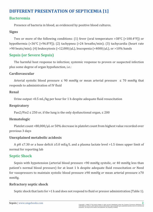
Table 1: Criteria for Sepsis Syndrome [2].
Screening Criteria Confirmatory Criteria
Presence of either (1) or (2): Presence of any 1 of the following, without an alternative explanation:
1. All 4 of the following: 1. PaO2/FiO2 < 280 (intubated) or whenusing a 40% face mask (not intubated)
Rectal temperature >38.3°C or <35.6°C 2. Arterial pH < 7.30Respiratory rate of >20/min or mechanical
ventilation 3. Urine output < 30 ml/hour
Heart rate > 90 beats/min
4. Systolic blood pressure < 90 mm Hg or fall in systolic blood pressure > 40 mm Hg
sustained for 2 hours despite fluid challenge
Clinical evidence of infection 5. Systemic vascular resistance < 800 dynes.s.cm2
2. One or more blood cultures positive for a pathogen at 48 hours after the onset of
sepsis
6. Prothrombin time or partial thromboplastin time greater than normal
or platelets > 100×106/l or platelets decreased to <50% of most recent measurement before current day
7. Deterioration in mental status within 24 hours
Etiological Agents
In the early 1960s, gram-negative bacilli were the most common cause of bacteremia and sepsis whereas in the last two decades, gram-positive cocci and yeasts have emerged as the major microbial pathogens in sepsis [3]. The systemic response to any class of microorganism can be harmful, because local inflammation can also elicit distant organ dysfunction and hypotension [1] (Table 2).
Table 2: Causative agents of septicemia [4-6].• Gram-positive bacteria- 52.9%• Gram-negative bacteria- 41.6%• Anaerobic organisms- 1.4%• Fungi- 4.1% [15].• Gram-negatives: Proteus, Serratia, Pseudomonas aeruginosa, Neisseria meningitudis, Escherichia coli Klebsiella pneumonia• Gram-positives: Staphylococcus aureus, coagulase-negative Staphylococcus, Streptococcus pyogenes, Streptococcus
pneumoniae, enterococci• Fungi: Candida albicans
Epidemiology
In the United States sepsis causes more than 200,000 deaths annually and is the second most common infectious cause of death in the United States after pneumonia [7]. The risk of death increased incrementally with the severity of illness (systemic inflammation 7%, sepsis, 16%, severe sepsis 20% and septic shock 46%) [8]. Recent epidemiologic data suggests that the incidence of sepsis is increasing at a rate of 8.7% annually [3] Patients who survive an episode of sepsis have an increased risk of dying during the subsequent five years when compared to controls [9] The average cost per case of sepsis was $22,100 with total costs of $16.7 billion nationally [10]. The rising incidence of severe sepsis in the United States has been attributable to the aging of the population, the increasing longevity of patients with chronic diseases, and the

4Sepsis | www.smgebooks.comCopyright Mittal S.This book chapter is open access distributed under the Creative Commons Attribution 4.0 International License, which allows users to download, copy and build upon published articles even for commercial purposes, as long as the author and publisher are properly credited.
relatively high frequency with which sepsis has occurred in patients with AIDS. The widespread use of immunosuppressive drugs, indwelling catheters, and mechanical devices has also played a role. In the aforementioned international ICU prevalence study, the case–fatality rate among infected patients (33%) greatly exceeded that among uninfected patients (15%) [1,11,12]
Pathophysiology
The 2001 International Sepsis Definitions Conference developed an expanded view of sepsis and developed the concept of a staging system for sepsis based on four separate characteristics designated by the acronym PIRO. P stands for the predisposition, indicating pre-existing co-morbid conditions that would reduce survival. I is the insult or infection, which reflects the clinical knowledge that some pathogenic organisms are more lethal than others. R represents the response to the infectious challenge. The last letter O stands for organ dysfunction and includes organ failure as well as the failure of a system such as the coagulation system [13].
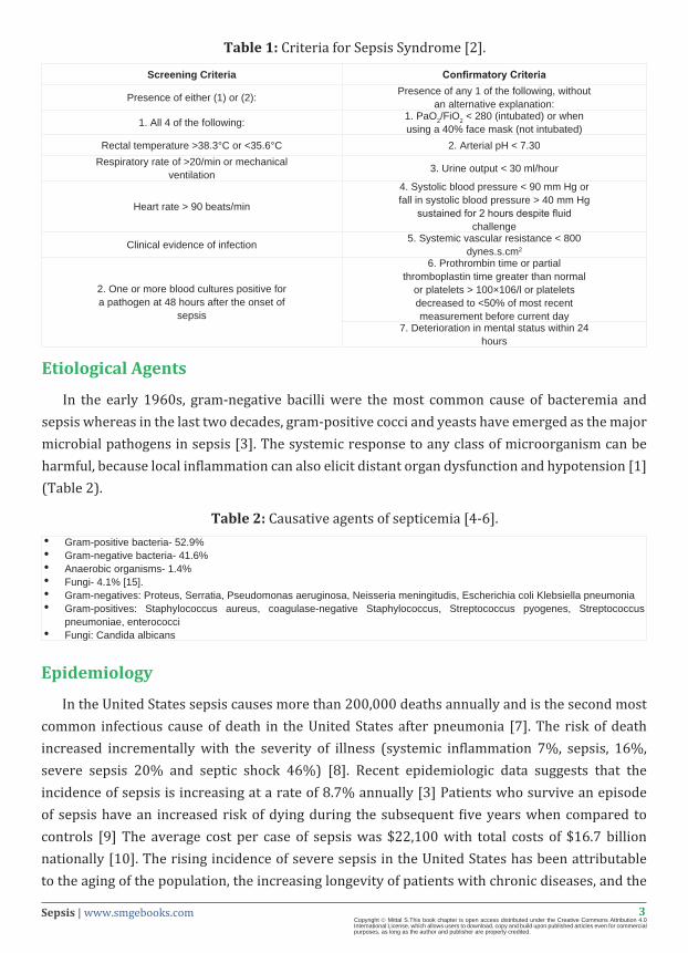
Sepsis originates as an isolated infection of microorganisms that become mobile in the circulatory system. Bacteremia is rarely associated with any signs or symptoms and most microorganisms are readily removed from circulation by the humoral immune system. When microbes begin to reproduce in the circulatory system and the body is unable to remove them at an adequate rate, septicemia develops, and a systemic inflammatory response is initiated. This syndrome, SIRS, is one of the primary disease patterns in sepsis. The progression from sepsis to septic shock follows the significant increase in serum levels of TNF-a, IFN-a, IL-1B, IL-8, and IL-6. Shock is a condition defined by inadequate tissue perfusion and emdash; in this case resulting from vasodilation due to increased cytokine levels and intravascular fluid shifting. TNF-a levels also increase, which is believed to be responsible for the onset of disseminated intravascular coagulation (DIC). These conditions, together with renal and liver failure, cause cardiac collapse and respiratory failure (ARDS) [10,14] (Figure 1).
5Sepsis | www.smgebooks.comCopyright Mittal S.This book chapter is open access distributed under the Creative Commons Attribution 4.0 International License, which allows users to download, copy and build upon published articles even for commercial purposes, as long as the author and publisher are properly credited.
Figure 1: Diagnostic sepsis phases.
CLINICAL SIGNS AND SYMPTOMS The signs and symptoms of sepsis vary according to the associated disease processes.
However, most symptomology is universal. Sepsis is preceded by a period of altered mental status for approximately 24 hours before other signs develop. Fatigue, malaise, myalgia, nausea, and vomiting are common early signs. Fever initially develops, but declines to hypothermia in late stages. Elevated heart rate and respiratory rate develop as blood pressure becomes erratic. Blood pressure eventually declines dramatically as plasma shifts and vasodilation worsens. Impaired renal function is evident with decreased urinary output, as is liver failure with jaundice [15-17].
DIAGNOSIS Clinical laboratory diagnosis is crucial to avoiding delay in treatment [18]. Thus, one-quarter
of patients with sepsis have inadequate treatment and worse prognosis as a consequence of a delayed diagnosis. [19,20]
In other words, laboratory tests should aim to identify compromised systems or organs, including indicators of inflammatory response in peripheral blood (pro-inflammatory mediators and acute phase indicators) and indicators of organic disorders. Enhanced serum lactate, cytokines, colony stimulating factor, and granulocyte markers of inflammation may be early indicators of more severe conditions, such as SIRS.
6Sepsis | www.smgebooks.comCopyright Mittal S.This book chapter is open access distributed under the Creative Commons Attribution 4.0 International License, which allows users to download, copy and build upon published articles even for commercial purposes, as long as the author and publisher are properly credited.
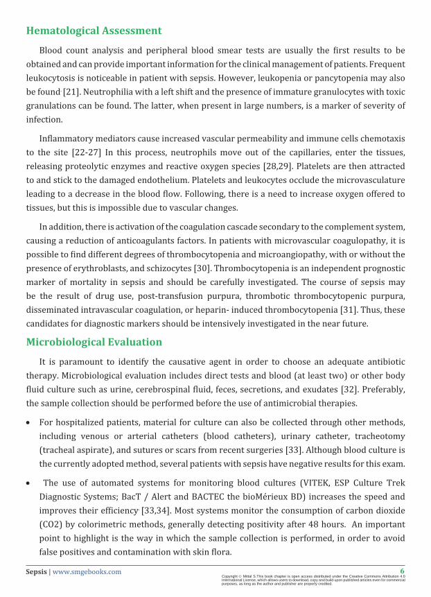
Hematological Assessment
Blood count analysis and peripheral blood smear tests are usually the first results to be obtained and can provide important information for the clinical management of patients. Frequent leukocytosis is noticeable in patient with sepsis. However, leukopenia or pancytopenia may also be found. [21]. Neutrophilia with a left shift and the presence of immature granulocytes with toxic granulations can be found. The latter, when present in large numbers, is a marker of severity of infection.
Inflammatory mediators cause increased vascular permeability and immune cells chemotaxis to the site [22-27] In this process, neutrophils move out of the capillaries, enter the tissues, releasing proteolytic enzymes and reactive oxygen species [28,29]. Platelets are then attracted to and stick to the damaged endothelium. Platelets and leukocytes occlude the microvasculature leading to a decrease in the blood flow. Following, there is a need to increase oxygen offered to tissues, but this is impossible due to vascular changes.
In addition, there is activation of the coagulation cascade secondary to the complement system, causing a reduction of anticoagulants factors. In patients with microvascular coagulopathy, it is possible to find different degrees of thrombocytopenia and microangiopathy, with or without the presence of erythroblasts, and schizocytes [30]. Thrombocytopenia is an independent prognostic marker of mortality in sepsis and should be carefully investigated. The course of sepsis may be the result of drug use, post-transfusion purpura, thrombotic thrombocytopenic purpura, disseminated intravascular coagulation, or heparin- induced thrombocytopenia [31]. Thus, these candidates for diagnostic markers should be intensively investigated in the near future.
Microbiological Evaluation
It is paramount to identify the causative agent in order to choose an adequate antibiotic therapy. Microbiological evaluation includes direct tests and blood (at least two) or other body fluid culture such as urine, cerebrospinal fluid, feces, secretions, and exudates [32]. Preferably, the sample collection should be performed before the use of antimicrobial therapies.
• For hospitalized patients, material for culture can also be collected through other methods, including venous or arterial catheters (blood catheters), urinary catheter, tracheotomy (tracheal aspirate), and sutures or scars from recent surgeries [33]. Although blood culture is the currently adopted method, several patients with sepsis have negative results for this exam.
• The use of automated systems for monitoring blood cultures (VITEK, ESP Culture Trek Diagnostic Systems; BacT / Alert and BACTEC the bioMérieux BD) increases the speed and improves their efficiency. [33,34]. Most systems monitor the consumption of carbon dioxide (CO2) by colorimetric methods, generally detecting positivity after 48 hours. An important point to highlight is the way in which the sample collection is performed, in order to avoid false positives and contamination with skin flora.
7Sepsis | www.smgebooks.comCopyright Mittal S.This book chapter is open access distributed under the Creative Commons Attribution 4.0 International License, which allows users to download, copy and build upon published articles even for commercial purposes, as long as the author and publisher are properly credited.
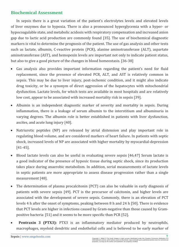
Biochemical Assessment
In sepsis there is a great variation of the patient’s electrolytes levels and elevated levels of liver enzymes due to hypoxia. There is also a pronounced hyperglycemia with a hyper- or hypocoagulable state, and metabolic acidosis with respiratory compensation and increased anion gap due to lactic acid production are commonly found [35]. The use of biochemical diagnostic markers is vital to determine the prognosis of the patient. The use of gas analysis and other tests such as lactate, albumin, C-reactive protein (PCR), alanine aminotransferase (ALT), aspartate aminotransferase (AST), and hemopexin levels are important not only to indicate patient status, but also to give a good picture of the changes in blood homeostasis. [36-38]
• Gas analysis also provides important information regarding the patient’s need for fluid replacement, since the presence of elevated PCR, ALT, and AST is relatively common in sepsis. This may be due to liver injury, post-ischemic condition, and it might also indicate drug toxicity, or be a synonym of direct aggression of the hepatocytes with mitochondrial dysfunction. Lactate levels, for which tests are available in most hospitals and are relatively low cost, appear to be associated with increased mortality risk in sepsis [39].
• Albumin is an independent diagnostic marker of severity and mortality in sepsis. During inflammation, there is a leakage of serum albumin to the interstitium and albuminuria in varying degrees. The albumin role is better established in patients with liver dysfunction, ascites, and acute lung injury [40].
• Natriuretic peptides (NP) are released by atrial distension and play important role in regulating blood volume, and are considered markers of heart failure. In patients with septic shock, increased levels of NP are associated with higher mortality by myocardial-depression [41-45].
• Blood lactate levels can also be useful in evaluating severe sepsis. [46,47] Serum lactate is a good indicator of the presence of hypoxic tissue during septic shock, since its production takes place during anaerobic metabolism. In addition, serial measurements of lactate levels in septic patients are more appropriate to assess disease progression rather than a single measurement [48].
• The determination of plasma procalcitonin (PCT) can also be valuable in early diagnosis of patients with severe sepsis [49]. PCT is the precursor of calcitonin, and higher levels are associated with the development of severe sepsis. Commonly, there is an elevation of PCT levels 4 h after the onset of symptoms, peaking between 8 h and 24 h [50]. There is evidence that PCT levels are higher in infections caused by Gram-negative than those caused by Gram-positive bacteria [51] and it seems to be more specific than PCR [52].
• Pentraxin 3 (PTX3): PTX3 is an inflammatory mediator produced by neutrophils, macrophages, myeloid dendritic and endothelial cells and is believed to be early marker of

8Sepsis | www.smgebooks.comCopyright Mittal S.This book chapter is open access distributed under the Creative Commons Attribution 4.0 International License, which allows users to download, copy and build upon published articles even for commercial purposes, as long as the author and publisher are properly credited.
severity and outcome in sepsis. PTX3 correlates with severity of sepsis, with sepsis-associated coagulation/fibrinolysis dysfunction and mortality especially over the first few days [53].
• Serum calprotectin : Serum Calprotectin (aka MRP8/14, calgranulin, cystic fibrosis-associated antigen), a complex of S100A8 and S100A9 is actively secreted via autocrine and paracrine mechanisms in phagocytes, endothelium, and other cells during stress response and it augments the inflammatory response in infections. It is an endogenous activator of TLR4 and promotes lethal, endotoxin-induced shock and a potent amplifier of inflammation in autoimmunity as well as in cancer development and tumor spread. Calprotectin protects cells against invasive microorganisms and regulates adhesion of leukocytes to the endothelium and extracellular matrix during the inflammatory process. Calprotectin is an early, accurate, and easy-to-use marker of neonatal sepsis [54].
• C-Reactive Protein: CRP is useful in the detection of sepsis and it is more sensitive than WBC although is now superceded by other better markers. The plasma CRP levels were significantly related to the infectious status (Negative, Unlikely, Probable or Definite). Concentrations of CRP in the Negative and Unlikely groups were significantly lower than in the Probable and Definite ones. A plasma CRP of 50 mg/l or more was highly suggestive of sepsis (sensitivity 98.5 %, specificity 75 %) [55].
• Lipopolysaccharide-binding protein : LPS-binding protein (LBP), mainly synthesized in the liver, is a polypeptide that binds LPS. The LPS-LBP complex initiates signal transduction according to LBP level. This complex complex has a dual action, enhancing and inhibiting LPS signaling at low and higher levels, respectively [ 56]. Serum LBP level increases several-fold in sepsis, making it useful for diagnosis [57,58]. It may also be effective as a predictive marker for disease severity and outcome [59,60]. However, LPS and LBP levels are affected by administration of antibiotics and generally do not correlate to the clinical course of sepsis [61]. Therefore, it is of limited use as a sepsis biomarker.
Immune Assessment
During an immune response, there is activation of various mechanisms of host defense against a pathogen, such as inflammation, complement and coagulation cascades, polymorphonuclear (PMN) activation, and chemoattraction to the site [62,63]. Among those markers used as mediators in sepsis there are cytokines such as tumor necrosis factor alpha (TNF)-α, interleukin (IL) -1, IL-6, IL-8, IL-10, and interferon (IFN)-γ, and the presence of bacterial products in the blood or the bacterium itself. IL-1 and TNF-α cytokines are the first cytokines released in an infection and stimulate cellular response [15,64-69].
• A good example of an acute mediator is IL-6, which is also a predictor of the severity and prognosis of sepsis.
• Mid-regional proadrenomedullin: Like PCT, proadrenomedullin (proADM) is a kind of

9Sepsis | www.smgebooks.comCopyright Mittal S.This book chapter is open access distributed under the Creative Commons Attribution 4.0 International License, which allows users to download, copy and build upon published articles even for commercial purposes, as long as the author and publisher are properly credited.
“hormokine” that encompasses the cytokine-like behavior of hormones during inflammation and infections. Adrenomedullin (ADM) is a 52-amino-acid peptide produced by the adrenal medulla. ADM is produced during physiological stress and has various functions including vasodilation and anti-inflammatory and antimicrobial effects [70]. Plasma ADM concentration and ADM gene expression increases in patients with sepsis [71]. However, ADM is rapidly cleared from the circulation, making measurements unreliable. Therefore, instead of ADM, serum quantification of the mid-regional fragment of proADM has been studied. Recent clinical data have shown that circulating mid-regional proADM levels are significantly higher in patients with sepsis than in patients with systemic inflammatory response syndrome (SIRS) [72]. A recent study of febrile patients with hematologic malignancies reported that proADM could predict localized bacterial infections and differentiate sepsis from SIRS [73]. In addition, proADM is responsible for hypotension associated with severe sepsis, which has been proposed as a good marker for risk assessment and predicting sepsis prognosis [ 72,74] . If further data support these findings on the predictive value of proADM, it could be useful as both a prognostic marker and a diagnostic marker for early stages of localized infections.
Note: It is necessary to process the sample immediately, because these proteins are labile and can easily change or be degraded.
Cytokine/chemokine biomarkers
TNF
TNF receptor signaling pathway (TNFR1 and TNFR2) plays a central role in the activation of innate immunity in response to pathogens. TNF is responsible for reduced neutrophil extravasation, migration to the infectious site and in neutrophil apoptosis. Absence of TNFR signaling leads to a decreased local and systemic inflammatory response with diminished organ injury. TNF levels are known to be significantly higher in septic shock than in sepsis without shock [75].
High Mobility Group Box Protein-1 (HMGB1)
HMGB1 is an important late mediator of endotoxin shock, intra abdominal sepsis, and acute lung injury, and a promising therapeutic target of severe sepsis [76].
Monocyte chemoattractant protein (MCP)-1
MCP-1, a prototype of CC chemokines, is a potent chemoattractant and a regulatory mediator involved in a variety of inflammatory diseases. Anti-MCP-1 strategies have shown promise in the treatment of sepsis and endotoxemia [77].
Growth-related oncogene-alpha (GRO alpha)
GRO-alpha, a member of the CXC chemokine family, induces endothelial dysfunction through oxidative stress and downregulation of Enos. High levels of GRO-alpha is seen in meningococcal sepsis/ septic shock and endotoxin induced shock [78].
10Sepsis | www.smgebooks.comCopyright Mittal S.This book chapter is open access distributed under the Creative Commons Attribution 4.0 International License, which allows users to download, copy and build upon published articles even for commercial purposes, as long as the author and publisher are properly credited.
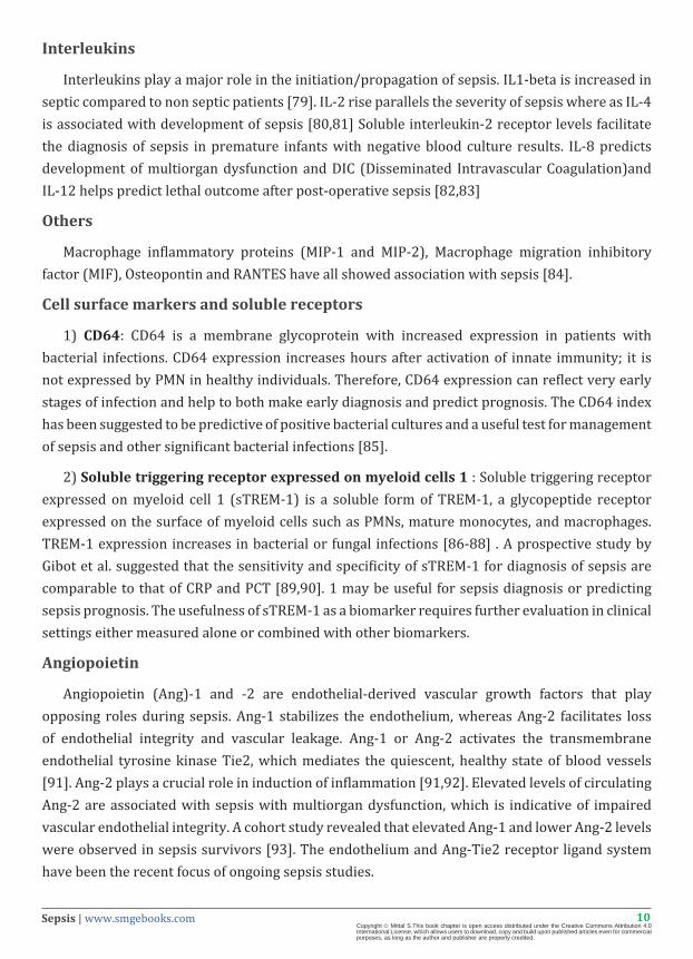
Interleukins
Interleukins play a major role in the initiation/propagation of sepsis. IL1-beta is increased in septic compared to non septic patients [79]. IL-2 rise parallels the severity of sepsis where as IL-4 is associated with development of sepsis [80,81] Soluble interleukin-2 receptor levels facilitate the diagnosis of sepsis in premature infants with negative blood culture results. IL-8 predicts development of multiorgan dysfunction and DIC (Disseminated Intravascular Coagulation)and IL-12 helps predict lethal outcome after post-operative sepsis [82,83]
Others
Macrophage inflammatory proteins (MIP-1 and MIP-2), Macrophage migration inhibitory factor (MIF), Osteopontin and RANTES have all showed association with sepsis [84].
Cell surface markers and soluble receptors
1) CD64: CD64 is a membrane glycoprotein with increased expression in patients with bacterial infections. CD64 expression increases hours after activation of innate immunity; it is not expressed by PMN in healthy individuals. Therefore, CD64 expression can reflect very early stages of infection and help to both make early diagnosis and predict prognosis. The CD64 index has been suggested to be predictive of positive bacterial cultures and a useful test for management of sepsis and other significant bacterial infections [85].
2) Soluble triggering receptor expressed on myeloid cells 1 : Soluble triggering receptor expressed on myeloid cell 1 (sTREM-1) is a soluble form of TREM-1, a glycopeptide receptor expressed on the surface of myeloid cells such as PMNs, mature monocytes, and macrophages. TREM-1 expression increases in bacterial or fungal infections [86-88] . A prospective study by Gibot et al. suggested that the sensitivity and specificity of sTREM-1 for diagnosis of sepsis are comparable to that of CRP and PCT [89,90]. 1 may be useful for sepsis diagnosis or predicting sepsis prognosis. The usefulness of sTREM-1 as a biomarker requires further evaluation in clinical settings either measured alone or combined with other biomarkers.
Angiopoietin
Angiopoietin (Ang)-1 and -2 are endothelial-derived vascular growth factors that play opposing roles during sepsis. Ang-1 stabilizes the endothelium, whereas Ang-2 facilitates loss of endothelial integrity and vascular leakage. Ang-1 or Ang-2 activates the transmembrane endothelial tyrosine kinase Tie2, which mediates the quiescent, healthy state of blood vessels [91]. Ang-2 plays a crucial role in induction of inflammation [91,92]. Elevated levels of circulating Ang-2 are associated with sepsis with multiorgan dysfunction, which is indicative of impaired vascular endothelial integrity. A cohort study revealed that elevated Ang-1 and lower Ang-2 levels were observed in sepsis survivors [93]. The endothelium and Ang-Tie2 receptor ligand system have been the recent focus of ongoing sepsis studies.
11Sepsis | www.smgebooks.comCopyright Mittal S.This book chapter is open access distributed under the Creative Commons Attribution 4.0 International License, which allows users to download, copy and build upon published articles even for commercial purposes, as long as the author and publisher are properly credited.
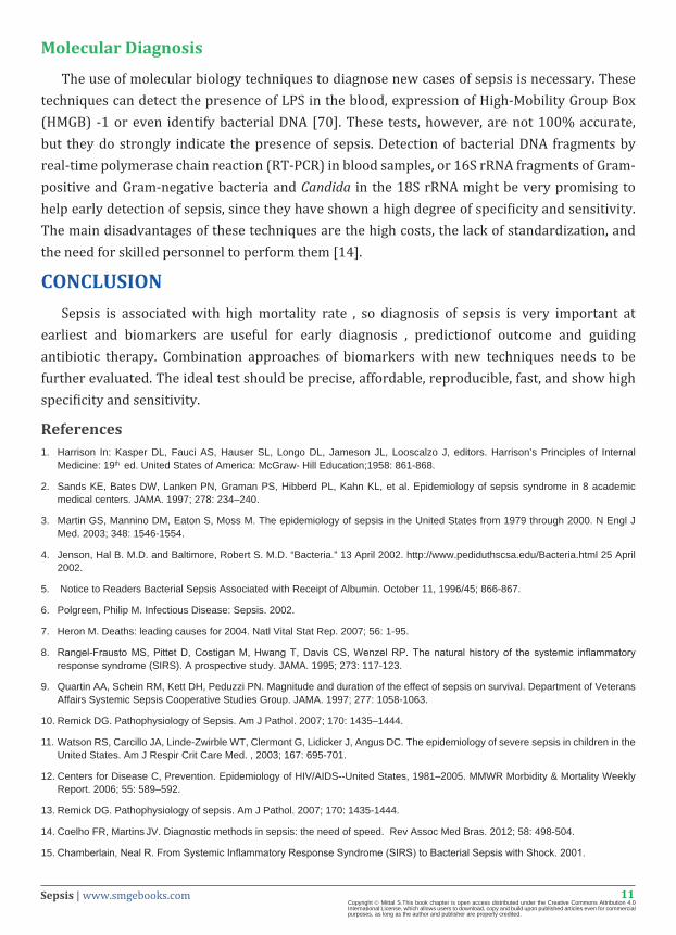
Molecular Diagnosis
The use of molecular biology techniques to diagnose new cases of sepsis is necessary. These techniques can detect the presence of LPS in the blood, expression of High-Mobility Group Box (HMGB) -1 or even identify bacterial DNA [70]. These tests, however, are not 100% accurate, but they do strongly indicate the presence of sepsis. Detection of bacterial DNA fragments by real-time polymerase chain reaction (RT-PCR) in blood samples, or 16S rRNA fragments of Gram-positive and Gram-negative bacteria and Candida in the 18S rRNA might be very promising to help early detection of sepsis, since they have shown a high degree of specificity and sensitivity. The main disadvantages of these techniques are the high costs, the lack of standardization, and the need for skilled personnel to perform them [14].
CONCLUSIONSepsis is associated with high mortality rate , so diagnosis of sepsis is very important at
earliest and biomarkers are useful for early diagnosis , predictionof outcome and guiding antibiotic therapy. Combination approaches of biomarkers with new techniques needs to be further evaluated. The ideal test should be precise, affordable, reproducible, fast, and show high specificity and sensitivity.
References1. Harrison In: Kasper DL, Fauci AS, Hauser SL, Longo DL, Jameson JL, Looscalzo J, editors. Harrison’s Principles of Internal
Medicine: 19th ed. United States of America: McGraw- Hill Education;1958: 861-868.
2. Sands KE, Bates DW, Lanken PN, Graman PS, Hibberd PL, Kahn KL, et al. Epidemiology of sepsis syndrome in 8 academic medical centers. JAMA. 1997; 278: 234–240.
3. Martin GS, Mannino DM, Eaton S, Moss M. The epidemiology of sepsis in the United States from 1979 through 2000. N Engl J Med. 2003; 348: 1546-1554.
4. Jenson, Hal B. M.D. and Baltimore, Robert S. M.D. “Bacteria.” 13 April 2002. http://www.pediduthscsa.edu/Bacteria.html 25 April 2002.
5. Notice to Readers Bacterial Sepsis Associated with Receipt of Albumin. October 11, 1996/45; 866-867.
6. Polgreen, Philip M. Infectious Disease: Sepsis. 2002.
7. Heron M. Deaths: leading causes for 2004. Natl Vital Stat Rep. 2007; 56: 1-95.
8. Rangel-Frausto MS, Pittet D, Costigan M, Hwang T, Davis CS, Wenzel RP. The natural history of the systemic inflammatory response syndrome (SIRS). A prospective study. JAMA. 1995; 273: 117-123.
9. Quartin AA, Schein RM, Kett DH, Peduzzi PN. Magnitude and duration of the effect of sepsis on survival. Department of Veterans Affairs Systemic Sepsis Cooperative Studies Group. JAMA. 1997; 277: 1058-1063.
10. Remick DG. Pathophysiology of Sepsis. Am J Pathol. 2007; 170: 1435–1444.
11. Watson RS, Carcillo JA, Linde-Zwirble WT, Clermont G, Lidicker J, Angus DC. The epidemiology of severe sepsis in children in the United States. Am J Respir Crit Care Med. , 2003; 167: 695-701.
12. Centers for Disease C, Prevention. Epidemiology of HIV/AIDS--United States, 1981–2005. MMWR Morbidity & Mortality Weekly Report. 2006; 55: 589–592.
13. Remick DG. Pathophysiology of sepsis. Am J Pathol. 2007; 170: 1435-1444.
14. Coelho FR, Martins JV. Diagnostic methods in sepsis: the need of speed. Rev Assoc Med Bras. 2012; 58: 498-504.
15. Chamberlain, Neal R. From Systemic Inflammatory Response Syndrome (SIRS) to Bacterial Sepsis with Shock. 2001.
12Sepsis | www.smgebooks.comCopyright Mittal S.This book chapter is open access distributed under the Creative Commons Attribution 4.0 International License, which allows users to download, copy and build upon published articles even for commercial purposes, as long as the author and publisher are properly credited.
16. Selim S. Sepsis. eMedicine Consumer. 2001.
17. http://www.emedicine.com/cgi-bin/foxweb.exe/showsection@d:/em/ga?book=emerg&topicid=533
18. Xing K, Murthy S, Liles WC, Singh JM. Clinical utility of biomarkers of endothelial activation in sepsis - a systematic review. Crit Care. 2012; 16: R7.
19. Ventetuolo CE, Levy MM. Biomarkers: diagnosis and risk assessment in sepsis. Clin Chest Med. 2008; 29: 591-603.
20. Angus DC, Linde-Zwirble WT, Lidicker J, Clermont G, Carcillo J, Pinsky MR. Epidemiology of severe sepsis in the United States: analysis of incidence, outcome, and associated costs of care. Crit Care Med. 2001; 29: 1303-1310.
21. Jacobi J. Pathophysiology of sepsis. Am J Health Syst Pharm. 2002; 15: S3-S8.
22. Oliveira C, Xavier RA, Anjos-Vallota E, Martins JO, Silveira VL, Gonçalves LR, et al. Effect of plant neutrophil elastase inhibitor on cell migration, adhesion and cytokine release on inflammatory conditions. Br J Pharmacol. 2010; 161: 899-910.
23. Oliveira WR, Cavassani SS, Maganhin CC, Carbonel AA, Simões MJ, Simões RS, et al. Histomorphologic and respiratory aspects of acute lung injury in rats induced by experimental sepsis and under pentoxifylline treatment. Rev Assoc Med Bras. 2009; 55: 127-1231.
24. Costantini TW, Deree J, Martins JO, Loomis W, Bansal V, Coimbra R. A novel fluid resuscitation strategy modulates pulmonary transcription factor activation after hemorrhagic shock. Clinics. 2010; 65: 621-628.
25. Sunahara KK, Martins JO. Alveolar macrophages in diabetes: friends or foes? J Leukoc Biol. 2012; 91: 871-876.
26. Legrand M, Max A, Peigne V, Mariotte E, Canet E, Debrumetz A, et al. Survival in neutropenic patients with severe sepsis or septic shock. Crit Care Med. 2012; 40: 43-49.
27. Alba-Loureiro TC, Munhoz CD, Martins JO, Cerchiaro GA, Scavone C, Curi R, et al. Neutrophil function and metabolism in individuals with diabetes mellitus. Braz J Med Biol Res. 2007; 40: 1037-1044.
28. Sunahara KKS, Sannomiya P, Martins JO. Briefs on insulin and innate immune response. Cell Physiol Biochem. 2012; 29: 1-8.
29. Souza YM, Fontes B, Martins JO, Sannomiya P, Brito GS, Younes RN, et al. Evaluation of the effects of ozone therapy in the treatment of intra-abdominal infection in rats. Clinics. 2010; 65: 195-202.
30. De Backer D, Donadello K, Taccone FS, Ospina-Tascon G, Salgado D, Vincent JL. Microcirculatory alterations: potential mechanisms and implications for therapy. Ann Intensive Care. 2011; 1: 27.
31. Wang Z, Yu Z, Su J, Cao L, Zhao X, Ruan C. Sepsis-induced disseminated intravascular coagulation with features of thrombotic thrombocytopenic purpura: a fatal fulminant syndrome. Clin Appl Thromb Hemost. 2011; 17: 251-253.
32. Riedel S, Carroll KC. Blood cultures: key elements for best practices and future directions. J Infect Chemother. 2010; 16: 301-316.
33. Mackenzie I, Lever A. Management of sepsis. BMJ. 2007; 335: 929-932.
34. Molina JM, Cordoba J, Ramirez P, Gobernado M. Deteccion automatica de bacterias y hongos en sangre. Enferm Infecc Microbiol Clin. 2008; 26: 75-80.
35. Levy MM, Fink MP, Marshall JC, Abraham E, Angus D, Cook D, et al. For the International Sepsis Definitions Conference. 2001 SCCM/ESICM/ ACCP/ATS/ SIS International Sepsis Definitions Conference. Crit Care Med. 2003; 31: 1250-1256.
36. Claessens YE, Schmidt J, Batard E, Grabar S, Jegou D, Hausfater P, et al. Can C-reactive protein, procalcitonin and mid-regional pro-atrial natriuretic peptide measurements guide choice of in-patient or out-patient care in acute pyelonephritis? Biomarkers In Sepsis (BIS) multicentre study. Clin Microbiol Infect. 2010; 16: 753-760.
37. Ceccon ME, Vaz FA, Diniz EM, Okay TS. Interleukins 6 and C-reactive protein for the diagnosis of late onset sepsis in the newborn infant. Rev Assoc Med Bras. 2006; 52: 79-85.
38. Hatzistilianou M. Diagnostic and prognostic role of procalcitonin in infections. ScientificWorldJournal. 2010; 10: 1941-1946.
39. Karon B, Scott R, Burritt M, Santrach P. Comparison of lactate values between point-of-care and central laboratory analyzers. Am J Clin Pathol. 2007;128: 168-171.
40. Han J, Martin GS. Does albumin fluid resuscitation in sepsis save lives? Crit Care Med. 2011; 39: 418-419.
41. Rudiger A, Gasser S, Fischler M, Hornemann T, Von Eckardstein A, Maggiorini M. Comparable increase of B-type natriuretic peptide and aminoterminal pro-B-type natriuretic peptide levels in patients with severe sepsis, septic shock, and acute heart failure. Crit Care Med. 2006; 34: 2140-2144.
42. Guinard-Barbier S, Grabar S, Chenevier-Gobeaux C, Quinquis L, Schmidt J, Kierzek G, et al. Is mid-regional pro-atrial natriuretic peptide (MRproANP) an accurate marker of bacteremia in pyelonephritis? Biomarkers. 2011; 16: 355-363.
13Sepsis | www.smgebooks.comCopyright Mittal S.This book chapter is open access distributed under the Creative Commons Attribution 4.0 International License, which allows users to download, copy and build upon published articles even for commercial purposes, as long as the author and publisher are properly credited.
43. Iapichino G, Marzorati S, Umbrello M, Baccalini R, Barassi A, Cainarca M, et al. Daily monitoring of biomarkers of sepsis in complicated long-term ICU-patients: can it support treatment decisions? Minerva Anestesiol. 2010; 76: 814-823.
44. Claessens YE, Mathevon T, Kierzek G, Grabar S, Jegou D, Batard E, et al. Accuracy of C-reactive protein, procalcitonin, and mid-regional pro-atrial natriuretic peptide to guide site of care of community-acquired pneumonia. Intensive Care Med. 2010; 36: 799-809.
45. Nguyen HB, Van Ginkel C, Batech M, Banta J, Corbett SW. Comparison of predisposition, insult/infection, response, and organ dysfunction, acute physiology and chronic health evaluation ii, and mortality in emergency department sepsis in patients meeting criteria for early goal-directed therapy and the severe sepsis resuscitation bundle. J Crit Care. 2011; 27: 362-369.
46. Friedman G, Berlot G, Kahn RJ, Vincent JL. Combined measurements of blood lactate concentrations and gastric intramucosal pH in patients with severe sepsis. Crit Care Med. 1995; 23: 1184-1193.
47. Hajjar LA, Nakamura RE, de Almeida JP, Fukushima JT, Hoff PM, Vincent JL, et al. Lactate and base deficit are predictors of mortality in critically ill patients with cancer. Clinics. 2011; 66: 2037-2042.
48. Arnold RC, Shapiro NI, Jones AE, Schorr C, Pope J, Casner E, et al. Multicenter study of early lactate clearance as a determinant of survival in patients with presumed sepsis. Shock. 2009; 32: 35-39.
49. Simon L, Saint-Louis P, Amre DK, Lacroix J, Gauvin F. Procalcitonin and C-reactive protein as markers of bacterial infection in critically ill children at onset of systemic inflammatory response syndrome. Pediatr Crit Care Med. 2008; 9: 407-413.
50. Kibe S, Adams K, Barlow G. Diagnostic and prognostic biomarkers of sepsis in critical care. J Antimicrob Chemother. 2011; 66: 1133-1140.
51. Charles PE, Ladoire S, Aho S, Quenot JP, Doise JM, Prin S, et al. Serum procalcitonin elevation in critically ill patients at the onset of bacteremia caused by either Gram-negative or Gram-positive bacteria. BMC Infect Dis. 2008; 8: 38.
52. Ferreira AM, Sakr Y. Organ dysfunction: general approach, epidemiology, and organ failure scores. Semin Respir Crit Care Med. 2011; 32: 543-551.
53. Mauri T, Bellani G, Patroniti N, Coppadoro A, Peri G, Cuccovillo I, et al. Persisting high levels of plasma pentraxin 3 over the first days after severe sepsis and septic shock onset are associated with mortality. Intensive Care Med. 2010; 36: 621-629.
54. Terrin G, Passariello A, Manguso F, Salvia G, Rapacciuolo L, Messina F, et al. Serum calprotectin: an antimicrobial peptide as a new marker for the diagnosis of sepsis in very low birth weight newborns. Clin Dev Immunol. 2011; 2011: 291085.
55. Póvoa P, Almeida E, Moreira P, Fernandes A, Mealha R, Aragão A, et al. C-reactive protein as an indicator of sepsis. Intensive Care Med. 1998; 24: 1052-1056.
56. Jerala R. Structural biology of the LPS recognition. Int J Med Microbiol. 2007; 297: 353–363.
57. Zweigner J, Gramm HJ, Singer OC, Wegscheider K, Schumann RR. High concentrations of lipopolysaccharide-binding protein in serum of patients with severe sepsis or septic shock inhibit the lipopolysaccharide response in human monocytes. Blood. 2001; 98: 3800–3808.
58. Gaïni S, Koldkjaer OG, Pedersen C, Pedersen SS. Procalcitonin, lipopolysaccharide-binding protein, interleukin-6 and C-reactive protein in community-acquired infections and sepsis: a prospective study. Crit Care. 2006; 10: R53.
59. Opal SM, Scannon PJ, Vincent JL, White M, Carroll SF, Palardy JE, et al. Relationship between plasma levels of lipopolysaccharide (LPS) and LPS-binding protein in patients with severe sepsis and septic shock. J Infect Dis. 1999; 180: 1584–1589.
60. Sakr Y, Burgett U, Nacul FE, Reinhart K, Brunkhorst F. Lipopolysaccharide binding protein in a surgical intensive care unit: a marker of sepsis? Crit Care Med. 2008; 36: 2014–2022.
61. Choi JH, Shin WS. Pathogenesis of sepsis and concepts of immunotherapy. Korean J Infect Dis. 2000; 32: 148–157.
62. Villar J, Flores C, Perez-Mendez L. Genetic determinants of survival in sepsis and acute lung injury. Minerva Anestesiol. 2008; 74: 341-345.
63. Jacobi J. Pathophysiology of sepsis. Am J Health Syst Pharm. 2002; 15: S3-S8.
64. Deree J, Martins JO, Melbostad H, Putnam JG, de Campos T, Hoyt DB, et al. Insights into the regulation of TNF-α production in human mononuclear cells: the effects of non-specific phosphodiesterase inhibition. Clinics. 2008; 63: 321-328.
65. Anjos-Valotta EA, Martins JO, Oliveira MA, Casolari DA, Britto LR, Tostes RC, et al. Inhibition of tumor necrosis factor-alpha-induced intercellular adhesion molecule-1 expression in diabetic rats: role of insulin. Inflamm Res. 2006; 55: 16-22.
66. Martins JO, Ferracini M, Ravanelli N, Landgraf RG, Jancar S. Insulin inhibits LPS-induced signaling pathways in alveolar macrophages. Cell Physiol Biochem. 2008; 21: 297-304.
67. Martins JO, Ferracini M, Ravanelli N, Landgraf RG, Jancar S. Insulin suppresses LPS-induced iNOS and COX-2 expression and NF-kappaB activation in alveolar macrophages. Cell Physiol Biochem. 2008; 22: 279-286.

14Sepsis | www.smgebooks.comCopyright Mittal S.This book chapter is open access distributed under the Creative Commons Attribution 4.0 International License, which allows users to download, copy and build upon published articles even for commercial purposes, as long as the author and publisher are properly credited.
68. Russell JA. Management of sepsis. N Engl J Med. 2006; 355: 1699-1713.
69. Schuerholz T, Marx G. Management of sepsis. Minerva Anestesiol. 2008; 74: 181-195.
70. Linscheid P, Seboek D, Zulewski H, Keller U, Müller B. Autocrine/paracrine role of inflammation-mediated calcitonin gene-related peptide and adrenomedullin expression in human adipose tissue. Endocrinology. 2005; 146: 2699–2708.
71. Hinson JP, Kapas S, Smith DM. Adrenomedullin, a multifunctional regulatory peptide. Endocr Rev. 2000; 21: 138–167.
72. Christ-Crain M, Morgenthaler NG, Struck J, Harbarth S, Bergmann A, Müller B. Mid-regional pro-adrenomedullin as a prognostic marker in sepsis: an observational study. Crit Care. 2005; 9: R816–R824.
73. Al Shuaibi M, Bahu RR, Chaftari AM, Al Wohoush I, Shomali W, Jiang Y, et al. Pro-adrenomedullin as a novel biomarker for predicting infections and response to antimicrobials in febrile patients with hematologic malignancies. Clin Infect Dis. 2013; 56: 943–950.
74. Suberviola B, Castellanos-Ortega A, Ruiz Ruiz A, Lopez-Hoyos M, Santibañez M. Hospital mortality prognostication in sepsis using the new biomarkers suPAR and proADM in a single determination on ICU admission. Intensive Care Med. 2013; 39: 1945–1952.
75. Secher T, Vasseur V, Poisson DM, Mitchell JA, Cunha FQ, Alves-Filho JC, et al. Crucial role of TNF receptors 1 and 2 in the control of polymicrobial sepsis. J Immunol 2009; 182: 7855-7864.
76. Suda K, Kitagawa Y, Ozawa S, Saikawa Y, Ueda M, Ebina M, et al. Anti-high-mobility group box chromosomal protein 1 antibodies improve survival of rats with sepsis. World J Surg. 2006; 30: 1755-1762.
77. Ramnath RD, Ng SW, Guglielmotti A, Bhatia M. Role of MCP-1 in endotoxemia and sepsis. Int Immunopharmacol. 2008; 8: 810-818.
78. Vermont CL, Hazelzet JA, de Kleijn ED, van den Dobbelsteen GP, de Groot R. CC and CXC chemokine levels in children with meningococcal sepsis accurately predict mortality and disease severity. Crit Care 2006; 10: R33.
79. Kurt AN, Aygun AD, Godekmerdan A, Kurt A, Dogan Y, Yilmaz E. Serum IL-1 beta, IL-6, IL-8, and TNF-alpha levels in early diagnosis and management of neonatal sepsis. Mediators Inflamm. 2007; 2007: 31397.
80. BalcI C, Sungurtekin H, Gurses E, Sungurtekin U, Kaptanoglu B. Usefulness of procalcitonin for diagnosis of sepsis in the intensive care unit. Crit Care. 2003; 7: 85-90.
81. DiPiro JT, Howdieshell TR, Goddard JK, Callaway DB, Hamilton RG, Mansberger AR Jr. Association of interleukin-4 plasma levels with traumatic injury and clinical course. Arch Surg. 1995; 130: 1159-1162.
82. El Maghraby SM, Moneer MM, Ismail MM, Shalaby LM, El Mahallawy HA. The diagnostic value of C-reactive protein, interleukin-8, and monocyte chemotactic protein in risk stratification of febrile neutropenic children with hematologic malignancies. J Pediatr Hematol Oncol. 2007; 29: 131-136.
83. Castellheim A, Thorgersen EB, Hellerud BC, Pharo A, Johansen HT, Brosstad F, et al. New biomarkers in an acute model of live Escherichia coli induced sepsis in pigs. Scand J Immunol. 2008; 68: 75-84.
84. Dilip Gude. Biomarkers in sepsis: A comprehensive review. Int J clin cases and investigations 2012; 3: 60-80.
85. Icardi M, Erickson Y, Kilborn S, Stewart B, Grief B, Scharnweber G. CD64 index provides simple and predictive testing for detection and monitoring of sepsis and bacterial infection in hospital patients. J Clin Microbiol. 2009; 47: 3914–3919.
86. Bouchon A, Facchetti F, Weigand MA, Colonna M. TREM-1 amplifies inflammation and is a crucial mediator of septic shock. Nature. 2001; 410: 1103–1107.
87. Sébastien Gibot. Clinical review: Role of triggering receptor expressed on myeloid cells-1 during sepsis. Critical Care. 2005; 9: 485-489.
88. Ford JW. McVicar DW. TREM and TREM-like receptors in inflammation and disease. Curr Opin Immunol. 2009; 21: 38–46.
89. Gibot S, Kolopp-Sarda MN, Béné MC, Cravoisy A, Levy B, Faure GC, et al. Plasma level of a triggering receptor expressed on myeloid cells-1: its diagnostic accuracy in patients with suspected sepsis. Ann Intern Med. 2004; 141: 9–15.
90. Zhang J, She D, Feng D, Jia Y, Xie L. Dynamic changes of serum soluble triggering receptor expressed on myeloid cells-1 (sTREM-1) reflect sepsis severity and can predict prognosis: a prospective study. BMC Infect Dis. 2011; 11: 53.
91. David S, Kümpers P, van Slyke P, Parikh SM. Mending leaky blood vessels: the angiopoietin-Tie2 pathway in sepsis. J Pharmacol Exp Ther. 2013; 345: 2–6.
92. Fiedler U, Reiss Y, Scharpfenecker M, Grunow V, Koidl S, Thurston G, et al. Angiopoietin-2 sensitizes endothelial cells to TNF-alpha and has a crucial role in the induction of inflammation. Nat Med. 2006; 12: 235–239.
93. Ricciuto DR, dos Santos CC, Hawkes M, Toltl LJ, Conroy AL, Rajwans N, et al. Angiopoietin-1 and angiopoietin-2 as clinically informative prognostic biomarkers of morbidity and mortality in severe sepsis. Crit Care Med. 2011; 39: 702–710.