Embed Size (px)
Citation preview
TDP-43 Proteinopathy and Motor Neuron Disease in ChronicTraumatic Encephalopathy
Ann C. McKee, MD, Brandon E. Gavett, PhD, Robert A. Stern, PhD, Christopher J. Nowinski,AB, Robert C. Cantu, MD, Neil W. Kowall, MD, Daniel P. Perl, MD, E. Tessa Hedley-Whyte,MD, Bruce Price, MD, Chris Sullivan, Peter Morin, MD, PhD, Hyo-Soon Lee, MD, Caroline A.Kubilus, Daniel H. Daneshvar, MA, Megan Wulff, MPH, and Andrew E. Budson, MDGeriatric Research Education Clinical Center (ACM, CS, PM, AEB), Bedford VeteransAdministration Hospital, Bedford; and Center for the Study of Traumatic Encephalopathy (ACM,BEG, RAS, CJN, RCC, NWK, DHD, MW) and Departments of Neurology (ACM, BEG, RAS, CS,PM, H-SL, CAK, AEB) and Pathology (ACM, NWK), Boston University School of Medicine, Boston,Massachusetts; National VA ALS Biorepository (ACM), Tucson, Arizona; Sports Legacy Institute(CJN, RCC), Waltham; Department of Neurosurgery (RCC), Boston University School of Medicine,Boston; Department of Neurosurgery (RCC), Emerson Hospital, Concord; and Geriatric ResearchEducation Clinical Center (NWK), Jamaica Plain Veterans Administration Medical Center, Boston,Massachusetts; Department of Pathology (Neuropathology) (DPP), Mount Sinai School of Medicine,New York, New York; and CS Kubik Laboratory for Neuropathology and Departments of Pathology(ETH-W) and Neurology (BP), Massachusetts General Hospital, Harvard Medical School, Boston;and Department of Neurology (BP), McLean Hospital, Belmont, Massachusetts
AbstractEpidemiological evidence suggests that the incidence of amyotrophic lateral sclerosis is increasedin association with head injury. Repetitive head injury is also associated with the development ofchronic traumatic encephalopathy (CTE), a tauopathy characterized by neurofibrillary tanglesthroughout the brain in the relative absence of β-amyloid deposits. We examined 12 cases of CTEand, in 10, found a widespread TAR DNA-binding protein of approximately 43 kd (TDP-43)proteinopathy affecting the frontal and temporal cortices, medial temporal lobe, basal ganglia,diencephalon, and brainstem. Three athletes with CTE also developed a progressive motor neurondisease with profound weakness, atrophy, spasticity, and fasciculations several years before death.In these 3 cases, there were abundant TDP-43–positive inclusions and neurites in the spinal cord inaddition to tau neurofibrillary changes, motor neuron loss, and corticospinal tract degeneration. TheTDP-43 proteinopathy associated with CTE is similar to that found in frontotemporal lobardegeneration with TDP-43 inclusions, in that widespread regions of the brain are affected. Akin tofrontotemporal lobar degeneration with TDP-43 inclusions, in some individuals with CTE, theTDP-43 proteinopathy extends to involve the spinal cord and is associated with motor neuron disease.This is the first pathological evidence that repetitive head trauma experienced in collision sportsmight be associated with the development of a motor neuron disease.
Send correspondence and reprint requests to: Ann C. McKee, MD, Bedford Veterans Administration Medical Center, 182-B, 200 SpringsRd, Bedford, MA 01730; [email protected] digital content is available for this article. Direct URL citations appear in the printed text and are provided in the HTMLand PDF versions of this article on the journal’s Web site (www.jneuropath.com).
NIH Public AccessAuthor ManuscriptJ Neuropathol Exp Neurol. Author manuscript; available in PMC 2010 October 7.
Published in final edited form as:J Neuropathol Exp Neurol. 2010 September ; 69(9): 918–929. doi:10.1097/NEN.0b013e3181ee7d85.
NIH
-PA Author Manuscript
NIH
-PA Author Manuscript
NIH
-PA Author Manuscript
KeywordsAmyotrophic lateral sclerosis; Chronic brain injury; Motor neuron disease; Sports; Tau proteins;TDP-43
INTRODUCTIONAmyotrophic lateral sclerosis (ALS) is a fatal progressive degeneration of motor neurons inthe brain and spinal cord. Whereas 90% to 95% of ALS cases are sporadic, gene mutations incopper/zinc superoxide dismutase 1, senataxin, dynactin, angiogenic, and TAR-DBP (the genefor transactive response DNA-binding protein of 43 kd [TDP-43] on chromosome 1) accountfor some familial forms of the disease (1,2). ALS is pathologically characterized by motorneuron loss and corticospinal tract degeneration. In addition, remaining motor neurons insporadic ALS often have ubiquitin- and TDP-43– immunoreactive inclusion bodies that appeareither as rounded hyaline inclusions or as skeinlike inclusions (3–6).
Although the etiology of sporadic ALS is unknown, it has long been suspected to involve acomplex interaction between multiple genetic and environmental risk factors. Manyenvironmental risk factors have been considered as possible triggers of the neurodegenerativecascade in ALS, including a history of trauma to the brain and spinal cord (7–13), a history ofparticipation in varsity athletics and a slim physique (14), strenuous physical activity (14–17), cigarette smoking (18–20), and exposure to heavy metals (21–28), radiation, electricalshocks (12,29), and pesticides (8,24,30,31). Yet, of all the putative environmental risk factors,trauma to the CNS emerges as one of the strongest and most consistent contenders for initiatingthe molecular cascades that result in ALS (9,32), as well as other neurodegenerative processes,such as Alzheimer disease (AD) (33–39) and Parkinson disease (40,41).
That mechanical or traumatic injury to the head, neck, or spine might be etiologically relatedto ALS has been suggested for more than 100 years (42,43). More recent literature pointstoward a trend not only between CNS trauma and the development of ALS but also betweena smaller number of years between the last injury and ALS diagnosis, and older age at the lastinjury and the development of ALS (9,32). In a case-control study of 109 cases of ALS and255 controls, Chen et al (9) found that having experienced repeated head injuries or havingbeen injured within the 10 years before diagnosis was associated with a more than 3-fold higherrisk of ALS (odds ratio [OR], 3.1; 95% confidence interval [95% CI], 1.2–8.1; and OR, 3.2;95% CI, 1.0–10.2, respectively), with a slightly elevated risk for the interval 11 to 30 years.The authors further performed a meta-analysis of 8 previous ALS studies and estimated apooled OR of 1.7 (95% CI, 1.3–2.2) for at least 1 previous head injury. Another case-controlstudy, which was not included in the meta-analysis, reported an increased risk of ALS whenthe last head injury occurred at an older age and closer to the time of diagnosis (10). ALSincidence and mortality are also reported to be unusually high among professional soccerplayers in Italy (16,17,44). ALS mortality for Italian professional soccer players was increased12-fold, whereas mortality from other causes was generally lower or comparable to that of thegeneral population (17). Moreover, an incidence study involving 7,325 Italian professionalsoccer players showed that the incidence of ALS was 6.5 times higher than expected (16). Anincreased incidence of ALS has also been reported in American football players (45), andclusters of ALS have been found in Canadian and National Football League players, including3 members of the 1970s San Francisco 49ers (46).
The risk of ALS was reported to be increased approximately 2-fold among veterans of the 1991Gulf War during the 10 years after the war (47–49). This elevated risk was evident amongdeployed military personnel who were on active duty, with statistically significant elevations
McKee et al. Page 2
J Neuropathol Exp Neurol. Author manuscript; available in PMC 2010 October 7.
NIH
-PA Author Manuscript
NIH
-PA Author Manuscript
NIH
-PA Author Manuscript
especially notable among those in the Air Force and Army. A second independent studyinvolving only Gulf War veterans younger than 45 years also found an elevated risk of ALSin this population (50). Recently, Schmidt et al (32) found that military veterans who hadexperienced head injuries during the last 15 years had an adjusted OR for the development ofALS of 2.33 (95% CI, 1.18–4.61) relative to veterans without any head injuries, and that thisassociation was strongest in APOE-ε4 carriers. Several specific attributes of the at-risk militaryveterans contributed to their higher prevalence of head injuries, including combat-relatedinjuries during deployment to major conflicts and participation in competitive sports.
Repetitive head injury is also associated with the development of chronic traumaticencephalopathy (CTE), a progressive tauopathy clinically associated with behavioral andpersonality changes, parkinsonism, and dementia (51). Recently, TDP-43 immunoreactivitywas found in the cerebral cortex in 3 cases of CTE associated with boxing (52). AlthoughTDP-43 was originally thought to be a specific marker for ALS and frontotemporal lobardegeneration (FTLD) with tau-negative ubiquitin-positive TDP-43–positive inclusions(FTLD-U, recently renamed FTLD-TDP) (5,53–57), TDP-43–positive inclusions have nowbeen found in a variety of other neurodegenerative disorders.
Through the brain donation program for Center for the Study of Traumatic Encephalopathy atBoston University School of Medicine and the Bedford VA Medical Center, we analyzed thebrains and spinal cords of 12 former athletes with CTE; 3 of the athletes with CTE also hadsigns and symptoms of motor neuron disease (MND). We compared the nature and distributionof tau and TDP-43 immunoreactivity in the brain and spinal cord of the 9 athletes with CTEwithout MND (CTE–no MND) with those of the 3 with CTE with MND (CTE + MND) andwith the findings in the spinal cords of 12 normal controls and 12 individuals with sporadicALS. We wanted to establish the extent of the TDP-43 proteinopathy in CTE, the relationshipof TDP-43 immunoreactivity to symptoms of MND, and whether the TDP-43–immunoreactivepathology colocalized with tau pathology.
MATERIALS AND METHODSSubjects
The brain and spinal cords from 11 of the 12 athletes with pathologically verified CTE werereceived through the brain donation program of the Boston University Alzheimer’s DiseaseCenter (ADC) and the Center for the Study of Traumatic Encephalopathy. Brain and spinalcord tissues from the remaining athlete (Case 3) were received from the MassachusettsAlzheimer’s Disease Research Center Brain Bank. Samples of paraffin-embedded brain andspinal cord sections from 12 age- and sex-matched subjects with pathologically verified ALSand 6 of the 12 normal controls were received from the National VA ALS biorepository.Samples of formalin-fixed spinal cord from the remaining 6 age- and sex-matched controlsubjects who were neurologically intact at time of death were received from the Mount SinaiADC.
For the athletes, the concussion history, injury history, history of cognitive, behavioral, andneurological abnormalities, motor symptoms, neurological examinations, and clinical status atthe time of death were determined through review of medical records and interviews with theplayers’ next of kin. When available, additional informants were interviewed to obtainconfirmation or the information provided. Interviews were conducted by a neuropsychologist(Robert Stern) who was blinded to the results of the neuropathologic examination at the timeof the interview. Informants were interviewed before receiving information concerning theresults of the neuropathologic examination. Medical records review was also conducted by aneurologist (Neil Kowall) and a neuropathologist (Ann McKee).
McKee et al. Page 3
J Neuropathol Exp Neurol. Author manuscript; available in PMC 2010 October 7.
NIH
-PA Author Manuscript
NIH
-PA Author Manuscript
NIH
-PA Author Manuscript
Neuropathologic ExaminationThe neuropathologic processing followed the procedures previously established for the BostonUniversity ADC, Mount Sinai ADC, and Massachusetts Alzheimer’s Disease Research CenterBrain Bank; these include a comprehensive analysis of all neurodegenerative conditions (58).Brain tissues from 3 athletes were received in fragments fixed in formalin after processing bymedical examiners; brain and spinal cord tissues from the other 9 were received either freshor fixed in formalin. Paraffin-embedded sections from at least 25 brain regions were stainedwith Luxol fast blue, hematoxylin and eosin, Bielschowsky silver, AT8 (a mouse monoclonalantibody directed against phosphoserine 202 and phosphothreonine 205 of PHF-tau, 1:2000;Pierce Endogen, Rockford, IL), PHF-1, a monoclonal antibody against phosphoserine 396 andphosphoserine 404 of hyperphosphorylated tau (1:1000; courtesy of Peter Davies), TDP-43(rabbit polyclonal to TAR DNA-binding protein, 1:1000; Abcam, Cambridge, MA), LN3(HLA-DR Class II [major histocompatibility complex], 1:2000; Zymed, San Francisco, CA),ubiquitin (rabbit polyclonal, 1:2000; Dako North America Inc, Carpinteria, CA), α-synuclein(rabbit polyclonal, 1:15,000; Chemicon, Temecula, CA), and β-amyloid (Aβ, mousemonoclonal, 1:2000, formic acid pretreatment; Dako North America Inc). In addition, multiplelarge coronal fragments were cut at 50 μm on a sledge microtome and stained as free-floatingsections using CP13, a monoclonal antibody directed against phosphoserine 202 of tau,considered to be the initial site of tau phosphorylation in neurofibrillary tangle (NFT) formation(1:200; courtesy of Peter Davies), glial fibrillary acidic protein (mouse monoclonal, 1:2000;Chemicon), TDP-43, PHF-1, ubiquitin, Aβ, and LN3, all counterstained with cresyl violet aspreviously described (51). For the control and ALS subjects, 10-μm-thick sections wereprepared from paraffin blocks and stained with Luxol fast blue, hematoxylin and eosin, AT8,and TDP-43.
Double immunofluorescence staining was carried out to assess colocalization by confocalimaging using a Leica SP5 laser scanning confocal microscope. Paraffin-embedded tissuesections were deparaffinized in xylene and rehydrated through alcohol before being subjectedto antigen retrieval in formic acid. Sections were blocked in Super Block buffer (ScyTek,Logan, UT) containing 5% serum before avidin/biotin blocking and incubation overnight at 4°C with the initial primary antibody. Sections were then incubated for 1 hour at roomtemperature with a biotinylated secondary antibody followed by avidin conjugated with eitherAlexa Fluor 488 or Alexa Fluor 555 (Invitrogen). This protocol (beginning with blockingprocedures) was repeated for secondary antigen detection.
Apolipoprotein E genotyping was conducted on 11 of the 12 athletes using restriction isotypingfor determining apolipoprotein E isoforms based on brain tissue samples.
RESULTSClinical Data
At death, the 12 athletes who developed CTE ranged in age from 42 to 85 years (mean, 65.4years; SD, 15.9 years) and included 7 former football players (6 of whom played professionallyand 1 in college), 4 retired professional boxers, and 1 professional hockey player (Tables 1 and2). The 3 athletes who developed clinically diagnosed MND included 2 retired professionalfootball players and a former professional boxer. The players with CTE + MND did not differfrom the 9 athletes with CTE–no MND with respect to age at death, total years of participationin the sport, age at retirement from the sport, or concussion histories (data not shown).
The 3 athletes with CTE + MND had clinical presentations characterized by profound muscleweakness, muscle atrophy, spasticity, and diffuse fasciculations (Table 1). Two developedmotor symptoms after the development of cognitive impairment or dementia and behavioral
McKee et al. Page 4
J Neuropathol Exp Neurol. Author manuscript; available in PMC 2010 October 7.
NIH
-PA Author Manuscript
NIH
-PA Author Manuscript
NIH
-PA Author Manuscript
changes; one developed parkinsonism in addition to dementia, behavioral changes, and MND;and one developed prominent symptoms of MND in the presence of behavioral changes anddepression but without evidence of cognitive decline or parkinsonism. In all 3, the MND beganwith bilateral involvement of the shoulder girdles, neck, and arms, involved the bulbarmusculature early in the disease course, and resulted in death within 2 to 3 years (see data,Supplemental Digital Content 1, http://links.lww.com/NEN/A161 for case summaries).
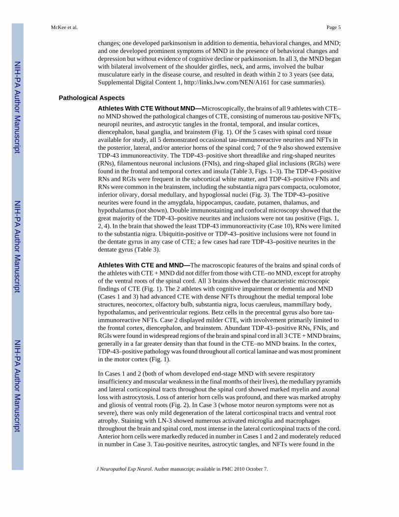
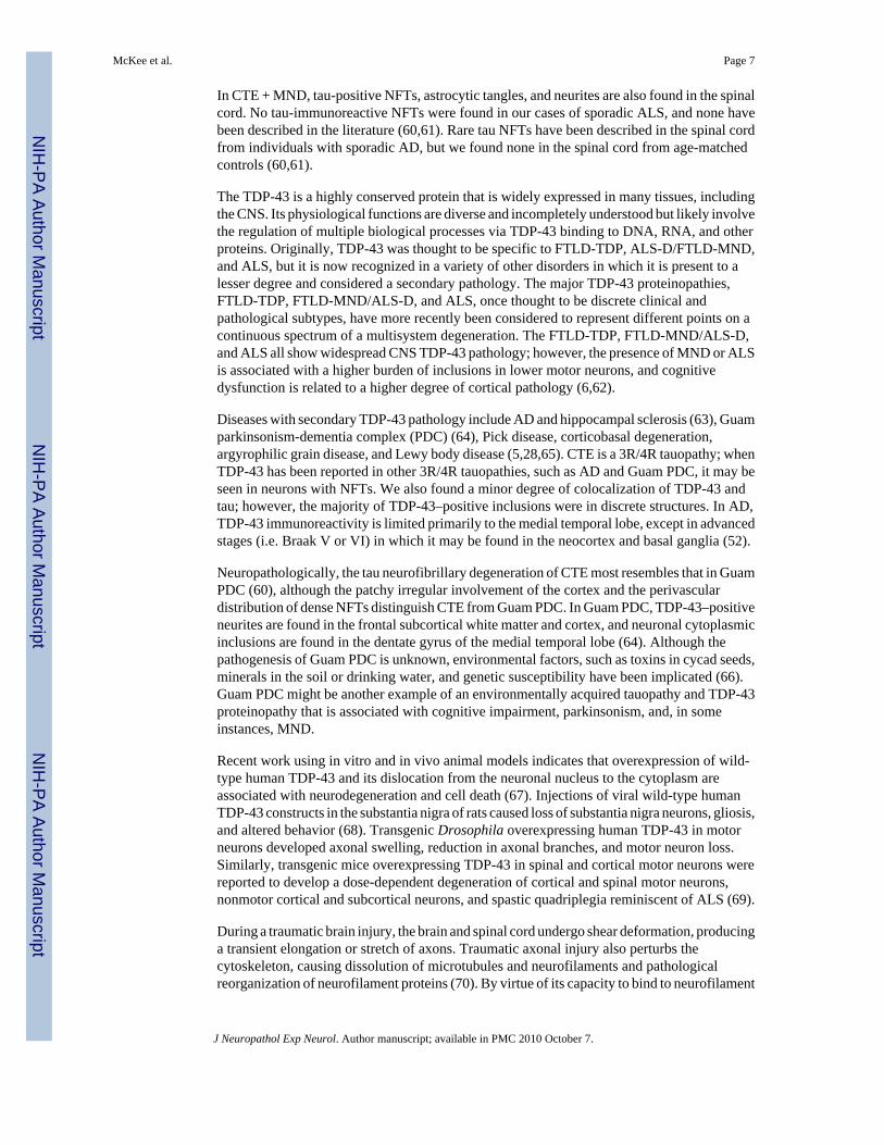
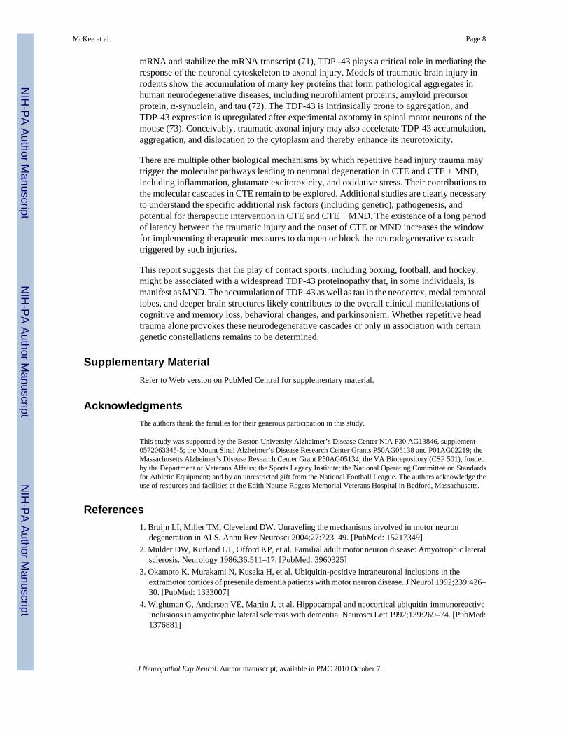
Pathological AspectsAthletes With CTE Without MND—Microscopically, the brains of all 9 athletes with CTE–no MND showed the pathological changes of CTE, consisting of numerous tau-positive NFTs,neuropil neurites, and astrocytic tangles in the frontal, temporal, and insular cortices,diencephalon, basal ganglia, and brainstem (Fig. 1). Of the 5 cases with spinal cord tissueavailable for study, all 5 demonstrated occasional tau-immunoreactive neurites and NFTs inthe posterior, lateral, and/or anterior horns of the spinal cord; 7 of the 9 also showed extensiveTDP-43 immunoreactivity. The TDP-43–positive short threadlike and ring-shaped neurites(RNs), filamentous neuronal inclusions (FNIs), and ring-shaped glial inclusions (RGIs) werefound in the frontal and temporal cortex and insula (Table 3, Figs. 1–3). The TDP-43–positiveRNs and RGIs were frequent in the subcortical white matter, and TDP-43–positive FNIs andRNs were common in the brainstem, including the substantia nigra pars compacta, oculomotor,inferior olivary, dorsal medullary, and hypoglossal nuclei (Fig. 3). The TDP-43–positiveneurites were found in the amygdala, hippocampus, caudate, putamen, thalamus, andhypothalamus (not shown). Double immunostaining and confocal microscopy showed that thegreat majority of the TDP-43–positive neurites and inclusions were not tau positive (Figs. 1,2, 4). In the brain that showed the least TDP-43 immunoreactivity (Case 10), RNs were limitedto the substantia nigra. Ubiquitin-positive or TDP-43–positive inclusions were not found inthe dentate gyrus in any case of CTE; a few cases had rare TDP-43–positive neurites in thedentate gyrus (Table 3).
Athletes With CTE and MND—The macroscopic features of the brains and spinal cords ofthe athletes with CTE + MND did not differ from those with CTE–no MND, except for atrophyof the ventral roots of the spinal cord. All 3 brains showed the characteristic microscopicfindings of CTE (Fig. 1). The 2 athletes with cognitive impairment or dementia and MND(Cases 1 and 3) had advanced CTE with dense NFTs throughout the medial temporal lobestructures, neocortex, olfactory bulb, substantia nigra, locus caeruleus, mammillary body,hypothalamus, and periventricular regions. Betz cells in the precentral gyrus also bore tau-immunoreactive NFTs. Case 2 displayed milder CTE, with involvement primarily limited tothe frontal cortex, diencephalon, and brainstem. Abundant TDP-43–positive RNs, FNIs, andRGIs were found in widespread regions of the brain and spinal cord in all 3 CTE + MND brains,generally in a far greater density than that found in the CTE–no MND brains. In the cortex,TDP-43–positive pathology was found throughout all cortical laminae and was most prominentin the motor cortex (Fig. 1).
In Cases 1 and 2 (both of whom developed end-stage MND with severe respiratoryinsufficiency and muscular weakness in the final months of their lives), the medullary pyramidsand lateral corticospinal tracts throughout the spinal cord showed marked myelin and axonalloss with astrocytosis. Loss of anterior horn cells was profound, and there was marked atrophyand gliosis of ventral roots (Fig. 2). In Case 3 (whose motor neuron symptoms were not assevere), there was only mild degeneration of the lateral corticospinal tracts and ventral rootatrophy. Staining with LN-3 showed numerous activated microglia and macrophagesthroughout the brain and spinal cord, most intense in the lateral corticospinal tracts of the cord.Anterior horn cells were markedly reduced in number in Cases 1 and 2 and moderately reducedin number in Case 3. Tau-positive neurites, astrocytic tangles, and NFTs were found in the
McKee et al. Page 5
J Neuropathol Exp Neurol. Author manuscript; available in PMC 2010 October 7.
NIH
-PA Author Manuscript
NIH
-PA Author Manuscript
NIH
-PA Author Manuscript
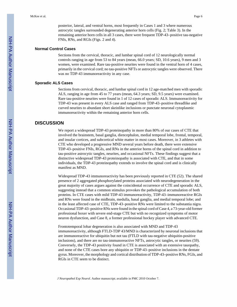
posterior, lateral, and ventral horns, most frequently in Cases 1 and 3 where numerousastrocytic tangles surrounded degenerating anterior horn cells (Fig. 2; Table 3). In theremaining anterior horn cells in all 3 cases, there were frequent TDP-43–positive tau-negativeFNIs, RNs, and RGIs (Figs. 2 and 4).
Normal Control CasesSections from the cervical, thoracic, and lumbar spinal cord of 12 neurologically normalcontrols ranging in age from 53 to 84 years (mean, 66.0 years; SD, 10.6 years), 9 men and 3women, were examined. Rare tau-positive neurites were found in the ventral horn of 4 cases,primarily in the cervical cord; no tau-positive NFTs or astrocytic tangles were observed. Therewas no TDP-43 immunoreactivity in any case.
Sporadic ALS CasesSections from cervical, thoracic, and lumbar spinal cord in 12 age-matched men with sporadicALS, ranging in age from 45 to 77 years (mean, 64.3 years; SD, 9.5 years) were examined.Rare tau-positive neurites were found in 1 of 12 cases of sporadic ALS. Immunoreactivity forTDP-43 was present in every ALS case and ranged from TDP-43–positive threadlike andcurved neurites to abundant short skeinlike inclusions or punctate neuronal cytoplasmicimmunoreactivity within the remaining anterior horn cells.
DISCUSSIONWe report a widespread TDP-43 proteinopathy in more than 80% of our cases of CTE thatinvolved the brainstem, basal ganglia, diencephalon, medial temporal lobe, frontal, temporal,and insular cortices, and subcortical white matter in most cases. Moreover, in 3 athletes withCTE who developed a progressive MND several years before death, there were extensiveTDP-43–positive FNIs, RGIs, and RNs in the anterior horns of the spinal cord in addition totau-positive astrocytic tangles, neurites, and occasional NFTs. These findings suggest that adistinctive widespread TDP-43 proteinopathy is associated with CTE, and that in someindividuals, the TDP-43 proteinopathy extends to involve the spinal cord and is clinicallymanifest as MND.
Widespread TDP-43 immunoreactivity has been previously reported in CTE (52). The sharedpresence of 2 aggregated phosphorylated proteins associated with neurodegeneration in thegreat majority of cases argues against the coincidental occurrence of CTE and sporadic ALS,suggesting instead that a common stimulus provokes the pathological accumulation of bothproteins. In CTE cases with mild TDP-43 immunoreactivity, TDP-43–immunoreactive RGIsand RNs were found in the midbrain, medulla, basal ganglia, and medial temporal lobe; andin the least affected case of CTE, TDP-43–positive RNs were limited to the substantia nigra.Occasional TDP-43–positive RNs were found in the spinal cord of Case 4, a 73-year-old formerprofessional boxer with severe end-stage CTE but with no recognized symptoms of motorneuron dysfunction, and Case 8, a former professional hockey player with advanced CTE.
Frontotemporal lobar degeneration is also associated with MND and TDP-43immunoreactivity, although FTLD-TDP 43/MND is characterized by neuronal inclusions thatare immunoreactive for ubiquitin but not tau (FTLD with tau-negative ubiquitin-positiveinclusions), and there are no tau-immunoreactive NFTs, astrocytic tangles, or neurites (59).Conversely, the TDP-43 positivity found in CTE is associated with an extensive tauopathy,and none of the CTE cases bore any ubiquitin or TDP-43–positive inclusions in the dentategyrus. Moreover, the morphology and cortical distribution of TDP-43–positive RNs, FGIs, andRGIs in CTE seem to be distinct.
McKee et al. Page 6
J Neuropathol Exp Neurol. Author manuscript; available in PMC 2010 October 7.
NIH
-PA Author Manuscript
NIH
-PA Author Manuscript
NIH
-PA Author Manuscript
In CTE + MND, tau-positive NFTs, astrocytic tangles, and neurites are also found in the spinalcord. No tau-immunoreactive NFTs were found in our cases of sporadic ALS, and none havebeen described in the literature (60,61). Rare tau NFTs have been described in the spinal cordfrom individuals with sporadic AD, but we found none in the spinal cord from age-matchedcontrols (60,61).
The TDP-43 is a highly conserved protein that is widely expressed in many tissues, includingthe CNS. Its physiological functions are diverse and incompletely understood but likely involvethe regulation of multiple biological processes via TDP-43 binding to DNA, RNA, and otherproteins. Originally, TDP-43 was thought to be specific to FTLD-TDP, ALS-D/FTLD-MND,and ALS, but it is now recognized in a variety of other disorders in which it is present to alesser degree and considered a secondary pathology. The major TDP-43 proteinopathies,FTLD-TDP, FTLD-MND/ALS-D, and ALS, once thought to be discrete clinical andpathological subtypes, have more recently been considered to represent different points on acontinuous spectrum of a multisystem degeneration. The FTLD-TDP, FTLD-MND/ALS-D,and ALS all show widespread CNS TDP-43 pathology; however, the presence of MND or ALSis associated with a higher burden of inclusions in lower motor neurons, and cognitivedysfunction is related to a higher degree of cortical pathology (6,62).
Diseases with secondary TDP-43 pathology include AD and hippocampal sclerosis (63), Guamparkinsonism-dementia complex (PDC) (64), Pick disease, corticobasal degeneration,argyrophilic grain disease, and Lewy body disease (5,28,65). CTE is a 3R/4R tauopathy; whenTDP-43 has been reported in other 3R/4R tauopathies, such as AD and Guam PDC, it may beseen in neurons with NFTs. We also found a minor degree of colocalization of TDP-43 andtau; however, the majority of TDP-43–positive inclusions were in discrete structures. In AD,TDP-43 immunoreactivity is limited primarily to the medial temporal lobe, except in advancedstages (i.e. Braak V or VI) in which it may be found in the neocortex and basal ganglia (52).
Neuropathologically, the tau neurofibrillary degeneration of CTE most resembles that in GuamPDC (60), although the patchy irregular involvement of the cortex and the perivasculardistribution of dense NFTs distinguish CTE from Guam PDC. In Guam PDC, TDP-43–positiveneurites are found in the frontal subcortical white matter and cortex, and neuronal cytoplasmicinclusions are found in the dentate gyrus of the medial temporal lobe (64). Although thepathogenesis of Guam PDC is unknown, environmental factors, such as toxins in cycad seeds,minerals in the soil or drinking water, and genetic susceptibility have been implicated (66).Guam PDC might be another example of an environmentally acquired tauopathy and TDP-43proteinopathy that is associated with cognitive impairment, parkinsonism, and, in someinstances, MND.
Recent work using in vitro and in vivo animal models indicates that overexpression of wild-type human TDP-43 and its dislocation from the neuronal nucleus to the cytoplasm areassociated with neurodegeneration and cell death (67). Injections of viral wild-type humanTDP-43 constructs in the substantia nigra of rats caused loss of substantia nigra neurons, gliosis,and altered behavior (68). Transgenic Drosophila overexpressing human TDP-43 in motorneurons developed axonal swelling, reduction in axonal branches, and motor neuron loss.Similarly, transgenic mice overexpressing TDP-43 in spinal and cortical motor neurons werereported to develop a dose-dependent degeneration of cortical and spinal motor neurons,nonmotor cortical and subcortical neurons, and spastic quadriplegia reminiscent of ALS (69).
During a traumatic brain injury, the brain and spinal cord undergo shear deformation, producinga transient elongation or stretch of axons. Traumatic axonal injury also perturbs thecytoskeleton, causing dissolution of microtubules and neurofilaments and pathologicalreorganization of neurofilament proteins (70). By virtue of its capacity to bind to neurofilament
McKee et al. Page 7
J Neuropathol Exp Neurol. Author manuscript; available in PMC 2010 October 7.
NIH
-PA Author Manuscript
NIH
-PA Author Manuscript
NIH
-PA Author Manuscript
mRNA and stabilize the mRNA transcript (71), TDP -43 plays a critical role in mediating theresponse of the neuronal cytoskeleton to axonal injury. Models of traumatic brain injury inrodents show the accumulation of many key proteins that form pathological aggregates inhuman neurodegenerative diseases, including neurofilament proteins, amyloid precursorprotein, α-synuclein, and tau (72). The TDP-43 is intrinsically prone to aggregation, andTDP-43 expression is upregulated after experimental axotomy in spinal motor neurons of themouse (73). Conceivably, traumatic axonal injury may also accelerate TDP-43 accumulation,aggregation, and dislocation to the cytoplasm and thereby enhance its neurotoxicity.
There are multiple other biological mechanisms by which repetitive head injury trauma maytrigger the molecular pathways leading to neuronal degeneration in CTE and CTE + MND,including inflammation, glutamate excitotoxicity, and oxidative stress. Their contributions tothe molecular cascades in CTE remain to be explored. Additional studies are clearly necessaryto understand the specific additional risk factors (including genetic), pathogenesis, andpotential for therapeutic intervention in CTE and CTE + MND. The existence of a long periodof latency between the traumatic injury and the onset of CTE or MND increases the windowfor implementing therapeutic measures to dampen or block the neurodegenerative cascadetriggered by such injuries.
This report suggests that the play of contact sports, including boxing, football, and hockey,might be associated with a widespread TDP-43 proteinopathy that, in some individuals, ismanifest as MND. The accumulation of TDP-43 as well as tau in the neocortex, medal temporallobes, and deeper brain structures likely contributes to the overall clinical manifestations ofcognitive and memory loss, behavioral changes, and parkinsonism. Whether repetitive headtrauma alone provokes these neurodegenerative cascades or only in association with certaingenetic constellations remains to be determined.
Supplementary MaterialRefer to Web version on PubMed Central for supplementary material.
AcknowledgmentsThe authors thank the families for their generous participation in this study.
This study was supported by the Boston University Alzheimer’s Disease Center NIA P30 AG13846, supplement0572063345-5; the Mount Sinai Alzheimer’s Disease Research Center Grants P50AG05138 and P01AG02219; theMassachusetts Alzheimer’s Disease Research Center Grant P50AG05134; the VA Biorepository (CSP 501), fundedby the Department of Veterans Affairs; the Sports Legacy Institute; the National Operating Committee on Standardsfor Athletic Equipment; and by an unrestricted gift from the National Football League. The authors acknowledge theuse of resources and facilities at the Edith Nourse Rogers Memorial Veterans Hospital in Bedford, Massachusetts.
References1. Bruijn LI, Miller TM, Cleveland DW. Unraveling the mechanisms involved in motor neuron
degeneration in ALS. Annu Rev Neurosci 2004;27:723–49. [PubMed: 15217349]2. Mulder DW, Kurland LT, Offord KP, et al. Familial adult motor neuron disease: Amyotrophic lateral
sclerosis. Neurology 1986;36:511–17. [PubMed: 3960325]3. Okamoto K, Murakami N, Kusaka H, et al. Ubiquitin-positive intraneuronal inclusions in the
extramotor cortices of presenile dementia patients with motor neuron disease. J Neurol 1992;239:426–30. [PubMed: 1333007]
4. Wightman G, Anderson VE, Martin J, et al. Hippocampal and neocortical ubiquitin-immunoreactiveinclusions in amyotrophic lateral sclerosis with dementia. Neurosci Lett 1992;139:269–74. [PubMed:1376881]
McKee et al. Page 8
J Neuropathol Exp Neurol. Author manuscript; available in PMC 2010 October 7.
NIH
-PA Author Manuscript
NIH
-PA Author Manuscript
NIH
-PA Author Manuscript
5. Dickson DW. Neuropathology of non-Alzheimer degenerative disorders. Int J Clin Exp Pathol2009;3:1–23. [PubMed: 19918325]
6. Geser F, Martinez-Lage M, Kwong LK, et al. Amyotrophic lateral sclerosis, frontotemporal dementiaand beyond: The TDP-43 diseases. J Neurol 2009;256:1205–14. [PubMed: 19271105]
7. Kondo K, Tsubaki T. Case-control studies of motor neuron disease: Association with mechanicalinjuries. Arch Neurol 1981;38:220–26. [PubMed: 7011280]
8. Deapen DM, Henderson BE. A case-control study of amyotrophic lateral sclerosis. Am J Epidemiol1986;123:790–99. [PubMed: 3962963]
9. Chen H, Richard M, Sandler DP, et al. Head injury and amyotrophic lateral sclerosis. Am J Epidemiol2007;166:810–16. [PubMed: 17641152]
10. Binazzi A, Belli S, Uccelli R, et al. An exploratory case-control study on spinal and bulbar forms ofamyotrophic lateral sclerosis in the province of Rome. Amyotroph Lateral Scler 2009;10:361–69.[PubMed: 19922125]
11. Kurtzke JF, Beebe GW. Epidemiology of amyotrophic lateral sclerosis: 1. A case-control comparisonbased on ALS deaths. Neurology 1980;30:453–62. [PubMed: 7189251]
12. Gawel M, Zaiwalla Z, Rose FC. Antecedent events in motor neuron disease. J Neurol NeurosurgPsychiatry 1983;46:1041–43. [PubMed: 6655478]
13. Strickland D, Smith SA, Dolliff G, et al. Physical activity, trauma, and ALS: A case-control study.Acta Neurol Scand 1996;94:45–50. [PubMed: 8874593]
14. Scarmeas N, Shih T, Stern Y, et al. Premorbid weight, body mass, and varsity athletics in ALS.Neurology 2002;59:773–75. [PubMed: 12221178]
15. Longstreth WT, McGuire V, Koepsell TD, et al. Risk of amyotrophic lateral sclerosis and history ofphysical activity: A population-based case-control study. Arch Neurol 1998;55:201–6. [PubMed:9482362]
16. Chiò A, Benzi G, Dossena M, et al. Severely increased risk of amyotrophic lateral sclerosis amongItalian professional football players. Brain 2005;128:472–76. [PubMed: 15634730]
17. Belli S, Vanacore N. Proportionate mortality of Italian soccer players: Is amyotrophic lateral sclerosisan occupational disease? Eur J Epidemiol 2005;20:237–42. [PubMed: 15921041]
18. Kamel F, Umbach DM, Munsat TL, et al. Association of cigarette smoking with amyotrophic lateralsclerosis. Neuroepidemiology 1999;18:194–202. [PubMed: 10364720]
19. Nelson LM, McGuire V, Longstreth WT, et al. Population-based case-control study of amyotrophiclateral sclerosis in western Washington state. I. Cigarette smoking and alcohol consumption. Am JEpidemiol 2000;151:156–63. [PubMed: 10645818]
20. Weisskopf MG, McCullough ML, Calle EE, et al. Prospective study of cigarette smoking andamyotrophic lateral sclerosis. Am J Epidemiol 2004;160:26–33. [PubMed: 15229114]
21. Campbell AM, Williams ER, Barltrop D. Motor neurone disease and exposure to lead. J NeurolNeurosurg Psychiatry 1970;33:877–85. [PubMed: 5497881]
22. Armon C, Kurland LT, Daube JR, et al. Epidemiologic correlates of sporadic amyotrophic lateralsclerosis. Neurology 1991;41:1077–84. [PubMed: 2067636]
23. Chancellor AM, Slattery JM, Fraser H, et al. Risk factors for motor neuron disease: A case-controlstudy based on patients from the Scottish Motor Neuron Disease Register. J Neurol NeurosurgPsychiatry 1993;56:1200–6. [PubMed: 8229031]
24. McGuire V, Longstreth WT, Nelson LM, et al. Occupational exposures and amyotrophic lateralsclerosis. A population-based case-control study. Am J Epidemiol 1997;145:1076–88. [PubMed:9199537]
25. Kamel F, Umbach DM, Munsat TL, et al. Lead exposure and amyotrophic lateral sclerosis.Epidemiology 2002;13:311–19. [PubMed: 11964933]
26. Felmus MT, Patten BM, Swanke L. Antecedent events in amyotrophic lateral sclerosis. Neurology1976;26:167–72. [PubMed: 946326]
27. Pierce-Ruhland R, Patten BM. Repeat study of antecedent events in motor neuron disease. Ann ClinRes 1981;13:102–7. [PubMed: 7235610]
28. Gresham LS, Molgaard CA, Golbeck AL, et al. Amyotrophic lateral sclerosis and occupational heavymetal exposure: A case-control study. Neuroepidemiology 1986;5:29–38. [PubMed: 3748267]
McKee et al. Page 9
J Neuropathol Exp Neurol. Author manuscript; available in PMC 2010 October 7.
NIH
-PA Author Manuscript
NIH
-PA Author Manuscript
NIH
-PA Author Manuscript
29. Haynal A, Regli F. Amyotrophic lateral sclerosis associated with accumulated electric injury [InGerman]. Confin Neurol 1964;24:189–98. [PubMed: 14154333]
30. Savettieri G, Salemi G, Arcara A, et al. A case-control study of amyotrophic lateral sclerosis.Neuroepidemiology 1991;10:242–45. [PubMed: 1798425]
31. Majoor-Krakauer D, Willems PJ, Hofman A. Genetic epidemiology of amyotrophic lateral sclerosis.Clin Genet 2003;63:83–101. [PubMed: 12630951]
32. Schmidt S, Kwee LC, Allen KD, et al. Association of ALS with head injury, cigarette smoking andAPOE genotypes. J Neurol Sci 2010;291:22–29. [PubMed: 20129626]
33. Fleminger S, Oliver DL, Lovestone S, et al. Head injury as a risk factor for Alzheimer’s disease: Theevidence 10 years on; a partial replication. J Neurol Neurosurg Psychiatry 2003;74:857–62.[PubMed: 12810767]
34. Mortimer JA, French LR, Hutton JT, et al. Head injury as a risk factor for Alzheimer’s disease.Neurology 1985;35:264–67. [PubMed: 3969219]
35. O’Meara ES, Kukull WA, Sheppard L, et al. Head injury and risk of Alzheimer’s disease byapolipoprotein E genotype. Am J Epidemiol 1997;146:373–84. [PubMed: 9290497]
36. Mehta KM, Ott A, Kalmijn S, et al. Head trauma and risk of dementia and Alzheimer’s disease: TheRotterdam Study. Neurology 1999;53:1959–62. [PubMed: 10599765]
37. Katzman R, Galasko DR, Saitoh T, et al. Apolipoprotein-epsilon4 and head trauma: Synergistic oradditive risks? Neurology 1996;46:889–91. [PubMed: 8618734]
38. Mayeux R, Ottman R, Maestre G, et al. Synergistic effects of traumatic head injury and apolipoprotein-epsilon 4 in patients with Alzheimer’s disease. Neurology 1995;45:555–57. [PubMed: 7898715]
39. Plassman BL, Havlik RJ, Steffens DC, et al. Documented head injury in early adulthood and risk ofAlzheimer’s disease and other dementias. Neurology 2000;55:1158–66. [PubMed: 11071494]
40. Bower JH, Maraganore DM, Peterson BJ, et al. Head trauma preceding PD: A case-control study.Neurology 2003;60:1610–15. [PubMed: 12771250]
41. Goldman SM, Tanner CM, Oakes D, et al. Head injury and Parkinson’s disease risk in twins. AnnNeurol 2006;60:65–72. [PubMed: 16718702]
42. Erb W. Zur Casuistik der bulbären Lähmungen: Über einen neuen wahrscheinlich bulbärenLähmungscomplex. Eur Arch Psychiatry Clin Neurosci 1879;9:325–50.
43. Hanisch R, Dworsky RL, Henderson BE. Letter: A search for clues to the cause of amyotrophic lateralsclerosis. Arch Neurol 1976;33:456–57. [PubMed: 938270]
44. Al-Chalabi A, Leigh PN. Trouble on the pitch: Are professional football players at increased risk ofdeveloping amyotrophic lateral sclerosis? Brain 2005;128:451–53. [PubMed: 15713848]
45. Abel EL. Football increases the risk for Lou Gehrig’s disease, amyotrophic lateral sclerosis. PerceptMot Skills 2007;104:1251–54. [PubMed: 17879657]
46. Wallis, C.; Dorfman, A. Medicine: Probing A Mysterious Cluster. TIME.com [Web site]. Feb 231987[Accessed September 28, 2008]. Available at:http://www.time.com/time/magazine/article/0,9171,963607-1,00.html
47. Horner RD, Kamins KG, Feussner JR, et al. Occurrence of amyotrophic lateral sclerosis among GulfWar veterans. Neurology 2003;61:742–49. [PubMed: 14504315]
48. Coffman CJ, Horner RD, Grambow SC, et al. Estimating the occurrence of amyotrophic lateralsclerosis among Gulf War (1990–1991) veterans using capture-recapture methods.Neuroepidemiology 2005;24:141–50. [PubMed: 15650320]
49. Weisskopf MG, O’Reilly EJ, McCullough ML, et al. Prospective study of military service andmortality from ALS. Neurology 2005;64:32–37. [PubMed: 15642900]
50. Haley RW. Excess incidence of ALS in young Gulf War veterans. Neurology 2003;61:750–56.[PubMed: 14504316]
51. McKee A, Cantu R, Nowinski C, et al. Chronic traumatic encephalopathy in athletes: Progressivetauopathy after repetitive head injury. J Neuropathol Exp Neurol 2009;68:709–35. [PubMed:19535999]
52. King A, Sweeney F, Bodi I, et al. Abnormal TDP-43 expression is identified in the neocortex in casesof dementia pugilistica, but is mainly confined to the limbic system when identified in high andmoderate stages of Alzheimer’s disease. Neuropathology. 2010 [Epub ahead of print].
McKee et al. Page 10
J Neuropathol Exp Neurol. Author manuscript; available in PMC 2010 October 7.
NIH
-PA Author Manuscript
NIH
-PA Author Manuscript
NIH
-PA Author Manuscript
53. Arai T, Hasegawa M, Akiyama H, et al. TDP-43 is a component of ubiquitin-positive tau-negativeinclusions in frontotemporal lobar degeneration and amyotrophic lateral sclerosis. Biochem BiophysRes Commun 2006;351:602–11. [PubMed: 17084815]
54. Neumann M, Sampathu DM, Kwong LK, et al. Ubiquitinated TDP-43 in frontotemporal lobardegeneration and amyotrophic lateral sclerosis. Science 2006;314:130–33. [PubMed: 17023659]
55. Hu WT, Josephs KA, Knopman DS, et al. Temporal lobar predominance of TDP-43 neuronalcytoplasmic inclusions in Alzheimer disease. Acta Neuropathol 2008;116:215–20. [PubMed:18592255]
56. Mackenzie IR, Baborie A, Pickering-Brown S, et al. Heterogeneity of ubiquitin pathology infrontotemporal lobar degeneration. Acta Neuropathol 2006;112:539–49. [PubMed: 17021754]
57. Sampathu DM, Neumann M, Kwong LK, et al. Pathological heterogeneity of frontotemporal lobardegeneration with ubiquitin-positive inclusions delineated by ubiquitin immunohistochemistry andnovel monoclonal antibodies. Am J Pathol 2006;169:1343–52. [PubMed: 17003490]
58. Vonsattel JP, Aizawa H, Ge P, et al. An improved approach to prepare human brains for research. JNeuropathol Exp Neurol 1995;54:42–56. [PubMed: 7815079]
59. Cairns NJ, Bigio EH, Mackenzie IRA, et al. Neuropathologic diagnostic and nosologic criteria forfrontotemporal lobar degeneration: Consensus of the Consortium for Frontotemporal LobarDegeneration. Acta Neuropathol 2007;114:5–22. [PubMed: 17579875]
60. Schmidt ML, Zhukareva V, Perl DP, et al. Spinal cord neurofibrillary pathology in Alzheimer diseaseand Guam parkinsonism-dementia complex. J Neuropathol Exp Neurol 2001;60:1075–86. [PubMed:11706937]
61. Umahara T, Hirano A, Kato S, et al. Demonstration of neurofibrillary tangles and neuropil thread-like structures in spinal cord white matter in parkinsonism-dementia complex on Guam and inGuamanian amyotrophic lateral sclerosis. Acta Neuropathol 1994;88:180–84. [PubMed: 7985499]
62. Geser F, Martinez-Lage M, Robinson J, et al. Clinical and pathological continuum of multisystemTDP-43 proteinopathies. Arch Neurol 2009;66:180–89. [PubMed: 19204154]
63. Amador-Ortiz C, Lin WL, Ahmed Z, et al. TDP-43 immunoreactivity in hippocampal sclerosis andAlzheimer’s disease. Ann Neurol 2007;61:435–45. [PubMed: 17469117]
64. Hasegawa M, Arai T, Akiyama H, et al. TDP-43 is deposited in the Guam parkinsonism-dementiacomplex brains. Brain 2007;130:1386–94. [PubMed: 17439983]
65. Nakashima-Yasuda H, Uryu K, Robinson J, et al. Co-morbidity of TDP-43 proteinopathy in Lewybody–related diseases. Acta Neuropathol 2007;114:221–29. [PubMed: 17653732]
66. Sieh W, Choi Y, Chapman NH, et al. Identification of novel susceptibility loci for Guamneurodegenerative disease: Challenges of genome scans in genetic isolates. Hum Mol Genet2009;18:3725–38. [PubMed: 19567404]
67. Barmada SJ, Skibinski G, Korb E, et al. Cytoplasmic mislocalization of TDP-43 is toxic to neuronsand enhanced by a mutation associated with familial amyotrophic lateral sclerosis. J Neurosci2010;30:639–49. [PubMed: 20071528]
68. Tatom JB, Wang D, Dayton R, et al. Mimicking aspects of frontotemporal lobar degeneration andLou Gehrig’s disease in rats via TDP-43 overexpression. Mol Ther 2009;17:607–13. [PubMed:19223871]
69. Wils H, Kleinberger G, Janssens J, et al. TDP-43 transgenic mice develop spastic paralysis andneuronal inclusions characteristic of ALS and frontotemporal lobar degeneration. Proc Natl AcadSci U S A 2010;107:3858–63. [PubMed: 20133711]
70. Serbest G, Burkhardt M, Siman R, et al. Temporal profiles of cytoskeletal protein loss followingtraumatic axonal injury in mice. Neurochem Res 2007;32:2006–14. [PubMed: 17401646]
71. Strong MJ, Volkening K, Hammond R, et al. TDP43 is a human low molecular weight neurofilament(hNFL) mRNA-binding protein. Mol Cell Neurosci 2007;35:320–27. [PubMed: 17481916]
72. Uryu K, Chen XH, Martinez D, et al. Multiple proteins implicated in neurodegenerative diseasesaccumulate in axons after brain trauma in humans. Exp Neurol 2007;208:185–92. [PubMed:17826768]
73. Moisse K, Mepham J, Volkening K, et al. Cytosolic TDP-43 expression following axotomy isassociated with caspase 3 activation in NFL−/− mice: Support for a role for TDP-43 in thephysiological response to neuronal injury. Brain Res 2009;1296:176–86. [PubMed: 19619516]
McKee et al. Page 11
J Neuropathol Exp Neurol. Author manuscript; available in PMC 2010 October 7.
NIH
-PA Author Manuscript
NIH
-PA Author Manuscript
NIH
-PA Author Manuscript
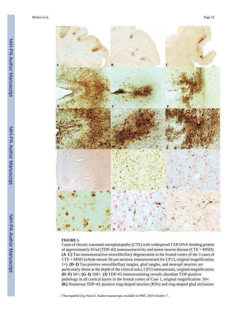
FIGURE 1.Cases of chronic traumatic encephalopathy (CTE) with widespread TAR DNA-binding proteinof approximately 43 kd (TDP-43) immunoreactivity and motor neuron disease (CTE + MND).(A–C) Tau-immunoreactive neurofibrillary degeneration in the frontal cortex of the 3 cases ofCTE + MND (whole-mount 50-μm sections immunostained for CP13, original magnification:1×). (D–I) Tau-positive neurofibrillary tangles, glial tangles, and neuropil neurites areparticularly dense at the depth of the cortical sulci, CP13 immunostain, original magnification:(D–F) 50×; (G–I) 100×. (J) TDP-43 immunostaining reveals abundant TDP-positivepathology in all cortical layers in the frontal cortex of Case 1, original magnification: 50×.(K) Numerous TDP-43–positive ring-shaped neurites (RNs) and ring-shaped glial inclusions
McKee et al. Page 12
J Neuropathol Exp Neurol. Author manuscript; available in PMC 2010 October 7.
NIH
-PA Author Manuscript
NIH
-PA Author Manuscript
NIH
-PA Author Manuscript
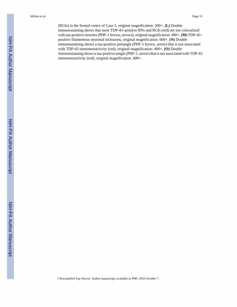
(RGIs) in the frontal cortex of Case 2, original magnification: 200×. (L) Doubleimmunostaining shows that most TDP-43–positive RNs and RGIs (red) are not colocalizedwith tau-positive neurites (PHF-1 brown, arrows), original magnification: 400×. (M) TDP-43–positive filamentous neuronal inclusions, original magnification: 600×. (N) Doubleimmunostaining shows a tau-positive pretangle (PHF-1 brown, arrow) that is not associatedwith TDP-43 immunoreactivity (red), original magnification: 400×. (O) Doubleimmunostaining shows a tau-positive tangle (PHF-1, arrow) that is not associated with TDP-43immunoreactivity (red), original magnification: 400×.
McKee et al. Page 13
J Neuropathol Exp Neurol. Author manuscript; available in PMC 2010 October 7.
NIH
-PA Author Manuscript
NIH
-PA Author Manuscript
NIH
-PA Author Manuscript
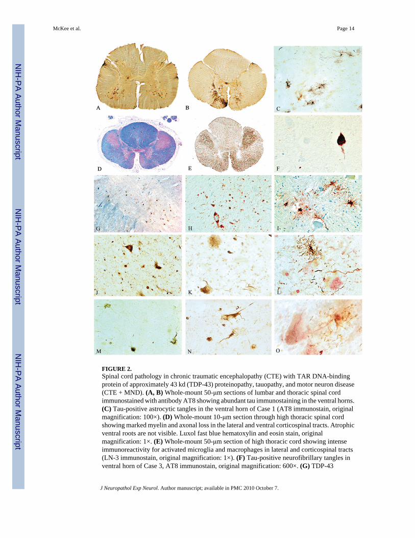
FIGURE 2.Spinal cord pathology in chronic traumatic encephalopathy (CTE) with TAR DNA-bindingprotein of approximately 43 kd (TDP-43) proteinopathy, tauopathy, and motor neuron disease(CTE + MND). (A, B) Whole-mount 50-μm sections of lumbar and thoracic spinal cordimmunostained with antibody AT8 showing abundant tau immunostaining in the ventral horns.(C) Tau-positive astrocytic tangles in the ventral horn of Case 1 (AT8 immunostain, originalmagnification: 100×). (D) Whole-mount 10-μm section through high thoracic spinal cordshowing marked myelin and axonal loss in the lateral and ventral corticospinal tracts. Atrophicventral roots are not visible. Luxol fast blue hematoxylin and eosin stain, originalmagnification: 1×. (E) Whole-mount 50-μm section of high thoracic cord showing intenseimmunoreactivity for activated microglia and macrophages in lateral and corticospinal tracts(LN-3 immunostain, original magnification: 1×). (F) Tau-positive neurofibrillary tangles inventral horn of Case 3, AT8 immunostain, original magnification: 600×. (G) TDP-43
McKee et al. Page 14
J Neuropathol Exp Neurol. Author manuscript; available in PMC 2010 October 7.
NIH
-PA Author Manuscript
NIH
-PA Author Manuscript
NIH
-PA Author Manuscript
immunoreactivity in ventral horn, original magnification: 50×. (H) TDP-43 immunoreactivefilamentous neuronal inclusions (FNIs), ring-shaped glial inclusions (RGIs), and ring-shapedneurites (RNs) in the ventral horns of the lumbar spinal cord in Case 3, original magnification:200×. (I) Tau-positive astrocytes and their processes surrounding degenerating anterior horncells in the thoracic spinal cord (AT8 immunostain, original magnification: 350×). (J) TDP-43–positive FNIs, RGIs, and RNs in the ventral horns of the lumbar spinal cord in Case 2, originalmagnification: 200×. (K) TDP-43–positive FNI in the anterior horn, original magnification:400×.(L) Double immunostained sections show tau-positive astrocytes (brown) and theirprocesses surrounding anterior horn neurons containing TDP-43–positive filamentousinclusions (red), PHF-1 and TDP-43 immunostains, original magnification: 400×. (M)TDP-43–positive FNIs, RGIs, and RNs in the lumbar ventral horns in Case 1, originalmagnification: 200×. (N) TDP-43–positive FNIs in the anterior horn, original magnification:400×. (O) Double immunostained sections showing tau-positive astrocytes (brown) and theirprocesses surrounding anterior horn neurons containing TDP-43–positive FNIs (red), PHF-1and TDP-43 immunostains, original magnification: 600×.
McKee et al. Page 15
J Neuropathol Exp Neurol. Author manuscript; available in PMC 2010 October 7.
NIH
-PA Author Manuscript
NIH
-PA Author Manuscript
NIH
-PA Author Manuscript
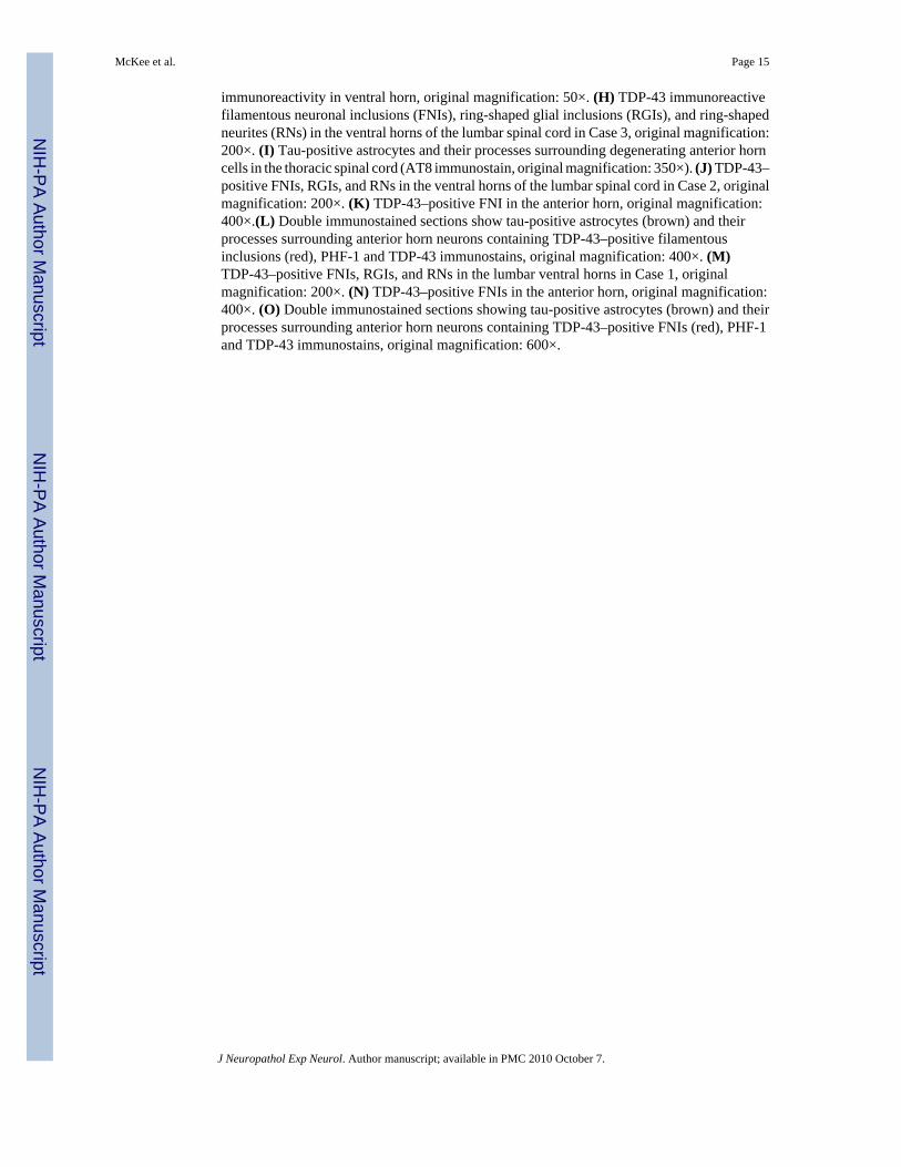
FIGURE 3.TAR DNA-binding protein of approximately 43 kd (TDP-43) immunoreactivity in chronictraumatic encephalopathy (CTE). TDP-43 immunoreactivity is found as glial cytoplasmicinclusions (GCIs) and neuropil neurites in multiple brainstem nuclei including hypoglossalnucleus (A), oculomotor nucleus (B), substantia nigra (C) (original magnification: 200×).TDP-43 immunoreactivity in the medial temporal lobe structures consists primarily of dotlikeneurites. (D) Hippocampus, CA1 (original magnification: 200×). TDP-43–positive dystrophicneurites and GCIs are also found in white matter. (E) Subcortical frontal white matter (originalmagnification: 200×). No ubiquitinated or TDP-43–positive inclusions are found in the dentategyrus of the hippocampus. (F) Dentate gyrus of the hippocampus (original magnification:400×).
McKee et al. Page 16
J Neuropathol Exp Neurol. Author manuscript; available in PMC 2010 October 7.
NIH
-PA Author Manuscript
NIH
-PA Author Manuscript
NIH
-PA Author Manuscript
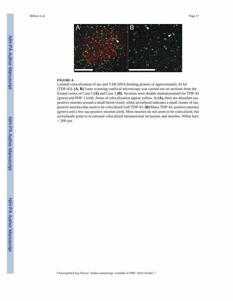
FIGURE 4.Limited colocalization of tau and TAR DNA-binding protein of approximately 43 kd(TDP-43). (A, B) Laser scanning confocal microscopy was carried out on sections from thefrontal cortex of Case 1 (A) and Case 2 (B). Sections were double immunostained for TDP-43(green) and PHF-1 (red). Areas of colocalization appear yellow. In (A), there are abundant tau-positive neurites around a small blood vessel; white arrowhead indicates a small cluster of tau-positive neurites that seem to be colocalized with TDP-43. (B) Many TDP-43–positive neurites(green) and a few tau-positive neurites (red). Most neurites do not seem to be colocalized, butarrowheads point to occasional colocalized intraneuronal inclusions and neurites. White bars= 200 μm.
McKee et al. Page 17
J Neuropathol Exp Neurol. Author manuscript; available in PMC 2010 October 7.
NIH
-PA Author Manuscript
NIH
-PA Author Manuscript
NIH
-PA Author Manuscript
NIH
-PA Author Manuscript
NIH
-PA Author Manuscript
NIH
-PA Author Manuscript
McKee et al. Page 18
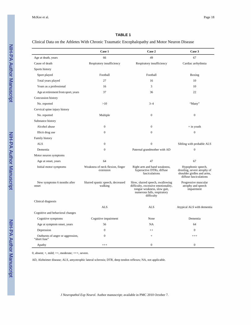
TABLE 1
Clinical Data on the Athletes With Chronic Traumatic Encephalopathy and Motor Neuron Disease
Case 1 Case 2 Case 3
Age at death, years 66 49 67
Cause of death Respiratory insufficiency Respiratory insufficiency Cardiac arrhythmia
Sports history
Sport played Football Football Boxing
Total years played 27 16 10
Years as a professional 16 3 10
Age at retirement from sport, years 37 36 22
Concussion history
No. reported >10 3–4 “Many”
Cervical spine injury history
No. reported Multiple 0 0
Substance history
Alcohol abuse 0 0 + in youth
Illicit drug use 0 0 0
Family history
ALS 0 0 Sibling with probable ALS
Dementia 0 Paternal grandmother with AD 0
Motor neuron symptoms
Age at onset, years 64 47 67
Initial motor symptoms Weakness of neck flexion, fingerextension
Right arm and hand weakness,hyperactive DTRs, diffuse
fasciculations
Hypophonic speech,drooling, severe atrophy ofshoulder girdles and arms,
diffuse fasciculations
New symptoms 6 months afteronset
Slurred spastic speech, decreasedwalking
Slow, slurred speech, swallowingdifficulty, excessive emotionality,
tongue weakness, slow gait,numerous falls, respiratory
difficulty
Progressive muscularatrophy and speech
impairment
Clinical diagnosis
ALS ALS Atypical ALS with dementia
Cognitive and behavioral changes
Cognitive symptoms Cognitive impairment None Dementia
Age at symptom onset, years 56 NA 64
Depression 0 ++ 0
Outbursts of anger or aggression,“short fuse”
0 + +++
Apathy +++ 0 0
0, absent; +, mild; ++, moderate; +++, severe.
AD, Alzheimer disease; ALS, amyotrophic lateral sclerosis; DTR, deep tendon reflexes; NA, not applicable.
J Neuropathol Exp Neurol. Author manuscript; available in PMC 2010 October 7.
NIH
-PA Author Manuscript
NIH
-PA Author Manuscript
NIH
-PA Author Manuscript
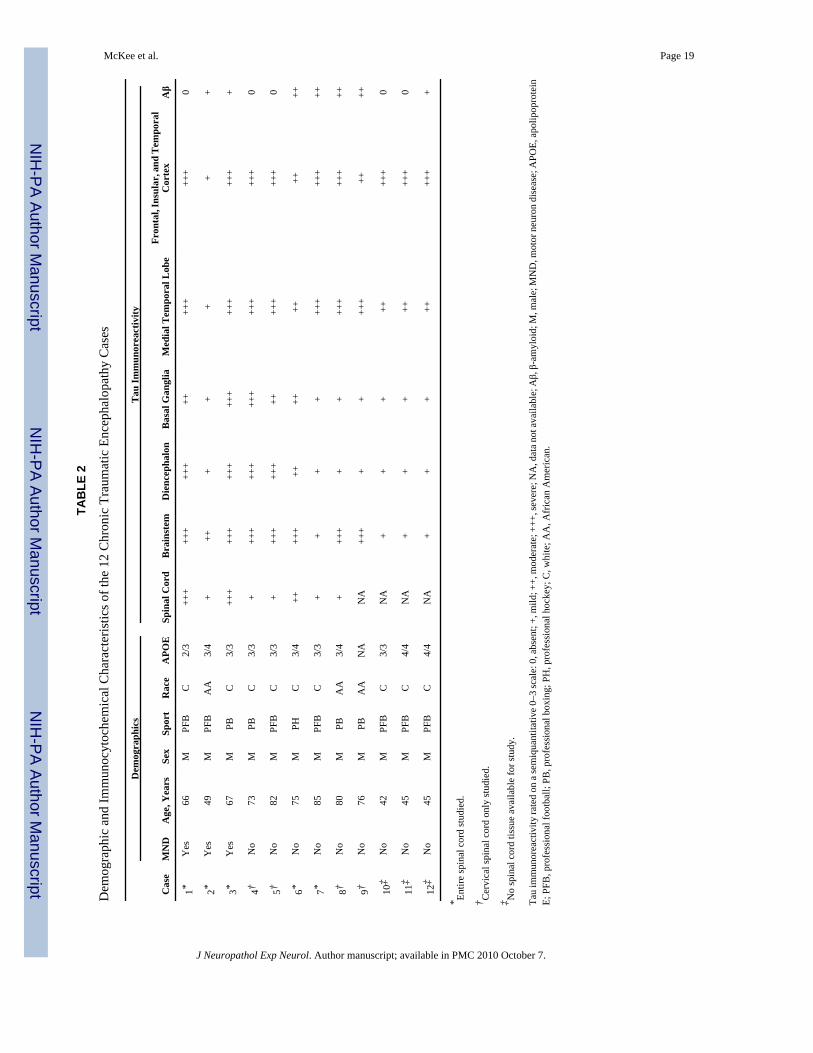
McKee et al. Page 19
TAB
LE 2
Dem
ogra
phic
and
Imm
unoc
ytoc
hem
ical
Cha
ract
eris
tics o
f the
12
Chr
onic
Tra
umat
ic E
ncep
halo
path
y C
ases
Cas
e
Dem
ogra
phic
sT
au Im
mun
orea
ctiv
ity
MN
DA
ge, Y
ears
Sex
Spor
tR
ace
APO
ESp
inal
Cor
dB
rain
stem
Die
ncep
halo
nB
asal
Gan
glia
Med
ial T
empo
ral L
obe
Fron
tal,
Insu
lar,
and
Tem
pora
lC
orte
xAβ
1*Y
es66
MPF
BC
2/3
+++
+++
+++
++++
+++
+0
2*Y
es49
MPF
BA
A3/
4+
+++
++
++
3*Y
es67
MPB
C3/
3++
+++
+++
+++
+++
+++
++
4†N
o73
MPB
C3/
3+
+++
+++
+++
+++
+++
0
5†N
o82
MPF
BC
3/3
+++
+++
+++
+++
+++
0
6*N
o75
MPH
C3/
4++
+++
++++
++++
++
7*N
o85
MPF
BC
3/3
++
++
+++
+++
++
8†N
o80
MPB
AA
3/4
+++
++
+++
+++
+++
9†N
o76
MPB
AA
NA
NA
+++
++
+++
++++
10‡
No
42M
PFB
C3/
3N
A+
++
++++
+0
11‡
No
45M
PFB
C4/
4N
A+
++
++++
+0
12‡
No
45M
PFB
C4/
4N
A+
++
++++
++
* Entir
e sp
inal
cor
d st
udie
d.
† Cer
vica
l spi
nal c
ord
only
stud
ied.
‡ No
spin
al c
ord
tissu
e av
aila
ble
for s
tudy
.
Tau
imm
unor
eact
ivity
rate
d on
a se
miq
uant
itativ
e 0–
3 sc
ale:
0, a
bsen
t; +,
mild
; ++,
mod
erat
e; +
++, s
ever
e; N
A, d
ata
not a
vaila
ble;
Aβ,
β-a
myl
oid;
M, m
ale;
MN
D, m
otor
neu
ron
dise
ase;
APO
E, a
polip
opro
tein
E; P
FB, p
rofe
ssio
nal f
ootb
all;
PB, p
rofe
ssio
nal b
oxin
g; P
H, p
rofe
ssio
nal h
ocke
y; C
, whi
te; A
A, A
fric
an A
mer
ican
.
J Neuropathol Exp Neurol. Author manuscript; available in PMC 2010 October 7.
NIH
-PA Author Manuscript
NIH
-PA Author Manuscript
NIH
-PA Author Manuscript
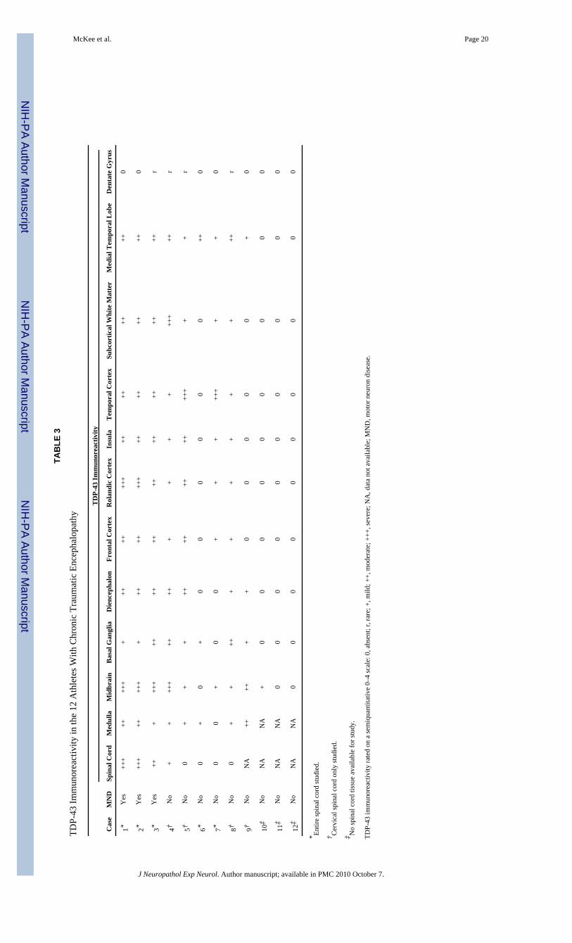
McKee et al. Page 20
TAB
LE 3
TDP-
43 Im
mun
orea
ctiv
ity in
the
12 A
thle
tes W
ith C
hron
ic T
raum
atic
Enc
epha
lopa
thy
Cas
eM
ND
TD
P-43
Imm
unor
eact
ivity
Spin
al C
ord
Med
ulla
Mid
brai
nB
asal
Gan
glia
Die
ncep
halo
nFr
onta
l Cor
tex
Rol
andi
c C
orte
xIn
sula
Tem
pora
l Cor
tex
Subc
ortic
al W
hite
Mat
ter
Med
ial T
empo
ral L
obe
Den
tate
Gyr
us
1*Y
es++
+++
+++
+++
++++
+++
++++
++0
2*Y
es++
+++
+++
+++
++++
+++
++++
++0
3*Y
es++
+++
+++
++++
++++
++++
++r
4†N
o+
+++
+++
+++
++
+++
+++
r
5†N
o0
++
+++
++++
++++
++
+r
6*N
o0
+0
+0
00
00
0++
0
7*N
o0
0+
00
++
+++
++
+0
8†N
o0
++
+++
++
++
+++
r
9†N
oN
A++
+++
+0
00
00
+0
10‡
No
NA
NA
+0
00
00
00
00
11‡
No
NA
NA
00
00
00
00
00
12‡
No
NA
NA
00
00
00
00
00
* Entir
e sp
inal
cor
d st
udie
d.
† Cer
vica
l spi
nal c
ord
only
stud
ied.
‡ No
spin
al c
ord
tissu
e av
aila
ble
for s
tudy
.
TDP-
43 im
mun
orea
ctiv
ity ra
ted
on a
sem
iqua
ntita
tive
0–4
scal
e: 0
, abs
ent;
r, ra
re; +
, mild
; ++,
mod
erat
e; +
++, s
ever
e; N
A, d
ata
not a
vaila
ble;
MN
D, m
otor
neu
ron
dise
ase.
J Neuropathol Exp Neurol. Author manuscript; available in PMC 2010 October 7.