Embed Size (px)
Citation preview

NewMethods
Introduction
Thoracoscopic surgery had become more and more popular in recent years. A lot of modifications of thoraco-scopic surgery had been proposed to treat the disease of the chest, including two-port, single-port approach, etc. Although the adequacy of single-port approach is to be determined, the techniques of single-port approach in thoracoscopic surgery were shown to be feasible and safe
in some recent studies.1–4) In our team, single-port thoraco-scopic approach had become our first-line approach since 2010. However, bilateral thoracoscopic surgery through unilateral single-port wound had not been reported. Here we reported a case of bilateral empyema resulting from a rare condition of septic embolization in the lung.
Case
A 28-year-old man complained mild dyspnea and soreness in the throat while swallowing two days prior to admission. He also complained generalized malaise, weakness and fever, up to 40.7°C. He once sought med-ical attention in the out-patient department in other hospital but his symptoms got even worse at home. On the day of admission, the patient was brought to our emergency department due to progressive dyspnea. Ini-tially, the patient’s consciousness was alert, cooperative and conversant. Body temperature was 38°C. Heart rate was 96 beats per minute and the blood pressure was 125/71 mmHg. The laboratory data were, white counts: 19300/ul with 3% band form; elevated C-reactive pro-tein: 33.87 and elevated D-dimer: 2329. Subsequent image study revealed multiple pulmonary nodule-like
Treatment of Bilateral Empyema Thoracis Using Unilateral Single-Port Thoracoscopic Approach
Chih-Hao Chen, MD,1,2,4 Wei-Sha Lin, MD,3 Ho Chang, PhD,1 Shih-Yi Lee, MD, MSc,3,4
Tzu-Ti Hung, BN,1,2,4 and Chih-Yin Tai, EdD5
A variety of disease in the chest can be treated with thoracoscopic surgery. Although with limited experience, thoracoscopic surgery can be performed with single-port approach. Theoretically, single-port approach can be applied in treating bilateral pleural effusions. Here we reported a case of 28-year-old man with the diagnosis of septic embolization of the lung with complications of bilateral empyema. He was treated with single-port thora-coscopic surgery for decortication of right pleural space and deloculation of left pleural space. After prolonged course of antibiotics for 21 days, the patient was discharged. After follow-up for 3 months, the patient recovered well and had no evidence of recurrence.
Keywords: empyema, SITS (single-incision thoracoscopic surgery), thoracoscopy/VATS, surgery/incision
1Graduate Institute of Mechanical and Electrical Engineering, National Taipei University of Technology, Taipei, Taiwan2Department of Thoracic Surgery, Mackay Memorial Hospital, Taipei, Taiwan3Division of Pulmonary and Critical Care Medicine, Mackay Memorial Hospital, Taipei, Taiwan4Mackay Medicine, Nursing and Management College, Taipei, Taiwan5Sports Science Center Research Institute, MusclePharm, Inc., Denver, Colorado, USA
Received: March 21, 2013; Accepted: May 1, 2013Corresponding author: Chih-Hao Chen, MD. No. 92, Section 2, Chung Shan North Road, Taipei, TaiwanEmail: [email protected]©2014 The Editorial Committee of Annals of Thoracic and Car-diovascular Surgery. All rights reserved.
1034 Ann Thorac Cardiovasc Surg Vol. 20, No. 6 (2014)
Ann Thorac Cardiovasc Surg 2014; 20: 1034–1037 Online June 18, 2013 doi: 10.5761/atcs.nm.13-00051
05_14_NM_Chen_051.indd 1034 2014/12/04 15:34:23
Unilateral Single-port Thoracoscopic Approach for Bilateral Empyema
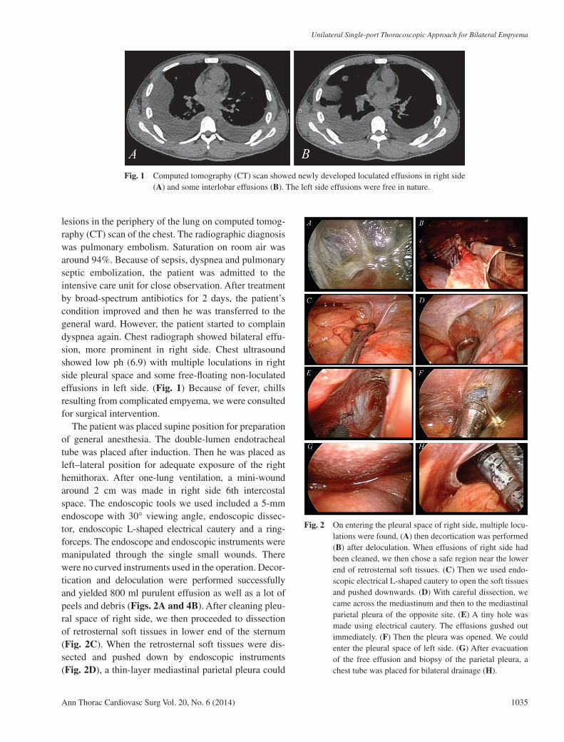
lesions in the periphery of the lung on computed tomog-raphy (CT) scan of the chest. The radiographic diagnosis was pulmonary embolism. Saturation on room air was around 94%. Because of sepsis, dyspnea and pulmonary septic embolization, the patient was admitted to the intensive care unit for close observation. After treatment by broad-spectrum antibiotics for 2 days, the patient’s condition improved and then he was transferred to the general ward. However, the patient started to complain dyspnea again. Chest radiograph showed bilateral effu-sion, more prominent in right side. Chest ultrasound showed low ph (6.9) with multiple loculations in right side pleural space and some free-floating non-loculated effusions in left side. (Fig. 1) Because of fever, chills resulting from complicated empyema, we were consulted for surgical intervention.
The patient was placed supine position for preparation of general anesthesia. The double-lumen endotracheal tube was placed after induction. Then he was placed as left–lateral position for adequate exposure of the right hemithorax. After one-lung ventilation, a mini-wound around 2 cm was made in right side 6th intercostal space. The endoscopic tools we used included a 5-mm endoscope with 30° viewing angle, endoscopic dissec-tor, endoscopic L-shaped electrical cautery and a ring- forceps. The endoscope and endoscopic instruments were manipulated through the single small wounds. There were no curved instruments used in the operation. Decor-tication and deloculation were performed successfully and yielded 800 ml purulent effusion as well as a lot of peels and debris (Figs. 2A and 4B). After cleaning pleu-ral space of right side, we then proceeded to dissection of retroste rnal soft tissues in lower end of the sternum (Fig. 2C). When the retrosternal soft tissues were dis-sected and pushed down by endoscopic instruments (Fig. 2D), a thin-layer mediastinal parietal pleura could
Fig. 1 Computed tomography (CT) scan showed newly developed loculated effusions in right side (A) and some interlobar effusions (B). The left side effusions were free in nature.
Fig. 2 On entering the pleural space of right side, multiple locu-lations were found, (A) then decortication was performed (B) after deloculation. When effusions of right side had been cleaned, we then chose a safe region near the lower end of retrosternal soft tissues. (C) Then we used endo-scopic electrical L-shaped cautery to open the soft tissues and pushed downwards. (D) With careful dissection, we came across the mediastinum and then to the mediastinal parietal pleura of the opposite site. (E) A tiny hole was made using electrical cautery. The effusions gushed out immediately. (F) Then the pleura was opened. We could enter the pleural space of left side. (G) After evacuation of the free effusion and biopsy of the parietal pleura, a chest tube was placed for bilateral drainage (H).
Ann Thorac Cardiovasc Surg Vol. 20, No. 6 (2014) 1035
05_14_NM_Chen_051.indd 1035 2014/12/04 15:34:23
Chen CH, et al.
be identified (Fig. 2E). The effusion gushed out immedi-ately after opening of the pleura (Fig. 2F). Then, the pleural space of left side was entered allowing drainage and collection of the effusions (Fig. 2G). Because the pleural space is not loculated, we placed a chest tube for later drainage of effusions (Fig. 2H). We artificially made additional side-holes in a more proximal site of the chest tube in order to ensure the drainage function of bilateral pleural space. Then the wound was closed after fixation of the chest tube. Three days after operation, the chest tube was removed. He was then treated by antibiotics for another two weeks and then discharged home. After follow-up for more than 3 months, he was alive and had no evidence of recurrence.
Culture from pleural effusion showed Fusobacterium varium and Prevotella species. Peel from decortication showed multiple foci of micro-abscess. There was no evidence of malignancy.
Comment
Single-port thoracoscopic surgery had become more and more feasible in some institutions as an alternative to conventional multiportal approach. In our team, single- port thoracoscopic surgery had become the first-line approach of thoracoscopic surgery. Such techniques had been proposed in the treatment of lung cancer, mediasti-nal tumor, pneumothorax and most common disease of the chest.1–5) With modifications of conventional thora-coscopic techniques, the operative field can be extended to the contralateral side of the pleural space. In the case we presented, peel formation and loculations were mainly located in right side pleural space while the left side effusion was relatively free-floating. On the day of CT scan, he was brought to the operative room for prepa-ration of thoracoscopic surgery. Initially, we plan to per-form bilateral thoracoscopic surgery. In consideration of the potential feasibility of bilateral exploration using a single-port wound from contralateral side, we planned to perform surgery first in the right side and then attempted to dissection to the left side allowing drainage and aspi-ration of the relatively free-floating fluids. If bilateral exploration is not possible, we planned to change posi-tion of the patient and then proceeded to left side thora-coscopic drainage of effusion.
The consideration of such procedure would be slightly different from that of unilateral single-port thoracos-copic surgery. The most important consideration is the wound location. In the methods we proposed previously,
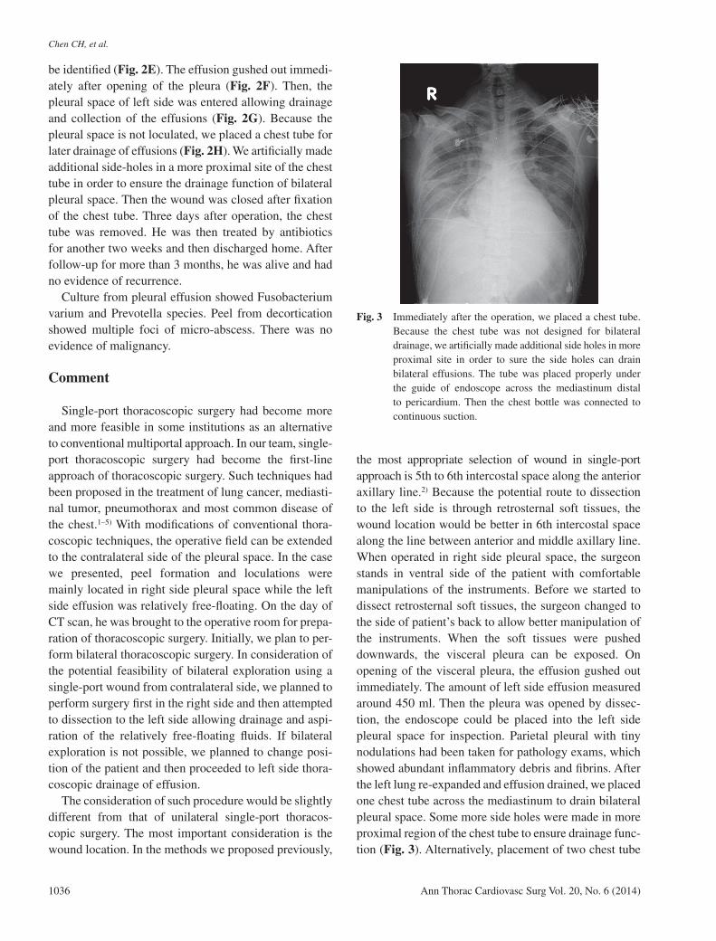
the most appropriate selection of wound in single-port approach is 5th to 6th intercostal space along the anterior axillary line.2) Because the potential route to dissection to the left side is through retrosternal soft tissues, the wound location would be better in 6th intercostal space along the line between anterior and middle axillary line. When operated in right side pleural space, the surgeon stands in ventral side of the patient with comfortable manipulations of the instruments. Before we started to dissect retrosternal soft tissues, the surgeon changed to the side of patient’s back to allow better manipulation of the instruments. When the soft tissues were pushed downwards, the visceral pleura can be exposed. On opening of the visceral pleura, the effusion gushed out immediately. The amount of left side effusion measured around 450 ml. Then the pleura was opened by dissec-tion, the endoscope could be placed into the left side pleural space for inspection. Parietal pleural with tiny nodulations had been taken for pathology exams, which showed abundant inflammatory debris and fibrins. After the left lung re-expanded and effusion drained, we placed one chest tube across the mediastinum to drain bilateral pleural space. Some more side holes were made in more proximal region of the chest tube to ensure drainage func-tion (Fig. 3). Alternatively, placement of two chest tube
Fig. 3 Immediately after the operation, we placed a chest tube. Because the chest tube was not designed for bilateral drainage, we artificially made additional side holes in more proximal site in order to sure the side holes can drain bilateral effusions. The tube was placed properly under the guide of endoscope across the mediastinum distal to pericardium. Then the chest bottle was connected to continuous suction.
1036 Ann Thorac Cardiovasc Surg Vol. 20, No. 6 (2014)
05_14_NM_Chen_051.indd 1036 2014/12/04 15:34:24

Unilateral Single-port Thoracoscopic Approach for Bilateral Empyema
in each pleural space is functionally equivalent. Because the wound is smaller, we decided to place only one tube.
The retrosternal route to contralateral pleural space is still limited in complete inspection. Therefore, with conventional rigid endoscope, the application of bilateral thoracoscopic surgery through unilateral single port wound will be confined in some simple conditions, such as drainage of non-loculated effusion in the case. If the endoscope and instruments can be more flexible and steerable, complete inspection and operation will be more feasible and can avoid bilateral wounds6–10) and a more complicated apicoposterior transmediastinal approach proposed by Cho and colleagues.11) An import-ant difference is that with the method we proposed is much faster than conventional bilateral thoracoscopy. Single-port approach, when operated with experienced surgeon’s hands, is much faster than conventional multi-port approach and has low conversion rate as we reported earlier.2,3,5)
When treating malignant pleural effusion, mediasti-nal metastasis may occur if the mediastinum is opened. Therefore, the adequacy of such procedure in treating malignant effusion is still not clear.
The importance of the case report established the possibility of potential applications of unilateral single- port thoracoscopic surgery in the treatment of bilateral pleural diseases in a more time-saving and lesser patient- burden method. The field of application is to be deter-mined in the future.
Disclosure Statement
All authors stated that there is no conflict of interest.
References
1) Chen CH, Chang H, Lee SY, et al. Single-port thoracoscopic surgery can be a first-line approach for
elective thoracoscopic surgery. Rev Port Pneumol 2012; 18: 278-84.
2) Chen CH, Lee SY, Chang H, et al. Technical aspects of single-port thoracoscopic surgery for lobectomy. J Cardiothorac Surg 2012; 7: 50.
3) Gonzalez-Rivas D, Paradela M, Fieira E, et al. Single- incision video-assisted thoracoscopic lobectomy: initial results. J Thorac Cardiovasc Surg 2012; 143: 745-7.
4) Chen CH, Lee SY, Chang H, et al. The adequacy of single-incisional thoracoscopic surgery as a first-line endoscopic approach for the management of recurrent primary spontaneous pneumothorax: a retrospective study. J Cardiothorac Surg 2012; 7: 99.
5) Chen CH, Chang H, Tseng PY, et al. A rare case of dysphagia and palpitation caused by the compression exerted by an enormous mediastinal lipoma. Rev Port Pneumol 2012; 18: 149-52.
6) Chen CH, Chang H, Yang LY, et al. A preliminary report of a disposable electrical non-fiberoptic endo-scope in thoracoscopic surgery. Int J Surg 2012; 10: 20-4.
7) Sachithanandan A, Nur Ezrin I, Badmanaban B. Single stage minimally invasive bilateral video assisted thoracoscopic surgery for simultaneous bilateral primary spontaneous pneumothorax. Med J Malaysia 2012; 67: 226-7.
8) Garrido M, Aguilera P. Bilateral spontaneous pneu-mothorax. West J Emerg Med 2012; 13: 213-4.
9) Ugolini D, Montinaro F, Ferrarello S. [Video-assisted thoracoscopic surgery for simultaneous spontaneus bilateral pneumothorax]. Minerva Chir 2011; 66: 371-2.
10) Wang KH, Chang CH, Cheng YJ, Chang YT, and Chang PC. Emergent bilateral thoracoscopy for massive hemopneumothorax with contralateral pneumothorax. Am J Emerg Med 2011; 29: 1235. e1-3.
11) Cho DG, Cho KD, Kang CU, et al. Thoracoscopic simultaneous bilateral bullectomy through apicop-osterior transmediastinal access for bilateral sponta-neous pneumothorax: a challenging approach. World J Surg 2011; 35: 2016-21.
Ann Thorac Cardiovasc Surg Vol. 20, No. 6 (2014) 1037
05_14_NM_Chen_051.indd 1037 2014/12/04 15:34:24