Embed Size (px)
Citation preview
Annals of Basic and Applied Sciences December 2017, Vol. 8, No. 1, ISSN 2277 – 8756.
Publication of St. Mary’s College, Thrissur-680020, Kerala, India.
(Re-Accredited by NAAC with A Grade)
Editor Dr C R Meera Department of Microbiology, St Mary’s College, Thrissur-680020,Kerala.
Associate Editors Dr Dhanya K C Department of Microbiology, St Mary’s College, Thrissur-680020, Kerala.
Dr Mabel Merlen Jacob Department of Microbiology, St Mary’s College, Thrissur-680020, Kerala.
Editorial Board Dr Regi Raphael K, Department of Botany, St Mary’s College, Thrissur-680020, Kerala.
Dr Sheeja T Tharakan, Department of Botany, Vimala College, Thrissur-680020, Kerala.
Dr Rekha K, Department of Botany, St Mary’s College, Thrissur-680020, Kerala.
Dr Manju Sebastian, Department of Chemistry,, St Mary’s College, Thrissur-680020, Kerala.
Dr Geetha T, Department of Chemistry, St Mary’s College, Thrissur-680020, Kerala.
Dr Manju Madhavan, Department of Botany, Vimala College, Thrissur-680020, Kerala.
Scientific Advisory Board Dr Sr Magie Jose, Principal, St Mary’s College, Thrissur-680020, Kerala.
Dr K K Janardhanan, FNABS, Professor & Head, Department of Microbiology, Amala Cancer Research Centre, Thrissur-680555, Kerala.
Dr CKK Nair, Director of Research, St. Gregorios Dental College & Research Centre, Kothamangalam- 686681, Kerala.
Dr Valsa A K, Asso. Professor & Head, Department of Biochemistry, Sree Sankara College, Kalady, Ernakulam-683574, Kerala.

Contents Page No:
Qualitative evaluation on phytochemical, antimicrobial and antioxidant profiles of Ethanolic extract of Carica papaya Elizabeth P Thomas and Greeshma Das
1-9
Antibacterial effect of citrus fruit juice against Enteric and Nonenteric pathogenic bacteria Geenat Paul, Rigi George A and Rahima N A
10-16
Green Synthesis of Silver nanoparticle using Aerva lanata Geetha T
17-22
Role of capping agent in the synthesis of stable nano ZnO colloid Litty Irimpan
23-27
Preliminary screening, isolation and characterization of cellulolytic bacteria from soil Rashmi Prakash, Roshni Poulose, Saranya Sasi, Sneha Das, Shilpa U Nair and Mabel Merlen Jacob
28-36
Synthesis of two novel Nnn-Donor Schiff bases and their characterization Manju Sebastian
37-44
Antioxidant Potential of Ethanolic and Hot Water Extracts of Pleurotus sajor-Caju Meera C R, Anjali Krishna, Anjali Variyar, Anju V T, Asha P Antony and Asna Ashraf
45-53
A comparison on probiotic characteristics of β- galactosidase producers from curd and raw milk Neenu Neelakantan
54-59
Physiochemical evaluation, phytochemical screening and pharmacognostic profile of leaves of Artocarpus hirsutus Lam. in different solvent extracts Ayana Elma Shaji and Regi Raphael K
60-72
Phytochemical analysis and antimicrobial effect of Azadirachta indica, Psidium guajava and Spinacia olerocea against Propionibacterium acne Ruveena T N, Sabha Kottayil, Sanu Simon, Rosemol Johnson, Sofiya P J and Nima Varghese
73-78
Comparative study on phytochemical, physiochemical and antioxidant properties of Sorghum bicolor and Triticum aestivum Rincy C K, Ranjitha P C, Priyanka K, Shahanas M, Aiswarya C G, Aneesha K, Aiswarya Das, Gayathri C V, Rahul Pramod K V and Smitha K R
79-87

Annals Of Basic And Applied Sciences, Vol. 8, No.1, Dec 2017 1
Qualitative evaluvatuion on phytochemical, antimicrobial and antioxidant profiles of Ethanolic extract of Carica papaya
Elizabeth P Thomas*, Greeshma Das
St Mary’s College, Thrissur-680020, Kerala, India.
Corresponding author: [email protected]:Ph-9746343500
Abstract
Ayurveda is a traditional Indian medicinal system being practiced thousands of years. Considerable research
on pharmacology, chemistry and clinical therapeutics has been carried out on ayurvedic medicinal plants. The
nontoxic or less toxic nature and the lesser known side effects of plant products has increased the popularity of plant
products as treatment modalities for various ailments. Traditionally, leaves of Carica papaya have been used for
treatment of a wide range of ailments, like in treatment of malaria, dengue, jaundice. The present study was designed
to observe antimicrobial, antioxidant activities of the ethanolic extract of Carica papaya. Phytochemical analysis of
extract was carried out.
Keywords: Ayurvedic medicines, Medicinal plants, Antimicrobial study
1.Introduction
Around the world, at least thirty five
thousand plant species are used for
medicinal purposes and virtually all
plant parts are usually consumed as food
for efficient supply of energy (Kong et
al., 2003). The study of disease and their
treatment have been existing since the
beginning of human civilization. Plant
kingdom is one of the major search areas
for effective works of recent days. The
importance of plants in search of new
drugs is increasing with the
advancements of medical sciences.
Despite the advent of modern medicine,
the popularity of plant natural products
as treatment modalities for various
ailments has increased worldwide due to
their nontoxic or less toxic nature and the
lesser known side effects than the
modern generic drugs. The burden
infectious diseases is a big challenge and
nuisance to human health and
Annals Of Basic And Applied Sciences, Vol. 8, No.1, Dec 2017 2
responsible for certain deaths on daily
basis. Plants have now scientifically
proven as effective, cheaper alternative
sources and have very least side effects
than commercially available synthetic
drugs. Carica papaya Linn is commonly
called as paw-paw and it belongs to the
family Caricaceae. Papaya possesses
excellent medicinal properties for
treatment of different ailments. (K.
Kayalvizhi et al., 2015)
The plant is native to tropical America
and was introduced to India in 16th
century. Young leaves are rich in
flavonoids (kaempferol and myricetin),
alkaloids (carpaine, pseudocarpaine,
dehydrocarpaine I and II), phenolic
compounds (ferulic acid, caffeic acid,
chlorogenic acid), the cynogenetic
compounds (benzylglucosinolate) found
in leaves. Both leaf and fruit of the
Carica papaya Linn. possess carotenoids
namely β- carotene, lycopene,
anthraquinones, glycoside, as compared
to matured leaves and hence possess
medicinal properties like anti-
inflammatory hypoglycaemic, anti-
fertility, abortifacient, hepato protective,
wound healing, recently its
antihypertensive and antitumor activities
have also been established. Leaves being
an important part of several traditional
formulations are undertaken for
standardization for various parameters
like moisture content, extractive values,
ash values, swelling index, etc. The
different parts of the Carica papaya
plant including leaves, seeds, latex and
fruit exhibited to have medicinal value.
2. Methodology
2.1 Collection of leaves
Fresh leaves of Carica papaya were
collectd from outskirts of Thrissur,
Kerala, India. The plant was identified at
Dept. of Botany, St Mary’s College
,Thrissur Kerala, India. The fresh leaves
were harvested properly washed in tap
water, and then rinsed in sterile distilled
water. The leaves were dried in the hot
air oven at 40º C for six hours. The dried
leaves were then crushed into powder
using electric grinder to obtain a
powered form. The powdered samples
were stored in airtight glass containers ,
prior to analysis.
2.2 Preparation of ethanolic extract
The crude powdered sample of 150 g
was defatted with petroleum ether in a
soxhlet apparatus for 6-8 hours,
repeatedly thrice. The ethanol extraction
was done sequentially. The dried
samples were then stored in air tight
bottles at 40C.(Baku, 2007)

Annals Of Basic And Applied Sciences, Vol. 8, No.1, Dec 2017 3
2.3 Phytochemical Screening
The ethanolic extract was analysed for
the presence of various phytochemicals
by the standard procedure of Sofowara
(1993);Trease and Evans(1989);and
harborne(1973)
2.4 Antibacterial activity
Antibacterial test were carried out by the
disc diffusion method with some
modification .The bacterial cultures
used for the test were Pseudomonas sp,
Bacillus sp, Klebsiella sp, Escherichia
coli, Salmonella sp, Serratia sp and
Proteus sp. From the overnight culture,
0.1 ml of culture was uniformly
distributed onto MHA plates. A filter
paper disc of 6 mm diameter was
punched out from a Whatmann No: 1
filter paper and sterilized. Then the discs
were placed on the surface of Mueller
Hinton agar plates at a distance of 2 cm
using sterile forceps. Drugs of different
concentrations (50,100,150,200,250
mg/ml) and a control (2 % DMSO) were
added on each disc with a micropipette.
Then the plates were incubated at 37˚c
for 24-48 hrs. After incubation zone
diameter was measured.
2.5 Antioxidant activity
2.5.1Total anti-oxidant capacity
The total anti-oxidant capacity was
measured according to
spectrophotometric method of Preito
(Preito et al.,1999) . 0.1 ml of the extract
(10 mg/ml)was dissolved in water was
combined in an eppendorf tube with 1 ml
of reagent solution(0.6 M H2SO4,
2.8mM Sodium phosphate and 4mM
Ammonium molybdate).The tubes were
capped and incubated in a thermal block
at 95˚C for 90 minutes. After cooling to
room temperature, the absorbance of the
aqueous solution was measured at 695
nm against blank .Vitamin C used as
standard.
2.5.2 Hydroxyl radical
scavenging activity
Hydroxyl radicals generated from Fe 3+
ascorbate-EDTA- H2O2system(Fenton’s
reaction) was eliminated by its
degradation of deoxyribose that resulted
in thiobarbituric acid reaction
substance(TBARS) (Elizabeth and
Rao,1990).The reaction mixture
contained deoxyribose (28 mM);

Annals Of Basic And Applied Sciences, Vol. 8, No.1, Dec 2017 4
Fe Cl3(1mM);KH2Po4 –KOH buffer(20
Mm ,pH 7.4); EDTA (1 mm); H2O2(1
Mm);Ascorbic acid (0.1 Mm) and
various concentrations of drug in a final
volume of 0.1 ml. The reaction mixture
incubated for 1 hour at 37˚C.The
TBARS formed was estimated by TBA
method. (Ohkawa et al.,1979).The
hydroxyl radical scavenging activity was
determined by comparing absorbance of
control with that of treatments. Vitamin
C was used as standard.
2.5.3 DPPH free radical
scavenging assay
In this method a commercially available
and stable free radical 2, 2, diphenyl-1-
picryl hydrazyl (DPPH+), soluble in
methanol was used(Aquino et al.,2001).
DPPH in its radical form has an
absorption peak at 515 nm, which
disappeared on reduction by an
antioxidant compound .An aliquot (25
µl) of the extracts was added to 1 ml of
freshly prepared DPPH solution(0.25g/l
in methanol).The sample were kept for
20 minutes in dark and the decrease in
absorbance was measured at 515 nm.
Vitamin C was used as the standard.
3.Results
Table 1:- Phytochemical profiling of ethanolic extract of Carica papaya
Phytochemicals Ethanolic
Alkaloids +
Anthraquinones _
Carbohydrates +
Cardiac glycosides _
Coumarin +
Flavanoids +
Phenols _
Phlobatannins _
Proteins _
Quinone _
Saponin _
Tannins _
Annals Of Basic And Applied Sciences, Vol. 8, No.1, Dec 2017 5
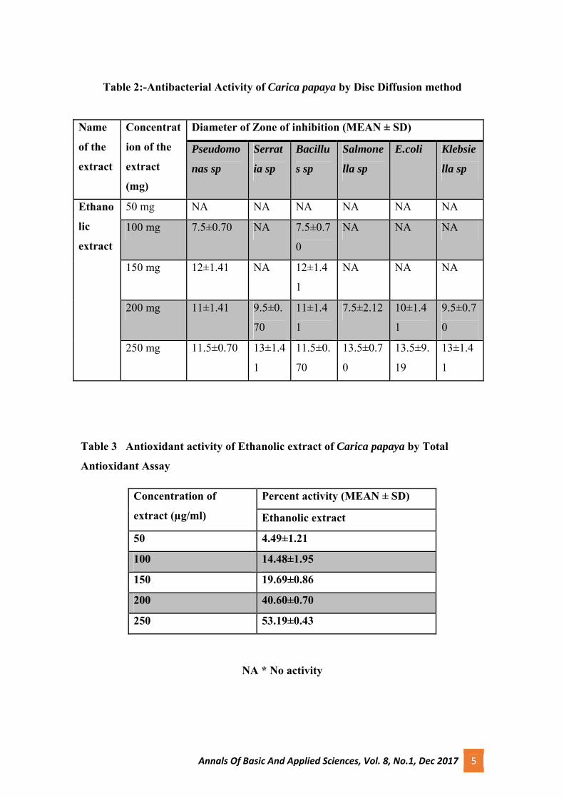
Table 2:-Antibacterial Activity of Carica papaya by Disc Diffusion method
Name
of the
extract
Concentrat
ion of the
extract
(mg)
Diameter of Zone of inhibition (MEAN ± SD)
Pseudomo
nas sp
Serrat
ia sp
Bacillu
s sp
Salmone
lla sp
E.coli Klebsie
lla sp
Ethano
lic
extract
50 mg NA NA NA NA NA NA
100 mg 7.5±0.70 NA 7.5±0.7
0
NA NA NA
150 mg 12±1.41 NA 12±1.4
1
NA NA NA
200 mg 11±1.41 9.5±0.
70
11±1.4
1
7.5±2.12 10±1.4
1
9.5±0.7
0
250 mg 11.5±0.70 13±1.4
1
11.5±0.
70
13.5±0.7
0
13.5±9.
19
13±1.4
1
Table 3 Antioxidant activity of Ethanolic extract of Carica papaya by Total
Antioxidant Assay
Concentration of
extract (µg/ml)
Percent activity (MEAN ± SD)
Ethanolic extract
50 4.49±1.21
100 14.48±1.95
150 19.69±0.86
200 40.60±0.70
250 53.19±0.43
NA * No activity

Annals Of Basic And Applied Sciences, Vol. 8, No.1, Dec 2017 6
Table 4 Antioxidant activity of Ethanolic extract of Carica papaya by DPPH Free
radical scavenging activity
Concentration of
extract (µg/ml) Percent activity (MEAN ± SD)
Ethanolic extract
50 20.1±0.25
100 24.09±0.84
150 26.7±0.07
200 28.07±0.84
250 29.42±0.17
NA * No activity
Table 5 Antioxidant activity of Ethanolic extract of Carica papaya by Hydroxyl
Radical Scavenging Activity
Concentration of
extract (µg/ml)
Percent activity (MEAN ± SD)
Ethanolic extract
50 11.41±0.30
100 16.7±0.15
150 33.07±0.14
200 34.69±0.30
250 44.28±0.14

Annals Of Basic And Applied Sciences, Vol. 8, No.1, Dec 2017 7
4. Discussion and Conclusion
Herbs have been a source of medical
compounds since time immemorial.
History of use of herbal medicine in
treatment of disease can be identified
with the history of medicine and with
the history of civilization itself. All parts
of plants were used in the Ayurvedic
,Unani Allopathic system of medicines
for the treatment of a number of human
diseases. The current study show that
ethanolic extracts of Carica papaya
posses significant antioxidant properties.
Phytochemical analysis showed the
presence of large number of biological
active plant constituents.
Phytochemicals such coumarin, saponin,
cardiac glycosides, tannins, phenols,
flavanoids, alkaloids, carbohydrates
were detected in the ethanolic extract.
Ethanolic extract of Carica papaya was
found to be effective against
Pseudomonas sp, Bacillus sp and E coli.
The maximum inhibitory activity was
against E coli.
The antioxidant activity for the extract
of Carica papaya were conducted using
Total Antioxidant Assay, DPPH
,Hydroxyl Radical Scavenging Assay.
The antioxidant activity was observed in
dose dependent manner. Carica papaya
have active components which are useful
in the straitening of antioxidant
properties (Kaleem et al.,2005).This was
moderately correlated to the high
phenolic content in the herb. However
the studies reveals that Carica papaya
can be a potential and effective source
for drug discovery. Further research may
pave way to the development of effective
antioxidant and antibacterial agents from
Carica papaya.
5. References
Abheri Das Sarma, Anisur Rahaman
Mallick and A. K. Ghosh,2010. Free
Radicals and Their Role in Different
Clinical Conditions: An Overview.
International Journal of Pharma Sciences
and Research (IJPSR).,185-192.
AliyuA.B, Brahim M.A, Brahim H,
Musa A.M., Lawal. A.Y, Oshamini. J.A,
Usman. M, Abdulkadir. I.E,
Oyewale.A.O,2012. Free radical
scavenging and total antioxidant
capacity of methanol extract of
Ethuliaconyzoides growing in
Nigeria,7458-7465
Anibijuwon and A.O. Udeze, 2009.
Antimicrobial Activity of Carica Papaya
(Pawpaw Leaf) on Some Pathogenic
Organisms of Clinical Origin from
Annals Of Basic And Applied Sciences, Vol. 8, No.1, Dec 2017 8
South-Western Nigeria. Ethnobotanical
Leaflets,1-12.
Arun Mathew, Venkat Paluri,
Venkateswaramurthy N, Sambathkumar
R, 2016.A review of newer therapy in
dengue fever. International Journal of
Research in Pharmacology&
Pharmacotherapeutics, 170-177.
Aravind G, Debjit Bhowmik, Duraivel.S
, Harish.G,2013.Traditional and
medicinal Uses of Carica papaya.
Journal of Medicinal Plants Studies,7-
15.
Aurelia Magdalena Pisoschi and
Gheorghe Petre Negulescu, 2011.
Methods for Total Antioxidant Activity
Determination: A Review. Biochemistry
and Analytical Biochemistry,1-10.
C. Gowda, N. Vijay Kumar, P. N.
Kasture, Sr. Medical Advisor, Medical
Services, K. H. Nagabhushan,2015. A
Pilot Study to Evaluate the Effectiveness
of Carica Papaya Leaf Extract in
Increasing the Platelet Count in Cases of
Dengue with Thrombocytopenia. Indian
Medical Gazette- Clinical
Evaluation.,105-116
Chandra Prakash Kala, 2012. Leaf Juice
of Carica papaya L.: A Remedy of
Dengue Fever. Medicinal & Aromatic
Plants.1-2.
Ekaiko Marshall U, Chiwendu Stephen,
Ukpabi Emmanuel O and Ezikpe
Chizaram A, 2015. Antimicrobial
screening and phytochemical analysis of
Carica papaya Leaf extracts, Standard
Research Journal of Microbiological
Sciences Vol 2(1): 001-004
Anjibijuwon and
A.O.Udeze.2009.Antimicrobial activity
of Carica papaya (Pawpaw Leaf)on some
pathogenic organisms of clinical origin
from South-Western Nigeria.850-864.
IrdaFidrianny,KhoirunnisaAyuParamith
a,Siti Kusmardiyani,2016.Anti-oxidant
activities from various leaves extracts of
three cultivars of Papaya from West
Java-Indonesia.299-303.
Jadhav S.S, Deshpande A.D. (2010)
“Antioxidant and anti-hyperglycemic
potential of methanolic extract of bark of
Mimusops elengi L in mice”, Research
Journal of Pharmacology and Biological
Chemical Sceince, Vol. 1, pp.67-7
Annals Of Basic And Applied Sciences, Vol. 8, No.1, Dec 2017 9
JyotsnaKiran Peter, Yashab Kumar,
PriyankaPandey and Harison Masih,
2014.Antibacterial Activity of Seed and
Leaf Extract of Carica Papaya var. Pusa
dwarf Linn. IOSR Journal of Pharmacy
and Biological Sciences (IOSR-
JPBS),29-37
.
K. Kayalvizhi1, Dr. L. Cathrine2, K.
Sahira Banu, 2015. Phytochemical and
antibacterial studies on the leaf
extractsof female Carica papaya. linn.
International Journal of PharmTech
Research, 166-170.
K L Krishna, M Paridhavi and Jagruti A
Patel, 2008.Review on nutritional,
medicinal and pharmacological
properties of Papaya (Carica papaya
Linn.).Natural product radiance,
Vol.7(4),364-373.
Mahendra C. Gunde, Nikhil D.
Amnerkar, 2016. Nutritional, medicinal
and pharmacological properties of
papaya (Carica papaya linn.): A review.
Journal of Innovations in
Pharmaceuticals and Biological
Sciences, 162-169.
Mahmoud A Saleh, PhD;Shavon Clark;
Brooke Woodard; Suizat Ayomide
Deolu-Sobogun, Anti-oxidant and Free
Radical Scavenging Activities of
Essential Oils
Maisarah, A.M., Nurul Amira, B.,
Asmah, R. and Fauziah, O, 2013.
Antioxidant analysis of different parts of
Carica papaya .International Food
Research Journal 20(3): 1043-1048
Michael Antolovich, Paul D. Prenzler,
Emilios Patsalides, Suzanne McDonald
and Kevin Robards,2001. Methods for
testing antioxidant activity, 183-198.
Michael Wink, 2015. Modes of Action of
Herbal Medicines and Plant Secondary
Metabolites. Mr. Nikhil Nishant, Mr
Pradyut Kumar Mohanty, Ms. Shilpa
Luthra, 2014.

Annals Of Basic And Applied Sciences, Vol.8, No.1. Dec 2017 10
Antibacterial Effect of Citrus Fruit Juice against Enteric
and Non Enteric Pathogenic Bacteria
* Geenat Paul , Rigi George A , Rahima N A
Department of Microbiology, St Mary’s College, Thrissur, Kerala – 680020, India
*Corresponding author- Email id: [email protected]:Ph- 9446589360
Abstract
The present study was carried out to determine the antimicrobial effect of three different citrus fruit juices against four
enteric and two non enteric pathogens named Escherichia coli , Salmonella , Proteus, Klebsiella ,Vibrio,and
Staphylococcus species. The study was done by well diffusion and disc diffusion method. The use of different
concentrations (100%, 75%, 50%, 25%) ofcitrus juice extracts had an effective antibacterial activity. Lemon juice was the
most effective against the test organisms in both undiluted and diluted concentration. Pomelo juice showed antimicrobial
activity only against Vibrio and Salmonella.Orange juice showed antimicrobial activity only against Staphylococcus and
Escherichia coli .
Key Words : citrus fruit ,, well diffusion , disc diffusion , antimicrobial effect
1. Introductionriiiiiiiiiii
iBacterial infections are among the
important infectious diseases. Hence,
over 50 years of extensive researches
have been launched for achieving new
antimicrobial medicines isolated from
different sources. Despite progress in
development of antibacterial agents, there
are still special needs to find new
antibacterial agents due to development
of multidrug resistant
bacteria(R.Wise,T.Hart,O.Cars et al.
,1998)
The emergence of resistant organisms
presents a major challenge for the
antimicrobial therapy of infectious
diseases and increases the incidence of
mortality and morbidity.The increase in
antibiotic resistance bacteria is largely
due to the widespread use of antibiotics

Annals Of Basic And Applied Sciences, Vol.8, No.1. Dec 2017 11
in medicine in animal care and agriculture
(Bansode et al 2012). )Citrus fruits have
been of interest for extraction of
antimicrobial metabolites by large
number of researchers (Kumar et al.,
2011; Kumar et al., 2010; Amandeep et
al., 2009) but the peels have been less
studied. Lemon juice has been used in the
treatment of oral thrush in HIV/AID
patients (Wright et al., 2009). The
antioxidant activities of citrus flavonoids
and phenolic compounds exhibited a
potent antibacterial activity which is
probably due to their ability to complex
with bacterial cell walls and disrupt
microbial membrane . Studies have
shown that concentrated or freshly
squeezed lemon juice has antibacterial
activity against Vibrio species
(Tomotake et al., 2006), Antibacterial
properties of plant extract have been a hot
topic for the researchers. Besides plants,
fruits also have been studied by the
researchers for the presence of bioac-tive
compounds close related to herbs,
commonly referred as phytochemicals
such as tannins, carotenoids, polyphe-nols
and anthocyanins (Khushwaha et al
2012). The current research focuses on
the extraction and assay of antibacterial
component from citrus fruit which are
easily available at very low cost.
2. Materials and Methods
2.1 Bacterial cultures - Escherichia coli ,
Salmonella , Proteus, Klebsiella ,Vibrio,
and Staphylococcus species. were kindly
provided from Poly Clinic Pvt Ltd,
Thrissur , Kerala , India.
2.2.Citrus fruits – Fresh lemon ,Orange ,
Pomelo were obtained from local market
of Thrissur.
2.3 Taking of juice from fruits
Surface of the fruit was disinfected using
ethanol. Fruit was pierced and juice was
aspirated and collected in beaker in sterile
condition.
2.4 Preparation of different
concentrations of fruit juice
Different concentrations were made by
adding sterile physiological saline(0.85%)
into the juice. To prepare 1ml of 25%
concentration of fruit juice,0.25 ml juice
was added to 0.75 ml of saline, 1ml of
50% concentraton,0.5 ml juice was added
to 0.5ml saline, for 1ml of 75%
concentration ,0.75 ml of juice was added
to 0.25ml of saline,and for 1 ml of 100%
concentration,1ml juice was used.The
physiological saline was used as control.
Annals Of Basic And Applied Sciences, Vol.8, No.1. Dec 2017 12
2.5. Anti-bacterial activity testing by
Well diffusion method
Antibacterial test were carried out by the
well diffusion method. Overnight
bacterial cultures were diluted in the
nutrient broth to obtain a bacterial
suspension of 10^8 CFU/ml. Petriplates
containing 20 ml of Muller-Hinton Agar
media were inoculated with diluted
cultures with a sterile cotton swab is
dipped into standardized bacterial test
suspension and used to evenly inoculate
the entire surface of the Muller-Hinton
Agar plates. After the agar surface has
dried for about 5 minutes, five wells, all
are have equal diameter were made in the
plate at equal distance. Citrus fruit juice
of different concentrations
(25,50,75,100µl)and a control (0.85%)
saline were poured on each well by using
micropipette. Plates were incubated at
37⁰C for 24h .The antibacterial activity
was determined by measuring the
inhibition zone.
2.6. Anti-bacterial activity testing by
Agar disc diffusion assay
The antibacterial activity of the citrus
fruits extracts was determined by the disc
diffusion method.Briefly ,overnight
bacterial culture were diluted in the
Muller-Hinton broth to obtain a bacterial
suspension of 10^8 CFU/ml. Petriplates
containing 20ml of Muller-Hinton Agar
media.A sterile cotton swab is dipped into
a standardized bacterial test suspension
and used to evenly inoculate the entire
surface of the Muller-Hinton Agar
plate.After the agar surface has dried for
about 5 minutes , Citrus fruit juice of
different concentrations (25,50,75,100µl)
were loaded on to the filter paper discs
(whatman No.1 ,6mm diameter) were
placed on the inoculated agar surface
were allowed to dry completely. Standard
saline (0.85%) was placed as control.
Plates were incubated at for 37⁰C 24h.
The antibacterial activity was determined
by measuring the inhibition zone.
(S.Sundar and koilpillai, 2015).
3. Result and Discussion
From three citrus fruits used in the study
for evaluation of their antimicrobial
activity against four enteric bacteria and
two non enteric bacteria initially
determined by Muller Hinton Agar well
diffusion method. All of the isolated
species showed activity against citrus
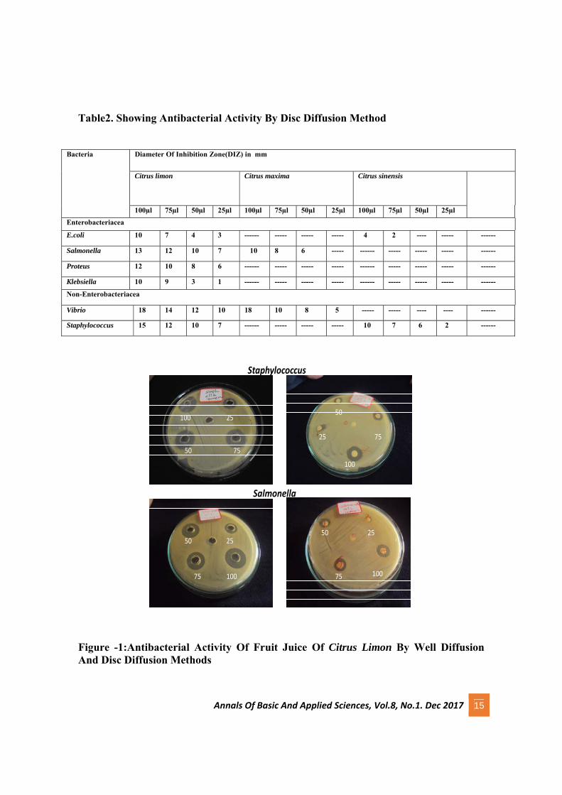
fruit juices. Juice of Citrus lemon

Annals Of Basic And Applied Sciences, Vol.8, No.1. Dec 2017 13
showed highest inhibitory effect against
Vibrio species with largest DIZ
(Diameter of Inhibition Zone) value
followed by Staphylococcus species
,Salmonella species and Proteus species.
Juice of Citrus maxima showed highest
inhibitory effect against Vibrio species
followed by Salmonella species. de
Castillo etal (2000) also reported that
freshly squeezed lemon juice inhibited
the growth of V. Cholerae . Juices of
Citrus sinesis showed highest inhibitory
effect against Staphylococcus species
followed by E.coli.
The antibacterial activity of these
pathogens are further determined by disc
diffusion method. All isolated bacteria
showed susceptibility against citrus fruit
juices. Juice of Citrus limon showed
highest inhibitory effect against Vibrio
species with largest DIZ followed by
Staphylococcus species ,salmonella
species and Proteus species. Juice
of Citrus maxima showed highest
inhibitory effect against Vibrio species
followed by Salmonella species. Citrus
sinesis showed highest inhibitory effect
against Staphylococcus species followed
by E.coli . Citrus limon showed
highest inhibitory activity against
Vibrio species while lowest activity
against Proteus species. The results
showed in Table 1 and Table 2.
Similar results for the various extracts
from citrus fruits have been reported by
many authors (Kumar et al., 2011; Kumar
et al., 2010; Amandeep et al., 2009;
Nurmahani et al., 2012) .On comparison
of antimicrobial activity of different
citrus fruits the results showed
that Citrus limon>Citrus
maxima>citrus sinensis. The results
of Table 1 and Table 2 revealed
that Citrus limon juice showed highest
activity against almost all enteric
pathogens and non enteric pathogens.
From the results obtained, the highest
inhibitory effect showed by 100µl
concentration and least effect by 25µl.
4. Conclusion
The study summarised here demonstrate
that the microbial load of human body
constitute enteric and non-enteric
pathogens.The bacterial isolates
identified in this study are common
human enteric and non-enteric
pathogens.This study emphasizes the

Annals Of Basic And Applied Sciences, Vol.8, No.1. Dec 2017 14
need of usage of citrus fruits in our
diet.Because the results showed that the
citrus fruit juice have high antibacterial
activity against almost all enteric and
non-enteric pathogens .The well diffusion
and disc diffusion methods using citrus
fruit juice showed antibacterial activity
against Vibrio, Salmonella,
Staphylococcus, E.coli, Proteus and
Klebsiella..The study points out that the
infections with enteric and non-enteric
pathogens can be successfully eliminated
by the use of easily available citrus fruit
juices. This approach however go a long
way in combining the rising tide of
antibacterial resistance.
Table 1: Showing Antibacterial Activity By Well Diffusion Method
Bacteria Diameter Of Inhibition Zone in mm.
Citrus limon Citrus maxima Citrus sinensis
100µl
75µl
50µl
25µl
100µl
75µl
50µl
25µl 100µl
75µl
50µl
25µl
Enterobacteriacea E.coli 15 10 11 7 ------ ----- ----- ----- 7 4 2 ----- ------
Salmonella 21 19 17 14 18 14 10 ----- ------
----- ----- ----- ------
Proteus 19 17 15 11 ------ ----- ----- ----- ------
----- ----- ----- ------
Klebsiella 13 11 10 3 ------ ----- ----- ----- ------
----- ----- ----- ------
Non-Enterobacteriacea
Vibrio 23 22 18 16 21 18 13 ----- -----
----- -----
---- ------
Staphylococcus 22 19 17 14 ------ ----- ----- ----- 12 10 8 7 ------
Annals Of Basic And Applied Sciences, Vol.8, No.1. Dec 2017 15
Table2. Showing Antibacterial Activity By Disc Diffusion Method
Bacteria Diameter Of Inhibition Zone(DIZ) in mm
Citrus limon Citrus maxima Citrus sinensis
100µl 75µl 50µl 25µl 100µl 75µl 50µl 25µl 100µl 75µl 50µl 25µl
Enterobacteriacea
E.coli 10 7 4 3 ------ ----- ----- ----- 4 2 ---- ----- ------
Salmonella 13 12 10 7 10 8 6 ----- ------ ----- ----- ----- ------
Proteus 12 10 8 6 ------ ----- ----- ----- ------ ----- ----- ----- ------
Klebsiella 10 9 3 1 ------ ----- ----- ----- ------ ----- ----- ----- ------
Non-Enterobacteriacea
Vibrio 18 14 12 10 18 10 8 5 ----- ----- ---- ---- ------
Staphylococcus 15 12 10 7 ------ ----- ----- ----- 10 7 6 2 ------
10075
50 25
25
50
75
100
25
50 75
100
2550
75 100
Staphylococcus
Salmonella
Figure -1:Antibacterial Activity Of Fruit Juice Of Citrus Limon By Well Diffusion And Disc Diffusion Methods
Annals Of Basic And Applied Sciences, Vol.8, No.1. Dec 2017 16
5. Reference
Amandeep Singh, Ahmed R. Bilal, In
vitro antibiotic activity of isolated volatile
oil of citrus Sinensis. International journal
of pharm. Res. And development. 2009;
7(1): 1-4.
Bansode D. S,Chavan M D,(2012)
Studies on antimicrobial activity and
phytochemical analysis of citrus fruit
juices against selected enteric pathogens.
IRJP.3(11):122-126.
De Castillo M C, de Allori C G , de
Gutierrez R C, de Saab O A, de
Fernandez N P, de Ruiz C S et al.(2000)
Bacterial activity of lemon juice and
lemon derivatives against Vibrio cholera.
BioPharma Bull.(10):1235-1238.
Khushwaha A,Singh R.P,Gupta V, Singh
M.(2012) Antimicrobial properties of
peels of citrus fruits .IJPLS .2(2):24-32
Kumar K. Ashok, Subanthini A.,
Jayakumar M. Antimicrobial Activity and
Phytochemical Analysis of Citrus Fruit
Peels -Utilization of Fruit Waste. 2011;
3(6): 5414-5421.
Kumar Vivek R, Anti Typhoid Activity
of Aqueous Extract of Fruit Peel Citrus
sinensis (L.). International Journal of
Pharm. Res. And Development. 2010;
2(9): 217-221.
Nurmahani MM, Osman A, Abdul Hamid
A, Mohamad Ghazali F and Pak Dek MS.
Antibacterial property of
Hylocereuspolyrhizus and
Hylocereusundatus peel extracts.
International Food Research Journal.
2012; 19(1): 77-84.
Wise,T.Hart,O.Cars et al.
,(1998).Antimicrobial resistance, is a
major threat to public health. BMJ 317
(7159) 609-10
Sundhar and Justin koilpillai (2015)
Antimicrobial potential of petroleum
ether extract and active column fraction
of the solanum incanum leaves. Journal of
Pure & Applied Microbiology.9(4):49-54
Tomotake H, Koga T, Yamato M, Kassu
A and Ota F (2006). Antibacterial activity
of citrus fruit juices against Vibrio
species. J. Nutr. Sci. Vitaminol. (Tokyo),
52(2): 157-160.
Wright SC, Maree JE and Sibanyoni M
(2009). Treatment of oral thrush in
HIV/AIDS patients with lemon juice and
lemon grass (Cymbopogon citratus) and
gentian violet. Phytomedicine, 16(2-3):
118-124.

Annals Of Basic And Applied Sciences, Vol.8, No.1. Dec 2017 17
Green Synthesis of Silver nanoparticle using Aerva lanata
Geetha T* Assistant Professor, Department of Chemistry, St. Mary’s College, Thrissur-680020, Kerala,
India.
*Corresponding author- [email protected]: 9495951805
Abstract
This study successfully synthesises silver nanoparticle using Aerva lanata (Cherrula) leaf extract. The leaf extract was
screened for phytochemicals. The pH and concentration of plant extract added to silver nitrate concentration was
optimised for the best result.
Key Words : Green Synthesis, Silver nanoparticles, Aerva lanata
1. Introduction
Among nanoparticles, metal nanoparticles
especially silver nanoparticles has attracted
considerable attention as a result of their
significant applications in the field of in
catalysis, sensors and medicine. Over the
last decades, silver nanoparticles have
found applications in catalysis, optics,
electronics and other areas due to their
unique size-dependent optical, electrical
and magnetic properties. Currently, most of
the applications of silver nanoparticles are
as antibacterial or antifungal agents in
biotechnology and bioengineering, textile
engineering, water treatment and silver
based consumer products. There is also an
effort to incorporate silver nanoparticles in
to wide range of medical devices like bone
cement, surgical instruments, surgical
masks, wound dressing, fluorescent
biological labels for important biological
markers and molecules in research and
diagnosis of diseases. They also find
application in the field of drug delivery
systems, gene delivery systems in gene
therapy, for biological detection of disease
causing organisms and diagnosis, detection
of proteins, isolation and purification of
biological molecules and cells in research,
probing of DNA structure, genetic and
tissue engineering, in MRI studies, in
pharmacokinetic studies etc. (Tiwari A. and
Syvà M, 2014, Wijnhoven, et.al., 2009).
Silver nanoparticles are nanoparticles of
silver i.e. silver particles between 1nm and
100nm in size. Although chemical and

Annals Of Basic And Applied Sciences, Vol.8, No.1. Dec 2017 18
physical methods may successfully produce
pure, well-defined nanoparticles, these are
quite expensive and potentially dangerous
to the environment. Use of biological
organisms such as microorganisms, plant
extract or plant biomass could be an
alternative to chemical and physical
methods for the production of nanoparticles
in an eco-friendly manner (Kumar &
Yadav, 2009).
Green syntheses of silver nanoparticles
when compared to chemical and physical
methods are environment friendly and cost
effective. In these methods, there is no need
to use high pressure, energy, temperature or
toxic chemicals. Thus synthetic methods
based on naturally occurring biomaterials
provide an alternative means for obtaining
nanoparticles and are preferable over the
traditional chemical or mechanical methods
for their simplicity, cost-effectiveness,
environment friendly nature and
reproducibility. (Mittal, et. al.,
2016). Synthesis of nanoparticles
especially silver using plant extracts has
been reported previously. Elumalai et al.
(2010) reported the synthesis of AgNp
using Euphorbia hirta leaves, Singha et al.,
(2010) using argemone mexicana leaf
extract, Prasanth et al. (2011) using
medicinal plant extracts and Mallikarjuna
et al. (2011) using Ocimum leaf extract.
Synthesis of AgNp from plant extract is
carried out by exploiting the reduction
capabilities of varied phytochemicals
present in them (Amarnath Kanchana et al.,
2011)
Here we attempted to find a cost effective
& ecofriendly method for the synthesis of
silver nanoparticles from 1mM AgNO3
solution through the extract of Aerva lanata
(Cherrula). The method involves reducing
the silver ions present in the solution of
silver nitrate by the different
phytochemicals present in the extract
followed by capping these nanoparticles.
Nanoparticles were characterized using
UV-visible absorption spectroscopy using
UV-visible spectrophotometer.
2. Materials & Methods
Silver nitrate is used for the synthesis of
silver nanoparticles. All glassware have
been washed with distilled water and dried
in oven before use. The leaf sample of
Aerva lanata (Cherrula) was collected
locally. The plant samples was cleaned of
all the impurities present by washing with
normal water followed by distilled water.
The sample was dried in shade by keeping
overnight in at room temperature.
2.1 Preparation of leaf extract
5 g of dried Aerva lanata (Cherrula) leafs
were weighed and transferred to a 250ml
beaker, 100ml of double distilled water was
added to it and a glass rod was placed inside
Annals Of Basic And Applied Sciences, Vol.8, No.1. Dec 2017 19
the beaker. Then the beaker was placed in
water bath, maintained at 80°c for thirty
minutes and cooled. The extract was then
filtered using Whatmann No: 1 filter paper
into a conical flask. This extract was stored
in refrigerator for future use.
2.2 Phytochemical screening of leaf extract
The plant extract was tested for
carbohydrates, glycosides, tannins,
saponins, phenols, flavonoids,
anthrocyanoside, and anthraquinonine.
2.3 Synthesis of silver nanoparticles
1mM aqueous solution of silver nitrate
(AgNO3) was prepared and used for the
synthesis of silver nanoparticles. 1.7g of
Silver nitrate was weighted accurately in an
electrical balance, transferred to the
standard flask and made up to 100ml. From
this solution, pipette out 50ml AgNO3
solution, and transfer to another 500ml
standard flask. Make up to the mark to
obtain 1mM AgNO3 solution.
To synthesize silver nanoparticles, the
solution of AgNO3 with plant extract in
varying ratios and at different pH. To
optimize the synthesize AgNP, three
different concentrations viz. 1:9, 2:8 and
3:7 were tested. The pH range was varied
and the experiment was carried out at pH 6,
8 and 12. The experiment was carried out in
conical flask covered with aluminium foil
was added to solution of AgNO3 drop wise
to prevent the entry of light. Leaf extract
slowly with constant shaking .This was
placed on shaker for 30 minutes. pH was
adjusted by adding necessary quantity of
NaOH.
2.4 UV-Visible spectra analysis
The formation of silver nanoparticles was
monitored by change in colour of the
solution to golden brown. The solution was
observed for colour change periodically
and allowed to rest in dark overnight if
needed. On observing a change in colour,
the solution was analyzed with UV
spectrophotometer (1800 shimadzu UV
spectrophotometer) after diluting a small
quantity of sample with distilled water.
3. Results & Conclusion
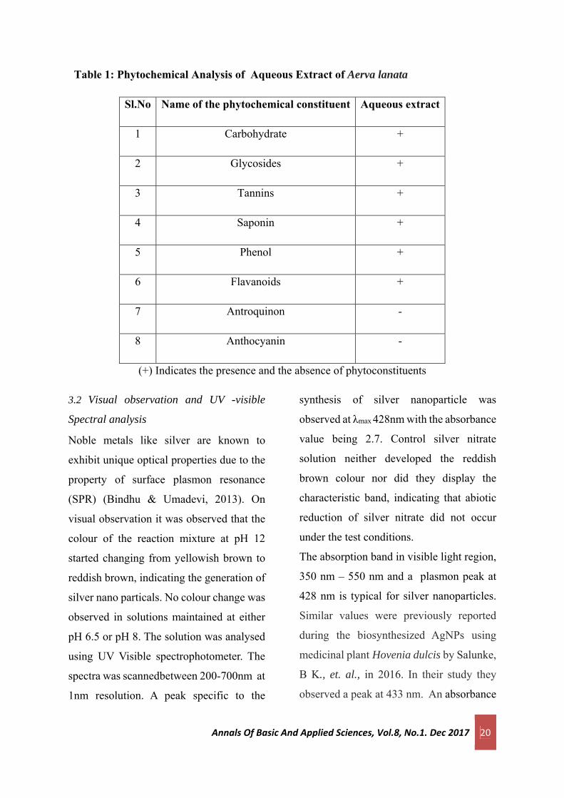
3.1 Phytochemical screening of leaf extract
Aqueous extracts of the plant Aerva lanata
(Cherrula) showed the presence of
carbohydrate, glycoside, tannin, saponin,
flavonoid, and phenol. Antroquinone and
anthocyanin were not found in the extract
of Aerva lanata (Cherrula). Table 1
showed the results of phytochemical
analysis of the aqueous extracts of Aerva
lanata (Cherrula).
Annals Of Basic And Applied Sciences, Vol.8, No.1. Dec 2017 20
Table 1: Phytochemical Analysis of Aqueous Extract of Aerva lanata
Sl.No Name of the phytochemical constituent Aqueous extract
1 Carbohydrate +
2 Glycosides +
3 Tannins +
4 Saponin +
5 Phenol +
6 Flavanoids +
7 Antroquinon -
8 Anthocyanin -
(+) Indicates the presence and the absence of phytoconstituents
3.2 Visual observation and UV -visible
Spectral analysis
Noble metals like silver are known to
exhibit unique optical properties due to the
property of surface plasmon resonance
(SPR) (Bindhu & Umadevi, 2013). On
visual observation it was observed that the
colour of the reaction mixture at pH 12
started changing from yellowish brown to
reddish brown, indicating the generation of
silver nano particals. No colour change was
observed in solutions maintained at either
pH 6.5 or pH 8. The solution was analysed
using UV Visible spectrophotometer. The
spectra was scannedbetween 200-700nm at
1nm resolution. A peak specific to the
synthesis of silver nanoparticle was
observed at λmax 428nm with the absorbance
value being 2.7. Control silver nitrate
solution neither developed the reddish
brown colour nor did they display the
characteristic band, indicating that abiotic
reduction of silver nitrate did not occur
under the test conditions.
The absorption band in visible light region,
350 nm – 550 nm and a plasmon peak at
428 nm is typical for silver nanoparticles.
Similar values were previously reported
during the biosynthesized AgNPs using
medicinal plant Hovenia dulcis by Salunke,
B K., et. al., in 2016. In their study they
observed a peak at 433 nm. An absorbance

Annals Of Basic And Applied Sciences, Vol.8, No.1. Dec 2017 21
peak at 450 nm was also reported by Jain,
et al., during synthesis of silver
nanoparticles using papaya fruit extract
(2009). The surface plasmon resonance of
the silver nanoparticles reduced by the
Spirulina aqueous extract was obtained at
431 nm in a study by Palanisamy, et.
al.,(2017).
It can be reasonably concluded that we
successfully synthesised silver nanoparticle
using extract of Aerva lanata (Cherrula).
The optimum pH and concentration for
biosynthesis of AgNp were pH 12 and 1:9
concentration.
4. References
Amarnath Kanchana, Isha Agarwal, Swetha
Sunkar, Jayshree Nellore, Karthick
Namasivayam (2011). Biogenic silver
nanoparticles from spinacia oleracea and
lactuca sativa and their potential
antimicrobial activity. Dig. J. Nanomate.
Bios. 6 (40): 1741-1750.
Elumalai EK, Prasad TNVKV,
Hemachandran J, Viviyan Therasa S,
Thirumalai T, David E (2010).
Extracellular synthesis of silver
nanoparticles using leaves of Euphorbia
hirta and their antibacterial activities. J.
Pharm. Sci. & Res. 2 (9): 549-554.
Jain D, Kumar Daima H, Kachhwaha S,
Kothari SL (2009). Dig. J. Nanomate. Bios.
4 (3): 557 – 563.
Kumar V & Yadav SK (2009). Plant‐
mediated synthesis of silver and gold
nanoparticles and their applications. J.
Chem. Technol. Biotechnol., 84(2): 151-
157.
Mallikarjuna K, Narasimha G, Dilip GR,
Praveen B, Shreedhar B, Sreelakshmi C,
Reddy B VS, Deva Prasad Raju B (2011)
Dig. J. Nanomate. Bios. 6 (1), 181 - 186.
Mittal J, Singh A, Batra A, Sharma MM.
(2016). Synthesis and characterization of
silver nanoparticles and their antimicrobial
efficacy. Particulate Science and
Technology. 7:1-8.
Palanisamy, SR, Anjali P. Rajasekar, E.
Kannapiran, B. Vaseeharan, and NM
Prabhu. (2017) Synthesis and Distribution
of Bioinspired Silver Nanoparticles Using
Spirulina Extract for Control of Vibrio
parahaemolyticus Infection in
Aquaculture. Asian J. Chem. 29, no. 4: 857.
Prasanth, Menaka R, Muthezhilan Navin
Kumar Sharma (2011). International
Journal of Engineering Science and
Technology. 3 (8): 6235 - 6250.
Annals Of Basic And Applied Sciences, Vol.8, No.1. Dec 2017 22
Salunke BK, Sawant SS, Kim BS. (2016)
Enhancement of Antibacterial Effect by
Biosynthesized Silver Nanoparticles with
Antibiotics. J. Nanosci. Nanotechnol.
1;16(7):7191-4.
Singha A, Jaina D, Upadhyaya MK,
Khandelwala N, Vermaa HN (2010). Green
synthesis of silver nanoparticles using
argemone mexicana leaf extract and
evaluation of their antimicrobial activities.
Dig. J. Nanomate. Bios. 5 (2) : 483 – 489.
Tiwari, A & Syvà M. (Eds.).
(2014). Advanced Materials for Agricultu-
re, Food and Environmental Safety. John
Wiley & Sons.
Wijnhoven S W. Peijnenburg, W. J.,
Herberts, C. A., Hagens, W. I., Oomen, A.
G., Heugens, E. H., ... & Dekkers, S.
(2009). Nano-silver–a review of available
data and knowledge gaps in human
and environmental risk
assessment. Nanotoxicology, 3(2), 109-
138.
Annals Of Basic And Applied Sciences, Vol. 8, No.1, Dec 2017
23
Role of capping agent in the synthesis of stable nano ZnO
colloid
Litty Irimpan Department of Physics, St. Mary’s College, Thrissur-680020, Kerala, India.
*Corresponding author- Email: [email protected] Ph: 9747864368
Abstract
In this article we present the Synthesis of Stable nano ZnO by colloidal chemical synthesis using a capping agent Poly
Vinyl Pyrrolidone (PVP). Absorption spectroscopy is a powerful tool in characterizing nanocrystal colloids and Particle
Size is calculated from the shift of absorption edge. Time dependent absorption spectroscopy gives us an idea about
Particle Growth over time and the effect of capping in the prevention of growth. It is found that the addition of PVP
effectively controls the growth. We found an optimum concentration of PVP to be added in one milli molar ZnO for
the effective capping.
Key words : Nanocolloid, Absorption spectroscopy, Capping
1.Introduction
Several approaches have been
considered to prepare size-controlled
semiconductor nanocrystallites with
narrow size distribution. Many of these
preparation methods control the growth
by the addition of stabilizers or capping
agents like Thiols, PVP etc. by
restricting the growth space in matrices
like Zeolites, reverse micelles, porous
silica matrix etc (Vossmeyer, 1994;
Yang, 2001; Wang, 1989; Pillai,1995;
Maeda, 1995). Surface-capped CdS
colloids have been prepared by different
thiols, for example, Henron et al. have
shown that CdS nanoclusters of various
sizes can be prepared by adjusting the
ratio of sulfide to thiophenol during
synthesis (Herron, 1990). Yang et al
(2001) have attempted preparing ZnO
quantum dots of different sizes by PVP
capping and we have attempted a
similar approach.
2.Theory
When quantum dots are excited with
energies larger than band gap energy,
electrons in the conduction band and
holes from the valence bands are
excited. Depending on the excitation
conditions, the coulomb attraction
Annals Of Basic And Applied Sciences, Vol. 8, No.1, Dec 2017
24
between a hole and electron might lead
to a bound state, the Wannier Exciton.
Just as the hydrogen atom, the exciton
states are characterized by a product
wave function consisting of a plane
wave part for the centre-of-mass motion
and hydrogen functions for the e-h
relative motion. The characteristic
length scale for the relative motion is
the exciton Bohr radius, which may be
of the order of 1-20 nm, depending on
the semiconductor material. Quantum
confinement effects arise, as soon as the
size of the quantum dot is comparable
to this exciton Bohr radius.
In the case of nanocrystallites, the
electrons, holes and excitons have
limited space to move and their limited
motion becomes possible only for
definite values of energy. The highest
occupies valence band and lowest
unoccupied conduction band are shifted
to a more negative and positive values
respectively resulting in widening of
band gap. This leads to a blue shift of
absorption band which be observed
through optical absorption and
transmission studies.
3.Experiment
Stable nano ZnO colloid is prepared by
colloidal chemical synthesis using a
capping agent Poly Vinyl Pyrrolidone
(PVP). One millimolar of zinc acetate is
dissolved in isopropyl alcohol (IPA-
Merck, HPLC grade)
by stirring at 500C in the presence of
capping agent poly vinyl pyrolidone
(PVP-Sisco) (Litty, 2007; Song, 2015).
ZnO colloid is formed when it is
hydrolysed with sodium hydroxide
under ultrasonification for 2 hours. The
ZnO colloids are characterized by time
dependent optical absorption
measurements recorded using a
spectrophotometer.
4.Results and Discussions
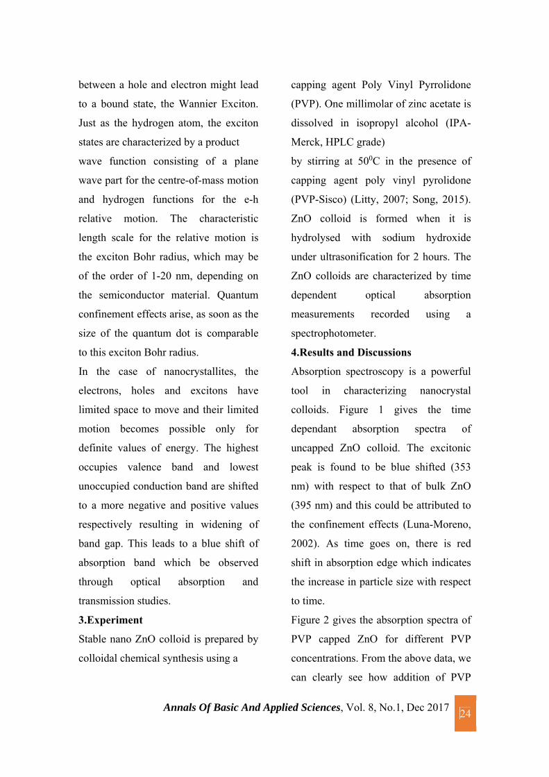
Absorption spectroscopy is a powerful
tool in characterizing nanocrystal
colloids. Figure 1 gives the time
dependant absorption spectra of
uncapped ZnO colloid. The excitonic
peak is found to be blue shifted (353
nm) with respect to that of bulk ZnO
(395 nm) and this could be attributed to
the confinement effects (Luna-Moreno,
2002). As time goes on, there is red
shift in absorption edge which indicates
the increase in particle size with respect
to time.
Figure 2 gives the absorption spectra of
PVP capped ZnO for different PVP
concentrations. From the above data, we
can clearly see how addition of PVP
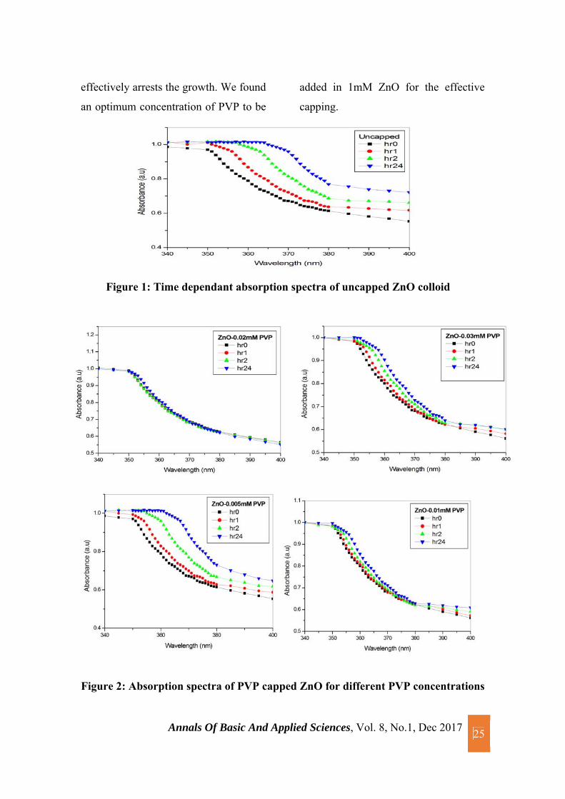
Annals Of Basic And Applied Sciences, Vol. 8, No.1, Dec 2017
25
effectively arrests the growth. We found
an optimum concentration of PVP to be
added in 1mM ZnO for the effective
capping.
Figure 1: Time dependant absorption spectra of uncapped ZnO colloid
Figure 2: Absorption spectra of PVP capped ZnO for different PVP concentrations

Annals Of Basic And Applied Sciences, Vol. 8, No.1, Dec 2017
26
Table 1: Variation of particle size of uncapped/capped ZnO for different PVP
concentrations
Particle Size (nm)
Uncapped
(1mM ZnO)
ZnO+
0.005mM
PVP
ZnO+
0.01mM
PVP
ZnO+
0.02mM
PVP
ZnO+
0.03mM
PVP
0hour 4.4 4.4 4.4 4.4 4.4
1 hour 5.24 4.85 4.5 4.4 4.67
2 hour 7.62 6.35 4.85 4.4 5.25
24 hour 16.37 10.7 5.25 4.67 6.25
The pronounced dependence of the
absorption band gap on the size of ZnO
nano crystals is used to determine the
particle size. The cluster sizes are
calculated from the absorption spectra
using the analytical formula given by
Ranjani et al (2004). To get a precise
measure of the shift, the first derivative
curve of the absorption spectrum is
taken and the point of inflection is taken
as the cut-off wavelength. From the cut-
off wavelength, the corresponding Eg is
calculated. The deviation of this from
the Eg of bulk ZnO gives gE and the
particle size, d is determined using the
equation,
12100 18.1 41.4 0.8gE d d
. Table
1 gives the variation of particle size of
uncapped/capped ZnO for different PVP
concentrations.
From the above table, it is clear that
uncapped ZnO is unstable since particle
size increases with time. It is found that
the addition of PVP effectively controls
the growth. We changed the
concentration of PVP from 0.001mM to
0.03mM and it is found that 0.02Mm
PVP capped ZnO is more stable. We
found an optimum concentration of
PVP to be added in 1mM ZnO for the
effective capping.
5.Conclusion
In this article we present the Synthesis
of Stable nano ZnO by colloidal
chemical synthesis using a capping
agent Poly Vinyl Pyrrolidone (PVP).
Absorption spectroscopy is a powerful
tool in characterizing nanocrystal
colloids. Even without sophisticated
tools like TEM, one can get an idea

Annals Of Basic And Applied Sciences, Vol. 8, No.1, Dec 2017
27
about the size range from the shift of
absorption edge. Time dependent
absorption spectroscopy gives us an
idea about particle growth over time and
the effect of capping in the prevention
of growth. It is found that the addition
of PVP effectively controls the growth.
We found an optimum concentration of
PVP to be added in 1mM ZnO for the
effective capping.
Acknowledgments
Author acknowledge Department of
Physics, Vimala College, Department of
Chemistry & Zoology, St. Mary’s
College, Thrissur for experimental
facility.
References
Vossmeyer et al (1994). J.Phys.Chem.
98: 7665
Yang C.L., Wang J.N., Ge W.K., Guo
L., Yang S.H, Shen D.Z. (2001)
J.Appl.Phys, 90(9): 4489-93
Wang et al (1989). J. Opt. Soc. Am. B,
6: 808
Pillai V,.Kumar P et al (1995).
Adv.Colloid &Int.Sci. 59: 241
Maeda.Y (1995).Phys.Rev.B 51: 1658
Herron et al (1990) J.Am Chem.Soc.
112:1322
Litty Irimpan, Bindu Krishnan, A
Deepthy, V P N Nampoori and P
Radhakrishnan (2007) J. Phys. D: Appl.
Phys. 40: 5670
Song Zhao et.al, (2015). Improving
permeability and antifouling
performance of polyethersulfone
ultrafiltration membrane by
incorporation of ZnO-DMF dispersion
containing nano- ZnO and
polyvinylpyrrolidone. Journal of
Membrane Science. 478: 105-116
Luna-Moreno D., De la Rosa-Cruz E.,
Cuevas F. J., Regalado L. E., Salas P.,
Rodríguez R. and Castano V. M (2002).
Opt. Mat. 19: 275
Ranjani Viswanatha, Sammer Sapra,
Satpati B., Satyam P.V., Dev B.N.,
Sharma D.D (2004). J.Mater.Chem,14:
661

Annals Of Basic And Applied Sciences, Vol. 8, N0.1, Dec 2017 28
Preliminary screening, isolation and characterization of
cellulolytic bacteria from soil
Rashmi Prakash, Roshni Poulose, Saranya Sasi, Sneha Das, Shilpa U Nair & Mabel
Merlen Jacob*
Department of Microbiology, St. Mary’s College, Thrissur- 680020. Kerala, India.
*Corresponding author- Email: [email protected] Ph: 8129442956.
Abstract
Microbial cellulases are an important group of enzymes that can have application in various industries such as food
processing, laundry industry, leather processing, bioremediation process and in textile industry. Soil is an important
source for isolation of microorganisms for novel industrial enzymes production. In this study the cellulase producing
bacteria were isolated from soil rich with degrading organic matter. Two potential isolates were identified to be cellulolytic
in CMC agar media by Congo Red Method and confirmed by Gram’s Iodine Method. These isolates were tentatively
identified by some common biochemical tests and microscopical observations. They were then subjected to submerged
fermentation for the production of cellulase enzyme and its activity was assayed. They were found to produce around 3 U
enzyme at 72 h. Further studies for maximization the enzyme production followed by its purification, characterization and
applications are to be undertaken.
Keywords: Cellulase, CMC agar, submerged fermentation, Congo Red, Gram’s Iodine.
1. Introduction
Cellulose is the primary product of
photosynthesis in terrestrial environments
and the most abundant renewable bio
resource produced in the biosphere.
Biotechnological conversion of cellulosic
biomass is potentially a sustainable
approach to develop novel bioprocesses
and products. Cellulose is commonly
degraded by an enzyme called cellulase
which hydrolyses the β-1,4- glycosidic
bonds in the polymer to release glucose
units. Cellulases are inducible enzymes
synthesized by large diversity of
microorganisms including both fungi and
Annals Of Basic And Applied Sciences, Vol. 8, N0.1, Dec 2017 29
bacteria during their growth on cellulosic
materials. These microorganisms can be
aerobic, anaerobic, mesophilic or
thermophilic. The genera of Clostridium,
Cellulomonas, Aspergillus Trichoderma
and Thermomonaspora are the most
extensively studied cellulase producers.
Microbial cellulases have become the
focal biocatalysts due to their complex
nature and widespread industrial
applications.
Cellulose containing wastes may be
agricultural, urban, or industrial in origin;
sewage sludge might also be considered a
source of cellulose since its cellulosic
content provides the carbon needed for
methane production in the anaerobic
digestion of sludge. Agricultural wastes
include crop residue, animal excreta and
crop-processing wastes, slashing
generated in logging, sawdust formed in
timber production, and wood products in
forestry originated activities.
Cellulase hydrolysis of the β-1,4-
glucosidic linkages yield glucose,
cellobiose and cello-oligosaccharides as
the primary products. Cellulases are the
most extensively studied multiple enzyme
complex comprising of endoglucanases
(EG), cellobiohydrolases (CBH), β-
glucosidases (BGL). Endoglucanases
produces nicks in the cellulose polymer
exposing reducing and non-reducing
ends, cellobiohydrolases acts upon these
reducing and non-reducing ends to
liberate cello-oligosaccharides and
cellobiose units and β-glucosidase cleaves
the cellobiose to liberate glucose, thereby
completing the hydrolysis. The complete
cellulase system comprises of CBH, EG
and BGL components which act
synergistically to convert crystalline
cellulose to glucose.
Cellulases were initially investigated
several decades back for the
bioconversion of biomass which gave
way to research in the industrial
applications of the enzyme in animal
feed, food, brewing and wine making,
agriculture, biomass refining, pulp and
paper, textile, and laundry. Cellulases are
currently the third largest industrial
enzymes worldwide. Cellulase due to its
massive applicability has been used in
various industrial processes such as
biofuels like bioethanol (Ekperigin, 2007;
Vaithanomsat et al, 2009), agricultural
and plant waste management (Lu et al,
Annals Of Basic And Applied Sciences, Vol. 8, N0.1, Dec 2017 30
2004; Mswaka, 1998); chiral separation
and ligand binding studies (Nutt, 1998).
The present study was on the screening of
cellulase producing isolates from soil,
followed by gram staining and different
biochemical tests in an attempt for
preliminary identification. Also,
fermentative production of cellulase
enzyme was carried out to quantify the
enzyme produced by the bacterial strains.
2. Materials and Methods
2.1 Sample Collection for Screening and
Isolation of Cellulolytic Bacteria
Cellulase producing bacteria were
isolated from soils collected from
different regions in the college campus.
The soil samples were serially diluted and
spread plate technique employed using
CMC selective agar media containing
0.2% NaNO3, 0.1% K2HPO4, 0.05%
MgSO4, 0.05% KCl, 0.2% carboxy
methyl cellulose (CMC) sodium salt,
0.02% peptone and 1.7% agar. The plates
were incubated at 37° C for 24-48 hours
followed by visualization of cellulose
hydrolysis zone.
2.2 Detection of Cellulolytic Bacteria –
Congo Red Method
To visualize the hydrolysis zone, the
plates were flooded with an aqueous
solution of 0.1% Congo red for 15min
and washed with 1M NaCl (Apun et al.,
2000). The diameter of the clear zone
around colonies on CMC agar was
measured which indicates the cellulose
activity of the organisms. The ratio of the
clear zone diameter to colony diameter
was measured in order to select for the
highest cellulose activity producer. The
largest ratio was assumed to contain the
highest activity.
2.3 Detection of Cellulolytic Bacteria -
Gram’s Iodine Method
The isolates with cellulose hydrolysis
zone detected by Congo red staining were
spot plated onto CMC agar and incubated
at 37°C for 3 days. The plates were then
flooded with Gram’s Iodine solution for
3-5 minutes according to Kasana et al.,
(2008) and zone of clearance around the
colony observed.
2.4 Identification of CDB isolates
The cellulose degrading bacterial isolates
were presumptively identified by means
of morphological examination and some
Annals Of Basic And Applied Sciences, Vol. 8, N0.1, Dec 2017 31
biochemical characterizations. The
parameters investigated included colony
morphology, Grams stain, microscopic
observations like morphology and
arrangement of cells, motility and
biochemical tests including catalase
production, IMViC reaction, Catalase,
Oxidase tests etc.
2.5 Fermentative Production of Cellulase
enzyme
Production medium contained (g/L)
glucose 0.5 gm, peptone 0.75 gm, FeSO4
0.01 gm, KH2PO40.5 gm, and MgSO4 0.5
gm. Fifty millilitres of medium were
taken in a 100mL conical flask. The
flasks were sterilized in autoclave at
121oC for 15 min, and after cooling, the
flask was inoculated with overnight
grown bacterial culture. The inoculated
medium was incubated at 37oC in shaker
incubator for 24-72 h. At the end of the
fermentation period, the culture medium
was filtered to obtain the crude extract,
which served as enzyme source.
2.6 Enzyme Assay
Cellulase activity was measured
following the method of Miller [18]. The
reaction mixture composed of crude
enzyme solution and carboxymethyl
cellulose (CMC) in 50 mM citrate buffer
which was incubated at 45oC in a water
bath for 30 min. The reaction was
terminated by adding 3mL of DNS
reagent. The colour was then developed
by boiling the mixture for 10min
followed by rapid cooling. OD of samples
was measured at 540nm against a blank.
Cellulase production was estimated by
using glucose calibration curve. One unit
(U) of enzyme activity is expressed as the
quantity of enzyme, which is required to
release 1mol of glucose per minute under
standard assay conditions.
3. Results and Discussion
Cellulose is the main building block of
plants and form major fraction of organic
carbon in soil. Microorganisms, which
live in soil, are accountable for recycling
of this organic carbon. Degradation of
cellulosic materials is a complex process
and requires participation of microbial
cellulolytic enzymes. Habitats where
these substrates are present are the best
sources for isolation of cellulolytic
microorganisms. The study aimed at
isolating and identifying a potential
source of cellulase enzyme initiated with
the screening of different environment
Annals Of Basic And Applied Sciences, Vol. 8, N0.1, Dec 2017 32
samples in CMC agar media to
qualitatively identify the enzyme source.
This was followed by the identification of
the major producer using various
morphological, staining and biochemical
characteristics, submerged fermentative
production of the enzyme and estimation
of its cellulolytic activity.
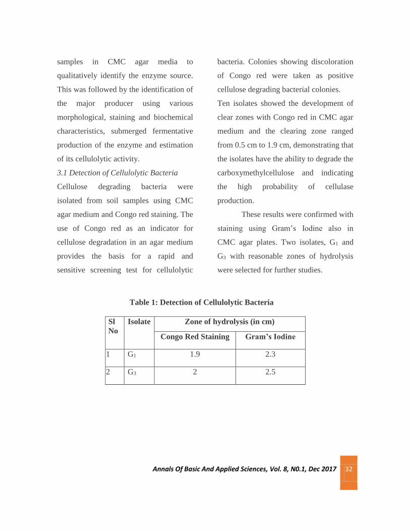
3.1 Detection of Cellulolytic Bacteria
Cellulose degrading bacteria were
isolated from soil samples using CMC
agar medium and Congo red staining. The
use of Congo red as an indicator for
cellulose degradation in an agar medium
provides the basis for a rapid and
sensitive screening test for cellulolytic
bacteria. Colonies showing discoloration
of Congo red were taken as positive
cellulose degrading bacterial colonies.
Ten isolates showed the development of
clear zones with Congo red in CMC agar
medium and the clearing zone ranged
from 0.5 cm to 1.9 cm, demonstrating that
the isolates have the ability to degrade the
carboxymethylcellulose and indicating
the high probability of cellulase
production.
These results were confirmed with
staining using Gram’s Iodine also in
CMC agar plates. Two isolates, G1 and
G3 with reasonable zones of hydrolysis
were selected for further studies.
Table 1: Detection of Cellulolytic Bacteria
Sl
No
Isolate Zone of hydrolysis (in cm)
Congo Red Staining Gram’s Iodine
1 G1 1.9 2.3
2 G3 2 2.5

Annals Of Basic And Applied Sciences, Vol. 8, N0.1, Dec 2017 33
Congo Red Method Gram’s Iodine Method
Figure 3: Detection of Cellulolytic Bacteria
3.2 Identification of CDB isolates
The results of staining and biochemical
examinations are summarized in Table 2.
From the observations G1 is presumably
identified as Microbacter and G3 is to be
further characterized.
Table 2: Presumptive Microscopic and biochemical identification of G1 and G3
Sl No Test G1 G3
1 Gram Staining Gram +ve rods Gram –ve cocci
2 Motility Motile Non motile
3 Catalase Negative Positive
4 Oxidase Negative Negative
5 Sugar Fermentation
a) Maltose
b) Lactose
c) Glucose
d) Sucrose
e) Mannitol
Acid
Acid
Acid
No change
Acid
Acid
Acid
Acid
No change
No change
6 IMViC
a) Indole Production
b) Methyl Red Test
c) Voges Proskaeur
d) Citrate Utilization
-
+
-
-
-
+
-
-

Annals Of Basic And Applied Sciences, Vol. 8, N0.1, Dec 2017 34
3.3 Fermentative Production of
Cellulase enzyme
The isolates G1 and G3 from soil which
gave a reasonable zone of hydrolysis in
the CMC agar plates were inoculated into
100ml production medium for a period of
72h with routine sampling done at 24 h
interval. The culture filtrates were
subjected to enzyme activity
determination which was calculated
against a glucose standard curve.
Cellulase system consist of 1,4-β-
endoglucanase, 1,4-β-exoglucanase, and
β-glucosidase which synergistically do
complete hydrolysis of cellulose to
glucose. DNS reagent was used to stop
the enzymatic reaction, and the reaction
product measured as a result of the
reaction between glucose and the DNS
reagent. One unit of enzyme activity was
defined as the amount of enzyme required
to release the 1µmole of glucose/min
under standard conditions.
Table 3 Cellulase activity during submerged fermentation
The activity was found to increase with
time and the maximum was recorded at
72 h. Both G1 and G3 showed similar kind
of enzyme activity with a yield of around
3 Units at the end of 72 h. Further
investigations are required to record the
enzymatic activity as a function of time
and to optimize the conditions for
maximization the enzyme production.
4. Conclusions
Microbial cellulases are an important
group of enzymes that can have
application in various industries such as
food processing, laundry industry, leather
processing, bioremediation process and in
textile industry. Cellulases are widespread
in nature; microbes serve as a preferred
source of these enzymes because of their
Time ( h) Cellulase activity
(U ml-1)
48 1.4 3.13
72 1.62 3.6

Annals Of Basic And Applied Sciences, Vol. 8, N0.1, Dec 2017 35
extreme habitat variability, rapid growth,
the limited space required for their
cultivation and the ease with which they
can be genetically manipulated to
generate new enzymes with altered
properties that are desirable for their
various applications. The future may
hold great prospects for lignocellulosic
biofuel; by combining our knowledge of
excellent cellulolytic and hemi
cellulolytic with technologies such as
directed evolution and co-culture, the
future of lignocellulolytic biofuel looks
potentially feasible.
Soil is an important source for isolation
of microorganisms for novel industrial
enzymes production. Hence, in this
present study also the cellulase producing
bacteria were attempted to be isolated
from soil. Two potential isolates
designated as G1 and G3 were found to be
cellulolytic in CMC agar media and were
tentatively identified by some common
biochemical tests and microscopical
observations. They were further subjected
to submerged fermentation for the
production of cellulase enzyme and its
activity was assayed. They were found to
produce around 3 U enzyme at 72 h.
Further studies like determining the time
course of enzymatic activity and
validating other parameters for
maximization the enzyme production
followed by its purification,
characterization, study of optimal
conditions of activity and its stability
followed by applications are to be
undertaken.
References
Apun K, Jong B C, and Salleh M A
(2000). “Screening and isolation of a
cellulolytic and amylolytic Bacillus
from sago pith waste,” Journal of General
and Applied Microbiology, (46), 263–
267.
Ekperigin M M (2007). “Preliminary
studies of cellulase production by
Acinetobacter anitratus and Branhamella
sp,” African Journal of Biotechnology,
(6), 28–33.
Kasana R C, Salwan R, Dhar H, Dutt S,
Gulati A (2008). “A rapid and easy
method for the detection of microbial
cellulases on agar plates using Gram’s
Annals Of Basic And Applied Sciences, Vol. 8, N0.1, Dec 2017 36
iodine” Current Microbiology, (55), 503–
507.
Lu W J, Wang H T, Nie Y F (2004).
“Effect of inoculating flower stalks and
vegetable waste with ligno-cellulolytic
microorganisms on the composting
process,” Journal of Environmental
Science and Health-B, (39), 871–887.
Miller G L (1959). “Use of
dinitrosalicylic acid reagent for
determination of reducing
sugar,”Analytical Chemistry,(31), 426–
428.
Mswaka A Y and Magan N (1998).
“Wood degradation, and cellulase and
ligninase production, by Trametes
and other wood-inhabiting
basidiomycetes from indigenous forests
of Zimbabwe,” Mycological Research,
(102), 1399-1404.
Nutt A, Sild V, Prtterson G and
Johansson G (1998). “Progress curve as a
means for functional classification of
cellulases,” Europian Journal of
Biochemistry, (258), 200.
Vaithanomsat P, Chuichulcherm S and
Apiwatanapi W (2009). “Bioethanol
production from enzymatically
saccharified sunflower stalks using steam
explosion as pretreatment,” Proceedings
of World Academy of Science,
Engineering and Technology, (37), 140–
143.

Annals Of Basic And Applied Sciences, Vol.8, No.1. Dec 2017 37
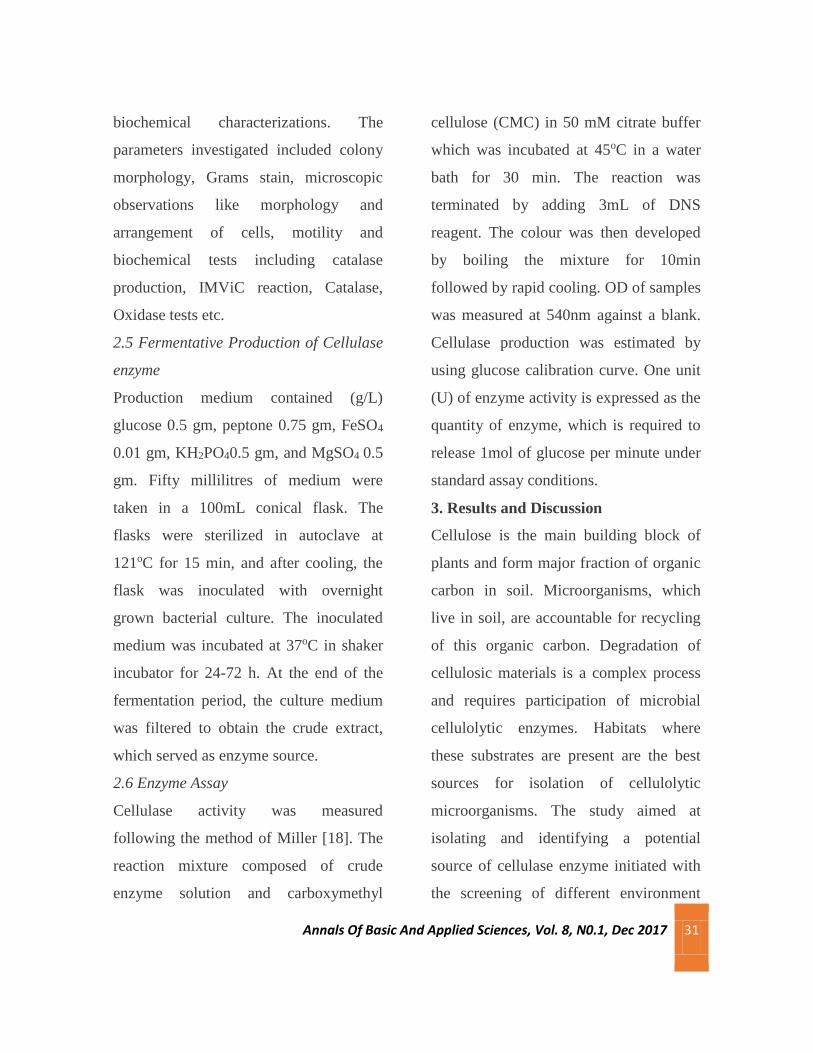
Synthesis of Two Novel Nnn-Donor Schiff Bases And Their
Characterization
Manju Sebastian*
Assistant Professor, Department of Chemistry, St. Mary’s College, Thrissur-20, Kerala, India
*Corresponding author- [email protected]: Ph-919495457933
Abstract
Two new Schiff bases, Quinoxaline-2-carboxalidine-2-aminopyrimidine and Quinoxaline-2-carboxalidine-3-
aminopyridine, have been synthesized by condensation from quinoxaline-2-carboxaldehyde and 2-aminopyrimidine / 3-
aminopyridine. The Schiff bases are characterized by elemental analysis, FT-IR, UV-Vis, TG, and NMR spectroscopy.
Spectroscopic characterization clearly reveals the tridentate NNN-donor behaviour of the Schiff base.
Keywords: Schiff base, quinoxaline-2-carboxaldehyde, 2-aminopyrimidine, 3-aminopyridine
1. Introduction
Multidentate ligands are widely used in
coordination chemistry, since they found
useful in the assembly of new
frameworks with attractive properties.
Among these ligands the transition metal
complexes containing nitrogen donor
atoms are of considerable interest in
inorganic and biomimetic chemistry due
to their possible application in catalysis,
medicine and material science (Gupta and
Sutar, 2008). These ligands are of interest
because of their ability to form transition
metal complexes which have varying
configurations, structural lability and
sensitivity to molecular environments
(Cozzi, 2004). They have proven to be
effective in constructing supramolecular
architectures such as coordination
polymers and helical assemblies.
Quinoxalines with other heterocyclic
amines groups are of particular interest

Annals Of Basic And Applied Sciences, Vol.8, No.1. Dec 2017 38
due to their wide applications (Sharma
and Varshney, 1991) (Cheeseman, 1982).
They can form complexes either in
bidentate NN form or in tridentate NNN
form. The synthesis and characterization
of Schiff bases derived from quinoxaline-
2-carboxaldehyde with various amines
and their complexes were previously
reported (Arun et al, 2009)(Sebastian et
al, 2010). The objective of current work is
to prepare new tridentate NNN donor
Schiff base from quinoxaline-2-
carboxaldehyde and 2-aminopyrimidine /
3-aminopyridine.
The Schiff base is synthesized by
condensation of aldehyde and amine. The
Schiff base is an NNN donor and it can
act as a monobasic or dibasic tridentate
ligand due to the possibility of different
coordinating nature of metal ion.
2. Materials and methods
2.1 Materials and physical measurements
2-aminopyrimidine (Aldrich) and 3-
aminopyridine (Aldrich) are used as
supplied. The preparation of
quinoxaline2carboxaldehyde is carried
out as reported in the literature (Mayadevi
et al, 2003). Elemental analyses of the
ligands were done on a Vario EL III
CHNS analyzer. The IR spectra were
recorded on a JASCO FTIR4100
spectrometer using KBr pellets in the
range 4004000 cm-1. The electronic
spectra of the ligands were recorded on a
Thermoelectron Nicolet evolution 300
UV-Vis spectrophotometer.
2.2 Synthesis of Schiff base Quinoxaline-
2-carboxalidine-2-aminopyrimidine
(Abbreviated as L1)
Take 1mmol of quinoxaldehyde
(1.5816g) in 50ml acetonitrile. To this
add 1mmol of 2-aminopyrimidine
(0.951g) in 30ml acetonitrile. The mixture
is refluxed in a 100ml R.B. for 6 hours.
Reduce the volume, cool to room
temperature. The crystals formed are
filtered out washed with water to remove
the traces of unreacted amine, followed
by petroleum ether. An orange colored
powder is obtained finally (Figure 1).
Yield: ~65%.
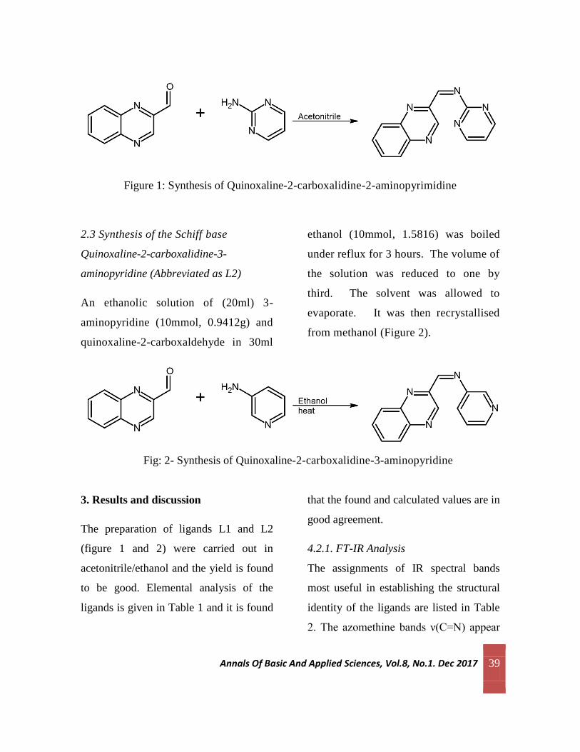
Annals Of Basic And Applied Sciences, Vol.8, No.1. Dec 2017 39
Figure 1: Synthesis of Quinoxaline-2-carboxalidine-2-aminopyrimidine
2.3 Synthesis of the Schiff base
Quinoxaline-2-carboxalidine-3-
aminopyridine (Abbreviated as L2)
An ethanolic solution of (20ml) 3-
aminopyridine (10mmol, 0.9412g) and
quinoxaline-2-carboxaldehyde in 30ml
ethanol (10mmol, 1.5816) was boiled
under reflux for 3 hours. The volume of
the solution was reduced to one by
third. The solvent was allowed to
evaporate. It was then recrystallised
from methanol (Figure 2).
Fig: 2- Synthesis of Quinoxaline-2-carboxalidine-3-aminopyridine
3. Results and discussion
The preparation of ligands L1 and L2
(figure 1 and 2) were carried out in
acetonitrile/ethanol and the yield is found
to be good. Elemental analysis of the
ligands is given in Table 1 and it is found
that the found and calculated values are in
good agreement.
4.2.1. FT-IR Analysis
The assignments of IR spectral bands
most useful in establishing the structural
identity of the ligands are listed in Table
2. The azomethine bands ν(C=N) appear

Annals Of Basic And Applied Sciences, Vol.8, No.1. Dec 2017 40
at 1600 - 1700 cm-1, is in agreement with
earlier reports. The quinoxaline ring C=N
stretching was found near 1580 cm-1. In
ligand L2, the in-plane and out-of-plane
deformation vibrations characteristic of
pyridyl ring are observed respectively in
the range 613-622 and 401-421cm-1
(Bermejo, 1999). Pyrimidines are
characterized with strong absorption in
the range 1600-1500 cm-1 due to the C=C
and C=N ring stretching vibrations.
Usually 2-substituted pyrimidines have
three medium to strong absorption bands
in the range 650-630, 580-475 and 515-
440 cm-1 (George, 2001). The infrared
spectrum of compound L1 contains three
bands in the above said range indicates
pyrimidyl moiety in the Schiff base.
Table 1: Analytical data of L1 and L2
Compound Stoichiometry Colour Yield
(%)
Analytical data. Found
(calculated)%
C N H
L1 C13H9N5 Violet 86 65.83
(66.37)
29.25
(29.77)
4.36
(3.85)
L2 C14H10N4 Yellow 81 71.55
(71.78)
23.21
(23.92)
4.36
(4.30)
4.2.1. FT-IR Analysis
The assignments of IR spectral bands
most useful in establishing the structural
identity of the ligands are listed in Table
2. The azomethine bands ν(C=N) appear
at 1600 - 1700 cm-1, is in agreement with
earlier reports. The quinoxaline ring C=N
stretching was found near 1580 cm-1. In
ligand L2, the in-plane and out-of-plane

Annals Of Basic And Applied Sciences, Vol.8, No.1. Dec 2017 41
deformation vibrations characteristic of
pyridyl ring are observed respectively in
the range 613-622 and 401-421cm-1
(Bermejo, 1999). Pyrimidines are
characterized with strong absorption in
the range 1600-1500 cm-1 due to the C=C
and C=N ring stretching vibrations.
Usually 2-substituted pyrimidines have
three medium to strong absorption bands
in the range 650-630, 580-475 and 515-
440 cm-1 (George, 2001). The infrared
spectrum of compound L1 contains three
bands in the above said range indicates
pyrimidyl moiety in the Schiff base.
Table 2: IR spectral data of L1 and L2
Assignments ν
(C=N) #
ν
(C=N)
*
ν (py)ip ν (py)op ν (pm)1 ν (pm)2 ν (pm)3
L1 1698 1587 - - 621 477 441
L2 1617 1574 624 403 - - -
# azomethine, * quinoxaline, py-pyridyl, pm-pyrimidyl
4.2.2. UV-Vis Spectral Analysis
The tentative assignments of the
significant electronic spectral bands of
ligands are presented in Table 3. The
electronic spectra of L1 and L2 in DMF
solution show the following intraligand
absorption maxima: Two bands
corresponding to π— π* transitions of the
pyridyl ring, pyrimidyl ring, benzene ring
and imine function of the schiff bases are
observed in the range 41840-43290 cm-1
and 33560- 38760 cm-1; the n— π*
transitions of the pyridyl ring and imine
function are observed in the range 29500-
30960 cm-1 (Neelakandan et al, 2008).
Table 3: Electronic spectral data
(in DMF)
Compound Absorption
maxima
Tentative
assignments

Annals Of Basic And Applied Sciences, Vol.8, No.1. Dec 2017 42
L1 42716,
44562
π— π*
L1 29530 n— π*
L2 42760,
43190
π— π*
32511 n— π*
4.2.3. NMR Spectral Analysis
The 1H NMR spectrum of L1 and L2
shows signals at 8.08 and 8.99 ppm
corresponding to azomethine CH protons
respectively. Aromatic protons appear as
a multiplet at 6.74-8.26 ppm. The detailed
result of NMR spectral analysis is listed
in table 4. The assignments are in
agreement with the values already
reported.
Table 4: 1H NMR Spectroscopic data
of L1 and L2
4. Conclusions
Synthesis and characterization of two
novel Schiff bases Quinoxaline-2-
carboxalidine-3-aminopyridine and
Quinoxaline-2-carboxalidine-2-
aminopyrimidine have been carried out.
Schiff bases are prepared by condensation
between aldehyde and primary amine.
Aldehyde selected for current work is
quinoxaline-2-carboxaldehyde which is a
heterocyclic aldehyde. The primary
amines used were 2-aminopyrimidine and
3-aminopyridine. Ligands are
characterized by elemental analysis,
infrared, UV-Vis and H1 NMR spectral
studies. Elemental analysis of ligands
shows that the found and calculated
values are in good agreement. An infrared
spectrum of Schiff bases gives a medium
band in the range 1600-1650 cm-1
characterized azomethine stretching of
Schiff base. Spectroscopic
characterizations clearly revealed the
Compound Chemical
shift, δ
(ppm)
Assignment
L1 8.78
6.74-8.22
(s, 1H, CH
azomethine)
(m, 8H, Ar)
L2 8.99
7.06-8.26
(s, 2H, CH
azomethine)
(m, 14H, Ar)
Annals Of Basic And Applied Sciences, Vol.8, No.1. Dec 2017 43
bidentate NN or tridentate NNN-donor
behaviour of the Schiff base.
References
Arun V, Robinson PP, Manju S, Leeju P,
Varsha G, Digna V, Yusuff KKM (2009)
A novel fluorescent bisazomethine dye
derived from 3-hydroxyquinoxaline-2-
carboxaldehyde and 2,3-
diaminomaleonitrile. Dyes and Pigments.
82: 268–275.
Bermejo E, Casteneiras A, Dominguez R,
Carballo R, Mossmer CM, Strahle J,
West DX (1999) Preparation, Structural
Characterization, and Antifungal
Activities of Complexes of Group 12
Metals with 2-Acetylpyridine- and 2-
Acetylpyridine-N-oxide-4N-
phenylthiosemicarbazones. Z. Anorg.
Allg.Chem. 625: 961-968.
Cheeseman GWH (1982) Recent
advances in quinoxaline chemistry in
Advances in heterocyclic chemistry,
Katritzky, A.R. (ed.), Vol. 2, Academic
press, New York.
Cozzi PG (2004). Metal–Salen Schiff
base complexes in catalysis: practical
aspects. Chem. Soc. Rev., 33 410-421
George S. (2001). Infrared and Raman
Characteristic Group Frequencies: Tables
and Charts. Wiley UK.
Gupta KC, Sutar AK (2008). catalytic
activities of Schiff base transition metal
complexes. Coord. Chem. Rev. 252:
1420–1450.
Mayadevi S, Prasad PG, Yusuff KKM
(2003). Studies on Some Transition Metal
Complexes of Schiff Bases Derived from
Quinoxaline-2-carboxaldehyde. Synth.
React. Inorg. Met.-Org. Chem. 33(3):
481-496
Neelakantan MA, Marriappan SS,
Dharmaraja J, Jeyakumar T,
Muthukumaran K (2008) Spectral, XRD,
SEM and biological activities of
transition metal complexes of polydentate
ligands containing thiazole moiety.
Spectrochim. Acta A, 71: 628.
Sebastian M, Arun V, Robinson PP,
Leeju P, Varghese D, Varsha G, Yusuff
Annals Of Basic And Applied Sciences, Vol.8, No.1. Dec 2017 44
KKM (2010) Synthesis, characterization,
and structure of a new cobalt(II) Schiff-
base complex with quinoxaline- 2-
carboxalidine-2-amino-5-methylphenol.
J. Coord. Chem. 63(2): 307-314.
Sharma RC, Varshney VK. (1991).
Antimicrobial and cytotoxic activity of
transition metal chelates with some
heterocyclic imines. J.Inorg. Biochem.
41:299-304.

Annals Of Basic And Applied Sciences, Vol. 8, No.1, Dec 2017 45
Antioxidant Potential of Ethanolic and Hot Water Extracts of
Pleurotus Sajor-Caju
Meera C R*, Anjali Krishna, Anjali Variyar, Anju V T, Asha P Antony & Asna Ashraf Department of Microbiology, St Mary’s College, Thrissur-680020, Kerala, India.
*Corresponding author- Email: [email protected] Ph: 9495369274
Abstract
Pleurotus species are commonly called oyster or paddy straw mushrooms. The genus Pleurotus comprises some most popular
edible mushrooms due to their favorable organoleptic and medicinal properties, vigorous growth and undemanding cultivation
techniques. In the present investigation, Pleurotus sajor‐caju fruiting bodies were cultivated on coir pith, ethanolic and hot water
extracts were prepared from it. The yield of the mushroom was found to be 24.33%. The yield of the ethanolic and hot
waterextract was found to be 10.1 and 7% respectively. The DPPH free radical scavenging activity of the ethanolic and hot water
extracts of the fruiting body was studied. Both extracts showed significant activity in a dose dependent manner. At the
maximum tested dose, ethanolic extract showed 90.11% activity and the hot water extract showed 90.87% activity. IC50 value of
ethanolic and hot water extracts were found to be 29 µg and 30 µg, respectively. Present study revealed the significant antioxidant
activity of the ethanolic as well as hot water extract from the fruiting body of the edible mushroom Pleurotus sajor‐caju. The
cultivation of the mushroom and isolation of active principles from the mushroom is simple, economical and can be carried out
with minimal effort. In this context, the active principles from Pleurotus sajor‐caju can be a suitable candidate as antioxidant
agent.
Key words: Mushrooms, antioxidant, Pleurotus sojor‐caju, free radicals
1. Introduction
The use of mushrooms as a food item is
probably as old as civilization.
Mushrooms contain many biologically
active compounds with high utility values
such as polysaccharides, oligosaccharides,
lipids, terpenoids and steroids in the
fruiting bodies and/or mycelium. The
longest tradition of using edible
mushrooms for medicinal purposes is in
China. Mushrooms represent the major as
yet largely untapped source of powerful
new pharmaceutical products. Of
approximately 10,000 species of
mushrooms, 2000 are safe and 300 are of
Annals Of Basic And Applied Sciences, Vol. 8, No.1, Dec 2017 46
pharmacological significance
(Hobbs,1995; Miles and Chang,1997).
Mushrooms have been reported to have
significant pharmacological effects and
physiological properties such as bio
regulation, maintenance of homeostasis,
regulation if biorhythm, cure if several
diseases like cancer, cerebral stroke, and
heart diseases. They have also
demonstrated to have effective substances
to decrease blood cholesterol,
hypolipidemic, antithrombotic,
hypotensive, and other applications
(Wasser, 1999).
The protein bound polysaccharides
isolated from the mushrooms have been
used as an immunmodulatory agent in the
treatment of cancer in Asia over 30 years.
A large number of mushroom-derived
compounds, both cellular components and
secondary metabolites, have been shown
to affect the immune system and could be
used to treat a variety of diseased states.
(Wasser, 1999). Unique anti-cancer
preparations were developed from
mushrooms such as Lentinan from
Lentinus edodes, Krestin from Trametes
versicolor, and Schizophyllan from
Schizophyllum commune (Mizuno, 1996
and 1999). Polysachharides from
mushrooms do not attack cancer cells
directly but produce their anti-tumour
effects by activating different immune
responses in the host.
Pleurotus species are commonly called
oyster or paddy straw mushrooms. The
genus Pleurotus comprises some most
popular edible mushrooms due to their
favorable organoleptic and medicinal
properties, vigorous growth and
undemanding cultivation techniques. It
can be cultivated on log and a wide variety
of agroforestry by-products, weeds, and
wastes for the production of food, feed,
enzymes and medicinal compounds, or for
waste degradation and detoxification.
Many different techniques and substrates
have been successfully utilized for
mushroom cultivation and biomass
production by means of solid state and
submerged liquid fermentation. Pleurotus
species are now widely consumed as food
in East and increasingly in the West
(Breene, 1990; Chang, 1993).
A number of polysaccharides or
exopolysaccharides (EPS) from cell wall,
fruiting body and culture filtrate of
Pleurotus sajor-caju have been reported
with some potential pharmaceutical
properties. Among these polymers, homo

Annals Of Basic And Applied Sciences, Vol. 8, No.1, Dec 2017 47
and heteroglucans, with β-(1→3), β-
(1→4) and β-(1→6) glycosidic linkages,
are supposed to play a key role in some
health aspects of mushrooms (Manzi &
Pizzoferrato, 2000).
In that two decades these have been an
explosive interest in the role of oxygen
free radicals, more generally known as
‘Reactive Oxygen Species’ (ROS) and ‘
Reactive Nitrogen Species’(RNS).A free
radical is an atom or molecule that has a
single unpaired electron in its outer shell.
They are highly reactive compounds that
are produced in the body during normal
metabolic functions or introduced from
environment. They are inherently
unstable, since they contain extra energy.
To reduce their energy load, free radicals
react with the certain chemicals in the
body and in the process interfere with the
normal functions of cells. The oxidants or
free radicals have very short life and
damaging activity towards
macromolecules like proteins, DNA and
lipids.
Antioxidants are a group of substances
which, when present at low
concentrations, in relation to oxidizable
substrates, significantly inhibit or delay
oxidative processes, while often being
oxidized themselves. In recent years, there
has been an increased interest in the
application of antioxidant to medical
treatment as information is constantly
gathered linking the development of
human diseases to oxidative stress. The
effect of reactive oxygen species and
nitrogen species is balanced by
antioxidants as well as by antioxidant
enzymes. Such antioxidant defenses are
extremely important as they represent the
direct removal of free radicals
(prooxidants).Enzymatic antioxidants
involve super oxide dismutase(SOD),
catalase (CAT), and glutathione
peroxidase(Gpx) (Maron et al., 1979). Non
enzymatic antioxidants involve vitamin C,
carotenoids thiol antioxidants
(glutathione, thioredoxin, and lipoic
acid);natural flavonoids; melatonin, a
hormonal product of pineal gland; and
other compounds (Mc Call & Frei, 1999).
Reducing exposure to free radicals and
increasing intake of antioxidant nutrient
have the potential to reduce the risk of free
radicals related health problems. Over the
past several years , nutritional antioxidants
have attracted considerable interest as
potential treatment for a wide variety of
disease states, including cancer,
Annals Of Basic And Applied Sciences, Vol. 8, No.1, Dec 2017 48
atherosclerosis, chronic diseases and
aging .Studies have shown that dietary
supplementation with extra antioxidants
can reduce the risk of developing cancer.
In the present investigation, Pleurotus
sajor-caju fruiting bodies were cultivated
on coir pith, ethanolic and hot water
extracts were prepared from it. An attempt
was made to evaluate the anti-oxidant
properties of the extracts prepared from
P.sajor-caju.
2. Materials and Methods
2.1. Pleurotus sajor-caju : The spawn
culture of P. sajor-caju was obtained
from Kerala Agricultural University,
Thrissur ,Kerala, India.
2.2. Cultivation of Pleurotus sajor-caju on
coir pith
Spawn culture of Pleurotus sajor-caju
was obtained from Kerala Agricultural
University, Thrissur, Kerala , India. Coir
pith was soaked overnight in water. It was
drained until approximately 60% of water
content retained and sterilized. Sterilized
coir pith was transferred to long sized well
aerated polythene bags. It was then layered
with the P. sajor-caju spawn culture.
Again a layer of sterilized coir pith was
placed following a layer of spawn culture.
This was repeated till the mouth part of
polythene bag. Mouth of the bag was tied
and kept in a raised platform in a well-
ventilated cropping room for about 15
days. At this time when mycelia became
visible inside the polythene bag over the
surface of coir pith , the bags were cut and
gently removed. Then the coir pith formed
a compact mass which was watered daily
in order to maintain the moisture. Within 3
days, first flash of the fruiting body
became apparent and it was harvested
when young. Subsequent harvesting was
done as the fruiting bodies are mature.
2.3. Preparation of Ethanolic extract of
Pleurotus sajor-caju
The fruiting body of the mushroom was
washed thoroughly and cut into small
pieces. It was then dried at 40-50oC in hot
air oven. The powdered material was
defatted with petroleum ether using
soxhlet apparatus for 8-10 hours. Then, the
powdered material was subjected to 70%
ethanolic extraction at 50-70o C for 9-10
hours using Soxhlet apparatus. The
extracts were pooled together and
evaporated to dryness.
Annals Of Basic And Applied Sciences, Vol. 8, No.1, Dec 2017 49
2.4. Preparation of Hot water extract of
Pleurotus sajor-caju
The fruiting body of the mushroom was
washed thoroughly and cut into small
pieces. It was then dried at 40-50oC in hot
air oven. The powdered material was
added to a little water in a beaker. The
beaker was placed in a boiling water bath
for 8-9 hours. The mixture was then
filtered and extract obtained was
evaporated to dryness.
2.5. DPPH free radical scavenging
activity (Antioxidant activity)
Free radical scavenging activity of the
extracts were studied using the method of
Aquino et al., 2001. DPPH in its radical
form has absorption at 515nm , which
disappeared on reduction by an
antioxidant compound. Various
concentrations of drug (50-2000µg)
combined with 1ml of freshly prepared
DPPH reagent solution. The samples were
kept for 20 minutes in dark and the
decrease in absorbance was measured at
515 nm. The percentage inhibition
activity was measured using the formula;
% inhibition = C-T/C×100; where C was
the absorbance of control and T was the
absorbance of treated/ standard. IC50 value
was calculated by plotting concentration
of the drug against percent activity.
3. Results
3.1. Cultivation of P. sajor-caju on coir
pith
After 14 days of incubation, there was
uniform growth of fruiting body along the
sides of the coir pith (Figure 1). 608.30
grams of fruiting body were harvested
from 2500 grams of coir pith. The yield of
the mushroom was found to be 24.33%.
3.2. Preparation of Ethanolic extract from
the fruiting body of P. sajor-caju
Extraction was done thrice, in batches for
9-10 hours. The extracts were pooled
together and evaporated to dryness. Dark
brown colored powder was obtained
which was maintained to 4°C, till use. The
yield of the extract was found to be 10.1%.
3.3. Preparation of hot water extract from
the fruiting body of P. sajor-caju
Extraction was done thrice, in batches for
8-9 hours in boiling water bath and
filtered, evaporated to dryness. Dark
brown colored filtrate was obtained. The
yield of the extract was found to be 7%.
3.4. DPPH free radical scavenging
activity
The DPPH free radical scavenging activity
of the ethanolic and hot water extracts of

Annals Of Basic And Applied Sciences, Vol. 8, No.1, Dec 2017 50
the fruiting body was studied. Both
extracts showed significant activity in a
dose dependent manner (Figure 2 and 3).
The ethanolic extract showed 90.11%
activity at the maximum tested dose of
2000µg. The hot water extract of P. sajor-
caju showed 90.87% activity at the
maximum tested dose of 2000µg. IC50
value of ethanolic and hot water extracts
were found to be 29 µg and 30 µg,
respectively.
Figure 1: Antioxidant activity of ethanolic extract of P. sajor-caju by DPPH free radical scavenging assay.
Figure 2: Antioxidant activity of hot water extract of P. sajor-caju by DPPH free radical scavenging assay.
78
80
82
84
86
88
90
92
50 100 500 1000 1500 2000
84
85
86
87
88
89
90
91
92
50 100 500 1000 1500 2000

Annals Of Basic And Applied Sciences, Vol. 8, No.1, Dec 2017 51
Figure 1: Stages of Mushroom Cultivation
4. Discussion
Mushrooms have a great potential for the
production of useful bioactive metabolites
and they are a prolific resource for drugs.
The spectrum of pharmacological activities
of mushrooms is fascinating. Discovery and
evaluation of metabolites from the various
medicinal mushrooms as new safe
compounds for treatment of various
diseases has become a hot research spot.
Different mushroom species possess
different constituents and in different
concentration, which account for the
different biological activities. Mushrooms
usually contain a wide variety of free
radical scavenging molecules, such as

Annals Of Basic And Applied Sciences, Vol. 8, No.1, Dec 2017 52
polysaccharides, proteins and polyphenols.
The mechanism underlying the free radical
scavenging activity exerted by these
molecules is still not fully understood.
Tsiapali etal.,(2001) speculated that the
abstraction of anomeric hydrogen from
monosaccharide was the reason for the free
radical scavenging ability of mushroom
polysaccharides. Polysaccharides enhanced
antioxidant activity over monosaccharides
because polysaccharides exhibited greater
ease of abstraction of the hydrogen from
one of the internal monosaccharide units.
In the present investigation, Pleurotus sajor
–caju was cultivated on the agro waste coir
pith. Fruiting bodies were produced
approximately 14 days after the bagging.
Antioxidant properties of the ethanolic as
well as hot water extract prepared from this
mushroom were studied. Present study
revealed profound antioxidant activity of
ethanolic and hot water extracts of
Pleurotus sajor –caju by DPPH free radical
scavenging activity. Both drugs showed
significant activity in a dose dependent
manner. Presence of the active principles
like polysaccharides and proteins in these
extracts may be responsible for the anti-
oxidant properties. However, this needs
further confirmation by adequate
experimental evidences.
5. Conclusion
Present study revealed the significant
antioxidant activity of the ethanolic as well
as hot water extract from the fruiting body
of the edible mushroom Pleurotus sajor-
caju. The cultivation of the mushroom and
isolation of active principles from the
mushroom is simple, economical and can be
carried out with minimal effort. In this
context, the active principles from
Pleurotus sajor-caju can be a suitable
candidate as antioxidant agent. However,
further research is necessary to elucidate the
structure and mechanism of action of these
active components.
6. References
Aquino R, Morelli S, Lauro M R, Abdo S,
Saija A and Tomaino A (2001). Phenolic
constituents and antioxidant activity of an
extract of Anthurium versicolor leaves.
Journal of Natural products. 64: 1019-
1023.
Breene W M (1990). Nutritional and
medicinal values of speciality mushrooms.
Journal of food protection. 53:883-894.
Chang S T (1993).The impact of mushroom
production and mushroom
products.Keynote lecture first Internatinal
Conference on mushroom biology and
Annals Of Basic And Applied Sciences, Vol. 8, No.1, Dec 2017 53
mushroom products. Mushroom Biology.
23-26.
Hobbs C (1995). Medicinal mushrooms-An
Exploration of Tradition, Healing and
Culture, Botanica Press, Santa Cruz, CA-
25.
Manzi, Pizzoferrato (2000). Beta-glucans in
edible mushrooms. Food Chemistry. 68:
315–318.
McCall M R, B Frei (1999).Can antioxidant
vitamins materially reduce oxidative
damage in human?. Free Radical Biology &
Medicine. 26:1034-1053.
Miles, Chang S T (1997). Mushroom
biology concise basics and current
developments. World Scientific, Singapore.
194-197.
Mizuno T (1999). The extraction and
development of antitumor polysaccharides
in Japan. International Journal of
Medicinal Mushrooms. 1: 9-29.
Mizuno T (1996). Development of
antitumor polysaccharides from mushroom
fungi. Food . Integrated.J (Japan). 167:69-
85.
Moron M A, Deiprre W, Mannervick B
(1979). Levels of glutathione reductase and
glutathione S-transferase activities in the rat
lung and liver. Biochemical Biophysica
Acta. 582:67-78.
Tsiapali E, Whaley S, Kalbfleisch J, Ensley
HE, Browder IW, Williams DL (2001).
Glucans exhibit weak antioxidant activity,
but stimulate macrophage free radical
activity. Free Radic. Biol. Med. 30: 393–
402.
Wasser S.P and Weis A.L (1999).
Medicinal properties of susbtances
occurring in higher Basidiomycetes, current
perspectives. International Journal of
Medicinal Mushrooms. 1: 47-50.

Annals Of Basic And Applied Sciences, Vol. 8, No.1, Dec 2017 54
A comparison on probiotic characteristics of beta- galactosidase producers from curd and raw milk: A review
Neenu Neelakantan
Dept.of Microbiology, St. Mary’s College – Thrissur- 680020-Thrissur, Kerala
Corresponding author- Email: [email protected] Ph:9605833184.
Abstract
Probiotics are defined as microbial food supplements which beneficially affect the host animal by improving its
intestinal microbial balance. The aim of probiotic approach is to repair the deficiencies in the micro flora and restore
the animal's resistance to disease. Lactose intolerance or inability to hydrolyze lactose is a problem prevalent in more
than half of world's population. Intolerance is mainly occurs due to the absence of galactosidase (rather the presence
of a nonfunctional galactosidase) an enzyme associated with the metabolism of lactose. The deficiency of functional
galactosidase in our body can be cured by the use of its probiotic producers in the form of easily available dietary
sources. Microorganisms are the important sources of many such enzymes, therefore efforts are now increasing to
produce these enzymes by microbial fermentation. Selection and isolation of the right organism from a right source
influence high yields of desirable enzymes. Major such dietary sources of beta‐gal are raw milk and curd. Among
these two sources, which one can act as the most beneficial and promising source of beta ‐ gal is to be compared and
confirmed for better commercial production the enzyme. This review focuses on the comparison of these two
different sources, yielding different probiotic producers of a single enzyme.
Key words: Beta‐galactosidase, Lactose intolerance, galacto – oligosaccharides, Ortho Nitro Phenyl beta‐D‐
Galactoside
1. Introduction
Beta-galactosidase (beta-gal)
catalyzes the hydrolysis of Beta-
galactosides (carbohydrates
containing galactose) into
monosaccharides through the
breaking of a glycosidic bond.
Substrates of this enzyme include
lactose, lactosylceramides, GM 1
and various glycoproteins (1). This
enzyme is also well known as
'lactase’. The lactose hydrolyzation
converts lactose in to galactose and

Annals Of Basic And Applied Sciences, Vol. 8, No.1, Dec 2017 55
glucose which will proceed in to
glycolysis. Substrate analogue
galactosides such as PETG (poly
ethylene terepthalate glycol) and
IPTG (Isopropyl Beta -D-1-Thio
Galactopyranoside ) will bind in the
shallow site (non-productive
binding) when the enzyme is in open
confirmation (2).
1.1 Microbial Sources of Beta-
galactosidase
The advantages of microbial Sources
over the other include easy handling,
higher multiplication rate and high
yield (3). Among the microbial
sources, bacterial sources are
preferred due to ease of
fermentation, high enzyme activity
and good stability. When there is no
enough glucose to sustain the growth
of bacteria but sufficient lactose is
present in the medium, beta-
galactosidase synthesis get turned
on.
Bacterial sources (4)
Lactobacillus bulgaricus
Lactobacillus delbruekii
Lactobacillus casei
Escherichia coli
Bacillus subtilis
Bacillus megatherium
Fungal sources
Alternaria alternate
Aspergillus Niger
Rhizobium sp.
Yeast sources
Candida sp.
Kluveromyces sp.
Plant sources
Apricot
Coffee berries
Peach
Tips of wild roses
1.2 Lactose intolerance and Beta-
galactosidase
As lactose intolerant people do not
have ability to synthesize
galactosidase enzyme, the treatment
of milk and it's derivatives with
galactosidase is required. Therefore,
products free of lactose or low
lactose content can be consumed
without any problems by lactose

Annals Of Basic And Applied Sciences, Vol. 8, No.1, Dec 2017 56
intolerant people (5). Yogurt is
usually better tolerated than fresh
milk products by maldigesters. This
is because the galactosidase in the
live yogurt bacteria can aid lactose
digestion in the colon. Therefore,
Lactic Acid Bacteria (LAB) which
constitute a diverse group of
lactococi, streptococci and
lactobacilli have become a focus of
studies (6).Beta-gal mediated trans
galactosylation reaction gives di-tri
or higher galacto - oligosaccharides
(GOS). GSO have a positive impact
on the growth and establishment of
Bifidobacterium in human intestine
and suppress potentially harmful
bacteria like Clostridia and
Bacteroid species in the gut. Health
benefits of GSO include anti-
carcinogenic effects, reduction in
serum cholesterol, improved liver
functioning, and improved intestinal
health. Therefore, the public demand
for Beta-galactosidase production is
significantly increasing together
with the development of effective
and inexpensive GOS production
(7).
Amongst the beta-gal enzymes thus
far studied, the Escherichia coli
enzyme has been well explored
essentially due to its use as a
molecular genetics tool, following
the discovery of the lactose operon
(8). But its industrial use is limited
due to the fact that it is not
considered safe for food
applications. Therefore, selection of
microorganisms which are safe for
human use and are capable of
producing high levels of Beta-gal
becomes an urgent and attractive
task.
In this regard, Lactic Acid Bacteria
and Bifidobacterium, which are
generally recognized as safe (GRAS)
organisms, have been regarded as
good sources of Beta-galactosidase,
especially for functional food
applications (9).
In addition, microorganisms used to
produce the beta-gal have varying
nutritional requirements and thus
they produce enzymes other than
beta-gal such as proteolytic and
lipolytic enzymes which can produce
inferior organoleptic properties or

Annals Of Basic And Applied Sciences, Vol. 8, No.1, Dec 2017 57
other quality defects in milk and
milk products (10). Consequently
there's a need for new microbial
sources that are capable of producing
economic quantities of Beta-gal with
the ability to function efficiently
under various conditions.
MRS agar is the preferred medium
for screening of this enzyme
producers. X-Gal and IPTG induced
blue color colony formation along
with the microscopic, biochemical
identification procedures are the
basic criteria underlying its isolation
and identification (11). Since it is an
intracellular enzyme, studies on
efficiency of different cell disruption
methods suggests that lysozyme
treatment is the promising one
followed by a glass bead treatment
(12). Characterization studies
concludes that the isolated
Lactobacilli should survive both the
acidic (pH 2.5) and alkaline (pH 8.5)
conditions (13). Beta-gal activity is
usually determined by enzyme assay
procedure, using ONPG (Ortho Nitro
Phenyl beta-D- Galactoside) as
substrate in the place of lactose. The
advantage of using ONPG as the
substrate instead of lactose is that,
the enzyme cleaves ONPG in to
galactose and a chromogenic
substance ( ONP ) having an yellow
color, which makes the assay easily
observable one. As the enzymatic
degradation continues, more and
more ONP get released and the
solution gets more yellowish.
Optical density at 420 is the measure
of the amount of ONP present in the
sample and in turn the beta-gal
activity and this is the principle of
this enzyme assay (14).
The probiotic character study
generally includes studying growth
at different temperature, resistance to
low pH, antimicrobial activity,
resistance to antibiotics, bile salt
resistance etc.
2. Conclusion
Even though both curd and raw milk
are sources of industrially important
as well probiotic beta-gal producers,
any detailed comparison will prefer
curd for the first position due the rich
amount of probiotic bacteria having
sufficient amount of highly active
beta-gal production.

Annals Of Basic And Applied Sciences, Vol. 8, No.1, Dec 2017 58
References
Juers, D.H, Hakda, S., Mathews,B. W., & Huber, R.E.(2003). Structural basis for the altered activity of Gly794 variants of Escherichia coli beta-gal. Biochemistry, 42(46), 13505-13511.
ÜstokÜstok, F.I. (2007). Production of beta-gal using lactic acid bacteria and optimization of fermentation parameters.
Wendorff, W.L.,Amundson, C.H.,Olson,N.F.,&Graver,J.C. (1971) Use of yeast beta-gal in milk and milk products. Journal of milk and Food Technology (JMFT), 34(6), 294-299.
Panesar,P.S., Panesar, R.,Singh,R.S.,Kennedy,J.F.,&Kumar.H.(2006). Microbial production, immobilization and applications of beta-gal. Journal of Chemical Technology and Biotechnology, 81(4), 530-543.
Furlan,S.A., Schneider, A.L., Merkle,R.,de Fatima Carvalhojonas, M.,& Jonas, R.(2000). Formulation of a lactose free, low cost culture medium for the production of beta-gal by Kluveromyces marxianus. Biotechnology Letters, 22(7), 589-593.
Somkuti.G. A., & Steinberg,D. H.(1979). Beta-galactohydrolase of Streptococcus thermophilus:
induction,purification and properties. J.Appl.Biochem, 1, 357-368.
Fai, A.E.C., da Silva, J.B., de Andrade, C.J., Bution, M.L., & Pastor, G.M. (2014). Production of prebiotic galactooligosaccharides from lactose by Pseudozymatsukubaensis and pichiakluyveri. Biocatalysis and Agricultural Biotechnology, 3(4), 343-350.
Jacobson, R.H., Zhang, X.J., DuBose, R.F., & Matthews,B. W. (1994). Three-dimensional structure of beta-gal from Escherichia coli. Nature, 369(6483), 761-766.
Geciova, J.,Bury, D., & Jelen, P.(2002). Methods for disruption of microbial cells for potential use in the dairy industry - a review. International dairy journal, 12(6), 541-553.
Soares, I., Barcelos, R.P., Baroni, S., & Tàvora, Z., (2012). Microorganisms-produced enzymes in the food industry. INTECH Open Access Publisher.
Gheytanchi, E., Heshmati, F., Search, B.K., Nowroozi, J., Movahedzadeh, F. (2010). Study on beta-gal enzyme produced by isolated Lactobacilli from milk and cheese. African Journal of Microbiology Research, 4(6), 454-458.

Annals Of Basic And Applied Sciences, Vol. 8, No.1, Dec 2017 59
Fatma, Tati, C., Üstok, F.I., & Harsa, S. (2010). Production of food grade beta-gal from artisanal yogurt strains. Food Biotechnology, 24(1). 78-94.
Howie, M.Z., Akter, F., Hossain, K.M., Rahman, M.S.M., Billah, M.M., & Islam, K.M.D. (2010). Isolation, identification and analysis of probiotic properties of Lactobacillus spp. from selective
regional yoghurts. World Journal of Dairy & Food sciences, 5(1), 39-46.
Richmond, M.L., Gray, J.I., & Stine, C.M. (1981). Beta-gal: review of recent research related to technological application, nutritional concerns, and immobilization. Journal of Dairy Science, 64(9), 1759-1771.
Annals Of Basic And Applied Sciences, Vol.8, No.1. Dec 2017 60
Physiochemical evaluation, phytochemical screening and Pharmacognostic profile of leaves of Artocarpus hirsutus Lam.
in different solvent extracts
Ayana Elma Shaji and Regi Raphael K* Department of Botany, St. Mary’s College, Thrissur- 20.
*Corresponding author: Email- [email protected] Ph: 9961738410
Abstract
A.hirsutus (wild jack) endoge‐neous to Kerala has wide medicinal values which is well documented in the third volume of
Hortus Malabaricus. The present work is an attempt to standardize the leaves by pharmacognostic studies and
phytochemical analysis. The preliminary phytochemical analysis of the plant extracts was carried out to identify the
primary and secondary metabolites present in the various alcoholic and aqueous extracts using standard protocol.
Pharmacognostic study includes macroscopic, microscopic studies, physicochemical parameters and florescence analysis.
The extraction ability of different solvents for recovering extractable components followed the order: methanol >
chloroform petroleum ether > ethyl acetate >distilled water. Phyto‐screening of leaves for secondary metabolites revealed
the presence of quinone , cardiac glycoside , terpenoids ,flavanoids , phenol, coumarin , alkaloid , steroids and
phlobatannin. The macroscopic and microscopic evaluation revealed characters that are of diagnostic value and useful in
authentication of the plant. The values of total ash , water soluble ash and acid insoluble ash were 7, 2.33 and 2 %
respectively.
Key words: Artocarpus hirsutus, standardization, phytochemicals, physicochemical
1. Introduction
As the crude drugs form the basis for the
manufacture of wide range of medicinal
preparations needed by people, the
development of pharmacognostical
research has become indispensable for
procuring therapeutically potent medicine
prepared from genuine drug material. The
pharmacognosists have a serious
responsibility, to take the initiative not
only in correctly locating the plant
mentioned in old treatises and
pharmacopeias but also making them
available to scientists in other disciplines
to test the use for which they are
acclaimed (Sawant et al,2010).

Annals Of Basic And Applied Sciences, Vol.8, No.1. Dec 2017 61
The value of medicinal plants to the
mankind is very well proven. India
harbors about 15 percent (3000 – 3500)
medicinal plants, out of 20,000 medicinal
plants of the world. About 90 percent of
these are found growing wild in different
climatic regions of the country. It is
estimated that 70 to 80% of the people
worldwide rely chiefly on traditional
health care system and largely on herbal
medicines (Shanley and Luz, 2003).
Nature has been a source of medicinal
plants for thousands of years and an
impressive number of modern drugs have
been isolated from natural sources.
Various medicinal plants have been used
for years in daily life to treat various
diseases all over the world. They have
been used as remedies and for health care
preparations. Phytochemistry deals with
the analysis of plant chemicals called
natural products, and with changes
occurring in such chemicals due to
alterations in environmental conditions.
These compounds are involved as well in
allelopathy, dealing with the interactions
between two plants, which process can
change depending upon variations in the
phytochemicals produced under particular
environmental conditions (Zobel et al.,
1999). The use of plant extracts and
phytochemicals, both with known
antimicrobial properties, can be of great
significance in therapeutic treatments. In
the last few years, a number of studies
have been conducted in different
countries to prove such efficiency. Many
plants have been used because of their
antimicrobial traits, which are chiefly due
to synthesized during secondary
metabolism of the plant (Prusti, 2008).
The interest in nature as a source of
potential chemotherapeutic agents
continues. Natural products and their
derivatives represent more than 50% of
all the drugs in clinical use in the world
today. Higher plants contribute no less
than 25% on the total ( Cragg and
Newman, 2005). In the last 40 years,
many potent drugs have been derived
from flowering plants.
Plants have been utilized as medicine for
thousands of years. These medicines
initially took the form of crude drugs
such as tinctures, teas, poultices, powders
and other herbal formulations. The
special plants to be used and the methods
of application for particular ailments were
Annals Of Basic And Applied Sciences, Vol.8, No.1. Dec 2017 62
passed down through oral tradition.
Eventually information regarding
medicinal plants was recorded in herbal
pharmacopoeias.
Artocarpus hirsutus belongs to the family
Moraceae and this family comprises of 50
different species. The name Artocarpus is
derived from the Greek words artos
(bread) and carpos(fruit) (Bailey,1942).
A.hirsutus are evergreen and deciduous
trees growing in the southern regions of
India. It is known by a variety of name s
such as Aani, Aini, Aini -maram, Anjili
and Anhili found in Karnataka, Kerala
and Tamil Nadu. The important source of
this plant is edible fruit, leaves, bark and
also economically good in timber. A.
hirsutus is distinctively well known as
sources of traditional medicine, food and
industry. Also it forms one of the major
keystone species of Western Ghats
(Nayar 1996,Issac ,2005). Dry leaves are
useful in treating bubose and hydrocele.
Burn the leaves and the ash is taken
internally to treat abdominal problems by
Paniya, Kurichia,Adiyan, Kattunaika
tribes of Wayanad district (Shyma and
Devi Prasad, 2012)
There are no reports on the
pharmacognostical studies of leaves .
Hence, the present work is an attempt in
this direction to standardize the leaves by
pharmacognostic studies and
phytochemical analysis, as these studies
stand as evident proof for confirming its
identity, authenticity and purity of the
plant.
2. Materials and methodology
2.1 Collection of plant materials.
The fresh leaves of Artocarpus hirsutus
Lam.were collected in the month of
December 2016 from the Kurumal,
Thrissur Kerala, India. Taxonomic
identification made with Flora of the
Presidency of Madras by JS Gamble. The
plant name checked with
www.theplantlist.org.
2.2 Preparation of extracts
Leaves of the plant were shade dried for
several days. The dried plant material was
ground to a course powder and 50 gm of
the powdered plant material was soaked
in solvents of increasing polarity starting
petroleum ether, chloroform, ethyl
acetate, methanol and distilled water (1:5)
Annals Of Basic And Applied Sciences, Vol.8, No.1. Dec 2017 63
for 72 hours .The solvent was then
removed by evaporation. Each residue
was weighed and the yield percentage
was determined. Percent of yield was
calculated as follows:
Extract yield %= (W1/W2)×100
Where, W1 is net weight of powder in
grams after extraction and W2 is total
weight of wood powder in grams taken
for extraction.
The dried extract was stored in
refrigerator for further studies.
2.3 Macroscopic study
The macroscopic study is the
morphological description of the plant
parts which are seen by naked eye or
magnifying lens.
2.4 Microscopic study
Free hand transverse sections of leaf and
petiole were taken and stained with
safranin and observed for their peculiar
characters.
2.5 Determination of stomatal index
As it is difficult to obtain satisfactory
epidermal peelings of the leaves of
A.hirsutus, a simple “fevicol impression
method” developed by Bai and
Jose(1981) in cassava was successfully
adopted for the study of stomatal
characters.
Stomatal index is the percentage of the
number of the stomata to the total number
of epidermal cells, each stoma being
counted as one cell. Stomatal index can
be calculated by using the following
formula.
S. I = S/ E+S X100
Where, S.I= Stomatal index
S= Number of stomata per unit area
E= Number of epidermal cells in the
same unit area
2.6 Physicochemical parameters (Kokate,
2003)
Ash values (Total ash, acid insoluble ash,
water soluble ash) were determined. Ash
values are used to determine quality and
purity of crude drug. It indicates presence
of various impurities like carbonate,
oxalate and silicate. The water soluble ash
is used to estimate the amount of
inorganic compound present in drugs.
The acid insoluble ash consist mainly
silica and indicate contamination with
earthy material.

Annals Of Basic And Applied Sciences, Vol.8, No.1. Dec 2017 64
2.7 Fluorescence analysis
A small quantity of dry plant powder is
placed on grease free clean petriplate and
1 or 2 drops of freshly prepared reagent
solution is added, mixed by gentle tilting
the petriplate and wait for few minutes.
Then the petriplate is placed inside the
UV chamber and observe the colour in
visible light, short (254 nm) and long
(365 nm) ultra violet radiations. The
colour observed by application of
different reagents in different radiations is
recorded . Generally the colour change is
noted in reagents like Powder + Distilled
water, Powder + CuSO4, Powder + 1 M
NaOH, Powder + Picric acid, Powder +
acetic acid, Powder + dil. HCl, Powder +
5% Iodine, Powder + 5% FeCl3, Powder +
HNO3 + 25% NH3, Powder + Methanol,
Powder + 50% HNO3, Powder +
Conc.H2SO4, Powder +Conc.HNO3,
Powder + Liquid ammonia etc. Some
constituents show fluorescence in the
visible range in daylight. The ultra violet
light produces fluorescence in many
natural products which do not visibly
fluorescent in daylight. If substance
themselves are not fluorescent, they may
often be converted into fluorescent
derivatives or decomposition products by
applying different reagents. Hence crude
drugs are often assessed qualitatively in
this way and it is an important parameter
for pharmacognostic evaluation of crude
drugs.
2.8 Phytochemical Screening
The preliminary phytochemical analysis
of the plant extracts was carried out to
identify the primary and secondary
metabolites present in the various
alcoholic and aqueous extracts of leaf of
A. hirsutus using standard protocol given
by Harborne, 1993.
3. Result
3.1Macroscopic characteristics
A large tree with branchlets horizontally
drooping and hairy (Fig
1). Leaves simple, clustered at twigs end.,
alternate, broadly egg-shaped or elliptic.
stipules to 4 cm long, lateral, ; petiole 10-
30 mm long 12-25x 7-15 cm, base
rounded , apex bluntly acute, margin
entire and wavy, leathery, with sparsely
scattered hairs above, densely hairy
below (Table 1). Male and female
flowers very minute, numerous, greenish

Annals Of Basic And Applied Sciences, Vol.8, No.1. Dec 2017 65
yellow, separately seen in different heads
of the same plant in the leaf axils. Mature
fruits globose to avoid, sorosis, covered
by numerous long spine-like projections,
ripening orange-yellow.
3.2 Macroscopic characteristics
3.2.1Transverse section of petiole
Outline of the petiole is almost round.
Outer epidermis with cuticle followed by
heterogeneous cortex with chlorenchyma
and paremchyma. Within the ground
tissue secondary vascular tissues forms a
continuous ring. It is conjoint, collateral
and open . Pith consists of parenchyma
cells. ( Fig.2 a &b)
3.2.2 Transverse section of lamina
Thin walled rectangular cells forms upper
and lower epidermis and it is
cuticularised. Anisocytic stomata present
on both surfaces. Upper surface contains
trichomes. Mesophyll tissue is divided
into compactly arranged, columnar
palisade and loosely arranged spongy
tissues. Well-developed vascular bundles
are also seen (Fig 2 c&d). Stomatal index
of upper leaf surface and lower surface
are 20.06 and 17.06 respectively.
3.3 Physicochemical Parameters
Leaves were subjected to ash test and results presented in Table 2.
3.4 Fluorescence analysis
Result of fluorescence analysis are
presented in Table 3.The characteristic
appearence of powder upon treated with
various chemicals and their difference in
daylight and UV lights are important
diagnostic characters for the proper
idendification of crude drug in its died
and powdered form.
3.5 Yield of extract
Comparatively, methanol extract
exhibited higher extraction yield (1.78g) .
The extraction ability of different solvents
for recovering extractable components
followed the order: methanol >
chloroform petroleum ether > ethyl
acetate >distilled water (Table 4).
3.6 Preliminary Phytochemical Screening
Phytochemical screening results (Table 5)
of the leaf extracts in petroleum ether
showed the presence of carbohydrate,
sugar, protein, flavanoids, and coumarin.
Chloroform extract contained ketose,
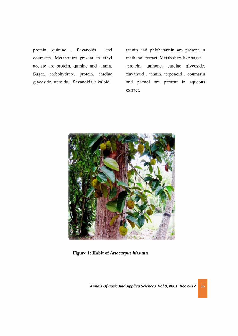
Annals Of Basic And Applied Sciences, Vol.8, No.1. Dec 2017 66
protein ,quinine , flavanoids and
coumarin. Metabolites present in ethyl
acetate are protein, quinine and tannin.
Sugar, carbohydrate, protein, cardiac
glycoside, steroids, , flavanoids, alkaloid,
tannin and phlobatannin are present in
methanol extract. Metabolites like sugar,
protein, quinone, cardiac glycoside,
flavanoid , tannin, terpenoid , coumarin
and phenol are present in aqueous
extract.
Figure 1: Habit of Artocarpus hirsutus

Annals Of Basic And Applied Sciences, Vol.8, No.1. Dec 2017 67
(a) Petiole (b) A portion enlarged
(c) T.S of leaf Midrib(d)T.S of lamina
Fig 2 Microscopic characters of leaves of Artocarpus hirsutus
Table 1: Morphology of the leaf of Artocarpus hirsutus
Morphological parameters Observation
Condition Fresh
Type Simple
Phyllotaxy Alternate
Length of lamina 23-25 cm
Width of lamina 13-15 cm
Venation Reticulate
Leaf shape Obovate
Surface of the leaf Glabrous
Tip of lamina Obtuse
Surface of leaf sheath Glabrous

Annals Of Basic And Applied Sciences, Vol.8, No.1. Dec 2017 68
Table 2. Physico chemical parameters of leaves of Artocarpus hirsutus
Table 3. Fluorescence analysis of leaves of Artocarpus hirsutus
Parameters Values
Total ash 7%
Water soluble Ash 2.33%
Acid soluble Ash 2%
Treatment Fluorescent light UV short UV long
Powder As Such Green Pale green Brownish green
Powder + Water Dark green Light green Black
Powder + CuSO4 Dark green Blackish green Black
Powder + 1m NaOH Black Brownish black Black
Powder + 1%Picric Acid Pale green Dark green Brown
Powder + Acetic Acid Yellowish green Dark green Brown
Powder + HCl Brownish black Dark green Black
Powder + 5%Iodine Brownish green Dark green Black
Powder + 5%FeCl3 Black green Blackish green Black
Powder + HNO3+ 25%NH3
Reddish orange Brownish green Black
Powder + Methanol Green Dark green Brownish black
Powder + 50%HNO3 Reddish orange Brown Black
Powder + H2SO4 Black Black Black
Powder + Conc.HNO3 Reddish orange Yellowish black Black
Powder + Liquid NH3 Brown Yellowish black Brown

Annals Of Basic And Applied Sciences, Vol.8, No.1. Dec 2017 69
Table 4. Percentage yield of Artocarpus hirsutus
4. DISCUSSION
In the present investigation, the detailed
macroscopic and microscopic characters
were studied which will be helpful for the
correct botanical identification.
Phytochemical study is helpful to isolate
the pharmacologically active principles
present in the drug.
Mixture of such chemicals shows a
spectrum of biological effects and
pharmacological properties. Tannins bind
to proline rich proteins and interfere with
the protein synthesis. Flavonoids are
hydroxylated phenolic substance known
to be synthesized by plants in response to
microbial infection and it should not be
surprising that they have been found in
vitro to be effective antimicrobial
substances against a wide array of
microorganisms. Their activity is
probably due to their ability to complex
with extracellular and soluble proteins
and to complex with bacterial cell walls.
Phenolic is one of the major groups of
phytochemical that can be found
ubiquitously in certain plants. Phenolic
compounds are potent antioxidants and
free radical scavenger which can act as
hydrogen donors, reducing agents, metal
chelators and singlet oxygen quenchers.
Studies have shown that phenolic
compounds such as catechin and
quercetin were very efficient in stabilising
phospholipid bilayers against
peroxidation induced by reactive oxygen
species (ROS).
It is concluded that the traditional plants
may represent new sources of
antimicrobials with stable, biologically
active components that can establish a
scientific base for the use of plants in
modern medicine. If the local ethno-
medical preparations and prescriptions is
scientifically evaluated and disseminated
properly the botanical preparation of
Petroleum ether Chloroform Ethyl acetate Methanol Distilled water
0.817% 1.12% 0.70% 1.78% 0.67%

Annals Of Basic And Applied Sciences, Vol.8, No.1. Dec 2017 70
traditional sources of medicinal plants can
be extended for future investigation into
the field of pharmacology,
phytochemistry, ethnobotany and other
biological actions for drug discovery.
Towards authentication and quality
assurance of medicinal plants,
pharmacognostic, physicochemical and
preliminary phytochemical studies of the
A. hirsutus were carried out. The
macroscopic and microscopic evaluation
revealed characters that are of diagnostic
value and useful in authentication of the
plant. Physicochemical analyses reveals
values for Total ash , water soluble ash
and acid insoluble ash are within the
World Health Organisation (WHO)
standards for crude drug from medicinal
plants.
Phyto-screening of leaves for secondary
metabolites revealed the presence of
quinone, cardiac glycoside, terpenoids
,flavanoids, phenol,coumarin , alkaloid ,
steroids and phlobatannin. Information
obtained from these studies can be used
as markers in the identification and
standardization of this plant as a herbal
remedy and also towards monograph
development of the plant.
5. REFERENCE
Bai K V, Jose J S (1981). A simple and
rapid method of screening stomatal
distribution and trichomes in Cassava,
Current Science. 50: 773-774.
Bailey LH (1942).The Standard
Encyclopedia of Horticulture,The
Macmillan Co. New York .401-402.
Cragg, G M and Newman DJ (2005).
Biodiversity.A continuing source of novel
drug Leads Pure Appl.Chem.77:7-24.
Harborne JR (1993). Introduction to
ecological biochemistry. 4th
edLondon,Elsevier;.89- 131
Isaac SR and Nair M A (2005). Litter
dynamics of six multipurpose trees in a
home garden in Southern Kerala,India,
Agroforestry Systems, Springer. 67: 203–
213.
Kokate C K ,Purohit A P and Gokhale S
B (2003) . Pharmacognosy, 25thedition,
NiraliPrakasan.77.
Nayar M.P (1996). Hot Spots of Endemic
Plants of India, Nepaland Bhutan,
Tropical Botanic Garden and Research
Institute, Trivandrum, India, ,251

Annals Of Basic And Applied Sciences, Vol.8, No.1. Dec 2017 71
Table 5. Qualitative analysis of the phytochemicals of leaves of Artocarpus hirsutus
+ indicate the presence of constituents and ─ indicate the absence of constituents
Primary/Secondary metabolites
Name of test Petroleum ether
chloro-form
Ethyl acetate
Methanol Distilled water
Carbohydrates Molisch’s Test + _ _ + _
Aldehyde Fehling Test _ _ _ _ _
Starch Iodine Test _ _ _ _ _
Sugar Benedict’s Test + _ _ + +
Ketose Seliwanoff’s test
_ + _ _ _
Proteins Lowry’s Method
+ + + + +
Aminoacid Ninhydrin test _ _ _ _ _
Fats Filter paper Test
_ _ _ _ _
Quinone H2SO4 Test _ + + _ +
Cardiac glycoside
Kellar –Killani test
_ _ _ + +
Steroids Salkowski test _ _ _ + _
Flavonoids Fluorescent Test
+ + _ + +
Phenols Folin Test _ _ _ _ +
Saponins Foam test _ _ _ _ _
Alkaloids Wagmens test _ _ _ + _
Tannin Iron salt test _ _ + + +
Phlobatanin Conc. Hcl test _ _ _ + _
Terpanoids Salkowski test _ _ _ _ +
Acid NaHCo3 test _ _ _ _ _
Coumarin Fecl3 test + + _ _ +

Annals Of Basic And Applied Sciences, Vol.8, No.1. Dec 2017 72
Prusti A (2008).Antibacterial activity of
some Indian Medicinal plants
Ethanobotanical leaflets.1(27):34-36
Shyma T B and Devi Prasad A G
(2012).TraditionalUse of Medicinal
Plants and its status among the tribes In
Mananthavady of Wayanad District,
Kerala, World Research Journal of
Medicinal & Aromatic Plants.1( 2), 2012,
22-26.
Sawant Laxman, PrabhakarBala,
Kachaeala Yusuf, Pandita Nancy (2010
).Pharmacognostical standardizationof
Enicostemma littotale blume.
J.Pharmacog. 12 (16): 12-20.
Shanley P and Luz L (2003). The impacts
of forest degradation on medicinal plant
use and implication for health care in
eastern Amazonia. Bioscience,53(6):573-
584.
Zobel, A M, Glowniak K, Lynch J E,
Dudka S, Alliota A (2000).
Phytochemistry of plants associated with
a 400-year-old stand of hemlock at Clear
Lake Reserve, Ontario.
Proceedings: Symposium on sustainable
management of hemlock ecosystems in
eastern North America. Newtown Square,
PA: U.S. Department of Agriculture,
Forest Service, Northeastern Forest
Experiment Station, 230-233.

Annals Of Basic And Applied Sciences, Vol. 8, No.1, Dec 2017 73
Phytochemical analysis and antimicrobial effect of Azadirachta indica, Psidium guajava and Spinacia olerocea
against Propionibacterium acne
Ruveena T N*, Sabha kottayil,Sanu Simon,Rosemol Johnson, Sofiya P J and Nima Varghese
Department of Microbiology, St Mary’s College,Thrissur-680020, Kerala, India. *Corresponding author:[email protected]. Ph:9497372574
Abstract
Acne vulgaris is the most common disorder of human skin that affects upto 80% of individuals in their lives.The
hypersecretion of hormone androgen stimulates higher sebum secretion in sebaceous gland. The daily use of many
cosmetics makes skin dull and the chemical contents have their own negative effects. This study is attempt to findout
the disease control potency of phytochemicals against acne causing bacteria Propinibacterium acne. Hot water extract
of Psidium guajava, Azadiracha indica and Spinacia oleracea are used for the study. Different concentrations of
extracts such as 1500 g/ml, 1000g/ml, 500g/ml and 100g/ml are used for the study. Phytochemical analysis
showed the presence of alkaloids, steroids, flavonoids, tannin and saponin. Agar Well diffusion method is used for
the detection of antimicrobial activity. Among the three plat extract Azadiracha indica showed excellent activity.
Azadiracha indica can be used in the treatment of infectious disease caused by P.acne.
Key words: Phytochemicals , Agar Well diffusion
1. Introduction
Acne is a skin disorder for which
dermatologists are still struggling since
years to treat successfully.It mainly
affects adolescents though it may present
at any age. Acne by definition is
multifactorial chronic inflammatory
disease of pilosebaceous units.It affects
the skin of the face,neck and upper
trunk.These particular sebaceous follicles
have capacious follicular channels and
voluminous, sebaceous glands.Acne
develops when these specialized follicles
undergo alteration that results in
formation of noninflammatory

Annals Of Basic And Applied Sciences, Vol. 8, No.1, Dec 2017 74
lesion(concedons) and inflammatory
lesions(papulese,postules and nodules)
black heads and wrinkles are also seen in
people suffering from acne vulgaris. (GM
White,1999)
Staphylococcus epidermis and
Propionibacterium acne are considered as
the major skin bacteria that cause the
formation of acne.P.acne is one among the
gram positive microaerophillic anaerobic
bacilli plays and it is implicated in
development of inflammatory acne by its
capability to activate complements and by
its ability to metabolize sebaceous
triglycerides into fatty acids which
chemotactically attracts
neutrophils.(G.SKumaret.al .,2007).
Plants have the ability to synthesize a
wide variety of chemical compounds that
are used to perform biological functions
and to defend against attack from
predators such as insects,fungi and
herbivorous mammals. Many of these
phytochemicals have beneficial effects are
long term health when consumed by
humans and can be used to treat human
diseases. The ingredients in tropical acne
treatments particularly herbs and naturally
derived compounds have received
considerable interest as they have fewer
adverse effects than synthetic agents.In
present study three essential plant extracts
of Azadirachta indica, Psidium guajava,
Spinacia olerocea were prepared which
have been traditionally used as
antimicrobial and anti inflammatory
agents were examined for antimicrobial
activities against P.acne.
Psidium guajava are very efficient in
treatment of acne and black spots.Guava
leaves are effective in eliminating acne
and black spots from the skin.They
contain an antiseptic that can kill acne
causing bacteria.For the treatment of
blackheads blend guava leaves with water
and use it as a scrub on face to remove
blackheads.Guava leaves contain
antioxidants which destroy the free
radicals that damage skin,thus protecting
skin from aging as well as improving skin
tone and texture.A decoction of mature
guava leaves can be applied on the skin for
tightening it.Guava leaves are an instant
cure for getting rid of itchiness as they
contain allergy blocking compounds.
The available products that are currently
in the market gaining it's significance in
providing relief for acne or pimple
problems are unable to prevent the

Annals Of Basic And Applied Sciences, Vol. 8, No.1, Dec 2017 75
reoccurance. The current research focuses
on antibacterial potency of some
medicinal plants against acne causing
organism Propionibacterium acne.
2. Materials and Method
2.2 The collection and processing of plant
extract
The three plants used in this study were
collected from local surrounding of
Thrissur District,Kerala . The leaves were
thoroughly washed in water and air dried
in room temperature for one week. They
were grind into powder and subjected to
extraction with hot water. The crude
powdered plant material of 10g was
washed and extracted with hot water in a
large beaker.The extract was prepared by
continuous boiling water for 810 hrs. The
extracts were concentrated and air dried.
The samples were then stored in airtight
bottles at 4°c.
2.1 Isolation and Identification of
Propionibacterium acne. The skin was first wiped with ethanol and
the material was extracted from skin
lesions and took up by sterile swab sticks
and inserted into Tryptone yeast extract
glucose agar(TYGA). The
Propionibacterium acne culture was
maintained in tryptone yeast extract
glucose agar (TYGA) plates and TYG
broth. The mother inoculum was
maintained anaerobically using candle jar
about 2 days.
The isolate was identified as
Propionibacterium acne by using various
biochemical test like Catalase test, Indole
test, Nitrate test and Carbohydrate
fermentation tests,
2.3 Phytochemical analysis
Test for alkaloids, flavonoids, steroids,
phenols, tannins, glycosides, terpenoids,
saponins, anthraquinones and cardiac
glycosides (kellerkillani test) were
performed for the detection of secondary
metabolites.
2.4 Anti microbial Assay The plant extracts of different plants were
tested against the pimple causing bacteria
Propionibacterium acne using well
diffusion method. About 5 wells in each
plates of 6mm diameter were punched in
TYG Agar surface for placing the plant
extract in a concentration of 1500 mg/ml,
1000mg/ml, 500mg/ml, 100mg/ml.
Aqueous extracts of 3 plants were mixed
with 2%DMSO and added in separate
places in consent. Clindamycine is used as
control. The plates were kept for

Annals Of Basic And Applied Sciences, Vol. 8, No.1, Dec 2017 76
incubation for about 24 to 48 hours. After
48 hours of incubation the zone of
inhibition was clearly visible and the
diameter of the zone was measured and
tabulated.
3.Result
Colonies isolated in modified TYGA
medium, the first colony isolation
identified as Propionibacterium acnes. It
grow on BHA (Brain Heart infusion Agar)
medium. Colony showed a cream colored
raised spread growth. Gram staining and
motility test showed that it is motile gram
positive rod shaped bacterium. It do not
produce endospore.Indole test, nitrate test,
catalase test and sugar fermentation test
are carried out. The organism showed
positive result to indole, nitrate and
catalase test. It can also ferment glucose.
Phytochemical analyses of aqueous
extracts of these plants were carried out by
using standard procedure. The tests were
based on qualitative analysis.
Phytochemical screening revealed the
presence of primary and sacondary
metabolites. The primary metabolites like
carbohydrate, reducing sugars and lipids
are found in each plants. The secondary
metabolites alkaloids, steroids,
flavonoids, tannin and saponin are found
in Azadirachta indica leaf, steroids,
flavinoids, tannins and cardiac glycoside
are found in Psidium guajava leaf and
phenol, flavinoids, tannin, glycoside and
saponin are found in Spinacia oleracea
seed.
Antibacterial testing of aqueous extracts
of the three plants against P. acnes was
done by well diffusion method. After
incubation, a zone of inhibition observed
in the plates and indicated the antibacterial
activity of these plants against P.
acnes(Swanson ,2003 )
Based on the aqueous plant extract and
their concentrations they showed different
zones of inhibition. The different
concentrations are 1500 g/ml,
1000g/ml, 500g/ml and 100g/ml. The
aqueous extracts of Azadirachta indica
showed a zone of inhibition 20 mm,
18mm, 17mm and 15mm. The aqueous
extracts of Psidium guajava showed a
zone of inhibition 20 mm, 15mm, 14mm
and 11mm. The aqueous extracts of
Spinacia oleracea showed a zone of
inhibition 16,14,13 and 12mm.
4.Discussion
Azadiracha indica showed excellent
antibacterial activity against the bacteria

Annals Of Basic And Applied Sciences, Vol. 8, No.1, Dec 2017 77
and showed the presence of alkaloides,
steroids, flavanoids and tanins. Flavanoids
and tanins present in the extract
contributed the major antibacterial
activity. Some of these secondary
metabolites are also present in the Psidium
guajava and Spinacia oleracea also have
anti bacterial property against
Propionibacterium.
Alkaloids, terpenoids, phenolics and
pigments can have therapeutic actions in
humans and which can be refined to
produce drugs. These phytochemicals
present in plant play a vital role in
contributing them the antiacne
property.(Nand et al., 2002)
5.Conclusion
The systemic research for useful bioactive
agent from the plants is now consider to be
a rational approach in nutraceuticals and
drug research.The results of
phytochemical analysis comprehensively
validate the presence of therapeuticaly
important and valuable secondary
metabolites. Acne induced by
Propionibacterium represents a fastidious
cutaneous problem .To get rid of this
problem, large amount of money is spent
every year on medicins and other agents
world wide but, attempt to completely
remove this problem are still a far of
target. Our present studies indicates that
the medicinal plant extract inhibits
antibacterial activity. Thus they can be
used in the treatment of infectious disease
caused by P.acne.(Mengesha et al ,2002)
The present investigation demonstrated
that extracts of premedicinal plants
Psidium guajava, Azadiracha indica,
Spinacia oleracea showed excellent
antibacterial activity against the bacteria.
so the phytochemicals present in plants
play a vital role in contributing them the
antibacterial action. So the combined
effects of these plants can provide
effective medicinal value and can be used
as a drug for skin disease causig organism
P.acne. To check effectiveness of
botanicals, detailed biomolecular,
characterisation, formulation of dosages,
clinical studies, safety and efficiency a lot
of more has yet to be explore. It is
concluded that these plants could be a
potential source of active antibacterial
agents
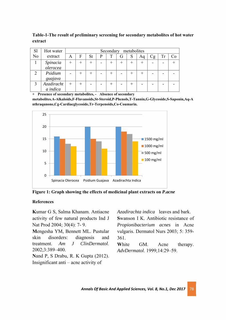
Annals Of Basic And Applied Sciences, Vol. 8, No.1, Dec 2017 78
Table-1-The result of preliminary screening for secondary metabolites of hot water extract
Sl No
Hot water extract
Secondary metabolites A F St P T G S Aq Cg Tr Co
1 Spinacia olerocea
+ + + - + + + + - - +
2 Psidium guajava
- + + - + - + + - - -
3 Azadirachta indica
+ + - - + - + - - - -
+ Presence of secondary metabolites, - Absence of secondary metabolites.A-Alkaloids,F-Flavanoids,St-Steroid,P-Phenols,T-Tannin,G-Glycoside,S-Saponin,Aq-Anthraqunone,Cg-Cardiacglycoside,Tr-Terpenoids,Co-Coumarin.
Figure 1: Graph showing the effects of medicinal plant extracts on P.acne
References
Kumar G S, Salma Khanam. Antiacne activity of few natural products Ind J Nat Prod 2004; 30(4): 7- 9. Mengesha YM, Bennett ML. Pustular skin disorders: diagnosis and treatment. Am J ClinDermatol. 2002;3:389–400. Nand P, S Drabu, R. K Gupta (2012). Insignificant anti – acne activity of
Azadirachta indica leaves and bark. Swanson I K. Antibiotic resistance of Propionibacterium acnes in Acne vulgaris. Dermatol Nurs 2003; 5: 359- 361. White GM. Acne therapy. AdvDermatol. 1999;14:29–59.
0
5
10
15
20
25
Spinacia Olerocea Psidium Guajava Azadirachta Indica
1500 mg/ml
1000 mg/ml
500 mg/ml
100 mg/ml

Annals Of Basic And Applied Sciences, Vol.8, No.1. Dec 2017 79
Comparative study on phytochemical, physiochemical and antioxidant
properties of Sorghum bicolor and Triticum aestivum
Rincy.C.K, Ranjitha.P.C, Priyanka.K. , Shahanas.M, Aiswarya .C.G, Aneesha.K, Aiswarya Das, Gayathri.C.V, Rahul Pramod K V and Smitha K R*
Department of Biochemistry, Sree Krishna College, Guruvayur.
*Corresponding author: Email id:[email protected], Ph: 9387005196
Abstract
Increased consumption of whole grains has been associated with reduced risk of major chronic diseases including
cardiovascular disease, type II diabetes, and some cancers. The unique phytochemicals in whole grains are responsible for
the health benefits of whole grain consumption. Triticum aestivum (Wheat) is a major crop and an important component
of the human diet, particularly in developing countries. Sorghum bicolor (Sorghum) is an important cereal widely
cultivated in tropical and subtropical regions. The objectives of this study were to compare the phytochemical profiles,
physiochemical properties and antioxidant activities of sorghum and wheat. The phytochemical studies revealed the
presence of various phytochemical such as carbohydrates, protein, tannins, flavonoids, terpenoids, steroids and alkaloids
etc. The results showed the gluten content is low in sorghum (7.855%) when compared to that of wheat (14.935%). Total
ash content (17.92% and 15.41% for sorghum and wheat, respectively) and moisture content (15.73% for sorghum and
12.18% for wheat) were also determined. Sorghum had the high total antioxidant activity with EC50 value 55.90 µg/mL and
that of wheat is 47.56 µg/mL. The methanolic extracts reduced the DPPH radical in a dose dependent manner with 50%
reduction in 21.51 and 27.69 µg/mL for sorghum and wheat respectively. Total reducing capacity is with response to the
increase in drug concentration in both extracts with EC50 value of 27.245 and 49.81 µg/mL for sorghum and wheat,
respectively. The study showed that the sorghum and wheat‐based diets can serve as functional foods, as evident from their
antioxidant capabilities in addition to their gross energy content.
Keywords: Phytochemicals; Gluten content; antioxidants; wheat; sorghum
Annals Of Basic And Applied Sciences, Vol.8, No.1. Dec 2017 80
1. Introduction
Plants are the richest resource of
drugs of traditional systems of medicine,
modern medicine, nutraceuticals, food
supplements, folk medicines,
pharmaceutical intermediates and
chemical entities for synthetic drugs
(Hammer et al., 1999). Medicinal plants
are valuable natural resources and
regarded as potential safe drugs and have
been tested for biological, antimicrobial
and hypoglycemic activity also play an
important role in modern medicine
(Hassawi and Kharma 2006, Bhat et al.,
2009). The phytochemical analysis of the
plant is very important commercially and
has great interest in pharmaceutical
companies for the production of many
new drugs for curing various diseases.
Phytochemicals such as alkaloids
terpenoids and flavonoids exhibit various
important pharmacological activities such
as anti-inflammatory, anticancer,
antimalarial, inhibition of cholesterol
synthesis, antiviral and antimicrobial
activities (Hagerman et al., 1998).
Dietary phytochemicals are commonly
found in plant-based foods such as fruits,
vegetables, grains and tea (Probst et al.,
2015). Whole grains in our food have a
role with reduced risk of major chronic
diseases including cardiovascular disease,
diabetes, and some cancers. Whole grains
are rich in nutrients
and phytochemicals with known health
benefits. Whole grains are enriched in
antioxidants including trace minerals and
phenolic compounds and these
compounds have been responsible for
disease prevention. It also contains
phytate, phyto-oestrogens such as lignan,
plant stanols and sterols, and vitamins
and minerals. The
unique phytochemicals in whole grains
are believed to be responsible for the
health benefits of whole grain
consumption (Slavin, 2004).
Sorghum bicolor (family Poaceae) is the
5th most important cereal globally in
terms of acreage and production. Most of
the phytochemicals in sorghum are
excellent antioxidants. Sorghum has great
drought-tolerence. Sorghum is gluten free
and thus can be consumed by people with
celiac disease. Triticum aestivum (Wheat)
is a major crop and an important

Annals Of Basic And Applied Sciences, Vol.8, No.1. Dec 2017 81
component of the human diet, particularly
in developing countries. Epidemiological
studies have associated the consumption
of whole grain and whole-grain products
with reduced incidence of chronic
degenerative diseases (De Moura, 2008).
Accordingly, dietary-plant-based drug
discovery still remains an important area,
remains unexplored, where a systematic
search may definitely provide important
leads for the pharmacological benefits.
2. Materials and methods
2.1 Plant material and extraction: The
fresh and healthy grains of Sorghum
bicolor and Triticum aestivum were
collected from the market Kunnamkulam,
Kerala. The fresh grains were washed,
dried in shades. The dried samples were
then crushed into powder. The crude
powdered plant material of 20 g was
extracted with 200 ml methanol and
stirred it using magnetic stirrer overnight.
After extraction the solvent was removed
to yield a viscous residue of extract. The
extract was filtered through Whatmann
No.1 filter paper. The extract were then
concentrated at 40-450 C in hot air oven
and dried.
2.2 Chemicals
2, 2-diphenyl-1-picryl hydrazyl (DPPH) is
procured from Sigma Aldrich. Nitric acid,
potassium di hydrogen phosphate,
potassium ferrocyanide, sodium
hydroxide, sodium phosphate and
sulphuric acid were purchased from
MERK specialities Pvt Ltd., Mumbai. All
other chemicals and reagents used were of
analytical reagent grade and purchased from
reputed Indian companies.
2.3 Methods
2.3.1 Phytochemical screening:
Phytochemical screening of various
extracts of U. narum leaves was carried
out according to standard procedures
(Smitha et al., 2014)
2.3.2 Physiochemical Properties
A.Determination of Percentage Gluten
in flour:
About 25 g of each composite flour
sample was weighed in to a petridish of
known weight and thoroughly mixed with
1 ml of water to form dough .The dough
is kneaded under running water to remove
starch and later put in to petridish and

Annals Of Basic And Applied Sciences, Vol.8, No.1. Dec 2017 82
weighed .It was then dried in an oven and
weighed after drying.
% Dry weight = Dry gluten weight / Dry
flour weight *100
A. Ash Determination of extracts:
Ash determination was by method of
ADAC (1990). Two grams of plant
powder was weighed crucible .The
crucible and content were ignited in a
preheated furnace to 600 degree for 2
hours. The crucible was loaded in a
desiccator, was weighed and percent ash
content was calculated
Ash content =Weight of ash /Weight of
sample * 100
A. Moisture Content Determination:
Moisture was determined by the method
of AOAC 1990. Two grams of plant
powder was weighed in to pre-dried and
weighed crucible and placed in
thermostatically regulated hot air oven at
169 degree for 9 hour. The crucible was
removed dried in a desiccator and
weighed. And the process was repeated
until a constant weight was obtained;
moisture content was regulated and
expressed as a percentage of initial
weight of the flour.
Moisture % = (Initial mass of sample –
Mass after drying) /weight of sample used
2.3.3 Antioxidant activities
A. DPPH radical scavenging
activity:
The scavenging activity of the extract on
2, 2-diphenyl-1-picrylhydrazyl (DPPH)
was analyzed by the method of (Aquino
et al., 2001). DPPH, in its radical form
has an absorption peak at 515 nm, which
disappears on reduction by an antioxidant
compound, evident from its transition
from purple to yellow colour. DPPH, in
solution was incubated in the presence of
extract at concentrations for 20 minutes at
room temperature in the dark and
absorbance was then read. Assay was
carried out in triplicate. The IC50 value
was determined.
B. Total antioxidant capacity assay:
The total antioxidant capacity was
measured according to
spectrophotometric method of Preito et
al; (1999). Various concentrations of
crude extract dissolved in distilled water

Annals Of Basic And Applied Sciences, Vol.8, No.1. Dec 2017 83
were combined with 1 ml of reagent
solution containing 0.6 M sulphuric acid,
2.8 mM sodium phosphate and 4mM
Ammonium molybdate. The tubes were
capped and incubated at 900 C for 90
minutes. After cooling to room
temperature, the absorbance was
measured at 695 nm against blank. The
activity of the drug was compared with
that of control.
C. Total reducing power activity :
The reducing power of sequential extracts
was determined by the method of Yen
and Duh (1994). Different concentrations
of extracts were mixed with 2.5 ml of
phosphate buffer (200 mM, pH 6.6) and
2.5 ml of 1% potassium ferricyanide. The
mixtures were incubated for 20 minutes at
50°C. After incubation, 2.5 ml of 10 %
Trichloroacetic acid were added to the
mixtures, followed by centrifugation at
650 rpm for 10 min. The upper layer
(5ml) was mixed with 5ml of distilled
water and 1ml of 0.1% ferric chloride and
the absorbance of the resultant solution
were measured at 700 nm. The activity of
the drug was compared with that of
control.
2.4 Statistical analysis
All in vitro assays were performed in
triplicate and data represented are mean ±
SD.
Phytochemicals Sorghum Wheat
Carbohydrates + +
Protein + +
Tannins + +
Flavonoids + +
Terpenoids + +
Steroids + +
Alkaloids + +
Saponins + +
Table 1: Phytochemical screening of Sorghum bicolor and Triticum aestivum

Annals Of Basic And Applied Sciences, Vol.8, No.1. Dec 2017 84
3.Results
3.1 Phytochemical analysis of Sorghum
bicolor and Triticum aestivum
Phytochemical screening of Sorghum
bicolor and Triticum aestivum methanolic
extracts revealed the presence of protein,
phenolic compounds, alkaloids,
terpenoids, saponins and tannins (Table
1).
3.2 Physiochemical properties of
Sorghum bicolor and Triticum
aestivum
A. Determination of Percentage Gluten
in Sorghum bicolor and Triticum
aestivum Flour
The gluten content of Sorghum bicolor
and Triticum aestivum flour were
determined and the results were shown in
Figure 1A.The results showed that
sorghum has low gluten content
compared to wheat.
B. Ash Determination of Sorghum bicolor
and Triticum aestivum Flour
The ash value of Sorghum bicolor is
17.92% and that of Triticum aestivum is
15.41% (Figure1B).
C. Moisture Content of Sorghum bicolor
and Triticum aestivum Flour
The moisture contents of Sorghum
bicolor and Triticum aestivum were 15.73
and 12.18% respectively. Results are
depicted in Figure 1C.
3.3 Antioxidant activities of Sorghum
bicolor and Triticum aestivum
A.DPPH radical scavenging activity of
Sorghum bicolor and Triticum aestivum
The methanolic extracts of Sorghum
bicolor and Triticum aestivum showed
potent DPPH radical inhibition. The
maximum activity was obtained in tested
concentration of 30 µg/mL showed 62.5
and 58.33% inhibition for sorghum and
wheat respectively. IC 50 value of
methanolic extract was reported as 21.51
and 27.69 µg/mL for sorghum and wheat
respectively. This indicates significant
DPPH inhibition of Sorghum bicolor and
Triticum aestivum extracts (Figure 2A).
B. Total Antioxidant activity of Sorghum
bicolor and Triticum aestivum
The results show the increase in
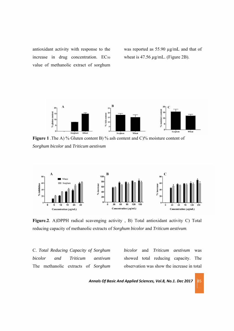
Annals Of Basic And Applied Sciences, Vol.8, No.1. Dec 2017 85
antioxidant activity with response to the
increase in drug concentration. EC50
value of methanolic extract of sorghum
was reported as 55.90 µg/mL and that of
wheat is 47.56 µg/mL. (Figure 2B).
Figure 1 .The A) % Gluten content B) % ash content and C)% moisture content of
Sorghum bicolor and Triticum aestivum
Figure.2. A)DPPH radical scavenging activity , B) Total antioxidant activity C) Total
reducing capacity of methanolic extracts of Sorghum bicolor and Triticum aestivum.
C. Total Reducing Capacity of Sorghum
bicolor and Triticum aestivum
The methanolic extracts of Sorghum
bicolor and Triticum aestivum was
showed total reducing capacity. The
observation was show the increase in total

Annals Of Basic And Applied Sciences, Vol.8, No.1. Dec 2017 86
reducing capacity with response to the
increase in drug concentration. EC50
value of the extracts were reported as
27.245 µg/mL for sorghum and 49.81
µg/mL for wheat respectively (Figure
2C).
4.Conclusion
The present study revealed the
antioxidant property of Sorghum bicolor
and Triticum aestivum which can defend
against free radicals. It was found that
dietary grains Sorghum bicolor and
Triticum aestivum possess numerous
phytoconstituents. The free radical
scavenging and antioxidant activities of
the grain extracts supported the evident of
identified phytochemicals present in it.
The gluten content is very less in the case
of Sorghum bicolor compared to Triticum
aestivum. So it can use in disease
condition such as celiac disease.
This reveals the antioxidant activities of
methanolic extracts of Sorghum bicolor
and Triticum aestivum which can be used
as a potential dietary substance as well as
pharmacological agent.
5.References
Bhat, S., M. Lobo, S.C. Kumar, K.V.
Sukesh and K.R. Chandrashekar, (2009).
Antimicrobial spectrum and
phytochemical study of Hopea parviflora
beddome saw dust extracts. J. Phytol., 1:
469–474
De Moura FF(2008). Whole Grain Intake
and Cardiovascular Disease and Whole
Grain Intake and Diabetes: A review.
Bethesda, MD: Life Sciences Research
Office; 1–79.
Hagerman, A.E., Riedl, K.M., Jones,
G.A., Sovik, K.N., Ritchard, N.T.,
Hartzfeld, P.W., Riechel, T.L.,(1998).
High molecular weight plant
polyphenolics (tannins) as biological
antioxidants. J. Agric. Food Chem. 46,
1887–1892.
Hammer KA, Carson CF, Riley TV
(1999). Antimicrobial activity of essential
oils and other plant extracts. J. Appl.
Microbiol. 86: 985-990.
Hassawi, D. and A. Kharma,( 2006).
Antimicrobial activity of medicinal plants
against Candida albicans. J. Biol. Sci., 6:
104‒109

Annals Of Basic And Applied Sciences, Vol.8, No.1. Dec 2017 87
Prieto P, Pineda M, Aguilar M. ( 1999).
Spectrophotometric quantitation of
antioxidant capacity through the
formation of a phosphomolybdenum
complex: Specific application to the
determination of vitamin E. Anal
Biochem. 269:337–41
Probst YC , Guan VX ,Kent K
(2017),Dietary phytochemical intake
from foods and health outcomes ; BMJ
open ; 5 ;(7);1-9
Probst, B.L., McCauley, L., Trevino, I.,
Wigley, W.C., and Ferguson, D.A.
(2015). 635 Cancer Cell Growth Is
Differentially Affected by Constitutive
Activation of 636 NRF2 by KEAP1
Deletion and Pharmacological Activation
of NRF2 by the 637 Synthetic
Triterpenoid, RTA 405. PLoS One.,
10(8), e0135257
Proestos C ;Boziaris , (2006).Analysis of
flavonoids and phenolic acids. Food
chem., 53, 1190 -1195 .
Slavin J, (2004). Whole grain and human
health, Nutr .Res Rev. 17(1); 99 -110 .
Smitha K. R. , Babu T. D. , Achuthan C.
R.(2014). Phytochemical screening and in
vitro cytotoxicity analysis of Uvaria
narum (dunal) wall. Asian Journal of
Phytomedicine and Clinical Research.
2(2), 2014, 40 - 47.
Valitova JN, Sulkarnayeva
AG, Minibayeva FV(2016). Plant Sterols:
Diversity, Biosynthesis, and
Physiological Functions. Biochemistry
(Mosc). ,81 (8):819-34.
Vasanthi HR, Shrishrimal N, Das DK,
(2012).Phytochemicals from plants to
combat cardiovascular disease. Curr Med
Chem. 19(14) ;2242 – 51
Yen GC, Duh PD.(1994). Scavenging
effect of methanolic extracts of peanut
hulls on free-radical and active-oxygen
species. J Agric Food Chem 42: 629–32.
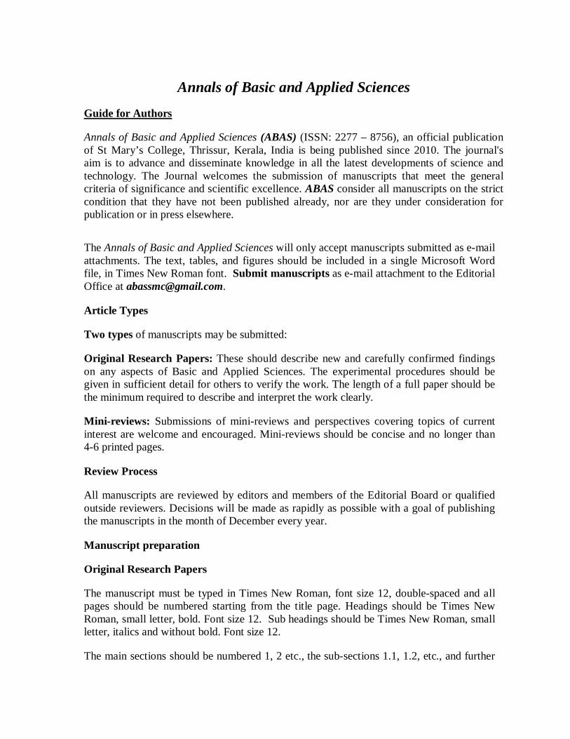
Annals of Basic and Applied Sciences
Guide for Authors
Annals of Basic and Applied Sciences (ABAS) (ISSN: 2277 – 8756), an official publication of St Mary’s College, Thrissur, Kerala, India is being published since 2010. The journal's aim is to advance and disseminate knowledge in all the latest developments of science and technology. The Journal welcomes the submission of manuscripts that meet the general criteria of significance and scientific excellence. ABAS consider all manuscripts on the strict condition that they have not been published already, nor are they under consideration for publication or in press elsewhere.
The Annals of Basic and Applied Sciences will only accept manuscripts submitted as e-mail attachments. The text, tables, and figures should be included in a single Microsoft Word file, in Times New Roman font. Submit manuscripts as e-mail attachment to the Editorial Office at [email protected].
Article Types
Two types of manuscripts may be submitted:
Original Research Papers: These should describe new and carefully confirmed findingson any aspects of Basic and Applied Sciences. The experimental procedures should be given in sufficient detail for others to verify the work. The length of a full paper should be the minimum required to describe and interpret the work clearly.
Mini-reviews: Submissions of mini-reviews and perspectives covering topics of current interest are welcome and encouraged. Mini-reviews should be concise and no longer than 4-6 printed pages.
Review Process
All manuscripts are reviewed by editors and members of the Editorial Board or qualified outside reviewers. Decisions will be made as rapidly as possible with a goal of publishing the manuscripts in the month of December every year.
Manuscript preparation
Original Research Papers
The manuscript must be typed in Times New Roman, font size 12, double-spaced and all pages should be numbered starting from the title page. Headings should be Times New Roman, small letter, bold. Font size 12. Sub headings should be Times New Roman, small letter, italics and without bold. Font size 12.
The main sections should be numbered 1, 2 etc., the sub-sections 1.1, 1.2, etc., and further

subsections (if necessary) 1.1.1, 1.1.2, etc.
The Title should be a brief phrase describing the contents of the paper. The Title Page should include
• Concise and informative title. (Times New Roman. Text font size 16)
• Author names and affiliations. (Times New Roman. Text font size 12)
• Name of Corresponding author (Times New Roman. Text font size 12) with telephone, e-mail address and the complete postal address. (Times New Roman. Text font size 11)
Structured Abstracts are required for all papers and should include objectives, key findings and major conclusions. It should be a single paragraph with not more than 250 words. References should be avoided in abstract.
Following the abstract, about 3 to 6 key words should be listed.
A list of non-standard Abbreviations should be added. Each abbreviation should be spelled out and introduced in parentheses the first time it is used in the text. Recommended SI units only should be used.
The Introduction should provide a background to the study and should clearly state the specific aims of the study. It should be understandable to the audience from a broad range of scientific disciplines. Approximate length is 500-1000 words.
Materials and methods should be complete enough to allow experiments to be reproduced. Methods in general use need not be described in detail. Subheadings should be used. Please include details of ethical approval in this section. Approximate length: 500-1000 words.
Results should be clear and concise with Graphs or Tables, may be inserted along with the matter or may be given as separate with the detailed title and in that case the places where the figures are to be inserted should be mentioned in the manuscript. Each figure and table should be numbered in Arabic numerals and mentioned in the text. Figures must be clearly lettered and suitable for reproduction to fit either one column width (8.5 cm) or two columns width (17 cm). Black and white photographs only are acceptable. The lettering in the figures should be readable. In addition to the inserted version in the word document, the figures can be supplied in electronic format as JPEG or TIFF. Tables should be kept to a minimum and be designed to be as simple as possible. Each table should be on a separate page, numbered consecutively (Table 1, Table 2 etc) and supplied with a heading and a legend. Tables should be prepared in Microsoft Word. The same data should not be presented in both table and graph forms.
The Discussion should interpret the findings in view of the results obtained in this and in past studies on this topic. A combined Results and Discussion section is also encouraged.

State the Conclusions in a few sentences at the end of the paper. The main conclusions of the study may be presented in a single paragraph.
The Acknowledgments of people, grants, funds, etc should be brief.
References: In the text, a reference identified by means of an author‘s name should be followed by the date of the reference in parentheses. When there are more than two authors, only the first author‘s name should be mentioned, followed by ’et al‘. In the event that an author cited has had two or more works published during the same year, the reference, both in the text and in the reference list, should be identified by a lower case letter like ’a‘ and ’b‘ after the date to distinguish the works.
Examples:
(Smith, 2000), (Chandra and Singh, 1992), (Blake et al., 2003), (Chege, 1998; Steddy, 1987a, b; Gold, 1993, 1995).
References should be listed at the end of the paper in alphabetical order. Authors are fully responsible for the accuracy of the references.
Reference to a journal publication:
Diaz E, Prieto MA (2000). Bacterial promoters triggering biodegradation of aromatic pollutants. Curr. Opin. Biotech. 11: 467-475.
Reference to a book:
Pitter P, Chudoba J (1990). Biodegradability of Organic Substances inthe Aquatic Environment. CRC press, Boca Raton, Florida, USA.
Reference to a chapter in a book:
Mandell GL, Petri WA, 1996. Antimicrobial Agents: Penicillins, Cephalosporins, and other ß-Lactam Antibiotics, In: Goodman and Gilman`s. The Pharmacological Basis of Therapeutics. 9th, Ed. J.G. Hardman and L.E. Limbird, McGraw-Hill: NY. Vol. 23; PP. 1073–1101.
Mini reviews
The format requirements for original research papers apply to reviews too.
Submission
Submission in electronic form of the final version of the manuscript to the email id [email protected].

ABAS December 2017Vol. 8, No. 1
Annals of Basic and Applied Sciences
December 2017 Volume 8 Number 1
St. Mary’s College Thrissur, Kerala, India.Re-Accredited by NAAC with A GradePhone: +91 487 2333485 Fax : +91 487 2334785Email: [email protected] : stmaryscollegethrissur.edu.in
ISSN 2277 – 8756