Embed Size (px)
Citation preview
i
ANNE CAROLINE COSTA OENNING
DIAGNOSIS OF EXTERNAL ROOT RESORPTION IN SECOND
MOLARS ASSOCIATED WITH IMPACTED THIRD MOLARS BY
PANORAMIC RADIOGRAPH AND TWO CONE BEAM
COMPUTED TOMOGRAPHY DEVICES
DIAGNÓSTICO DA REABSORÇÃO RADICULAR EXTERNA EM
SEGUNDOS MOLARES ASSOCIADA A TERCEIROS MOLARES
IMPACTADOS POR MEIO DE RADIOGRAFIAS
PANORÂMICAS E DOIS SISTEMAS DE TOMOGRAFIA
COMPUTADORIZADA DE FEIXE CÔNICO
PIRACICABA
2014

ii
iii
UNIVERSIDADE ESTADUAL DE CAMPINAS
FACULDADE DE ODONTOLOGIA DE PIRACICABA
ANNE CAROLINE COSTA OENNING
DIAGNOSIS OF EXTERNAL ROOT RESORPTION IN SECOND MOLARS ASSOCIATED WITH
IMPACTED THIRD MOLARS BY PANORAMIC RADIOGRAPH AND TWO CONE BEAM
COMPUTED TOMOGRAPHY DEVICES
DIAGNÓSTICO DA REABSORÇÃO RADICULAR EXTERNA EM SEGUNDOS MOLARES
ASSOCIADA A TERCEIROS MOLARES IMPACTADOS POR MEIO DE RADIOGRAFIAS
PANORÂMICAS E DOIS SISTEMAS DE TOMOGRAFIA COMPUTADORIZADA DE FEIXE
CÔNICO
Thesis presented to the Piracicaba Dental School of the
University of Campinas in partial fulfillment of the requirements
for the degree of Doctor in Oral Radiology, in the Oral Radiology
area
Tese apresentada à Faculdade de Odontologia de Piracicaba da
Universidade Estadual de Campinas como parte dos requisitos
exigidos para obtenção do título de Doutora em Radiologia
Odontológica, na área de Radiologia Odontológica
Orientador: Prof. Dr. Francisco Haiter Neto
Este exemplar corresponde à versão final da tese defendida pela aluna Anne Caroline Costa Oenning, e orientada pelo Prof. Dr. Francisco Haiter Neto ___________________ Assinatura do Orientador
PIRACICABA
2014
iv
v

vi
vii
ABSTRACT
The aim of this study was to compare a two-dimensional method - panoramic radiography
- and a three-dimensional modality - cone beam computed tomography (CBCT) - on the
assessment of external root resorption (ERR) of second molars associated with impacted
third molars. In addition, we aimed to relate the third molar inclination (Winter´s
classification) with the detection of ERR on the second molar. First, the sample was
consisted of 188 impacted third molars (66 individuals). Panoramic radiography
(Orthopantomograph OP100 D) and CBCT imaging (i-CAT Classic) were obtained of all
patients. Two oral radiologists investigated the presence of ERR on the adjacent second
molar and the inclination of the third molar. Statistical analysis was performed using chi-
square test, Fisher exact test, two-proportion Z test and simple logistic regression
(significance level was set at 5%). A significantly higher number of ERR was diagnosed on
CBCT images (n=43) than on panoramic radiographs (n=10) (P=0.0001). The agreement
between panoramic radiographs and CBCT for diagnosing ERR was 4.3% (n=8). The
mandibular third molars on mesioangular and horizontal inclinations were more related to
ERR lesions on the second molars. Therefore, 174 mandibular third molars on these two
inclinations were evaluated in a second sample comprising of 116 CBCT images acquired in
two units: i-CAT Classic e Picasso Trio. Age and sex of individuals and depth of third
molars (subjective analysis and Pell and Gregory classification) were also recorded.
Statistical analysis was performed using ANOVA and Mann-Whitney tests (numerical data)
and chi-square test (qualitative data). There were no statistically significant differences in
the detection of ERR in images from both devices (p>0.05). Therefore, subsequent
analyzes were performed on the total sample. The prevalence of ERR on this sample was
49.43%. There was no difference between mesioangular and horizontal inclination in the
detection of ERR. Third molars of older patients (over 24 y-o) and in Pell and Gregory Class
A and Class B were more associated with the presence of ERR. The results showed that
CBCT should be indicated when a direct contact between the mandibular second and third
molars is observed on panoramic radiography, especially in patients aged over 24
viii
presenting with mesioangular or horizontal impactions, and Class A or Class B of Pell &
Gregory.
Keywords: Third molar; Root resorption; Cone-beam computed tomography; Panoramic
radiography.
ix
RESUMO
O presente estudo propôs-se a comparar um método radiográfico bidimensional, a
radiografia panorâmica, com uma modalidade de imagem tridimensional, a tomografia
computadorizada de feixe cônico (TCFC), no diagnóstico da reabsorção radicular externa
(RRE) nos segundos molares relacionada à impactação dos terceiros molares. Buscou-se
também relacionar a inclinação do terceiro molar, de acordo com a classificação proposta
por Winter, com a presença da RRE no segundo molar. Primeiramente, a amostra foi
composta por 188 terceiros molares impactados (66 indivíduos) observados na radiografia
panorâmica (Orthopantomograph OP100 D) e nas imagens de TCFC obtidas no
equipamento i-CAT Classic. Dois cirurgiões-dentistas, especialistas em Radiologia
Odontológica, registraram a presença da RRE no segundo molar e a inclinação do terceiro
molar impactado. Os dados foram tabulados e submetidos à análise estatística por meio
dos testes de qui-quadrado, teste exato de Fisher, teste Z para duas proporções e
regressão logística simples (nível de significância de 5%). Um número significativamente
maior de casos de RRE foi diagnosticado na TCFC (n=43) quando comparada à radiografia
panorâmica (n=10) (P=0,0001). Além disso, a concordância entre os métodos para o
diagnóstico da RRE foi de apenas 4,3% (n=8). Terceiros molares inferiores e nas posições
mesioangular e horizontal foram mais relacionados à presença da RRE nos segundos
molares. Por esse motivo, 174 terceiros molares inferiores nessas duas inclinações foram
avaliados em uma segunda amostra formada por 116 imagens de TCFC obtidas em dois
diferentes equipamentos: i-CAT Classic e Picasso Trio. Além da presença da RRE,
informações acerca da idade, sexo dos indivíduos e profundidade de terceiros molares
(análise subjetiva e classificação de Pell & Gregory) foram registradas pelos dois
avaliadores. Os dados numéricos foram submetidos aos testes ANOVA e Mann-Whitney, e
os dados categóricos, aos testes do qui-quadrado para análises de contingência e qui-
quadrado de aderência. Não houve diferenças estatisticamente significantes na detecção
da RRE nos dois equipamentos e entre as inclinações mesioangular e horizontal (p>0,05).
A prevalência da condição na amostra total de dentes foi de 49,43%. Os terceiros molares
x
pertencentes a pacientes de maior idade e posicionados mais superiormente (classes A e
B de Pell & Gregory) estiveram mais associados à presença da RRE nos dentes adjacentes.
Concluiu-se que a TCFC deve ser indicada quando for observado um contato direto entre o
segundo e o terceiro molar inferiores na radiografia panorâmica, principalmente nos casos
de impactações mesioangulares e horizontais, em classes A e B de Pell & Gregory e de
pacientes com idade superior a 24 anos.
Palavras-Chave: Terceiro molar, Reabsorção da raiz, Tomografia computadorizada de
feixe cônico, Radiografia panorâmica.

xi
SUMÁRIO
DEDICATÓRIA .................................................................................................................... ..xiii
AGRADECIMENTOS ........................................................................................................... ...xv
INTRODUÇÃO .................................................................................................................... ....1
CAPÍTULO 1: External root resorption of the second molar associated with third molar
impaction: comparison of panoramic radiography and cone beam computed
tomography ...................................................................................................................... ...9
CAPÍTULO 2: The mesial inclination of impacted third molars and its propensity to
stimulate external root resorption in second molars – a cone beam CT evaluation ....... ..28
CONSIDERAÇÕES GERAIS ................................................................................................. ..47
CONCLUSÃO ...................................................................................................................... ..51
REFERÊNCIAS .................................................................................................................... ..52
APÊNDICE 1: Metodologia detalhada ............................................................................... ..55
ANEXO 1: Certificado de aprovação do Protocolo de Pesquisa pelo CEP da FOP-
UNICAMP (Artigo 1) .......................................................................................................... ..63
ANEXO 2: Certificado de aprovação do Protocolo de Pesquisa pelo CEP da FOP-
UNICAMP (Artigo 2) .......................................................................................................... ..64
ANEXO 3: Carta de confirmação da submissão do artigo intitulado “External root
resorption of the second molar associated with third molar impaction: comparison of
panoramic radiography and cone beam computed tomography” ao periódico
International Journal of Oral and Maxillofacial Surgery .................................................. ..65
ANEXO 4: Declaração de não infração dos dispositivos da Lei nº 9.610/98 .................... ..66

xii
xiii
Dedico este trabalho Aos meus pais Miriam e Gilberto, às minhas irmãs Ingrid e Milena e ao meu namorado Luis Henrique.

xiv
xv
AGRADECIMENTOS INSTITUCIONAIS
À Coordenação de Aperfeiçoamento de Pessoal de Nível Superior – CAPES, pela
concessão das bolsas DS (Demanda Social).
Agradeço à Universidade Estadual de Campinas, na pessoa do Reitor, Prof. Dr. José Tadeu
Jorge.
À Pró-Reitoria de Pós-Graduação (PRPG) da Universidade Estadual de Campinas, pela
concessão das bolsas PED B e C (Programa de Estágio Docente).
À Faculdade de Odontologia de Piracicaba, na pessoa do seu Diretor, Prof. Dr. Jackes
Jorge Júnior.
Ao Programa de Pós-Graduação em Radiologia Odontológica, na pessoa da
Coordenadora, Profa. Dra. Gláucia Maria Bovi Ambrosano.
AGRADECIMENTOS
Agradeço a Deus por toda a luz, proteção, e por sempre me colocar no caminho certo.
Aos meus pais Miriam de Lourdes Costa Oenning e Gilberto Oenning, pelo amor
incondicional, incentivo, e pelo exemplo de dedicação à Odontologia e ao Magistério.
Às minhas irmãs Ingrid Costa Oenning e Milena Costa Oenning pela grande amizade e
compreensão.
Ao meu namorado Luis Henrique Penteado Gullo, por estar ao meu lado em todos os
momentos.
xvi
Ao meu irmão, grande amigo, conselheiro e responsável direto pela minha mudança de
Florianópolis para Piracicaba, Saulo Leonardo Sousa Melo. Obrigada por me fazer
acreditar sempre.
Ao meu orientador, Prof. Dr. Francisco Haiter Neto. Agradeço pela disponibilidade,
atenção e por tudo o que eu aprendi, não somente sobre Radiologia e docência, mas
também por ser exemplo de alguém que trabalha com afinco e mantém a humildade.
Obrigada, sobretudo, pela confiança em mim depositada.
Aos demais professores do Programa de Pós-Graduação em Radiologia Odontológica,
Prof. Dr. Frab Norberto Boscolo, Profa. Dra. Solange Maria de Almeida, Profa. Dra.
Deborah Queiroz de Freitas França e Profa. Dra. Glaucia Maria Bovi Ambrosano.
Agradeço pelos valiosos ensinamentos transmitidos.
Ao Prof. Dr. Francisco Carlos Groppo pelo auxílio na execução do presente trabalho e
pelas palavras de motivação.
Ao Prof. Dr. Márcio de Moraes, Profa. Dra. Solange Maria de Almeida, Profa. Dra
Deborah Queiroz de Freitas França, Profa. Dra. Rivea Inês Ferreira, Prof. Dr. Luiz
Roberto Coutinho Manhães Júnior, Prof. Dr. Matheus Lima de Oliveira, Prof. Dr. Felippe
Bevilacqua Prado, Prof. Dr. Sergio Lins De Azevedo Vaz e Prof. Dr. Sérgio Lúcio Pereira
de Castro Lopes pela contribuição e disponibilidade para participação nos exames de
qualificação e defesa de tese.
À secretária da Radiologia Luciane Sattolo, aos funcionários e técnicos da clínica de
Radiologia Waldeck Moreira, Fernando Andrade e Giselda Gonçalves, e às funcionárias
do laboratório de Morfologia e Histologia Maria Aparecida Varela e Eliene Narvaes.
Obrigada pela amizade e pela dedicação com a qual vocês trabalham.

xvii
Aos alunos do curso de graduação da Faculdade de Odontologia de Piracicaba, agradeço
pela oportunidade de aprender a ensinar. Obrigada pelos questionamentos que eu pude
responder, pois me fizeram acreditar na minha formação como professora. No entanto,
agradeço especialmente pelas perguntas que eu não soube responder, pois estas me
fizeram estudar ainda mais.
Aos professores e amigos da Universidade Federal de Santa Catarina, Prof. Dr. Cleo
Nunes de Sousa, Profa. Dra. Liliane Janete Grando, Prof. Dr. Filipe Modolo, Profa. Dra.
Maria Inês Meurer, Profa. Dra. Elena Riet Rivero, Prof. Dr. Marcio Correa, Prof. Dr.
Edemir Costa, Prof. Dr. Murillo Abreu Júnior, Profa. Dra. Inês Vilain, que sempre me
incentivaram e me mostraram o melhor caminho.
A todos os amigos que conheci na FOP/UNICAMP. Nestes quatro anos de jornada, tive a
oportunidade de conhecer e conviver com pessoas inesquecíveis que se tornaram parte
de um dos períodos mais importante da minha vida. Agradeço a todas elas,
especialmente aos amigos Sérgio Lins de Azevedo Vaz, Karla Rovaris, Carolina Cintra,
Débora de Melo Távora, Ilana Queiroga, Maria Augusta Visconti, Monikelly Nascimento,
pela confiança, cumplicidade e pelos momentos incríveis que compartilhamos!
Agradeço aos amigos Matheus Oliveira, Daniela Brait, Carolina Cintra, Luciana Aguiar,
Amanda Araújo, Manuella Belém, Luana Bastos, Monikelly Nascimento, Débora Távora,
Carla Klamt, Frederico Neves, Isabela Crusoé, Laura Sotelo pela excelente recepção, por
terem feito eu sentir-me em casa.
Agradeço aos amigos Gabriella Rezende, Phillipe Nogueira, Frederico Neves, Rodrigo
Prado, Alexandre Freire, Sergio Lins de Azevedo Vaz, Saulo Leonardo Sousa Melo,
Thiago Gamba e Maria Beatriz Alonso pela parceria e importante contribuição nos
trabalhos científicos.
xviii
À minha família de Limeira, Zilá Penteado de Oliveira, Humberto Gullo, Humberto Gullo
Júnior, Paula Gullo, Gabriela Gullo, Thiago Gullo e Vania Molke por tornar meus dias por
aqui mais alegres.
Aos amigos de longe, mas sempre presentes, Carolina Faust, Daniela Pita, Karen Melina
Trichês, Lilian Búrigo, Lúcio Kurita, Michelle Kaster, Pâmela Ribas e Vanessa Thiesen.
À Sonia Argollo pelo inestimável trabalho de revisão de texto.
1
INTRODUÇÃO
As impactações dentárias constituem importantes condições em Odontologia,
em virtude da sua alta frequência, além da possibilidade de desenvolvimento de
manifestações clínicas associadas que podem ter severidade variável, sendo de ordem
local, regional ou sistêmica (Almendros-Marqués et al., 2008). Dentes impactados são
aqueles que não irromperam normalmente no arco dentário dentro do tempo esperado,
em decorrência de uma barreira física localizada na trajetória de erupção. Nesse contexto,
os terceiros molares, por serem os últimos dentes a erupcionar nos arcos, são muito
sujeitos à impactação, já que, muitas vezes, o espaço presente é insuficiente para o
posicionamento adequado (Peterson, 2000). Esse provável fator causal, associado a
outros descritos na literatura, tais como a limitação do crescimento esquelético, a erupção
mais distal da dentição, o crescimento da cabeça da mandíbula no sentido vertical e o
aumento do tamanho das coroas dentárias, contribui para que 40% dos terceiros molares
tornem-se parcialmente ou completamente impactados no osso alveolar, aparecendo
como os primeiros em frequência dentre as impactações dentárias, seguidos pelos
caninos superiores e pré-molares inferiores (Yamalik & Bozkaya, 2008). Acredita-se, ainda,
que o processo de evolução maxilomandibular ao longo das gerações, com a substituição
da alimentação de consistência mais resistente por uma dieta constituída por alimentos
cozidos, cremosos ou pastosos, que exigem pouco esforço mastigatório, tenha interferido
sobremaneira no menor desenvolvimento das estruturas maxilomandibulares e, como
consequência, nessa maior taxa de impactação dentária (Weismann, 1990).
Frequentemente, os cirurgiões-dentistas, em seus planos de tratamento,
decidem pela remoção ou manutenção de um terceiro molar impactado. Sabe-se que
esses dentes apresentam um potencial de produzir condições patológicas associadas que
podem variar desde lesões de cárie nos terceiros ou segundos molares adjacentes até
graves infecções sistêmicas originadas em lesões de pericoronarite (Yamalik & Bozkaya,
2008). No entanto, um terceiro molar pode permanecer impactado sem que haja qualquer
2
repercussão patológica durante seu ciclo de vida, fato este que, por muitos anos, tem
colocado a literatura em discussão acerca da real necessidade da remoção profilática
desses dentes (Lysell &Rohlin, 1988; Kahl et al., 1994; Yamaoka et al.; 1995; van der
Linden et al., 1995; Knutsson et al., 1996; Al-Khateeb &Batained, 2006). Ainda assim,
estima-se que a proporção de terceiros molares impactados que são extraídos sem
qualquer justificativa clínica esteja entre 18 e 57%, o que demonstra que a remoção
profilática ainda figura como uma prática universalmente aceita (Adeymo, 2006). Em
virtude disso, diversas pesquisas foram realizadas com o propósito de investigar a
ocorrência e determinar a prevalência dessas condições patológicas associadas,
correlacionando dados clínicos com exames radiográficos (Yamaoka et al. 1999; Al-
Katheeb et al, 2006; Arkaslan & Kocabay, 2009; Falci et al. 2012). Além das lesões de cárie
e de pericoronarite já mencionadas, os terceiros molares impactados podem contribuir
para a perda óssea na região, infecção periapical, desenvolvimento de lesões císticas e
tumorais, edema e ulceração da mucosa e a reabsorção externa da raiz do dente
adjacente (Kahl et al., 1994; Van der Linden et al., 1995; Knutsson et al., 1996; Nemcosky
et al., 1996; Yamaoka et al. 1999; Al-Katheeb et al., 2006; Arkaslan & Kocabay, 2009; Falci
et al. 2012).
A reabsorção radicular externa (RRE) em dentes permanentes pode ser
causada por estímulos mecânicos ou inflamatórios (Benefatti, 1997). O diagnóstico,
classificação e tratamento das RREs são diretamente dependentes da determinação desse
fator de estímulo (Fuss et al., 2003). Dentre as possíveis etiologias descritas na literatura
capazes de suscitar esse processo patológico de reabsorção encontram-se: a força
promovida por aparatos ortodônticos, trauma dental, cistos ou tumores, infecção pulpar,
regeneração ineficiente do periodonto em dentes transplantados ou reimplantados e a
presença de um dente retido em íntimo contato com a raiz dentária (Benefatti, 1997;
Yamaoka et al., 1999; Fuss et al., 2003; Armas et al., 2008). A RRE associada a um dente
impactado é classificada como uma reabsorção por estímulo mecânico, sendo esta uma
condição asséptica, geralmente assintomática, cujo dente envolvido apresenta vitalidade
3
pulpar, a menos que o fator etiológico (dente impactado) esteja localizado próximo ao
forame apical, interferindo, dessa forma, no suprimento sanguíneo (Fuss et al., 2003).
Estudos histológicos demonstraram que injúrias localizadas no ligamento periodontal e
cemento radicular podem causar reabsorção de superfície, por substituição ou
inflamatória (Andreasen, 1980; Andreasen, 1987; Nemcovsky et al. , 1997). Dentre estas, a
reabsorção radicular de superfície foi encontrada com maior frequência em segundos
molares em íntimo contato com terceiros molares impactados, estando associada em
algumas situações às reabsorções inflamatórias e de substituição, especialmente nos
casos mais avançados (Nemcovsky et al., 1997). Esse tipo de reabsorção é geralmente
detectado radiograficamente no sítio de contato da impactação, o que pode indicar que a
pressão exercida pelo dente impactado seja um fator desencadeante para o processo de
reabsorção (Nitzan et al., 1981; Yamaoka et al., 1999). Ademais, o defeito reabsortivo é
geralmente “preenchido” pelo dente impactado (Fuss et al., 2009). No entanto, o
mecanismo pelo qual um dente impactado induz à reabsorção no dente adjacente não
está completamente esclarecido na literatura, e alguns autores também consideram a
possibilidade de ocorrência de um mecanismo similar ao que ocorre de maneira fisiológica
em dentes decíduos (Nitzan et al., 1981; Nemcosky et al., 1997; Kakuta et al., 2010). A
presença de uma inflamação local (p. ex.: bolsa periodontal) pode ainda complicar,
acelerar a reabsorção ou até mesmo estimular o epitélio reduzido do dente impactado a
secretar mediadores inflamatórios envolvidos com o recrutamento de células clásticas
(Nemcosky et al., 1997; Yamaoka et al., 1999). Apesar dessas possíveis etiologias e
mecanismos biológicos serem descritos na literatura como hipóteses que, de maneira
isolada ou combinada, sejam responsáveis pela reabsorção que ocorre em dentes
adjacentes a outros impactados, a pressão mecânica exercida pela força eruptiva de um
dente impactado aparece como o fator etiológico mais aceito em estudos clínicos (Bjerklin
& Ericson, 2006; Alqerban et al., 2011; Kakuta et al., 2010; Pai & Khosla, 2012; Strbac et
al., 2013).
4
Relatos publicados nos anos de 1970 chamaram a atenção dos cirurgiões-
dentistas para a particularidade da RRE que ocorre em dentes adjacentes a terceiros
molares impactados, podendo levar à perda do segundo molar reabsorvido de maneira
insidiosa, sem manifestar qualquer sinal ou sintoma clínico (Romero, 1971; Oles, 1979).
Além disso, a sobreposição radiográfica entre a coroa do dente em erupção e a raiz do
dente adjacente torna o diagnóstico precoce uma tarefa difícil, fazendo com que essas
reabsorções sejam detectadas somente quando já atingiram uma grande parte do dente.
Holcomb e colaboradores (1983) descreveram o procedimento de apicificação, com trocas
sucessivas de medicação intracanal para estimular o fechamento do ápice, em um
segundo molar severamente reabsorvido no sítio de contato com o terceiro molar
impactado. Os autores obtiveram excelentes resultados com o tratamento endodôntico,
tanto em relação à regeneração óssea perirradicular como na interrupção do processo de
reabsorção. Anos depois, Girdler (1992) relatou um caso em que um acompanhamento
radiográfico realizado 3 anos após a radiografia inicial não foi suficiente para evitar a
migração do terceiro molar para a região do segundo molar, causando a reabsorção
completa das raízes deste dente. Já Wang (1992) alertou para a possibilidade de
ocorrência desse tipo de reabsorção até mesmo em segundos molares superiores, e
defendeu a exodontia do segundo molar com a preservação do terceiro molar em erupção
como a melhor alternativa de tratamento para os casos de reabsorções avançadas.
Recentemente, Vivekananda & Khosla (2012) também descreveram a reabsorção em um
segundo molar superior causada por um terceiro molar impactado, no entanto, optaram
pela extração do terceiro molar e ressecção radicular parcial do segundo molar associada
ao tratamento endodôntico.
Além dos relatos de casos supracitados, é possível encontrar na literatura
estudos retrospectivos e de caso-controle que descreveram a ocorrência e prevalência da
RRE em segundos molares na região de contato com terceiros molares impactados,
detectada em exames radiográficos bidimensionais. Nessas condições, a RRE foi pouco
diagnosticada em muitos estudos, com taxas de detecção inferiores a 1% das amostras
5
estudadas (van der Linden et al., 1995; Yamaoka et al., 1999; Chu et al., 2003; Al-Khateeb
& Bataineh, 2006). Apesar disso, porcentagens superiores, que variam de 7 a 24%, são
também descritas na literatura em pesquisas retrospectivas e estudos longitudinais que
utilizaram radiografias periapicais e panorâmicas (Nitzan et al., 1981; Kahl et al., 1994;
Nemcovsky et al., 1995). É importante também salientar a dificuldade que existe na
distinção entre uma lesão cariosa envolvendo a face distal de um segundo molar e uma
lesão de reabsorção radicular. Além disso, a dinâmica infecciosa da cavidade oral favorece
a infecção secundária de lesões primariamente assépticas (Stanley et al., 1988; Chu et al.,
2003).
A posição anatômica de um terceiro molar impactado é uma variável
importante para a determinação da dificuldade de extração e o risco de complicações pós-
operatórias, além de influenciar no aparecimento de condições patológicas associadas à
impactação desses dentes (Peterson, 2000; Yamalik & Bozkaya; 2008; Akarslan & Kocabay,
2009). Tradicionalmente, os terceiros molares são classificados de acordo com o sistema
de Pell & Gregory, que está baseado na profundidade do terceiro molar e relação com o
ramo mandibular, e a classificação de Winter, que se fundamenta na inclinação do
terceiro molar em relação ao segundo molar adjacente. Ambas são baseadas em
parâmetros clínicos e principalmente radiográficos, sendo que, dentre estas, a
classificação de Winter, além de mais simples, tem demonstrado melhores valores de
reprodutibilidade intra e inter-examinador (Almendros-Marqués et al., 2008). As
pesquisas também têm mostrado que as inclinações mesiais aumentam a probabilidade
de um terceiro molar impactado desencadear uma lesão associada, estando entre estas a
RRE do dente adjacente (Knutsson et al., 1996; Nemcovsky et al., 1996, Akarslan &
Kocabay, 2009).
Com o advento da Tomografia Computadorizada de Feixe Cônico (TCFC) no
final da década de 90, novas possibilidades clínicas e científicas surgiram na Odontologia
(Mozzo et al., 1998; Arai et al., 1999). Antes disso, a Tomografia Computadorizada Axial ou
6
de Feixe em Leque (TC), utilizada principalmente para fins médicos, figurava como o único
recurso tridimensional computadorizado para avaliação dos tecidos ósseos, dentários e
espaços aéreos maxilomandibulares, sendo prioritariamente solicitada na Odontologia por
cirurgiões bucomaxilofaciais e implantodontistas, para a avaliação de lesões amplas
envolvendo o complexo maxilomandibular, pacientes politraumatizados, planejamento de
cirurgias ortognáticas e avaliação da espessura óssea do rebordo alveolar edêntulo. A
introdução da TCFC na Odontologia possibilitou a indicação de imagens tridimensionais
pelas diversas especialidades odontológicas com propósitos adicionais distintos dos
habituais, uma vez que a mesma apresenta importantes vantagens sobre a TC,
especialmente no que concerne à menor dose de radiação ionizante emitida e ao menor
custo para o paciente (Ziegler et al., 2002; Miracle et al., 2009). Segundo Chau & Fung
(2009), a dose nas glândulas salivares pela TC (7,49 mGy) é oito vezes maior do que pela
TCFC (0,96mGy). Ademais, as imagens por TCFC apresentam resoluções espacial e de
contraste adequadas para o diagnóstico dos tecidos com densidade óssea e dentária,
prestando-se, dessa forma, para a análise de alterações discretas envolvendo os dentes e
estruturas de suporte, tais como lesões inflamatórias periapicais em estágio inicial,
fraturas radiculares e reabsorções dentárias internas e externas (Melo et al., 2010; De-
Azevedo-Vaz et al., 2012; Liang et al.,2013; Ren et al., 2013).
Em relação ao diagnóstico das reabsorções radiculares, a TCFC tem
demonstrado desempenho superior quando comparada aos exames radiográficos
bidimensionais, em estudos in vitro e in vivo (Vasconcelos et al., 2012; Ren et al., 2013). A
possibilidade de avaliação dos dentes e estruturas adjacentes livres de sobreposições e
nos três planos do espaço traz benefícios inegáveis ao diagnóstico de alterações na
morfologia e na integridade das raízes dentárias em íntimo contato com lesões ósseas e
dentes retidos (Alqerban et al., 2011). Nesse contexto, os estudos disponíveis na literatura
que abordaram a RRE que ocorre em dentes adjacentes a outros impactados envolveram,
em sua grande maioria, caninos retidos e sua repercussão em incisivos centrais e laterais,
bem como a alteração no planejamento cirúrgico e ortodôntico mediante a avaliação
7
tridimensional realizada por meio de imagens de TC e TCFC (Ericson, 2000; Walker et
al.,2005; Bjerklin & Ericson, 2006; Liu et al., 2008; Alqerban et al., 2009; Haney et al.,
2010; Alqerban et al., 2011; ). Nesses casos, sabe-se que as imagens tridimensionais são
capazes de agregar informações valiosas para o diagnóstico e planejamento, aumentar a
previsibilidade das intervenções e consequentemente o sucesso do tratamento (Kapila et
al., 2011).
Como previamente mencionado, até o momento os estudos que abordaram a
RRE em segundos molares adjacentes a terceiros molares impactados relataram casos
clínicos, enfocaram as alternativas de tratamento e descreveram a ocorrência e
prevalência da condição baseada em dados clínicos, análises histológicas e exames
radiográficos convencionais. A análise tridimensional da RRE causada por dentes
impactados restringe-se, na literatura, à avaliação de caninos retidos no contexto da
Ortodontia. Diante do exposto, o presente trabalho propôs-se a comparar a radiografia
panorâmica com a TCFC no diagnóstico da RRE em segundos molares associada a terceiros
molares impactados. Adicionalmente, buscou-se relacionar a localização (arco superior ou
inferior) e a inclinação do terceiro molar de acordo com a classificação proposta por
Winter com a presença da RRE no segundo molar. Em terceiros molares inferiores
mesioinclinados, que produzem maiores superfícies de contato com os segundos molares,
investigou-se também a relação entre a idade, sexo dos indivíduos e profundidade do
terceiro molar (análise subjetiva e classificação de Pell & Gregory) com a presença da RRE.
Com isso, foi possível a investigação da quantidade de informação adicional
obtida por meio da modalidade tridimensional de imagem (TCFC), fornecendo, dessa
forma, subsídios aos cirurgiões-dentistas e à comunidade científica acerca dos critérios de
indicação da TCFC em Odontologia e da necessidade de remoção profilática de um
terceiro molar impactado assintomático.
Este trabalho foi desenvolvido no formato alternativo, conforme Deliberação
número 002/06 da Comissão Central de Pós-Graduação (CCPG) da Universidade Estadual

8
de Campinas (UNICAMP), que permite a inclusão de artigos já publicados ou submetidos
para publicação em revistas científicas como capítulos da tese.
9
CAPÍTULO 1
Este artigo foi submetido à apreciação, visando publicação, pelo periódico
International Journal of Oral and Maxillofacial Surgery, considerado Qualis A1 pela CAPES.
A formatação do artigo baseou-se nas “Instruções aos Autores” preconizadas pela editora
do periódico.
External root resorption of the second molar associated with third molar impaction:
comparison of panoramic radiography and cone beam computed tomography
Anne Caroline Costa Oenning1, Frederico Sampaio Neves1, Phillipe Nogueira Barbosa
Alencar1, Rodrigo Freire Prado1, Francisco Carlos Groppo2, Francisco Haiter-Neto1
1Department of Oral Diagnosis, Division of Oral Radiology, Piracicaba Dental School, State
University of Campinas, Piracicaba, São Paulo, Brazil
2Department of Morphology, Division of Pharmacology, Piracicaba Dental School, State
University of Campinas, Piracicaba, São Paulo, Brazil
Address correspondence to: Anne Caroline Costa Oenning
Piracicaba Dental School, State University of Campinas, Department of Oral Diagnosis, Av.
Limeira, 901, PO Box 52, 13414-903 Piracicaba, SP, Brazil;
e-mail: [email protected]
Key words: third molar; external root resorption; cone-beam computed tomography;
panoramic radiography.
Short Title: Root resorption caused by third molar impaction
Abstract
The aim of this study was to compare panoramic radiography and cone beam computed
tomography (CBCT) for the assessment of external root resorption (ERR) of second molars
associated with impacted third molars. The sample consisted of 66 individuals with
impacted third molars (n=188) seen on panoramic and CBCT images. The presence of ERR
10
on the adjacent second molar was investigated and the position of maxillary and
mandibular third molars was determined using the Winter’s classification (vertical,
horizontal, mesioangular, distoangular, and transverse). Statistical analysis was performed
using the chi-square test, Fisher exact test, two-proportion Z test and simple logistic
regression (significance level was set at 5%). There was a statistically significant difference
between teeth that showed ERR and those without ERR in both images (P = 0.0001). A
significantly higher number of cases of ERR was diagnosed by CBCT images than on
panoramic radiographs (P=0.0001). The agreement between the panoramic radiographs
and CBCT for diagnosing ERR was 4.3%. Mandibular third molars in mesioangular and
horizontal inclinations were more likely to cause resorption of the adjacent teeth. The
results showed that CBCT should be indicated when a direct contact between the
mandibular second and third molars is observed on panoramic radiography, especially
mesioangular or horizontal impactions.
Introduction
An impacted tooth is one that is precluded from erupting normally because of
malposition, lack of space, or when there is a physical barrier in its eruption trajectory.
These impacted teeth can lead to various symptoms and pathologic conditions. Impacted
third molars (average eruption age, 17–21 years) rank first in frequency of impacted
teeth1. The literature shows that these teeth can be associated with some pathologic
conditions such as pericoronitis, swelling, cheek ulceration2–4, caries lesions2,4-7, bone
loss2,4,5, odontogenic cysts or tumors2,4,6, and resorption of the adjacent teeth 2,4–6,8–13.
External root resorption (ERR) of permanent teeth is believed to result from
mechanical or inflammatory factors, such as the force pressure of orthodontic appliances,
dental trauma, cysts or tumors, chronic apical periodontitis, and poor regeneration of the
periodontium in reimplanted or transplanted teeth14,15. Another factor frequently
associated with ERR of second molars is the presence of a nonerupted third molar in close
proximity to the root of the second molar4,6,11,13,16. This type of root resorption has been
found at the site of contact with the impacted tooth, which could indicate that the
11
pressure exerted by the impacted tooth participates in the resorption process9,13.
Although this mechanism of resorption is not yet entirely clear, some authors consider it
similar to the mechanism involved in the resorption of deciduous teeth9,16. An
inflammatory process (e.g. periodontal loss) can complicate the condition or even
stimulate the reduced dental epithelium of the impacted tooth to secrete inflammatory
mediators involved in osteoclast recruitment and mineralized tissue resorption13,16.
Experimental animal studies17,18 and finite element analysis19 have shown that the dental
follicle is involved in bone resorption and formation of an eruption pathway. However, the
underlying mechanism of root resorption has not yet been defined20.
Three-dimensional imaging techniques, such as computed tomography (CT),
have proved to be superior to two-dimensional techniques such as radiography, with a
50% higher detection rate of ERR associated with unerupted maxillary canines21–23.
However, the use of CT for routine analysis of impacted teeth is not justified because of
the higher effective dose of radiation24. Cone beam computed tomography (CBCT) has
been found to be extremely useful in the diagnosis of this type of root resorption25,26.
CBCT scans produce an adequate image quality of the maxillofacial region using lower
doses of radiation than CT developed for medical applications27,28. Other advantages of
CBCT over CT are the relatively low cost and fewer artifacts29,30.
Most of the available data on ERR of second molars associated with unerupted
third molars comes from case reports8,10,11 and retrospective studies on panoramic and
periapical radiographs2,4–6,9,12,13. In addition, there has been no direct comparison of
panoramic imaging and CBCT, especially related to the amount of extra information
provided by three-dimensional images. The information available is still limited, which
emphasizes the need for further research on this condition. Therefore, the aim of this
study was to compare panoramic radiography and CBCT images for the assessment of ERR
of second molars associated with impacted third molars.

12
Materials and methods
This retrospective study included 111 patients (42 males and 69 females)
selected on the basis of data from the Dental School, who had undergone preoperative
examination of one or more impacted third molars (panoramic radiography and CBCT)..
The study was carried out after ethical committee approval (protocol number 077/2011).
Images of third molars with less than two-thirds of the root developed were excluded
from the study; patients with radiologic evidence of intraosseous pathologies (e.g., cysts
or tumors) associated with the third molars and extensive caries lesions on the second
molars were also excluded. In some situations, the presence of high density materials
(e.g., restoration, intracanal post, orthodontic or surgical screw) can produce beam
hardening artifacts jeopardizing the CBCT images quality. If these artifacts were
superimposed to the second molar roots precluding the correct diagnostic, the image was
excluded. The final sample comprised 66 patients (25 males and 41 females), aged
between 15 and 76 years (average 22.8 years), and 188 second molars (91 maxillary and
97 mandibular) adjacent to impacted third molars.
Digital panoramic radiographs were obtained using an Orthopantomograph
OP100 D unit (Instrumentarium Corp., Imaging Division, Tuusula, Finland), operating at 66
kVp, 2.5 mA with an exposure time of 17.6 s. The images were exported in TIFF format
from the original software. The tomographic images were acquired using a Classic i-CAT
CBCT unit (Imaging Sciences International, Inc, Hatfield, PA, USA), operating at 120 kVp, 8
mA, with 0.25 mm voxel size and field of view of 13 X 17 cm, in order to include maxillary
and mandibular third molars in the same scanning. The panoramic radiographs and the
CBCT images were evaluated by two oral radiologists independently, on a computer
monitor (21-inch LCD monitor, spatial resolution of 1280×1024 pixels), under dim lighting
conditions. To be included in the study as observers, the oral radiologists had to have at
least 3 years of experience with CBCT diagnosis. The panoramic radiographs were
examined using ImageJ software (National Institutes of Health, USA) and all the CBCT
images were analyzed on Xoran software (Xoran CAT, version 3.0.34, Xoran Technologies,
13
Ann Arbor, MI). The observers were free to use the software enhancement tools
according to their own preference, in order to simulate a clinical situation. In cases of
disagreement, the two observers discussed their findings until a final consensus was
reached.
The observers were asked to state whether ERR on the distal surface of the
second molars was present or absent. ERR was registered according to the criteria of Al-
Khateeb and Bataineh6 when a clear loss of substance in the root of an adjacent second
molar was detected. In addition, the position of the third molar was determined using a
modification of Winter’s classification: vertical, horizontal, mesioangular, distoangular,
and transverse (rare inverted positions were not considered). The findings from the
panoramic radiographs and the CBCT images were then subjected to statistical analysis.
The data were analyzed using Systat 13.0 (Cranes Software International,
Chicago, IL, USA) and the panoramic and CBCT findings were compared using the chi-
square test, the Fisher exact test, the two-proportion Z test, and simple logistic regression.
The significance level was set at 5%.
Results
The complete profile of the final sample of third molars (number, distribution
on the upper and lower jaws, and inclination) is shown in Table 1. The number of teeth
with ERR was significantly lower than those without ERR (chi-square test, P<0.0001) in
both images (panoramic = 10 cases, 1 maxillary teeth and 9 mandibular teeth; CBCT= 43
cases, 13 maxillary teeth and 30 mandibular teeth). In addition, CBCT images showed
significantly more ERR than the panoramic radiographs (chi-square test, P<0.0001) (Table
2). The prevalence of ERR was 5.31% in the panoramic radiographs and 22.88% in the
CBCT images (Table 2). Thus, CBCT images detected 4.3 times more ERR than the
panoramic radiographs.

14
Table 1. Descriptive analyses of the third molars included in the sample.
Inclination
Upper
third
molars
Lower
third
molars
Total (%)
Distoangular 16 1 17 (9)
Horizontal 0 9 9 (4.8)
Mesioangular 6 59 65 (34.6)
Transverse 1 1 2 (1.1)
Vertical 68 27 95 (50.5)
Total 91 97 188 (100)
Table 2. Presence and absence of ERR in second molars detected in panoramic
radiographs and CBCT images.
Image modality With ERR(%) Without ERR(%) Total(%)
Panoramic radiographs 10 (5.31) 178 (94.69) 188 (100)
CBCT Images 43 (22.88)* 145 (77.12) 188 (100)
*Statistically significant difference, Chi-square test: p<0.0001
Agreement between both methods regarding the presence of ERR occurred in
8 of 188 cases (4.3%), but overall agreement (teeth with and without resorption) occurred
in 151 of 188 cases (80.3%) as shown in Table 3. Figure 1 presents a case of disagreement
between the methods, where ERR was only detected on CBCT; an agreement situation is
shown in Figure 2.

15
Table 3. Comparison between panoramic radiographs and CBCT images in the detection of
ERR.
Fig. 1. The panoramic radiograph (A) did not show the ERR lesion of the mandibular left
second molar. Arrows indicate ERR detected on the CBCT images (B: sagittal view; C: axial
view).
CBCT images
Panoramic radiography Absence (%) Presence (%) Total (%)
Absence 143 (76) 35 (18.6) 178 (94.6)
Presence 2 (1.1) 8 (4.3) 10 (5.4)
Total 145 (77.1) 43 (22.9) 188 (100)

16
Fig. 2. Arrows indicate ERR of the mandibular left second molar detected on the
panoramic radiograph (A) and CBCT images (B: sagittal; C: axial).
The frequency of ERR according to localization (upper or lower jaw) of the
second molar is summarized in Table 4. ERR was most frequent in lower second molars in
both panoramic radiographs (9 cases, Fisher exact test, P=0.0189) and CBCT images (30
cases, chi-square test, P=0.011). Figure 3 show a case of a rare maxillary ERR of the
sample.
Fig. 3. Detection of ERR in the maxillary right second molar: Panoramic radiography (A)
did not show the ERR lesion that was observed on CBCT images (B: sagittal; C: axial).

17
Table 4. Cases of ERR according to the localization (upper or lower jaw) of the second
molar
Tooth position
Imaging technique Presence
of ERR
Upper
second molar
(%)
Lower
second molar
(%)
Panoramic radiography No 90 (99) 88 (90.7)
Yes 1 (1) 9 (9.3)*
CBCT No 78 (85.7) 67 (69)
Yes 13 (14.3) 30 (31)*
*Statistically significant difference, Chi-square test
The presence of ERR in the second molar was more correlated with
mesioangular third molars than vertical third molars (two-proportion Z test; P=0.0195,
panoramic radiographs; P=0.0131, CBCT). However, mesioangular third molars had a
similar proportion of ERR in the second molar than horizontal third molars (P=0.9753,
panoramic radiographs; P=0.5325, CBCT). Horizontal third molars had a higher proportion
of ERR in the second molar than vertical third molars on CBCT images (P=0.0446) but not
on the panoramic radiographs (P=0.1229), as shown in Table 5. In general, mesioangular
and horizontally impacted third molars were most often associated with ERR.
18
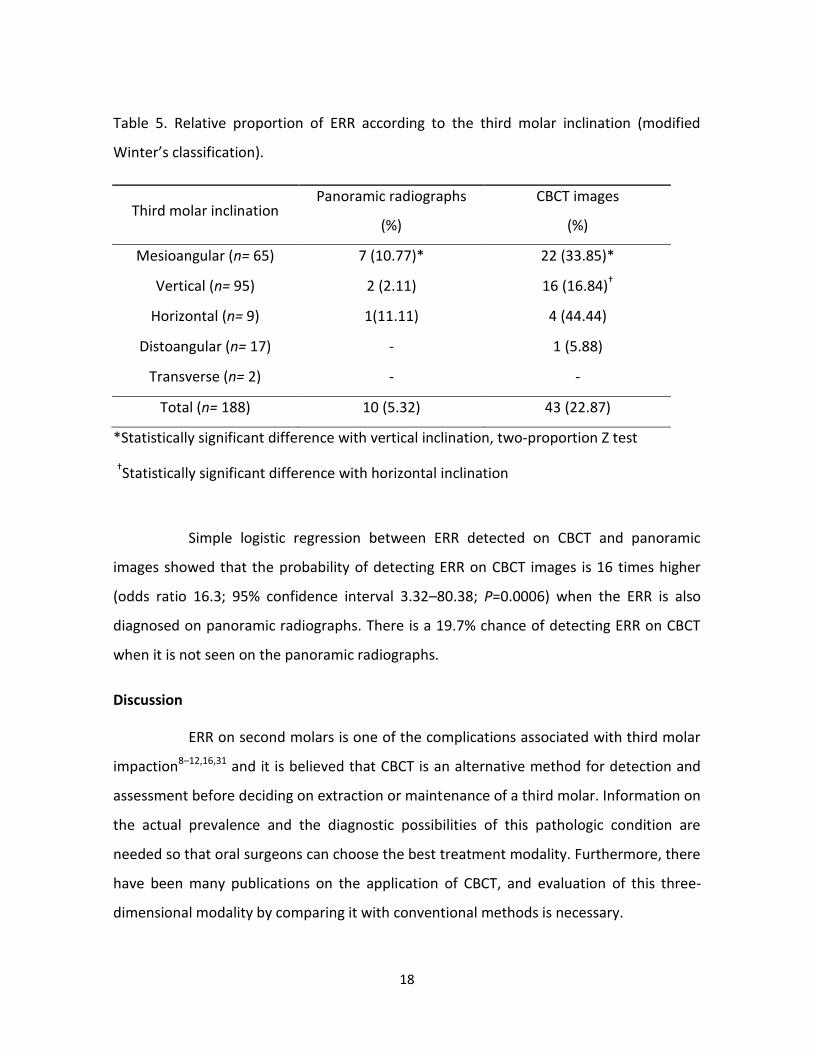
Table 5. Relative proportion of ERR according to the third molar inclination (modified
Winter’s classification).
Third molar inclination Panoramic radiographs
(%)
CBCT images
(%)
Mesioangular (n= 65) 7 (10.77)* 22 (33.85)*
Vertical (n= 95) 2 (2.11) 16 (16.84)†
Horizontal (n= 9) 1(11.11) 4 (44.44)
Distoangular (n= 17) - 1 (5.88)
Transverse (n= 2) - -
Total (n= 188) 10 (5.32) 43 (22.87)
*Statistically significant difference with vertical inclination, two-proportion Z test
†Statistically significant difference with horizontal inclination
Simple logistic regression between ERR detected on CBCT and panoramic
images showed that the probability of detecting ERR on CBCT images is 16 times higher
(odds ratio 16.3; 95% confidence interval 3.32–80.38; P=0.0006) when the ERR is also
diagnosed on panoramic radiographs. There is a 19.7% chance of detecting ERR on CBCT
when it is not seen on the panoramic radiographs.
Discussion
ERR on second molars is one of the complications associated with third molar
impaction8–12,16,31 and it is believed that CBCT is an alternative method for detection and
assessment before deciding on extraction or maintenance of a third molar. Information on
the actual prevalence and the diagnostic possibilities of this pathologic condition are
needed so that oral surgeons can choose the best treatment modality. Furthermore, there
have been many publications on the application of CBCT, and evaluation of this three-
dimensional modality by comparing it with conventional methods is necessary.
19
The correct diagnosis of dental impaction requires a detailed anamnesis,
clinical examination, and the use of complementary tests to ensure that correct treatment
decisions are made. A precise radiographic diagnosis is essential to evaluate the possible
outcomes related to unerupted third molars32. In oral and maxillofacial surgery,
panoramic radiography is initially requested to assess unerupted third molars and to
estimate the risk of inferior alveolar nerve damage33. In addition, it can be used to
evaluate the presence of ERR, distal caries, and periodontal bone loss in second molars5.
However, this imaging modality, like all two-dimensional radiographs, involves several
potential sources of misinterpretation, such as image enlargement, distortion due to
projection errors, blurred images, and complex maxillofacial structures when projected
onto a two-dimensional plane20,34. Intraoral images, such as periapical radiographs, are
also indicated to evaluate the integrity of the distal surface of the second molar9,12,13. In
the present study, periapical radiographs were not performed to avoid further radiation
exposure and because they have the same constraints as panoramic imaging34.
Assessment of root resorption and changes in root surface morphology usually
requires three-dimensional information, especially at the early stages20. The present study
highlights the fact that the CBCT allowed validation of the relationship between the
second and third molars. In 35 cases, ERR of the second molars was observed in the CBCT
images but not in the panoramic radiographs. The two methods agreed on the ERR status
in only 8 cases (4.3%). Similar results were found by Alqerban et al.34 in a study on canine
impaction. Comparing panoramic radiographs with two CBCT units, these authors
detected more lateral incisor root resorption using three-dimensional images. This could
be attributed to the fact the CBCT images provide diagnostic information in the sagittal,
axial, and coronal planes without overlap of structures; panoramic radiographs, on the
other hand, lack the third dimension.
In our study, based on CBCT images, 13 cases of ERR were associated with
maxillary second molars and 30 cases were associated with mandibular second molars,
giving a ratio of maxilla to mandible of 1:2.3. A higher number of cases of resorption in
20
lower second molars was also found in similar studies using periapical radiographs9 and
panoramic radiographs5. Our results can be justified by the complex process of third molar
eruption, particularly the mandibular teeth. The factors related to impaction of third
molars are lack of space, limited skeletal growth, distal eruption of dentition, vertical
condylar growth, increased crown size, and late maturation of third molars3,35. Other
authors have attributed this high rate of impingement, especially in mandibular teeth, to
late formation and the phylogeny of the mandible, which result in lack of space for normal
eruption36.
Knutsson et al.2 evaluated 666 patients before removal of mandibular third
molars and found that 548 patients had pathologic conditions associated with this
situation. Root resorption of the second molar was found on the radiographs of only 1% of
these cases (7 second molars). This prevalence is low in most studies ranging from <1% to
4.7%37, 0.3%6, 0.9%5, 1.4%4, and 7%9. Nemcovsky et al.12 found a higher prevalence of root
resorption (24.2%) in a sample of 186 periapical radiographs of third/second molars,
similar to the value in the present study on CBCT images (22.87%). Nemcovsky et al.12
believe that their choice of a sample of completely nonerupted third molars contributed
to this higher prevalence, because partially erupted teeth could extend the inflammatory
process to lower resistance areas without calcified tissues, thus reducing the pressure on
the roots of the second molar. In the present research, unlike the study of Nemcovsky et
al.12, partially erupted third molars were included as well as completely nonerupted teeth.
Therefore, this criterion cannot be used to justify the higher prevalence of ERR on the
CBCT images in our study. We believe that the results of the present research can be
explained by the better diagnostic ability of CBCT images compared with two-dimensional
radiographs25,34.
The literature shows that the position and angulation of third molars have an
impact on the pathologies and symptoms associated with this condition3,4. In the present
research, mesioangular and horizontal third molars were more associated with ERR on the
second molars, although the horizontal inclination is not so prevalent. These findings

21
corroborate studies that evaluated this relationship on conventional radiographs2,12. The
mesial inclinations, mesioangular and horizontal, have a bigger area of contact between
the third and second molars, leading us to believe that the inflammatory process is more
severe and has a greater potential to damage the dental surface.
The presence of root caries lesions on the distal surface of the second molars
included in this research cannot be completely discounted. However, some signs are more
associated with caries lesions, such as the relation between radiolucency and the oral
environment and the presence of a gap between the third and second molars that favors
food retention7,38. In addition, in a histologic study of 8 teeth diagnosed with ERR by
conventional intraoral radiographs, resorption was apparent histologically in all teeth16.
The results of this study indicate that radiographic distinction between ERR and distal
caries on second molars in proximity to impacted third molars is generally reliable.
For many years, removing or retaining unerupted third molars has been a
subject of discussion in the dental literature. The decision to remove third molars
associated with pathologic changes is often straightforward6,12,36,38, but prophylactically
removing an asymptomatic third molar may not be an easy decision. The detrimental
effect on the adjacent second molar should be considered during the decision-making
process12. We believe that when the diagnosis of ERR is confirmed, especially on CBCT
images, the oral surgeon should carefully evaluate the case and consider extraction of the
third molar followed by restoration, endodontic treatment of the second molar, or even
resection of the affected root, if necessary31. However, in some cases, a thin layer of
dentin appears between the resorptive defect and the pulp or the apical locations of this
defect make it unrestorable8,12. Fused roots or high trifurcation can also preclude root
resection8. In all these situations of severe resorption, especially those accompanied by
loss of periodontal support, extraction of the second molar and maintenance of the third
molar (spontaneous or active eruption) could be considered. CBCT images play an
important role in this treatment decision.
22
One limitation of the present research is that ERR was diagnosed only by
radiographs and tomographs, without correlation with clinical or intraoperative
information. This was because direct visualization of the defect during and after extraction
of the third molar is difficult. In addition, for ethical reasons, the histopathological analysis
of the resorbed defects was not indicated. In order to provide better scientific evidence
for the conclusions in this study, longitudinal research must be conducted by evaluating
patients over time.
CBCT should not be used routinely for assessment of unerupted teeth in the
context of root resorption diagnosis, but it may be indicated when conventional intraoral
radiography does not supply adequate information39. However, the use of CBCT for
localizing lower third molars in relation to the mandibular canal is well established in the
literature to improve surgical planning and inform the patient more adequately about the
risk of nerve damage40. Images acquired for that specific indication (relation between
canal and third molar root) may also be used to evaluate the integrity or resorption of the
distal surface of second molars. Using CBCT to provide the maximum data possible would
benefit patients and avoid additional radiation exposure.
According to our results and taking into consideration the three-dimensional
information obtained from CBCT, we found that a significantly higher number of cases of
ERR were detected by means of CBCT. The most common inclinations of third molars
associated with ERR in the second molar were mesioangular and horizontal. In conclusion,
when direct contact between the second molar and an unerupted mandibular third molar
is observed on panoramic radiographs, especially in mesioangular or horizontal third
molars, CBCT should be used to provide better evaluation of the case.
References
1. Tymofiyev O, Rottner K, Jakob PM, Richter E-J, Proff P. Three-dimensional
localization of impacted teeth using magnetic resonance imaging. Clin Oral Investig
2010: 14: 169–176.

23
2. Knutsson K, Brehmer B, Lysell L, Rohlin M. Pathoses associated with mandibular
third molars subjected to removal. Oral Surg Oral Med Oral Pathol Oral Radiol
Endod 1996: 82: 10–17.
3. Yamalik K, Bozkaya S. The predictivity of mandibular third molar position as a risk
indicator for pericoronitis. Clin Oral Investig 2008: 12: 9–14.
4. Akarslan ZZ, Kocabay C. Assessment of the associated symptoms, pathologies,
positions and angulations of bilateral occurring mandibular third molars: is there
any similarity? Oral Surg Oral Med Oral Pathol Oral Radiol Endod 2009: 108: e26–
e32.
5. Van der Linden W, Cleaton-Jones P, Lownie M. Diseases and lesions associated
with third molars. Review of 1001 cases. Oral Surg Oral Med Oral Pathol Oral
Radiol Endod 1995: 79: 142–145.
6. Al-Khateeb TH, Bataineh AB. Pathology associated with impacted mandibular third
molars in a group of Jordanians. J Oral Maxillofac Surg 2006: 64: 1598–1602.
7. Falci SG, de Castro CR, Santos RC, de Souza Lima LD, Ramos-Jorge ML, Botelho AM,
Dos Santos CR. Association between the presence of a partially erupted
mandibular third molar and the existence of caries in the distal of the second
molars. Int J Oral Maxillofac Surg 2012: 41: 1270–1274.
8. Oles RD. Root resorption associated with impacted third molars. Oral Surg Oral
Med Oral Pathol. 1979: 48: 281.
9. Nitzan D, Keren T, Marmary Y. Does an impacted tooth cause root resorption of the
adjacent one? Oral Surg Oral Med Oral Pathol 1981: 51: 221–224.
10. Girdler NM. The unpredictability of impacted third molar development- the
danger of passive observation. Br Dent J 1990: 168: 92.
24
11. Wang HY. Root resorption associated with impacted maxillary third molar. Oral
Surg Oral Med Oral Pathol 1992: 73: 765–766.
12. Nemcovsky CE, Libfeld H, Zubery Y. Effect of non-erupted 3rd molars on distal roots
and supporting structures of approximal teeth. A radiographic survey of 202 cases.
J Clin Periodontol. 1996: 23: 810–815.
13. Yamaoka M, Furusawa K, Ikeda M, Hasegawa T. Root resorption of mandibular
second molar teeth associated with the presence of the third molars. Aust Dent J.
1999: 44: 112–116.
14. Benenati FW. Root resorption: types and treatment. Gen Dent 1997: 45: 42–45.
15. Armas JM, Savarrio L, Brocklebank LM. External apical root resorption: two case
reports. Int Endod J 2008: 41: 997–1004.
16. Nemcovsky CE, Tal H, Pitaru S. Effect of non-erupted third molars on roots of
approximal teeth. A radiographic, clinical and histologic study. J Oral Pathol Med
1997: 26: 464–469.
17. Marks SC Jr, Cahill DR. Experimental study in the dog of the non-active role of the
tooth in the eruptive process. Arch Oral Biol 1984: 29: 311–322.
18. Ericson S, Bjerklin K, Falahat B. Does the canine dental follicle cause resorption of
permanent incisor roots? A computed tomographic study of erupting maxillary
canines. Angle Orthod 2002: 72: 95–104.
19. Sarrafpour B, Swain M, Li Q, Zoellner H. Tooth eruption results from bone
remodelling driven by bite forces sensed by soft tissue dental follicles: a finite
element analysis. PLoS One 2013: 8: e58803.
25
20. Strbac GD, Foltin A, Gahleitner A, Bantleon HP, Watzek G, Bernhart T. The
prevalence of root resorption of maxillary incisors caused by impacted maxillary
canines. Clin Oral Investig 2013: 17: 553–564.
21. Ericson S, Kurol PJ. Resorption of incisors after ectopic eruption of maxillary
canines: a CT study. Angle Orthod 2000: 70: 415–423.
22. Bjerklin K, Ericson S. How a computerized tomography examination changed the
treatment plans of 80 children with retained and ectopically positioned maxillary
canines. Angle Orthod 2006: 76: 43–51.
23. Alqerban A, Jacobs R, Souza PC, Willems G. In-vitro comparison of 2 cone-beam
computed tomography systems and panoramic imaging for detecting simulated
canine impaction-induced external root resorption in maxillary lateral incisors. Am
J Orthod Dentofacial Orthop 2009: 136: 764.e1–764.e11.
24. Scarfe WC, Farman AG, Sukovic P: Clinical applications of cone-beam computed
tomography in dental practice. J Can Dent Assoc 2006: 72: 75–80.
25. Liu DG, Zhang WL, Zhang ZY, Wu YT, Ma XC. Localization of impacted maxillary
canines and observation of adjacent incisor resorption with cone-beam computed
tomography. Oral Surg Oral Med Oral Pathol Oral Radiol Endod 2008: 105: 91–98.
26. Haney E, Gansky SA, Lee JS, Johnson E, Maki K, Miller AJ, Huang JC. Comparative
analysis of traditional radiographs and cone-beam computed tomography
volumetric images in the diagnosis and treatment planning of maxillary impacted
canines. Am J Orthod Dentofacial Orthop 2010: 137: 590–597.
27. Loubele M, Maes F, Jacobs R, van Steenberghe D, White SC, Suetens P.
Comparative study of image quality for MSCT and CBCT scanners for
26
dentomaxillofacial radiology applications. Radiat Prot Dosimetry 2008: 129: 222–
226.
28. Suomalainen A, Kiljunen T, Käser Y, Peltola J, Kortesniemi M. Dosimetry and image
quality of four dental cone beam computed tomography scanners compared with
multislice computed tomography scanners. Dentomaxillofac Radiol 2009: 38: 367–
378.
29. Ziegler CM, Woertche R, Brief J, Hassfeld S. Clinical indications for digital volume
tomography in oral and maxillofacial surgery. Dentomaxillofac Radiol 2002: 31:
126–130.
30. Miracle AC, Mukherji SK. Conebeam CT of the head and neck, part 1: physical
principles. AJNR Am J Neuroradiol 2009: 30: 1088–1095.
31. Pai AV, Khosla M. Root resection under the surgical field employed for extraction
of impacted tooth and management of external resorption. J Conserv Dent 2012:
15: 298–302.
32. Almendros-Marqués N, Berini-Aytés L, Gay-Escoda C. Influence of lower third
molar position on the incidence of preoperative complications. Oral Surg Oral Med
Oral Pathol Oral Radiol Endod 2006: 102: 725–732.
33. Neves FS, de Almeida SM, Bóscolo FN, Haiter-Neto F, Alves MC, Crusoé-Rebello I,
Campos PS. Risk assessment of inferior alveolar neurovascular bundle by
multidetector computed tomography in extractions of third molars. Surg Radiol
Anat 2012: 34: 619–624.
34. Alqerban A, Jacobs R, Fieuws S, Willems G. Comparison of two cone beam
computed tomographic systems versus panoramic imaging for localization of
27
impacted maxillary canines and detection of root resorption. Eur J Orthod 2011:
33: 93–102.
35. Hattab FN. Positional changes and eruption of impacted mandibular third molars in
young adults. A radiographic 4-year follow-up study. Oral Surg Oral Med Oral
Pathol Oral Radiol Endod 1997: 84: 604–608.
36. Lysell L, Rohlin M. A study of indications used for removal of the mandibular third
molar. Int J Oral Maxillofac Surg 1988: 17: 161–164.
37. Nordenram A, Hultin M, Kjellman O, Ramström G. Indications for surgical removal
of the mandibular third molar. Study of 2,630 cases. Swed Dent J 1987: 11: 23–29.
38. Ozeç I, Hergüner Siso S, Taşdemir U, Ezirganli S, Göktolga G. Prevalence and factors
affecting the formation of second molar distal caries in a Turkish population. Int J
Oral Maxillofac Surg 2009: 38: 1279–1282.
39. SedentextCT Project. Radiation protection: cone beam CT for dental and
maxillofacial radiology. Evidence-based guidelines.
http://www.sedentexct.eu/files/radiation_protection_172.pdf [Accessibility
verified May 13, 2013]
40. Ghaeminia H, Meijer GJ, Soehardi A, Borstlap WA, Mulder J, Vlijmen OJ, Bergé SJ,
Maal TJ. The use of cone beam CT for the removal of wisdom teeth changes the
surgical approach compared with panoramic radiography: a pilot study. Int J Oral
Maxillofac Surg 2011: 40: 834–339.
28
CAPÍTULO 2
Este artigo será submetido à apreciação, visando publicação pelo periódico
Journal of Oral and Maxillofacial Surgery, considerado Qualis A2 pela CAPES. A formatação
do artigo baseou-se nas “Instruções aos Autores” preconizadas pela editora do periódico.
The mesial inclination of impacted third molars and its propensity to stimulate external
root resorption in second molars – a cone beam CT evaluation
Anne Caroline Costa Oenning1, Saulo Leonardo Sousa Melo2, Francisco Carlos Groppo3,
Francisco Haiter-Neto1
1 Department of Oral Diagnosis, Division of Oral Radiology, Piracicaba Dental School,
University of Campinas, São Paulo, Brazil.
2 Department of Oral Pathology, Radiology and Medicine, University of Iowa College of
Dentistry, Iowa City, IA.
3Department of Morphology, Division of Pharmacology, Piracicaba Dental School,
University of Campinas, São Paulo, Brazil.
Corresponding author:
Anne Caroline Costa Oenning
Faculdade de Odontologia de Piracicaba, Departamento de Diagnóstico Oral
Av Limeira, 901, Caixa postal 52
Piracicaba/SP, Brazil, 13414-903
e-mail: [email protected]
Key-words: Third molar; root resorption; cone beam computed tomography.
Abstract
Objective: To investigate the presence of external root resorption (ERR) in second molars
adjacent to horizontally and mesioangularly impacted mandibular third molars by CBCT

29
imaging. In addition, subject characteristics (age and sex) and third molar depth was
correlated with the presence of ERR.
Methods: The sample consisted of 116 scans: 58 were acquired on i-CAT classic and 58 on
Picasso-Trio. The sample included 70 women and 46 men with a mean age of 23.7 years.
Two observers recorded the presence of ERR in the second molars; the inclination and the
depth of third molars in relation to bone and soft tissues; and third molars classification
according to Pell & Gregory. The observers indicated the location and severity of ERR as
well. Data were analyzed by ANOVA, Mann-Whitney and chi-square tests. The Kappa test
was used to analyze the intra-observer agreement.
Results: Men and women presented with ERR in the same proportion (p = 0.9575). The
overall prevalence was 49.43%. There were no statistically significant differences in the
detection of ERR in images from both devices and between mesioangular and horizontal
inclinations (p> 0.05). The Kappa test showed excellent reproducibility values (Kappa =
0.7778). There was a lower proportion of affected individuals aged 14-24 years and ERR in
teeth adjacent to Class C third molars.
Conclusions: Mesially inclined third molars have a greater potential of being associated
with ERR in second molars, which was demonstrated by the high prevalence of the
condition in the overall sample. Class A and Class B third molars of patients over 24 years
were more associated with the presence of ERR in the adjacent teeth.
Introduction
The impaction of upper and lower third molars may be associated with breaks
in the integrity of the distal aspect of the adjacent second molar due to dental caries or
external root resorption (ERR).1–5 Unlike carious lesions, ERR is usually asymptomatic and
aseptic, unless the pulp cavity has been involved or the lesion has been secondarily
infected.6 The literature attributes the occurrence of this type of resorption to the
pressure from the impacted tooth. This pressure itself, with associated factors derived
30
from the follicle that contribute to inflammation, is able to activate clastic cells
responsible for triggering resorption.2,7,8
Studies on periapical and panoramic radiographs have investigated the
presence of ERR in second molars adjacent to impacted third molars, and other
pathological conditions associated to such impacted teeth.1–4,9–11 The majority of these
studies revealed a low prevalence of ERR in second molars (0.3 to 7 %).3,4,7,10 However, in
a previous study comparing Cone Beam CT (CBCT) and panoramic images, the detection of
ERR on second molars was 4.3 times greater when the three-dimensional images were
used. It has been proven that multiplanar images, which are free of overlapping, provide
more accurate diagnoses in relation to root resorption associated with impacted
teeth.12,13
It is known that the position and inclination of third molars predispose to the
development of pathological conditions.4,14 While the presence of a gap between retained
and adjacent teeth allows biofilm accumulation, and consequent development of carious
lesions,5 direct contact between these teeth favors the occurrence of resorption.15
Although to date most studies revealed a low prevalence of resorption, it is believed that
horizontally and mesioangularly impacted third molars (i.e. larger contact surfaces) should
be associated with a higher frequency of ERR on second molars.1,11,16 The fact that those
were studies performed using bidimensional images and heterogeneous samples may be
adding bias to their findings.1,3,4,7,10,11
Therefore, the aim of this research was to investigate the presence of ERR in
second molars adjacent to horizontally and mesioangularly impacted mandibular third
molars by CBCT imaging. In addition, subject characteristics (age and sex) and third molar
depth was correlated with the presence of ERR.
Materials and methods
The study protocol was reviewed and approved by the Institutional Review
Board of the University of Campinas, School of Dentistry at Piracicaba, São Paulo, Brazil.
31
A retrospective cross-sectional study was designed to evaluate CBCT images
from a dental radiology service database. A total of 217 CBCT scans of patients referred
for evaluation of third molars were available: 120 scans acquired using an i-CAT classic
unit (Imaging Sciences International, Hatfield, PA) and 97 on a Picasso-Trio unit (E-WOO
Technology, Giheung-gu, Republic of Korea). Scans to be included in the sample were
required to present a second molar adjacent to a horizontally or mesioangularly impacted
mandibular third molar in the FOV. Images of completely erupted third molars, impacted
teeth associated with cystic or tumor lesions, non-odontogenic tumors or bone defects
extending to the posterior mandible, third molars with less than 2/3 of root developed,
and/or second molars showing extensive carious lesions were excluded. The final sample
comprised CBCT scans of 70 women and 46 men, with a mean age of 23.7 years (range: 14
to 62 years).
Image acquisition protocols were standardized for each unit according to
manufacturer recommendations, as follows: i-CAT unit was set up at 120 kVp, 8 mA, 13 x
17 cm FOV and 0.25 mm voxel size; and the Picasso-Trio unit at 90 kVp, 5 mA, 8.5 x 12 cm
FOV, 0.2 mm voxel size and metal artifact reduction algorithm. All resulting images were
displayed on a 21-inch LCD monitor with a matrix resolution of 1280 x 1024. The
evaluations were performed in the proprietary software of each device: Xoran for i-CAT
(Xoran CAT, version 3.0.34, Xoran Technologies, Ann Arbor, MI), and EZ3D for Picasso-Trio
(E-WOO Technology, Giheung-gu, Republic of Korea).
The images were coded and shown to the observers in random order, and
under dim-light conditions. The observers were two previously trained oral and
maxillofacial radiologists with at least 3 years of experience in CBCT diagnosis. They were
instructed to consider the presence of ERR on the second molar only when a clear loss of
substance could be detected on the root surface, as preconized by Al-Khateeb &
Bataineh.3 Once detected, ERR was classified according to its location (cervical, middle or
apical root thirds) and severity (mild, moderate or severe). This last classification was

32
based on Ericson et al. classification of slight (involving less than half the thickness dentin),
moderate (involving half of dentin or more) and severe (involving the pulp cavity).18
The observers also indicated both inclination (mesioangular or horizontal) and
depth of the third molars, according to the classification of Winter and Pell & Gregory
respectively. According to Winter, a third molar crown is mesioangular when it is inclined
to the mesial with the long axis at an angle range 31-60° to the occlusal plane; and it is
horizontal when its axis is in a 0-30° angle to a tangent to the occlusal plane of the
adjacent teeth.17 Pell & Gregory categorize third molars according to their horizontal and
vertical positions regarding the mandibular ramus, amelocemental junction and the
occlusal plane of the second molar. In the present study, the following classes that
indicate the vertical position of the third molars were adopted: A - the third molar is near
the occlusal plane of the second molar, B - the third molar is located in a level between
the occlusal plane and amelocemental junction of the second molar, and C - the third
molar is below the amelocemental junction of the second molar. The depth of the third
molar was also classified in relation to bone and soft tissues. In this case the tooth could
be considered unerupted, when completely surrounded by bone tissue; or partially
erupted, when hidden by soft tissue or partially visible in the oral cavity.
All analyses and records were performed independently by each observer.
Disagreement between their answers was resolved by consensus in a second moment.
After 30 days, 36 scans were re-evaluated (18 for each device, corresponding to
approximately 20% of the total sample). Cohen’s kappa was used to calculate
intraobserver agreement (poor agreement, 0.40; moderate agreement, 0.40-0.59; good
agreement, 0.60-0.74; excellent agreement, 0.75-1.00). All numerical data was compared
by one way analysis of variance (ANOVA), in cases of normal distribution, and Mann-
Whitney, when a nonparametric test was required. Categorical data was compared by a
chi-square contingency test or goodness of fit chi-square test. The level of significance was
set at p < 0.05. Data analyses were performed using Prism (v. 6.0, GraphPad Software Inc.,
33
La Jolla, CA) and BioEstat (v. 5.0, Manuel Ayres Software Informer, Belem, PA, Brazil)
software.
Results
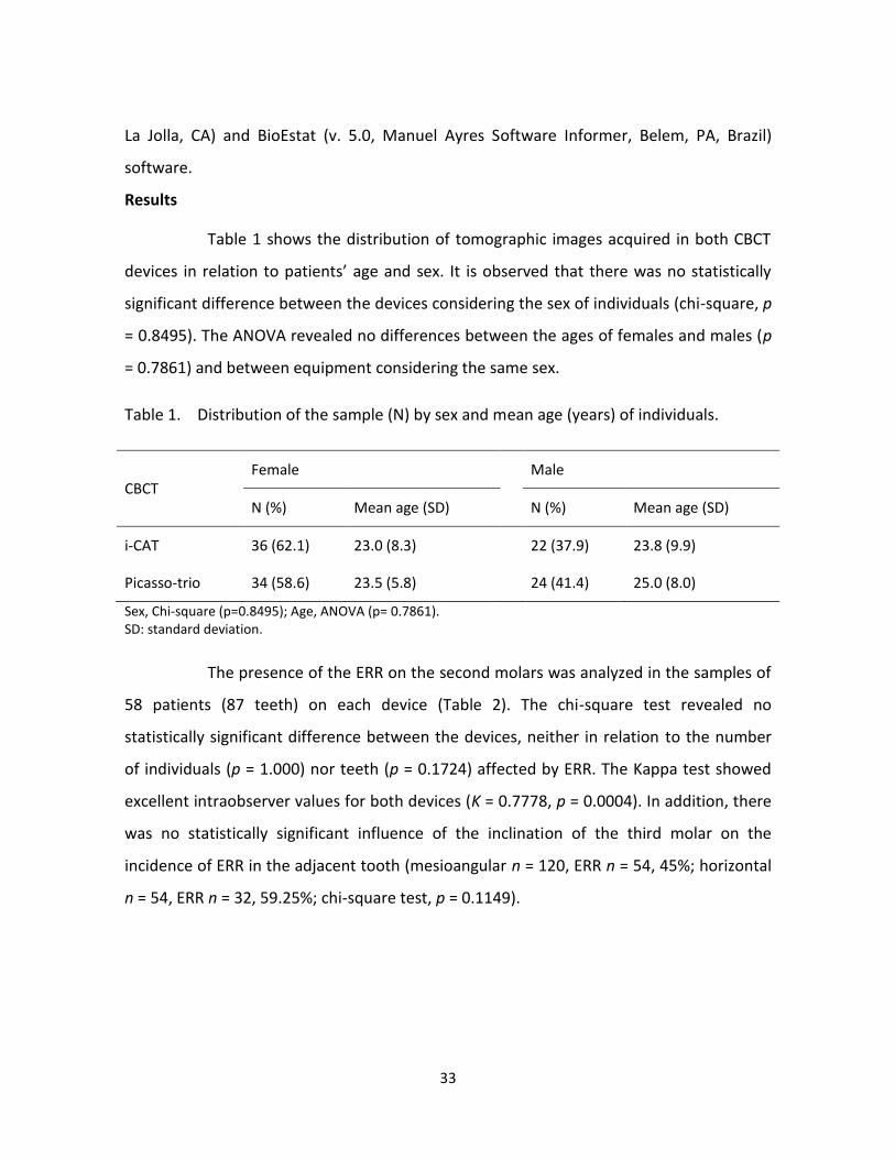
Table 1 shows the distribution of tomographic images acquired in both CBCT
devices in relation to patients’ age and sex. It is observed that there was no statistically
significant difference between the devices considering the sex of individuals (chi-square, p
= 0.8495). The ANOVA revealed no differences between the ages of females and males (p
= 0.7861) and between equipment considering the same sex.
Table 1. Distribution of the sample (N) by sex and mean age (years) of individuals.
CBCT Female Male
N (%) Mean age (SD) N (%) Mean age (SD)
i-CAT 36 (62.1) 23.0 (8.3) 22 (37.9) 23.8 (9.9)
Picasso-trio 34 (58.6) 23.5 (5.8) 24 (41.4) 25.0 (8.0)
Sex, Chi-square (p=0.8495); Age, ANOVA (p= 0.7861). SD: standard deviation.
The presence of the ERR on the second molars was analyzed in the samples of
58 patients (87 teeth) on each device (Table 2). The chi-square test revealed no
statistically significant difference between the devices, neither in relation to the number
of individuals (p = 1.000) nor teeth (p = 0.1724) affected by ERR. The Kappa test showed
excellent intraobserver values for both devices (K = 0.7778, p = 0.0004). In addition, there
was no statistically significant influence of the inclination of the third molar on the
incidence of ERR in the adjacent tooth (mesioangular n = 120, ERR n = 54, 45%; horizontal
n = 54, ERR n = 32, 59.25%; chi-square test, p = 0.1149).
34
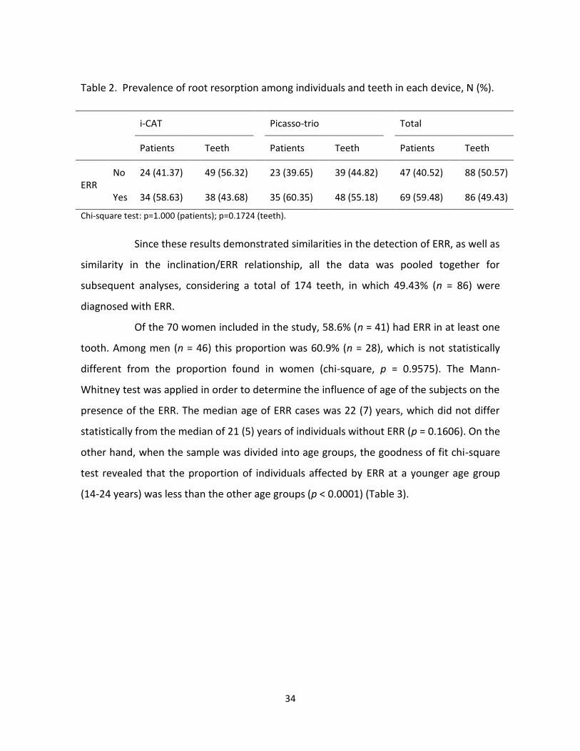
Table 2. Prevalence of root resorption among individuals and teeth in each device, N (%).
i-CAT Picasso-trio Total
Patients Teeth Patients Teeth Patients Teeth
ERR No 24 (41.37) 49 (56.32) 23 (39.65) 39 (44.82) 47 (40.52) 88 (50.57)
Yes 34 (58.63) 38 (43.68) 35 (60.35) 48 (55.18) 69 (59.48) 86 (49.43)
Chi-square test: p=1.000 (patients); p=0.1724 (teeth).
Since these results demonstrated similarities in the detection of ERR, as well as
similarity in the inclination/ERR relationship, all the data was pooled together for
subsequent analyses, considering a total of 174 teeth, in which 49.43% (n = 86) were
diagnosed with ERR.
Of the 70 women included in the study, 58.6% (n = 41) had ERR in at least one
tooth. Among men (n = 46) this proportion was 60.9% (n = 28), which is not statistically
different from the proportion found in women (chi-square, p = 0.9575). The Mann-
Whitney test was applied in order to determine the influence of age of the subjects on the
presence of the ERR. The median age of ERR cases was 22 (7) years, which did not differ
statistically from the median of 21 (5) years of individuals without ERR (p = 0.1606). On the
other hand, when the sample was divided into age groups, the goodness of fit chi-square
test revealed that the proportion of individuals affected by ERR at a younger age group
(14-24 years) was less than the other age groups (p < 0.0001) (Table 3).
35
Table 3. Age distribution of the sample.
Age group N (%) ERR (%)
14 – 24 84 (100) 48 (57.1)*
25 – 35 24 (100) 16 (66.7)
36+ 8 (100) 5 (62.5)
*Chi-square test: p<0.0001
Among 174 third molars that had been included in the study sample, 147 were
classified as partially erupted and 27 as unerupted. In these two groups, 51.7 % (n = 76)
and 37.03 % (n = 10) of third molars were stimulating ERR on the adjacent teeth,
respectively. The chi-square test did not show a relation between the depth of the third
molar and the presence or absence of the RRE in second molars (p = 0.2335). However,
according to the classification of Pell & Gregory, teeth positioned in class C were less
associated with ERR in second molars when compared to other classes (p = 0.0047). The
teeth classified as A and B did not differ statistically (p = 0.0953) (Table 4). Although not
statistically associated with ERR, figure 1 shows two examples of class C third molars that
notably injured the adjacent second molar.
Table 4. Third molars distribution according to Pell & Gregory classification and ERR
3rd molar (Pell & Gregory)
Total (%) A (%) B (%) C (%)
ERR No 13 (32.5) 38 (48,7) 37 (66,1) 88 (50.6)
Yes 27 (67.5) 40 (51,3) 19 (33.9)* 86 (49.4)
Total 40 (100) 78 (100) 56 (100) 174 (100)
*Statistically significant difference from A and B (chi-square; p=0.0047)
36
Figure 1. Two examples of notable ERR on right second molars, with adjacent
third molars in Pell & Gregory class C (below the cervical line of the adjacent
tooth). TCFC images A1 (sagittal) and A2 (axial) were acquired in the i-CAT
device, images B1 and B2 in Picasso-trio.
There was significantly more slight resorption (p <0.0001), as well as
resorption located in the cervical third of the root (chi-square, p <0.0001). This
relationship can be appreciated in Table 5, as it shows that over half of the sample of
resorbed second molars (n = 49, 56.97%) presented both situations simultaneously.
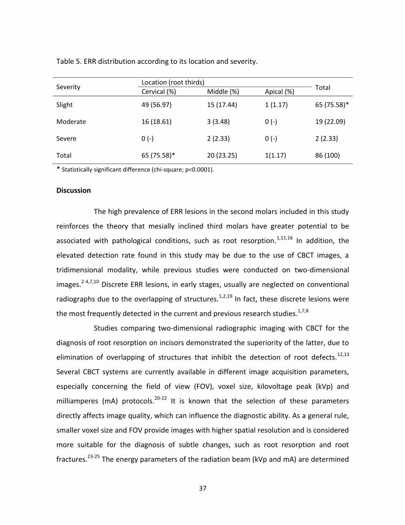
37
Table 5. ERR distribution according to its location and severity.
Severity Location (root thirds)
Total Cervical (%) Middle (%) Apical (%)
Slight 49 (56.97) 15 (17.44) 1 (1.17) 65 (75.58)*
Moderate 16 (18.61) 3 (3.48) 0 (-) 19 (22.09)
Severe 0 (-) 2 (2.33) 0 (-) 2 (2.33)
Total 65 (75.58)* 20 (23.25) 1(1.17) 86 (100)
* Statistically significant difference (chi-square; p<0.0001).
Discussion
The high prevalence of ERR lesions in the second molars included in this study
reinforces the theory that mesially inclined third molars have greater potential to be
associated with pathological conditions, such as root resorption.1,11,16 In addition, the
elevated detection rate found in this study may be due to the use of CBCT images, a
tridimensional modality, while previous studies were conducted on two-dimensional
images.2-4,7,10 Discrete ERR lesions, in early stages, usually are neglected on conventional
radiographs due to the overlapping of structures.1,2,19 In fact, these discrete lesions were
the most frequently detected in the current and previous research studies.1,7,8
Studies comparing two-dimensional radiographic imaging with CBCT for the
diagnosis of root resorption on incisors demonstrated the superiority of the latter, due to
elimination of overlapping of structures that inhibit the detection of root defects.12,13
Several CBCT systems are currently available in different image acquisition parameters,
especially concerning the field of view (FOV), voxel size, kilovoltage peak (kVp) and
milliamperes (mA) protocols.20-22 It is known that the selection of these parameters
directly affects image quality, which can influence the diagnostic ability. As a general rule,
smaller voxel size and FOV provide images with higher spatial resolution and is considered
more suitable for the diagnosis of subtle changes, such as root resorption and root
fractures.23-25 The energy parameters of the radiation beam (kVp and mA) are determined
38
according to the device and shall not exceed the limits that can significantly increase the
radiation dose to the patient.26,27 However, very low values can impair the final image
quality by increasing the noise.28 In this study, all patients were scanned according to the
School’s protocol and by following manufacturer’s recommendations. The standardization
of same parameters for different devices is an arduous task, since each system has a
specific configuration that should be followed depending on the case.20 For this study,
FOVs that included both ipsilateral maxillary and mandibular third molars, smallest voxel
size and the average energy factors recommended by the manufacturers were selected.
One should take into account the fact that the database was built with images of different
individuals. However, for ethical reasons related to patient exposure to ionizing radiation,
the same individuals could not be scanned on both devices at the same time. Although
there were inherent differences on the protocols adopted for each device, there was no
statistically significant difference in the detection of ERR. In fact, it allowed us to consider
the entire sample for subsequent analyses.
Over half the sample (59.48 %) presented with at least one adjacent second
molar resorbed by a mesioangularly or horizontally impacted third molar. The horizontal
inclination of the third molar is one of the least frequently encountered positioning in a
population.9-11,29 The data available on this inclination usually comprise small samples that
are often difficult to analyze. However, even with a reasonable increase of sample size in
this study (n = 54), horizontal third molars presented no statistically significant difference
in the presence of ERR in the second molar compared with mesioangular third molars.
This result suggests that the inflammatory process triggering resorption in second molars
may be similar in both situations. Moreover, a slightly larger contact surface between
teeth, as observed in horizontal impaction, may not represent an important component
for the occurrence of ERR in second molars.
The lack of space for eruption of the third molars is cited as a major cause for
impaction of these teeth, especially the mandibular third molars that appear often
overlapped by the second molars.14,30 Although studies have reported a higher frequency
39
of ERR in second molars adjacent to impacted third molar in male subjects, it is unclear
whether the difference in size of the teeth and/or jaws may be an important factor for
these results.2,7 Nitzan et al. reported that sex hormones may have interfered in their
findings of a higher prevalence of ERR in second molars in men.7 Our results correlating
sex and ERR does not contribute to this discussion, since men and women showed lesions
in the same proportion.
CBCT does not offer an adequate contrast resolution for a proper appreciation
of anatomical or pathological changes in the soft tissue; in addition, image noise can
decrease the outline definition of this tissue.31,32 For this reason, in this study, the third
molars hidden by soft tissue and those partially visible in the oral cavity were grouped as
partially erupted. It was observed that there was no difference between unerupted
(completely surrounded by bone tissue) and partially erupted teeth regarding the
presence of ERR. However, when the depth of the third molar was classified according to
Pell & Gregory, class C third molars (located below the amelocemental junction of the
adjacent tooth) were less associated with the presence ERR in the second molar. Lesions
of ERR were more frequently detected in the cervical region of the second molars when
the third molars were more superiorly positioned (classes A and B). These findings
highlight the fact that the cervical portion of the root, i.e. amelocemental junction, is
more susceptible to the inflammatory process which culminates in ERR.7 On the other
hand, Nemcovsky et al., who only evaluated completely unerupted third molars, reported
that the apical region of the adjacent second molar was the most susceptible to ERR
injuries.1 These authors also stated that the pressure on the periodontal ligament and
roots of the second molar is reduced when third molars are partially erupted.
Research has shown that the decision to maintain an asymptomatic impacted
third molar has increased the risk of periodontal disease in young adults and middle-aged
individuals.33-35 The higher frequency of ERR lesions in individuals aged over 24 years
found in this study also draws attention to the possible risk of passive observation of
impacted third molars, a fact already reported in previous studies with conventional
40
radiographs.1,2,16,19,36,37 The research published by Sarrafpour et al. indicates that eruptive
tooth movements do not cease with completion of root formation and closure of the
apex, which means that a retained tooth continues to exert pressure on the adjacent
tooth, therefore stimulating root resorption and/or its progression.38 However,
asymptomatic third molar removal represents a surgical procedure, with related cost and
risk of complications.39,40 Unfortunately, there is not a consensus guideline for the
management, either removal or active surveillance, of impacted third molars yet.37 A
systematic review published in the Cochrane database concluded that there is insufficient
scientific evidence to justify prophylactic removal of asymptomatic third molars.41
Contrarily, the American Association of Oral and Maxillofacial Surgeons (AAOMS) recently
published a guideline recommending the extraction of the asymptomatic third molars
based on clinical studies that investigated the occurrence and progression of lesions
related to impacted teeth.42-44 The AAOMS also recommends that new studies should be
conducted in order to determine the prevalence of pathological conditions associated
with third molars and adjacent second molars. Longitudinal studies known as “Third
Molars Clinical Trials” have been encouraged and sponsored for this purpose.45
It is completely justifiable the continued interest of surgeons and researchers
on evidences to support the management of third molars. Based on the present results, it
should be suggested that: (1) mesially inclined third molars have a greater potential of
being associated with ERR in second molars, which was demonstrated by the high
prevalence of the condition (49.43%) in the overall sample; (2) there was no greater
predilection between mesioangularly or horizontally impacted third molar inclination in
inducing ERR on second molars; and (3) class A and B third molars of patients over 24
years were more associated with the presence of ERR in the adjacent teeth.
References
1. Nemcovsky CE, Libfeld H, Zubery Y: Effect of non-erupted 3rd molars on distal roots and
supporting structures of approximal teeth. A radiographic survey of 202 cases. J Clin
Periodontol 23: 810, 1996
41
2. Yamaoka M, Furusawa K, Ikeda M, et al: Root resorption of mandibular second
molar teeth associated with the presence of the third molars. Aust Dent J 44: 112,
1999
3. Al-Khateeb TH, Bataineh, AB. Pathology associated with impacted mandibular third
molars in a group of Jordanians. J Oral Maxillofac Surg 64: 1598, 2006
4. Akarslan ZZ, Kocabay C: Assessment of the associated symptoms, pathologies,
positions and angulations of bilateral occurring mandibular third molars: is there
any similarity? Oral Surg Oral Med Oral Pathol Oral Radiol Endod 108: e26, 2009
5. Falci SG, de Castro CR, Santos RC, et al: Association between the presence of a
partially erupted mandibular third molar and the existence of caries in the distal of
the second molars. Int J Oral Maxillofac Surg 41: 1270, 2012
6. Fuss Z, Tsesis I, Lin S: Root resorption--diagnosis, classification and treatment
choices based on stimulation factors. Dent Traumatol 19: 175, 2003
7. Nitzan D, Keren T, Marmary Y: Does an impacted tooth cause root resorption of the
adjacent one? Oral Surg Oral Med Oral Pathol 51: 221, 1981
8. Nemcovsky CE, Tal H, Pitaru S: Effect of non-erupted third molars on roots of
approximal teeth. A radiographic, clinical and histologic study. J Oral Pathol Med 26:
464, 1997
9. Kahl B, Gerlach KL, Hilgers RD: A long-term, follow-up, radiographic evaluation of
asymptomatic impacted third molars in orthodontically treated patients. Int J Oral
Maxillofac Surg 23: 279, 1994
42
10. Van der Linden W, Cleaton-Jones P, Lownie M: Diseases and lesions associated with
third molars. Review of 1001 cases. Oral Surg. Oral Med Oral Pathol Oral Radiol
Endod 79:142, 1995
11. Knutsson K, Brehmer B, Lysell L: Pathoses associated with mandibular third molars
subjected to removal. Oral Surg Oral Med Oral Pathol Oral Radiol Endod 82: 10,
1996
12. Alqerban A, Jacobs R, Souza PC: In-vitro comparison of 2 cone-beam computed
tomography systems and panoramic imaging for detecting simulated canine
impaction-induced external root resorption in maxillary lateral incisors. Am J
Orthod Dentofacial Orthop 136: 764, 2009
13. Alqerban A, Jacobs R, Fieuws S. et al: Comparison of two cone beam computed
tomographic systems versus panoramic imaging for localization of impacted
maxillary canines and detection of root resorption. Eur J Orthod 33: 93, 2011
14. Yamalik K, Bozkaya S: The predictivity of mandibular third molar position as a risk
indicator for pericoronitis. Clin Oral Investig 12: 9, 2008
15. Strbac GD, Foltin A, Gahleitner A, et al: The prevalence of root resorption of
maxillary incisors caused by impacted maxillary canines. Clin Oral Investig 17: 553,
2013
16. Romero CJ: Root resorption of second molar related to impacted 3rd molar. Oral
Surg Oral Med Oral Pathol 32: 502, 1971
17. Ericson S, Bjerklin K, Falahat B: Does the canine dental follicle cause resorption of
permanent incisor roots? A computed tomographic study of erupting maxillary
canines. Angle Orthod 72: 95, 2002
43
18. Almendros-Marqués N, Berini-Aytés L, Gay-Escoda C: Evaluation of intraexaminer
and interexaminer agreement on classifying lower third molars according to the
systems of Pell and Gregory and of Winter. J Oral Maxillofac Surg 66: 893, 2008
19. Wang HY: Root resorption associated with impacted maxillary third molar. Oral Surg
Oral Med Oral Patho 73: 765, 1992
20. Alqerban A, Jacobs R, Fieuws S, et al: Comparison of 6 cone-beam computed
tomography systems for image quality and detection of simulated canine
impaction-induced external root resorption in maxillary lateral incisors. Am J
Orthod Dentofacial Orthop 140: e129, 2011
21. Kamburoğlu K, Murat S, Kolsuz E, et al: Comparative assessment of subjective
image quality of cross-sectional cone-beam computed tomography scans. J Oral Sci
53: 501, 2011
22. Bamba J, Araki K, Endo A, et al: Image quality assessment of three cone beam CT
machines using the SEDENTEXCT CT phantom. Dentomaxillofac Radiol 42:
20120445, 2013.
23. Scarfe WC, Farman AG, Sukovic P: Clinical applications of cone-beam computed
tomography in dental practice. J Can Dent Assoc 72: 75, 2006
24. Melo SLS; Bortoluzzi EA, Abreu M, et al: Diagnostic ability of a cone-beam
computed tomography scan to assess longitudinal root fractures in prosthetically
treated teeth. J Endod 36: 1879, 2010
25. Shelley A M, Brunton P, Horner K: Subjective image quality assessment of cross
sectional imaging methods for the symphyseal region of the mandible prior to
dental implant placement. J Dent 39: 764, 2011

44
26. Palomo JM, Rao PS, Hans MG: Influence of CBCT exposure conditions on radiation
dose. Oral Surg Oral Med Oral Pathol Oral Radiol Endod 105: 773, 2008
27. Panmekiate S, Apinhasmit W, Petersson A: Effect of electric potential and current
on mandibular linear measurements in cone beam CT. Dentomaxillofac Radiol 41:
578, 2012
28. Sur J, Seki K, Koizumi, H, Nakajima K, Okano T: Effects of tube current on cone-beam
computerized tomography image quality for presurgical implant planning in vitro.
Oral Surg Oral Med Oral Pathol Oral Radiol Endod 110: e29, 2010
29. Peterson LJ, Ellis E, Hupp JR, Tucker MR: Contemporary Oral and Maxillofacial
Surgery. St. Louis. MO. Mosby, 1993,pp239.
30. Hattab FN: Positional changes and eruption of impacted mandibular third molars in
young adults. A radiographic 4-year follow-up study. Oral Surg Oral Med Oral Pathol
Oral Radiol Endod 84: 604, 1997
31. Arai Y, Tammisalo E, Iwai K, et al: Development of a compact computed
tomographic apparatus for dental use. Dentomaxillofac Radiol 28: 245, 1999
32. Scarfe WC, Farman AG: What is cone-beam CT and how does it work? Dent Clin
North Am 52: 707, 2008
33. Blakey GH Marciani RD, Haug RH, et al: Periodontal pathology associated with
asymptomatic third molars. J Oral Maxillofac Surg 60: 1227, 2002
34. Phillips C, Norman J, Jaskolka M, et al: Changes over time in position and
periodontal probing status of retained third molars. J Oral Maxillofac Surg 65: 2011,
2007
45
35. Offenbacher S, Beck JD, Moss KL, et al: What are the local and systemic implications
of third molar retention? J Oral Maxillofac Surg 70: S58, 2012
36. Oles RD: Root resorption associated with impacted third molars. Oral Surg Oral Med
Oral Pathol 48: 281,1979
37. Girdler NM: The unpredictability of impacted third molar development-the danger
of passive observation. Br Dent J 168: 92, 1990
38. Sarrafpour B, Swain M, Li Q, et al: Tooth eruption results from bone remodelling
driven by bite forces sensed by soft tissue dental follicles: a finite element analysis.
PLoS One 8: e58803, 2013
39. Dodson TB: Surveillance as a management strategy for retained third molars: is it
desirable? J Oral Maxillofac Surg 70: S20, 2012
40. Nunn ME, Fish MD, Garcia RI, et al: Retained asymptomatic third molars and risk for
second molar pathology. J Dent Res 92: 1095, 2013
41. Mettes TG, Nienhuijs MEL, van der Sanden WJM, et al: Interventions for treating
asymptomatic impacted wisdom teeth in adolescents and adults. Cochrane
database Syst Rev 18: CD003879, 2005
42. American Association of Oral and Maxillofacial Surgeons: The Management of
Impacted Third Molar Teeth. URL acessed on 11/20/2013 at:
http://www.aaoms.org/docs/practice_mgmt/condition_statements/impacted_thir
d_molars.pdf, 2013
43. Blakey GH, Golden BA, White RP, et al: Changes over time in the periodontal status
of young adults with no third molar periodontal pathology at enrollment. J Oral
Maxillofac Surg 67: 2425, 2009
46
44. Garaas RN, Fisher EL, Wilson GH, et al: Prevalence of third molars with caries
experience or periodontal pathology in young adults. J Oral Maxillofac Surg 70: 507,
2012
45. White RP: Progress report on third molar clinical trials. J Oral Maxillofac Surg 65:
377, 2007

47
CONSIDERAÇÕES GERAIS
Com o presente trabalho espera-se contribuir não somente com o diagnóstico
da RRE associada à impactação dos terceiros molares, como também na resolução de uma
importante dúvida clínica comum nos consultórios odontológicos: a remoção ou
manutenção de um terceiro molar retido assintomático. Essa questão ainda é muito
discutida nos meios clínico e científico (Gomes et al., 2004). A dicotomia existente nas
opiniões da Associação Americana de Cirurgiões Bucomaxilofaciais, que recomenda a
remoção profilática em indivíduos jovens para garantir o reparo ideal, e da Associação
Americana de Saúde Pública, que condena a exposição de indivíduos à morbidade do
procedimento e a custos desnecessários, demonstra que esse assunto ainda necessita de
evidência científica (Nunn et al., 2013).
Os cirurgiões dentistas conhecem os riscos e custos envolvidos nas cirurgias de
remoção de terceiros molares retidos. Problemas podem surgir por complicações
inflamatórias, infecções trans ou pós-operatórias, hemorragias, injúrias transitórias ou
permanentes a estruturas anatômicas (nervos ou dentes), defeitos periodontais, fraturas
mandibulares ou de tuberosidade maxilar e comunicações buco-sinusais persistentes.
Além do custo do tratamento cirúrgico, deve-se também levar em consideração os custos
indiretos relacionados a possíveis perdas temporárias de produtividade no trabalho ou
escola (Dodson, 2012). Por esse motivo, os cirurgiões dentistas devem avaliar os riscos e
benefícios do tratamento cirúrgico contrapondo-os aos riscos e custos adicionais da
manutenção e acompanhamento do terceiro molar. A necessidade de um controle
clínico-radiográfico periódico e a possibilidade de desenvolvimento tardio de uma
condição patológica na região devem ser levadas em consideração e esclarecidas aos
pacientes nos quais optou-se pela manutenção do dente. Além disso, um terceiro molar
assintomático não é necessariamente um dente livre de doença (Dodson, 2012). Algumas
condições patológicas, como as reabsorções radiculares em dentes adjacentes, podem
surgir e progredir sem provocarem dor, edema ou limitação de movimento. Nesse
48
contexto, destaca-se a importância do diagnóstico radiográfico dos terceiros molares
nessa tomada de decisão.
Entretanto, o primeiro artigo apresentado nesta tese demonstrou que muitas
lesões de RRE foram diagnosticadas somente no exame tomográfico, não sendo
detectadas na radiografia panorâmica. Ao confrontar esse resultado com as informações
expostas nos primeiros parágrafos desta sessão, pode-se concluir que a opção pela
realização da TCFC é capaz de interferir diretamente na decisão clínica, uma vez que a
detecção da doença aponta para a terapia intervencionista. De maneira geral, as lesões
de RRE são detectadas nas radiografias panorâmicas como defeitos irreversíveis levando à
restauração, tratamento endodôntico ou até mesmo à perda do dente reabsorvido. Por
meio da TCFC foi possível a detecção das lesões discretas em estágios iniciais. Apesar de a
progressão dessas lesões ser questionável (Nitzan et al., 1991; Nemcovsky et al., 1996),
acredita-se que a manutenção do estímulo (dente impactado) contribua para a evolução
da RRE (Girdler, 1990; Wang, 1990). Já foi comprovado que os movimentos eruptivos não
cessam com o final da formação radicular (Sarrafpour et al., 2013), o que pode significar
que terceiros molares impactados continuam exercendo pressão nos dentes adjacentes,
até mesmo após o desenvolvimento completo das raízes.
A ausência da informação clínica (padrão-ouro) representa uma limitação do
presente estudo. No entanto, as pesquisas de Alqerban e colaboradores já comprovaram
que a TCFC representa uma ferramenta confiável e acurada para o diagnóstico de
reabsorções em raízes de incisivos adjacentes a caninos retidos (Alqerban et al., 2009,
Alqerban et al., 2011). Os resultados desses estudos, que demonstram taxas de
concordância superiores a 90% e ausência de diferenças entre equipamentos, reforçam as
conclusões da presente pesquisa.
A TCFC foi indicada no primeiro artigo principalmente para o diagnóstico de
terceiros molares inferiores e inclinados para mesial, ou seja, nas situações onde as lesões
de RRE em segundos molares foram mais frequentes. Neste momento, surge um
49
questionamento: Se a TCFC pode interferir na decisão clínica, por que não indicá-la para o
diagnóstico de todos os terceiros molares retidos? Diretrizes internacionais de
radioproteção condenam a utilização rotineira da TCFC para a avaliação de todos os
dentes retidos (SedentextCT Project, 2013). Apesar de a dose de radiação emitida por
esses equipamentos ser substancialmente inferior àquela emitida por equipamentos de
TC, a indicação desses exames não deve ser indiscriminada. Os pacientes devem ser
avaliados individualmente e a opção pelo método bidimensional, de menor dose, deve ser
feita quando o mesmo fornecer informações suficientes.
A maior frequência das lesões de RRE encontradas em dentes adjacentes a um
grupo de terceiros molares (inferiores e mesioinclinados) originou a proposta da segunda
etapa do estudo. Com o número de dentes reabsorvidos passando de 43 (primeiro artigo)
para 86, envolvendo quase 50% da amostra, foi possível a correlação com outras variáveis
que ampliaram as conclusões e agregaram novos dados aos critérios de indicação da TCFC
para terceiros molares retidos: pacientes com idade superior a 24 anos e com terceiros
molares em classes A e B de Pell & Gregory.
É importante ressaltar que estudos que investiguem a possibilidade de
progressão dessas lesões de RRE são necessários para que a condição seja compreendida
em sua totalidade. Nesse contexto, destaca-se a importância dos estudos longitudinais
incentivados pela Associação Americana de Cirurgiões Bucomaxilofaciais, que têm
buscado dados clínicos e biológicos para embasar o tratamento de terceiros molares
retidos (White, 2007; Nunn et al., 2013). Recomendam-se, também, estudos que
investiguem a manutenção do potencial de indução inflamatória do folículo pericoronário
e pesquisas envolvendo simulações biomecânicas acerca dos terceiros molares retidos,
para melhor compreensão da etiopatogenia da RRE em segundos molares. Por meio dos
estudos retrospectivos apresentados nos capítulos da presente tese, buscou-se fornecer
dados relativos ao diagnóstico e prevalência dessa modalidade de reabsorção. Assim,
espera-se também contribuir para a definição dos protocolos de indicação cirúrgica versus

50
acompanhamento dos terceiros molares, bem como para as futuras publicações
relacionadas.

51
CONCLUSÃO
A prevalência da RRE em segundos molares encontrada na TCFC foi maior do que a
observada em radiografias panorâmicas.
Terceiros molares inferiores e nas inclinações mesioangular e horizontal estiveram
mais associados à presença de RRE nos segundos molares.
Os terceiros molares pertencentes a pacientes mais velhos (idade superior a 24
anos) e em classes A e B de Pell & Gregory estiveram mais associados à presença
da RRE nos dentes adjacentes.
A TCFC deve ser indicada para o diagnóstico da RRE quando for observado um
contato direto entre o segundo e o terceiro molar inferiores na radiografia
panorâmica, principalmente nos casos de impactações mesioangulares e
horizontais, em classes A e B de Pell & Gregory e de pacientes com idade superior a
24 anos.
52
REFERÊNCIAS
1. Adeyemo WL. Do pathologies associated with impacted lower third molars justify
prophylactic removal? A critical review of the literature. Oral Surg Oral Med Oral Pathol
Oral Radiol Endod. 2006; 102: 448-452.
2. Andreasen JO. Analysis of topography of surface and inflammatory root resorption
after replantation of mature permanent incisors in monkeys. Swed Dent J 1980: 4: 135-
144.
3. Andreasen JO. Experimental dental traumatology: development of a model for
external root resorption. Endod Dent Traumatol. 1987: 3:269-287.
4. Chau ACM, Fung K. Comparison of radiation dose for implant imaging using
conventional spiral tomography, computed tomography, and cone-beam computed
tomography. Oral Surg Oral Med Oral Pathol Oral Radiol Endod 2009; 107: 559-565.
5. Chu FC, Li TK, Lui VK, Newsome PR, Chow RL, Cheung LK. Prevalence of impacted
teeth and associated pathologies--a radiographic study of the Hong Kong Chinese
population. Hong Kong Med J. 2003; 9: 158-163.
6. de Azevedo Vaz SL, Vasconcelos TV, Neves FS, de Freitas DQ, Haiter-Neto F.
Influence of cone-beam computed tomography enhancement filters on diagnosis of
simulated external root resorption. J Endod. 2012; 38: 305-308.
De acordo com a norma da UNICAMP/FOP, baseadas na norma do International Committee of Medical
Journal Editors – Grupo de Vancouver. Abreviatura dos periódicos em conformidade com o Medline.
53
7. Gomes ACA, Silva EDO, Bezerra TP, Pontual MM, Vasconcelos ZR. Terceiros
molares: O que fazer? Rev Cir Traumatol Buco-maxilo-facial. 2004; 3:137-143.
8. Holcomb JB, Dodds RN, England MC. Endodontic treatment modalities for external
root resorption associated with impacted mandibular third molars. J Endod. 1983; 9: 335-
337.
9. Kakuta H, Sone S, Matsumoto H, Tanaka M. Severe root resorption associated with
ectopically erupting teeth: a case report. J Dent Child. 2010; 77: 118-122.
10. Kapila S, Conley RS, Harrell WE Jr. The current status of cone beam computed
tomography imaging in orthodontics. Dentomaxillofac Radiol. 2011; 40: 24-34.
11. Liang YH, Jiang L, Gao XJ, Shemesh H, Wesselink PR, Wu MK. Detection and
measurement of artificial periapical lesions by cone-beam computed tomography. Int
Endod J; no prelo 2013.
12. Mozzo P, Procacci C, Tacconi A, Martini PT, Andreis IA. A new volumetric CT
machine for dental imaging based on the cone-beam technique: preliminary results. Eur
Radiol. 1998; 8: 1558-1564.
13. Peterson, LJ. Normas de conduta em dentes impactados. In: Peterson LJ, Ellis III E,
Hupp JR, Tucker MR. Cirurgia oral e maxillofacial contemporânea. 3. ed. Rio de Janeiro:
Guanabara Koogan; 2000. p. 230-247.
14. Ren H, Chen J, Deng F, Zheng L, Liu X, Dong Y. Comparison of cone-beam computed
tomography and periapical radiography for detecting simulated apical root resorption.
Angle Orthod. 2013; 83: 189-195.

54
15. Stanley HR, Alattar M, Collett WK, Stringfellow HR Jr, Spiegel EH. Pathological
sequelae of "neglected" impacted third molars. J Oral Pathol. 1988; 17: 113-7.
16. Vasconcelos K de F, Nejaim Y, Haiter Neto F, Bóscolo FN. Diagnosis of invasive
cervical resorption by using cone beam computed tomography: report of two cases. Braz
Dent J. 2012; 23: 602-607.
17. Walker L, Enciso R, Mah J. Three-dimensional localization of maxillary canines with
cone-beam computed tomography. Am J Orthod Dentofacial Orthop. 2005; 128: 418-423.
18. Weismann R. Dentes retidos. In: Zanini SA. Cirurgia e Traumatologia
Bucomaxilofacial. São Paulo: Revinter; 1990. p. 219-235.
55
APÊNDICE 1: Metodologia detalhada
A presente pesquisa foi idealizada durante o curso de doutorado em
Radiologia Odontológica, fazendo parte da linha de pesquisa Métodos Avançados de
Imagens em Odontologia do grupo de pesquisa Imaginologia em Odontologia do
Programa de Pós-Graduação em Radiologia da FOP/UNICAMP. Os projetos de pesquisa
foram encaminhados ao Comitê de Ética em pesquisa com seres humanos da instituição,
sendo aprovados com os Protocolos de números 077/2011 (Anexo 1) e 103/2013 (Anexo
2).
O primeiro trabalho consistiu em um estudo retrospectivo que incluiu imagens
radiográficas e tomográficas de 111 pacientes pertencentes ao banco de imagens do
Curso de Pós-Graduação em Radiologia da FOP/UNICAMP. Todos os pacientes
selecionados submeteram-se a exames pré-operatórios para avaliação de um ou mais
terceiros molares impactados. O banco de imagens mencionado é formado por
radiografias panorâmicas digitais e tomografias computadorizadas de feixe cônico (TCFC)
dos mesmos pacientes adquiridas nos equipamentos Ortopantomograph OP100D
(Instrumentarium Corp., Imaging Division, Tuusula, Finlândia) e i-CAT Classic (Imaging
Sciences International, Inc, Hatfield, PA, EUA), respectivamente.
Todas as imagens foram pré-avaliadas para confirmação e registro da presença
de um ou mais terceiros molares, bem como para avaliação da qualidade geral das
imagens. Dessa forma, foram incluídos no estudo os terceiros molares que apresentavam
pelo menos dois terços de raiz formados (estágio 8 de Nolla). Terceiros molares
completamente irrompidos não foram considerados nas avaliações. Além disso, foram
excluídas as radiografias e tomografias inadequadas para o diagnóstico (p. ex: mau
posicionamento do paciente e artefatos de movimento). Nas imagens tomográficas, a
presença de materiais de alta densidade pode produzir artefatos do tipo beam hardening.
Nas situações em que esses artefatos estiveram sobrepostos às raízes dos segundos
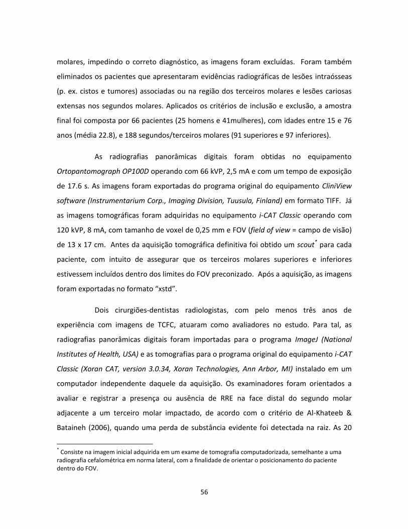
56
molares, impedindo o correto diagnóstico, as imagens foram excluídas. Foram também
eliminados os pacientes que apresentaram evidências radiográficas de lesões intraósseas
(p. ex. cistos e tumores) associadas ou na região dos terceiros molares e lesões cariosas
extensas nos segundos molares. Aplicados os critérios de inclusão e exclusão, a amostra
final foi composta por 66 pacientes (25 homens e 41mulheres), com idades entre 15 e 76
anos (média 22.8), e 188 segundos/terceiros molares (91 superiores e 97 inferiores).
As radiografias panorâmicas digitais foram obtidas no equipamento
Ortopantomograph OP100D operando com 66 kVP, 2,5 mA e com um tempo de exposição
de 17.6 s. As imagens foram exportadas do programa original do equipamento CliniView
software (Instrumentarium Corp., Imaging Division, Tuusula, Finland) em formato TIFF. Já
as imagens tomográficas foram adquiridas no equipamento i-CAT Classic operando com
120 kVP, 8 mA, com tamanho de voxel de 0,25 mm e FOV (field of view = campo de visão)
de 13 x 17 cm. Antes da aquisição tomográfica definitiva foi obtido um scout* para cada
paciente, com intuito de assegurar que os terceiros molares superiores e inferiores
estivessem incluídos dentro dos limites do FOV preconizado. Após a aquisição, as imagens
foram exportadas no formato “xstd”.
Dois cirurgiões-dentistas radiologistas, com pelo menos três anos de
experiência com imagens de TCFC, atuaram como avaliadores no estudo. Para tal, as
radiografias panorâmicas digitais foram importadas para o programa ImageJ (National
Institutes of Health, USA) e as tomografias para o programa original do equipamento i-CAT
Classic (Xoran CAT, version 3.0.34, Xoran Technologies, Ann Arbor, MI) instalado em um
computador independente daquele da aquisição. Os examinadores foram orientados a
avaliar e registrar a presença ou ausência de RRE na face distal do segundo molar
adjacente a um terceiro molar impactado, de acordo com o critério de Al-Khateeb &
Bataineh (2006), quando uma perda de substância evidente foi detectada na raiz. As 20
* Consiste na imagem inicial adquirida em um exame de tomografia computadorizada, semelhante a uma
radiografia cefalométrica em norma lateral, com a finalidade de orientar o posicionamento do paciente dentro do FOV.

57
primeiras imagens das duas modalidades foram avaliadas pelos dois examinadores ao
mesmo tempo, para que, nesse momento, fossem uniformizados os critérios de avaliação
e solucionadas as dúvidas com os pesquisadores envolvidos. Os demais exames foram
avaliados de maneira independente por cada avaliador, sendo avaliadas primeiramente
todas as radiografias panorâmicas e posteriormente as imagens por TCFC, que foram
aleatorizadas de maneira distinta em cada modalidade. Os avaliadores foram
aconselhados a realizar a avaliação de 20 dentes por dia, de forma a evitar fadiga visual.
As avaliações ocorreram em condições de baixa iluminação, em um mesmo
monitor LCD de 21 polegadas com resolução espacial de 1280 X 1024 pixels. Nas
radiografias panorâmicas foram investigados, além da RRE no segundo molar, o
posicionamento/inclinação do terceiro molar adjacente de acordo com a classificação
proposta por Winter (1926): vertical, mesioangular, distoangular, horizontal, transverso e
invertido. Por não haver terceiros molares em posição invertida, a denominação foi
eliminada da classificação final proposta pelo estudo, visando à simplificação dos dados.
Os avaliadores foram orientados a utilizar os escores (0) e (1) para a ausência e presença
de RRE, respectivamente, e escores de 1 a 5 para a inclinação do terceiro molar como
observado na Figura 1.
Nas imagens por TCFC a avaliação ocorreu de maneira dinâmica no modo de
visualização multiplanar (MPR), no qual os cortes axiais, sagitais e coronais foram
observados de maneira simultânea. Os examinadores foram encorajados a utilizar as
ferramentas de giro da imagem, possibilitando o posicionamento das linhas de orientação
do software de modo a favorecer a investigação da região dos terceiros molares. Da
mesma forma, atribuíram-se escores de avaliação 0 e 1 para ausência e presença de RRE,
respectivamente.
58
Figura 1: Demonstração das inclinações dos terceiros molares encontradas no estudo e os
respectivos escores atribuídos para cada uma delas (1=vertical, 2=mesioangular,
3=horizontal, 4=distoangular, 5=transverso)
Tanto nas avaliações radiográficas como nas tomográficas foram permitidas
aos examinadores a manipulação de brilho, contraste, ampliação de imagem (zoom) e a
aplicação de filtros de realce ou suavização de acordo com a preferência e experiência
pessoal. Tal conduta foi adotada com o objetivo de simular situações da rotina clínica de
diagnóstico. Ao final dessa etapa, as avaliações foram comparadas e, nos casos em que
houve discordância entre os avaliadores, tanto para o diagnóstico da RRE quanto para a
inclinação dos terceiros molares, os mesmos discutiram seus achados e obtiveram um
consenso final.
Os escores finais atribuídos pelos avaliadores após o consenso foram
tabulados utilizando o programa Microsoft Office Excel 2010 (Microsoft Corporation by
Impressa Systems, Santa Rosa, CA, EUA) e transferidos para o programa Systat 13.0
59
(Cranes Software International, Chicago, IL, EUA) para análise estatística. O teste do qui-
quadrado foi utilizado para a comparação entre presença e ausência de RRE em ambas as
modalidades de imagem. Utilizou-se o mesmo teste para confrontar a detecção de RRE na
panorâmica e na TCFC. Uma tabela de contingência (2X2) com os dados de ausência e
presença de RRE em ambos os métodos foi elaborada com o objetivo de facilitar a
compreensão dos dados. Para a análise da distribuição dos casos de RRE nos arcos
superior e inferior e investigação da possibilidade de associações estatisticamente
significantes foram utilizados os testes de qui-quadrado e exato de Fisher na dependência
do tamanho da amostra, com emprego do teste exato Fisher nas tabelas em que o qui-
quadrado não pôde ser aplicado. As análises das possíveis associações entre as inclinações
dos terceiros molares (classificação de Winter) e a presença e ausência de RRE foram
realizadas pelo teste Z para duas proporções, sendo os percentuais para cada inclinação
comparados dois a dois. A regressão logística simples foi realizada para investigar a
possível relação de dependência existente entre a detecção da RRE na radiografia
panorâmica e na TCFC, bem como a chance de detecção da RRE nas duas modalidades de
maneira isolada e associada. O nível de significância adotado para todas as análises foi de
5%.
Para a segunda parte do estudo, que envolveu somente imagens de TCFC,
foram utilizados 217 exames tomográficos, também provenientes do banco de imagens da
instituição, adquiridos em dois diferentes equipamentos: 120 no equipamento i-CAT
Classic e 97 no Picasso trio (E-WOO Technology, Giheung-gu, Republic of Korea). Foram
incluídas nessa avaliação as tomografias que continham segundos molares adjacentes a
terceiros molares inferiores impactados nas posições mesioangular e horizontal. Da
mesma forma, foram excluídos do estudo os terceiros molares completamente
irrompidos, com menos de 2/3 de raiz formada e associados a lesões císticas ou tumorais.
Uma lesão de cárie extensa no segundo molar inferior também representou um critério de
exclusão. As tomografias as quais a presença de beam hardening prejudicou o diagnóstico
não foram consideradas. Aplicados os critérios de inclusão e exclusão acima descritos, a

60
amostra final foi composta por 116 tomografias, 58 adquiridas no i-CAT classic e 58 no
Picasso trio, sendo 70 mulheres e 46 homens com idades entre 14 e 62 anos (média 23,7).
Para o equipamento Picasso trio os parâmetros de exposição e aquisição das
imagens foram 90 kVp, 5 mA, voxel de 0,20 mm, FOV de 8,5 x 12 cm e aplicação do
algoritmo para redução de artefatos metálicos. As imagens do i-CAT Classic foram
adquiridas de acordo com o protocolo já descrito. As imagens foram avaliadas pelos
mesmos avaliadores, sob as condições técnicas e de calibração previamente relatadas, nos
softwares específicos para cada equipamento: o Xoran CAT para o i-CAT e o EZ3D (E-WOO
Technology, Giheung-gu, Republic of Korea) para o Picasso Trio.
Os examinadores registraram a ausência ou presença da RRE no segundo
molar, atribuindo os escores 0 e 1, respectivamente. Foi também apontado o terço
radicular no qual a lesão de RRE se localizava em sua maior extensão: terços cervical (1),
médio(2) ou apical(3). De acordo com o sistema proposto por Ericson et al. (2002), as
lesões de RRE foram classificadas em relação ao grau de envolvimento da estrutura
radicular em: (1) Discreta – envolvendo menos da metade da espessura da dentina, (2)
Moderada – envolvendo a metade da dentina ou mais, sem comprometer a cavidade
pulpar, (3) Severa – caracterizada por envolvimento pulpar.
Também foram indicadas a inclinação dos terceiros molares (mesioangular ou
horizontal), conforme demonstrado na Figura 1 (escores 2 e 3), e a profundidade dos
mesmos de acordo com a classificação de Pell & Gregory. A classificação de Pell & Gregory
categoriza os terceiros molares de acordo com as posições horizontal (relação com o ramo
mandibular) e vertical (relação com a junção cemento-esmalte e plano oclusal do segundo
molar) (Almendros-Marqués et al., 2008). Foram utilizadas no presente estudo as classes
A, B e C, que apontam a posição vertical/profundidade dos terceiros molares (Figura 2).

61
Figura 2. Imagens de TCFC ilustrando a classificação de Pell & Gregory relacionada à
profundidade vertical do terceiro molar: (A) o terceiro molar está próximo ou ao nível do
plano oclusal do segundo molar, (B) o terceiro molar está entre o plano oclusal e a junção
cemento-esmalte do segundo molar, (C) o terceiro molar está sob a junção cemento-
esmalte do segundo molar
A localização dos terceiros molares em relação aos tecidos ósseo e mole foi
também avaliada, sendo os mesmos classificados como semi-inclusos (escore = 0) ou
inclusos (escore=1). Neste estudo, um dente foi considerado incluso quando, durante a
avaliação tridimensional das imagens, os examinadores constataram que o mesmo estava
completamente encoberto por tecido ósseo. Nas situações onde os dentes estavam
parcialmente irrompidos na cavidade bucal ou encobertos por tecido mole, os mesmos
foram registrados como semi-inclusos.
Trinta dias após o término das avaliações e obtenção do consenso nos casos
de discordância, 36 dentes foram reavaliados pelos dois examinadores, sendo 18 para
cada equipamento. Os dados dessa análise foram comparados aos dados iniciais e
submetidos ao teste Kappa para a análise da concordância intraobservador, levando-se
em consideração os valores de referência: 0.40 – concordância fraca; 0.40–0.59 –
concordância moderada; 0.60–0.74 – boa concordância; 0.75–1.00 – concordância
excelente.
62
Para a análise estatística foram utilizados os programas Prism 6.0 (GraphPad
software, Inc., La Jolla, CA, EUA) e BioEstat 5.0 (Manuel Ayres Software Informer,
Belem/PA, Brasil). O nível de significância adotado foi de 5%. Os dados numéricos
relacionados à idade dos indivíduos foram submetidos ao teste ANOVA nas situações de
normalidade. O teste Mann-Whitney foi utilizado quando o teste paramétrico não pôde
ser aplicado. Já os testes de qui-quadrado em tabelas de contingência e qui-quadrado de
aderência foram utilizados para avaliar a associação existente entre as variáveis
qualitativas: (1) presença e ausência da RRE, (2) inclinação dos terceiros molares, (3)
Classes de Pell & Gregory, (4) profundidade dos terceiros molares em relação aos tecidos
ósseo e mole, (5) localização das lesões de RRE e (6) severidade das lesões de RRE. Em
tabelas de contingência, o teste de qui-quadrado buscou as possíveis divergências entre as
frequências observadas e esperadas para os eventos. O teste de qui-quadrado de
aderência para proporções esperadas desiguais verificou se as variáveis em estudo
exercem influência na distribuição de um determinado evento.
REFERÊNCIAS
1. Al-Khateeb TH, Bataineh AB. Pathology associated with impacted mandibular third
molars in a group of Jordanians. J Oral Maxillofac Surg. 2006, 64: 1598–1602.
2. Almendros-Marqués N, Berini-Aytés L, Gay-Escoda C. Evaluation of intraexaminer
and interexaminer agreement on classifying lower third molars according to the
systems of Pell and Gregory and of Winter. J Oral Maxillofac Surg. 2008; 66: 893-
899.
3. Ericson, S., Bjerklin, K. & Falahat, B. Does the canine dental follicle cause
resorption of permanent incisor roots? A computed tomographic study of
erupting maxillary canines. Angle Orthod. 2002, 72: 95–104.
4. Winter GB, ed. Principles of exodontia as applied to the impacted third molar. St
Louis: American medical Books, 1926.
63
A
NEX
O 1
: Ap
rovação
do
Pro
toco
lo d
e Pe
squ
isa pelo
CEP
da FO
P-U
NIC
AM
P (A
rtigo 1
)
64
A
NEX
O 2
: Ap
rovação
do
Pro
toco
lo d
e Pe
squ
isa pelo
CEP
da FO
P-U
NIC
AM
P (A
rtigo 2
)
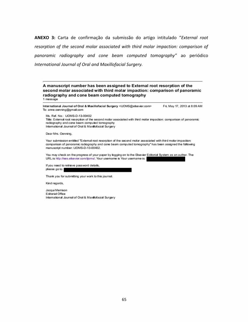
65
ANEXO 3: Carta de confirmação da submissão do artigo intitulado “External root
resorption of the second molar associated with third molar impaction: comparison of
panoramic radiography and cone beam computed tomography” ao periódico
International Journal of Oral and Maxillofacial Surgery.
66
ANEXO 4: Declaração de não infração dos dispositivos da Lei nº 9.610/98