Embed Size (px)
Citation preview
Annexin A6-Balanced Late Endosomal Cholesterol Controls InfluenzaA Replication and Propagation
Agnes Musiol,a Sandra Gran,a Christina Ehrhardt,b Stephan Ludwig,b Thomas Grewal,c Volker Gerke,a Ursula Reschera
Institute of Medical Biochemistry, Centre for Molecular Biology of Inflammation and Interdisciplinary Clinical Research Centre, University of Münster, Münster, Germanya;Institute of Molecular Virology, Centre for Molecular Biology of Inflammation, University of Münster, Münster, Germanyb; Faculty of Pharmacy A15, University of Sydney,Sydney, NSW, Australiac
ABSTRACT Influenza is caused by influenza A virus (IAV), an enveloped, negative-stranded RNA virus that derives its envelopelipids from the host cell plasma membrane. Here, we examined the functional role of cellular cholesterol in the IAV infectioncycle. We show that shifting of cellular cholesterol pools via the Ca2�-regulated membrane-binding protein annexin A6 (AnxA6)affects the infectivity of progeny virus particles. Elevated levels of cellular AnxA6, which decrease plasma membrane and in-crease late endosomal cholesterol levels, impaired IAV replication and propagation, whereas RNA interference-mediated AnxA6ablation increased viral progeny titers. Pharmacological accumulation of late endosomal cholesterol also diminished IAV viruspropagation. Decreased IAV replication caused by upregulated AnxA6 expression could be restored either by exogenous replen-ishment of host cell cholesterol or by ectopic expression of the late endosomal cholesterol transporter Niemann-Pick C1 (NPC1).Virus released from AnxA6-overexpressing cells displayed significantly reduced cholesterol levels. Our results show that IAVreplication depends on maintenance of the cellular cholesterol balance and identify AnxA6 as a critical factor in linking IAV tocellular cholesterol homeostasis.
IMPORTANCE Influenza A virus (IAV) is a major public health concern, and yet, major host-pathogen interactions regulating IAVreplication still remain poorly understood. It is known that host cell cholesterol is a critical factor in the influenza virus life cycle.The viral envelope is derived from the host cell membrane during the process of budding and, hence, equips the virus with a spe-cial lipid-protein mixture which is high in cholesterol. However, the influence of host cell cholesterol homeostasis on IAV infec-tion is largely unknown. We show that IAV infection success critically depends on host cell cholesterol distribution. Cholesterolsequestration in the endosomal compartment impairs progeny titer and infectivity and is associated with reduced cholesterolcontent in the viral envelope.
Received 2 August 2013 Accepted 8 October 2013 Published 5 November 2013
Citation Musiol A, Gran S, Ehrhardt C, Ludwig S, Grewal T, Gerke V, Rescher U. 2013. Annexin A6-balanced late endosomal cholesterol controls influenza A replication andpropagation. mBio 4(6):e00608-13. doi:10.1128/mBio.00608-13.
Invited Editor Stephan Pleschka, Justus-Liebig-University Giessen Editor Vincent Racaniello, Columbia University College of Physicians & Surgeons
Copyright © 2013 Musiol et al. This is an open-access article distributed under the terms of the Creative Commons Attribution-Noncommercial-ShareAlike 3.0 Unportedlicense, which permits unrestricted noncommercial use, distribution, and reproduction in any medium, provided the original author and source are credited.
Address correspondence to Ursula Rescher, [email protected].
Influenza A virus (IAV) remains a major public health concern,not only by causing thousands of deaths because of annual epi-
demics and rare but often severe pandemics but also by leading toenormous economic loss every year (1). As agents directed againstviral components select for resistant mutants, new antiviral ther-apeutic approaches might target the interaction of virus with hostcell components. Despite the enormous progress in influenza-related research in the last decade, major host-pathogen interac-tions regulating IAV replication and propagation still remainpoorly understood. The virus is characterized by a segmented,single-stranded RNA genome with negative orientation, and itsgenome encodes up to 12 viral structural and nonstructural pro-teins (2). The virus genome is enclosed by an envelope that isderived from the host cell membrane during the process of bud-ding and, hence, equips the virus with a special lipid-protein mix-ture. As this process is heavily dependent on the presence of spe-cialized and cholesterol-enriched lipid microdomains, or so-called “rafts,” this leads to a high level of cholesterol, a majorcomponent of those raft domains, in the virus envelope (3–7).
Host cell cholesterol is a critical factor in IAV replication andpropagation. It is known that viral assembly and budding, as wellas infectivity, are strongly dependent on cellular cholesterol levels,indicating the great importance of this host factor for virus infec-tion (7–11). However, the molecular mechanisms of these regula-tory interactions are largely unknown. This is partly due to thelimited knowledge about intracellular cholesterol transport be-tween distinct membrane compartments in the host cell that reg-ulates cholesterol homeostasis, as well as cholesterol-sensitiveprotein trafficking (12).
Recently, annexin A6 (AnxA6) has emerged as an importantplayer in the maintenance of cellular cholesterol homeostasis (13–16). Annexin A6 is a member of the annexin protein family ofstructurally highly conserved, Ca2�-regulated membrane-binding proteins that have been linked to the regulation of mem-brane recognition and trafficking (17–20). All annexins share acommon structure composed of two domains: a conserved corethat is responsible for Ca2� and phospholipid binding and anN-terminal tail that is unique for each annexin. Due to their role in
RESEARCH ARTICLE
November/December 2013 Volume 4 Issue 6 e00608-13 ® mbio.asm.org 1
on March 2, 2020 by guest
http://mbio.asm
.org/D
ownloaded from
membrane dynamics, annexins have already been shown to beinvolved in the life cycles of several pathogens, including diverseviruses. Regarding infections with IAV, proteomic analysis of in-fluenza virions revealed the incorporation of annexins A1, A2, A4,A5, and A11 into IAV particles (21). For AnxA2, it was even re-ported that the protein has a supportive role for IAV replication(22, 23). Recently, AnxA6 was proposed to be negatively involvedin IAV replication (24). Here, we elucidate the molecular mecha-nism through which annexin A6 exerts a strong antiviral effect.We show that AnxA6 affects the infectivity of progeny virus par-ticles through shifting intracellular cholesterol pools. This effectwas independent of the plasma membrane-associated pool ofAnxA6 and could be reversed either through exogenous replen-ishment of host cell cholesterol or by overexpression of the lateendosomal cholesterol transporter NPC1. These studies support arole for AnxA6 in IAV replication and propagation and indicatethat cellular cholesterol homeostasis is critically linked to the in-fectivity of the virus.
RESULTSAnnexin A6 negatively modulates influenza virus replication.To examine the function of AnxA6 in IAV replication, we em-ployed the human epithelial carcinoma cell line A431 (hereinaftercalled A431wt, for A431 wild type), which naturally lacks endog-enous AnxA6, and the A431-A6 cell line, which has stable expres-sion of AnxA6 (25, 26). Western blot analysis of cell lysates con-firmed that the expression of AnxA6 was only detectable inA431-A6 cells (Fig. 1A, bottom). A431wt and A431-A6 cells wereinfected with the avian IAV isolate A/FPV/Bratislava/79 (H7N7;FPV) at a multiplicity of infection (MOI) of 0.01, and progenyvirus titers were monitored over a time period of 48 h (Fig. 1A).The infectious titers of viruses produced by these cells and releasedinto the cell culture supernatants were measured by a standardplaque assay technique. Both cell lines were permissive for IAVreplication; however, in A431-A6 cells, the virus titers were im-
paired at every time point analyzed. This correlated with a reducedexpression of virion-associated matrix protein 1 (M1), as assessedby Western blotting (Fig. 1A, bottom).
To confirm this result in a cell model relevant for infections ofthe upper respiratory tract and to exclude an aberrant phenotypeas a consequence of clonal selection during A431-A6 cell line gen-eration, we repeated this experiment using human A549 lung ep-ithelial cells transiently transfected with green fluorescent protein(GFP)-tagged AnxA6 (A6-GFP) or GFP alone as a control. GFPand A6-GFP expression were verified by fluorescence microscopy(data not shown) and Western blotting (see Fig. S1 in the supple-mental material). At 24 h after transfection, cells were infected asdescribed above. Again, virus titers were significantly reduced inAnxA6-overexpressing cells (Fig. 1B). Furthermore, impaired vi-ral progeny titers again correlated with reduced viral M1 proteinexpression. This observation strengthened the finding that ele-vated AnxA6 expression negatively influences IAV replication.
To further verify an involvement of AnxA6 in IAV replication,we performed small interfering RNA (siRNA)-mediated knock-down of AnxA6 in A549 cells using a pool of AnxA6-specificsiRNA duplexes and nontargeting siRNA as a control. At 48 h aftertransfection, cells were infected with FPV at an MOI of 0.1, andvirus replication was allowed to proceed for 24 h. Efficient andreproducible AnxA6 knockdown was confirmed by Western blotanalysis (see Fig. S1 in the supplemental material). In line with arole of AnxA6 as a negative regulator, downregulation of AnxA6resulted in significantly enhanced progeny virus titers in cell cul-ture supernatants compared to the titers in control siRNA-treatedcells (Fig. 1B, top right). This correlated with higher M1 virusprotein expression.
Additional experiments revealed that AnxA6 also inhibited thereplication of other influenza strains, such as the H1N1 IAV strainA/Puerto Rico/8/34 (PR8) and a mouse-adapted strain (H1N1v;HH/04-3rd) of the 2009 pandemic swine-origin influenza A virus
FIG 1 AnxA6 negatively modulates influenza A virus replication. A431 wild-type (wt) and A431 cells stably overexpressing AnxA6 (A6) (A), as well as A549 cellstransiently overexpressing GFP or AnxA6-GFP (B, left), were infected with the avian IAV isolate A/FPV/Bratislava/79 (H7N7; FPV) at an MOI of 0.01. At theindicated time points postinfection (p.i.), progeny virus titers were determined by standard plaque assay. IAV M1 and AnxA6 protein levels in cell lysates weredetermined by Western blotting. Equal protein loading was verified by using �-tubulin. (B, right) A549 cells transfected with AnxA6 siRNA (si_A6) ornontargeting control siRNA (si_ct) were infected with FPV for 24 h at an MOI of 0.1, and viral progeny were determined by standard plaque assay. Levels of M1and AnxA6 proteins were monitored by Western blotting, and blots were probed for STAT3 to verify equal protein loading. Mean values � SEM of at least threeindependent experiments were calculated and assessed for statistically significant differences using a two-tailed t test. *, P � 0.05; **, P � 0.01; ***, P � 0.001.
Musiol et al.
2 ® mbio.asm.org November/December 2013 Volume 4 Issue 6 e00608-13
on March 2, 2020 by guest
http://mbio.asm
.org/D
ownloaded from
subtype H1N1 variant (H1N1v) strain A/Hamburg/04/2009 (seeFig. S2 in the supplemental material).
Annexin A6 overexpression inhibits IAV particle productionand decreases virus infectivity. So far, our results indicated animpact of cellular AnxA6 levels on IAV infection. To determinewhether the decrease in progeny virus titers induced by AnxA6overexpression was due to reduced infectivity or a decreasedamount of infectious virus particles, we first performed hemag-glutination (HA) assays to determine the levels of IAV particlespresent in the sample. For this purpose, we infected A431wt andA431-A6 cells with FPV at an MOI of 0.1, allowed the infection toproceed for 24 h, and used the resulting supernatants for the HAassay. As shown by the results in Fig. 2A, supernatants from in-fected A431wt cells (FPV_wt) caused hemagglutination of redblood cells up to the 1:32 dilution, whereas viral particles pro-duced by A431-A6 cells (FPV_A6) needed the 1:16 dilution.Therefore, the amount of viral particles produced by A431-A6cells was decreased by approximately 50% compared to theamount produced by controls. This strongly indicated that highAnxA6 levels inhibit the production of viral particles per se. How-ever, an additional effect on infectivity could not be excluded, asthe decrease in infectious titers in AnxA6-overexpressing cells ap-peared to be greater than the 50% decrease in the total production
of IAV particles. To address the infectivity of IAV particles, wetherefore reinfected A549 cells with virus obtained from infectedA431wt (FPV_wt) or A431-A6 (FPV_A6) cells. To obtain one cy-cle of IAV replication, we allowed the infection to proceed for 8 h.Although A549 cells were infected with the same MOI of the re-spective virus (MOI of 0.1), the virus titers were still decreased incells infected with virus that originated from A431-A6 cells(Fig. 2B, left). Next, we investigated the entry process of the re-spective virus into the host cell. Therefore, virion-associated ma-trix protein was detected by Western blot analysis, as describedpreviously (27). We infected A549 cells with FPV_wt or FPV_A6at an MOI of 5 and followed IAV internalization by detection ofviral M1 protein (Fig. 2B, right). A549 cells infected with FPV_A6showed a strong decrease in the amount of M1 protein comparedto the amount in cells infected with FPV_wt, indicating that thevirus produced in A431-A6 cells displayed reduced capability inthe entry process and, hence, exhibited decreased infectivity ofviral particles.
In conclusion, high levels of AnxA6 not only decreased theamount of viral particles produced by the host cell but, in addi-tion, decreased the infectivity of those particles.
Plasma membrane-associated functions of AnxA6 are not re-sponsible for its antiviral activity. Next, we aimed to investigate
FIG 2 AnxA6 upregulation decreases the amount and infectivity of viral particles. (A) A431wt and A431-A6 cells were infected with FPV (MOI of 0.1 for 24 h),and supernatants obtained from either A431wt (FPV_wt) or A431-A6 (FPV_A6) cells were analyzed by hemagglutination assay. � control, positive control; �control, negative control. Arrows indicate the greatest serum dilution giving a visible agglutination. (B) A431wt and A431-A6 cells were infected with FPV (MOIof 0.1 for 48 h), and progeny virus titers were determined by standard plaque assay. A549 cells were then infected for 8 h with the virus replicated in A431wt(FPV_wt) or A431-A6 cells (FPV_A6). For determination of infectious viral titers, an MOI of 0.1 was used. To monitor virus entry, A549 cells were infected withan MOI of 5 for 15 to 360 min, as indicated. IAV M1 protein levels in cell lysates were monitored by Western blotting. Equal protein loading was verified using�-tubulin. Mean values � SEM of at least three independent experiments were calculated and assessed for statistically significant differences using a two-tailedt test. *, P � 0.05.
Late Endosomal Cholesterol in IAV Infection
November/December 2013 Volume 4 Issue 6 e00608-13 ® mbio.asm.org 3
on March 2, 2020 by guest
http://mbio.asm
.org/D
ownloaded from
the underlying mechanism by which high AnxA6 levels decreasedthe infectivity of IAV particles. Upon cell activation, AnxA6 bindsto negatively charged phospholipids that are found predomi-nantly in the plasma membrane but also in endosomal mem-branes. As AnxA6 is mainly localized at the plasma membrane, wefirst addressed a role for plasma membrane-associated AnxA6 forantiviral activity.
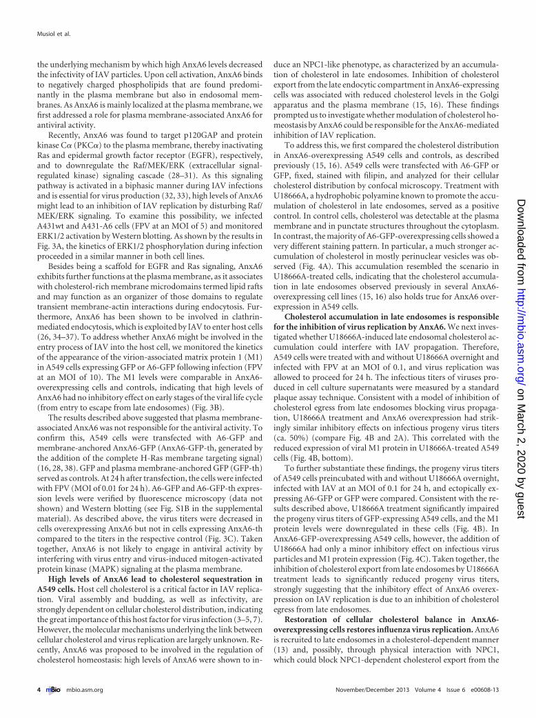
Recently, AnxA6 was found to target p120GAP and proteinkinase C� (PKC�) to the plasma membrane, thereby inactivatingRas and epidermal growth factor receptor (EGFR), respectively,and to downregulate the Raf/MEK/ERK (extracellular signal-regulated kinase) signaling cascade (28–31). As this signalingpathway is activated in a biphasic manner during IAV infectionsand is essential for virus production (32, 33), high levels of AnxA6might lead to an inhibition of IAV replication by disturbing Raf/MEK/ERK signaling. To examine this possibility, we infectedA431wt and A431-A6 cells (FPV at an MOI of 5) and monitoredERK1/2 activation by Western blotting. As shown by the results inFig. 3A, the kinetics of ERK1/2 phosphorylation during infectionproceeded in a similar manner in both cell lines.
Besides being a scaffold for EGFR and Ras signaling, AnxA6exhibits further functions at the plasma membrane, as it associateswith cholesterol-rich membrane microdomains termed lipid raftsand may function as an organizer of those domains to regulatetransient membrane-actin interactions during endocytosis. Fur-thermore, AnxA6 has been shown to be involved in clathrin-mediated endocytosis, which is exploited by IAV to enter host cells(26, 34–37). To address whether AnxA6 might be involved in theentry process of IAV into the host cell, we monitored the kineticsof the appearance of the virion-associated matrix protein 1 (M1)in A549 cells expressing GFP or A6-GFP following infection (FPVat an MOI of 10). The M1 levels were comparable in AnxA6-overexpressing cells and controls, indicating that high levels ofAnxA6 had no inhibitory effect on early stages of the viral life cycle(from entry to escape from late endosomes) (Fig. 3B).
The results described above suggested that plasma membrane-associated AnxA6 was not responsible for the antiviral activity. Toconfirm this, A549 cells were transfected with A6-GFP andmembrane-anchored AnxA6-GFP (AnxA6-GFP-th, generated bythe addition of the complete H-Ras membrane targeting signal)(16, 28, 38). GFP and plasma membrane-anchored GFP (GFP-th)served as controls. At 24 h after transfection, the cells were infectedwith FPV (MOI of 0.01 for 24 h). A6-GFP and A6-GFP-th expres-sion levels were verified by fluorescence microscopy (data notshown) and Western blotting (see Fig. S1B in the supplementalmaterial). As described above, the virus titers were decreased incells overexpressing AnxA6 but not in cells expressing AnxA6-thcompared to the titers in the respective control (Fig. 3C). Takentogether, AnxA6 is not likely to engage in antiviral activity byinterfering with virus entry and virus-induced mitogen-activatedprotein kinase (MAPK) signaling at the plasma membrane.
High levels of AnxA6 lead to cholesterol sequestration inA549 cells. Host cell cholesterol is a critical factor in IAV replica-tion. Viral assembly and budding, as well as infectivity, arestrongly dependent on cellular cholesterol distribution, indicatingthe great importance of this host factor for virus infection (3–5, 7).However, the molecular mechanisms underlying the link betweencellular cholesterol and virus replication are largely unknown. Re-cently, AnxA6 was proposed to be involved in the regulation ofcholesterol homeostasis: high levels of AnxA6 were shown to in-
duce an NPC1-like phenotype, as characterized by an accumula-tion of cholesterol in late endosomes. Inhibition of cholesterolexport from the late endocytic compartment in AnxA6-expressingcells was associated with reduced cholesterol levels in the Golgiapparatus and the plasma membrane (15, 16). These findingsprompted us to investigate whether modulation of cholesterol ho-meostasis by AnxA6 could be responsible for the AnxA6-mediatedinhibition of IAV replication.
To address this, we first compared the cholesterol distributionin AnxA6-overexpressing A549 cells and controls, as describedpreviously (15, 16). A549 cells were transfected with A6-GFP orGFP, fixed, stained with filipin, and analyzed for their cellularcholesterol distribution by confocal microscopy. Treatment withU18666A, a hydrophobic polyamine known to promote the accu-mulation of cholesterol in late endosomes, served as a positivecontrol. In control cells, cholesterol was detectable at the plasmamembrane and in punctate structures throughout the cytoplasm.In contrast, the majority of A6-GFP-overexpressing cells showed avery different staining pattern. In particular, a much stronger ac-cumulation of cholesterol in mostly perinuclear vesicles was ob-served (Fig. 4A). This accumulation resembled the scenario inU18666A-treated cells, indicating that the cholesterol accumula-tion in late endosomes observed previously in several AnxA6-overexpressing cell lines (15, 16) also holds true for AnxA6 over-expression in A549 cells.
Cholesterol accumulation in late endosomes is responsiblefor the inhibition of virus replication by AnxA6. We next inves-tigated whether U18666A-induced late endosomal cholesterol ac-cumulation could interfere with IAV propagation. Therefore,A549 cells were treated with and without U18666A overnight andinfected with FPV at an MOI of 0.1, and virus replication wasallowed to proceed for 24 h. The infectious titers of viruses pro-duced in cell culture supernatants were measured by a standardplaque assay technique. Consistent with a model of inhibition ofcholesterol egress from late endosomes blocking virus propaga-tion, U18666A treatment and AnxA6 overexpression had strik-ingly similar inhibitory effects on infectious progeny virus titers(ca. 50%) (compare Fig. 4B and 2A). This correlated with thereduced expression of viral M1 protein in U18666A-treated A549cells (Fig. 4B, bottom).
To further substantiate these findings, the progeny virus titersof A549 cells preincubated with and without U18666A overnight,infected with IAV at an MOI of 0.1 for 24 h, and ectopically ex-pressing A6-GFP or GFP were compared. Consistent with the re-sults described above, U18666A treatment significantly impairedthe progeny virus titers of GFP-expressing A549 cells, and the M1protein levels were downregulated in these cells (Fig. 4B). InAnxA6-GFP-overexpressing A549 cells, however, the addition ofU18666A had only a minor inhibitory effect on infectious virusparticles and M1 protein expression (Fig. 4C). Taken together, theinhibition of cholesterol export from late endosomes by U18666Atreatment leads to significantly reduced progeny virus titers,strongly suggesting that the inhibitory effect of AnxA6 overex-pression on IAV replication is due to an inhibition of cholesterolegress from late endosomes.
Restoration of cellular cholesterol balance in AnxA6-overexpressing cells restores influenza virus replication. AnxA6is recruited to late endosomes in a cholesterol-dependent manner(13) and, possibly, through physical interaction with NPC1,which could block NPC1-dependent cholesterol export from the
Musiol et al.
4 ® mbio.asm.org November/December 2013 Volume 4 Issue 6 e00608-13
on March 2, 2020 by guest
http://mbio.asm
.org/D
ownloaded from
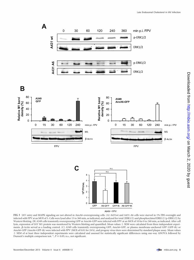
FIG 3 IAV entry and MAPK signaling are not altered in AnxA6-overexpressing cells. (A) A431wt and A431-A6 cells were starved in 1% FBS overnight andinfected with FPV at an MOI of 5. Cells were lysed after 15 to 360 min, as indicated, and analyzed for total (ERK1/2) and phosphorylated ERK1/2 (p-ERK1/2) byWestern blotting. (B) A549 cells transiently overexpressing GFP or AnxA6-GFP were infected with FPV at an MOI of 10 for 0 to 240 min, as indicated. After celllysis, expression of IAV M1 protein was monitored by Western blotting and quantified. Mean values � SEM were calculated from three independent experi-ments. �-Actin served as a loading control. (C) A549 cells transiently overexpressing GFP, AnxA6-GFP, or plasma membrane-anchored GFP (GFP-th) orAnxA6-GFP (AnxA6-GFP-th) were infected with FPV (MOI of 0.01 for 24 h), and progeny virus titers were determined by standard plaque assay. Mean values� SEM of at least three independent experiments were calculated and assessed for statistically significant differences using one-way ANOVA followed byDunnett’s multiple comparison test. *, P � 0.05; n.s., not significant.
Late Endosomal Cholesterol in IAV Infection
November/December 2013 Volume 4 Issue 6 e00608-13 ® mbio.asm.org 5
on March 2, 2020 by guest
http://mbio.asm
.org/D
ownloaded from
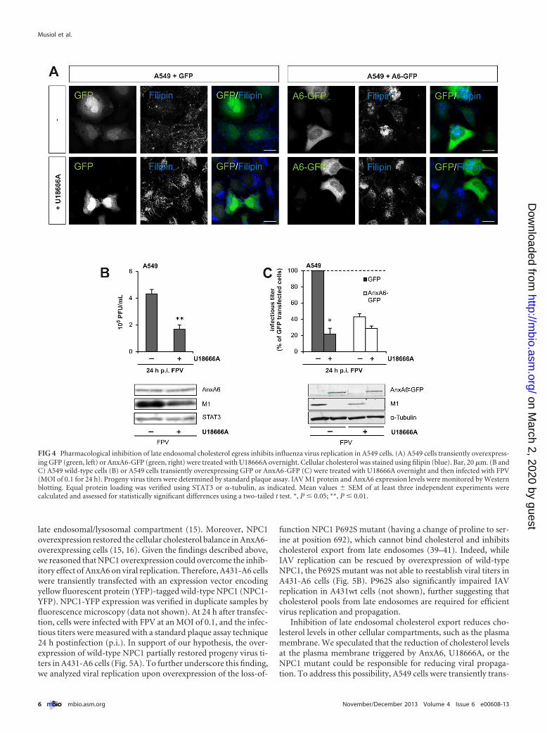
late endosomal/lysosomal compartment (15). Moreover, NPC1overexpression restored the cellular cholesterol balance in AnxA6-overexpressing cells (15, 16). Given the findings described above,we reasoned that NPC1 overexpression could overcome the inhib-itory effect of AnxA6 on viral replication. Therefore, A431-A6 cellswere transiently transfected with an expression vector encodingyellow fluorescent protein (YFP)-tagged wild-type NPC1 (NPC1-YFP). NPC1-YFP expression was verified in duplicate samples byfluorescence microscopy (data not shown). At 24 h after transfec-tion, cells were infected with FPV at an MOI of 0.1, and the infec-tious titers were measured with a standard plaque assay technique24 h postinfection (p.i.). In support of our hypothesis, the over-expression of wild-type NPC1 partially restored progeny virus ti-ters in A431-A6 cells (Fig. 5A). To further underscore this finding,we analyzed viral replication upon overexpression of the loss-of-
function NPC1 P692S mutant (having a change of proline to ser-ine at position 692), which cannot bind cholesterol and inhibitscholesterol export from late endosomes (39–41). Indeed, whileIAV replication can be rescued by overexpression of wild-typeNPC1, the P692S mutant was not able to reestablish viral titers inA431-A6 cells (Fig. 5B). P962S also significantly impaired IAVreplication in A431wt cells (not shown), further suggesting thatcholesterol pools from late endosomes are required for efficientvirus replication and propagation.
Inhibition of late endosomal cholesterol export reduces cho-lesterol levels in other cellular compartments, such as the plasmamembrane. We speculated that the reduction of cholesterol levelsat the plasma membrane triggered by AnxA6, U18666A, or theNPC1 mutant could be responsible for reducing viral propaga-tion. To address this possibility, A549 cells were transiently trans-
FIG 4 Pharmacological inhibition of late endosomal cholesterol egress inhibits influenza virus replication in A549 cells. (A) A549 cells transiently overexpress-ing GFP (green, left) or AnxA6-GFP (green, right) were treated with U18666A overnight. Cellular cholesterol was stained using filipin (blue). Bar, 20 �m. (B andC) A549 wild-type cells (B) or A549 cells transiently overexpressing GFP or AnxA6-GFP (C) were treated with U18666A overnight and then infected with FPV(MOI of 0.1 for 24 h). Progeny virus titers were determined by standard plaque assay. IAV M1 protein and AnxA6 expression levels were monitored by Westernblotting. Equal protein loading was verified using STAT3 or �-tubulin, as indicated. Mean values � SEM of at least three independent experiments werecalculated and assessed for statistically significant differences using a two-tailed t test. *, P � 0.05; **, P � 0.01.
Musiol et al.
6 ® mbio.asm.org November/December 2013 Volume 4 Issue 6 e00608-13
on March 2, 2020 by guest
http://mbio.asm
.org/D
ownloaded from
fected with A6-GFP or GFP, followed by the addition of exoge-nous cholesterol to replenish plasma membrane cholesterol. Next,cholesterol-treated and nontreated A549 cells were infected withFPV at an MOI of 0.1, and the infectious titers were measured withthe standard plaque assay technique. Indeed, cholesterol replen-ishment completely restored the progeny virus titers in A6-GFP-overexpressing A549 cells (Fig. 5C), further supporting a rolefor AnxA6 to modulate cholesterol-dependent steps during viralreplication. In conclusion, restoration of the cellular cholesterolbalance via cholesterol replenishment using exogenous choles-terol or the ectopic expression of wild-type NPC1 in AnxA6-overexpressing cells improves the ability of IAV to replicate andpropagate.
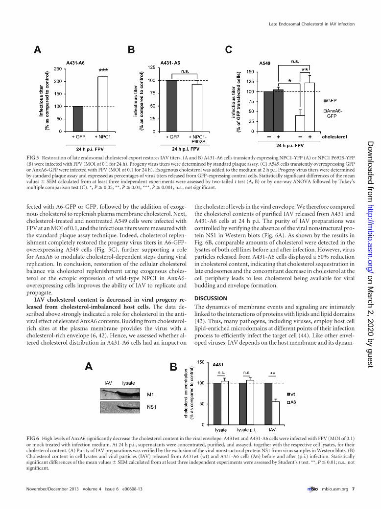
IAV cholesterol content is decreased in viral progeny re-leased from cholesterol-imbalanced host cells. The data de-scribed above strongly indicated a role for cholesterol in the anti-viral effect of elevated AnxA6 contents. Budding from cholesterol-rich sites at the plasma membrane provides the virus with acholesterol-rich envelope (6, 42). Hence, we assessed whether al-tered cholesterol distribution in A431-A6 cells had an impact on
the cholesterol levels in the viral envelope. We therefore comparedthe cholesterol contents of purified IAV released from A431 andA431-A6 cells at 24 h p.i. The purity of IAV preparations wascontrolled by verifying the absence of the viral nonstructural pro-tein NS1 in Western blots (Fig. 6A). As shown by the results inFig. 6B, comparable amounts of cholesterol were detected in thelysates of both cell lines before and after infection. However, virusparticles released from A431-A6 cells displayed a 50% reductionin cholesterol content, indicating that cholesterol sequestration inlate endosomes and the concomitant decrease in cholesterol at thecell periphery leads to less cholesterol being available for viralbudding and envelope formation.
DISCUSSION
The dynamics of membrane events and signaling are intimatelylinked to the interactions of proteins with lipids and lipid domains(43). Thus, many pathogens, including viruses, employ host celllipid-enriched microdomains at different points of their infectionprocess to efficiently infect the target cell (44). Like other envel-oped viruses, IAV depends on the host membrane and its dynam-
FIG 5 Restoration of late endosomal cholesterol export restores IAV titers. (A and B) A431-A6 cells transiently expressing NPC1-YFP (A) or NPC1 P692S-YFP(B) were infected with FPV (MOI of 0.1 for 24 h). Progeny virus titers were determined by standard plaque assay. (C) A549 cells transiently overexpressing GFPor AnxA6-GFP were infected with FPV (MOI of 0.1 for 24 h). Exogenous cholesterol was added to the medium at 2 h p.i. Progeny virus titers were determinedby standard plaque assay and expressed as percentages of virus titers released from GFP-expressing control cells. Statistically significant differences of the meanvalues � SEM calculated from at least three independent experiments were assessed by two-tailed t test (A, B) or by one-way ANOVA followed by Tukey’smultiple comparison test (C). *, P � 0.05; **, P � 0.01; ***, P � 0.001; n.s., not significant.
FIG 6 High levels of AnxA6 significantly decrease the cholesterol content in the viral envelope. A431wt and A431-A6 cells were infected with FPV (MOI of 0.1)or mock treated with infection medium. At 24 h p.i., supernatants were concentrated, purified, and assayed, together with the respective cell lysates, for theircholesterol content. (A) Purity of IAV preparations was verified by the exclusion of the viral nonstructural protein NS1 from virus samples in Western blots. (B)Cholesterol content in cell lysates and viral particles (IAV) released from A431wt (wt) and A431-A6 cells (A6) before and after (p.i.) infection. Statisticallysignificant differences of the mean values � SEM calculated from at least three independent experiments were assessed by Student’s t test. **, P � 0.01; n.s., notsignificant.
Late Endosomal Cholesterol in IAV Infection
November/December 2013 Volume 4 Issue 6 e00608-13 ® mbio.asm.org 7
on March 2, 2020 by guest
http://mbio.asm
.org/D
ownloaded from
ics at several stages of the viral life cycle. Annexins constitute afamily of Ca2�-dependent host cell membrane proteins that havedifferent lipid specificities and, thus, associate with different targetmembranes in the cell (17–19, 45). Annexins have already beenshown to act in viral infections (46), and IAV carries several an-nexins in its particle, most likely as a consequence of budding atraftlike domains enriched with annexins (22, 23).
Recently, AnxA6 has been proposed to negatively regulate IAVinfection through interaction with the IAV matrix protein M2(24). Here, we show that AnxA6 levels in the host cell negativelycorrelate with IAV replication and reveal that aberrant cholesterolaccumulation in late endosomes, reminiscent of an NPC1mutant-like phenotype, in AnxA6-expressing cells causes antiviralactivity. Consistently, when cells were treated with the hydropho-bic polyamine U18666A, a drug commonly used to mimic theabnormal accumulation of unesterified cholesterol seen in lateendosomes of NPC1 mutant cells (47), a strong reduction in virustiters was observed. These findings suggest that AnxA6 interfereswith NPC1-dependent cholesterol trafficking. In accordancewith this, increased expression of wild-type NPC1, known tocorrect the NPC1 mutant-like phenotype in AnxA6-expressingcells (15, 16), significantly improved the virus titers in AnxA6-overexpressing cells. Cellular cholesterol is synthesized de novo inthe endoplasmic reticulum (ER) and transported to the plasmamembrane independently of NPC1 (47). Subsequently, it reinter-nalizes to the ER or other cellular compartments and/or recyclesback to the plasma membrane. Dysfunctional NPC1 disturbs theintracellular distribution of cholesterol, leading to its accumula-tion in late endosomes and a secondary reduction of cholesterollevels in other cellular sites, such as the Golgi apparatus and theplasma membrane. This is also observed in AnxA6-overexpressing cells (15). The fact that AnxA6 coimmunoprecipi-tates with NPC1 suggests that a direct interaction of these proteinsin the late endocytic compartment interferes with the ability ofNPC1 to bind and transfer cholesterol across the late endosomal/lysosomal membrane. Our finding that exogenous cholesterolpartially reversed the inhibitory effect of AnxA6 on IAV propaga-tion indicates that cellular cholesterol trafficking from the lateendosome to other sites, most likely the plasma membrane, isrequired for efficient IAV replication. These results also argueagainst a major impact of other lipids, such as sphingosine, thathave been found to accumulate abnormally together with choles-terol in the endo-/lysosomes of NPC mutant cells (48–50).
Over the past decade, cholesterol has been shown to be a cru-cial host factor for IAV. It is assumed that this essential host mem-brane component plays a decisive role during virus replication(10, 11, 42). Within the host cell membrane, cholesterol functionsin intracellular transport and cell signaling, acting through lipidrafts. These cholesterol-enriched membrane microdomains havebeen implicated in several steps of the viral life cycle, including theassembly and budding of progeny virus particles at the plasmamembrane (42). However, experimental evidence for the impor-tance of rafts in IAV replication is mainly derived from detergentextraction experiments, which drastically change the distributionand potential clustering of certain lipids and proteins. More-conclusive evidence that plasma membrane rafts are involved inHA clustering is drawn from recent improvements in fluorescencemicroscopy techniques that allowed the analysis of protein-raftassociation in a more-physiological cellular membrane environ-ment (51).
In contrast to other enveloped viruses that depend on host cellcomponents to ensure budding and the release of viral progeny,IAV uses the virus-encoded M2 protein to mediate scission ofbuds (52). Although IAV is thought to bud from cholesterol-richmembrane domains, M2 was shown to partition into rafts onlywhen clustered with HA (51). Physical association of AnxA6 withM2 has been reported previously (24) and was proposed to impairIAV replication in AnxA6-overexpressing cells. However, our datastrongly suggest that AnxA6-mediated changes in cholesterol ho-meostasis also have to be considered, as the restoration of cellularcholesterol distribution, through NPC1 overexpression or the ad-dition of exogenous cholesterol, reversed impaired IAV replica-tion in AnxA6-overexpressing cells. This may point at AnxA6 ex-erting multiple functions in different cellular locations that eitherdirectly (M2) or indirectly (cholesterol) inhibit viral replicationand propagation. It is tempting to speculate that the drop in virustiters observed in the previous study (24) was also accompanied bymajor alterations in cholesterol distribution caused by AnxA6up-/downregulation, as described here. As AnxA6 displays en-hanced binding to membranes with elevated cholesterol levels (13,26, 53, 54), the interaction of AnxA6 with M2 could serve to facil-itate M2 targeting to the IAV bud zone to ensure the assembly ofvirus components.
Host membrane-derived IAV envelope is enriched in lipidsgenerally found in raft microdomains, including cholesterol. Infact, 44% of the total virus lipid is cholesterol, which representsapproximately 12% of the total mass of the virion (6, 42). Buddingof virus particles through rafts equips the particle with an appro-priate lipid mixture that protects particles from environmentaldamage and, in the case of cholesterol, might promote membranefusion upon virus entry. Hence, virus treated with cholesterol-depleting agents shows reduced infectivity (10). Our results nowdemonstrate that the AnxA6-mediated endosomal cholesterol se-questration that leads to reduced cholesterol contents in theplasma membrane (15) is associated with strongly reduced IAVcholesterol contents and impaired infectivity.
A precise understanding of the molecular mechanisms thatunderlie virus-host cell interactions is a prerequisite for targetinghost cell components and could open up efficient therapeuticstrategies. Antiviral drugs could circumvent the time-consumingvaccine development and the viral resistance due to rapid anti-genic mutation. Host cell factor targeting has recently emerged asa promising approach (reviewed in reference 55), and recent find-ings strongly suggest that modulating the host immune responsereduces mortality rates (56, 57). Thus, combination therapy oftwo or more antiviral drugs with different modes of action (i.e.,targeting the virus and the host cell) to prevent and control acuteinfection could become the therapeutic approach of choice notonly for IAV but also for other microbial infections. Statin treat-ment of hospitalized influenza patients is associated with reducedmortality (58), although it is difficult to distinguish between theimmunosuppressive and the cholesterol-lowering effects. Target-ing host cell components could provide an elegant approach todissect and functionally address the influence of cellular choles-terol levels on IAV infection. Collectively, our data provide evi-dence that host cell factors, such as AnxA6, involved in maintain-ing proper cholesterol homeostasis have a major impact on IAVreplication. We conclude that AnxA6 indirectly regulates IAV rep-lication by reducing the availability of cholesterol at the plasmamembrane, thereby equipping the budding virus with an envelope
Musiol et al.
8 ® mbio.asm.org November/December 2013 Volume 4 Issue 6 e00608-13
on March 2, 2020 by guest
http://mbio.asm
.org/D
ownloaded from
that is strongly reduced in cholesterol. Thus, targeting the cellularcholesterol balance might ameliorate IAV infection. It remains tobe determined whether additional defects in the delivery and/orassembly of viral components and cell surface molecules engagedin influenza release contribute to the reduced IAV replication.
MATERIALS AND METHODSCells, viruses, and infection conditions. The human alveolar epithelialcell line A549, the human epithelial carcinoma cell line A431 (A431wt),and A431-derived A431-A6 cells were cultivated in Dulbecco’s modifiedEagle’s medium (DMEM). The generation of stable AnxA6-expressingA431 (A431-A6) cells has been described previously (31). Madin-Darbycanine kidney (MDCK) cells were cultured in minimal essential medium(MEM). Cell culture media were supplemented with 10% heat-inactivated fetal bovine serum, 100 U/ml penicillin, and 0.1 mg/ml strep-tomycin. All cell lines were cultured at 37°C in a humidified 5% CO2
atmosphere.The avian influenza virus A/FPV/Bratislava/79 (H7N7; FPV) and the
human prototype strain A/Puerto Rico/8/34 (H1N1) (PR8) were origi-nally obtained from the virus strain collection of the Institute of Virology,Giessen, Germany. The mouse-adapted S-OIV A/Hamburg/04/2009(H1N1v) strain (H1N1v; HH/04-3rd), adapted to efficient propagation inmice by sequential lung-to-lung passages, was generated in house (59). Allviruses were propagated in MDCKII cells. For infection, cells were washedwith phosphate-buffered saline (PBS) and incubated with the respectivevirus at the indicated multiplicities of infection (MOI) diluted in PBS-BA(PBS containing 0.2% bovine serum albumin [BSA; MP Biomedicals],1 mM MgCl2, 0.9 mM CaCl2, 100 U/ml penicillin, and 0.1 mg/ml strep-tomycin) at 37°C. After 30 min, the inoculum was aspirated, and cells werewashed with PBS and incubated with DMEM-BA (DMEM containing0.2% BSA, 1 mM MgCl2, 0.9 mM CaCl2, 100 U/ml penicillin, and0.1 mg/ml streptomycin) for the times indicated.
Plaque titration. To quantify virus production, supernatants of in-fected cells were collected at the indicated times postinfection (p.i.) induplicate experiments to assess the number of infectious particles by astandard plaque assay technique. For this purpose, MDCK cells grown toa monolayer in six-well dishes were washed with PBS and infected withserial dilutions of the respective supernatants in PBS-BA for 30 min at37°C. The inoculum was replaced with 2 ml MEM-BA (MEM containing0.2% BSA, 1 mM MgCl2, 0.9 mM CaCl2, 100 U/ml penicillin, and0.1 mg/ml streptomycin) containing 0.6% agar (Oxoid, Hampshire,United Kingdom), 0.3% DEAE-dextran (Amersham Pharmacia Biotech,Freiburg, Germany), and 1.5% NaHCO3 (Gibco Invitrogen, Karlsruhe,Germany) and incubated at 37°C. After 2 days, virus plaques were visual-ized by staining with neutral red. Virus titers were depicted as PFU/ml.
HA assay. Hemagglutination (HA) assays were performed inV-bottomed microtiter plates. Briefly, serial 2-fold dilutions of virus su-pernatants in PBS were prepared in microtiter plates in a volume of 50 �l.Additionally, PBS was used as a negative control and purified virus as apositive control. Amounts of 50 �l of chicken erythrocytes were added tothe wells and were analyzed following 1 h of incubation at 4°C. Hemag-glutination was observed with the unaided eye and monitored by photog-raphy.
Virus purification. For purification of IAV particles, harvested cellculture supernatants were first clarified by centrifugation (10 min at 700� g) and then concentrated by using Centricon plus 70 filter devices(Millipore). For this purpose, the filter devices were coated with 1 mg/mlBSA overnight prior to virus concentration according to the manufactur-er’s instructions.
Cholesterol quantification. The cholesterol contents in cell lysatesand IAV preparations were measured by using the Amplex red cholesterolassay kit (Invitrogen) according to the manufacturer’s protocol. The re-sults were normalized to total cellular protein.
Transient transfections, plasmids, and siRNAs. A549 and A431 celllines were transfected with plasmid or siRNA using Lipofectamine 2000
(Invitrogen) according to the manufacturer’s protocol. Human AnxA6was expressed from the plasmid pEGFP-N1 (38), and murine NPC1 wasexpressed from the plasmid pEYFP-N2 (39). pEGFP-N3 served as a con-trol. AnxA6 fused to the H-Ras membrane anchor was expressed from thepC1-based GFP-th plasmid. The cloning is described in detail in reference38. Transfected cells were incubated for 24 h before the start of experi-ments. Transfection efficiency was controlled by fluorescence micros-copy, as well as by detection of the respective proteins with Western blot-ting. For knockdown of AnxA6 protein expression, siRNA against humanAnxA6 (siGENOME SMART pool, human ANXA6; Dharmacon) wasused. Nontargeting siRNA (ON-Target plus siControl; Dharmacon)served as a negative control. Transfected cells were incubated for 48 hbefore the start of experiments, and transfection efficiency was controlledby using Western blots.
Cell lysis and Western blotting. After infection for the indicatedtimes, cells were washed and harvested in 1.5 ml PBS and subsequentlypelleted by centrifugation (15,000 � g for 1 min), resuspended in anappropriate amount of 8 M urea, and sonified. The protein concentrationin the lysates was determined by the Bradford method. Cell lysates wereused for protein expression analysis by SDS-PAGE and Western blotting.The primary antibodies used for detection of the respective proteins weremouse anti-influenza M1 monoclonal antibody (MAb) (AbD Serotec),mouse anti-annexin A6 MAb (BD Transduction Laboratories), mouseanti-�-tubulin MAb (Sigma-Aldrich), rabbit anti-GFP polyclonal anti-body (PAb) (Invitrogen), rabbit anti-�-actin PAb (Sigma-Aldrich), rabbitanti-STAT3 MAb (Cell Signaling), mouse anti-MAPK p44/42 MAb(L34F12; Cell Signaling), and rabbit anti-phospho-MAPK p44/42 MAb(Thr202/Tyr204, D13.14.4E; Cell Signaling). Rabbit anti-AnxA6 PAb wasprepared in our laboratory and has been described elsewhere (13, 26).IRDye secondary antibodies (LI-COR) labeled with near infrared (NIR)fluorescent dyes used for direct, nonenzymatic detection of primary anti-bodies were as follows: IRDye 680CW donkey anti-mouse IgG (H�L),IRDye 800CW donkey anti-mouse IgG (H�L), IRDye 680CW donkeyanti-rabbit IgG (H�L), and IRDye 800CW donkey anti-rabbit IgG(H�L). The Odyssey infrared imaging system (LI-COR) was used for NIRfluorescence detection.
Quantification of Western blots. Western blots were quantified usingthe Odyssey infrared imaging system software version 3.0.25. The totalband densities were measured against the local background. M1 signalintensities were normalized to �-actin. All data are expressed as the meansof three independent transfection and infection experiments.
Filipin staining and microscopy. A549 cells destined for fluorescencemicroscopy were fixed with 4% paraformaldehyde (PFA)-PBS for 10 minat room temperature. For visualization of free cholesterol, fixed cells wereblocked with 2% BSA for 30 min, incubated with filipin (filipin complexfrom Streptomyces filipinensis, diluted 1:50 in heat-inactivated fetal calfserum; Sigma-Aldrich), and washed with PBS. Confocal microscopy wascarried out using an LSM 710 META microscope (Carl Zeiss, Jena, Ger-many) equipped with a Plan-Apochromat 63�/1.4 oil immersion objec-tive.
Treatment with exogenous cholesterol or U18666A. For cholesterolreplenishment experiments, water-soluble cholesterol (45 mg cholesterolcomplexed with 955 mg methyl-�-cyclodextrin [M�CD]; Sigma) wasused. In brief, cholesterol was premixed for 60 min in DMEM-BA (30 �g/ml) and added to cells to a final concentration of 5 �g/ml 2 h after infec-tion. For the accumulation of cholesterol in late endosomes, cells weretreated for 16 h with 2 �g/ml U18666A (Biomol). If the cells were used forvirus experiments, the infection of cells was followed by renewed treat-ment with 2 �g/ml U18666A. Filipin staining was used to control the lateendosomal accumulation of free cholesterol upon U18666A treatment.
Statistical analysis. All experiments were performed at least threetimes, and mean values � standard errors of the means (SEM) were cal-culated. Statistical significance was evaluated by two-tailed t test or byone-way analysis of variance (ANOVA) followed by either Tukey’s or
Late Endosomal Cholesterol in IAV Infection
November/December 2013 Volume 4 Issue 6 e00608-13 ® mbio.asm.org 9
on March 2, 2020 by guest
http://mbio.asm
.org/D
ownloaded from
Dunnett’s multiple comparison test. A P value of �0.05 indicated a sta-tistically significant difference.
SUPPLEMENTAL MATERIALSupplemental material for this article may be found at http://mbio.asm.org/lookup/suppl/doi:10.1128/mBio.00608-13/-/DCSupplemental.
Figure S1, TIF file, 0.2 MB.Figure S2, TIF file, 0.3 MB.
ACKNOWLEDGMENTS
This work was supported by grants from the Interdisciplinary ClinicalResearch Center (IZKF; grant RE2/017/10) and the German ResearchFoundation (GRK 1409, RE2611/2-1, SFB 1009/A6, and SFB 629/A1) toU.R. and V.G. T.G. acknowledges support from the National Health andMedical Research Council of Australia (NHMRC; grant 510294) and theUniversity of Sydney (grant 2010-02681).
REFERENCES1. Forrest HL, Webster RG. 2010. Perspectives on influenza evolution and
the role of research. Anim. Health Res. Rev. 11:3–18.2. Cheung TK, Poon LL. 2007. Biology of influenza A virus. Ann. N. Y.
Acad. Sci. 1102:1–25.3. Scheiffele P, Rietveld A, Wilk T, Simons K. 1999. Influenza viruses select
ordered lipid domains during budding from the plasma membrane. J.Biol. Chem. 274:2038 –2044.
4. Simpson-Holley M, Ellis D, Fisher D, Elton D, McCauley J, Digard P.2002. A functional link between the actin cytoskeleton and lipid raftsduring budding of filamentous influenza virions. Virology 301:212–225.
5. Zhang J, Pekosz A, Lamb RA. 2000. Influenza virus assembly and lipidraft microdomains: a role for the cytoplasmic tails of the spike glycopro-teins. J. Virol. 74:4634 – 4644.
6. Lenard J, Compans RW. 1974. The membrane structure of lipid-containing viruses. Biochim. Biophys. Acta 344:51–94.
7. Nayak DP, Barman S. 2002. Role of lipid rafts in virus assembly andbudding. Adv. Virus Res. 58:1–28.
8. Nayak DP, Hui EK, Barman S. 2004. Assembly and budding of influenzavirus. Virus Res. 106:147–165.
9. Gerl MJ, Sampaio JL, Urban S, Kalvodova L, Verbavatz JM, BinningtonB, Lindemann D, Lingwood CA, Shevchenko A, Schroeder C, SimonsK. 2012. Quantitative analysis of the lipidomes of the influenza virusenvelope and MDCK cell apical membrane. J. Cell Biol. 196:213–221.
10. Sun X, Whittaker GR. 2003. Role for influenza virus envelope cholesterolin virus entry and infection. J. Virol. 77:12543–12551.
11. Veit M, Thaa B. 2011. Association of influenza virus proteins with mem-brane rafts. Adv. Virol. 2011:370606. doi:10.1155/2011/370606.
12. Ikonen E. 2008. Cellular cholesterol trafficking and compartmentaliza-tion. Nat. Rev. Mol. Cell Biol. 9:125–138.
13. de Diego I, Schwartz F, Siegfried H, Dauterstedt P, Heeren J, BeisiegelU, Enrich C, Grewal T. 2002. Cholesterol modulates the membranebinding and intracellular distribution of annexin 6. J. Biol. Chem. 277:32187–32194.
14. Cubells L, Vilà de Muga S, Tebar F, Bonventre JV, Balsinde J, Pol A,Grewal T, Enrich C. 2008. Annexin A6-induced inhibition of cytoplasmicphospholipase A2 is linked to caveolin-1 export from the Golgi. J. Biol.Chem. 283:10174 –10183.
15. Cubells L, Vilà de Muga S, Tebar F, Wood P, Evans R, Ingelmo-TorresM, Calvo M, Gaus K, Pol A, Grewal T, Enrich C. 2007. AnnexinA6-induced alterations in cholesterol transport and caveolin export fromthe Golgi complex. Traffic 8:1568 –1589.
16. Reverter M, Rentero C, de Muga SV, Alvarez-Guaita A, Mulay V,Cairns R, Wood P, Monastyrskaya K, Pol A, Tebar F, Blasi J, Grewal T,Enrich C. 2011. Cholesterol transport from late endosomes to the Golgiregulates t-SNARE trafficking, assembly, and function. Mol. Biol. Cell22:4108 – 4123.
17. Gerke V, Moss SE. 2002. Annexins: from structure to function. Physiol.Rev. 82:331–371.
18. Gerke V, Creutz CE, Moss SE. 2005. Annexins: linking Ca2� signallingto membrane dynamics. Nat. Rev. Mol. Cell Biol. 6:449 – 461.
19. Rescher U, Gerke V. 2004. Annexins— unique membrane binding pro-teins with diverse functions. J. Cell Sci. 117:2631–2639.
20. Enrich C, Rentero C, de Muga SV, Reverter M, Mulay V, Wood P,
Koese M, Grewal T. 2011. Annexin A6-linking ca(2�) signaling withcholesterol transport. Biochim. Biophys. Acta 1813:935–947.
21. Shaw ML, Stone KL, Colangelo CM, Gulcicek EE, Palese P. 2008.Cellular proteins in influenza virus particles. PLoS Pathog. 4:e1000085.doi:10.1371/journal.ppat.1000085.
22. LeBouder F, Morello E, Rimmelzwaan GF, Bosse F, Péchoux C, DelmasB, Riteau B. 2008. Annexin II incorporated into influenza virus particlessupports virus replication by converting plasminogen into plasmin. J. Vi-rol. 82:6820 – 6828.
23. LeBouder F, Lina B, Rimmelzwaan GF, Riteau B. 2010. Plasminogenpromotes influenza A virus replication through an annexin 2-dependentpathway in the absence of neuraminidase. J. Gen. Virol. 91:2753–2761.
24. Ma H, Kien F, Manière M, Zhang Y, Lagarde N, Tse KS, Poon LL, NalB. 2012. Human annexin A6 interacts with influenza A virus protein M2and negatively modulates infection. J. Virol. 86:1789 –1801.
25. King IC, Sartorelli AC. 1986. The relationship between epidermal growthfactor receptors and the terminal differentiation of A431 carcinoma cells.Biochem. Biophys. Res. Commun. 140:837– 843.
26. Grewal T, Heeren J, Mewawala D, Schnitgerhans T, Wendt D, SalomonG, Enrich C, Beisiegel U, Jäckle S. 2000. Annexin VI stimulates endocy-tosis and is involved in the trafficking of low density lipoprotein to theprelysosomal compartment. J. Biol. Chem. 275:33806 –33813.
27. Eierhoff T, Ludwig S, Ehrhardt C. 2009. The influenza A virus matrixprotein as a marker to monitor initial virus internalisation. Biol. Chem.390:509 –515.
28. Koese M, Rentero C, Kota BP, Hoque M, Cairns R, Wood P, Vilà deMuga S, Reverter M, Alvarez-Guaita A, Monastyrskaya K, Hughes WE,Swarbrick A, Tebar F, Daly RJ, Enrich G, Grewal T. 2012. Annexin A6is a scaffold for PKCalpha to promote EGFR inactivation. Oncogene 32:2858 –2872.
29. Grewal T, Koese M, Rentero C, Enrich C. 2010. Annexin A6 —regulatorof the EGFR/ras signalling pathway and cholesterol homeostasis. Int. J.Biochem. Cell Biol. 42:580 –584.
30. Grewal T, Enrich C. 2006. Molecular mechanisms involved in rasinactivation: the annexin A6-p120GAP complex. Bioessays 28:1211–1220.
31. Grewal T, Evans R, Rentero C, Tebar F, Cubells L, de Diego I, KirchhoffMF, Hughes WE, Heeren J, Rye KA, Rinninger F, Daly RJ, Pol A, EnrichC. 2005. Annexin A6 stimulates the membrane recruitment of p120GAPto modulate ras and raf-1 activity. Oncogene 24:5809 –5820.
32. Eierhoff T, Hrincius ER, Rescher U, Ludwig S, Ehrhardt C. 2010. Theepidermal growth factor receptor (EGFR) promotes uptake of influenza Aviruses (IAV) into host cells. PLoS Pathog. 6:e1001099. doi:10.1371/journal.ppat.1001099.
33. Pleschka S, Wolff T, Ehrhardt C, Hobom G, Planz O, Rapp UR, LudwigS. 2001. Influenza virus propagation is impaired by inhibition of the raf/MEK/ERK signalling cascade. Nat. Cell Biol. 3:301–305.
34. Kamal A, Ying Y, Anderson RG. 1998. Annexin VI-mediated loss ofspectrin during coated pit budding is coupled to delivery of LDL to lyso-somes. J. Cell Biol. 142:937–947.
35. Lin HC, Südhof TC, Anderson RG. 1992. Annexin VI is required forbudding of clathrin-coated pits. Cell 70:283–291.
36. Patterson S, Oxford JS, Dourmashkin RR. 1979. Studies on the mecha-nism of influenza virus entry into cells. J. Gen. Virol. 43:223–229.
37. Matlin KS, Reggio H, Helenius A, Simons K. 1981. Infectious entrypathway of influenza virus in a canine kidney cell line. J. Cell Biol. 91:601– 613.
38. Monastyrskaya K, Babiychuk EB, Hostettler A, Wood P, Grewal T,Draeger A. 2009. Plasma membrane-associated annexin A6 reducesCa2� entry by stabilizing the cortical actin cytoskeleton. J. Biol. Chem.284:17227–17242.
39. Ohgami N, Ko DC, Thomas M, Scott MP, Chang CC, Chang TY. 2004.Binding between the Niemann-Pick C1 protein and a photoactivatablecholesterol analog requires a functional sterol-sensing domain. Proc. Natl.Acad. Sci. U. S. A. 101:12473–12478.
40. Du X, Kumar J, Ferguson C, Schulz TA, Ong YS, Hong W, Prinz WA,Parton RG, Brown AJ, Yang H. 2011. A role for oxysterol-bindingprotein-related protein 5 in endosomal cholesterol trafficking. J. Cell Biol.192:121–135.
41. Millard EE, Gale SE, Dudley N, Zhang J, Schaffer JE, Ory DS. 2005. Thesterol-sensing domain of the Niemann-Pick C1 (NPC1) protein regulatestrafficking of low density lipoprotein cholesterol. J. Biol. Chem. 280:28581–28590.
42. Barman S, Nayak DP. 2007. Lipid raft disruption by cholesterol depletion
Musiol et al.
10 ® mbio.asm.org November/December 2013 Volume 4 Issue 6 e00608-13
on March 2, 2020 by guest
http://mbio.asm
.org/D
ownloaded from
enhances influenza A virus budding from MDCK cells. J. Virol. 81:12169 –12178.
43. Rajendran L, Simons K. 2005. Lipid rafts and membrane dynamics. J. CellSci. 118:1099 –1102.
44. Suzuki T, Suzuki Y. 2006. Virus infection and lipid rafts. Biol. Pharm.Bull. 29:1538 –1541.
45. Gerke V, Moss SE. 1997. Annexins and membrane dynamics. Biochim.Biophys. Acta 1357:129 –154.
46. Backes P, Quinkert D, Reiss S, Binder M, Zayas M, Rescher U, Gerke V,Bartenschlager R, Lohmann V. 2010. Role of annexin A2 in the produc-tion of infectious hepatitis C virus particles. J. Virol. 84:5775–5789.
47. Liscum L, Ruggiero RM, Faust JR. 1989. The intracellular transport oflow density lipoprotein-derived cholesterol is defective in Niemann-Picktype C fibroblasts. J. Cell Biol. 108:1625–1636.
48. Pagano RE. 2003. Endocytic trafficking of glycosphingolipids in sphingo-lipid storage diseases. Philos. Trans. R. Soc. Lond. B Biol. Sci. 358:885– 891.
49. Mukherjee S, Maxfield FR. 2004. Lipid and cholesterol trafficking inNPC. Biochim. Biophys. Acta 1685:28 –37.
50. Lloyd-Evans E, Morgan AJ, He X, Smith DA, Elliot-Smith E, SillenceDJ, Churchill GC, Schuchman EH, Galione A, Platt FM. 2008.Niemann-Pick disease type C1 is a sphingosine storage disease that causesderegulation of lysosomal calcium. Nat. Med. 14:1247–1255.
51. Veit M, Engel S, Thaa B, Scolari S, Herrmann A. 2013. Lipid domainassociation of influenza virus proteins detected by dynamic fluorescencemicroscopy techniques. Cell. Microbiol. 15:179 –189.
52. Rossman JS, Jing X, Leser GP, Lamb RA. 2010. Influenza virus M2protein mediates ESCRT-independent membrane scission. Cell 142:902–913.
53. Domon MM, Matar G, Strzelecka-Kiliszek A, Bandorowicz-Pikula J,Pikula S, Besson F. 2010. Interaction of annexin A6 with cholesterol richmembranes is pH-dependent and mediated by the sterol OH. J. ColloidInterface Sci. 346:436 – 441.
54. Ayala-Sanmartin J. 2001. Cholesterol enhances phospholipid bindingand aggregation of annexins by their core domain. Biochem. Biophys. Res.Commun. 283:72–79.
55. Müller KH, Kakkola L, Nagaraj AS, Cheltsov AV, Anastasina M, KainovDE. 2012. Emerging cellular targets for influenza antiviral agents. TrendsPharmacol. Sci. 33:89 –99.
56. Walsh KB, Teijaro JR, Wilker PR, Jatzek A, Fremgen DM, Das SC,Watanabe T, Hatta M, Shinya K, Suresh M, Kawaoka Y, Rosen H,Oldstone MB. 2011. Suppression of cytokine storm with a sphingosineanalog provides protection against pathogenic influenza virus. Proc. Natl.Acad. Sci. U. S. A. 108:12018 –12023.
57. Teijaro JR, Walsh KB, Cahalan S, Fremgen DM, Roberts E, Scott F,Martinborough E, Peach R, Oldstone MB, Rosen H. 2011. Endothelialcells are central orchestrators of cytokine amplification during influenzavirus infection. Cell 146:980 –991.
58. Vandermeer ML, Thomas AR, Kamimoto L, Reingold A, Gershman K,Meek J, Farley MM, Ryan P, Lynfield R, Baumbach J, Schaffner W,Bennett N, Zansky S. 2012. Association between use of statins and mor-tality among patients hospitalized with laboratory-confirmed influenzavirus infections: a multistate study. J. Infect. Dis. 205:13–19.
59. Seyer R, Hrincius ER, Ritzel D, Abt M, Mellmann A, Marjuki H, KühnJ, Wolff T, Ludwig S, Ehrhardt C. 2012. Synergistic adaptive mutationsin the hemagglutinin and polymerase acidic protein lead to increased vir-ulence of pandemic 2009 H1N1 influenza A virus in mice. J. Infect. Dis.205:262–271.
Late Endosomal Cholesterol in IAV Infection
November/December 2013 Volume 4 Issue 6 e00608-13 ® mbio.asm.org 11
on March 2, 2020 by guest
http://mbio.asm
.org/D
ownloaded from