Embed Size (px)
Citation preview
Structure of human Niemann–Pick C1 proteinXiaochun Lia,b,1, Jiawei Wangc, Elias Coutavasa,b, Hang Shia,b, Qi Haoa,b, and Günter Blobela,b,1
aLaboratory of Cell Biology, The Rockefeller University, New York, NY 10065; bHoward Hughes Medical Institute, The Rockefeller University, New York, NY10065; and cState Key Laboratory of Membrane Biology, Center for Structural Biology, School of Life Sciences, Tsinghua University, Beijing 100084, China
Contributed by Günter Blobel, May 16, 2016 (sent for review May 2, 2016; reviewed by Frederick R. Maxfield, Suzanne R. Pfeffer, and James E. Rothman)
Niemann–Pick C1 protein (NPC1) is a late-endosomal membrane pro-tein involved in trafficking of LDL-derived cholesterol, Niemann–Pickdisease type C, and Ebola virus infection. NPC1 contains 13 trans-membrane segments (TMs), five of which are thought to representa “sterol-sensing domain” (SSD). Although present also in other keyregulatory proteins of cholesterol biosynthesis, uptake, and signal-ing, the structure and mechanism of action of the SSD are unknown.Here we report a crystal structure of a large fragment of humanNPC1 at 3.6 Å resolution, which reveals internal twofold pseudosym-metry along TM 2–13 and two structurally homologous domains thatprotrude 60 Å into the endosomal lumen. Strikingly, NPC1’s SSDforms a cavity that is accessible from both the luminal bilayer leafletand the endosomal lumen; computational modeling suggests thatthis cavity is large enough to accommodate one cholesterol mole-cule. We propose a model for NPC1 function in cholesterol sensingand transport.
endosomal membrane | cholesterol traffic | sterol-sensing domain |crystal structure | allostery
Cholesterol is a critical component of cellular membranes, and itis either synthesized de novo or supplied from the diet. Al-
though amphiphilic, cholesterol is only poorly soluble in water.Therefore, cholesterol associates with soluble proteins for transportbetween compartments (1), either as a single molecule or in theform of large lipoprotein particles (2). Cholesterol also functions asa covalently attached ligand in hedgehog-mediated signal trans-duction (3). Not surprisingly, many proteins involved in cholesterolbiosynthesis, transport, or signaling pathways are polytopic, integralmembrane proteins (4). Structures of the critical transmembraneregion of these polytopic membrane proteins have thus far not beendetermined, except for an NADPH-dependent reductase in thecholesterol biosynthetic pathway (5), the structure of which yieldedinsights into the mechanism of intramembrane catalysis.A subgroup of the polytopic integral membrane proteins of
cholesterol-related pathways shares a highly conserved regioncomprised of five transmembrane segments (TMs) that are thoughtto represent a key regulatory element in response to cholesterol inthe bilayer (6). This transmembrane region has been termed the“sterol-sensing domain” (SSD) (7, 8). Because crystal structures ofthese SSDs have not been determined, it has remained unclearprecisely how an SSD detects cholesterol in the bilayer and conveysthis information to the rest of the protein to influence its activity,stability, or trafficking.Niemann–Pick C1 protein (NPC1) is an SSD-containing, ubiq-
uitous, cholesterol-trafficking protein in the cholesterol uptakepathway (9). Cholesterol is transported throughout the body ascholesterol esters that are packaged into lipoprotein particles in-cluding “low-density lipoprotein” (LDL) (2, 10). LDL is endocy-tosed and transported to late endosomes and lysosomes, where the∼25-nm particle is subject to lipolysis by lysosomal acid lipase (11,12) at the low pH of this compartment. The liberated cholesterol isthen bound, one molecule at a time, to a soluble intralysosomalprotein, termed Niemann–Pick type C2 (NPC2) (13). A crystalstructure of the 15-kD NPC2 revealed a cholesterol-binding pocketwith distinct polarity: The iso-octyl moiety is located at the deepend and the hydroxyl group at the opening (14). The NPC2-boundcholesterol is then thought to be transferred to another pocket
located in the ∼230-residue, N-terminal domain (NTD) of NPC1that binds cholesterol in an opposite orientation (15–17) (Fig. 1A).Notably, the NTD of NPC1 is connected via a proline-rich regionto TM 1 (17). This flexible region has been suggested to facilitatecholesterol transfer from NPC1’s NTD pocket to another, down-stream site within NPC1 (15). Although the exact location andstructure of this second cholesterol binding site has not been de-termined precisely, its existence is strongly implied from multiple,independent lines of evidence (18–20). However, the relationshipof these two binding sites, and if they interact directly, is not clear.Mutations in NPC1 cause excessive accumulation of cholesterol
and glycosphingolipids in lysosomes within multiple tissues, in-cluding the liver, spleen, and nervous system. This accumulationinduces a fatal lysosomal storage disease termed Niemann–Pick typeC (NPC), most often diagnosed in early childhood and usuallyleading to death before age 20 (21, 22). In addition, NPC1 has beenshown to be required for Ebola and Marburg virus entry (23, 24)into the cytoplasm, serving as an intraluminal receptor after theseviruses have been internalized. Recently a crystal structure of acomplex between the middle luminal domain (MLD) of NPC1 andthe cleaved glycoprotein (GP) of the Ebola virus has been reported(25, 26). Some NPC1 inhibitors can prevent viral infection (27), andthe Ebola virus was completely noncontagious in Npc1−/−mice (28).In this paper, we report the crystal structure of a large fragment
of human NPC1 that contains 12 transmembrane helices, the SSD,and two luminal domains, one of which is the Ebola virus-bindingdomain. The SSD includes a two-way cavity open to both theendosomal lumen and the luminal leaflet of the lipid bilayer. Thesefeatures could explain how NPC1 transports cholesterol across thelipid bilayer.
Significance
Niemann–Pick C1 protein (NPC1) is a late-endosomal membraneprotein required for transport of LDL-derived cholesterol into cellsand Ebola virus entry; mutations cause Niemann–Pick type C dis-ease. NPC1 contains a “sterol-sensing domain” (SSD) that also ap-pears in several key regulatory proteins of cholesterol biosynthesis,uptake, and signaling. We present here the crystal structure of alarge portion of human NPC1, which reveals the architecture of theSSD, including a cavity that is accessible both vertically to theendosome lumen and laterally to the “luminal” leaflet of the lipidbilayer. We propose that NPC1’s SSD functions in a pocket-relaysystem for cholesterol transport, the activity of which is regulatedby the cholesterol concentration of the adjacent lipid bilayer.
Author contributions: X.L. designed research; X.L., J.W., H.S., and Q.H. performed re-search; X.L., J.W., E.C., and G.B. analyzed data; and X.L., E.C., and G.B. wrote the paper.
Reviewers: F.R.M., Weill Cornell Medical College; S.R.P., Stanford University School ofMedicine; and J.E.R., Yale University.
The authors declare no conflict of interest.
Freely available online through the PNAS open access option.
Data deposition: The crystallography, atomic coordinates, and structure factors have beendeposited in the Protein Data Bank, www.pdb.org (PDB ID code 5I31).
See Commentary on page 7941.1To whom correspondence may be addressed. Email: [email protected] or [email protected].
This article contains supporting information online at www.pnas.org/lookup/suppl/doi:10.1073/pnas.1607795113/-/DCSupplemental.
8212–8217 | PNAS | July 19, 2016 | vol. 113 | no. 29 www.pnas.org/cgi/doi/10.1073/pnas.1607795113
Results and DiscussionExpression and Crystallization of NPC1. We subcloned cDNA ofhuman NPC1 into different vectors for eukaryotic expression andeventually chose the baculovirus-mediated gene transductionof mammalian cells (BacMam) system (29). Infected cells yield∼0.5–1 mg/L of full-length protein. After screening detergents in thepurification process, the full-length protein (NPC1FL) was purifiedand eluted as a monodisperse peak during gel filtration (Fig. S1).NPC1FL failed to crystallize, which led us to search for a crystal-lizable large fragment. Digestion with various proteases producedsuitably large and stable fragments of NPC1 as assessed by sizeexclusion chromatography (Fig. S1). Mass spectrometric analyses ofa proteinase K fragment, designated NPC1*, showed that it iscomprised of residues 314 to the carboxyl-terminus (1278), in-cluding 12 of the 13 TMs with two soluble domains facing the ly-sosomal lumen; it lacks only the NTD (for which a crystal structure
has been reported) (15) and its adjacent downstream TM 1 (Fig.1A). Notably, NPC1 without the NTD has been reported to bindsterols (20). Moreover, mutations in the SSD (P691S) abolishsterol binding (18, 19). Therefore, NPC1 harbors another sterol-binding site besides that in the NTD, most likely within the SSD.
Overall Structure of NPC1. After screening more than 10,000 condi-tions, we eventually obtained suitable crystals of NPC1* for X-raydata collection. NPC1* was crystallized in two space groups. Theinitial phases in the C2221 space group were determined byTa6Br12-based single-wavelength anomalous dispersion (SAD). Wethen performed cross-crystal electron density averaging with the C2space group and extended this density to the native data of 3.6 Åresolution for building the initial model (Fig. S2 and Table S1).Using the coordinates of the MLD (26) improved the electrondensity of the luminal domain and confirmed the assignment of
CytoplasmC
E
B
90°
Lumen
3
CTD
MLD
9
D
TM3-7 TM9-13
1278
TM2TM1
1 24
NTD MLD CTDTM8SS
A
Proteinase K
~10Å~24Å
2
84
3
5
6
7
13
1211
10
7
CTD
MLD
90°
41 14 1
Cytoplasm
Lumen
~60Å
9
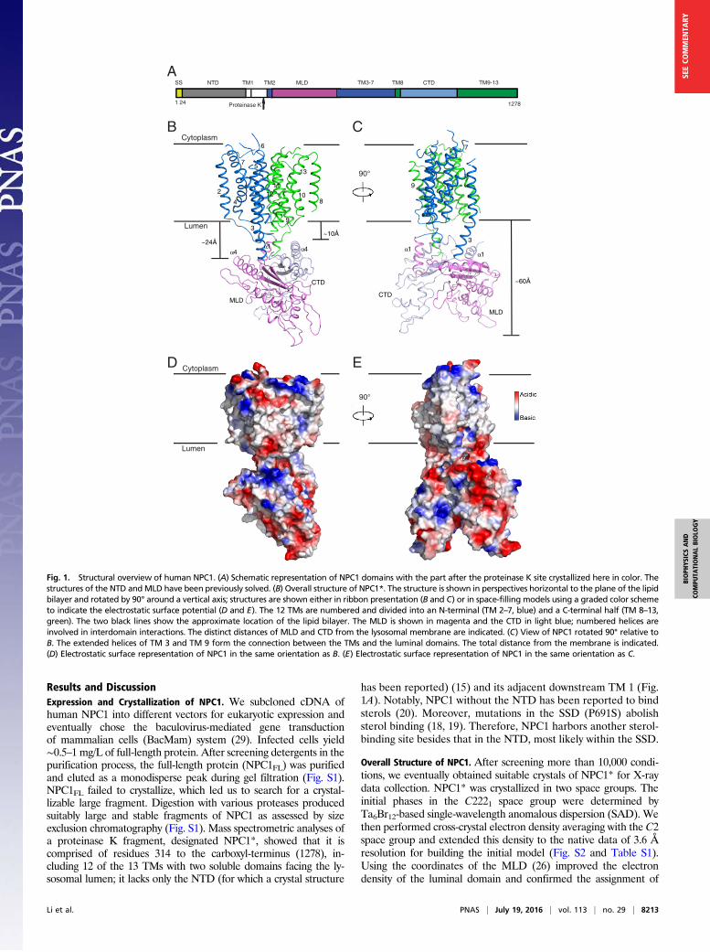
Fig. 1. Structural overview of human NPC1. (A) Schematic representation of NPC1 domains with the part after the proteinase K site crystallized here in color. Thestructures of the NTD andMLD have been previously solved. (B) Overall structure of NPC1*. The structure is shown in perspectives horizontal to the plane of the lipidbilayer and rotated by 90° around a vertical axis; structures are shown either in ribbon presentation (B and C) or in space-filling models using a graded color schemeto indicate the electrostatic surface potential (D and E). The 12 TMs are numbered and divided into an N-terminal (TM 2–7, blue) and a C-terminal half (TM 8–13,green). The two black lines show the approximate location of the lipid bilayer. The MLD is shown in magenta and the CTD in light blue; numbered helices areinvolved in interdomain interactions. The distinct distances of MLD and CTD from the lysosomal membrane are indicated. (C) View of NPC1 rotated 90° relative toB. The extended helices of TM 3 and TM 9 form the connection between the TMs and the luminal domains. The total distance from the membrane is indicated.(D) Electrostatic surface representation of NPC1 in the same orientation as B. (E) Electrostatic surface representation of NPC1 in the same orientation as C.
Li et al. PNAS | July 19, 2016 | vol. 113 | no. 29 | 8213
BIOPH
YSICSAND
COMPU
TATIONALBIOLO
GY
SEECO
MMEN
TARY
residues. Due to its higher resolution, we analyzed the structure ofthe C2 space group in this manuscript (Fig. S2).The overall dimensions of NPC1* are 105 × 55 × 45 Å (Fig. 1 B
and C). Two regions can be distinguished in the structure: (i) alarge and compact membrane-spanning domain formed by 12 TMsshowing internal pseudotwofold symmetry and (ii) a large exten-sion formed by two lumen-exposed domains, MLD and C-terminalluminal domain (CTD), each folding into a structurally similar,elongated curved rod, with the two rods intertwining with eachother at their concave surfaces and forming a 60 Å-long protrusioninto the lysosomal lumen (Fig. 1 B–E, Fig. S3, and Movie S1).
The Transmembrane Domain. The TMs of NPC1* display internal,pseudotwofold symmetry around a central axis parallel to the TMs(Fig. 2A). Strikingly, TM 3–5 of NPC1’s SSD delineate a largecavity that is accessible both from the lipid bilayer and the lysosomelumen (Fig. 2 B and C). This largely hydrophobic cavity measures∼24 × 8 × 8 Å (Movie S1). AutoDock (30) analysis showed thatthis SSD cavity is large enough to accommodate one molecule ofcholesterol (Fig. 2D). This cavity also can be fitted with U18666A,a cholesterol-trafficking inhibitor (Fig. S4).A Dali server search (31) for homologous structures showed that
the transmembrane domain of NPC1 shares a similar transmembrane
helix arrangement with prokaryotic Resistance–Nodulation–CellDivision (RND)-type transporters [AcrB (32), MexB (33), CusA(34), and SecDF (35)] (Fig. 2 E and F). The root-mean-squaredeviation (rmsd) of the superimposition of Cα traces of trans-membrane domains of NPC1 and RND transporters is about 3 Å(z score above 10), which is consistent with previously noted se-quence similarities and predicted topologies (36, 37). For the evo-lutionary history of NPC1 (and of the similar, intestine-localized,NPC1-like1), we suggest that several regions of DNA were recom-bined to code for NPC1’s distinct domains: (i) a novel cholesterol-engulfing domain (NTD) that became membrane-anchored via itsdownstream TM 1 and (ii) two RND-derived domains but arrangedin reverse order—the primary structure of RND transporter TM7–12 resembles more closely TM 2–7 of NPC1 and vice versa, andthat of RND transporter TM 1–6 is more similar to TM 8–13 ofNPC1. This structural similarity is reflected by the sequence align-ment of human NPC1 with proteins of known structure; TM 8–13 inNPC1 show ∼40% similarity with TM 1–6 in AcrB or MexB.
Luminal Domains. The MLD and CTD are attached to the mem-brane region of NPC1 via “stalks” that consist primarily of ex-tended regions of TM 2 and 3 linking the MLD and TM 8 and 9connecting the CTD (Fig. 1 B and C). The MLD and CTD foldinto similar, elongated, curved structures (Fig. 3 A and B). Theyintertwine with each other through their concave surfaces, espe-cially at their membrane-proximal ends, and protrude 60 Å into theendosomal lumen (Fig. 1 B and C and Movie S1). The electrostaticsurface potential of the luminally exposed domains is primarilynegative. Notably, the longer stalk region of the MLD (∼24 Å)leaves a larger gap between the MLD and the luminal aspect of themembrane region (i.e., the SSD) than the shorter stalk (∼10 Å)connecting the CTD to the luminal membrane (Fig. 1B).There are two interfaces between the MLD and CTD: (i) the
N-terminal end of the extended TM 9 helix mediates the interactionwith α1 of MLD and α4 of CTD, and the loops between α1 and β1of the MLD and CTD (referred to as L1) make direct contact (Fig.3C, upper box), and (ii) α1 of the CTD binds to β1 and β3 of theMLD (Fig. 3C, lower box), and this interaction directly contributesto keeping the MLD farther away from the membrane.
Structural Insights into Previous Studies. Our work sheds light onother SSD-containing proteins (Fig. S5). For example, Y298 inSCAP is part of a YIYF tetrapeptide motif that is necessary forSCAP (38) and HMGCR (39) binding to the protein Insig. Y298/I299 is conserved in the SSD of NPC1 (Y634/I635 in TM 3), ex-posed to the membrane at the edge of the transmembrane domain,suggesting that the corresponding helix of SCAP or HMGCR couldpotentially have enough room to bind Insig directly (Fig. 4 A and B).P691, in the center of TM 5, could potentially influence the rigidityof the SSD cavity (Fig. 4B).Previous studies have shown that the MLD of NPC1 binds NPC2
in vitro and in vivo and that two disease-causing mutations (R404Qand especially R518Q) in NPC1 disrupt the NPC2–MLD in-teraction (40). In our structure, R518 is exposed at the end of theshort helix α3 on top of the MLD, clearly free to interact withNPC2 (Fig. 4A). R404 is buried in the MLD, interacting with E606in the part of the stalk that is the amino terminus of TM 3 (Fig. 4A).Mutation of this residue to glutamine would likely destabilize theconformation of the protein.A recently reported structure of the MLD bound to a GP of the
Ebola virus (26) is essentially identical to our MLD structure; theviral GP binding sites consist of loops located at the most mem-brane-distant region of the MLD (Fig. 4A). By docking the crystalstructure of GP to our NPC1* (Fig. 4C), we could visualize thisinteraction as separate from the CTD (about 40 Å away) andconfirm the MLD alone is involved in viral entry (25). Our data donot provide information regarding whether or not the Ebola virusGP also interacts with the NPC1 NTD.
2
8
3
45
6
7
13
12
11
10
9
5
4
3
Cavity in SSD Docked CholesterolNPC1 TM2-7
3
4
100° 90°
D5
lumen
2 810
11
9
12
13
7
6 7
6
3
12
4
5
7
2
6
A B
C
E F
100° 90°
lumen
cytoplasm
cytoplasm
MexB-TMs
3
4
5
12
NPC1-SSD SecDF-TMs
123
5
4
NPC1-SSDNPC1 TM2,8-13 NPC1 TM2,8-13
1313
887
7
2
2
10 10
9 9
11 11
P691
Fig. 2. Structure of NPC1 transmembrane domains. (A) View from the lumen ofthe transmembrane helices perpendicular to the view in Fig. 1B. The pseudosym-metry center is labeled by a red oval. (B) Side (membrane) view of the trans-membrane helices after two 90° rotations relative to A. (C) The cavity in the SSD inthe same orientation as A. The transmembrane cavity (purple mesh) opens to thelumen and is continuous with the membrane inner leaflet. (D) Modeling of cho-lesterol (yellow) in the SSD binding pocket after two 90° rotations relative to C. Thered line approximates the middle of the lipid bilayer. Docking of cholesterol wasperformed by AutoDock (30). (E) Structural comparison between NPC1 and SecDF.(F) Structural comparison between NPC1 and MexB in the same orientation as A.The SSD of NPC1 is shown in blue, and the other TMs of NPC1 are shown in gray.TM 4 of NPC1 undergoes an obvious movement to open the cavity in the SSD.
8214 | www.pnas.org/cgi/doi/10.1073/pnas.1607795113 Li et al.
Model for Cholesterol Sensing. Based on our findings, we proposea working model for cholesterol transport across the lysosome
membrane (Fig. 5). Unesterified cholesterol (state 1, top right)binds first to NPC2, which then docks to the NTD of NPC1,
A
B
C
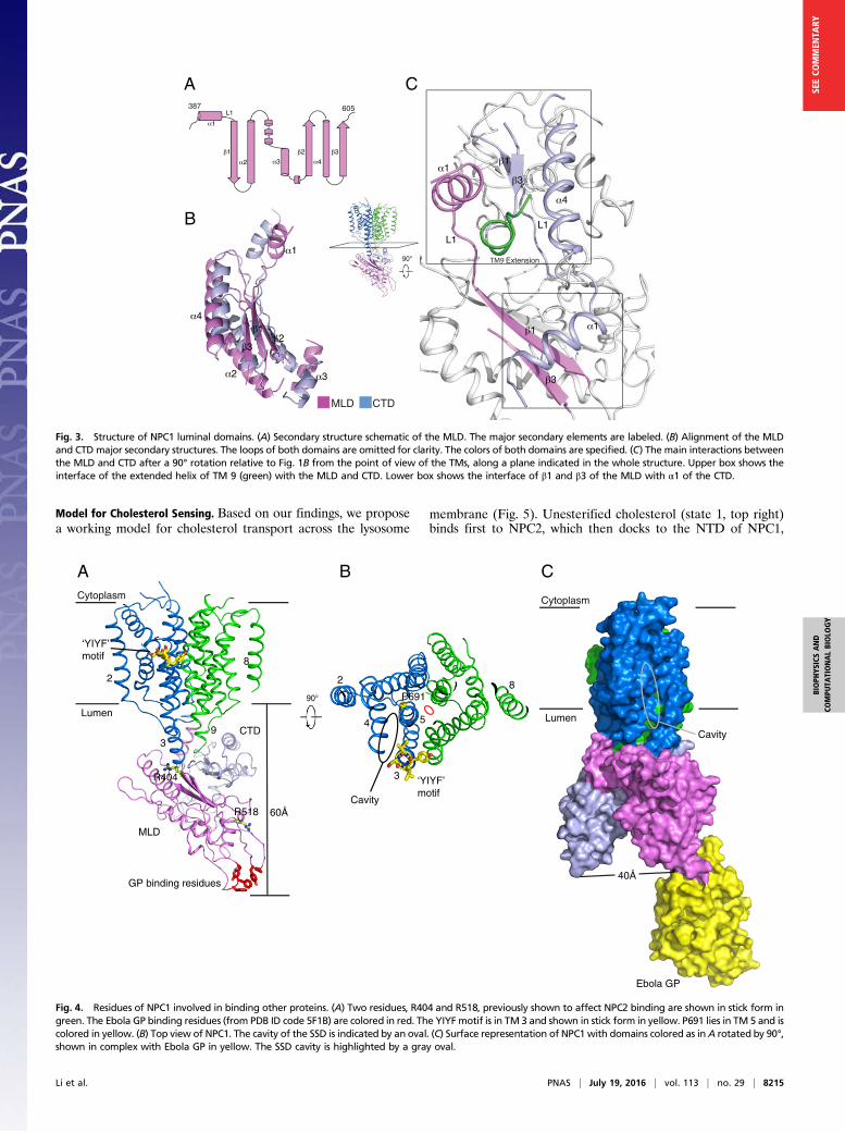
Fig. 3. Structure of NPC1 luminal domains. (A) Secondary structure schematic of the MLD. The major secondary elements are labeled. (B) Alignment of the MLDand CTD major secondary structures. The loops of both domains are omitted for clarity. The colors of both domains are specified. (C) The main interactions betweenthe MLD and CTD after a 90° rotation relative to Fig. 1B from the point of view of the TMs, along a plane indicated in the whole structure. Upper box shows theinterface of the extended helix of TM 9 (green) with the MLD and CTD. Lower box shows the interface of β1 and β3 of the MLD with α1 of the CTD.
Cytoplasm
Lumen
8
2
CTD
MLD
60ÅR518
39
R404
‘YIYF’motif
‘YIYF’motif
P691 90°
Cavity
2
3
4 5
Cytoplasm
Lumen
40Å
Cavity
GP binding residues
Ebola GP
8
A B C
Fig. 4. Residues of NPC1 involved in binding other proteins. (A) Two residues, R404 and R518, previously shown to affect NPC2 binding are shown in stick form ingreen. The Ebola GP binding residues (from PDB ID code 5F1B) are colored in red. The YIYF motif is in TM 3 and shown in stick form in yellow. P691 lies in TM 5 and iscolored in yellow. (B) Top view of NPC1. The cavity of the SSD is indicated by an oval. (C) Surface representation of NPC1 with domains colored as in A rotated by 90°,shown in complex with Ebola GP in yellow. The SSD cavity is highlighted by a gray oval.
Li et al. PNAS | July 19, 2016 | vol. 113 | no. 29 | 8215
BIOPH
YSICSAND
COMPU
TATIONALBIOLO
GY
SEECO
MMEN
TARY
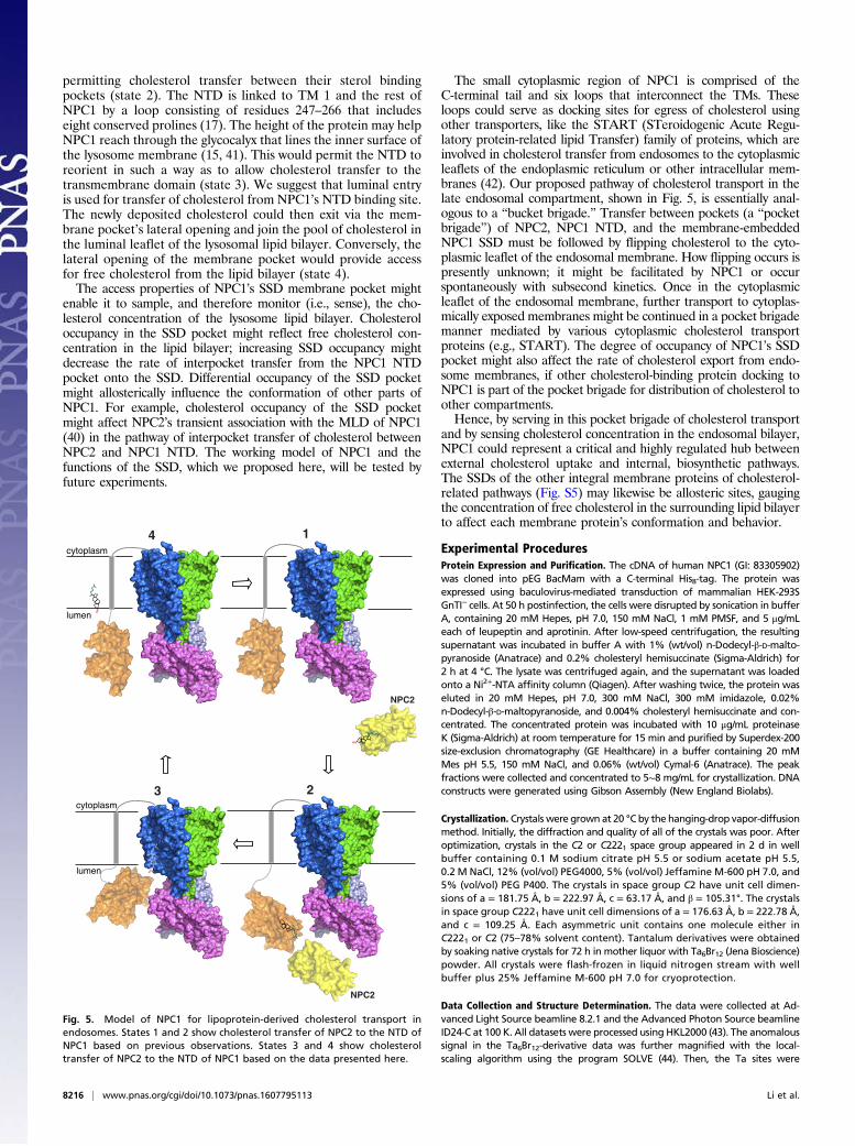
permitting cholesterol transfer between their sterol bindingpockets (state 2). The NTD is linked to TM 1 and the rest ofNPC1 by a loop consisting of residues 247–266 that includeseight conserved prolines (17). The height of the protein may helpNPC1 reach through the glycocalyx that lines the inner surface ofthe lysosome membrane (15, 41). This would permit the NTD toreorient in such a way as to allow cholesterol transfer to thetransmembrane domain (state 3). We suggest that luminal entryis used for transfer of cholesterol from NPC1’s NTD binding site.The newly deposited cholesterol could then exit via the mem-brane pocket’s lateral opening and join the pool of cholesterol inthe luminal leaflet of the lysosomal lipid bilayer. Conversely, thelateral opening of the membrane pocket would provide accessfor free cholesterol from the lipid bilayer (state 4).The access properties of NPC1’s SSD membrane pocket might
enable it to sample, and therefore monitor (i.e., sense), the cho-lesterol concentration of the lysosome lipid bilayer. Cholesteroloccupancy in the SSD pocket might reflect free cholesterol con-centration in the lipid bilayer; increasing SSD occupancy mightdecrease the rate of interpocket transfer from the NPC1 NTDpocket onto the SSD. Differential occupancy of the SSD pocketmight allosterically influence the conformation of other parts ofNPC1. For example, cholesterol occupancy of the SSD pocketmight affect NPC2’s transient association with the MLD of NPC1(40) in the pathway of interpocket transfer of cholesterol betweenNPC2 and NPC1 NTD. The working model of NPC1 and thefunctions of the SSD, which we proposed here, will be tested byfuture experiments.
The small cytoplasmic region of NPC1 is comprised of theC-terminal tail and six loops that interconnect the TMs. Theseloops could serve as docking sites for egress of cholesterol usingother transporters, like the START (STeroidogenic Acute Regu-latory protein-related lipid Transfer) family of proteins, which areinvolved in cholesterol transfer from endosomes to the cytoplasmicleaflets of the endoplasmic reticulum or other intracellular mem-branes (42). Our proposed pathway of cholesterol transport in thelate endosomal compartment, shown in Fig. 5, is essentially anal-ogous to a “bucket brigade.” Transfer between pockets (a “pocketbrigade”) of NPC2, NPC1 NTD, and the membrane-embeddedNPC1 SSD must be followed by flipping cholesterol to the cyto-plasmic leaflet of the endosomal membrane. How flipping occurs ispresently unknown; it might be facilitated by NPC1 or occurspontaneously with subsecond kinetics. Once in the cytoplasmicleaflet of the endosomal membrane, further transport to cytoplas-mically exposed membranes might be continued in a pocket brigademanner mediated by various cytoplasmic cholesterol transportproteins (e.g., START). The degree of occupancy of NPC1’s SSDpocket might also affect the rate of cholesterol export from endo-some membranes, if other cholesterol-binding protein docking toNPC1 is part of the pocket brigade for distribution of cholesterol toother compartments.Hence, by serving in this pocket brigade of cholesterol transport
and by sensing cholesterol concentration in the endosomal bilayer,NPC1 could represent a critical and highly regulated hub betweenexternal cholesterol uptake and internal, biosynthetic pathways.The SSDs of the other integral membrane proteins of cholesterol-related pathways (Fig. S5) may likewise be allosteric sites, gaugingthe concentration of free cholesterol in the surrounding lipid bilayerto affect each membrane protein’s conformation and behavior.
Experimental ProceduresProtein Expression and Purification. The cDNA of human NPC1 (GI: 83305902)was cloned into pEG BacMam with a C-terminal His8-tag. The protein wasexpressed using baculovirus-mediated transduction of mammalian HEK-293SGnTI− cells. At 50 h postinfection, the cells were disrupted by sonication in bufferA, containing 20 mM Hepes, pH 7.0, 150 mM NaCl, 1 mM PMSF, and 5 μg/mLeach of leupeptin and aprotinin. After low-speed centrifugation, the resultingsupernatant was incubated in buffer A with 1% (wt/vol) n-Dodecyl-β-D-malto-pyranoside (Anatrace) and 0.2% cholesteryl hemisuccinate (Sigma-Aldrich) for2 h at 4 °C. The lysate was centrifuged again, and the supernatant was loadedonto a Ni2+-NTA affinity column (Qiagen). After washing twice, the protein waseluted in 20 mM Hepes, pH 7.0, 300 mM NaCl, 300 mM imidazole, 0.02%n-Dodecyl-β-D-maltopyranoside, and 0.004% cholesteryl hemisuccinate and con-centrated. The concentrated protein was incubated with 10 μg/mL proteinaseK (Sigma-Aldrich) at room temperature for 15 min and purified by Superdex-200size-exclusion chromatography (GE Healthcare) in a buffer containing 20 mMMes pH 5.5, 150 mM NaCl, and 0.06% (wt/vol) Cymal-6 (Anatrace). The peakfractions were collected and concentrated to 5∼8 mg/mL for crystallization. DNAconstructs were generated using Gibson Assembly (New England Biolabs).
Crystallization. Crystals were grown at 20 °C by the hanging-drop vapor-diffusionmethod. Initially, the diffraction and quality of all of the crystals was poor. Afteroptimization, crystals in the C2 or C2221 space group appeared in 2 d in wellbuffer containing 0.1 M sodium citrate pH 5.5 or sodium acetate pH 5.5,0.2 M NaCl, 12% (vol/vol) PEG4000, 5% (vol/vol) Jeffamine M-600 pH 7.0, and5% (vol/vol) PEG P400. The crystals in space group C2 have unit cell dimen-sions of a = 181.75 Å, b = 222.97 Å, c = 63.17 Å, and β = 105.31°. The crystalsin space group C2221 have unit cell dimensions of a = 176.63 Å, b = 222.78 Å,and c = 109.25 Å. Each asymmetric unit contains one molecule either inC2221 or C2 (75–78% solvent content). Tantalum derivatives were obtainedby soaking native crystals for 72 h in mother liquor with Ta6Br12 (Jena Bioscience)powder. All crystals were flash-frozen in liquid nitrogen stream with wellbuffer plus 25% Jeffamine M-600 pH 7.0 for cryoprotection.
Data Collection and Structure Determination. The data were collected at Ad-vanced Light Source beamline 8.2.1 and the Advanced Photon Source beamlineID24-C at 100 K. All datasets were processed using HKL2000 (43). The anomaloussignal in the Ta6Br12-derivative data was further magnified with the local-scaling algorithm using the program SOLVE (44). Then, the Ta sites were
HO
14
NPC2
cytoplasm
lumen
2
lumen
HO
3cytoplasm
NPC2
HO
HO
Fig. 5. Model of NPC1 for lipoprotein-derived cholesterol transport inendosomes. States 1 and 2 show cholesterol transfer of NPC2 to the NTD ofNPC1 based on previous observations. States 3 and 4 show cholesteroltransfer of NPC2 to the NTD of NPC1 based on the data presented here.
8216 | www.pnas.org/cgi/doi/10.1073/pnas.1607795113 Li et al.
determined using the program SHELXD (45). The identified sites were refinedand the initial phases were generated in the program PHASER (46) with thesingle-wavelength anomalous dispersion (SAD) experimental phasing module.Cross-crystal density averaging along with solvent flattening and histogrammatching were performed using DMMulti (47). The initial model was built inCrystallographic Object-Oriented Toolkit (COOT) (48) manually. Initially, largearomatic/hydrophobic residues were assigned to facilitate the register of thetransmembrane helices. Because the membrane domain of NPC1 has a topol-ogy similar to RND transporters, the sequence homology between NPC1 andstructurally known RND transporters was used to facilitate model building ofthe transmembrane domain. The coordinates of the MLD (26) improved theelectron density of the luminal domain and confirmed the assignment of resi-dues. The structure was refined with PHENIX.REFINE (Python-Based HierarchicalEnvironment for Integrated Xtallography) (49) at a 3.6 Å resolution. Due to
flexibility and limited resolution, the six linker regions and three loops of theCTD were not visible in the electron density map and were not included in thefinal structure (Fig. S3). Model validation was performed with MolProbity (50).All figures were generated with PyMOL.
ACKNOWLEDGMENTS. We thank George F. Gao for sharing the coordinatesof the free NPC1-C protein (MLD). We also thank Erik Debler and Yi Ren forconstructive comments in structure refinement and manuscript preparation.We acknowledge the staff of the Berkeley Center for Structural Biology at theAdvanced Light Source and the Northeastern Collaborative Access Team at theAdvanced Photon Source for assistance with data collection. This work wassupported by funds from the Rockefeller University and the Howard HughesMedical Institute (G.B., Investigator). X.L. is a Gordon and Betty MooreFoundation Fellow of the Life Sciences Research Foundation.
1. Maxfield FR, van Meer G (2010) Cholesterol, the central lipid of mammalian cells. CurrOpin Cell Biol 22(4):422–429.
2. Brown MS, Goldstein JL (1986) A receptor-mediated pathway for cholesterol ho-meostasis. Science 232(4746):34–47.
3. Porter JA, Young KE, Beachy PA (1996) Cholesterol modification of hedgehog sig-naling proteins in animal development. Science 274(5285):255–259.
4. Chang TY, Chang CC, Ohgami N, Yamauchi Y (2006) Cholesterol sensing, trafficking,and esterification. Annu Rev Cell Dev Biol 22:129–157.
5. Li X, Roberti R, Blobel G (2015) Structure of an integral membrane sterol reductasefrom Methylomicrobium alcaliphilum. Nature 517(7532):104–107.
6. Hua X, Nohturfft A, Goldstein JL, Brown MS (1996) Sterol resistance in CHO cellstraced to point mutation in SREBP cleavage-activating protein. Cell 87(3):415–426.
7. Kuwabara PE, Labouesse M (2002) The sterol-sensing domain: Multiple families, aunique role? Trends Genet 18(4):193–201.
8. Goldstein JL, DeBose-Boyd RA, Brown MS (2006) Protein sensors for membrane ste-rols. Cell 124(1):35–46.
9. Carstea ED, et al. (1997) Niemann-Pick C1 disease gene: Homology to mediators ofcholesterol homeostasis. Science 277(5323):228–231.
10. Roth MG (2006) Clathrin-mediated endocytosis before fluorescent proteins. Nat RevMol Cell Biol 7(1):63–68.
11. Goldstein JL, Dana SE, Faust JR, Beaudet AL, Brown MS (1975) Role of lysosomal acidlipase in the metabolism of plasma low density lipoprotein. Observations in culturedfibroblasts from a patient with cholesteryl ester storage disease. J Biol Chem 250(21):8487–8495.
12. Du H, Grabowski GA (2004) Lysosomal acid lipase and atherosclerosis. Curr OpinLipidol 15(5):539–544.
13. Naureckiene S, et al. (2000) Identification of HE1 as the second gene of Niemann-PickC disease. Science 290(5500):2298–2301.
14. Xu S, Benoff B, Liou HL, Lobel P, Stock AM (2007) Structural basis of sterol binding byNPC2, a lysosomal protein deficient in Niemann-Pick type C2 disease. J Biol Chem282(32):23525–23531.
15. Kwon HJ, et al. (2009) Structure of N-terminal domain of NPC1 reveals distinct sub-domains for binding and transfer of cholesterol. Cell 137(7):1213–1224.
16. Infante RE, et al. (2008) Purified NPC1 protein. I. Binding of cholesterol and oxysterolsto a 1278-amino acid membrane protein. J Biol Chem 283(2):1052–1063.
17. Infante RE, et al. (2008) Purified NPC1 protein: II. Localization of sterol binding to a240-amino acid soluble luminal loop. J Biol Chem 283(2):1064–1075.
18. Ohgami N, et al. (2004) Binding between the Niemann-Pick C1 protein and a pho-toactivatable cholesterol analog requires a functional sterol-sensing domain. ProcNatl Acad Sci USA 101(34):12473–12478.
19. Lu F, et al. (2015) Identification of NPC1 as the target of U18666A, an inhibitor oflysosomal cholesterol export and Ebola infection. eLife 4:4.
20. Ohgane K, Karaki F, Dodo K, Hashimoto Y (2013) Discovery of oxysterol-derivedpharmacological chaperones for NPC1: Implication for the existence of second sterol-binding site. Chem Biol 20(3):391–402.
21. Yanjanin NM, et al. (2010) Linear clinical progression, independent of age of onset, inNiemann-Pick disease, type C. Am J Med Genet B Neuropsychiatr Genet 153B(1):132–140.
22. Pentchev PG (2004) Niemann-Pick C research from mouse to gene. Biochim BiophysActa 1685(1-3):3–7.
23. Carette JE, et al. (2011) Ebola virus entry requires the cholesterol transporter Niemann-Pick C1. Nature 477(7364):340–343.
24. Côté M, et al. (2011) Small molecule inhibitors reveal Niemann-Pick C1 is essential forEbola virus infection. Nature 477(7364):344–348.
25. Miller EH, et al. (2012) Ebola virus entry requires the host-programmed recognition ofan intracellular receptor. EMBO J 31(8):1947–1960.
26. Wang H, et al. (2016) Ebola viral glycoprotein bound to its endosomal receptorNiemann-Pick C1. Cell 164(1-2):258–268.
27. Rhein BA, Maury WJ (2015) Ebola virus entry into host cells: Identifying therapeuticstrategies. Curr Clin Microbiol Rep 2(3):115–124.
28. Herbert AS, et al. (2015) Niemann-pick C1 is essential for ebolavirus replication andpathogenesis in vivo. MBio 6(3):e00565-15.
29. Dukkipati A, Park HH, Waghray D, Fischer S, Garcia KC (2008) BacMam system forhigh-level expression of recombinant soluble and membrane glycoproteins forstructural studies. Protein Expr Purif 62(2):160–170.
30. Morris GM, et al. (2009) AutoDock4 and AutoDockTools4: Automated docking withselective receptor flexibility. J Comput Chem 30(16):2785–2791.
31. Holm L, Rosenstrom P (2010) Dali server: Conservation mapping in 3D. Nucleic AcidsRes 38(Web Server issue):W545–W549.
32. Murakami S, Nakashima R, Yamashita E, Yamaguchi A (2002) Crystal structure ofbacterial multidrug efflux transporter AcrB. Nature 419(6907):587–593.
33. Nakashima R, et al. (2013) Structural basis for the inhibition of bacterial multidrugexporters. Nature 500(7460):102–106.
34. Long F, et al. (2010) Crystal structures of the CusA efflux pump suggest methionine-mediated metal transport. Nature 467(7314):484–488.
35. Tsukazaki T, et al. (2011) Structure and function of a membrane component SecDFthat enhances protein export. Nature 474(7350):235–238.
36. Tseng TT, et al. (1999) The RND permease superfamily: An ancient, ubiquitous anddiverse family that includes human disease and development proteins. J MolMicrobiol Biotechnol 1(1):107–125.
37. Davies JP, Chen FW, Ioannou YA (2000) Transmembrane molecular pump activity ofNiemann-Pick C1 protein. Science 290(5500):2295–2298.
38. Adams CM, et al. (2004) Cholesterol and 25-hydroxycholesterol inhibit activation ofSREBPs by different mechanisms, both involving SCAP and Insigs. J Biol Chem 279(50):52772–52780.
39. Sever N, Yang T, Brown MS, Goldstein JL, DeBose-Boyd RA (2003) Accelerated deg-radation of HMG CoA reductase mediated by binding of insig-1 to its sterol-sensingdomain. Mol Cell 11(1):25–33.
40. Deffieu MS, Pfeffer SR (2011) Niemann-Pick type C 1 function requires lumenal do-main residues that mediate cholesterol-dependent NPC2 binding. Proc Natl Acad SciUSA 108(47):18932–18936.
41. Li J, Deffieu MS, Lee PL, Saha P, Pfeffer SR (2015) Glycosylation inhibition reducescholesterol accumulation in NPC1 protein-deficient cells. Proc Natl Acad Sci USA112(48):14876–14881.
42. Soccio RE, Breslow JL (2003) StAR-related lipid transfer (START) proteins: Mediators ofintracellular lipid metabolism. J Biol Chem 278(25):22183–22186.
43. Otwinowski ZMW (1997) Processing of X-ray diffraction data collected in oscillationmode. Methods Enzymol 276:307–326.
44. Terwilliger TC, Berendzen J (1999) Automated MAD and MIR structure solution. ActaCrystallogr D Biol Crystallogr 55(Pt 4):849–861.
45. Schneider TR, Sheldrick GM (2002) Substructure solution with SHELXD. ActaCrystallogr D Biol 58(Pt 10 Pt 2):1772–1779.
46. McCoy AJ, et al. (2007) Phaser crystallographic software. J Appl Cryst 40(Pt 4):658–674.47. Cowtan K (1994) ‘dm’: An automated procedure for phase improvement by density
modification. Joint CCP4 ESF-EACBM Newslett J Prot Crystallogr 31:34–38.48. Emsley P, Cowtan K (2004) Coot: Model-building tools for molecular graphics. Acta
Crystallogr D Biol Crystallogr 60(Pt 12 Pt 1):2126–2132.49. Adams PD, et al. (2010) PHENIX: A comprehensive Python-based system for macro-
molecular structure solution. Acta Crystallogr D Biol Crystallogr 66(Pt 2):213–221.50. Chen VB, et al. (2010) MolProbity: All-atom structure validation for macromolecular
crystallography. Acta Crystallogr D Biol Crystallogr 66(Pt 1):12–21.
Li et al. PNAS | July 19, 2016 | vol. 113 | no. 29 | 8217
BIOPH
YSICSAND
COMPU
TATIONALBIOLO
GY
SEECO
MMEN
TARY


![GÖQ - tip.kocaeli.edu.trtip.kocaeli.edu.tr/.../NIEMANN-PICKTIPC.pdf · x Genetik inceleme Niemann Pick d] Z ofRfvf }R µo v fX. Niemann Pick , ofRf (NPH) Niemann Pick , ofRfV ol](https://img.pdfslide.net/doc/110x75/5c6c994f09d3f2fe088b4cea/goeq-tip-x-genetik-inceleme-niemann-pick-d-z-ofrfvf-r-o-v-fx-niemann.jpg)