Embed Size (px)
Citation preview
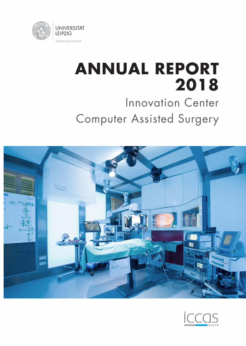
ANNUAL REPORT2018
Innovation Center Computer Assisted Surgery
IMPRINT
EDITORLeipzig UniversityFaculty of MedicineInnovation Center Computer Assisted Surgery (ICCAS)
Semmelweisstraße 1404103 LeipzigGermany
E-Mail: [email protected]: www.iccas.de
EXECUTIVE DIRECTORProf. Dr. Andreas Melzer
CONCEPT & LAYOUTKathrin ScholzSimon RosenowChistoph Zeumer
COVERICCAS’s intelligent and fully networked demonstration operating room.
PHOTOSICCAS, Leonie Lang, Swen Reichhold
GRAPHIC ARTSSimon Rosenow
DISCLAIMERAll data in this report is to the Institutes specifications.No responsibility can be accepted for the correctness of this information.

CONTENTS
PREFACE ................................................................................................................................................
INSTITUTIONAL FACTS .......................................................................................................................Timeline ..................................................................................................................................................Facts and Figures ....................................................................................................................................
ACTIVITIES ............................................................................................................................................Highlights ................................................................................................................................................External Presentations ............................................................................................................................Selected Events ...................................................................................................................................... Guest Talks ..............................................................................................................................................Collaboration Works ...............................................................................................................................Project Launches ....................................................................................................................................Honors and Awards ................................................................................................................................
RESEARCH AREAS AND RELATED PROJECT PROFILES ....................................................................Model-Based Automation and Integration ............................................................................................Digital Patient- and Process Model ........................................................................................................Intraoperative Multimodal Imaging .......................................................................................................Computer-Assisted Image-Guided Interventions ...................................................................................Life Support Systems ..............................................................................................................................
PUBLICATIONS .....................................................................................................................................Journal and Book Publications, First- and Senior Authorship .................................................................Co-Authorship ........................................................................................................................................Conference Proceedings .........................................................................................................................
EVENTS ...................................................................................................................................................In-House Events ......................................................................................................................................Conferences, Symposia, Workshops ......................................................................................................Presentations at Fairs .............................................................................................................................Project- and Cooperation Work .............................................................................................................University Courses .................................................................................................................................Graduations ..........................................................................................................................................
ORGANIZATION ...................................................................................................................................
COOPERATION PARTNERS ..................................................................................................................
2
335
779
1011121314
151731415571
79798181
84848690919293
94
97

1 PREFACE
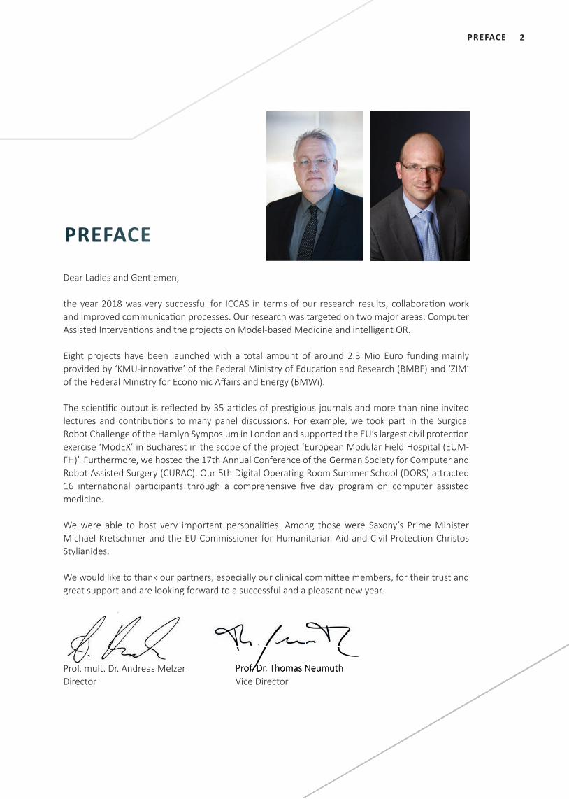
2PREFACE
Dear Ladies and Gentlemen,
the year 2018 was very successful for ICCAS in terms of our research results, collaborati on work and improved communicati on processes. Our research was targeted on two major areas: Computer Assisted Interventi ons and the projects on Model-based Medicine and intelligent OR.
Eight projects have been launched with a total amount of around 2.3 Mio Euro funding mainly provided by ‘KMU-innovati ve’ of the Federal Ministry of Educati on and Research (BMBF) and ‘ZIM’ of the Federal Ministry for Economic Aff airs and Energy (BMWi).
The scienti fi c output is refl ected by 35 arti cles of presti gious journals and more than nine invited lectures and contributi ons to many panel discussions. For example, we took part in the Surgical Robot Challenge of the Hamlyn Symposium in London and supported the EU’s largest civil protecti on exercise ‘ModEX’ in Bucharest in the scope of the project ‘European Modular Field Hospital (EUM-FH)’. Furthermore, we hosted the 17th Annual Conference of the German Society for Computer and Robot Assisted Surgery (CURAC). Our 5th Digital Operati ng Room Summer School (DORS) att racted 16 internati onal parti cipants through a comprehensive fi ve day program on computer assisted medicine.
We were able to host very important personaliti es. Among those were Saxony’s Prime Minister Michael Kretschmer and the EU Commissioner for Humanitarian Aid and Civil Protecti on Christos Stylianides.
We would like to thank our partners, especially our clinical committ ee members, for their trust and great support and are looking forward to a successful and a pleasant new year.
Yours sincerely,Yours sincerely,
PREFACE
Prof. mult. Dr. Andreas MelzerDirector
Prof. Dr. Thomas NeumuthVice DirectorProf. Dr. Thomas Neumuth
3
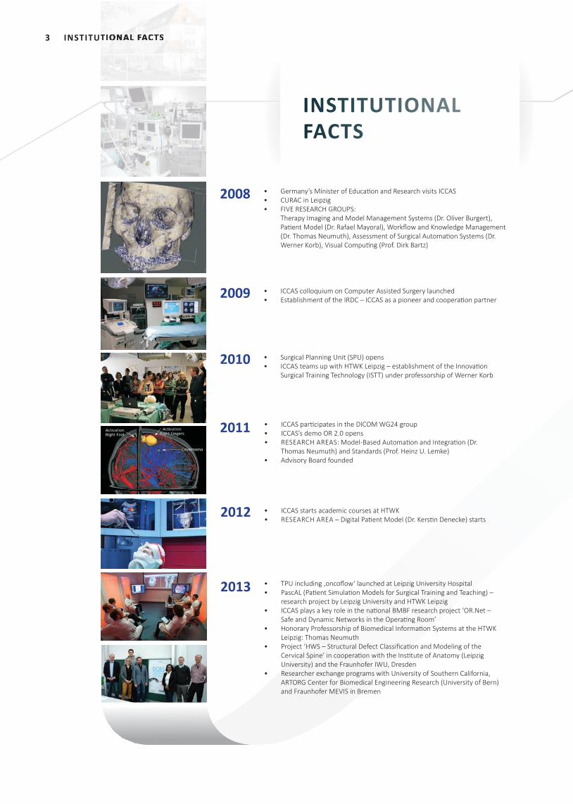
• Germany’s Minister of Educati on and Research visits ICCAS • CURAC in Leipzig• FIVE RESEARCH GROUPS:
Therapy Imaging and Model Management Systems (Dr. Oliver Burgert), Pati ent Model (Dr. Rafael Mayoral), Workfl ow and Knowledge Management (Dr. Thomas Neumuth), Assessment of Surgical Automati on Systems (Dr. Werner Korb), Visual Computi ng (Prof. Dirk Bartz)
• Surgical Planning Unit (SPU) opens• ICCAS teams up with HTWK Leipzig – establishment of the Innovati on
Surgical Training Technology (ISTT) under professorship of Werner Korb
2008
2010
2009 • ICCAS colloquium on Computer Assisted Surgery launched• Establishment of the IRDC – ICCAS as a pioneer and cooperati on partner
2013
• ICCAS parti cipates in the DICOM WG24 group• ICCAS’s demo OR 2.0 opens • RESEARCH AREAS: Model-Based Automati on and Integrati on (Dr.
Thomas Neumuth) and Standards (Prof. Heinz U. Lemke)• Advisory Board founded
• TPU including ‚oncofl ow‘ launched at Leipzig University Hospital• PascAL (Pati ent Simulati on Models for Surgical Training and Teaching) –
research project by Leipzig University and HTWK Leipzig• ICCAS plays a key role in the nati onal BMBF research project ‘OR.Net –
Safe and Dynamic Networks in the Operati ng Room’ • Honorary Professorship of Biomedical Informati on Systems at the HTWK
Leipzig: Thomas Neumuth• Project ‘HWS – Structural Defect Classifi cati on and Modeling of the
Cervical Spine’ in cooperati on with the Insti tute of Anatomy (Leipzig University) and the Fraunhofer IWU, Dresden
• Researcher exchange programs with University of Southern California, ARTORG Center for Biomedical Engineering Research (University of Bern) and Fraunhofer MEVIS in Bremen
• ICCAS starts academic courses at HTWK • RESEARCH AREA – Digital Pati ent Model (Dr. Kersti n Denecke) starts
2012
2011
INSTITUTIONAL FACTSINSTITUTIONAL FACTS
INSTITUTIONALFACTS
4INSTITUTIONAL FACTS
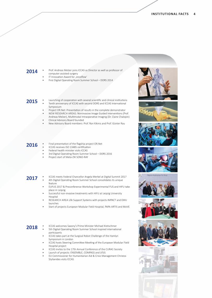
• Prof. Andreas Melzer joins ICCAS as Director as well as professor of computer assisted surgery
• IT Innovati on Award for ‚oncofl ow‘• First Digital Operati ng Room Summer School – DORS 2014
• Final presentati on of the fl agship project OR.Net• ICCAS receives ISO 13485 certi fi cati on• Federal health minister visits ICCAS• 3rd Digital Operati ng Room Summer School – DORS 2016 • Project start of Meta-ZIK SONO-RAY
2014
2016
2015
2017
2018
• Launching of cooperati on with several scienti fi c and clinical insti tuti ons• Tenth anniversary of ICCAS with second DORS and ICCAS Internati onal
Symposium• Project OR.Net: Presentati on of results in the complete demonstrator• NEW RESEARCH AREAS: Noninvasive Image Guided Interventi ons (Prof.
Andreas Melzer), Multi modal Intraoperati ve Imaging (Dr. Claire Chalopin)• Clinical Advisory Board founded• New Advisory Board members: Prof. Ron Kikinis and Prof. Günter Rau
• ICCAS meets Federal Chancellor Angela Merkel at Digital Summit 2017 • 4th Digital Operati ng Room Summer School consolidates its unique
feature• EUFUS 2017 & Preconference Workshop Experimental FUS and HIFU take
place in Leipzig• Successful non-invasive treatments with HIFU at Leipzig University
Hospital• RESEARCH AREA Life Support Systems with projects IMPACT and EMU
launches• Start of projects European Modular Field Hospital, PAPA-ARTIS and MoVE
• ICCAS welcomes Saxony’s Prime Minister Michael Kretschmer• 5th Digital Operati ng Room Summer School inspired internati onal
parti cipants• ICCAS takes part at the Surgical Robot Challenge of the Hamlyn
Symposium in London• ICCAS hosts Steering Committ ee Meeti ng of the European Modular Field
Hospital project• ICCAS invites to the 17th Annual Conference of the CURAC-Society • Launch of projects: ENSEMBLE, COMPASS and LYSiS• EU Commissioner for Humanitarian Aid & Crisis Management Christos
Stylianides visits ICCAS
INSTITUTIONALFACTS
5 INSTITUTIONAL FACTS
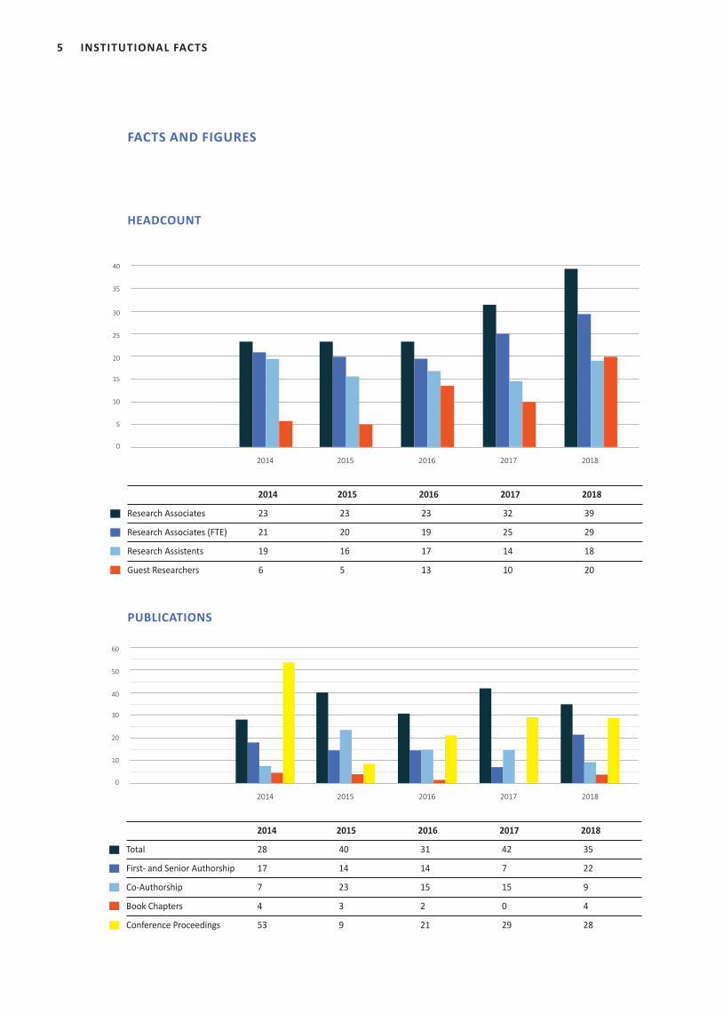
2014 2015 2016 2017 2018
Research Associates 23 23 23 32 39
Research Associates (FTE) 21 20 19 25 29
Research Assistents 19 16 17 14 18
Guest Researchers 6 5 13 10 20
FACTS AND FIGURES
HEADCOUNT
PUBLICATIONS
0
5
10
15
20
25
35
30
40
2014 2015 2016 2017 2018
Total 28 40 31 42 35
First- and Senior Authorship 17 14 14 7 22
Co-Authorship 7 23 15 15 9
Book Chapters 4 3 2 0 4
Conference Proceedings 53 9 21 29 28
2014 2015 2016 2017 2018
2014 2015 2016 2017 2018
0
10
20
30
40
50
60
6INSTITUTIONAL FACTS
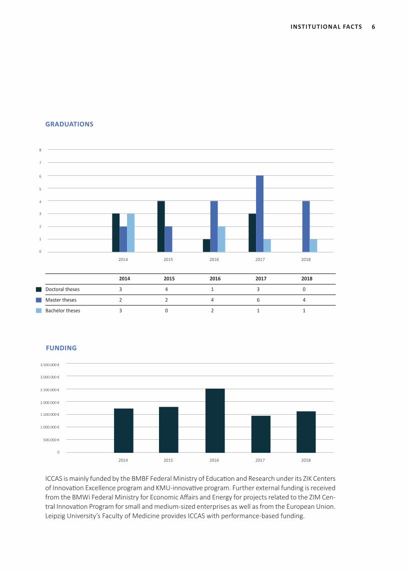
2014 2015 2016 2017 2018
Doctoral theses 3 4 1 3 0
Master theses 2 2 4 6 4
Bachelor theses 3 0 2 1 1
GRADUATIONS
0
500.000 €
1.000.000 €
1.500.000 €
2.000.000 €
2.500.000 €
3.000.000 €
3.500.000 €
FUNDING
ICCAS is mainly funded by the BMBF Federal Ministry of Education and Research under its ZIK Centers of Innovation Excellence program and KMU-innovative program. Further external funding is received from the BMWi Federal Ministry for Economic Affairs and Energy for projects related to the ZIM Cen-tral Innovation Program for small and medium-sized enterprises as well as from the European Union. Leipzig University’s Faculty of Medicine provides ICCAS with performance-based funding.
0
1
2
3
4
5
7
6
8
2014 2015 2016 2017 2018
2014 2015 2016 2017 2018 2014 2015 2016 2017 2018
2014 2015 2016 2017 2018
7 ACTIVITIES
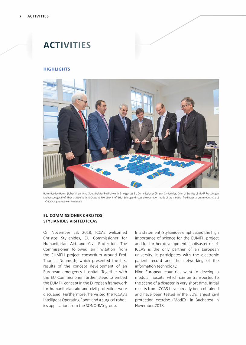
EU COMMISSIONER CHRISTOS STYLIANIDES VISITED ICCAS
On November 23, 2018, ICCAS welcomed Christos Stylianides, EU Commissioner for Humanitarian Aid and Civil Protecti on. The Commissioner followed an invitati on from the EUMFH project consorti um around Prof. Thomas Neumuth, which presented the fi rst results of the concept development of an European emergency hospital. Together with the EU Commissioner further steps to embed the EUMFH concept in the European framework for humanitarian aid and civil protecti on were discussed. Furthermore, he visited the ICCAS’s Intelligent Operati ng Room and a surgical robot-ics applicati on from the SONO-RAY group.
In a statement, Stylianides emphasized the high importance of science for the EUMFH project and for further developments in disaster relief. ICCAS is the only partner of an European university. It parti cipates with the electronic pati ent record and the networking of the informati on technology. Nine European countries want to develop a modular hospital which can be transported to the scene of a disaster in very short ti me. Initi al results from ICCAS have already been obtained and have been tested in the EU’s largest civil protecti on exercise (ModEX) in Bucharest in November 2018.
ACTIVITIES
Harm-Basti an Harms (Johanniter), Gino Claes (Belgian Public Health Emergency), EU Commissioner Christos Stylianides, Dean of Studies of MedF Prof. Jürgen Meixensberger, Prof. Thomas Neumuth (ICCAS) and Prorector Prof. Erich Schröger discuss the operati on mode of the modular fi eld hospital on a model. (f.l.t.r.) | © ICCAS, photo: Swen Reichhold
HIGHLIGHTS
8ACTIVITIES
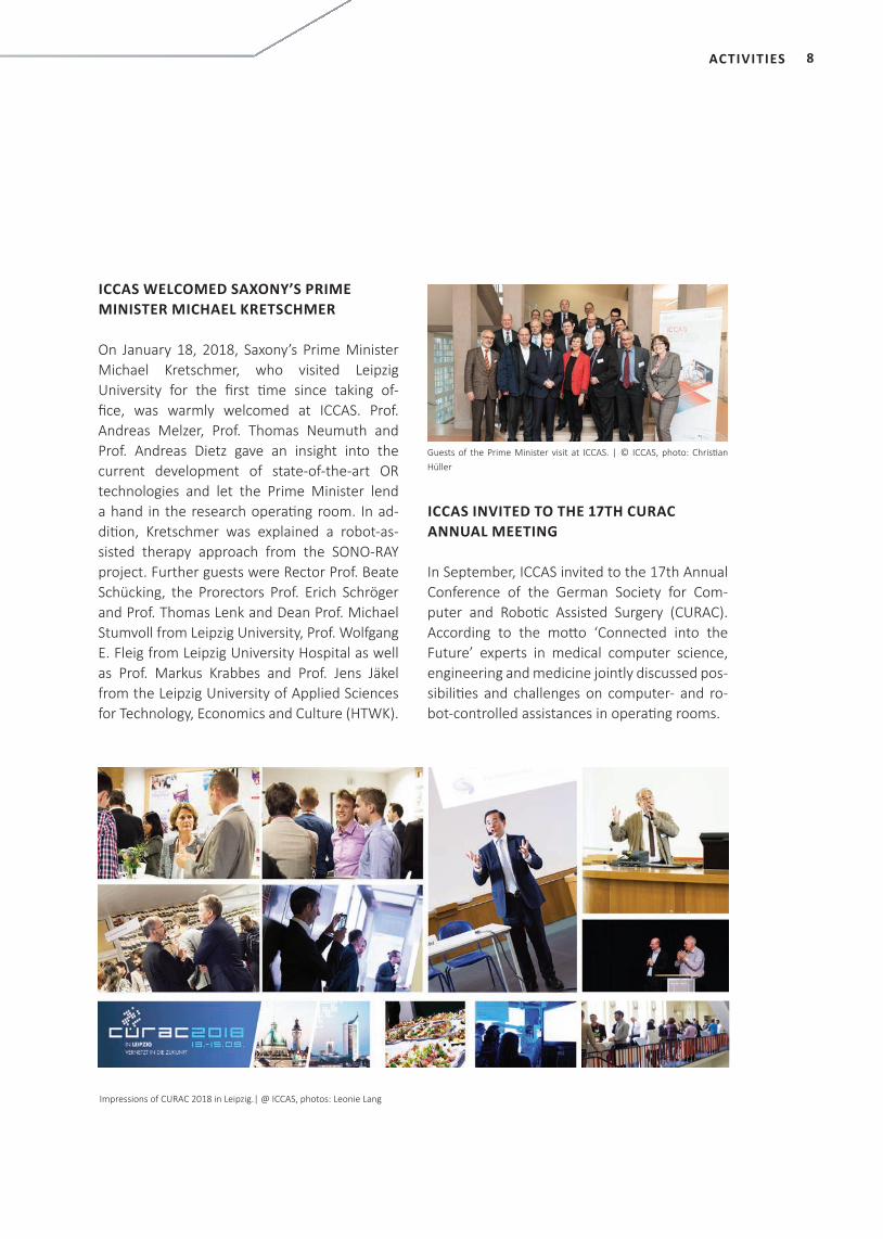
ICCAS WELCOMED SAXONY’S PRIME MINISTER MICHAEL KRETSCHMER
On January 18, 2018, Saxony’s Prime Minister Michael Kretschmer, who visited Leipzig University for the fi rst ti me since taking of-fi ce, was warmly welcomed at ICCAS. Prof. Andreas Melzer, Prof. Thomas Neumuth and Prof. Andreas Dietz gave an insight into the current development of state-of-the-art OR technologies and let the Prime Minister lend a hand in the research operati ng room. In ad-diti on, Kretschmer was explained a robot-as-sisted therapy approach from the SONO-RAY project. Further guests were Rector Prof. Beate Schücking, the Prorectors Prof. Erich Schröger and Prof. Thomas Lenk and Dean Prof. Michael Stumvoll from Leipzig University, Prof. Wolfgang E. Fleig from Leipzig University Hospital as well as Prof. Markus Krabbes and Prof. Jens Jäkel from the Leipzig University of Applied Sciences for Technology, Economics and Culture (HTWK).
Guests of the Prime Minister visit at ICCAS. | © ICCAS, photo: Christi an Hüller
ICCAS INVITED TO THE 17TH CURAC ANNUAL MEETING
In September, ICCAS invited to the 17th Annual Conference of the German Society for Com-puter and Roboti c Assisted Surgery (CURAC). According to the mott o ‘Connected into the Future’ experts in medical computer science, engineering and medicine jointly discussed pos-sibiliti es and challenges on computer- and ro-bot-controlled assistances in operati ng rooms.
Impressions of CURAC 2018 in Leipzig.| @ ICCAS, photos: Leonie Lang
9 ACTIVITIES
ICCAS’S ‘INTELLIGENT OR’ AT XPOMET© CONVENTION 2018 March 21 – 23, 2018, Leipzig
Dr. Stefan Franke explained the functionalities of ICCAS’s ‘Intelligent Operating Room’ to Saxony’s Minister for Social Affairs and Consumer Protection Barbara Klepsch at the XPOMET© Convention exhibition booth.
ICCAS SUPPORTED EU’S LARGEST CIVIL PROTECTION EXERCISE ‘MODEX’ October 14 – 18, 2018, Bucharest (Romania)
Prof. Thomas Neumuth and Erik Schreiber supported the project team ‘European Modular Field Hospital’ (EUMFH) during the EU’s largest civil protection exercise ‘ModEX’ in Bucharest. They evaluated the digital patient file for disaster relief developed at ICCAS under real conditions.
EXTERNAL PRESENTATIONS
ICCAS TOOK PART IN THE SURGICAL ROBOT CHALLENGE OF THE HAMLYN SYMPOSIUM June 24, 2018, London (UK)
Scientists from the SONO-RAY group competed with international leading research groups at the Surgical Robot Challenge of the renowned Hamlyn Symposium in London. They presented a robot system for supporting the ultrasound-guided removal of tissue samples.
© ModEX 2017/2018
10ACTIVITIES
SELECTED EVENTS
GIRLS’DAY April 26, 2018 | ICCAS, Leipzig
On the occassion of the nationwide Girl’s Day, Dr. Lisa Landgraf and Dr. Claire Chalopin gave female pupils an insight into research projects in the field of medical informatics at ICCAS.
OPEN DAY June 6, 2018 | ICCAS, Leipzig
Leipzig University’s Prorector for Develop-ment and Transfer Prof. Thomas Lenk and project coordinator Moritz Waschbüsch were two of the 100 visitors of the Open Day. ICCAS’s scientists from all research areas had invited to demonstrate medical technologies in a close and simple way.
LONG NIGHT OF SCIENCES June 22, 2018 | BBZ, Leipzig
At the Long Night of Sciences, the scientists of the Life Support Systems group experi-mentally explained the function of Electrical Impedance Tomography (EIT). Many guests came especially to fasten the measuring belt and to watch their own lung function in the live image.
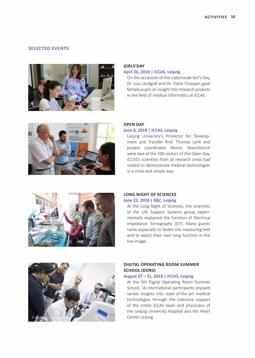
DIGITAL OPERATING ROOM SUMMER SCHOOL (DORS) August 27 – 31, 2018 | ICCAS, Leipzig
At the 5th Digital Operating Room Summer School, 16 international participants enjoyed varied insights into state-of-the-art medical technologies through the intensive support of the entire ICCAS team and physicians of the Leipzig University Hospital and the Heart Center Leipzig. © ICCAS, photo: Leonie Lang
11 ACTIVITIES
INVITED TALK AT DGU CONGRESS September 27, 2018, Dresden
At the opening plenary of this year’s Congress of the German Society for Urology‚ Prof. Andreas Melzer held an invited talk on aspects of the future operating room.
KEY NOTE LECTURE AT BMT September 26, 2018, Aachen
At this year’s Annual Conference of the German Society for Biomedical Engineering, Prof. Thomas Neumuth gave a keynote lecture on ‘Patient Specific Model Guided Therapy‘.
PANEL DISCUSSION AT FRAUNHOFER INTERACTIVE EXHIBITION October 9, 2018, Berlin
At the Fraunhofer Interactive Exhibition ‚Future Work‘, Prof. Andreas Melzer was one of the experts of the panel discussion ‚Future Work Health‘. Top-level representatives from science and politics talked about the workplace of the future in the fields of health and nursing care.
PANEL DISCUSSION AT SZ CONGRESS September 25, 2018, Munich
Prof. Thomas Neumuth was one of the experts of the panel discussion on ‘Smart Hospital’ at the Süddeutsche Zeitung Congress ‘Digital Health’. Together with participants from science, industry and medicine he discussed how digital technologies can simplify therapy- and administration processes.
GUEST TALKS
© DGU e.V.
© Svea Pietschmann
© SZ-Kongress Digital Health, photo: A. McMaster
12ACTIVITIES
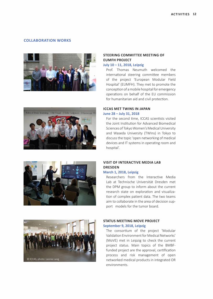
STEERING COMMITTEE MEETING OF EUMFH PROJECT July 10 – 11, 2018, Leipzig
Prof. Thomas Neumuth welcomed the international steering committee members of the project ‘European Modular Field Hospital’ (EUMFH). They met to promote the conception of a mobile hospital for emergency operations on behalf of the EU commission for humanitarian aid and civil protection.
ICCAS MET TWINS IN JAPAN June 28 – July 31, 2018
For the second time, ICCAS scientists visited the Joint Institution for Advanced Biomedical Sciences of Tokyo Women’s Medical University and Waseda University (TWIns) in Tokyo to discuss the topic ‘open networking of medical devices and IT systems in operating room and hospital’.
VISIT OF INTERACTIVE MEDIA LAB DRESDEN March 1, 2018, Leipzig
Researchers from the Interactive Media Lab at Technische Universität Dresden met the DPM group to inform about the current research state on exploration and visualiza-tion of complex patient data. The two teams aim to collaborate in the area of decision sup-port models for the tumor board.
STATUS MEETING MOVE PROJECT September 9, 2018, Leipzig
The consortium of the project ‘Modular Validation Environment for Medical Networks’ (MoVE) met in Leipzig to check the current project status. Main topics of the BMBF- funded project are the approval, certification process and risk management of open networked medical products in integrated OR environments.
COLLABORATION WORKS
© ICCAS, photo: Leonie Lang
13 ACTIVITIES
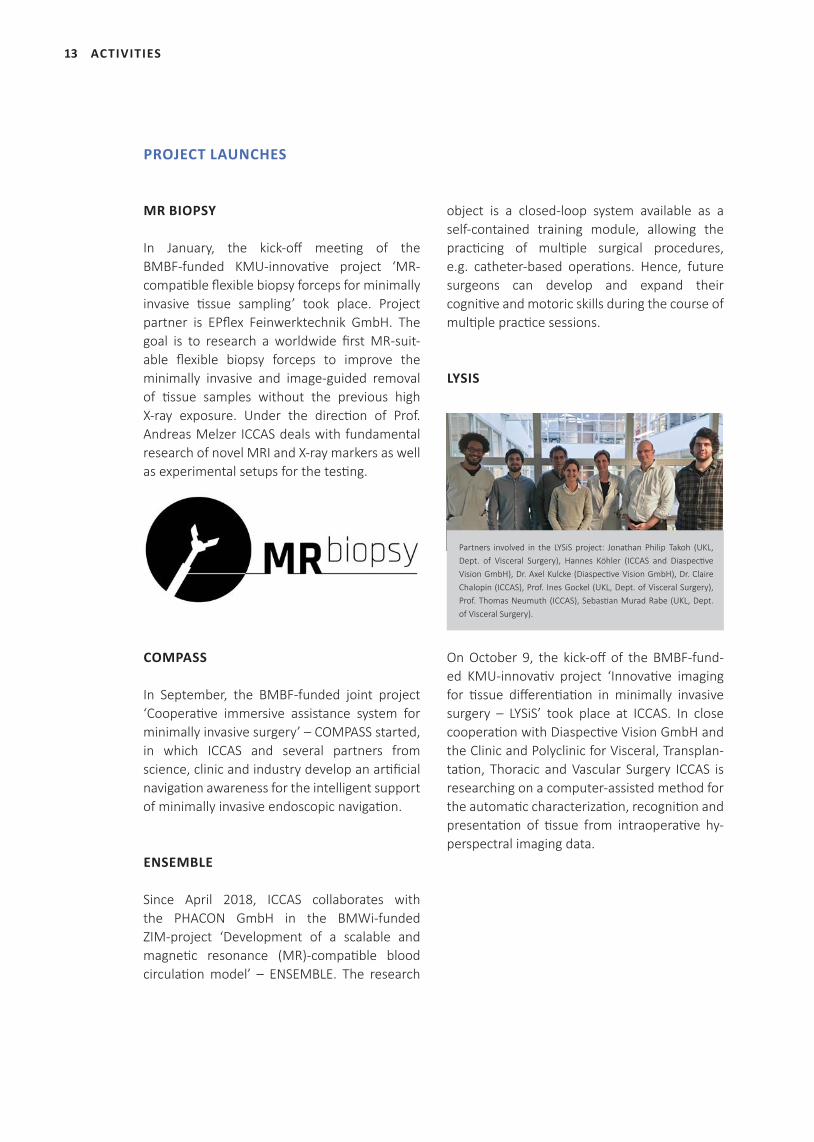
MR BIOPSY
In January, the kick-off meeting of the BMBF-funded KMU-innovative project ‘MR- compatible flexible biopsy forceps for minimally invasive tissue sampling’ took place. Project partner is EPflex Feinwerktechnik GmbH. The goal is to research a worldwide first MR-suit-able flexible biopsy forceps to improve the minimally invasive and image-guided removal of tissue samples without the previous high X-ray exposure. Under the direction of Prof. Andreas Melzer ICCAS deals with fundamental research of novel MRI and X-ray markers as well as experimental setups for the testing.
COMPASS
In September, the BMBF-funded joint project ‘Cooperative immersive assistance system for minimally invasive surgery’ – COMPASS started, in which ICCAS and several partners from science, clinic and industry develop an artificial navigation awareness for the intelligent support of minimally invasive endoscopic navigation.
ENSEMBLE
Since April 2018, ICCAS collaborates with the PHACON GmbH in the BMWi-funded ZIM-project ‘Development of a scalable and magnetic resonance (MR)-compatible blood circulation model’ – ENSEMBLE. The research
object is a closed-loop system available as a self-contained training module, allowing the practicing of multiple surgical procedures, e.g. catheter-based operations. Hence, future surgeons can develop and expand their cognitive and motoric skills during the course of multiple practice sessions.
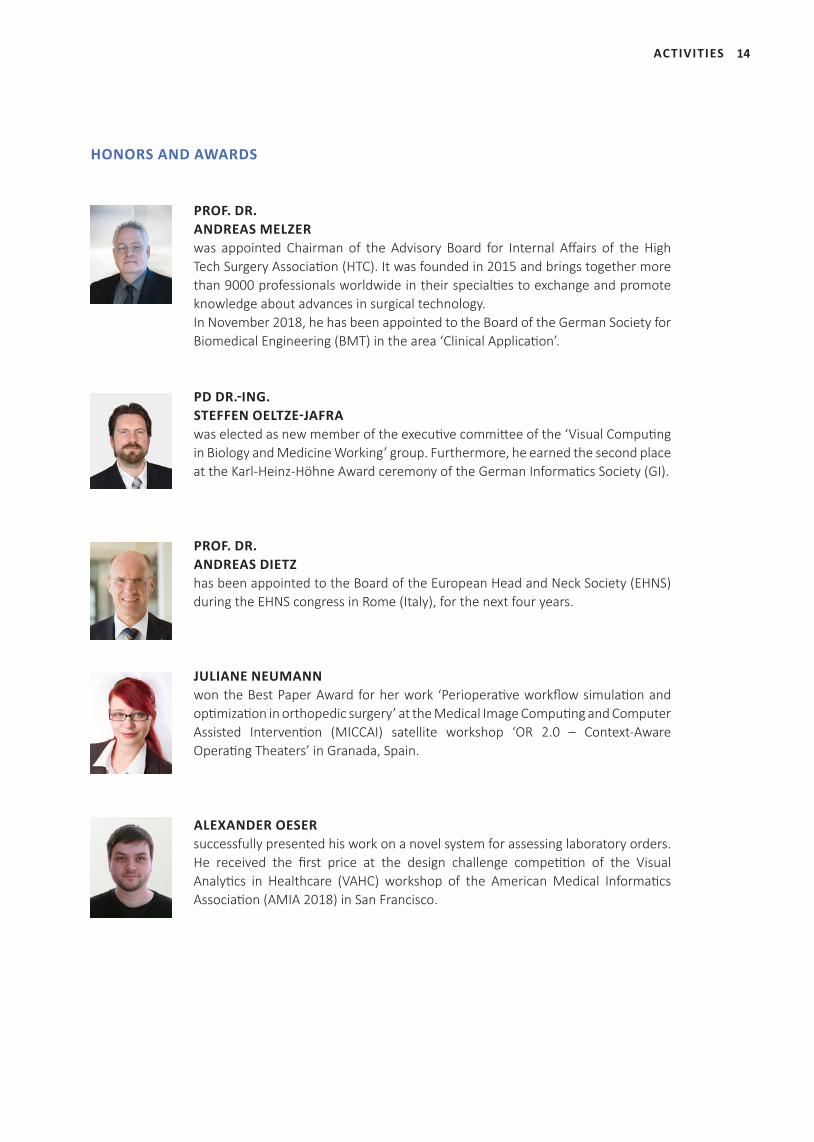
LYSIS
On October 9, the kick-off of the BMBF-fund-ed KMU-innovativ project ‘Innovative imaging for tissue differentiation in minimally invasive surgery – LYSiS’ took place at ICCAS. In close cooperation with Diaspective Vision GmbH and the Clinic and Polyclinic for Visceral, Transplan-tation, Thoracic and Vascular Surgery ICCAS is researching on a computer-assisted method for the automatic characterization, recognition and presentation of tissue from intraoperative hy-perspectral imaging data.
PROJECT LAUNCHES
Partners involved in the LYSiS project: Jonathan Philip Takoh (UKL, Dept. of Visceral Surgery), Hannes Köhler (ICCAS and Diaspective Vision GmbH), Dr. Axel Kulcke (Diaspective Vision GmbH), Dr. Claire Chalopin (ICCAS), Prof. Ines Gockel (UKL, Dept. of Visceral Surgery), Prof. Thomas Neumuth (ICCAS), Sebastian Murad Rabe (UKL, Dept. of Visceral Surgery).
14ACTIVITIES
PROF. DR. ANDREAS MELZER was appointed Chairman of the Advisory Board for Internal Affairs of the High Tech Surgery Association (HTC). It was founded in 2015 and brings together more than 9000 professionals worldwide in their specialties to exchange and promote knowledge about advances in surgical technology. In November 2018, he has been appointed to the Board of the German Society for Biomedical Engineering (BMT) in the area ‘Clinical Application’.
PD DR.-ING. STEFFEN OELTZE-JAFRAwas elected as new member of the executive committee of the ‘Visual Computing in Biology and Medicine Working’ group. Furthermore, he earned the second place at the Karl-Heinz-Höhne Award ceremony of the German Informatics Society (GI).
PROF. DR.ANDREAS DIETZ has been appointed to the Board of the European Head and Neck Society (EHNS) during the EHNS congress in Rome (Italy), for the next four years.
JULIANE NEUMANN won the Best Paper Award for her work ‘Perioperative workflow simulation and optimization in orthopedic surgery’ at the Medical Image Computing and Computer Assisted Intervention (MICCAI) satellite workshop ‘OR 2.0 – Context-Aware Operating Theaters’ in Granada, Spain.
ALEXANDER OESER successfully presented his work on a novel system for assessing laboratory orders. He received the first price at the design challenge competition of the Visual Analytics in Healthcare (VAHC) workshop of the American Medical Informatics Association (AMIA 2018) in San Francisco.
HONORS AND AWARDS
15
RESEARCH AREAS AND RELATED PROJECT PROFILES
RESEARCH AREAS AND RELATED PROJECT PROFILES
LIFE SUPPORT SYSTEMS
INTRAOPERATIVE MULTIMODAL IMAGING
16
MODEL-BASED AUTOMATION AND INTEGRATION
COMPUTER-ASSISTED IMAGE-GUIDED INTERVENTIONS
SONO-RAY
DIGITAL PATIENT- AND PROCESS MODEL
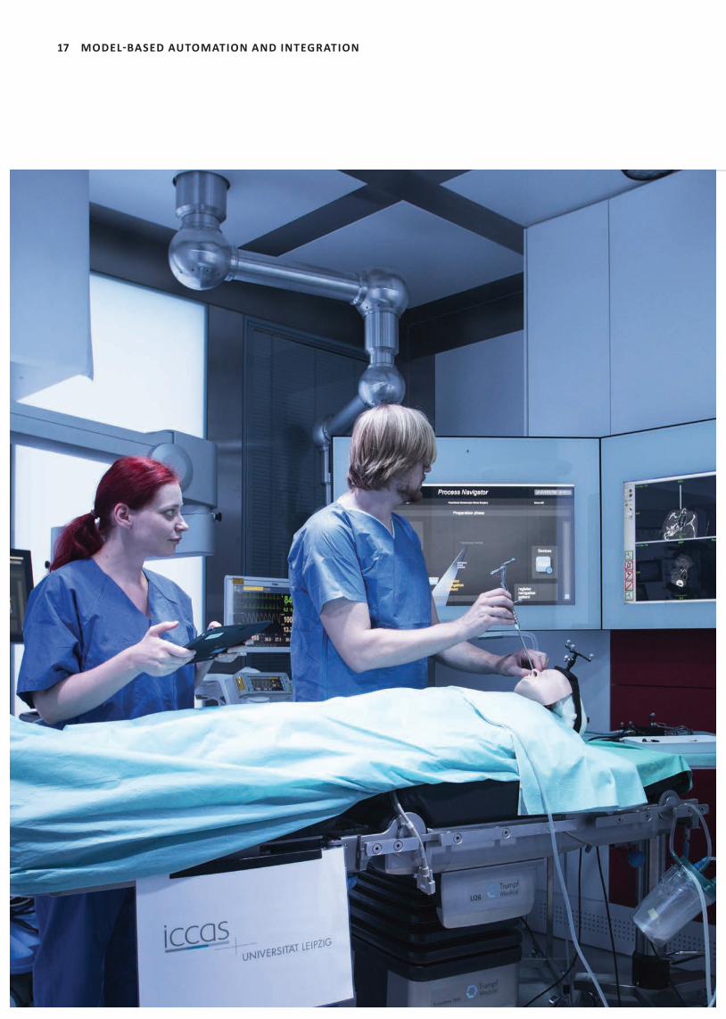
17 MODEL-BASED AUTOMATION AND INTEGRATION
18
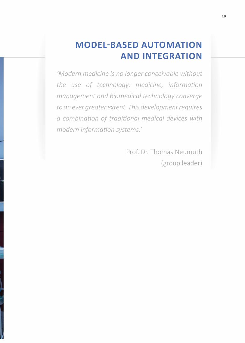
‘Modern medicine is no longer conceivable without
the use of technology: medicine, informati on
management and biomedical technology converge
to an ever greater extent. This development requires
a combinati on of traditi onal medical devices with
modern informati on systems.‘
Prof. Dr. Thomas Neumuth
(group leader)
MODEL-BASED AUTOMATION AND INTEGRATION
19 MODEL-BASED AUTOMATION AND INTEGRATION
SELECTED PUBLICATIONS
Neumuth T, Franke S. Clear oxygen-level forecasts during
anaesthesia. Nat Biomed Eng. 2018; 2(10): 715-6.
Franke S, Rockstroh M, Hofer M, Neumuth T. The
intelligent OR: design and validation of a context-aware
surgical working environment. Int J Comput Radiol Surg.
2018; 13(8): 1301-8.
Unger M, Black D, Fischer NM, Neumuth T, Glaser B.
Design and evaluation of an eye tracking support system
for the scrub nurse. Int J Med Robot. 2018 [Epub ahead
of print].
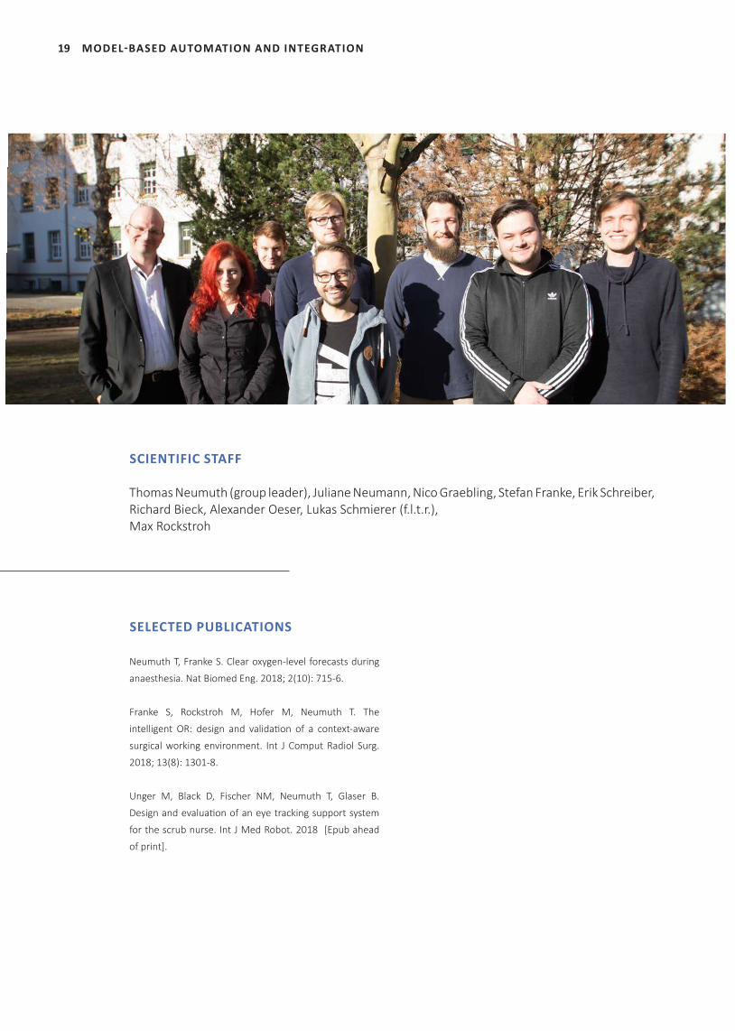
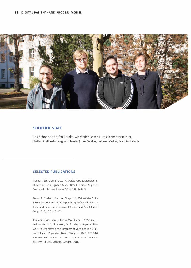
SCIENTIFIC STAFF
Thomas Neumuth (group leader), Juliane Neumann, Nico Graebling, Stefan Franke, Erik Schreiber, Richard Bieck, Alexander Oeser, Lukas Schmierer (f.l.t.r.), Max Rockstroh
20
PERIOPERATIVE WORKFLOW SIMULATION AND OPTIMIZATION IN ORTHOPEDIC SURGERY
INTRODUCTIONOperati ng room management aims at the ef-fi cient coordinati on of surgical procedures by maximizing the number of surgical cases while minimizing the required surgery ti me, with the main goal of improving the pati ent outcome. Discrete Event Simulati on (DES) can be uti lized to describe, analyze and predict the impact of procedural changes in perioperati ve process-es. The aim of this work is to provide a DES ap-proach for a holisti c perioperati ve process op-ti mizati on with a focus on the combinati on of behavioral, temporal, operati onal and structur-al perspecti ve. Two diff erent process simulati on techniques, namely Business Process Simula-ti on (BPS) and 3D Process Flow Simulati on were uti lized. DES models were implemented with perioperati ve data from Total Hip Replacement (THR) and Total Knee Replacement (TKR) sur-geries. The opti mizati on objecti ve is to increase the number of surgeries to three cases per day by reducing the intraoperati ve ti me through the opti mizati on of the OR layout. Furthermore, the processes for surgery follow-up and OR prepa-rati on should be streamlined.
MATERIAL AND METHODSFor the intraoperati ve simulati on 15 THR and 7 TKR surgeries and for pre- and postopera-ti ve simulati on 30 (total or parti al) knee- and hip replacement surgeries were recorded. The pre- and postoperati ve acti viti es of all OR team members were modeled in BPMN format and simulated in diff erent scenarios. The pre- and postoperati ve processes were streamlined with methods of Business Process Re-engineering and the simulati on scenarios were repeated. In the second step, the intraoperati ve process opti mizati on was performed. Aft erwards, the simulati on was repeated with improved intra-operati ve surgery ti mes.
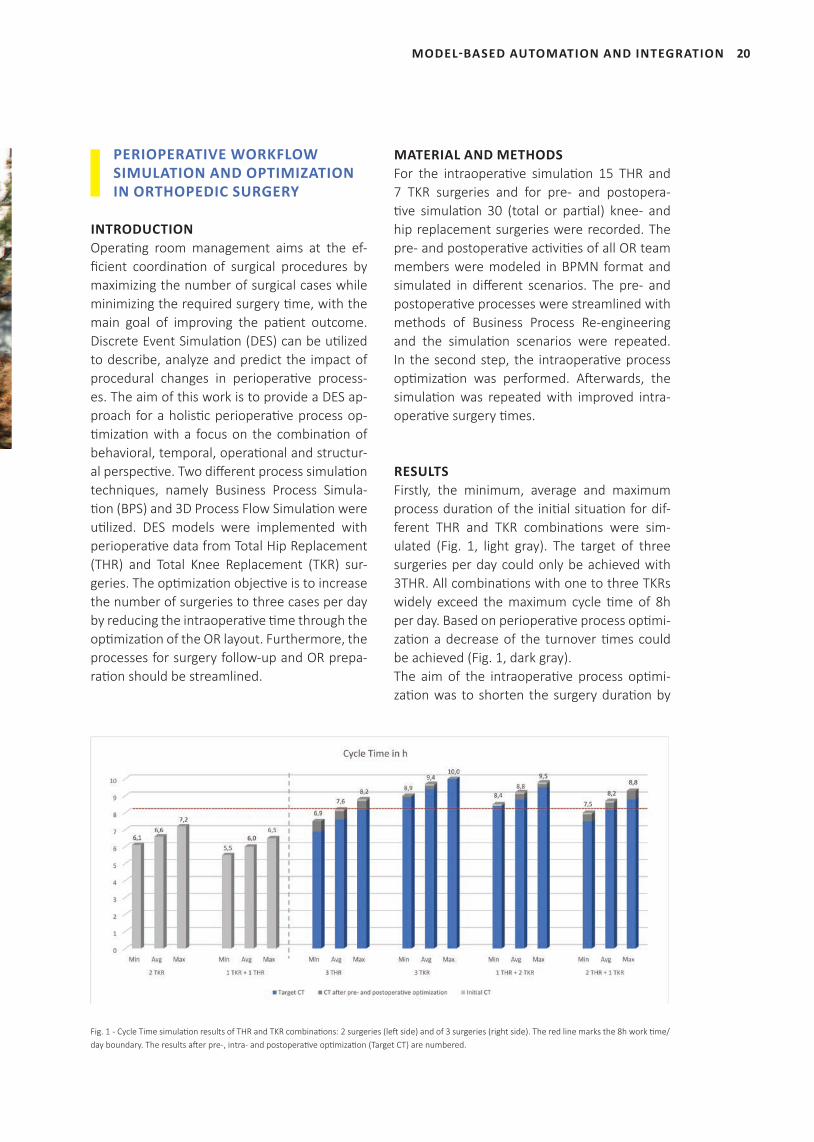
RESULTS Firstly, the minimum, average and maximum process durati on of the initi al situati on for dif-ferent THR and TKR combinati ons were sim-ulated (Fig. 1, light gray). The target of three surgeries per day could only be achieved with 3THR. All combinati ons with one to three TKRs widely exceed the maximum cycle ti me of 8h per day. Based on perioperati ve process opti mi-zati on a decrease of the turnover ti mes could be achieved (Fig. 1, dark gray).The aim of the intraoperati ve process opti mi-zati on was to shorten the surgery durati on by
MODEL-BASED AUTOMATION AND INTEGRATION
Fig. 1 - Cycle Time simulati on results of THR and TKR combinati ons: 2 surgeries (left side) and of 3 surgeries (right side). The red line marks the 8h work ti me/day boundary. The results aft er pre-, intra- and postoperati ve opti mizati on (Target CT) are numbered.
21 MODEL-BASED AUTOMATION AND INTEGRATION
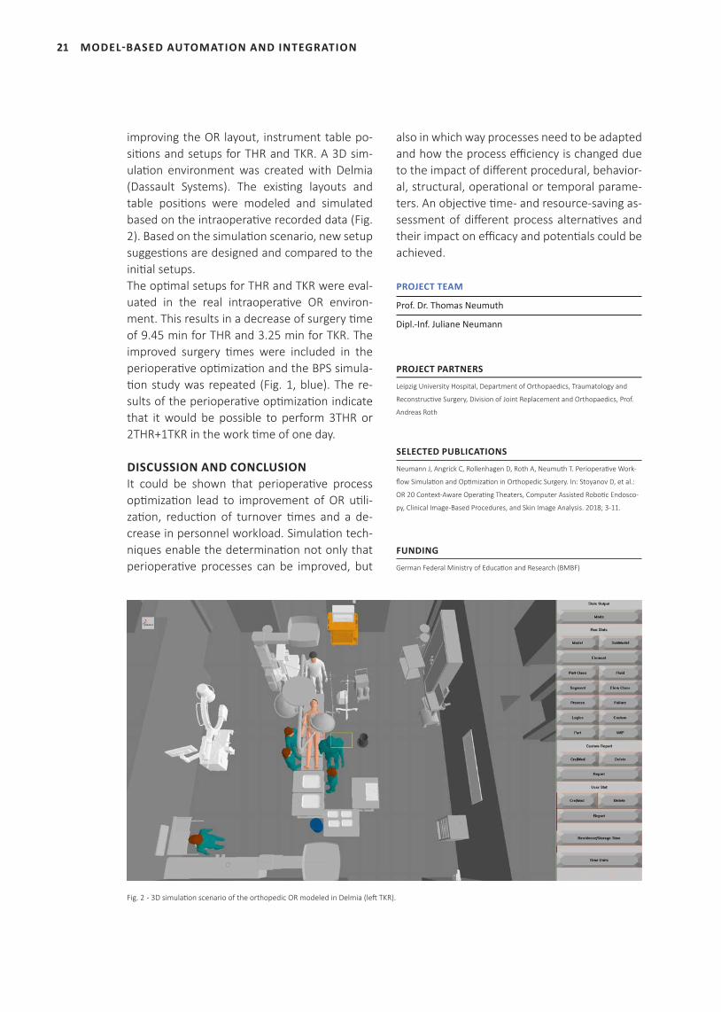
improving the OR layout, instrument table po-sitions and setups for THR and TKR. A 3D sim-ulation environment was created with Delmia (Dassault Systems). The existing layouts and table positions were modeled and simulated based on the intraoperative recorded data (Fig. 2). Based on the simulation scenario, new setup suggestions are designed and compared to the initial setups. The optimal setups for THR and TKR were eval-uated in the real intraoperative OR environ-ment. This results in a decrease of surgery time of 9.45 min for THR and 3.25 min for TKR. The improved surgery times were included in the perioperative optimization and the BPS simula-tion study was repeated (Fig. 1, blue). The re-sults of the perioperative optimization indicate that it would be possible to perform 3THR or 2THR+1TKR in the work time of one day.
DISCUSSION AND CONCLUSIONIt could be shown that perioperative process optimization lead to improvement of OR utili-zation, reduction of turnover times and a de-crease in personnel workload. Simulation tech-niques enable the determination not only that perioperative processes can be improved, but
also in which way processes need to be adapted and how the process efficiency is changed due to the impact of different procedural, behavior-al, structural, operational or temporal parame-ters. An objective time- and resource-saving as-sessment of different process alternatives and their impact on efficacy and potentials could be achieved.
PROJECT TEAM
Prof. Dr. Thomas Neumuth
Dipl.-Inf. Juliane Neumann
PROJECT PARTNERS
Leipzig University Hospital, Department of Orthopaedics, Traumatology and
Reconstructive Surgery, Division of Joint Replacement and Orthopaedics, Prof.
Andreas Roth
SELECTED PUBLICATIONS
Neumann J, Angrick C, Rollenhagen D, Roth A, Neumuth T. Perioperative Work-
flow Simulation and Optimization in Orthopedic Surgery. In: Stoyanov D, et al.:
OR 20 Context-Aware Operating Theaters, Computer Assisted Robotic Endosco-
py, Clinical Image-Based Procedures, and Skin Image Analysis. 2018; 3-11.
FUNDING
German Federal Ministry of Education and Research (BMBF)
Fig. 2 - 3D simulation scenario of the orthopedic OR modeled in Delmia (left TKR).
22
CONTEXT-AWARE HUMAN-MACHINE INTERFACES IN INTEGRATED OPERATING ROOMS
INTRODUCTIONAn increasing number of medical devices pro-vide communicati on interfaces, but yet they only show very limited cooperati ve behavior. The lack of contextual informati on during sur-gery hinders autonomous intelligent system’s adaptati on. We implemented an intraopera-ti ve context-awareness pipeline and propose a modeling approach for the realizati on of online dynamic human-machine interfaces in the OR.
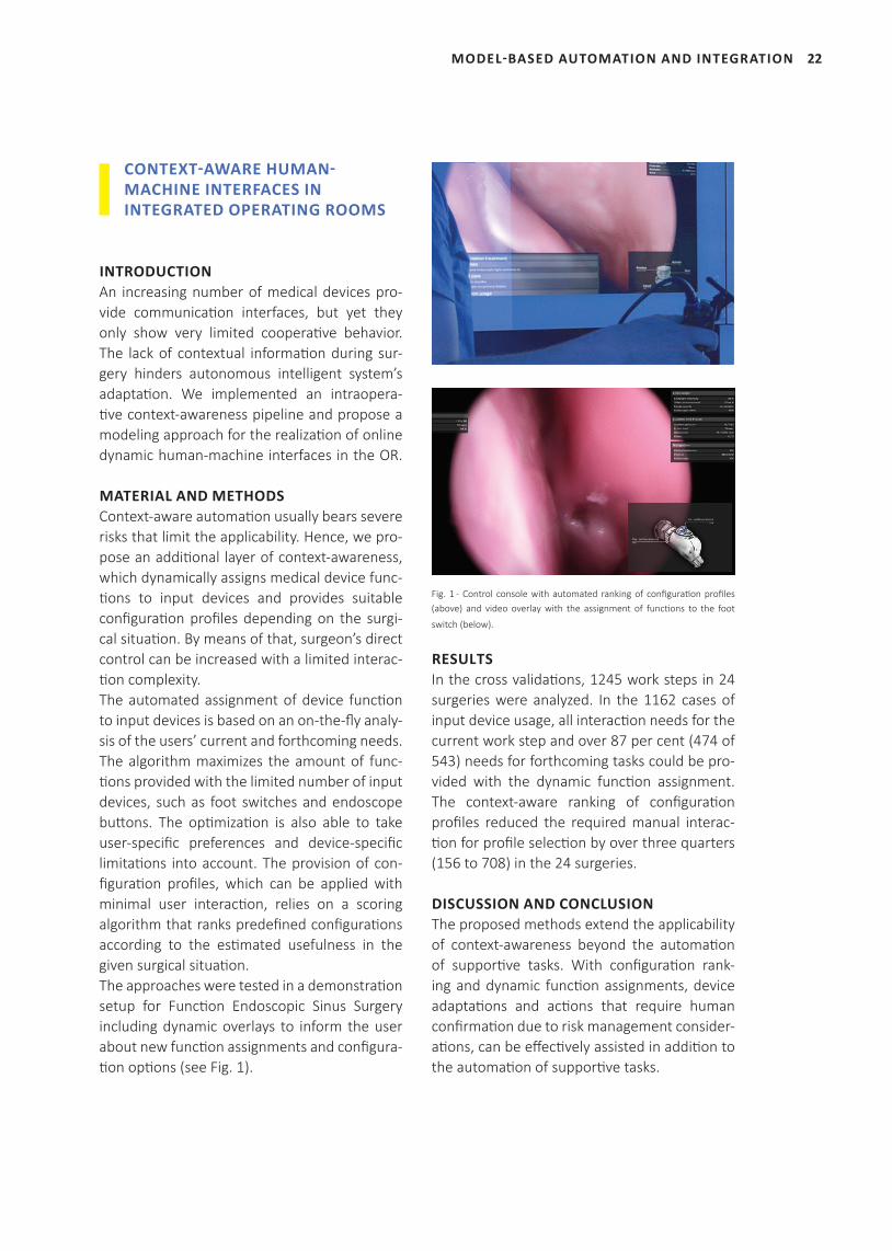
MATERIAL AND METHODSContext-aware automati on usually bears severe risks that limit the applicability. Hence, we pro-pose an additi onal layer of context-awareness, which dynamically assigns medical device func-ti ons to input devices and provides suitable confi gurati on profi les depending on the surgi-cal situati on. By means of that, surgeon’s direct control can be increased with a limited interac-ti on complexity. The automated assignment of device functi on to input devices is based on an on-the-fl y analy-sis of the users’ current and forthcoming needs. The algorithm maximizes the amount of func-ti ons provided with the limited number of input devices, such as foot switches and endoscope butt ons. The opti mizati on is also able to take user-specifi c preferences and device-specifi c limitati ons into account. The provision of con-fi gurati on profi les, which can be applied with minimal user interacti on, relies on a scoring algorithm that ranks predefi ned confi gurati ons according to the esti mated usefulness in the given surgical situati on.The approaches were tested in a demonstrati on setup for Functi on Endoscopic Sinus Surgery including dynamic overlays to inform the user about new functi on assignments and confi gura-ti on opti ons (see Fig. 1).
Fig. 1 - Control console with automated ranking of confi gurati on profi les (above) and video overlay with the assignment of functi ons to the foot
switch (below).
RESULTSIn the cross validati ons, 1245 work steps in 24 surgeries were analyzed. In the 1162 cases of input device usage, all interacti on needs for the current work step and over 87 per cent (474 of 543) needs for forthcoming tasks could be pro-vided with the dynamic functi on assignment. The context-aware ranking of confi gurati on profi les reduced the required manual interac-ti on for profi le selecti on by over three quarters (156 to 708) in the 24 surgeries.
DISCUSSION AND CONCLUSIONThe proposed methods extend the applicability of context-awareness beyond the automati on of supporti ve tasks. With confi gurati on rank-ing and dynamic functi on assignments, device adaptati ons and acti ons that require human confi rmati on due to risk management consider-ati ons, can be eff ecti vely assisted in additi on to the automati on of supporti ve tasks.
MODEL-BASED AUTOMATION AND INTEGRATION
23 MODEL-BASED AUTOMATION AND INTEGRATION
PROJECT TEAM
Prof. Dr. Thomas Neumuth
Dr.-Ing. Stefan Franke
Dipl.-Inf. Max Rockstroh
SELECTED PUBLICATIONS
Franke S, Rockstroh M, Hofer M, Neumuth T. The intelligent OR: design and
validati on of a context-aware surgical working environment. Int J Comput Radiol
Surg. 2018; 13(8): 1301-8.
Franke S, Rockstroh M, Kasparick M, Neumuth T. A Method for the Con-
text-Aware Assignment of Medical Device Functi ons to Input Devices in
Integrated Operati ng Rooms. In: Stoyanov D, et al.: OR 20 Context-Aware Op-
erati ng Theaters, Computer Assisted Roboti c Endoscopy, Clinical Image-Based
Procedures, and Skin Image Analysis. 2018; 12-19.
Franke S, Rockstroh M, Neumuth T. Context-awareness for control consoles in
integrated operati ng rooms. Curr Directi ons Biomed Eng, 2018; 4(1): 291-5.
FUNDING
German Federal Ministry of Educati on and Research (BMBF)
BIOPASS – IMAGE-, ONTOLOGY- AND PROCESS-BASED ASSISTANCE FOR MINIMALLY INVASIVE ENDOSCOPIC SURGERY
INTRODUCTIONIn the BIOPASS project a novel localizati on ap-proach for a markerless navigati on system was developed to reduce the navigati on hardware while assisti ng the surgeon with adapted navi-gati on assistance. A criti cal aspect for the devel-opment of such an intelligent system was the defi niti on of situati onal knowledge. Therefore, the goals of the project were: a multi modal acquisiti on of informati on in the OR, a com-prehensive descripti on as well as a consistent human-machine-interacti on with a new naviga-ti on approach.
MATERIAL AND METHODSWe combined a process modeling approach for temporal classifi cati on of visited anatomical landmarks during previous procedures with a semanti c knowledge management system us-ing an ontology framework. We used Hidden
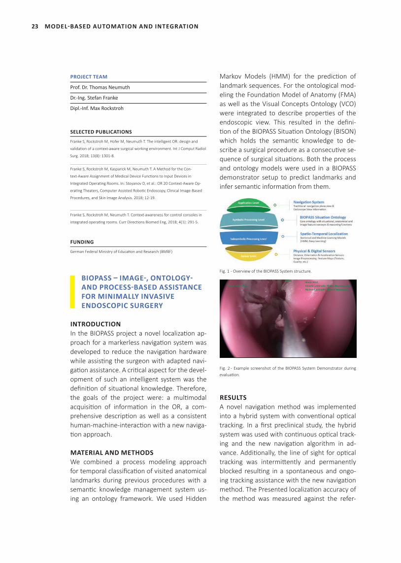
Markov Models (HMM) for the predicti on of landmark sequences. For the ontological mod-eling the Foundati on Model of Anatomy (FMA) as well as the Visual Concepts Ontology (VCO) were integrated to describe properti es of the endoscopic view. This resulted in the defi ni-ti on of the BIOPASS Situati on Ontology (BISON) which holds the semanti c knowledge to de-scribe a surgical procedure as a consecuti ve se-quence of surgical situati ons. Both the process and ontology models were used in a BIOPASS demonstrator setup to predict landmarks and infer semanti c informati on from them.
Fig. 1 - Overview of the BIOPASS System structure.
Fig. 2 - Example screenshot of the BIOPASS System Demonstrator during evaluati on.
RESULTSA novel navigati on method was implemented into a hybrid system with conventi onal opti cal tracking. In a fi rst preclinical study, the hybrid system was used with conti nuous opti cal track-ing and the new navigati on algorithm in ad-vance. Additi onally, the line of sight for opti cal tracking was intermitt ently and permanently blocked resulti ng in a spontaneous and ongo-ing tracking assistance with the new navigati on method. The Presented localizati on accuracy of the method was measured against the refer-
24
ence values defined in the CT model. Accuracy was calculated to have a mean error of 5 mm.
DISCUSSION AND CONCLUSIONWe successfully integrated the novel navigation method into a conventional tracking setup and validated and evaluated the hybrid system in an ideal laboratory setup. Tracking functionality was identified to be comparable with conven-tional tracking technology. The missing quanti-tative navigation information, e.g. distance to regions of interest or CT-based allocentric local-ization, is substituted by an abstract qualitative navigation assistance, e.g. landmark-based ego-centric orientation. How this new navigation as-sistance method actually benefits surgeon nav-igation experience and technology acceptance is still unclear. Further work needs to be done to enable better surgeon-machine-interaction along the navigation process.
PROJECT TEAM
Prof. Dr. Thomas Neumuth
M. Sc. Richard Bieck
PROJECT PARTNERS
Zuse Institute for Information Technology, Berlin
LOCALITE GmbH, St. Augustin
Dornheim Medical Imaging, Magdeburg
SELECTED PUBLICATIONS
Bieck R, Heuermann K, Hofer M, Neumuth T. From Passive Tool To Active Guid-
ance: Requirements For Computational Navigation Intelligence In Computer-As-
sisted Functional Endoscopic Sinus Surgery. Int J Comput Assist Radiol Surg.
[Epub ahead of print].
Heuermann K, Bieck R, Dietz A, Uciteli A, Franke S, Herre H, Neumuth T, Fischer
M. Ein neuartiges Navigationssystem für die endoskopische funktionelle Nasen-
nebenhöhlenchirurgie. 87. Deutscher HNO-Kongress, Düsseldorf, Germany;
2018.
Siemoleit S, Uciteli A, Bieck R, Herre H. Ontological Modelling of Situational
Awareness in Surgical Interventions. The Joint Ontology Workshops, Bozen-Bol-
zano, Italy; 2017.
Siemoleit S, Uciteli A, Bieck R, Herre H. Processual Reasoning over Sequences
of Situations in Endoscopic Surgery. Stud Health Technol Inform. 2017; 243:
222-226.
Heuermann K, Bieck R, Hofer M, Dietz A, Neumuth T. Intelligent navigation
strategies of a markerless FESS navigation by prioritizing multimodal context
information. Laryngo-Rhino-Otologie 2018; 97(S02): 42.
Bieck R, Heuermann K, Hofer M, Neumuth T. From Passive Tool To Active Guid-
ance: Requirements For Computational Navigation Intelligence In Computer-As-
sisted Functional Endoscopic Sinus Surgery. Int J Comput Assist Radiol Surg.
[Epub ahead of print].
FUNDING
German Federal Ministry of Education and Research (BMBF)
PIMPAP – PATIENT-BASED INDIVIDUAL MODELING OF PARASPINAL COLLATERAL NETWORK PERFUSION AFTER SEGMENTAL ARTERY OCCLUSION
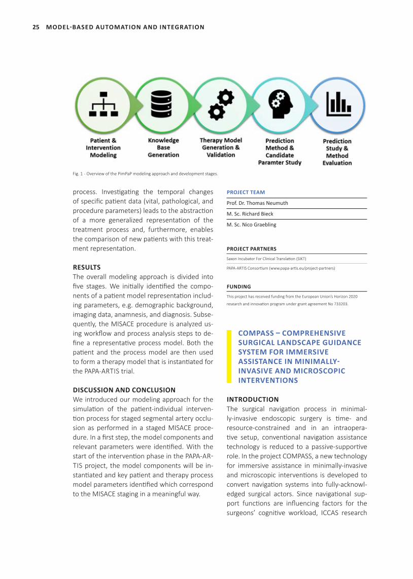
INTRODUCTIONThe repair of large thoracoabdominal aortic an-eurysms (TAAA) is done by using endovascular minimally-invasive surgery with stent grafting. However, a successful intervention still poses risks of paraplegia or death due to ischaemic reactions in the spinal cord. The minimally-in-vasive, selective segmental artery coil embo-lization (MISACE) is a procedure employed to reduce these risks by preemptively closing supplying segmental arteries of the aorta be-fore stenting. Since the procedure is still in an early application phase there exist no general guidelines and the effectiveness is currently be-ing investigated in a multi-centric clinical trial in the PAPA-ARTIS EU-project. In the subproject PimPaP, a patient and an intervention process hybrid model is developed to investigate the in-fluence of varying coiling patterns and their im-pact on the convalescence of spinal perfusion and the clinical outcome using computational modeling strategies.
MATERIAL AND METHODSMultimodal patient-specific information is ac-quired in various stages including before, during and after the MISACE staging as well as one month and twelve months post-operatively af-ter aneurysm repair. Using a digital patient mod-eling approach, a software-internal representa-tion of the individual patient state is generated at different time points during the treatment
MODEL-BASED AUTOMATION AND INTEGRATION
25 MODEL-BASED AUTOMATION AND INTEGRATION
process. Investigating the temporal changes of specific patient data (vital, pathological, and procedure parameters) leads to the abstraction of a more generalized representation of the treatment process and, furthermore, enables the comparison of new patients with this treat-ment representation.
RESULTSThe overall modeling approach is divided into five stages. We initially identified the compo-nents of a patient model representation includ-ing parameters, e.g. demographic background, imaging data, anamnesis, and diagnosis. Subse-quently, the MISACE procedure is analyzed us-ing workflow and process analysis steps to de-fine a representative process model. Both the patient and the process model are then used to form a therapy model that is instantiated for the PAPA-ARTIS trial.
DISCUSSION AND CONCLUSIONWe introduced our modeling approach for the simulation of the patient-individual interven-tion process for staged segmental artery occlu-sion as performed in a staged MISACE proce-dure. In a first step, the model components and relevant parameters were identified. With the start of the intervention phase in the PAPA-AR�TIS project, the model components will be in-stantiated and key patient and therapy process model parameters identified which correspond to the MISACE staging in a meaningful way.
PROJECT TEAM
Prof. Dr. Thomas Neumuth
M. Sc. Richard Bieck
M. Sc. Nico Graebling
PROJECT PARTNERS
Saxon Incubator For Clinical Translation (SIKT)
PAPA-ARTIS Consortium (www.papa-artis.eu/project-partners)
FUNDING
This project has received funding from the European Union’s Horizon 2020
research and innovation program under grant agreement No 733203.
COMPASS – COMPREHENSIVE SURGICAL LANDSCAPE GUIDANCE SYSTEM FOR IMMERSIVE ASSISTANCE IN MINIMALLY-INVASIVE AND MICROSCOPIC INTERVENTIONS
INTRODUCTIONThe surgical navigation process in minimal-ly-invasive endoscopic surgery is time- and resource-constrained and in an intraopera-tive setup, conventional navigation assistance technology is reduced to a passive-supportive role. In the project COMPASS, a new technology for immersive assistance in minimally-invasive and microscopic interventions is developed to convert navigation systems into fully-acknowl-edged surgical actors. Since navigational sup-port functions are influencing factors for the surgeons’ cognitive workload, ICCAS research
Fig. 1 - Overview of the PimPaP modeling approach and development stages.
26
in COMPASS is focused on the investi gati on of a modeling approach that considers surgical cog-niti on for intelligent navigati on assistance.
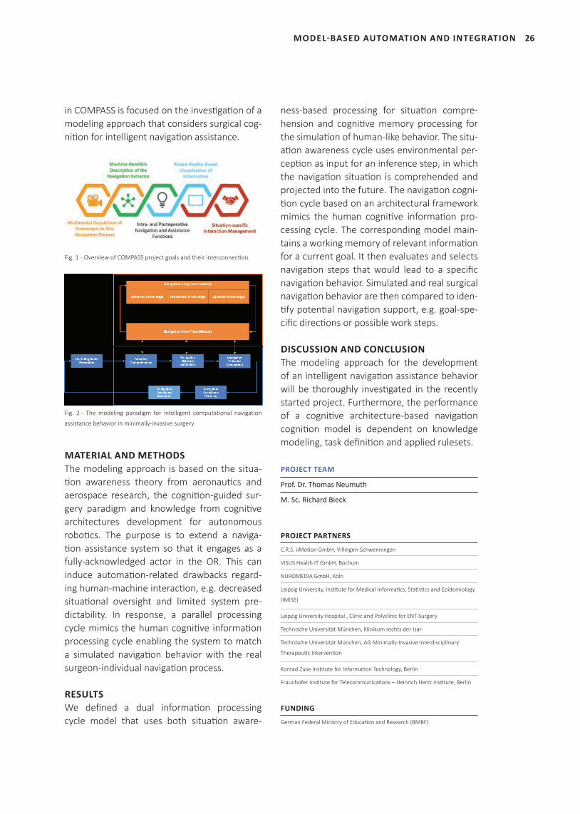
Fig. 1 - Overview of COMPASS project goals and their interconnecti on.
Fig. 2 - The modeling paradigm for intelligent computational navigation
assistance behavior in minimally-invasive surgery.
MATERIAL AND METHODSThe modeling approach is based on the situa-ti on awareness theory from aeronauti cs and aerospace research, the cogniti on-guided sur-gery paradigm and knowledge from cogniti ve architectures development for autonomous roboti cs. The purpose is to extend a naviga-ti on assistance system so that it engages as a fully-acknowledged actor in the OR. This can induce automati on-related drawbacks regard-ing human-machine interacti on, e.g. decreased situati onal oversight and limited system pre-dictability. In response, a parallel processing cycle mimics the human cogniti ve informati on processing cycle enabling the system to match a simulated navigati on behavior with the real surgeon-individual navigati on process.
RESULTSWe defi ned a dual informati on processing cycle model that uses both situati on aware-
ness-based processing for situati on compre-hension and cogniti ve memory processing for the simulati on of human-like behavior. The situ-ati on awareness cycle uses environmental per-cepti on as input for an inference step, in which the navigati on situati on is comprehended and projected into the future. The navigati on cogni-ti on cycle based on an architectural framework mimics the human cogniti ve informati on pro-cessing cycle. The corresponding model main-tains a working memory of relevant informati on for a current goal. It then evaluates and selects navigati on steps that would lead to a specifi c navigati on behavior. Simulated and real surgical navigati on behavior are then compared to iden-ti fy potenti al navigati on support, e.g. goal-spe-cifi c directi ons or possible work steps.
DISCUSSION AND CONCLUSIONThe modeling approach for the development of an intelligent navigati on assistance behavior will be thoroughly investi gated in the recently started project. Furthermore, the performance of a cogniti ve architecture-based navigati on cogniti on model is dependent on knowledge modeling, task defi niti on and applied rulesets.
PROJECT TEAM
Prof. Dr. Thomas Neumuth
M. Sc. Richard Bieck
PROJECT PARTNERS
C.R.S. iiMoti on GmbH, Villingen-Schwenningen
VISUS Health IT GmbH, Bochum
NUROMEDIA GmbH, Köln
Leipzig University, Insti tute for Medical Informati cs, Stati sti cs and Epidemiology
(IMISE)
Leipzig University Hospital , Clinic and Polyclinic for ENT-Surgery
Technische Universität München, Klinikum rechts der Isar
Technische Universität München, AG Minimally-Invasive Interdisciplinary
Therapeuti c Interventi on
Konrad Zuse Insti tute for Informati on Technology, Berlin
Fraunhofer Insti tute for Telecommunicati ons – Heinrich Hertz Insti tute, Berlin
FUNDING
German Federal Ministry of Educati on and Research (BMBF)
MODEL-BASED AUTOMATION AND INTEGRATION
27 MODEL-BASED AUTOMATION AND INTEGRATION
MOVE – MODULAR VALIDATION ENVIRONMENT FOR MEDICAL NETWORKS
INTRODUCTIONThe integration and networking of medi-cal equipment has become an indispensable component of modern operating theaters. At present, the market is characterized by closed solutions, which are regulatorily approved as monolithic settings. The aim of the project is therefore to develop methods that support the development of openly integrated medical de-vices as well as the approval process by means of a test environment. The project aims to ease the access of SMEs to the market with innova-tive technologies.
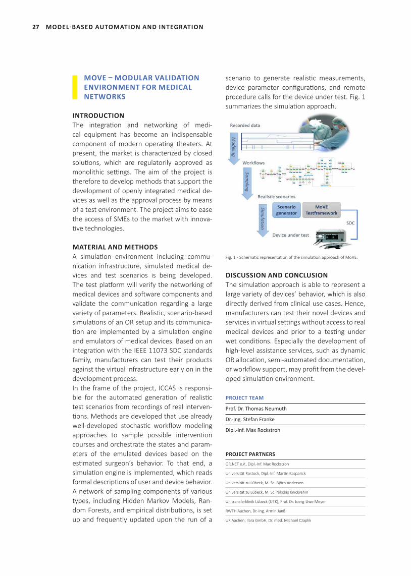
MATERIAL AND METHODSA simulation environment including commu-nication infrastructure, simulated medical de-vices and test scenarios is being developed. The test platform will verify the networking of medical devices and software components and validate the communication regarding a large variety of parameters. Realistic, scenario-based simulations of an OR setup and its communica-tion are implemented by a simulation engine and emulators of medical devices. Based on an integration with the IEEE 11073 SDC standards family, manufacturers can test their products against the virtual infrastructure early on in the development process.In the frame of the project, ICCAS is responsi-ble for the automated generation of realistic test scenarios from recordings of real interven-tions. Methods are developed that use already well-developed stochastic workflow modeling approaches to sample possible intervention courses and orchestrate the states and param-eters of the emulated devices based on the estimated surgeon’s behavior. To that end, a simulation engine is implemented, which reads formal descriptions of user and device behavior. A network of sampling components of various types, including Hidden Markov Models, Ran-dom Forests, and empirical distributions, is set up and frequently updated upon the run of a
scenario to generate realistic measurements, device parameter configurations, and remote procedure calls for the device under test. Fig. 1 summarizes the simulation approach.
Fig. 1 - Schematic representation of the simulation approach of MoVE.
DISCUSSION AND CONCLUSIONThe simulation approach is able to represent a large variety of devices’ behavior, which is also directly derived from clinical use cases. Hence, manufacturers can test their novel devices and services in virtual settings without access to real medical devices and prior to a testing under wet conditions. Especially the development of high-level assistance services, such as dynamic OR allocation, semi-automated documentation, or workflow support, may profit from the devel-oped simulation environment.
PROJECT TEAM
Prof. Dr. Thomas Neumuth
Dr.-Ing. Stefan Franke
Dipl.-Inf. Max Rockstroh
PROJECT PARTNERS
OR.NET e.V., Dipl.-Inf. Max Rockstroh
Universität Rostock, Dipl.-Inf. Martin Kasparick
Universität zu Lübeck, M. Sc. Björn Andersen
Universität zu Lübeck, M. Sc. Nikolas Knickrehm
Unitransferklinik Lübeck (UTK), Prof. Dr. Joerg-Uwe Meyer
RWTH Aachen, Dr.-Ing. Armin Janß
UK Aachen, Ilara GmbH, Dr. med. Michael Czaplik
28
Synagon GmbH, Dr.-Ing. Andreas Zimolong
HEBU medical GmbH, Dipl.-Ing. (FH) Thomas Butsch
Fritz Stephan GmbH, Dipl.-Phys. Wolfgang Braun
Localite GmbH, Dipl.-Phys. Sven Arnold
Steute Schaltgeräte GmbH, Dipl.-Ing. Guido Becker
SurgiTAIX AG, Dr.-Ing. Frank Portheine
GADV mbH, Dr.-Ing. Gregor Diehl
qcmed GmbH, Dipl.-Ing. Sandra Fiehe
FUNDING
German Federal Ministry of Education and Research (BMBF)
ENSEMBLE – DEVELOPMENT OF A SCALABLE AND MAGNETIC RESONANCE (MR)-COMPATIBLE BLOOD CIRCULATION MODEL
INTRODUCTIONMedical residents accompany proficient staff in the operating room and fulfill a supporting role while observing and memorizing the pro-cedure, in order to receive training in their sur-gical field. During this time, they start their sur-gical training by performing elementary suture and cutting tasks before moving on to more complex techniques and basic operations. In order to develop their surgical skills outside of real surgeries, trainees have the opportunity to simulate procedures on phantoms or specially prepared bodies. However, for the training of catheter-based surgery, they require a training model with a realistic vascular tree and an ac-tive blood circulatory system. The project EN-SEMBLE aims to provide such models, by using CT images and automatically segmented blood vessel areas throughout the combined data set. Together with a pump and a blood-like fluid, the resulting 3D representation will be used to manufacture an artificial vascular system.
MATERIAL AND METHODSThe segmentation of the vascular system is to be performed by a segmentation algorithm, us-ing a set of CT images as input.The first step towards this goal was the acquire-
ment of a sufficient amount of anonymized data, which was used for preliminary develop-ment, testing and result comparison. Afterward, the project group engaged in further research, to establish an assortment of segmentation ap-proaches by comparing their uses and results in state-of-the-art research. After the initial re-search, the team focused heavily on the use of active contour models and level-set algorithms. Furthermore, an additional manual segmen-tation was done, which is utilized as a ground truth that the employed methods will be com-pared to.
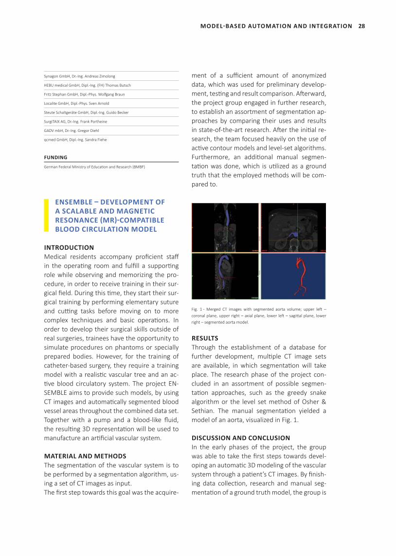
Fig. 1 - Merged CT images with segmented aorta volume; upper left – coronal plane, upper right – axial plane, lower left – sagittal plane, lower right – segmented aorta model.
RESULTSThrough the establishment of a database for further development, multiple CT image sets are available, in which segmentation will take place. The research phase of the project con-cluded in an assortment of possible segmen-tation approaches, such as the greedy snake algorithm or the level set method of Osher & Sethian. The manual segmentation yielded a model of an aorta, visualized in Fig. 1.
DISCUSSION AND CONCLUSIONIn the early phases of the project, the group was able to take the first steps towards devel-oping an automatic 3D modeling of the vascular system through a patient’s CT images. By finish-ing data collection, research and manual seg-mentation of a ground truth model, the group is
MODEL-BASED AUTOMATION AND INTEGRATION
29 MODEL-BASED AUTOMATION AND INTEGRATION
now in the process of using automatic segmen-tation to compare their corresponding results.
PROJECT TEAM
Prof. Dr. Thomas Neumuth
Dipl.-Inf. Juliane Neumann
M. Sc. Reinhard Fuchs
PROJECT PARTNERS
PHACON GmbH, Hendrik Moeckel
FUNDING
This project has received funding from the ZIM program of the German Federal
Ministry of Economic Affairs and Energy (BMWi).
EUMFH – BIOMEDICAL INFORMATION TECHNOLOGY FOR THE EUROPEAN MEDICAL FIELD HOSPITAL
INTRODUCTIONThe project European Modular Field Hospital (EUMFH) aims to explore how the medical ca-pacity of the Union Civil Protection Mechanism can be improved. Different Member States of the European Union combine their expertise and build a common deployable Emergency Medical Team (EMT) level 3 for disaster relief missions. Current developments show that there is a clear lack of active deployable level 3 Emergency Medical Teams, i.e. referral hos-pitals in the field. Therefore, there is a need for a high-level medical module that can be de-ployed for a longer-term mission without put-ting the burden on one single Member State or organization.
MATERIAL AND METHODSDuring the project, ICCAS was commissioned with the conceptualization and provision of an electronic patient record (EPR) for EMTs. As first step, a comprehensive requirements analysis was conducted. Subsequently, a concept for an EPR was derived, taking the special demands (e.g. lightweight, high flexibility, robustness) of EMTs into account. After implementation, an
early version of the EPR was tested during the ModEX exercise in Bucharest. The participating personnel was interviewed regarding suitability, performance and operational capabilities of the developed EPR.
RESULTSTwenty-one team members have been inter-viewed. Fourteen of them with medical roles (physicians and nurses) and seven of them with supportive roles (Management, Logistics, or Training). Among the fourteen medical inter-view partners were three medical team leaders and all participants came from nine different European countries. The system was evaluated very positive.
DISCUSSION AND CONCLUSIONThe evaluation of the EPR during the ModEX ex-ercise was very successful, considering the pos-itive user feedback. Despite this success, there were various lessons learned on how to further improve the EPR to cope with the challenges of EMT missions. After EPR optimization, it will be tested under realistic conditions during another EMT exercise at the beginning of 2019.
PROJECT TEAM
Prof. Dr. Thomas Neumuth
M. Sc. Erik Schreiber
PROJECT PARTNERS
Italian Civil Protection Department
Belgian Ministry of Health
Danish Emergency Management Agency
Estonian Health Board
French DG for Civil Protection and Crisis Management
Romanian Ministry of Internal Affairs
Johanniter, Germany
Leipzig University, Germany
Association of Slovak Samaritans

30
SELECTED PUBLICATIONS
Neumuth T. The European Modular Field Hospital: Herausforderungen für IT
beim Einsatz mobiler Krankenhäuser nach Naturkatastrophen. Deutsche Ge-
sellschaft für Medizinische Informatik, Biometrie und Epidemiologie. 62. Jahre-
stagung der Deutschen Gesellschaft für Medizinische Informatik, Biometrie und
Epidemiologie e.V. (GMDS). Oldenburg, Germany; 2017. Düsseldorf: German
Medical Science GMS Publishing House; 2017. DocAbstr. 143.
FUNDING
General Directorate for European Civil Protection and Humanitarian Aid Operations: ECHO/SUB/2016/739964/PREP14
MODEL-BASED AUTOMATION AND INTEGRATION
31 DIGITAL PATIENT- AND PROCESS MODEL
32
‘The growing number of medical screening opti ons
and forms of treatment for complex diseases
requires more pati ent-specifi c therapy decisions and
treatment processes that increase the chance of a
bett er clinical outcome. Digital pati ent and process
models integrated in clinical decision support
systems address these problems. They represent
the disease-specifi c therapeuti c decision-making
and therapy processes and are instanti ated with
pati ent-specifi c data for personalized medicine.’
PD Dr.-Ing. Steff en Oeltze-Jafra
(group leader)
DIGITAL PATIENT- AND PROCESS MODEL
33 DIGITAL PATIENT- AND PROCESS MODEL
SELECTED PUBLICATIONS
Gaebel J, Schreiber E, Oeser A, Oeltze-Jafra S. Modular Ar-
chitecture for Integrated Model-Based Decision Support.
Stud Health Technol Inform. 2018; 248: 108-15.
Oeser A, Gaebel J, Dietz A, Wiegand S, Oeltze-Jafra S. In-
formation architecture for a patient-specific dashboard in
head and neck tumor boards. Int J Comput Assist Radiol
Surg. 2018; 13:8 1283-90.
Multani P, Niemann U, Cypko MA, Kuehn J-P, Voelzke H,
Oeltze-Jafra S, Spiliopoulou, M. Building a Bayesian Net-
work to Understand the Interplay of Variables in an Epi-
demiological Population-Based Study. In: 2018 IEEE 31st
International Symposium on Computer-Based Medical
Systems (CBMS). Karlstad, Sweden; 2018.
SCIENTIFIC STAFF
Erik Schreiber, Stefan Franke, Alexander Oeser, Lukas Schmierer (f.l.t.r.), Steffen Oeltze-Jafra (group leader), Jan Gaebel, Juliane Müller, Max Rockstroh
34
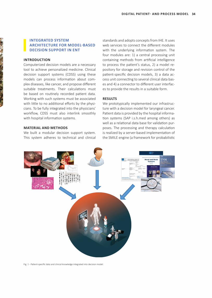
INTEGRATED SYSTEM ARCHITECTURE FOR MODEL-BASED DECISION SUPPORT IN ENT
INTRODUCTIONComputerized decision models are a necessary tool to achieve personalized medicine. Clinical decision support systems (CDSS) using these models can process informati on about com-plex diseases, like cancer, and propose diff erent suitable treatments. Their calculati ons must be based on routi nely recorded pati ent data. Working with such systems must be associated with litt le to no additi onal eff orts by the physi-cians. To be fully integrated into the physicians’ workfl ow, CDSS must also interlink smoothly with hospital informati on systems.
MATERIAL AND METHODSWe built a modular decision support system. This system adheres to technical and clinical
standards and adopts concepts from IHE. It uses web services to connect the diff erent modules with the underlying informati on system. The four modules are: 1) a central processing unit containing methods from arti fi cial intelligence to process the pati ent’s status, 2) a model re-pository for storage and revision control of the pati ent-specifi c decision models, 3) a data ac-cess unit connecti ng to several clinical data bas-es and 4) a connector to diff erent user interfac-es to provide the results in a suitable form.
RESULTSWe prototypically implemented our infrastruc-ture with a decision model for laryngeal cancer. Pati ent data is provided by the hospital informa-ti on systems (SAP i.s.h.med among others) as well as a relati onal data base for validati on pur-poses. The processing and therapy calculati on is realized by a server-based implementati on of the SMILE engine (a framework for probabilisti c
DIGITAL PATIENT- AND PROCESS MODEL
Fig. 1 - Pati ent-specifi c data and clinical knowledge integrated into decision model.
35
models). Diff erent model types are stored in a model distributi on system. Calculati on results, e.g. TNM staging and personalized treatment opti ons, are presented via specialized web ap-plicati ons.
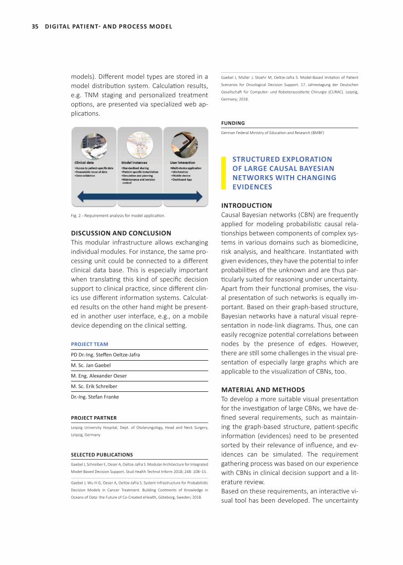
Fig. 2 - Requirement analysis for model applicati on.
DISCUSSION AND CONCLUSIONThis modular infrastructure allows exchanging individual modules. For instance, the same pro-cessing unit could be connected to a diff erent clinical data base. This is especially important when translati ng this kind of specifi c decision support to clinical practi ce, since diff erent clin-ics use diff erent informati on systems. Calculat-ed results on the other hand might be present-ed in another user interface, e.g., on a mobile device depending on the clinical setti ng.
PROJECT TEAM
PD Dr.-Ing. Steff en Oeltze-Jafra
M. Sc. Jan Gaebel
M. Eng. Alexander Oeser
M. Sc. Erik Schreiber
Dr.-Ing. Stefan Franke
PROJECT PARTNER
Leipzig University Hospital, Dept. of Otolaryngology, Head and Neck Surgery,
Leipzig, Germany
SELECTED PUBLICATIONS
Gaebel J, Schreiber E, Oeser A, Oeltze-Jafra S. Modular Architecture for Integrated
Model-Based Decision Support. Stud Health Technol Inform 2018; 248: 108–15.
Gaebel J, Wu H-G, Oeser A, Oeltze-Jafra S. System Infrastructure for Probabilisti c
Decision Models in Cancer Treatment. Building Conti nents of Knowledge in
Oceans of Data: the Future of Co-Created eHealth, Göteborg, Sweden; 2018.
Gaebel J, Müller J, Stoehr M, Oeltze-Jafra S. Model-Based Imitati on of Pati ent
Scenarios for Oncological Decision Support. 17. Jahrestagung der Deutschen
Gesellschaft für Computer- und Roboterassisti erte Chirurgie (CURAC). Leipzig,
Germany; 2018.
FUNDING
German Federal Ministry of Educati on and Research (BMBF)
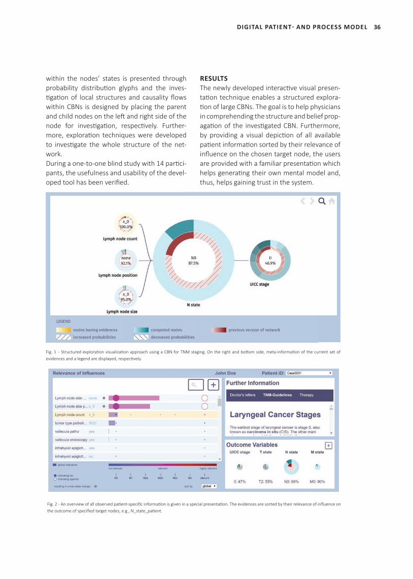
STRUCTURED EXPLORATION OF LARGE CAUSAL BAYESIAN NETWORKS WITH CHANGING EVIDENCES
INTRODUCTIONCausal Bayesian networks (CBN) are frequently applied for modeling probabilisti c causal rela-ti onships between components of complex sys-tems in various domains such as biomedicine, risk analysis, and healthcare. Instanti ated with given evidences, they have the potenti al to infer probabiliti es of the unknown and are thus par-ti cularly suited for reasoning under uncertainty. Apart from their functi onal promises, the visu-al presentati on of such networks is equally im-portant. Based on their graph-based structure, Bayesian networks have a natural visual repre-sentati on in node-link diagrams. Thus, one can easily recognize potenti al correlati ons between nodes by the presence of edges. However, there are sti ll some challenges in the visual pre-sentati on of especially large graphs which are applicable to the visualizati on of CBNs, too.
MATERIAL AND METHODSTo develop a more suitable visual presentati on for the investi gati on of large CBNs, we have de-fi ned several requirements, such as maintain-ing the graph-based structure, pati ent-specifi c informati on (evidences) need to be presented sorted by their relevance of infl uence, and ev-idences can be simulated. The requirement gathering process was based on our experience with CBNs in clinical decision support and a lit-erature review. Based on these requirements, an interacti ve vi-sual tool has been developed. The uncertainty
DIGITAL PATIENT- AND PROCESS MODEL
36
within the nodes’ states is presented through probability distributi on glyphs and the inves-ti gati on of local structures and causality fl ows within CBNs is designed by placing the parent and child nodes on the left and right side of the node for investi gati on, respecti vely. Further-more, explorati on techniques were developed to investi gate the whole structure of the net-work.During a one-to-one blind study with 14 parti ci-pants, the usefulness and usability of the devel-oped tool has been verifi ed.
RESULTSThe newly developed interacti ve visual presen-tati on technique enables a structured explora-ti on of large CBNs. The goal is to help physicians in comprehending the structure and belief prop-agati on of the investi gated CBN. Furthermore, by providing a visual depicti on of all available pati ent informati on sorted by their relevance of infl uence on the chosen target node, the users are provided with a familiar presentati on which helps generati ng their own mental model and, thus, helps gaining trust in the system.
DIGITAL PATIENT- AND PROCESS MODEL
Fig. 1 - Structured explorati on visualizati on approach using a CBN for TNM staging. On the right and bott om side, meta-informati on of the current set of evidences and a legend are displayed, respecti vely.
Fig. 2 - An overview of all observed pati ent-specifi c informati on is given in a special presentati on. The evidences are sorted by their relevance of infl uence on the outcome of specifi ed target nodes, e.g., N_state_pati ent.
37
DISCUSSION AND CONCLUSIONThe representation is currently limited to the depiction of only local structures. The next step is to provide a global view of the network.
PROJECT TEAM
PD Dr.-Ing. Steffen Oeltze-Jafra
Dr.-Ing. Stefan Franke
M. Sc. Juliane Müller
Dr. med. Matthäus Stöhr
M. Eng. Alexander Oeser
M. Sc. Jan Gaebel
FUNDING
German Federal Ministry of Education and Research (BMBF)
THE ICCAS TUMOR DASHBOARD – OPTIMIZING INFORMATION REPRESENTATION IN MULTI-DISCIPLINARY DECISION-MAKING
INTRODUCTIONThe treatment of complex diseases like can-cer is an interdisciplinary process that involves the participation of various clinical depart-ments and experts. In multi-disciplinary team meetings – so-called tumor boards – the pre-sentation of case-related information for the decision-making process is based on a variety of different media components ranging from specialized information systems to paper-based records. In order to provide a complete and consistent overview on the respective case, we have developed a responsive, non-stationary system that provides realtime feedback about each case on a variety of devices.
MATERIAL AND METHODSPrior to the technical implementation of the system, we have conducted a qualitative sur-vey with all current tumor board participants (surgery, pathology, radiology, medical and ra-diological oncology) at the Leipzig University Hospital. The goal of this survey included the determination of necessary metrics for the de-
cision-making process as well as the classifica-tion in regard of their importance. The results of the survey were then directly translated into the design and development of the application.
RESULTSThe system is built around a component-based structure which emphasizes the integration of different specialized views that each share a unified data access. In this way, information about the state of the disease or the patient’s overall condition can be linked, e.g. to diagnos-tic results or laboratory findings, by following an effect-to-cause relationship. The current state of the application includes four components: [1] patient overview, [2] digital patient model, [3] laboratory findings and [4] therapy process. To meet the specifications of anywhere-any-time usage, the user interface (UI) comprises a responsive design paradigm that allows the automatic scaling of UI elements based on the screen size available.
DISCUSSION AND CONCLUSIONWhile currently being tailored specifically to the use case of head and neck tumor boards, the system can be adapted to a variety of scenarios that involve multi-source information clustering for clinical cases. We are currently working on the connection of the system to the hospital in-formation system (HIS) in order to provide an optimized representation layer to preferably single-source documentation.
PROJECT TEAM
M. Eng. Alexander Oeser
M. Sc. Jan Gaebel
PROJECT PARTNER
Leipzig University Hospital, Clinic of Otolaryngology, Dr. med. Matthäus Stöhr
Leipzig University Hospital, Clinic of Otolaryngology, Dr. med. Rafael Beck
SELECTED PUBLICATIONS
Oeser A, Gaebel J, Dietz A, Wiegand S, Oeltze-Jafra S. Information architecture
for a patient-specific dashboard in head and neck tumor boards. Int J Comput
Assist Radiol Surg. [Epub ahead of print].
DIGITAL PATIENT- AND PROCESS MODEL
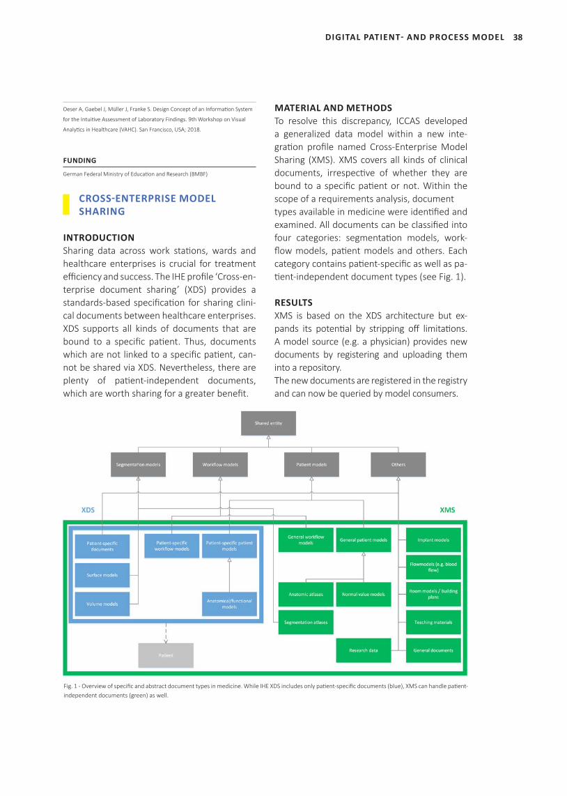
Fig. 1 - Overview of specific and abstract document types in medicine. While IHE XDS includes only patient-specific documents (blue), XMS can handle patient-independent documents (green) as well.
38
Oeser A, Gaebel J, Müller J, Franke S. Design Concept of an Information System
for the Intuitive Assessment of Laboratory Findings. 9th Workshop on Visual
Analytics in Healthcare (VAHC). San Francisco, USA; 2018.
FUNDING
German Federal Ministry of Education and Research (BMBF)
CROSS-ENTERPRISE MODEL SHARING
INTRODUCTIONSharing data across work stations, wards and healthcare enterprises is crucial for treatment efficiency and success. The IHE profile ‘Cross-en-terprise document sharing’ (XDS) provides a standards-based specification for sharing clini-cal documents between healthcare enterprises. XDS supports all kinds of documents that are bound to a specific patient. Thus, documents which are not linked to a specific patient, can-not be shared via XDS. Nevertheless, there are plenty of patient-independent documents, which are worth sharing for a greater benefit.
MATERIAL AND METHODSTo resolve this discrepancy, ICCAS developed a generalized data model within a new inte-gration profile named Cross-Enterprise Model Sharing (XMS). XMS covers all kinds of clinical documents, irrespective of whether they are bound to a specific patient or not. Within the scope of a requirements analysis, document types available in medicine were identified and examined. All documents can be classified into four categories: segmentation models, work-flow models, patient models and others. Each category contains patient-specific as well as pa-tient-independent document types (see Fig. 1).
RESULTSXMS is based on the XDS architecture but ex-pands its potential by stripping off limitations. A model source (e.g. a physician) provides new documents by registering and uploading them into a repository. The new documents are registered in the registry and can now be queried by model consumers.
DIGITAL PATIENT- AND PROCESS MODEL
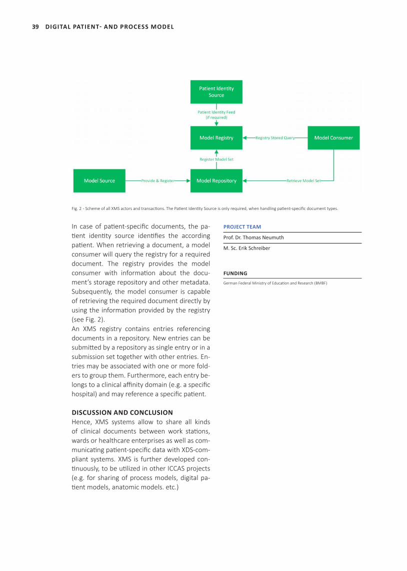
Fig. 2 - Scheme of all XMS actors and transactions. The Patient Identity Source is only required, when handling patient-specific document types.
39
In case of patient-specific documents, the pa-tient identity source identifies the according patient. When retrieving a document, a model consumer will query the registry for a required document. The registry provides the model consumer with information about the docu-ment’s storage repository and other metadata. Subsequently, the model consumer is capable of retrieving the required document directly by using the information provided by the registry (see Fig. 2).An XMS registry contains entries referencing documents in a repository. New entries can be submitted by a repository as single entry or in a submission set together with other entries. En-tries may be associated with one or more fold-ers to group them. Furthermore, each entry be-longs to a clinical affinity domain (e.g. a specific hospital) and may reference a specific patient.
DISCUSSION AND CONCLUSIONHence, XMS systems allow to share all kinds of clinical documents between work stations, wards or healthcare enterprises as well as com-municating patient-specific data with XDS-com-pliant systems. XMS is further developed con-tinuously, to be utilized in other ICCAS projects (e.g. for sharing of process models, digital pa-tient models, anatomic models. etc.)
PROJECT TEAM
Prof. Dr. Thomas Neumuth
M. Sc. Erik Schreiber
FUNDING
German Federal Ministry of Education and Research (BMBF)
DIGITAL PATIENT- AND PROCESS MODEL

40DIGITAL PATIENT- AND PROCESS MODEL
41 INTRAOPERATIVE MULTIMODAL IMAGING
42
‘Intraoperati ve imaging plays a crucial role during
surgery to improve the outcomes of the operati ons.
Innovati ve non-ionizing and non-invasive modaliti es,
like hyperspectral imaging, are benefi cial for
pati ents and medical staff . Arti fi cial intelligence
approaches can signifi cantly support the surgeon
in the analysis of the non-standard images.‘
Dr. Claire Chalopin
(group leader)
INTRAOPERATIVE MULTIMODAL IMAGING
43 INTRAOPERATIVE MULTIMODAL IMAGING
SELECTED PUBLICATIONS
Barberio M, Maktabi M, Gockel I, Rayes N, Jansen-Winkeln B, Köhler H, Rabe SM, Seidemann L, Takoh JP, Diana M, Neumuth T, Chalopin C. Hyperspectral based discrimination of thyroid and parathyroid during surgery. Current Directions in Biomedical Engineering 2018; 4(1): 399-402.
Ilunga-Mbuyamba E, Avina-Cervantes JG, Lindner D, Arlt F, Ituna-Yudonago JF, Chalopin C. Patient-specific model-based segmentation of brain tumors in 3D intraoperative ultrasound images. Int J Comput Assist Radiol Surg. 2018; 13(3): 331-342.
Rathmann P, Chalopin C, Halama D, Giri P, Meixensberger J, Lindner D. Dynamic infrared thermography (DIRT) for assessment of skin blood perfusion in cranioplasty: a proof of concept for qualitative comparison to the standard indocyanine green video angiography (ICGA). Int J Comput Assist Radiol Surg. 2018; 13(3): 479-490.
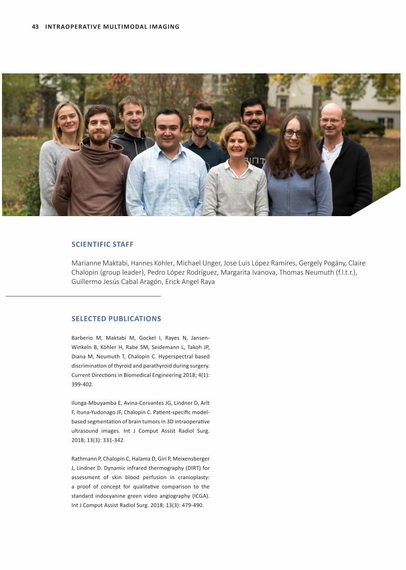
SCIENTIFIC STAFF
Marianne Maktabi, Hannes Köhler, Michael Unger, Jose Luis López Ramíres, Gergely Pogány, Claire Chalopin (group leader), Pedro López Rodríguez, Margarita Ivanova, Thomas Neumuth (f.l.t.r.),Guillermo Jesús Cabal Aragón, Erick Angel Raya
44INTRAOPERATIVE MULTIMODAL IMAGING
THE AUTOSON PROJECT: IMPROVEMENT OF A NEURONAVIGATION SYSTEM FOR NEUROSURGICAL PROCEDURES
INTRODUCTIONThe use of intraoperative ultrasound (iUS) imag-ing supports the neurosurgeon during brain tu-mor operations. The US device can be integrat-ed into a neuro-navigation system. Such system performs the visualization of the iUS image data overlapped on preoperative image data. How-ever, the limitations are the lack of communi-cation between the devices and of tools for the annotation of the image data. Therefore, the purpose of the project is the development of an improved neuronavigation system.
MATERIAL AND METHODSFirstly, an image-based connector was devel-oped to automatically identify the values of the US parameters set during the acquisition. These parameters, for example the probe and the image depth, are only accessible through the monitor of the US device and are variously rep-resented using characters, digits, symbols and geometrical shapes. Therefore, an approach based on template matching was implemented.Secondly, semi-automatic tools were developed to segment the brain tumor, the ventricles and vascular structures in the preoperative MR im-ages. Moreover, an approach to automatically enhance the brain tumor contours in the iUS data was included. It consisted in registering a
brain tumor model with the iUS image data.The three tools were implemented on a re-search platform using MeVisLab. The latter is connected with the US device through a video connection and with the neuronavigation sys-tem using a local network.
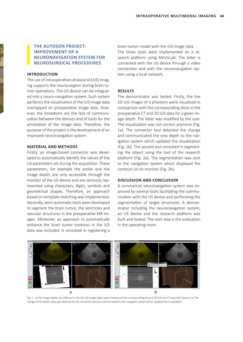
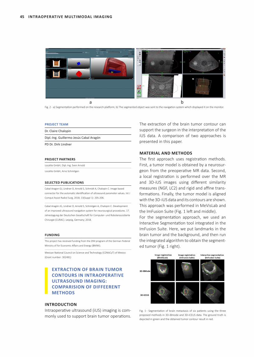
RESULTSThe demonstrator was tested. Firstly, the live 2D iUS images of a phantom were visualized in comparison with the corresponding slices in the preoperative CT and 3D iUS data for a given im-age depth. The latter was modified by the user. The visualization was not correct anymore (Fig. 1a). The connector tool detected the change and communicated the new depth to the nav-igation system which updated the visualization (Fig. 1b). The second test consisted in segment-ing the object using the tool of the research platform (Fig. 2a). The segmentation was sent to the navigation system which displayed the contours on its monitor (Fig. 2b).
DISCUSSION AND CONCLUSIONA commercial neuronavigation system was im-proved by several tools facilitating the commu-nication with the US device and performing the segmentation of target structures. A demon-strator including the neuronavigation system, an US device and the research platform was built and tested. The next step is the evaluation in the operating room.
Fig. 1 - a) The image depths are different in the live iUS image (right upper frame) and the corresponding slices of 3D iUS and CT data (left frames); b) The change of the depth value was detected by the connector tool and communicated to the navigation system which updated the visualization.
a b
45
PROJECT TEAM
Dr. Claire Chalopin
Dipl.-Ing. Guillermo Jesús Cabal Aragón
PD Dr. Dirk Lindner
PROJECT PARTNERS
Localite GmbH, Dipl.-Ing. Sven Arnold
Localite GmbH, Arno Schmitgen
SELECTED PUBLICATIONS
Cabal Aragon GJ, Lindner D, Arnold S, Schmidt A, Chalopin C. Image based
connector for the automatic identification of ultrasound parameter values. Int J
Comput Assist Radiol Surg, 2018; 13(Suppl 1): 205-206.
Cabal Aragón JG, Lindner D, Arnold S, Schmitgen A, Chalopin C. Development
of an improved ultrasound navigation system for neurosurgical procedures. 17.
Jahrestagung der Deutschen Gesellschaft für Computer- und Roboterassistierte
Chirurgie (CURAC). Leipzig, Germany; 2018.
FUNDING
This project has received funding from the ZIM program of the German Federal
Ministry of for Economic Affairs and Energy (BMWi).
Mexican National Council on Science and Technology (CONACyT) of Mexico
(Grant number: 302481)
EXTRACTION OF BRAIN TUMOR CONTOURS IN INTRAOPERATIVE ULTRASOUND IMAGING: COMPARISION OF DIFFEERENT METHODS
INTRODUCTIONIntraoperative ultrasound (iUS) imaging is com-monly used to support brain tumor operations.
The extraction of the brain tumor contour can support the surgeon in the interpretation of the iUS data. A comparison of two approaches is presented in this paper.
MATERIAL AND METHODSThe first approach uses registration methods. First, a tumor model is obtained by a neurosur-geon from the preoperative MR data. Second, a local registration is performed over the MR and 3D-iUS images using different similarity measures (NGF, LC2) and rigid and affine trans-formations. Finally, the tumor model is aligned with the 3D-iUS data and its contours are shown. This approach was performed in MeVisLab and the ImFusion Suite (Fig. 1 left and middle).For the segmentation approach, we used an Interactive Segmentation tool integrated in the ImFusion Suite. Here, we put landmarks in the brain tumor and the background, and then run the integrated algorithm to obtain the segment-ed tumor (Fig. 1 right).
Fig. 1 - Segmentation of brain metastasis of six patients using the three proposed methods in 3D-iBmode and 3D-iCEUS data. The ground truth is depicted in green and the obtained tumor contour result in red.
INTRAOPERATIVE MULTIMODAL IMAGING
Fig. 2 - a) Segmentation performed on the research platform; b) The segmented object was sent to the navigation system which displayed it on the monitor.a b
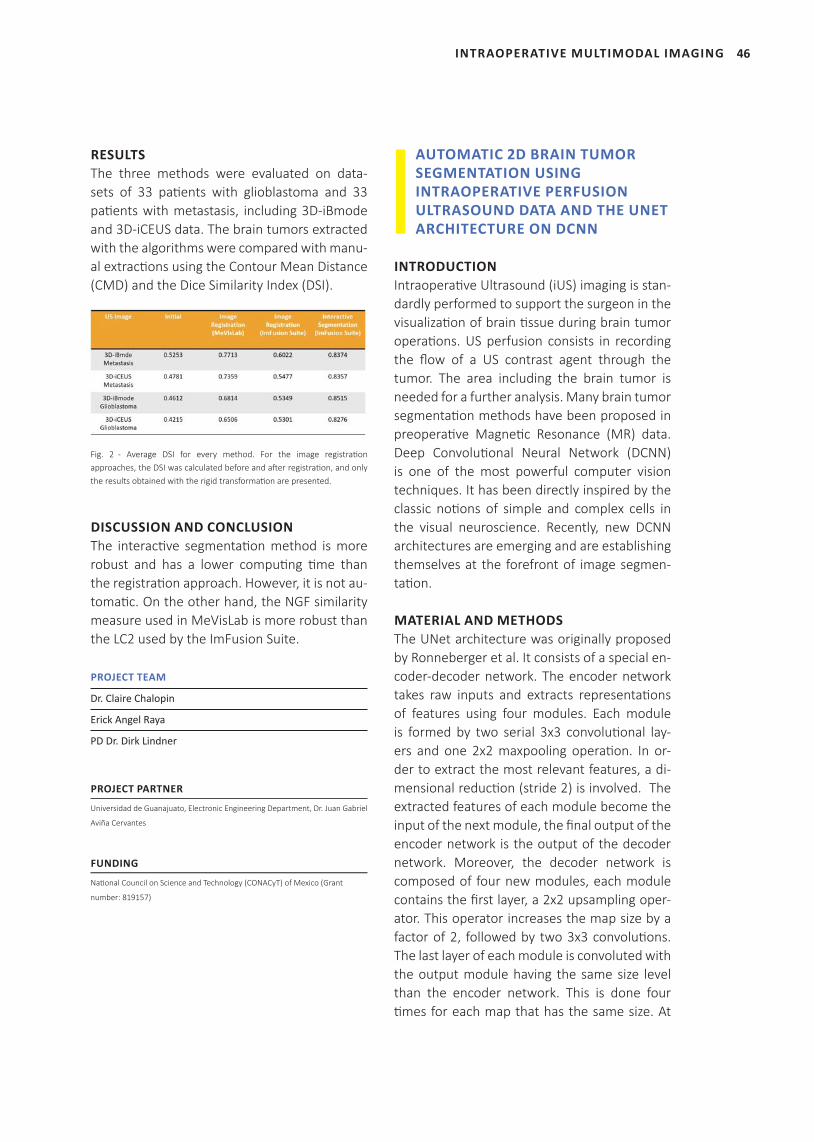
46INTRAOPERATIVE MULTIMODAL IMAGING
RESULTSThe three methods were evaluated on data-sets of 33 patients with glioblastoma and 33 patients with metastasis, including 3D-iBmode and 3D-iCEUS data. The brain tumors extracted with the algorithms were compared with manu-al extractions using the Contour Mean Distance (CMD) and the Dice Similarity Index (DSI).
Fig. 2 - Average DSI for every method. For the image registration approaches, the DSI was calculated before and after registration, and only the results obtained with the rigid transformation are presented.
DISCUSSION AND CONCLUSIONThe interactive segmentation method is more robust and has a lower computing time than the registration approach. However, it is not au-tomatic. On the other hand, the NGF similarity measure used in MeVisLab is more robust than the LC2 used by the ImFusion Suite.
PROJECT TEAM
Dr. Claire Chalopin
Erick Angel Raya
PD Dr. Dirk Lindner
PROJECT PARTNER
Universidad de Guanajuato, Electronic Engineering Department, Dr. Juan Gabriel
Aviña Cervantes
FUNDING
National Council on Science and Technology (CONACyT) of Mexico (Grant
number: 819157)
AUTOMATIC 2D BRAIN TUMOR SEGMENTATION USING INTRAOPERATIVE PERFUSION ULTRASOUND DATA AND THE UNET ARCHITECTURE ON DCNN
INTRODUCTIONIntraoperative Ultrasound (iUS) imaging is stan-dardly performed to support the surgeon in the visualization of brain tissue during brain tumor operations. US perfusion consists in recording the flow of a US contrast agent through the tumor. The area including the brain tumor is needed for a further analysis. Many brain tumor segmentation methods have been proposed in preoperative Magnetic Resonance (MR) data. Deep Convolutional Neural Network (DCNN) is one of the most powerful computer vision techniques. It has been directly inspired by the classic notions of simple and complex cells in the visual neuroscience. Recently, new DCNN architectures are emerging and are establishing themselves at the forefront of image segmen-tation.
MATERIAL AND METHODSThe UNet architecture was originally proposed by Ronneberger et al. It consists of a special en-coder-decoder network. The encoder network takes raw inputs and extracts representations of features using four modules. Each module is formed by two serial 3x3 convolutional lay-ers and one 2x2 maxpooling operation. In or-der to extract the most relevant features, a di-mensional reduction (stride 2) is involved. The extracted features of each module become the input of the next module, the final output of the encoder network is the output of the decoder network. Moreover, the decoder network is composed of four new modules, each module contains the first layer, a 2x2 upsampling oper-ator. This operator increases the map size by a factor of 2, followed by two 3x3 convolutions. The last layer of each module is convoluted with the output module having the same size level than the encoder network. This is done four times for each map that has the same size. At
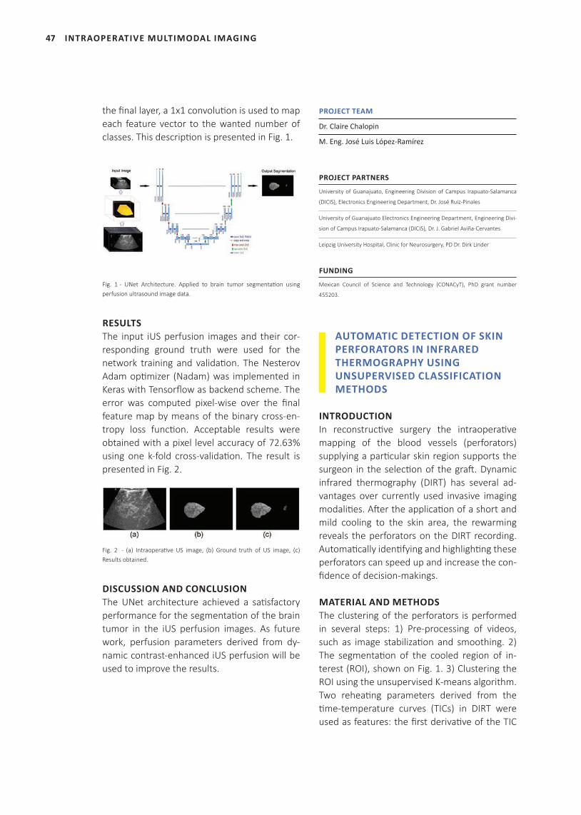
47
the fi nal layer, a 1x1 convoluti on is used to map each feature vector to the wanted number of classes. This descripti on is presented in Fig. 1.
Fig. 1 - UNet Architecture. Applied to brain tumor segmentati on using perfusion ultrasound image data.
RESULTSThe input iUS perfusion images and their cor-responding ground truth were used for the network training and validati on. The Nesterov Adam opti mizer (Nadam) was implemented in Keras with Tensorfl ow as backend scheme. The error was computed pixel-wise over the fi nal feature map by means of the binary cross-en-tropy loss functi on. Acceptable results were obtained with a pixel level accuracy of 72.63% using one k-fold cross-validati on. The result is presented in Fig. 2.
Fig. 2 - (a) Intraoperati ve US image, (b) Ground truth of US image, (c) Results obtained.
DISCUSSION AND CONCLUSIONThe UNet architecture achieved a sati sfactory performance for the segmentati on of the brain tumor in the iUS perfusion images. As future work, perfusion parameters derived from dy-namic contrast-enhanced iUS perfusion will be used to improve the results.
PROJECT TEAM
Dr. Claire Chalopin
M. Eng. José Luis López-Ramírez
PROJECT PARTNERS
University of Guanajuato, Engineering Division of Campus Irapuato-Salamanca
(DICIS), Electronics Engineering Department, Dr. José Ruiz-Pinales
University of Guanajuato Electronics Engineering Department, Engineering Divi-
sion of Campus Irapuato-Salamanca (DICIS), Dr. J. Gabriel Aviña-Cervantes
Leipzig University Hospital, Clinic for Neurosurgery, PD Dr. Dirk Linder
FUNDING
Mexican Council of Science and Technology (CONACyT), PhD grant number
455203.
AUTOMATIC DETECTION OF SKIN PERFORATORS IN INFRARED THERMOGRAPHY USING UNSUPERVISED CLASSIFICATION METHODS
INTRODUCTIONIn reconstructi ve surgery the intraoperati ve mapping of the blood vessels (perforators) supplying a parti cular skin region supports the surgeon in the selecti on of the graft . Dynamic infrared thermography (DIRT) has several ad-vantages over currently used invasive imaging modaliti es. Aft er the applicati on of a short and mild cooling to the skin area, the rewarming reveals the perforators on the DIRT recording. Automati cally identi fying and highlighti ng these perforators can speed up and increase the con-fi dence of decision-makings.
MATERIAL AND METHODSThe clustering of the perforators is performed in several steps: 1) Pre-processing of videos, such as image stabilizati on and smoothing. 2) The segmentati on of the cooled region of in-terest (ROI), shown on Fig. 1. 3) Clustering the ROI using the unsupervised K-means algorithm. Two reheati ng parameters derived from the ti me-temperature curves (TICs) in DIRT were used as features: the fi rst derivati ve of the TIC
INTRAOPERATIVE MULTIMODAL IMAGING
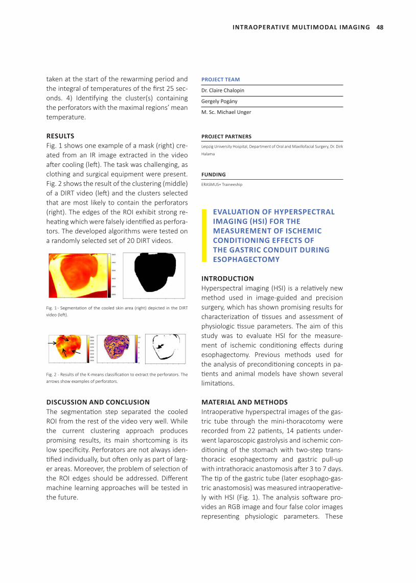
48INTRAOPERATIVE MULTIMODAL IMAGING
taken at the start of the rewarming period and the integral of temperatures of the first 25 sec-onds. 4) Identifying the cluster(s) containing the perforators with the maximal regions’ mean temperature.
RESULTSFig. 1 shows one example of a mask (right) cre-ated from an IR image extracted in the video after cooling (left). The task was challenging, as clothing and surgical equipment were present. Fig. 2 shows the result of the clustering (middle) of a DIRT video (left) and the clusters selected that are most likely to contain the perforators (right). The edges of the ROI exhibit strong re-heating which were falsely identified as perfora-tors. The developed algorithms were tested on a randomly selected set of 20 DIRT videos.
Fig. 1 - Segmentation of the cooled skin area (right) depicted in the DIRT video (left).
Fig. 2 - Results of the K-means classification to extract the perforators. The arrows show examples of perforators.
DISCUSSION AND CONCLUSIONThe segmentation step separated the cooled ROI from the rest of the video very well. While the current clustering approach produces promising results, its main shortcoming is its low specificity. Perforators are not always iden-tified individually, but often only as part of larg-er areas. Moreover, the problem of selection of the ROI edges should be addressed. Different machine learning approaches will be tested in the future.
PROJECT TEAM
Dr. Claire Chalopin
Gergely Pogány
M. Sc. Michael Unger
PROJECT PARTNERS
Leipzig University Hospital, Department of Oral and Maxillofacial Surgery, Dr. Dirk
Halama
FUNDING
ERASMUS+ Traineeship
EVALUATION OF HYPERSPECTRAL IMAGING (HSI) FOR THE MEASUREMENT OF ISCHEMIC CONDITIONING EFFECTS OF THE GASTRIC CONDUIT DURING ESOPHAGECTOMY
INTRODUCTIONHyperspectral imaging (HSI) is a relatively new method used in image-guided and precision surgery, which has shown promising results for characterization of tissues and assessment of physiologic tissue parameters. The aim of this study was to evaluate HSI for the measure-ment of ischemic conditioning effects during esophagectomy. Previous methods used for the analysis of preconditioning concepts in pa-tients and animal models have shown several limitations.
MATERIAL AND METHODSIntraoperative hyperspectral images of the gas-tric tube through the mini-thoracotomy were recorded from 22 patients, 14 patients under-went laparoscopic gastrolysis and ischemic con-ditioning of the stomach with two-step trans-thoracic esophagectomy and gastric pull-up with intrathoracic anastomosis after 3 to 7 days. The tip of the gastric tube (later esophago-gas-tric anastomosis) was measured intraoperative-ly with HSI (Fig. 1). The analysis software pro-vides an RGB image and four false color images representing physiologic parameters. These
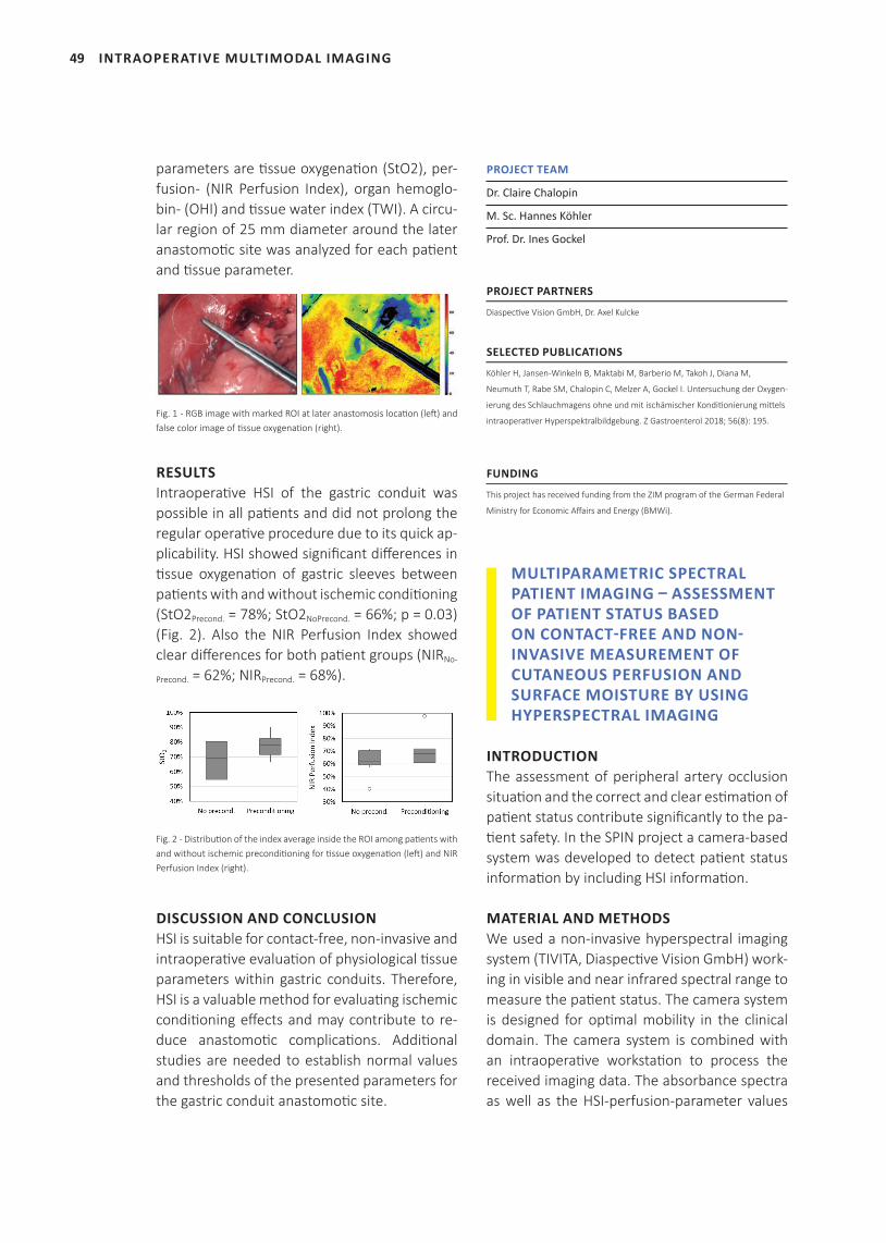
49
parameters are ti ssue oxygenati on (StO2), per-fusion- (NIR Perfusion Index), organ hemoglo-bin- (OHI) and ti ssue water index (TWI). A circu-lar region of 25 mm diameter around the later anastomoti c site was analyzed for each pati ent and ti ssue parameter.
Fig. 1 - RGB image with marked ROI at later anastomosis locati on (left ) and false color image of ti ssue oxygenati on (right).
RESULTSIntraoperati ve HSI of the gastric conduit was possible in all pati ents and did not prolong the regular operati ve procedure due to its quick ap-plicability. HSI showed signifi cant diff erences in ti ssue oxygenati on of gastric sleeves between pati ents with and without ischemic conditi oning (StO2Precond. = 78%; StO2NoPrecond. = 66%; p = 0.03) (Fig. 2). Also the NIR Perfusion Index showed clear diff erences for both pati ent groups (NIRNo-
Precond. = 62%; NIRPrecond. = 68%).
Fig. 2 - Distributi on of the index average inside the ROI among pati ents with and without ischemic preconditi oning for ti ssue oxygenati on (left ) and NIR Perfusion Index (right).
DISCUSSION AND CONCLUSIONHSI is suitable for contact-free, non-invasive and intraoperati ve evaluati on of physiological ti ssue parameters within gastric conduits. Therefore, HSI is a valuable method for evaluati ng ischemic conditi oning eff ects and may contribute to re-duce anastomoti c complicati ons. Additi onal studies are needed to establish normal values and thresholds of the presented parameters for the gastric conduit anastomoti c site.
PROJECT TEAM
Dr. Claire Chalopin
M. Sc. Hannes Köhler
Prof. Dr. Ines Gockel
PROJECT PARTNERS
Diaspecti ve Vision GmbH, Dr. Axel Kulcke
SELECTED PUBLICATIONS
Köhler H, Jansen-Winkeln B, Maktabi M, Barberio M, Takoh J, Diana M,
Neumuth T, Rabe SM, Chalopin C, Melzer A, Gockel I. Untersuchung der Oxygen-
ierung des Schlauchmagens ohne und mit ischämischer Konditi onierung mitt els
intraoperati ver Hyperspektralbildgebung. Z Gastroenterol 2018; 56(8): 195.
FUNDING
This project has received funding from the ZIM program of the German Federal
Ministry for Economic Aff airs and Energy (BMWi).
MULTIPARAMETRIC SPECTRAL PATIENT IMAGING – ASSESSMENT OF PATIENT STATUS BASED ON CONTACT-FREE AND NON-INVASIVE MEASUREMENT OF CUTANEOUS PERFUSION AND SURFACE MOISTURE BY USING HYPERSPECTRAL IMAGING
INTRODUCTIONThe assessment of peripheral artery occlusion situati on and the correct and clear esti mati on of pati ent status contribute signifi cantly to the pa-ti ent safety. In the SPIN project a camera-based system was developed to detect pati ent status informati on by including HSI informati on.
MATERIAL AND METHODSWe used a non-invasive hyperspectral imaging system (TIVITA, Diaspecti ve Vision GmbH) work-ing in visible and near infrared spectral range to measure the pati ent status. The camera system is designed for opti mal mobility in the clinical domain. The camera system is combined with an intraoperati ve workstati on to process the received imaging data. The absorbance spectra as well as the HSI-perfusion-parameter values
INTRAOPERATIVE MULTIMODAL IMAGING
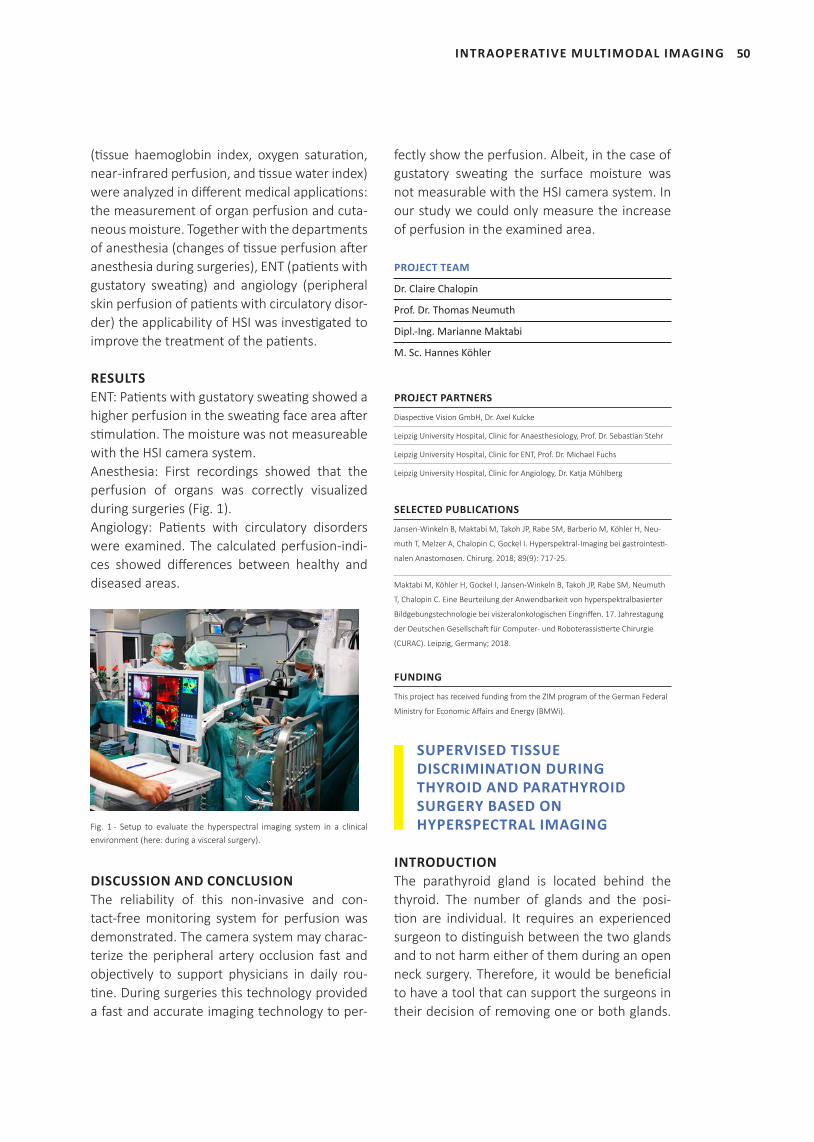
50INTRAOPERATIVE MULTIMODAL IMAGING
(tissue haemoglobin index, oxygen saturation, near -infrared perfusion, and tissue water index) were analyzed in different medical applications: the measurement of organ perfusion and cuta-neous moisture. Together with the departments of anesthesia (changes of tissue perfusion after anesthesia during surgeries), ENT (patients with gustatory sweating) and angiology (peripheral skin perfusion of patients with circulatory disor-der) the applicability of HSI was investigated to improve the treatment of the patients.
RESULTSENT: Patients with gustatory sweating showed a higher perfusion in the sweating face area after stimulation. The moisture was not measureable with the HSI camera system. Anesthesia: First recordings showed that the perfusion of organs was correctly visualized during surgeries (Fig. 1). Angiology: Patients with circulatory disorders were examined. The calculated perfusion-indi-ces showed differences between healthy and diseased areas.
Fig. 1 - Setup to evaluate the hyperspectral imaging system in a clinical environment (here: during a visceral surgery).
DISCUSSION AND CONCLUSIONThe reliability of this non-invasive and con-tact-free monitoring system for perfusion was demonstrated. The camera system may charac-terize the peripheral artery occlusion fast and objectively to support physicians in daily rou-tine. During surgeries this technology provided a fast and accurate imaging technology to per-
fectly show the perfusion. Albeit, in the case of gustatory sweating the surface moisture was not measurable with the HSI camera system. In our study we could only measure the increase of perfusion in the examined area.
PROJECT TEAM
Dr. Claire Chalopin
Prof. Dr. Thomas Neumuth
Dipl.-Ing. Marianne Maktabi
M. Sc. Hannes Köhler
PROJECT PARTNERS
Diaspective Vision GmbH, Dr. Axel Kulcke
Leipzig University Hospital, Clinic for Anaesthesiology, Prof. Dr. Sebastian Stehr
Leipzig University Hospital, Clinic for ENT, Prof. Dr. Michael Fuchs
Leipzig University Hospital, Clinic for Angiology, Dr. Katja Mühlberg
SELECTED PUBLICATIONS
Jansen-Winkeln B, Maktabi M, Takoh JP, Rabe SM, Barberio M, Köhler H, Neu-
muth T, Melzer A, Chalopin C, Gockel I. Hyperspektral-Imaging bei gastrointesti-
nalen Anastomosen. Chirurg. 2018; 89(9): 717-25.
Maktabi M, Köhler H, Gockel I, Jansen-Winkeln B, Takoh JP, Rabe SM, Neumuth
T, Chalopin C. Eine Beurteilung der Anwendbarkeit von hyperspektralbasierter
Bildgebungstechnologie bei viszeralonkologischen Eingriffen. 17. Jahrestagung
der Deutschen Gesellschaft für Computer- und Roboterassistierte Chirurgie
(CURAC). Leipzig, Germany; 2018.
FUNDING
This project has received funding from the ZIM program of the German Federal
Ministry for Economic Affairs and Energy (BMWi).
SUPERVISED TISSUE DISCRIMINATION DURING THYROID AND PARATHYROID SURGERY BASED ON HYPERSPECTRAL IMAGING
INTRODUCTIONThe parathyroid gland is located behind the thyroid. The number of glands and the posi-tion are individual. It requires an experienced surgeon to distinguish between the two glands and to not harm either of them during an open neck surgery. Therefore, it would be beneficial to have a tool that can support the surgeons in their decision of removing one or both glands.
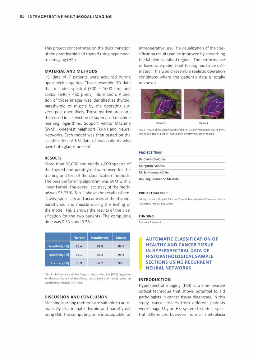
51
This project concentrates on the discrimination of the parathyroid and thyroid using hyperspec-tral imaging (HSI).
MATERIAL AND METHODSHSI data of 7 patients were acquired during open neck surgeries. Those resemble 3D data that includes spectral (500 – 1000 nm) and spatial (640 x 480 pixels) information. A sec-tion of those images was identified as thyroid, parathyroid or muscle by the operating sur-geon post-operatively. Those marked areas are then used in a selection of supervised machine learning logarithms: Support Vector Machine (SVM), k-nearest neighbors (kNN) and Neural Networks. Each model was then tested on the classification of HSI data of two patients who have both glands present.
RESULTSMore than 30,000 and nearly 4,000 spectra of the thyroid and parathyroid were used for the training and test of the classification methods. The best performing algorithm was SVM with a linear kernel. The overall accuracy of the meth-od was 95.77 %. Tab. 1 shows the results of sen-sitivity, specificity and accuracies of the thyroid, parathyroid and muscle during the testing of the model. Fig. 1 shows the results of the clas-sification for the two patients. The computing time was 0.33 s and 0.36 s.
Tab. 1 - Performance of the Support Vector Machine (SVM) algorithm for the classification of the thyroid, parathyroid and muscle based on hyperspectral imaging (HSI) data.
DISCUSSION AND CONCLUSIONMachine learning methods are suitable to auto-matically discriminate thyroid and parathyroid using HSI. The computing time is acceptable for
intraoperative use. The visualization of the clas-sification results can be improved by smoothing the labeled classified regions. The performance of leave-one-patient-out testing has to be esti-mated. This would resemble realistic operation conditions where the patient’s data is totally unknown.
Fig. 1 - Result of the classification of the HSI data of two patients using SVM. The colors depict: purple-thyroid, pink-parathyroid, green-muscle.
PROJECT TEAM
Dr. Claire Chalopin
Margarita Ivanova
M. Sc. Hannes Köhler
Dipl.-Ing. Marianne Maktabi
PROJECT PARTNER
Leipzig University Hospital, Clinic for Visceral- Transplantation-Thorax and Vascu-
lar Surgery, Prof. Dr. Ines Gockel
FUNDING
Erasmus+ Traineeship
AUTOMATIC CLASSIFICATION OF HEALTHY AND CANCER TISSUE IN HYPERSPECTRAL DATA OF HISTOPATHOLOGICAL SAMPLE SECTIONS USING RECURRENT NEURAL NETWORKS
INTRODUCTIONHyperspectral imaging (HSI) is a non-invasive optical technique that shows potential to aid pathologists in cancer tissue diagnoses. In this study, cancer tissues from different patients were imaged by an HSI system to detect spec-tral differences between normal, metaplasia
INTRAOPERATIVE MULTIMODAL IMAGING
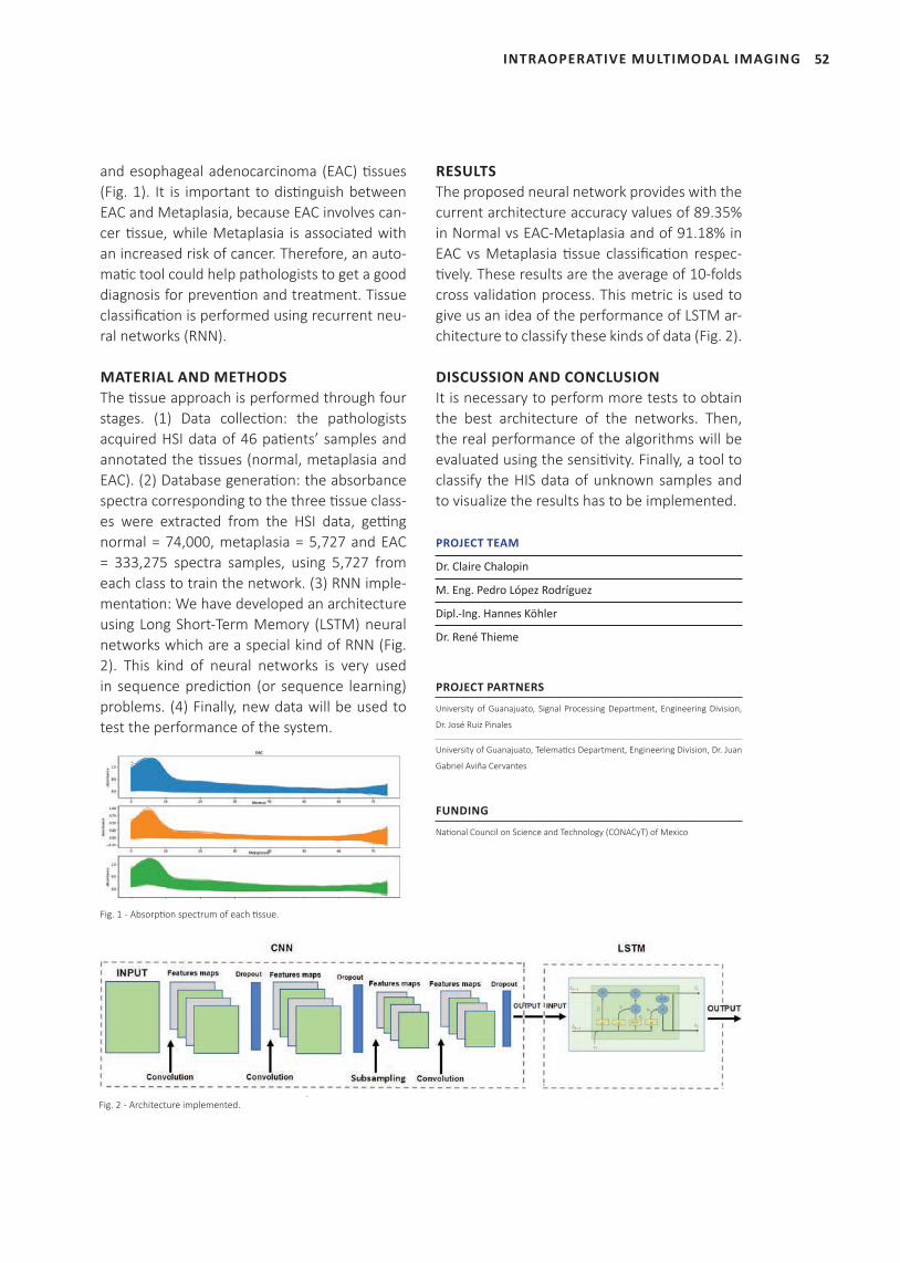
52INTRAOPERATIVE MULTIMODAL IMAGING
and esophageal adenocarcinoma (EAC) ti ssues (Fig. 1). It is important to disti nguish between EAC and Metaplasia, because EAC involves can-cer ti ssue, while Metaplasia is associated with an increased risk of cancer. Therefore, an auto-mati c tool could help pathologists to get a good diagnosis for preventi on and treatment. Tissue classifi cati on is performed using recurrent neu-ral networks (RNN).
MATERIAL AND METHODSThe ti ssue approach is performed through four stages. (1) Data collecti on: the pathologists acquired HSI data of 46 pati ents’ samples and annotated the ti ssues (normal, metaplasia and EAC). (2) Database generati on: the absorbance spectra corresponding to the three ti ssue class-es were extracted from the HSI data, getti ng normal = 74,000, metaplasia = 5,727 and EAC = 333,275 spectra samples, using 5,727 from each class to train the network. (3) RNN imple-mentati on: We have developed an architecture using Long Short-Term Memory (LSTM) neural networks which are a special kind of RNN (Fig. 2). This kind of neural networks is very used in sequence predicti on (or sequence learning) problems. (4) Finally, new data will be used to test the performance of the system.
Fig. 1 - Absorpti on spectrum of each ti ssue.
RESULTSThe proposed neural network provides with the current architecture accuracy values of 89.35% in Normal vs EAC-Metaplasia and of 91.18% in EAC vs Metaplasia ti ssue classifi cati on respec-ti vely. These results are the average of 10-folds cross validati on process. This metric is used to give us an idea of the performance of LSTM ar-chitecture to classify these kinds of data (Fig. 2).
DISCUSSION AND CONCLUSIONIt is necessary to perform more tests to obtain the best architecture of the networks. Then, the real performance of the algorithms will be evaluated using the sensiti vity. Finally, a tool to classify the HIS data of unknown samples and to visualize the results has to be implemented.
PROJECT TEAM
Dr. Claire Chalopin
M. Eng. Pedro López Rodríguez
Dipl.-Ing. Hannes Köhler
Dr. René Thieme
PROJECT PARTNERS
University of Guanajuato, Signal Processing Department, Engineering Division,
Dr. José Ruiz Pinales
University of Guanajuato, Telemati cs Department, Engineering Division, Dr. Juan
Gabriel Aviña Cervantes
FUNDING
Nati onal Council on Science and Technology (CONACyT) of Mexico
Fig. 2 - Architecture implemented.
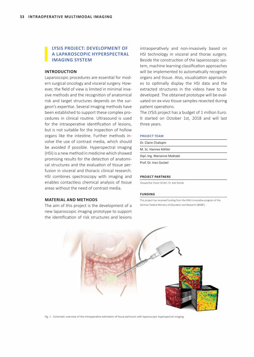
53
LYSIS PROJECT: DEVELOPMENT OF A LAPAROSCOPIC HYPERSPECTRAL IMAGING SYSTEM
INTRODUCTIONLaparoscopic procedures are essenti al for mod-ern surgical oncology and visceral surgery. How-ever, the fi eld of view is limited in minimal inva-sive methods and the recogniti on of anatomical risk and target structures depends on the sur-geon’s experti se. Several imaging methods have been established to support these complex pro-cedures in clinical routi ne. Ultrasound is used for the intraoperati ve identi fi cati on of lesions, but is not suitable for the inspecti on of hollow organs like the intesti ne. Further methods in-volve the use of contrast media, which should be avoided if possible. Hyperspectral imaging (HSI) is a new method in medicine which showed promising results for the detecti on of anatomi-cal structures and the evaluati on of ti ssue per-fusion in visceral and thoracic clinical research. HSI combines spectroscopy with imaging and enables contactless chemical analysis of ti ssue areas without the need of contrast media.
MATERIAL AND METHODSThe aim of this project is the development of a new laparoscopic imaging prototype to support the identi fi cati on of risk structures and lesions
intraoperati vely and non-invasively based on HSI technology in visceral and thorax surgery. Beside the constructi on of the laparoscopic sys-tem, machine learning classifi cati on approaches will be implemented to automati cally recognize organs and ti ssue. Also, visualizati on approach-es to opti mally display the HSI data and the extracted structures in the videos have to be developed. The obtained prototype will be eval-uated on ex-vivo ti ssue samples resected during pati ent operati ons. The LYSiS project has a budget of 1 million Euro. It started on October 1st, 2018 and will last three years.
PROJECT TEAM
Dr. Claire Chalopin
M. Sc. Hannes Köhler
Dipl.-Ing. Marianne Maktabi
Prof. Dr. Ines Gockel
PROJECT PARTNERS
Diaspecti ve Vision GmbH, Dr. Axel Kulcke
FUNDING
This project has received funding from the KMU-Innovati ve program of the
German Federal Ministry of Educati on and Research (BMBF).
Fig. 1 - Schemati c overview of the intraoperati ve esti mati on of ti ssue perfusion with laparoscopic hyperspectral imaging.
INTRAOPERATIVE MULTIMODAL IMAGING

54
INTRAOPERATIVE MULTIMODAL IMAGING
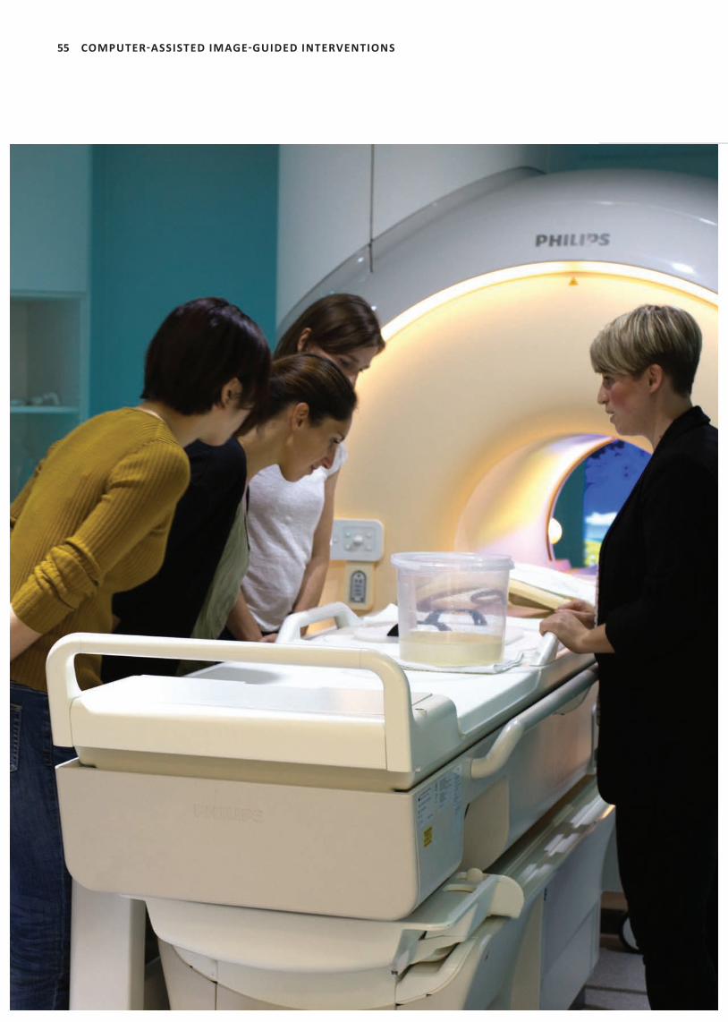
55 COMPUTER-ASSISTED IMAGE-GUIDED INTERVENTIONS
56
‘ICCAS researches on new technologies for image-
guided procedures and therapeuti c assistance
systems in the fi eld of MRI-guided and non-invasive
interventi ons – MR-guided endomyocardial
biopsy – novel applicati on of focused ultrasound
(FUS) under the guidance of magneti c resonance
therapy (MRgFUS) – roboti c-assisted MRgFUS and
interventi onal techniques under PET-MRI guidance
– combined FUS and radiati on therapy to support
the treatment of cancer (SONO-RAY, BMBF project).’
Prof. Dr. Andreas Melzer
(group leader)
COMPUTER-ASSISTED IMAGE-GUIDED INTERVENTIONS
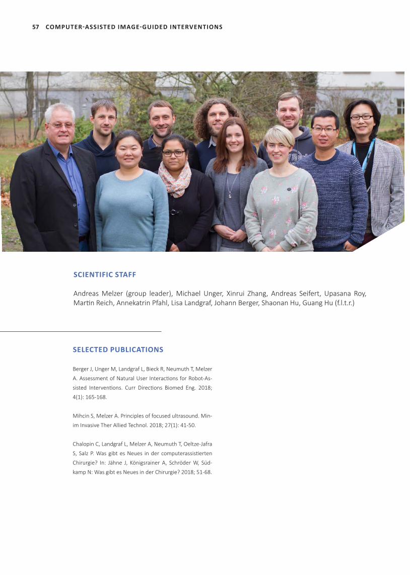
57 COMPUTER-ASSISTED IMAGE-GUIDED INTERVENTIONS
SELECTED PUBLICATIONS
Berger J, Unger M, Landgraf L, Bieck R, Neumuth T, Melzer
A. Assessment of Natural User Interactions for Robot-As-
sisted Interventions. Curr Directions Biomed Eng. 2018;
4(1): 165-168.
Mihcin S, Melzer A. Principles of focused ultrasound. Min-
im Invasive Ther Allied Technol. 2018; 27(1): 41-50.
Chalopin C, Landgraf L, Melzer A, Neumuth T, Oeltze-Jafra
S, Salz P. Was gibt es Neues in der computerassistierten
Chirurgie? In: Jähne J, Königsrainer A, Schröder W, Süd-
kamp N: Was gibt es Neues in der Chirurgie? 2018; 51-68.
SCIENTIFIC STAFF
Andreas Melzer (group leader), Michael Unger, Xinrui Zhang, Andreas Seifert, Upasana Roy, Martin Reich, Annekatrin Pfahl, Lisa Landgraf, Johann Berger, Shaonan Hu, Guang Hu (f.l.t.r.)
58COMPUTER-ASSISTED IMAGE-GUIDED INTERVENTIONS
1 META CENTER FOR INNOVATION COMPETENCE (ZIK) BETWEEN ICCAS (LEIPZIG) AND ONCORAY (DRESDEN): SONO-RAY – Combination Therapy of MR-Guided Focused Ultrasound-Hyperthermia (FUS-HT) and Radiation Therapy (RT) for the Treatment of Cancer | project part ICCAS
INTRODUCTIONImage-guided procedures gain more and more clinical acceptance, especially in the field of cancer diagnosis and treatment. The imaging via ultrasound (US), X-ray computed tomogra-phy (CT) or magnetic resonance tomography (MRI) allows for minimal- and non-invasive pro-cedures like biopsies, thermal ablation, emboli-zation as well as minimally invasive surgery. The imaging leads to smaller incisions and thus re-duces side effects like inflammation and short-ens the time of hospitalization. Focused ultrasound or high intensity focused ultrasound (FUS/HIFU) describes a unique tech-nique using ultrasound beams to heat a target tissue inside the body in a totally non-invasive way. In contrast to diagnostic ultrasound imag-ing, a special form or e.g. an array of multiple transducers submit a large number of ultra-sound waves into the focus point. The ablation of tissue using HIFU is already approved by the Food and Drug Administration (FDA) for the treatment of benign tumors of the uterine, of prostate carcinoma, in palliative treatment of bone metastasis and for therapy of essential tremor. During these procedures, treatment planning and control can be performed either by US-guidance or by MRI-guidance (MRgHIFU). In 2016, the MRgHIFU system Sonalleve (Profound medical), funded by ICCAS, was installed at the Leipzig University Hospital. Fourteen patients with uterine fibroids were treated in 2018 in Leipzig as teamwork of interventional radiology and gynecology. Furthermore, the first desmoid tumor was successfully treated with MRgHIFU. At the side of our project partner OncoRay in
Dresden, the transurethral ultrasound system TULSA-PRO (Profound medical) was successful-ly installed into the clinical PET/MRI. In 2018, the progress of the SONO-RAY project at ICCAS in Leipzig included the further investigation of radiosensitizing effects of FUS-induced hy-perthermia in vitro and the development of a next generation in vitro cell applicator together with Fraunhofer IBMT (St. Ingbert). In collabo-ration with oncology in Leipzig (Prof. Lordick, Prof. Hacker), first tests for immunostimulating events caused by FUS are underway.For the realization of in vivo treatment in the SONO-RAY project in Leipzig, an MR-compatible small animal FUS probe was investigated in dif-ferent preclinical MRI scanners in cooperation with Helmholtz Center (HZDR) and Fraunhofer IZI located in Leipzig. First steps for real-time MR-thermometry were implemented by a new postdoc. With regard to a clinical translation of observations in the SONO-RAY project new concepts for robotically assisted FUS treat-ment combined with radiation treatment were evolved.
PROJECT TEAM
Prof. Dr. Andreas Melzer
Dr. Lisa Landgraf
Dr. Upasana Roy
Dr. Guang Hu
Dr. Ina Patties
M. Sc. Johann Berger
M. Sc. Xinrui Zhang
M. Sc. Shaonan Hu
M. Sc. Michael Unger
PROJECT PARTNERS
Dresden University Hospital, Center for Radiation Research in Oncology (On-
coRay), Prof. Dr. Mechthild Krause
Dresden University Hospital, Center for Radiation Research in Oncology (On-
coRay), Dr. Damian McLeod
Leipzig University Hospital, Dep. of Nuclear Medicine, Prof. Dr. Osama Sabri, Prof.
Dr. Bernhard Sattler and Dr. Thies Jochimsen
Fraunhofer Institute for Cell Therapy and Immunology IZI, Leipzig, Dr. Thomas
Grunwald, Dr. Sebastian Greiser and Dr. Franziska Lange
59
Leipzig University Hospital, Dep. of Radiation Therapy, Prof. Dr. Rolf-Dieter Kort-
mann
Leipzig University Hospital, Dep. of Radiation Therapy, Dr. Annegret Glasow
Leipzig University Hospital, Dep. of Diagnostic and Interventional Radiology, Dr.
Patrick Stumpp
Leipzig University Hospital, Dep. of Diagnostic and Interventional Radiology, Dr.
Harald Busse
Leipzig University, Saxon Incubator for Clinical Translation (SIKT), Dr. Vuk Savkovic
Helmholtz Center Dresden Rossendorf, Neuroradiopharmaceuticals, Prof. Dr.
Peter Brust
Fraunhofer IBMT, Ultrasound Department, M. Sc. Steffen Tretbar
Fraunhofer IBMT, Ultrasound Department, Dr. Marc Fournelle
SELECTED PUBLICATIONS
Berger J , Unger M , Keller J, Bieck R, Landgraf L, Neumuth T, Melzer A. Kollab-
orative Interaktion für die roboterassistierte ultraschallgeführte Biopsie. 17.
Jahrestagung der Deutschen Gesellschaft für Computer- und Roboterassistierte
Chirurgie (CURAC). Leipzig, Germany; 2018.
Berger J , Unger M, Bieck R, Landgraf L, Neumuth T, Melzer A. Assessment of
Natural User Interactions for Robot-Assisted Interventions, Curr Directions
Biomed Eng. 2018; 4(1): 165-168.
Xiao X, Huang Z, Rube MA, Melzer A. Investigation of active tracking for robotic
arm assisted magnetic resonance guided focused ultrasound ablation. Int J Med
Robot. 2017;13(3).
FUNDING
German Federal Ministry of Education and Research (BMBF)
1.1 RADIOSENSITIZATION OF CANCER CELL LINES BY FOCUSED ULTRASOUND HYPERTHERMIA IN VITRO
INTRODUCTIONHyperthermia describes the heating of tumor cells to temperatures ranging between 41-46°C. Radiosensitizing events like the reduction of non-oxygenated hypoxic tumor areals or a triggered drug uptake caused by hyperthermal heating were detected in different preclinical studies. Currently, there is no device for hyper-thermia used in clinical routine based on the lack of real-time temperature control and heat-ing which is only limited to the target region. In this context, image-guided focused ultrasound is for the first time a relevant method to gener-ate hyperthermia precisely and non-invasively
in the tumor region. Based on that knowledge, goals in Sono-Ray are the detection of optimal temperature for a pleasant short heating pe-riod but leading to radiosensitizing effects on cancer cell lines in vitro while avoiding necro-sis. Furthermore, the impact of the time regime between the two treatment modalities FUS-in-duced hyperthermia and radiation (RT) needs to be investigated.
MATERIALS AND METHODSHuman head and neck (FaDu), glioblastoma (T98G) and prostate carcinoma (PC-3) cell lines were cultured in special ultrasound-pen-etrable 96-well cell culture plates (Greiner bio one GmbH, Germany) for high throughput FUS treatment. They were cultured in the desired medium in an incubator at 37°C and 5% CO2. To generate FUS hyperthermia in the well plates an existing FUS in vitro system (Imsat, Dundee) with a customized 1.14 MHz single transducer made by piezoelectric ceramic materials (Meg-gitt-Ferroperm Piezoceramics, Kvistgaard, Den-mark) was modified to sonicate 3 wells in paral-lel at the acoustic intensity of 214 W/cm2. The transducer was mounted into a water container with circulating degassed water and a tempera-ture of 34°C to avoid overheating. In prelimi-nary studies, the target temperature of 45°C was the most homogenous temperature which could be held for a desired duration. Labview (National Instruments, Newbury, UK) and a mo-tion system (VELMEX Inc., Bloomfield, NY, USA) were utilized to control the movement of the plate. An infrared thermal camera (PI450, Op-tris GmbH, Germany) and imaging software (PI connect version 2.10) monitored the tempera-ture inside the wells in real time during FUS-HT treatment. A feedback loop from the camera to the motor enabled translation of the plate when the temperature reached the desired tar-get. Hyperthermia in a thermal cycler (Master-cycler gradient, Eppendorf, Germany) worked as a control for the comparison of mechanical effects of FUS-HT and HT alone. To determine optimal temperatures of HT for radiosensitiza-tion and to analyze effects of different combi-
COMPUTER-ASSISTED IMAGE-GUIDED INTERVENTIONS
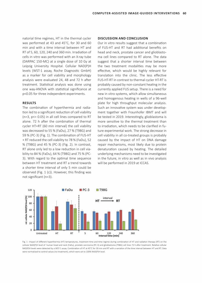
60
natorial time regimes, HT in the thermal cycler was performed at 43 and 45°C, for 30 and 60 min and with a time interval between HT and RT of 5, 60, 120, 240 and 360 min. Irradiation of cells in vitro was performed with an X-ray tube (DARPAC 150-MC) at a single dose of 10 Gy at Leipzig University Hospital. Cellular NAD(P)H levels (WST-1 assay, Roche Diagnostic GmbH) as a marker for cell viability and morphology analysis were evaluated 24, 48 and 72 h after treatment. Statistical analysis was done using one way-ANOVA with statistical significance at p<0.05 for three independent experiments.
RESULTSThe combination of hyperthermia and radia-tion led to a significant reduction of cell viability (n=3, p<= 0.05) in all cell lines compared to RT alone. 72 h after the combination of thermal cycler HT+RT (60 min interval) the cell viability was decreased to 55 % (FaDu), 27 % (T98G) and 59 % (PC-3) (Fig. 1). The combination of FUS-HT + RT reduced the cell viability to 78 % (FaDu), 52 % (T98G) and 45 % (PC-3) (Fig. 2). In contrast, RT alone only led to a low reduction in cell via-bility to 84 % (FaDu), 64 % (T98G) and 75 % (PC-3). With regard to the optimal time sequence between HT treatment and RT a trend towards a shorter time interval of only 5 min could be observed (Fig. 1 (c)). However, this finding was not significant (n=3).
DISCUSSION AND CONCLUSIONOur in vitro results suggest that a combination of FUS-HT and RT had additional benefits on head and neck, prostate cancer and glioblasto-ma cell lines compared to RT alone. The data suggest that a shorter interval time between the two treatment modalities may be more effective, which would be highly relevant for translation into the clinic. The less effective FUS-HT-RT in contrast to thermal cycler HT-RT is probably caused by non-constant heating in the currently applied FUS setup. There is a need for new in vitro systems, which allow simultaneous and homogenous heating in wells of a 96-well plate for high throughput molecular analysis. Such an innovative system was under develop-ment together with Fraunhofer IBMT and will be tested in 2019. Interestingly, glioblastoma is more sensitive to the thermal treatment than to irradiation, which needs to be clarified in fu-ture experimental work. The strong decrease in cell viability in all co-treated groups is probably caused by the impact of HT on DNA damage repair mechanisms, most likely due to protein denaturation caused by heating. The detailed underlying mechanisms need to be investigated in the future, in vitro as well as in vivo analysis will be performed in 2019 at ICCAS.
COMPUTER-ASSISTED IMAGE-GUIDED INTERVENTIONS
Fig. 1 - Impact of different hyperthermia (HT) temperatures, treatment time and time regime during combination of HT and radiation therapy (RT) on the cellular NAD(P)H level of human head and neck (FaDu), prostate carcinoma (PC-3) and glioblastoma (T98G) cell lines 72 h after treatment. Relative cellular NAD(P)H levels were detected by a WST-1 assay. Combination of HT at 45°C for 30 min and RT with a variation of the time interval between HT and RT. Data were normalized to control values (no treatment), which were set to 100% NAD(P)H level.
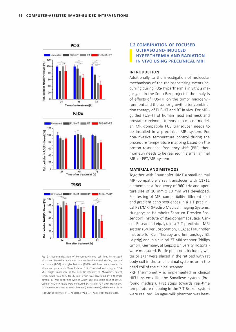
61
Fig. 2 - Radiosensitization of human carcinoma cell lines by focused ultrasound hyperthermia in vitro. Human head and neck (FaDu), prostate carcinoma (PC-3) and glioblastoma (T98G) cell lines were seeded in ultrasound penetrable 96-well plates. FUS-HT was induced using an 1.14 MHz single transducer at the acoustic intensity of 214W/cm2. Target temperature was 45°C for 30 min which was controlled by a thermal camera. RT was performed with an X-ray tube at a single dose of 10 Gy. Cellular NAD(P)H levels were measured 24, 48 and 72 h after treatment. Data were normalized to control values (no treatment), which were set to
100% NAD(P)H level; n= 3, *p< 0.05; **p<0.01; #p<0.001; ##p< 0.0001.
1.2 COMBINATION OF FOCUSED ULTRASOUND-INDUCED HYPERTHERMIA AND RADIATION IN VIVO USING PRECLINICAL MRI
INTRODUCTIONAdditionally to the investigation of molecular mechanisms of the radiosensitizing events oc-curring during FUS- hyperthermia in vitro a ma-jor goal in the Sono-Ray project is the analysis of effects of FUS-HT on the tumor microenvi-ronment and the tumor growth after combina-tion therapy of FUS-HT and RT in vivo. For MRI- guided FUS-HT of human head and neck and prostate carcinoma tumors in a mouse model, an MRI-compatible FUS transducer needs to be installed in a preclinical MRI system. For non-invasive temperature control during the procedure temperature mapping based on the proton resonance frequency shift (PRF) ther-mometry needs to be realized in a small animal MRI or PET/MRI system.
MATERIAL AND METHODSTogether with Fraunhofer IBMT a small animal MRI-compatible array transducer with 11×11 elements at a frequency of 960 kHz and aper-ture size of 10 mm x 10 mm was developed. For testing of MRI compatibility different spin and gradient echo sequences in a 1 T preclini-cal PET/MRI (Mediso Medical Imaging Systems, Hungary; at Helmholtz-Zentrum Dresden-Ros-sendorf, Institute of Radiopharmaceutical Can-cer Research, Leipzig), in a 7 T preclinical MRI system (Bruker Corporation, USA; at Fraunhofer Institute for Cell Therapy and Immunology IZI, Leipzig) and in a clinical 3T MRI scanner (Philips GmbH, Germany; at Leipzig University Hospital) were measured. Bottle phantoms including wa-ter or agar were placed in the rat bed with rat body coil in the small animal systems or in the head coil of the clinical scanner.PRF thermometry is implemented in clinical HIFU systems like the Sonalleve system (Pro-found medical). First steps towards real-time temperature mapping in the 7 T Bruker system were realized. An agar-milk phantom was heat-
COMPUTER-ASSISTED IMAGE-GUIDED INTERVENTIONS
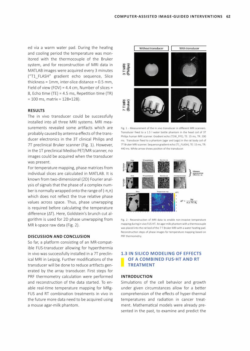
62
ed via a warm water pad. During the heati ng and cooling period the temperature was mon-itored with the thermocouple of the Bruker system, and for reconstructi on of MRI data in MATLAB images were acquired every 3 minutes (“T1_FLASH” gradient echo sequence, Slice thickness = 1mm, inter-slice distance = 0.5 mm, Field of view (FOV) = 4.4 cm, Number of slices = 8, Echo ti me (TE) = 4.5 ms, Repeti ti on ti me (TR) = 100 ms, matrix = 128×128).
RESULTSThe in vivo transducer could be successfully installed into all three MRI systems. MRI mea-surements revealed some arti facts which are probably caused by antenna eff ects of the trans-ducer electronics in the 3T clinical Philips and 7T preclinical Bruker scanner (Fig. 1). However, in the 1T preclinical Mediso PET/MR scanner, no images could be acquired when the transducer was present. For temperature mapping, phase matrices from individual slices are calculated in MATLAB. It is known from two-dimensional (2D) Fourier anal-ysis of signals that the phase of a complex num-ber is normally wrapped onto the range of (-π,π) which does not refl ect the true relati ve phase values across space. Thus, phase unwrapping is required before calculati ng the temperature diff erence (ΔT). Here, Goldstein’s brunch cut al-gorithm is used for 2D phase unwrapping from MR k-space raw data (Fig. 2).
DISCUSSION AND CONCLUSIONSo far, a platf orm consisti ng of an MR-compat-ible FUS-transducer allowing for hyperthermia in vivo was successfully installed in a 7T preclin-ical MRI in Leipzig. Further modifi cati ons of the transducer will be done to reduce arti facts gen-erated by the array transducer. First steps for PRF thermometry calculati on were performed and reconstructi on of the data started. To en-able real-ti me temperature mapping for MRg-FUS and RT combinati on treatments in vivo in the future more data need to be acquired using a mouse agar-milk phantom.
Fig. 1 - Measurement of the in vivo transducer in diff erent MRI scanners. Transducer fi xed to a 1.5 l water bott le phantom in the head coil of 3T Philips human MRI scanner. Gradient echo (T2W_FFE), TE: 15 ms, TR: 200 ms. Transducer fi xed to a phantom (agar and Lego) in the rat body coil of 7T Bruker MRI scanner. Sequence gradient echo (T1_FLASH), TE: 15 ms, TR: 440 ms. White arrow shows positi on of the transducer.
Fig. 2 - Reconstructi on of MRI data to enable non-invasive temperature mapping during in vivo FUS-HT. An agar-milk phantom with a thermocouple was placed into the rat bed of the 7 T Bruker MRI with a water heati ng pad. Reconstructi on steps of phase images for temperature mapping based on PRF thermometry.
1.3 IN SILICO MODELING OF EFFECTS OF A COMBINED FUS-HT AND RT TREATMENT
INTRODUCTIONSimulati ons of the cell behavior and growth under given circumstances allow for a bett er comprehension of the eff ects of hyper-thermal temperatures and radiati on in cancer treat-ment. Mathemati cal models were already pre-sented in the past, to examine and predict the
COMPUTER-ASSISTED IMAGE-GUIDED INTERVENTIONS
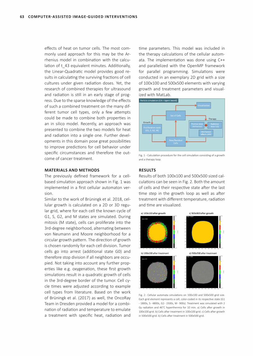
63
effects of heat on tumor cells. The most com-monly used approach for this may be the Ar-rhenius model in combination with the calcu-lation of t_43 equivalent minutes. Additionally, the Linear-Quadratic model provides good re-sults in calculating the surviving fractions of cell cultures under given radiation doses. Yet, the research of combined therapies for ultrasound and radiation is still in an early stage of prog-ress. Due to the sparse knowledge of the effects of such a combined treatment on the many dif-ferent tumor cell types, only a few attempts could be made to combine both properties in an in silico model. Recently, an approach was presented to combine the two models for heat and radiation into a single one. Further devel-opments in this domain pose great possibilities to improve predictions for cell behavior under specific circumstances and therefore the out-come of cancer treatment.
MATERIALS AND METHODSThe previously defined framework for a cell-based simulation approach shown in Fig. 1 was implemented in a first cellular automaton ver-sion.Similar to the work of Brüningk et al. 2018, cel-lular growth is calculated on a 2D or 3D regu-lar grid, where for each cell the known cycle of G1, S, G2, and M states are simulated. During mitosis (M state), cells can proliferate into the 3rd-degree neighborhood, alternating between von Neumann and Moore neighborhood for a circular growth pattern. The direction of growth is chosen randomly for each cell division. Tumor cells go into arrest (additional state G0) and therefore stop division if all neighbors are occu-pied. Not taking into account any further prop-erties like e.g. oxygenation, these first growth simulations result in a quadratic growth of cells in the 3rd-degree border of the tumor. Cell cy-cle times were adjusted according to example cell types from literature. Based on the work of Brüningk et al. (2017) as well, the OncoRay Team in Dresden provided a model for a combi-nation of radiation and temperature to emulate a treatment with specific heat, radiation and
time parameters. This model was included in the therapy calculations of the cellular autom-ata. The implementation was done using C++ and parallelized with the OpenMP framework for parallel programming. Simulations were conducted in an exemplary 2D grid with a size of 100x100 and 500x500 elements with varying growth and treatment parameters and visual-ized with MatLab.
Fig. 1 - Calculation procedure for the cell simulation consisting of a growth and a therapy loop.
RESULTSResults of both 100x100 and 500x500 sized cal-culations can be seen in Fig. 2. Both the amount of cells and their respective state after the last time step in the growth loop as well as after treatment with different temperature, radiation and time are visualized.
Fig. 2 - Cellular automata simulations on 100x100 and 500x500 grid size. Each grid element represents a cell, color-coded in its respective state (G1 - 1800s, S - 4800s, G2 - 1500s, M - 900s). Treatment was simulated with 2 Gy radiation and 46°C hyperthermia for 10 min. a) Cells after growth in 100x100 grid. b) Cells after treatment in 100x100 grid. c) Cells after growth in 500x500 grid. b) Cells after treatment in 500x500 grid.
COMPUTER-ASSISTED IMAGE-GUIDED INTERVENTIONS
64
DISCUSSION AND CONCLUSIONThe presented cellular automaton is able to simulate a simplified cell growth (quadratic pro-liferation) and treatment outcome for different cell types and treatment modalities. It provides a solid base for further development to adjust the parameters for more realistic calculations. Factors like the amount of available oxygen and proliferation cycles before and after treatment, measured in experiments have to be taken into account.
1.4 CONCEPTS FOR ROBOTIC GUIDED FOCUSED ULTRASOUND HYPERTHERMIA AND RADIATION THERAPY IN THE CLINIC
INTRODUCTIONRobotic systems become more and more im-portant in medicine because they provide pre-cise and reproducible positioning of surgical tools and equipment. In this context, robotic systems are used in clinics to assist, for exam-ple, during biopsies and ablation treatment. Regarding the non-invasive therapy applying FUS/HIFU in combination with radiation thera-py for treatment of tumors at various locations or moving organs a precise positioning of the transducer is crucial for the efficacy and safety. Available HIFU systems like Profound Sonalleve only allow the treatment in very specific regions and are an uncomfortable procedure to realize a combination of FUS-HT and RT. Hence, a ro-botic arm positioning the FUS system according to the treatment planning is more versatile. For further studies on combined FUS-RT treatment, the robotic arm system needs to be integrated into existing clinical infrastructures like a PET-MRI system or radiation room.
MATERIALS AND METHODSTo apply a combination of FUS-hyperthermia and RT (i) a robotic workflow consisting of an ultrasound-guided FUS system needs to be in-tegrated into the radiation room or using MRI guidance into (ii) MR-guided radiation devices
(e.g. MR-linac, Elekta).To improve the usability and reduce complexi-ty, collaborative robotics will be used to install a dual-arm robot setup (i). First evaluations were conducted with a mobile platform, utilizing a KUKA LBR iiwa 7 robotic arm and a Clarius L7 wireless ultrasound probe (Fig. 1) for the use case of ultrasound-guided biopsies. Touch ges-ture interaction concepts as well as a phantom study with 9 participants of varying technical and clinical expertise were performed. Ven-dor-independent communication with other devices was achieved using the surgical device communication protocol (SDC).The realization of a FUS-RT combination in a PET/MRI-guided setup (ii) was achieved in Leipzig by integrating a PET/MR-compatible robot (InnoMotion by InnoMedic GmbH) into a Biograph mMR PET/MRI system (Siemens Healthineers) (Fig. 2). By virtualizing the hard-ware containing the planning software of the robotic arm with an Oracle VirtualBox, the sys-tem was successfully integrated into the clini-cal infrastructure. Placing the virtual machine on the post-processing and data storing Linux server attached to the PET/MRI system, image data can be transferred easily to the planning software which acts as a DICOM receiver.
RESULTSRegarding the first concept (Fig. 1 (a)) for US-guided FUS in a clinical radiation room based on use case-guided biopsies, the mean time for the localization of the target was 24 seconds and the mean time to hit the target was 42 seconds in case of the unassisted intervention. During the assisted procedure the mean time to plan the intervention was 23 seconds and the mean time to adjust the needle guide and hit the target was 51 seconds. Unassisted, the par-ticipants had to insert the needle two times on average to successfully hit the target, whereas the participants hit the target on the first nee-dle insertion during the assisted intervention. The participants rated the needle guidance as an improvement of the workflow and rated the interaction concept as intuitive.
COMPUTER-ASSISTED IMAGE-GUIDED INTERVENTIONS
65
Fig. 1 - Concept for collaborative robot-guided focused ultrasound hyperthermia and radiation (a) and usability of a KUKA LBR iiwa 7 robotic arm with wireless ultrasound imaging probe (b). Touch gesture recognition tests of four touch gestures (Double Taps and Pushes) resulted in a total accuracy of 89.8% (c).
First steps for MR-guided FUS-HT and RT were made by successful modification of the MR-compatible Innomotion arm to fit in the Biograph mMR MR-PET system (Fig. 2 (a)) and the robotic arm system was integrated into the clinical IT infrastructure.
Fig. 2 - Integration of the robotic arm into the MR-PET system (a). Attenuation map of the robotic arm visualized by two perpendicular maximum intensity projections of the linear attenuation coefficient of 511-keV PET annihilation photons (in 1/cm) obtained by a stand-alone PET scanner equipped with Ge-/Ga-68 rod sources (b). PET imaging of 5 MBq F18-FDG injected into a pomelo. PET data was reconstructed into a 256×256 matrix (voxel size: 1.40×1.40×2.03mm^3) using the built-in 3D ordered subset expectation maximization (OSEM) algorithm with 8 iterations, 21 subsets, and a 3-mm Gaussian filter (c).
To correct for attenuation of gamma radiation by the robotic hardware in the PET field of view and test its performance, the integrated robot attenuation was measured with a stand-alone PET system equipped with Ge68-transmission sources emitting 511 keV gamma radiation (Fig. 2 (b)). MR-guided FUS-HT and RT therapy may be combined with PET imaging to evaluate the efficacy of these treatments during the proce-dure and further improve therapy strategies. An example of PET imaging is given in Fig. 2 (c).
DISCUSSION AND CONCLUSIONWe successfully implemented two previously developed interaction functions into a collabo-rative robotic assistance setup for intervention-al needle placement procedures. The interac-tion quality was evaluated in comparison with an unassisted needle placement. With qualitative feedback on the interaction, it can preliminarily be assumed that collabo-rative robotic assistance with close-proximity intuitive control mechanisms benefits system acceptance and perceived guidance support. Using MR-PET imaging technique improves the evaluation of the efficacy of a combined FUS-RT treatment. Due to the early stage of these com-bined therapies, MR-capable robotic systems are hardly available. Therefore, these systems lack a standard for clinical integration. Studies with imaging and therapeutic ultrasound trans-ducers will be conducted in 2019.
2 MR-SAFE FLEXIBLE BIOPSY FORCEPS FOR MINIMALLY INVASIVE TISSUE SAMPLING
INTRODUCTIONThe procedure of minimally invasive endomyo-cardial biopsy (EMB) under X-ray fluoroscopy guidance implies radiation, nephrotoxic con-trast agent, and poor tissue contrast. Magnet-ic Resonance Imaging (MRI) guidance offers a promising option to overcome these disad-vantages. So far, however, biopsy forceps are mostly metallic and as per ASTM/ISO regulation
COMPUTER-ASSISTED IMAGE-GUIDED INTERVENTIONS
66
MR Unsafe because of radiofrequency heat-ing. To improve the intervention, funding by the ‘KMU-innovativ: Medizintechnik’ program could be raised. Therefore, the objective of the joint project MR-Biopsy is the development and evaluation of a flexible and MR Safe/Conditional biopsy forceps along with a corresponding clini-cal workflow to realize MRI-guided EMB.
MATERIAL AND METHODSA design of the biopsy jaws has been realized that allows the processing of non-magnetic, non-metallic, and non-conductive materials. To enable MRI visibility and subsequently as-sess the opening state and spatial orientation of the jaw components under MR guidance, the jaws have been equipped with passive markers. Standard and real-time cardiac MRI sequences were obtained in an 1.5T MRI system (Siemens MAGNETOM Aera) and evaluated. For intensive ex-vivo suitability tests, a detailed and life-size phantom of the vessel system from groin to neck has been modeled and manufactured (Fig. 1). Therefore, anonymous medical imaging data of a patient was segmented (Materialise Mim-ics Innovation Suite) and served as blueprint for additive 3D printing.
RESULTSMRI visibility investigations show promising re-sults in terms of appropriate selection of size, position, and concentration of the markers to identify the opening state: a clear difference
regarding the artifact sizes can be observed vi-sually when comparing the prototypes 2 and 5 in Fig. 2.
Fig. 2 - A: test setup with opened jaw prototypes by EPflex GmbH of different materials, with various marker positions and concentrations (1-5), one jaw set without markers (6), and an equal sized reference volume (7). B: coronal MR image of the test setup with opened jaws in a saline solution. C: coronal MR image of the test setup with closed jaws in a saline solution. (TR= 40.95ms/ TE=1.1.6ms/ FA=74°/ slice thickness=6mm/ matrix=192\0\0\96/ pixel scaling=1.77\1.77mm²/ pixel bandwith=930Hz/px). Below the image, the dimensions of the MRI artefacts of the opened/closed jaws 2, 5, 6 are listed.
DISCUSSION AND CONCLUSIONMR Safe jaw prototypes have been fabricated, equipped with passive markers, and evaluated under MRI, showing the feasibility to discrimi-nate the opening state of the forceps. Further MRI and ex-vivo studies are currently conduct-ed to ensure the applicability in terms of reliable MRI guidance and reproducible biopsy quality. Later, in-vivo investigations will be carried out
COMPUTER-ASSISTED IMAGE-GUIDED INTERVENTIONS
Fig. 1 - Left: 3D model of the phantom. Right: life size silicone phantom (55x25x20cm) by Phacon GmbH. A replaceable fresh porcine heart can be integrated into the phantom, as depicted in the computer model.
Position 2 5 6open closed open closed open closed
Height [mm] 5.5 6 7 10 8.1 7.8Width [mm] 4.8 10.1 4.4 9.2 5.9 4.8
67
accordingly. Another task is to implement a clin-ical workflow for MRI-guided EMB, comprising optimized MRI sequences, MRI operation, in-tra-interventional communication, and patient safety.
PROJECT TEAM
Prof. Dr. Andreas Melzer
M. Sc. C. Martin Reich
Dipl.-Ing. (FH) Andreas Seifert
M. Sc. Annekatrin Pfahl
M. Sc. Johann Berger
M. Sc. Michael Unger
PROJECT PARTNERS
EPflex Feinwerktechnik GmbH, Dettingen an der Erms
Leipzig University Hospital, Clinic and Polyclinic for Cardiology
Leipzig University Hospital, Clinic and Polyclinic for Diagnostic and Interventional
Radiology
University Medical Center Goettingen, Clinic for Cardiology and Pneumology
University Medical Center Goettingen, Institute for Diagnostic and
Interventional Radiology
PHACON GmbH, Leipzig
SELECTED PUBLICATIONS
Reich CM, Pfahl A, Lehnhardt M, Uihlein B, Petershans S. Unterberg-Buchwald
C, Uecker M, Melzer A. Interventionelle MR-geführte Herzbiopsie: Entwicklung
einer innovativen MR-tauglichen Biopsiezange. 17. Jahrestagung der Deutschen
Gesellschaft für Computer- und Roboterassistierte Chirurgie (CURAC). Leipzig,
Germany; 2018.
Reich CM, Pfahl A, Lehnhardt M, Uihlein B, Petershans S, Unterberg-Buchwald
C, Uecker M, Melzer A. Status-quo and demands for MR-guided endomyocardial
biopsies. 12th Interventional MRI Symposium (iMRI ). Boston, USA; 2018.
FUNDING
This project has received funding from the program “KMU-innovativ: Medizin-
technik” of the German Federal Ministry of Education and Research (BMBF).
3 MRI-GUIDED FOCUSED ULTRASOUND IN THE TREATMENT OF UTERINE FIBROIDS AND BEYOND
INTRODUCTIONIn 2017 the Philips/Profound medical Sonalleve MR-HIFU System was put into operation at the Department of Radiology (Leipzig University Hospital) to establish an MR-HIFU service pri-marily to provide a clinical therapy option for
cancer treatment and contribute to the growing body of evidence in the clinical as well as the research community. The Sonalleve MR-HIFU System is approved to treat uterine fibroids and bone metastases. We realized our aim by building up a multispecialty treatment center for uterine fibroids with MR-HIFU option. Our initial procedural and clinical experience with our first applications showed good results. Es-pecially for young women in childbearing age, who are still wishing for a family, HIFU is import-ant, e.g. to avoid uterine scars. Beyond that, we extended the field of HIFU ap-plication on desmoid tumors. Desmoid tumors are locally infiltrative and may cause pain and dysfunction. Standard therapies, including sur-gical resection, radiation, and systemic therapy, suffer from excessive side effects when consid-ering their limited efficacy for treating desmoid tumors. We were able to demonstrate that MR-HIFU treatment of desmoids is feasible and that this technique may be used to control the growth of symptomatic desmoid tumors, even of the abdomen and in analgesia without seda-tion.
MATERIALS AND METHODSUterine fibroids:A total of 60 patients with symptomatic uterine fibroids have been screened in our multispecial-ty Fibroid Center since 2017. After proper con-sultation and consensus for a therapy option, fourteen of them underwent MR-HIFU (Sonal-leve, Profound Medical, Mississauga, ON) in a diagnostic 3 T MRI (Ingenia 3.0T, Philips Health-care, Best, Netherlands). One patient with des-moid tumor was treated via MR-HIFU. Patients received an analgesic and sedative medication and their vital parameters were monitored continuously. T2-weighted and special bowel sequences were used for planning. Therapeu-tic and adverse tissue heating during sonication was controlled by multiplanar MR temperature mapping (6 slices in 3.5 s) in the treatment cell as well as the so-called near (skin) and far field (sacrum).
COMPUTER-ASSISTED IMAGE-GUIDED INTERVENTIONS
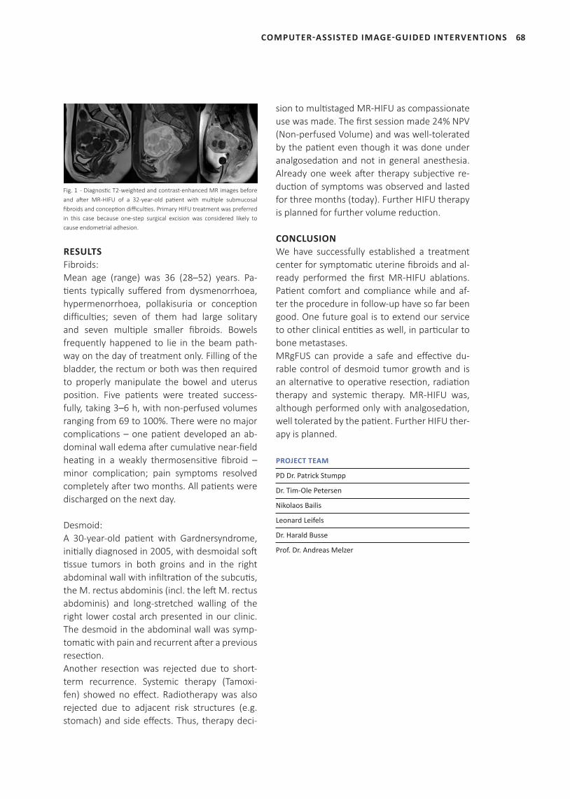
68
Fig. 1 - Diagnostic T2-weighted and contrast-enhanced MR images before and after MR-HIFU of a 32-year-old patient with multiple submucosal fibroids and conception difficulties. Primary HIFU treatment was preferred in this case because one-step surgical excision was considered likely to cause endometrial adhesion.
RESULTSFibroids:Mean age (range) was 36 (28–52) years. Pa-tients typically suffered from dysmenorrhoea, hypermenorrhoea, pollakisuria or conception difficulties; seven of them had large solitary and seven multiple smaller fibroids. Bowels frequently happened to lie in the beam path-way on the day of treatment only. Filling of the bladder, the rectum or both was then required to properly manipulate the bowel and uterus position. Five patients were treated success-fully, taking 3–6 h, with non-perfused volumes ranging from 69 to 100%. There were no major complications – one patient developed an ab-dominal wall edema after cumulative near-field heating in a weakly thermosensitive fibroid – minor complication; pain symptoms resolved completely after two months. All patients were discharged on the next day.
Desmoid:A 30-year-old patient with Gardnersyndrome, initially diagnosed in 2005, with desmoidal soft tissue tumors in both groins and in the right abdominal wall with infiltration of the subcutis, the M. rectus abdominis (incl. the left M. rectus abdominis) and long-stretched walling of the right lower costal arch presented in our clinic. The desmoid in the abdominal wall was symp-tomatic with pain and recurrent after a previous resection.Another resection was rejected due to short-term recurrence. Systemic therapy (Tamoxi-fen) showed no effect. Radiotherapy was also rejected due to adjacent risk structures (e.g. stomach) and side effects. Thus, therapy deci-
sion to multistaged MR-HIFU as compassionate use was made. The first session made 24% NPV (Non-perfused Volume) and was well-tolerated by the patient even though it was done under analgosedation and not in general anesthesia. Already one week after therapy subjective re-duction of symptoms was observed and lasted for three months (today). Further HIFU therapy is planned for further volume reduction.
CONCLUSIONWe have successfully established a treatment center for symptomatic uterine fibroids and al-ready performed the first MR-HIFU ablations. Patient comfort and compliance while and af-ter the procedure in follow-up have so far been good. One future goal is to extend our service to other clinical entities as well, in particular to bone metastases.MRgFUS can provide a safe and effective du-rable control of desmoid tumor growth and is an alternative to operative resection, radiation therapy and systemic therapy. MR-HIFU was, although performed only with analgosedation, well tolerated by the patient. Further HIFU ther-apy is planned.
PROJECT TEAM
PD Dr. Patrick Stumpp
Dr. Tim-Ole Petersen
Nikolaos Bailis
Leonard Leifels
Dr. Harald Busse
Prof. Dr. Andreas Melzer
COMPUTER-ASSISTED IMAGE-GUIDED INTERVENTIONS
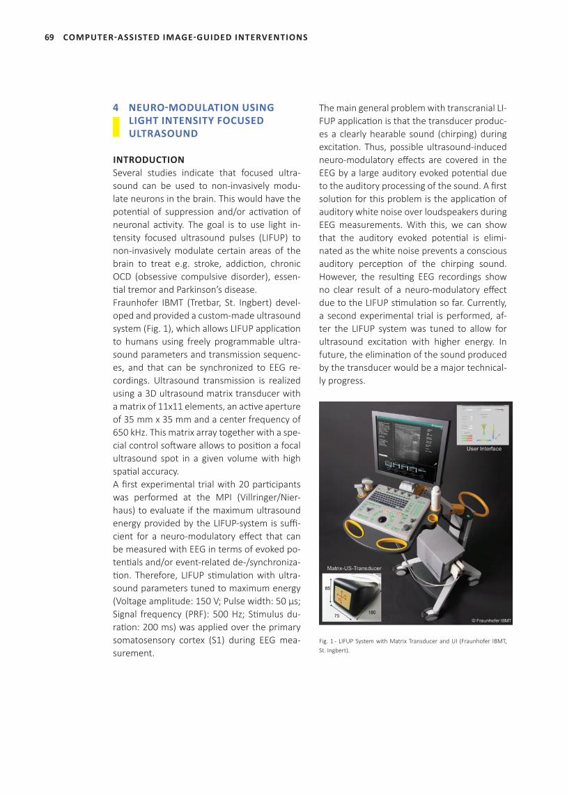
69
4 NEURO-MODULATION USING LIGHT INTENSITY FOCUSED ULTRASOUND
INTRODUCTIONSeveral studies indicate that focused ultra-sound can be used to non-invasively modu-late neurons in the brain. This would have the potenti al of suppression and/or acti vati on of neuronal acti vity. The goal is to use light in-tensity focused ultrasound pulses (LIFUP) to non-invasively modulate certain areas of the brain to treat e.g. stroke, addicti on, chronic OCD (obsessive compulsive disorder), essen-ti al tremor and Parkinson’s disease.Fraunhofer IBMT (Tretbar, St. Ingbert) devel-oped and provided a custom-made ultrasound system (Fig. 1), which allows LIFUP applicati on to humans using freely programmable ultra-sound parameters and transmission sequenc-es, and that can be synchronized to EEG re-cordings. Ultrasound transmission is realized using a 3D ultrasound matrix transducer with a matrix of 11x11 elements, an acti ve aperture of 35 mm x 35 mm and a center frequency of 650 kHz. This matrix array together with a spe-cial control soft ware allows to positi on a focal ultrasound spot in a given volume with high spati al accuracy. A fi rst experimental trial with 20 parti cipants was performed at the MPI (Villringer/Nier-haus) to evaluate if the maximum ultrasound energy provided by the LIFUP-system is suffi -cient for a neuro-modulatory eff ect that can be measured with EEG in terms of evoked po-tenti als and/or event-related de-/synchroniza-ti on. Therefore, LIFUP sti mulati on with ultra-sound parameters tuned to maximum energy (Voltage amplitude: 150 V; Pulse width: 50 µs; Signal frequency (PRF): 500 Hz; Sti mulus du-rati on: 200 ms) was applied over the primary somatosensory cortex (S1) during EEG mea-surement.
The main general problem with transcranial LI-FUP applicati on is that the transducer produc-es a clearly hearable sound (chirping) during excitati on. Thus, possible ultrasound-induced neuro-modulatory eff ects are covered in the EEG by a large auditory evoked potenti al due to the auditory processing of the sound. A fi rst soluti on for this problem is the applicati on of auditory white noise over loudspeakers during EEG measurements. With this, we can show that the auditory evoked potenti al is elimi-nated as the white noise prevents a conscious auditory percepti on of the chirping sound. However, the resulti ng EEG recordings show no clear result of a neuro-modulatory eff ect due to the LIFUP sti mulati on so far. Currently, a second experimental trial is performed, af-ter the LIFUP system was tuned to allow for ultrasound excitati on with higher energy. In future, the eliminati on of the sound produced by the transducer would be a major technical-ly progress.
Fig. 1 - LIFUP System with Matrix Transducer and UI (Fraunhofer IBMT, St. Ingbert).
COMPUTER-ASSISTED IMAGE-GUIDED INTERVENTIONS

70
PROJECT TEAM
Prof. Dr. Arno Villringer
Dr. Till Nierhaus
Dipl.-Ing. Steffen Tretbar
Prof. Dr. Andreas Melzer
PROJECT PARTNERS
Max Planck Institute for Humane Cognitive and Brain Sciences, Leipzig
Fraunhofer Institute for Biomedical Engineering - IBMT, St. Ingbert
COMPUTER-ASSISTED IMAGE-GUIDED INTERVENTIONS
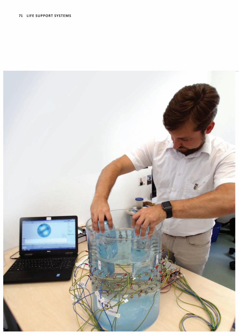
71 LIFE SUPPORT SYSTEMS

72
‘The non-invasive and radiati on-free Electrical
Impedance Tomography has the potenti al to off er
direct insight into the pati ent’s lung conditi on and
lung functi on for physicians in emergency medicine,
intensive care and neonatal care. We develop
innovati ve analysis and monitoring algorithms
to advance individualized therapy selecti on and
steering for neonatal and adult pati ents with
respiratory disorders in both the prehospital and
the clinical setti ng.’
Prof. Dr. Andreas Reske
(group leader)
LIFE SUPPORT SYSTEMS
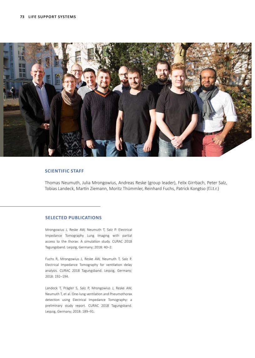
73 LIFE SUPPORT SYSTEMS
SELECTED PUBLICATIONS
Mrongowius J, Reske AW, Neumuth T, Salz P. Electrical
Impedance Tomography Lung Imaging with partial
access to the thorax: A simulation study. CURAC 2018
Tagungsband. Leipzig, Germany; 2018: 40–2.
Fuchs R, Mrongowius J, Reske AW, Neumuth T, Salz P.
Electrical Impedance Tomography for ventilation delay
analysis. CURAC 2018 Tagungsband. Leipzig, Germany;
2018: 192–194.
Landeck T, Prägler S, Salz P, Mrongowius J, Reske AW,
Neumuth T, et al. One-lung ventilation and Pneumothorax
detection using Electrical Impedance Tomography: a
preliminary study report. CURAC 2018 Tagungsband.
Leipzig, Germany; 2018: 189–91.
SCIENTIFIC STAFF
Thomas Neumuth, Julia Mrongowius, Andreas Reske (group leader), Felix Girrbach, Peter Salz, Tobias Landeck, Martin Ziemann, Moritz Thümmler, Reinhard Fuchs, Patrick Kongtso (f.l.t.r.)
74
ELECTRICAL IMPEDANCE TOMOGRAPHY ELECTRODE HANDLING WITH PARTIAL ACCESS TO THE THORAX
INTRODUCTIONElectrical impedance tomography (EIT) is a functi onal imaging method capable of monitor-ing lung venti lati on and can potenti ally be used as bedside applicati on at intensive care units. In the IMPACT (Improved Multi PArametric moni-toring of blunt Chest Trauma) project at ICCAS this imaging method is transferred to the use in prehospital emergency scenarios and a mobile EIT system is developed in collaborati on with Fritz Stephan GmbH, ITP GmbH and the Leipzig University of Applied Sciences.In emergency situati ons, such as car crashes, the emergency physician has to handle situa-ti ons with trapped pati ents and only parti al ac-cess to the thorax. In this case, only a part of the belt and few electrodes can be placed on the thorax, but the system is sti ll required to detect problems with or missing lung venti lati on.
MATERIALS AND METHODSIn order to evaluate situati ons with a reduced number of electrodes, a virtual simulati on study was conducted. A cylindrical phantom containing a single object with lower conducti v-ity is created in a simulati on environment. Sim-ulati ng no access to the dorsal part of a pati ent, the electrodes on only one side of the object are gradually reduced and the same object is reconstructed in each step.A prototypic phantom was built by the Leipzig University of Applied Sciences in order to test the developed EIT hardware functi onality. For that reason, a cylindrical tank made from acrylic glass is equipped with 96 screws, acti ng as elec-trodes (see Fig. 1). The tank can be connected to the EIT hardware and test measurements can be recorded when the phantom is fi lled with conducti ve liquid, i.e. saline water.
RESULTSThe fi rst simulati on results showed a severe reducti on in image quality when reducing the number of electrodes on one side of the object. Even for a small number of missing electrodes the original simulated object was heavily dis-torted and could not be properly recognized (see Fig. 2).
Fig. 1 - Image of the EIT phantom, fi lled with saline water.
Fig. 2 - EIT image results of the simulati on study of reduced electrodes on one side of the object. Results for 16, 14, 12 and 10 electrodes are depicted.
With the phantom, EIT measurements in dif-ferent test situati ons can be created. Applying objects of diff erent conducti vity, impedance
LIFE SUPPORT SYSTEMS
75 LIFE SUPPORT SYSTEMS
changes can be recorded, reconstructed and visualized. The connection of electrodes is vari-able and different electrode configurations of the 96 electrodes are possible.
DISCUSSION AND CONCLUSIONSevere degradation of image quality was ob-served when simulating a reduced set of elec-trodes. Until now, purely virtual simulations were conducted. The developed EIT phantom is the optimal environment to generate actual measurements with varying electrode configu-ration. Consequently, next steps in the project are the evaluation of the simulation results with the developed EIT phantom, in order to devel-op algorithms handling a decreased and rear-ranged situation of electrodes.
PROJECT TEAM
Prof. Dr. Thomas Neumuth
Prof. Dr. Andreas Reske
Dr. Peter Salz
Dr. Dominic Schneider
Dr. Felix Girrbach
M. Sc. Reinhard Fuchs
M. Sc. Patrick Kongtso
M. Sc. Julia Mrongowius
M. Sc. Moritz Thümmler
PROJECT PARTNERS
Leipzig University of Applied Sciences (HTWK), Faculty of Electrical Engineering
and Information Technology, Prof. Dr. Andreas Pretschner
ITP GmbH, Chemnitz, Klaus Richter
Fritz Stephan GmbH, Gackenbach, Wolfgang Braun
SELECTED PUBLICATIONS
Mrongowius J, Reske AW, Neumuth T, Salz P. Electrical Impedance Tomography
Lung Imaging with partial access to the thorax: A simulation study. 17. Jahre-
stagung der der Deutschen Gesellschaft für Computer- und Roboterassistierte
Chirurgie (CURAC). Leipzig, Germany; 2018.
FUNDING
This project has received funding from the KMU innovative program of the
German Federal Ministry of Education and Research (BMBF).
PRELIMINARY STUDY RESULTS FROM THE IMPACT PROJECT WITH CLINICAL RELEVANCE
INTRODUCTIONState-of-the-art approaches for recognition and regulation of impaired breathing and irregular ventilation encompass auscultation, sonog-raphy and spirometry. Yet, these methods are either insufficient for a conclusive detection and quantification of abnormal lung behavior or require time-intensive effort. Electrical Im-pedance Tomography (EIT) presents an alterna-tive, visualizing pulmonary ventilation through a combination of small-voltage stimulations and measurements, performed by a multitude of electrodes around the patient’s thorax. In IMPACT (Improved MultiPArametric monitor-ing of blunt Chest Trauma) the research group used EIT to establish a database for analyzing not well known artefacts during one-lung venti-lation (OLV) or pneumothorax (PTX) and devel-oping algorithms for their automatic detection.
Fig. 1: EIT-trend lines of left (grey) and right (black) image side.
Fig. 2: Left: Ventilation image reconstructed through EIT, Right: Delay Map acquired by compairing the pixels behavior to a global signal (blue – ventilation advance, red – ventilation delay).
MATERIAL AND METHODSThe data was collected through a clinical trial with thoracic surgery patients, where OLV is necessary for the procedure and thoracotomy is leading to an intended and controlled PTX. An
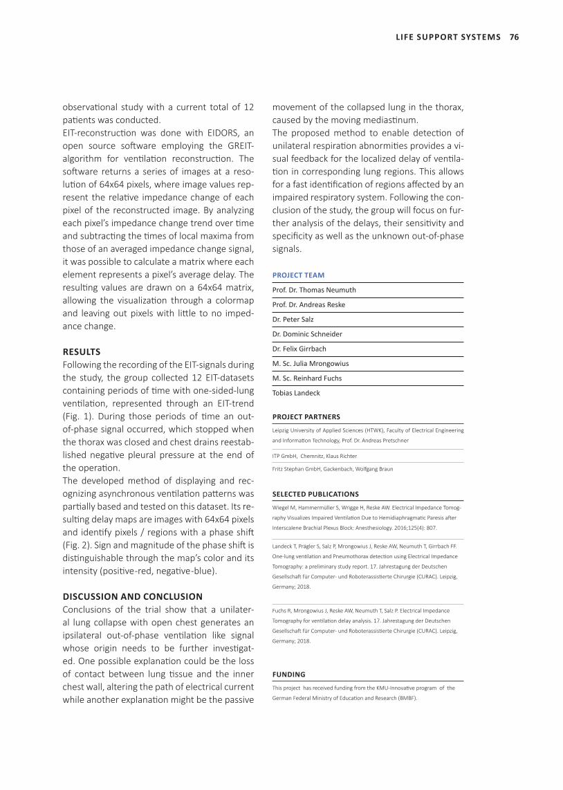
76
observational study with a current total of 12 patients was conducted.EIT-reconstruction was done with EIDORS, an open source software employing the GREIT- algorithm for ventilation reconstruction. The software returns a series of images at a reso-lution of 64x64 pixels, where image values rep-resent the relative impedance change of each pixel of the reconstructed image. By analyzing each pixel’s impedance change trend over time and subtracting the times of local maxima from those of an averaged impedance change signal, it was possible to calculate a matrix where each element represents a pixel’s average delay. The resulting values are drawn on a 64x64 matrix, allowing the visualization through a colormap and leaving out pixels with little to no imped-ance change.
RESULTSFollowing the recording of the EIT-signals during the study, the group collected 12 EIT-datasets containing periods of time with one-sided-lung ventilation, represented through an EIT-trend (Fig. 1). During those periods of time an out-of-phase signal occurred, which stopped when the thorax was closed and chest drains reestab-lished negative pleural pressure at the end of the operation.The developed method of displaying and rec-ognizing asynchronous ventilation patterns was partially based and tested on this dataset. Its re-sulting delay maps are images with 64x64 pixels and identify pixels / regions with a phase shift (Fig. 2). Sign and magnitude of the phase shift is distinguishable through the map’s color and its intensity (positive -red, negative -blue).
DISCUSSION AND CONCLUSIONConclusions of the trial show that a unilater-al lung collapse with open chest generates an ipsilateral out-of-phase ventilation like signal whose origin needs to be further investigat-ed. One possible explanation could be the loss of contact between lung tissue and the inner chest wall, altering the path of electrical current while another explanation might be the passive
movement of the collapsed lung in the thorax, caused by the moving mediastinum.The proposed method to enable detection of unilateral respiration abnormities provides a vi-sual feedback for the localized delay of ventila-tion in corresponding lung regions. This allows for a fast identification of regions affected by an impaired respiratory system. Following the con-clusion of the study, the group will focus on fur-ther analysis of the delays, their sensitivity and specificity as well as the unknown out-of-phase signals.
PROJECT TEAM
Prof. Dr. Thomas Neumuth
Prof. Dr. Andreas Reske
Dr. Peter Salz
Dr. Dominic Schneider
Dr. Felix Girrbach
M. Sc. Julia Mrongowius
M. Sc. Reinhard Fuchs
Tobias Landeck
PROJECT PARTNERS
Leipzig University of Applied Sciences (HTWK), Faculty of Electrical Engineering
and Information Technology, Prof. Dr. Andreas Pretschner
ITP GmbH, Chemnitz, Klaus Richter
Fritz Stephan GmbH, Gackenbach, Wolfgang Braun
SELECTED PUBLICATIONS
Wiegel M, Hammermüller S, Wrigge H, Reske AW. Electrical Impedance Tomog-
raphy Visualizes Impaired Ventilation Due to Hemidiaphragmatic Paresis after
Interscalene Brachial Plexus Block: Anesthesiology. 2016;125(4): 807.
Landeck T, Prägler S, Salz P, Mrongowius J, Reske AW, Neumuth T, Girrbach FF.
One-lung ventilation and Pneumothorax detection using Electrical Impedance
Tomography: a preliminary study report. 17. Jahrestagung der Deutschen
Gesellschaft für Computer- und Roboterassistierte Chirurgie (CURAC). Leipzig,
Germany; 2018.
Fuchs R, Mrongowius J, Reske AW, Neumuth T, Salz P. Electrical Impedance
Tomography for ventilation delay analysis. 17. Jahrestagung der Deutschen
Gesellschaft für Computer- und Roboterassistierte Chirurgie (CURAC). Leipzig,
Germany; 2018.
FUNDING
This project has received funding from the KMU-Innovative program of the
German Federal Ministry of Education and Research (BMBF).
LIFE SUPPORT SYSTEMS
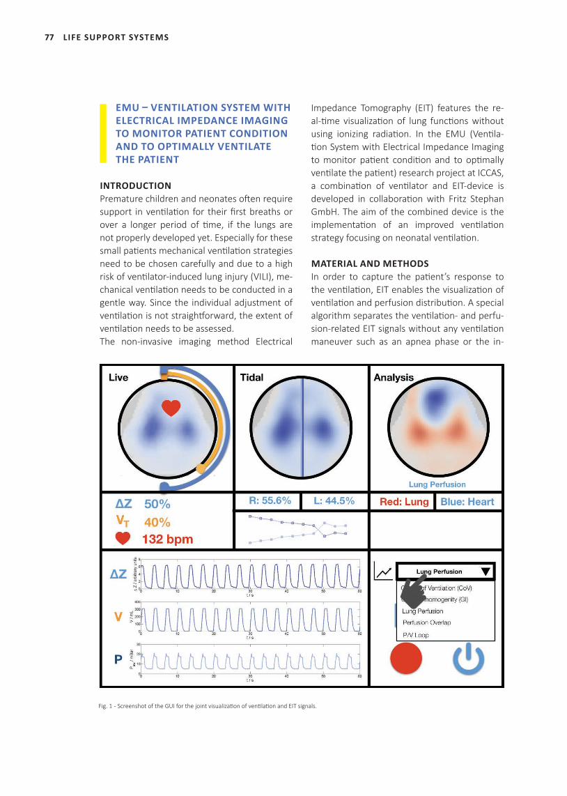
77 LIFE SUPPORT SYSTEMS
EMU – VENTILATION SYSTEM WITH ELECTRICAL IMPEDANCE IMAGING TO MONITOR PATIENT CONDITION AND TO OPTIMALLY VENTILATE THE PATIENT
INTRODUCTIONPremature children and neonates oft en require support in venti lati on for their fi rst breaths or over a longer period of ti me, if the lungs are not properly developed yet. Especially for these small pati ents mechanical venti lati on strategies need to be chosen carefully and due to a high risk of venti lator-induced lung injury (VILI), me-chanical venti lati on needs to be conducted in a gentle way. Since the individual adjustment of venti lati on is not straightf orward, the extent of venti lati on needs to be assessed. The non-invasive imaging method Electrical
Impedance Tomography (EIT) features the re-al-ti me visualizati on of lung functi ons without using ionizing radiati on. In the EMU (Venti la-ti on System with Electrical Impedance Imaging to monitor pati ent conditi on and to opti mally venti late the pati ent) research project at ICCAS, a combinati on of venti lator and EIT-device is developed in collaborati on with Fritz Stephan GmbH. The aim of the combined device is the implementati on of an improved venti lati on strategy focusing on neonatal venti lati on.
MATERIAL AND METHODSIn order to capture the pati ent’s response to the venti lati on, EIT enables the visualizati on of venti lati on and perfusion distributi on. A special algorithm separates the venti lati on- and perfu-sion-related EIT signals without any venti lati on maneuver such as an apnea phase or the in-
Fig. 1 - Screenshot of the GUI for the joint visualizati on of venti lati on and EIT signals.
78
jection of a saline bolus. Based on these dis-tributions different parameters describing the patient’s condition can be derived. Examples for these parameters are the extraction of re-gions with collapse and overdistension, the lo-cal delay and pendelluft phenomena, the heart rate and the regional overlap of ventilation and perfusion.
RESULTSA combined monitoring system consisting of the Sophie ventilator from Fritz Stephan GmbH, the Swisstom OEM EIT module and the Swiss-tom Baby EIT belt is developed and bundles the visualization of ventilator and EIT information in a joint “all-in-one” touch monitor. An innovative GUI (see Fig. 1) gives an easy access and analy-sis to the ventilator and EIT signals.
DISCUSSION AND CONCLUSIONThe developed device offers improved mechan-ical ventilation strategies for neonates without the requirement of X-ray and ionizing radiation. With this device, the aim of improved oxygen-ation with a reduced risk of VILI can be achieved through a real-time response with EIT. It even allows for monitoring of ventilation and perfu-sion in situations where standard oxygenation measurement is not available or of poor signal quality. This may be the case for sustained infla-tions in the first seconds after the delivery.
PROJECT TEAM
Prof. Dr. Thomas Neumuth
Prof. Dr. Andreas Reske
Dr. Peter Salz
M. Sc. Julia Mrongowius
PROJECT PARTNERS
Fritz Stephan GmbH, Wolfgang Braun
FUNDING
This project has received funding from the ZIM program of the German Federal
Ministry for Economic Affairs and Energy (BMWi).
LIFE SUPPORT SYSTEMS
79
JOURNAL AND BOOK PUBLICATIONS, FIRST- AND SENIOR AUTHORSHIP
Barberio M, Maktabi M, Gockel I, Rayes N, Jansen-Winkeln B,
Köhler H, Rabe, SM, Seidemann L, Takoh JP, Diana M, Neumuth
T, Chalopin C. Hyperspectral based discriminati on of thyroid
and parathyroid during surgery. Curr Directi ons Biomed Eng.
2018; 4(1): 399-402.
Berger J, Unger M, Landgraf L, Bieck R, Neumuth T, Melzer
A. Assessment of Natural User Interacti ons for Robot-Assisted
Interventi ons. Curr Directi ons Biomed Eng. 2018; 4(1): 165-
168.
Bieck R, Heuermann K, Hofer M, Neumuth T. From Passive
Tool To Acti ve Guidance: Requirements For Computati onal
Navigati on Intelligence In Computer-Assisted Functi onal En-
doscopic Sinus Surgery. Int J Comput Assist Radiol Surg. [Epub
ahead of print].
Black D, Unger M, Fischer N, Kikinis R, Hahn H, Neumuth T,
Glaser B. Auditory display as feedback for a novel eye-tracking
system for sterile operati ng room interacti on. Int J Comput As-
sist Radiol Surg. 2018; 13(1): 37-45.
Cabal Aragon GJ, Lindner D, Arnold S, Schmidt A, Chalopin C.
Image based connector for the automati c identi fi cati on of ul-
trasound parameter values. Int J Comput Assist Radiol Surg,
2018; 13(Suppl 1): 205-206.
Chalopin C, Landgraf L, Melzer A, Neumuth T, Oeltze-Jafra S,
Salz P. Was gibt es Neues in der computerassisti erten Chirur-
gie? In: Jähne J, Königsrainer A, Schröder W, Südkamp N: Was
gibt es Neues in der Chirurgie? 2018; 51-68.
Czauderna T, Haga J, Kim J, Klapperstück M, Klein K, Kuhlen
T, Oeltze-Jafra S, Sommer B, Schreiber F. Immersive Analyt-
ics Applicati ons in Life and Health Sciences. In: Marriot K et
al.: Immersive Analyti cs. Lecture Notes in Computer Science,
2018; 289-330.
Franke S, Rockstroh M, Hofer M, Neumuth T. The intelligent
OR: design and validati on of a context-aware surgical working
environment. Int J Comput Radiol Surg. 2018; 13(8): 1301-8.
Franke S, Rockstroh M, Neumuth T. Context-awareness for
control consoles in integrated operati ng rooms. Curr Direc-
ti ons Biomed Eng, 2018; 4(1): 291-5.
Franke S, Rockstroh M, Kasparick M, Neumuth T. A Method for
the Context-Aware Assignment of Medical Device Functi ons
to Input Devices in Integrated Operati ng Rooms. In: Stoyanov
D, et al.: OR 20 Context-Aware Operati ng Theaters, Computer
Assisted Roboti c Endoscopy, Clinical Image-Based Procedures,
and Skin Image Analysis. 2018; 12-19.
Gaebel J, Schreiber E, Oeser A, Oeltze-Jafra S. Modular Archi-
tecture for Integrated Model-Based Decision Support. Stud
Health Technol Inform. 2018; 248: 108-15.
Girrbach FF, Hilbig F, Michael M, Bernhard M. Systemati c anal-
ysis of airway registries in emergency medicine. Anaesthesist.
2018; 67(9): 664-73.
Golatowski F, Janss A, Leucker M, Neumuth T. OR.NET: Secure
dynamic networks in the operati ng room and clinic. Biomed
Tech (Berl). 2018; 63(1): 1-3.
Heuermann K, Bieck R, Hofer M, Dietz A, Neumuth T. Intel-
ligent navigati on strategies of a markerless FESS navigati on
by prioriti zing multi modal context informati on. Laryngo-Rhi-
no-Otologie 2018; 97(S02): 42.
Ilunga-Mbuyamba E, Avina-Cervantes JG, Lindner D, Arlt F,
Ituna-Yudonago JF, Chalopin C. Pati ent-specifi c model-based
segmentati on of brain tumors in 3D intraoperati ve ultrasound
images. Int J Comput Radiol Surg. 2018; 13(3): 331-42.
PUBLICATIONS
PUBLICATIONS
80
Neumann J, Angrick C, Rollenhagen D, Roth A, Neumuth T.
Perioperati ve Workfl ow Simulati on and Opti mizati on in Or-
thopedic Surgery. In: Stoyanov D, et al.: OR 20 Context-Aware
Operati ng Theaters, Computer Assisted Roboti c Endoscopy,
Clinical Image-Based Procedures, and Skin Image Analysis.
2018; 3-11.
Karakitsios I, Mihcin S, Melzer A. Reference-less MR ther-
mometry on pre-clinical thiel human cadaver for liver surgery
with MRgFUS. Minim Invasive Ther Allied Technol. 2018; 1-7.
Kasparick M, Andersen B, Ulrich H, Franke S, Schreiber E,
Rockstroh M, Golatowski F, Timmermann D, Ingenerf J, Neu-
muth T. IEEE 11073 SDC and HL7 FHIR: Emerging Standards for
Interoperability of Medical Systems. Int J Comput Radiol Surg.
2018 [Epub ahead of print].
Köhler H, Jansen-Winkeln B, Maktabi M, Barberio M, Takoh J,
Diana M, Neumuth T, Rabe SM, Chalopin C, Melzer A, Gockel
I. Untersuchung der Oxygenierung des Schlauchmagens ohne
und mit ischämischer Konditi onierung mitt els intraoperati ver
Hyperspektralbildgebung. Z Gastroenterol 2018; 56(8): 195.
Mihcin S, Melzer A. Principles of focused ultrasound. Minim
Invasive Ther Allied Technol. 2018; 27(1): 41-50.
Neumuth T, Rockstroh M, Franke S. Context-aware medical
technologies: Relief or burden for clinical users? Curr Direc-
ti ons Biomed Eng. 2018; 4(1): 119-22.
Neumuth T, Franke S. Clear oxygen-level forecasts during an-
aesthesia. Nat Biomed Eng. 2018; 2(10): 715-6.
Oeltze-Jafra S, Meuschke M, Neugebauer M, Saalfeld S,
Lawonn K, Janiga G, Hege HC, Zacow S, Preim B. Generati on
and Visual Explorati on of Medical Flow Data: Survey, Research
Trends and Future Challenges: Medical Flow Visualizati on.
Comput Graph Forum. 2018 [Epub ahead of print].
Oeser A, Gaebel J, Dietz A, Wiegand S, Oeltze-Jafra S. Infor-
mati on architecture for a pati ent-specifi c dashboard in head
and neck tumor boards. Int J Comput Assist Radiol Surg. 2018;
13(8): 1283-1290..
Rabe S, Köhler H, Maktabi M, Barberio M, Takoh J, Rayes N,
Diana M, Neumuth T, Jansen-Winkeln B, Gockel I, Chalopin C.
Intraoperati ve Gewebe-Charakterisierung und Klassifi kati on
mitt els Hyperspektralbildgebung: Erste Ergebnisse. Z Gastro-
enterol. 2018; 56(08):223.
Rathmann P, Chalopin C, Halama D, Giri P, Meixensberger J,
Lindner D. Dynamic infrared thermography (DIRT) for as-
sessment of skin blood perfusion in cranioplasty: a proof of
concept for qualitati ve comparison with the standard indo-
cyanine green video angiography (ICGA). Int J Comput Assist
Radiol Surg. 2018; 13(3): 479-90.
Rockstroh M, Franke S, Dees R, Merzweiler A, Schneider G,
Dingler M, Dietz C, Pfeifer J, Kühn F, Schmitz M, Mildner A,
Janß A, Dell’Anna Pudlik J, Köny M, Andersen B, Bergh B, Neu-
muth T. From SOMDA to applicati on: Integrati on strategies in
the OR.NET demonstrati on sites. Biomed Tech (Berl). 2018;
63(1): 69-80.
Unger M, Black D, Fischer NM, Neumuth T, Glaser B. Design
and evaluati on of an eye tracking support system for the scrub
nurse. Int J Med Robot. 2018 [Epub ahead of print].
Unger M, Markfort M, Halama D, Chalopin C. Automati c de-
tecti on of perforator vessels using infrared thermography in
reconstructi ve surgery. Int J Comput Radiol Surg. 2018 [Epub
ahead of print].
PUBLICATIONS
81
CO-AUTHORSHIP
Andersen B, Kasparick M, Ulrich H, Franke S, Schlamelcher J,
Rockstroh M, Ingenerf J. Connecting the clinical IT infrastruc-
ture to a service-oriented architecture of medical devices.
Biomed Tech (Berl). 2018; 63(1): 57-68.
Gibaud B, Forestier G, Feldmann C, Ferrigno G, Gonçalves
P, Haidegger T, Julliard C, Katić D, Kenngott H, Maier-Hein L,
März K, de Momi E, Nagy DA, Nakawala H, Neumann J, Neu-
muth T, Rojas Balderrama J, Speidel S, Wagner M, Jannin P.
Toward a standard ontology of surgical process models. Int J
Comput Radiol Surg. 2018; 13(9): 1397-408.
Gockel I, Takoh JP, Chalopin C, Jansen-Winkeln B. Die intra-
operative Hyperspektral-Bildgebung in der Viszeralchirurgie.
Nicht-invasive Beurteilung physiologischer Gewebe-Parame-
ter gastrointestinaler Anastomosen. Ärzteblatt Sachsen. 2018;
7: 291-294.
Hempel G, Simon P, Salz P, Wrigge H. Respiratory Failure: Inno-
vations in Diagnostics and Therapy. Anasthesiol Intensivmed
Notfallmed Schmerzther. 2018; 53(2): 126-140.
Jansen-Winkeln B, Holfert N, Takoh J, Rabe S, Köhler H, Makta-
bi M, Chalopin C, Neumuth T, Melzer A, Gockel I. Überprüfung
der chirurgischen Resektionsgrenze bei Kolonresektionen
mittels Hyperspektralkamera. Z Gastroenterol. 2018; 56(8):
225-225.
Jansen-Winkeln B, Maktabi M, Takoh JP, Rabe SM, Barberio M,
Köhler H, Neumuth T, Melzer A, Chalopin C, Gockel I. Hypers-
pektral-Imaging bei gastrointestinalen Anastomosen. Chirurg.
2018; 89(9): 717-25.
Kasparick M, Schmitz M, Andersen B, Rockstroh M, Franke S,
Schlichting S, Golatkowski F, Timmermann D. OR.NET: a ser-
vice-oriented architecture for safe and dynamic medical de-
vice interoperability. Biomed Tech (Berl). 2018; 63(1): 11-30.
Mehdorn M, Rabe S, Köhler H, Maktabi M, Takoh J, Neumuth
T, et al. Intraoperative Beurteilung grenzwertig perfundierten
Darms im Rahmen der akuten Mesenterialischämie mittels
Hyperspektralbildgebung: Eine Falldarstellung. Z Gastroenter-
ol. 2018; 56(8): 224.
Meuschke M, Oeltze-Jafra S, Beuing O, Preim B, Lawonn K.
Classification of Blood Flow Patterns in Cerebral Aneurysms.
IEEE Trans Vis Comput Graph. 2018 [Epub ahead of print].
.
CONFERENCE PROCEEDINGS
Berger J, Unger M, Landgraf L, Zhang X, Hu S, McLeod D, Neu-
muth T, Melzer A. Towards integrating robotics for combined
focused ultrasound and radiation therapy into the treatment
process. 18th Annual International Symposium for Therapeu-
tic Ultrasound (ISTU). Nashville, USA; 2018.
Berger J, Unger M, Keller J, Bieck R, Landgraf L, Neumuth T,
Melzer A. Natural User Interaction for Collaborative Robot-As-
sisted Needle Targeting. The 12th Hamlyn Symposium on
Medical Robotics. London, UK; 2018.
Berger J, Unger M, Keller J, Bieck R, Landgraf L, Neumuth T,
Melzer A. Kollaborative Interaktion für die roboterassistierte
ultraschallgeführte Biopsie. 17. Jahrestagung der Deutschen
Gesellschaft für Computer- und Roboterassistierte Chirurgie
(CURAC). Leipzig, Germany; 2018.
Berger J, Unger M, Jochimsen T, Sattler B, Roy U, Landgraf L,
Neumuth T, Sabri O, Melzer A. Concepts for robot-assisted
focused ultrasound to support radiation therapy. 6th Interna-
tional Symposium on Focused Ultrasound. Reston, USA; 2018.
Berger J, Unger M, Jochimse T, Sattler B, Landgraf L, Sabri O,
Melzer A. Approaches to support radiation therapy by ro-
bot-assisted focused ultrasound. SMIT2018-IBEC2018 Joint
Conference. Seoul, Korea; 2018.
Berger J, Unger M, Landgraf L, Neumuth T, Melzer A. Towards
integration of robotics for focused ultrasound (FUS) surgery
and radiation therapy into the clinical treatment process. 26th
International European Association for Endoscopic Surgery
Congress (EAES). London, UK; 2018.
Cabal Aragón JG, Lindner D, Arnold S, Schmitgen A, Chalo-
pin C. Development of an improved ultrasound navigation
system for neurosurgical procedures. 17. Jahrestagung der
Deutschen Gesellschaft für Computer- und Roboterassistierte
Chirurgie (CURAC). Leipzig, Germany; 2018.
Fuchs R, Mrongowius J, Reske AW, Neumuth T, Salz P. Elec-
trical Impedance Tomography for ventilation delay analysis.
17. Jahrestagung der Deutschen Gesellschaft für Computer-
und Roboterassistierte Chirurgie (CURAC). Leipzig, Germany;
2018.
PUBLICATIONS
82PUBLICATIONS
Gaebel J, Müller J, Stoehr M, Oeltze-Jafra S. Model-Based Im-
itation of Patient Scenarios for Oncological Decision Support.
17. Jahrestagung der Deutschen Gesellschaft für Computer-
und Roboterassistierte Chirurgie (CURAC). Leipzig, Germany;
2018.
Gaebel J, Oeser A, Dietz A, Oeltze-Jafra S. Bayesian Networks
for Oncological Therapy Decision Support. International So-
ciety for Bayesian Analysis World Meeting (ISBA). Edinburgh,
UK; 2018.
Gaebel J, Wu H-G, Oeser A, Oeltze-Jafra S. System Infrastruc-
ture for Probabilistic Decision Models in Cancer Treatment.
Medical Informatics Europe (MIE). Gothenburg, Sweden;
2018.
Gaebel J, Oeser A, Müller J, Schreiber E, Oeltze-Jafra S. Prob-
abilistic Patient Modeling for Therapeutic Decision Support in
Oncology. 23rd International Conference on Telemedicine and
eHealth (ISfTeH). Helsinki, Finland; 2018.
Gaebel J, Wu H-G, Oeser A, Schreiber E, Oeltze-Jafra S. Mod-
ular Architecture for Integrated Model-Based Decision Sup-
port. 12th Annual Conference on Health Informatics meets
eHealth. Vienna, Austria; 2018.
Graebling N, Neumuth T, Franke S. Generierung und Verwend-
ung künstlicher Trainingsdaten für CNNs zur Erkennung von
Operationsinstrumenten. 17. Jahrestagung der Deutschen
Gesellschaft für Computer- und Roboterassistierte Chirurgie
(CURAC). Leipzig, Germany; 2018.
Köhler H, Maktabi M, Rabe SM, Barberio M, Takoh JP, Rayes
N, Diana M, Neumuth T, Jansen-Winkeln B, Gockel I, Chalo-
pin C. Intraoperative tissue characterization and classification
in hyperspectral imaging: first results. 17. Jahrestagung der
Deutschen Gesellschaft für Computer- und Roboterassistierte
Chirurgie (CURAC). Leipzig, Germany; 2018.
Landeck T, Prägler S, Salz P, Mrongowius J, Reske AW, Neumuth
T, Girrbach FF. One-lung ventilation and Pneumothorax detec-
tion using Electrical Impedance Tomography: a preliminary
study report. 17. Jahrestagung der Deutschen Gesellschaft für
Computer- und Roboterassistierte Chirurgie (CURAC). Leipzig,
Germany; 2018.
Maktabi M, Köhler H, Gockel I, Jansen-Winkeln B, Takoh JP,
Rabe SM, Neumuth T, Chalopin C. Eine Beurteilung der An-
wendbarkeit von hyperspektralbasierter Bildgebungstechnol-
ogie bei viszeralonkologischen Eingriffen. 17. Jahrestagung
der Deutschen Gesellschaft für Computer- und Roboterassist-
ierte Chirurgie (CURAC). Leipzig, Germany; 2018.
Mrongowius J, Reske AW, Neumuth T, Salz P. Electrical Imped-
ance Tomography Lung Imaging with partial access to the tho-
rax: A simulation study. 17. Jahrestagung der der Deutschen
Gesellschaft für Computer- und Roboterassistierte Chirurgie
(CURAC). Leipzig, Germany; 2018.
Multani P, Niemann U, Cypko MA, Kuehn JP, Voelzke H, Oel-
tze-Jafra S. Building a Bayesian Network to Understand the
Interplay of Variables in an Epidemiological Population-Based
Study. 31st International Symposium on Computer-Based
Medical Systems (IEEE). Karlstad, Sweden; 2018.
Oeser A, Gaebel J, Müller J, Franke S. Design Concept of an
Information System for the Intuitive Assessment of Laborato-
ry Findings. 9th Workshop on Visual Analytics in Healthcare
(VAHC). San Francisco, USA; 2018.
Oeser A, Gaebel J, Oeltze-Jafra S, Dietz A. Towards Structural
Learning of Bayesian Networks in Head and Neck Oncology.
International Society for Bayesian Analysis World Meeting
(ISBA). Edinburgh, UK; 2018.
Pirlich M, Weber-Chüo T, Rockstroh M, Dietz A, Neumuth T,
Hofer M. Clinical study on qualitative and quantitative eval-
uation of OR.NET: An integrated operating room of the fu-
ture. 89. Jahresversammlung der Deutschen Gesellschaft für
HNO-Heilkunde, Kopf- und Hals-Chirurgie e.V. Lübeck, Germa-
ny; 2018.
Reich CM, Pfahl A, Lehnhardt M, Uihlein B, Petershans S. Un-
terberg-Buchwald C, Uecker M, Melzer A. Interventionelle
MR-geführte Herzbiopsie: Entwicklung einer innovativen
MR-tauglichen Biopsiezange. 17. Jahrestagung der Deutschen
Gesellschaft für Computer- und Roboterassistierte Chirurgie
(CURAC). Leipzig, Germany; 2018.
83
Reich CM, Pfahl A, Lehnhardt M, Uihlein B, Petershans S,
Unterberg-Buchwald C, Uecker M, Melzer A. Status-quo and
demands for MR-guided endomyocardial biopsies. 12th Inter-
ventional MRI Symposium (iMRI ). Boston, USA; 2018.
Zhang X, Landgraf L, Unger M, Roy U, Patties I, McLeod D,
Melzer A. Focused ultrasound-induced hyperthermia and ra-
diation therapy for combined treatment of brain and prostate
cancer cells in vitro. SMIT2018-IBEC2018 Joint Conference.
Seoul, Korea; 2018.
Zhang X, Landgraf L, Unger M, Roy U, Patties I, McLeod D,
Neumuth T, Melzer A. Focused ultrasound-hyperthermia and
radiation therapy for combined treatment of brain and pros-
tate tumors: Preliminary studies in vitro. In: FUS Foundation
symposium. 6th International Symposium on Focused Ultra-
sound. Reston, USA; 2018.
Zhang X, Landgraf L, Patties I, Hu S, McLeod D, Fournelle M,
Tretbar S, Neumuth T, Melzer A. Ultrasound-induced hyper-
thermia: Radiosensitization in glioblastoma, prostate and
head and neck cancer: preliminary studies. ESHO. Berlin, Ger-
many; 2018.
PUBLICATIONS
84
IN-HOUSE EVENTS
ICCAS STATUS SEMINAR 2017 January 18, 2018 | ICCAS, LeipzigPresentati on of ICCAS’s research results and highlights
in 2017
1ST SONO-RAY STATUS SEMINAR January 18, 2018 | ICCAS, LeipzigPresentati on of the Meta-ZIK’s (ICCAS and OncoRay)
research results in 2017
14TH LEIPZIG RESEARCH FESTIVAL FOR LIFE SCIENCE
January 19, 2018 | Medical Study Center, Leipzig University Jesús Guillermo Cabal Aragón | poster: ‘Image based
connector for the automati c confi gurati on of 3D ultra-
sound data acquisiti on’
Stefan Franke | poster: ‘Realizati on of context-aware
technical assistance in integrated operati ng rooms’
Reinhard Fuchs | poster: ‘Development and Evalua-
ti on of an Instrument-Recogniti on-System for intraop-
erati ve acti vity-tracking through a multi -sensor arm-
band’
Jan Gaebel |poster: ‘Modular Infrastructure for the
Integrati on of Decision Models into Hospital Informa-
ti on Systems’
Norbert Lang | poster: ‘Auditory feedback system for
intraoperati ve navigati on during craniotomy in neuro-
surgery’
Marianne Maktabi | poster: ‘Hyperspektrale Untersu-
chungen von koagulierten ti erischen Gewebeproben’
Juliane Neumann | poster: ‘Extending BPMN 2.0
Modeling Language for Surgical Interventi ons’
Alexander Oeser | poster: ‘Development of a pa-
ti ent-specifi c dashboard applicati on for decision-mak-
ing in oncology’
Max Rockstroh | poster: ‘Qualitati ve Evaluati on of an
integrated operati ng room based on the IEEE11073-
SDC’
Erik Schreiber | poster: ‘Prioriti zed Presentati on of
Surgical informati on to counteract informati on over-
load in the operati ng room’
Michael Unger | poster: ‘Integrati on of a MR-compati -
ble roboti c arm into the clinical infrastructure’
Jan Berger | poster: ‘Scorpius – Robot assisted needle
guidance and US monitoring’
Richard Bieck | poster: ‘Towards Computati onal Navi-
gati on Intelligence In Computer-Assisted Minimally-In-
vasive ENT Surgery’
Julia Mrongowius | poster: ‘Improved Multi PAramet-
ric monitoring of blunt Chest Trauma, IMPACT – a proj-
ect presentati on’
Juliane Müller | poster: ‘Requirements and Visualiza-
ti on Methods for Visual Verifi cati on of Bayesian Net-
works’
Xinrui Zhang | poster: ‘An in vitro study of ultra-
sound-induced HT combined with radiati on therapy
for glioblastoma cells’
KUNSTSTOFF TRIFFT MEDIZINTECHNIK CONFERENCE
January 31, 2018 | ICCAS, LeipzigMax Rockstroh | presentati on of ICCAS’s Intelligent
Operati ng Room
ICCAS COLLOQUIUM March 12, 2018 | Leipzig University HospitalUlf Leser | invited lecture: ‘PREDICT: Knowledge Man-
agement for Precision Oncology’
SENIOR-ACADEMY April 10, 2018 | Leipzig UniversityThomas Neumuth | lecture: ‘Roboter in der Medizin‘
GIRLS’DAY April 26, 2018 | ICCAS, LeipzigClaire Chalopin, Lisa Landgraf | Insight into medical
informati cs for female pupils
ICCAS OPEN DAYJune 7, 2018 | ICCAS, LeipzigPresentati on of ICCAS’s research areas to the public
EVENTS
EVENTS
85 EVENTS
LONG NIGHT OF SCIENCES June 22, 2018 | BBZ, LeipzigLSS group | presentation of Electrical Impedance To-
mography (EIT)
Peter Salz | lecture: From Idea to Device – Today’s Med-
ical Technology Production
5TH DIGITAL OPERATING SUMMER SCHOOL (DORS)August 27 – 31, 2018 | ICCAS, Leipzig University Hospi-tal, Heart Center LeipzigUnique introduction to various topics of computer-as-
sisted medicine.
17TH ANNUAL CONEFRENCE OF THE GERMAN SOCIETY FOR COMPUTER AND ROBOTIC ASSISTED SURGERY (CURAC)
September 13 – 15, 2018 | ICCAS, Leipzig University Hospital and Medical Study Center Andreas Melzer | welcome speech; session chair: ‘Nav-
igation’; lecture: ‘Was gibt es Neues in iFUS?’
Thomas Neumuth | welcome speech; session chair:
‘Workflowanalyse und Prädiktion’
Claire Chalopin | session chair: ‘Navigation’
Stefan Franke | session chair: ’Chirurgische Assis-
tenzsysteme’
Hannes Köhler | lecture: ‘Intraoperative tissue charac-
terization and classification in hyperspectral imaging:
first results’
Nico Graebling | lecture: ’Generierung und Verwend-
ung künstlicher Trainingsdaten für CNNs zur Erkennung
von Operationsinstrumenten’
Julia Mrongowius | lectures: ‘Electrical Impedance To-
mography Lung Imaging with partial access to the tho-
rax: A simulation study’, ‘Electrical Impedance Tomogra-
phy for ventilation delay analysis’
Johann Berger | lecture: ’Kollaborative Interaktion für
die roboterassistierte ultraschallgeführte Biopsie‘
Richard Bieck | lecture: ‘Simulating the Patient-Individ-
ual Intervention Process for Staged Segmental Artery
Occlusion – A Modeling Approach
Marianne Maktabi | lecture: ’Eine Beurteilung der An-
wendbarkeit von hyperspektralbasierter Bildgebungs-
technologie bei viszeralonkologischen Eingriffen’
Jesús G. Cabal Aragón | lecture: ‘Development of an
improved ultrasound navigation system for neurosurgi-
cal procedures’
Martin Reich | lecture: ’Interventionelle MR-geführte
Herzbiopsie: Entwicklung einer innovativen MR-taugli-
chen Biopsiezange‘
Tobias Landeck | lecture: ‘One-lung ventilation and
Pneumothorax detection using Electrical Impedance To-
mography: a preliminary study report’
Jan Gaebel | lecture: ‘Model-Based Imitation of Patient
Scenarios for Oncological Decision Support’
EUMFH PROJECT MEETING INCLUDING VISIT OF THE EUROPEAN COMMISSIONER FOR HUMANITARIAN AID & CRISIS MANAGEMENT
November 23, 2018 | ICCAS Thomas Neumuth | member: steering committee; pre-
sentation of ICCAS and results of the project European
Modular Field Hospital (EUMFH)
86
CONFERENCES, SYMPOSIA, WORKSHOPS
EUROPEAN ASSOCIATION FOR ENDOSCOPIC SURGERY AND OTHER INTERVENTIONAL TECHNIQUES (EAES) –TECHNOLOGY WINTER MEETING 2018
February 3, 2018 | Frankfurt am Main, GermanyProf. Dr. Andreas Melzer | director symposium
Prof. Dr. Thomas Neumuth | invited lecture: ‘AI to Im-
prove the Workflow in the OR. Fully Integrated OR – the
German OR Network’
SYMPOSIUM INTENSIVMEDIZIN UND INTENSIVPFLEGEFebruary 14 – 16, 2018 | BremenAndreas Reske | invited lecture: ’Einsatz der Elek-
trischen Impedanztomografie im klinischen Alltag’
VDE-TAGUNG ‘SOFTWARE IN DER MEDIZIN’February 28, 2018 | Goethe-Universität, Frankfurt a. M.Thomas Neumuth, Richard Bieck | presentation of
ICCAS’s research topics
EUROPEAN CONGRESS OF RADIOLOGY (ECR) February 28 – March 4, 2018 | Vienna, AustriaAndreas Melzer | representation of ICCAS
DGBMT IM VDE (FACHAUSSCHUSS OPTISCHE VERFAHREN IN DER MEDIZIN (OPTOMED) – WORKSHOP: ‘VERGLEICHBARKEIT UND STANDARDISIERUNG OPTISCHER VERFAHREN IN DER MEDIZIN: WELCHE INFORMATIONEN BENÖTIGT DER ANWENDER? – WELCHE GRÖßEN MESSEN WIR?‘
March 1 – 2, 2018 | Technische Universität Dresden, DresdenDr. Claire Chalopin | technical implementation of work-
shop: ‘Dermatologie/Wunddiagnostik’
CONFERENCE ‘HOSPITAL CONCEPT’ BY OSTBAYERISCHE TECHNISCHE HOCHSCHULE AMBERG-WEIDEN (OTH)
March 5, 2018 | WeidenMax Rockstroh | invited talk: ‘OR.NET: Safe and Dy-
namic Technology Integration in Operating Rooms and
Hospitals’
48. KONGRESS DER DEUTSCHEN GESELLSCHAFT FÜR ENDOSKOPIE UND BILDGEBENDE VERFAHREN E.V. (DGE-BV) – ‘ENDOSKOPIE LIVE 2018’ & ‘INTEGRATION’
March 15 – 17, 2018 | MunichAndreas Melzer | member: board; opening and wel-
come speech; session chair: ‘DGBMT – Bildgebung und
-analyse in der Endoskopie und Chirurgie’; lectures:
‘Computerassistierte Chirurgie und -therapie’, ‘Magic
Lens AR and tablet based planning of surgical and inter-
ventional robotic assisted procedures‘
23RD INTERNATIONAL CONFERENCE ON TELEMEDICINE AND EHEALTH (ISFTEH 2018)
March 15 – 17, 2018 | Helsinki, FinlandJan Gaebel | lecture: ‘Probabilistic Patient Modeling for
Therapeutic Decision Support in Oncology’
EUROPEAN RESEARCH COUNCIL (ERC)March 19 – 23, 2018 | Brussels, BelgiumAndreas Melzer | member: ERC Starting Panel 2018
FG-MEETING VISUAL COMPUTING FOR BIOLOGY AND MEDICINE (VCBM)
April 11 – 13, 2017 | UlmJuliane Müller | invited talk: DPM Visualization
CONNECTING HEALTHCARE IT (CONHIT)April, 17 – 19, 2018 | BerlinThomas Neumuth | session chair: ‘Intelligente Technol-
ogie im Krankenhaus – Konvergenz von Medizintechnik
und IT’
135TH CONCRESS OF THE GERMAN SOCIETY OF SURGERY (DGCH)
April 17 – 20, 2018 | BerlinAndreas Melzer | invited lecture: ‘Computerassistierte
Chirurgie’
Heinz U. Lemke | invited lecture: Model-based Surgery
42ND CONGRESS OF THE KOREAN SOCIETY OF ENDOSCOPIC & LAPAROSCOPIC SURGEONS (KSELS) & 8TH INTERNATIONAL SYMPOSIUM
April 19 – 21, 2018 | Jeju, South KoreaAndreas Melzer | session chair: ‘AI in MIS’; invited key-
note lecture: ‘Artificial and Machine Intelligence in the
OR of the Future’
EVENTS
87
29TH MEDICAL INFORMATICS EUROPE (MIE)April 24 – 26, 2018 | Gothenburg, SwedenJan Gaebel | lecture: ‘System Infrastructure for Proba-
bilistic Decision Models in Cancer Treatment’
14TH EUROPEAN CONFERENCE ON PEDIATRIC AND NEONATAL MECHANICAL VENTILATION (EPNV)
April 25 – 28, 2018 | Montreux, SwitzerlandPeter Salz | workshop presentation: ’The current EMU
Prototype’
DEUTSCHER ANÄSTHESIE CONGRESS (DAC)April 25 – 27, 2018 | NurembergMax Rockstroh | lecture: ‘Mehrwert durch Vernetzung
– Der Patient im Fokus‘
VDE/DGBMT FOKUS-WORKSHOP ‘MODELLGESTÜTZTE PERSONALISIERTE MEDIZINTECHNIK‘
April 26, 2018 | Frankfurt a. M.Thomas Neumuth | lecture: ‘Grundidee, Zielstellung,
Modellvarianten und -klassifikationen‘
RESEARCH SEMINAR AT UPC UNIVERSITAT POLITECNICA DE CATALUNYA
May 05, 2018 | Barcelona, SpainSteffen Oeltze-Jafra | invited lecture: ’Therapy Decision
Support’
12TH ANNUAL CONFERENCE ON HEALTH INFORMATICS MEETS EHEALTH (EHEALTH)
May, 8 – 9, 2018 | Vienna, AustriaJan Gaebel | lecture: ‘Modular Architecture for Inte-
grated Model-Based Decision Support’
99. DEUTSCHER RÖNTGENKONGRESSMay 9 – 12, 2018 | LeipzigAndreas Melzer | representation of ICCAS
18TH INTERNATIONAL SYMPOSIUM FOR THERAPEUTIC ULTRASOUND (ISTU 2018)
May 14 – 17, 2018 | Nashville, TN, USAAndreas Melzer | session chair: ‘Thermal Therapies’
Lisa Landgraf | session chair: ‘Thermal Therapies’; lec-
ture: ‘In Vitro Effects of Ultrasound Hyperthermia and
Radiation Therapy on Glioblastoma, Prostate and Head
and Neck Cancer Cell Lines: Preliminary Studies’; poster
presentation: ‘Towards Integrating Robotics for Com-
bined Focused Ultrasound and Radiation Therapy into
the Treatment Process’
32ND ANNUAL MEETING OF THE EUROPEAN SOCIETY FOR HYPERTHERMIC ONCOLOGY (ESHO 2018)
May 16 – 19, 2018 | BerlinXinrui Zhang | poster ‘Flash’ presentation: ‘Ultra-
sound-induced hyperthermia – radiosensitisation in
glioblastoma, prostate and head and neck cancer – pre-
liminary studies’
26TH INTERNATIONAL CONGRESS OF EUROPEAN ASSOCIATION FOR ENDOSCOPIC SURGERY (E.A.E.S.)
May 30 – June 1, 2018 | London, UKAndreas Melzer | course director hands on: ‘Ultrasound
for Surgeons’; session chair: ‘Gerhard Buess technology
award’; invited lecture
Johann Berger | project presentation: ‘SONO-RAY’
20TH EUROGRAPHICS WORKING GROUP ON DATA VISUALIZATION (EG/VGTC) AT CONFERENCE ON VISUALIZATION (EUROVIS)
June 4 – 8, 2018 | Brno, Czech RepublicSteffen Oeltze-Jafra | presentation of Computer Graph-
ics article
CHARITÉ INHOUSE EVENT ‘INTENSIVE CARE’June 6, 2018 | Charité BerlinMax Rockstroh | invited lecture: ‚Intensive Care of the
Future’
INTERNATIONAL CONFERENCE ON ENGINEERING, TECHNOLGOGY AND INNOVATION (ICE/IEEE ITMC)
19 – 20 June, 2018 | StuttgartMax Rockstroh | session chair: ‘IEEE Konferenz Work-
shop Regulatory Health’; lecture: ‘OR.NET: Short intro-
duction to medical device interoperability based on the
IEEE11073-SDC standard family’
32ND INTERNATIONAL CONGRESS AND EXHIBITION OF COMPUTER ASSISTED RADIOLOGY AND SURGERY (CARS)
June 20 – 23, 2018 | BerlinAndreas Melzer | session chair: ‘Clinical Decision Mak-
ing’
Thomas Neumuth | session chair: ’Technical Standard-
ization in the OR – Standards, Use Cases, Benefits and
Potentials’; lectures: ‘Medical information technology
standards in the operating room’, ‘IEEE 11073 SDC and
HL7 FHIR – Emerging Standards for Interoperability of
Medical Systems’
EVENTS
88
Steffen Oeltze-Jafra | lecture: ‘Information Architec-
ture for a Patient-Specific Dashboard in Head and Neck
Tumor Boards’
Claire Chalopin | lectures: ‘Measurement of moisture
at skin surface with hyperspectral imaging’, ‘Image
based connector for the automatic identification of ul-
trasound parameter values’
Stefan Franke | lecture: ‘The intelligent OR – Design
and validation of a context-aware surgical working en-
vironment’
Chris Unger | lecture: ‘A Knowledge-based Data Entry
Form for High Quality Clinical Data Collection’
Erik Schreiber | session chair: ’IHE Session’; lectures:
‘Current Work Items for IHE Integration Profiles.
Cross-enterprise Model Sharing (XMS) and Consistent
Presentation of Surgical Information’, ‘IHE Surgery – In-
troduction, goals and current work items’
Richard Bieck | lecture: ‘From Passive Tool to Active
Guidance – Requirements for Navigation Intelligence in
Computer-Assisted Endoscopic ENT Surgery’
Heinz U. Lemke | invited lecture: ’Machine Intelligence
and CAS’
11TH HAMLYN SYMPOSIUM ON MEDICAL ROBOTICSJune 24 – 27, 2018 | London, UKAndreas Melzer | co-chair: ‘Image Guided Therapies
(IGT)’; invited talk: ‘Ultrasound and MR Guided Focused
Ultrasound Therapy: SONO-RAY’
Johann Berger, Johannes Keller | robotic presentation
at ‘Surgical Robotic Challenge’
INTERNATIONAL SOCIETY FOR BAYESIAN ANALYSIS WORLD MEETING (ISBA)
June 25 – 29, 2018 | Edinburgh, UKJan Gaebel | poster presentation: ‘Bayesian Networks
for Oncological Therapy Decision Support’
SYMPOSIUM ON ‘AI IN MEDICAL IMAGING’ NATIONAL INSTITUTES OF HEALTH AND BIOMEDICAL IMAGING/BIOENGINEERING (NIH/NIBIB)
August 24, 2018 | Bethesda, MD, USAHeinz U. Lemke | panelist and speaker in session ‘The
Human-Machine System’
TAG DER MEDIZINTECHNIKSeptember 12, 2018 | Fraunhofer Forum, BerlinAndreas Melzer | representation of ICCAS
21ST INTERNATIONAL CONFERENCE ON MEDICAL IMAGE COMPUTING AND COMPUTER ASSISTED INTERVENTION (MICCAI)
September 16 – 20, 2018 | Granada, SpainStefan Franke | lecture: ‘A method for the context-aware
assignment of medical device functions to input devices
in integrated operating rooms’
Steffen Oeltze-Jafra, Jan Gaebel | tutorial: ‘ModelsCDS:
Digital Therapy and Patient Models for Clinical Decision
Support’
Juliane Neumann | lecture: ‘Perioperative workflow
simulation and optimization in orthopedic surgery’
CONFERENCE OF EUROPEAN CARDIOVASCULAR AND INTERVENTIONAL RADIOLOGICAL SOCIETY (CIRSE)
September 22 – 25, 2018, 2018 | Lisbon, PortugalLisa Landgraf | poster presentations: ‘Towards the inte-
gration of robotic concepts into the interventional on-
cology workflow of combined focused ultrasound and
radiation therapy’, ‘Ultrasound therapy system for high
throughput in vitro analysis’
SZ-KONGRESS: ‘DIGITAL HEALTH – GESUNDHEIT NEU DENKEN. WIE VERNETZUNG DAS GESUNDHEITSWESEN VERÄNDERT‘
September 25 – 26, 2018 | Munich, GermanyThomas Neumuth | panel discussion: ‘Smart Hospital:
Wie digitale Anwendungen & Technologien Behand-
lungs- und Verwaltungsabläufe erleichtern‘
52ND ANNUAL CONFERENCE OF THE GERMAN SOCIETY FOR BIOMEDICAL ENGINEERING (DGBMT WITHIN VDE)
September 26 – 28, 2018 | AachenAndreas Melzer | chair: ‘MR Safety and Compatibility
of Medical Devices’, lecture: ‘New Devices for MR Inter-
ventional procedures’
Thomas Neumuth | keynote lecture: ‘Patient Specific
Model Guided Therapy’; session chair: ‘Personalized
model-based medical technology’, ‘Computer Assisted
Surgery’; lecture: ‘Context-aware medical technologies
- relief or burden for clinical users?’
Xinrui Zhang | lecture: ‘Combination of focused ultra-
sound hyperthermia (FUS-HT) and radiation therapy:
Validation of in vitro effects in a preliminary study’
Johann Berger | lecture: ‘Assessment of Natural User
Interactions for Robot-Assisted Interventions’
Claire Chalopin | lecture: ‘Hyperspectral based discrim-
ination of thyroid and parathyroid during surgery’
EVENTS
89
ANNUAL MEETING GERMAN SOCIETY OF UROLOGY (DGU) September 26 – 29, 2018 | Dresden
Andreas Melzer | invited keynote during opening ple-
nary
Stefan Franke | lectures: ‘Roboterassistierte OP-Techni-
ken: Morgen‘; ‘Der OP-Saal von morgen‘
12TH INTERVENTIONAL MRI SYMPOSIUMOctober 5 – 6, 2018 | Boston, MA, USAAndreas Melzer | faculty member; chair: ‘Special Ses-
sion for European Focused Ultrasound Charitable Soci-
ety’; invited talk: ‘Novel Technologies and Applications
of MR Guided FUS and HIFU’
FRAUNHOFER INTERACTIVE EXHIBITION #ZUKUNFTSARBEIT
October 8 – 12, 2018 | BerlinAndreas Melzer | panelist: panel discussion #Zukunfts-
arbeit Gesundheit
ANNUAL CONGRESS EUROPEAN ASSOCIATION OF NUCLEAR MEDICINE (EANM)
October 13 – 17, 2018 | DüsseldorfAndreas Melzer | lecture: ‘Trends in Computer Assisted
Intervention’; talk: ‘ROUND TABLE: Incorporating Imag-
ing and Machine Intelligence in the Operating Room –
Portable and/or Fixed Equipment? Will Interventional
Workflows be Affected?‘
Heinz U. Lemke | session chair: EANM-IFCARS Joint
Symposium on Computer Assisted Radioguided Inter-
vention in Surgery and Radiology
EUROPEAN SOCIETY FOR MAGNETIC RESONANCE IN MEDICINE AND BIOLOGY (ESMRMB) SUMMER SCHOOL
October 18 – 20, 2018 | Vienna, AustriaAndreas Melzer | lecture: ‘MR-safety issues in Interven-
tional MR’
6TH INTERNATIONAL SYMPOSIUM ON FOCUSED ULTRASOUND
October 21 – 25, 2018 | Reston, VA, USAAndreas Melzer | poster presentations: ‘Focused ul-
trasound-hyperthermia and radiation therapy for com-
bined treatment of brain and prostate tumors – pre-
liminary studies in vitro’, ‘Concepts for robot-assisted
focused ultrasound to support radiation therapy’; rep-
resentation of ICCAS at a booth
INNOVATIONSFORUM FLEXMEDOctober 24 – 25, 2018 | DresdenAlexander Oeser | representation of ICCAS
TRI-ANNUAL IMAGE-GUIDED-THERAPIES (IGT) NETWORK AND MEETING
November 1, 2018 | University of Dundee, UKAndreas Melzer | welcome speech; session chair:
‘Research Talks’, ‘Plenary Talk’, ‘Poster Prize and Final
Words’
ANNUAL SYMPOSIUM AMERICAN MEDICAL INFORMATICS ASSOCIATION (AMIA) AND 9TH WORKSHOP ON VISUAL ANALYTICS IN HEALTHCARE (VAHC 2018)
November 3 – 7, 2018 | San Francisco, CA, USAAlexander Oeser | lecture: ‘Design Concept of an Infor-
mation System for the Intuitive Assessment of Labora-
tory Findings’
30TH CONFERENCE INTERNATIONAL SOCIETY FOR MEDICAL INNOVATION AND TECHNOLOGY (ISMIT 2018)
November 8 – 10, 2018 | Seoul, South KoreaAndreas Melzer | international scientific committee
member; invited lecture: ‘Future Operating Room &
Safety’
NATIONAL CANCER CENTER KOREANovember 12 – 14, 2018 | Goyang, KoreaAndreas Melzer | invited lectures: ‘Intelligent OR solu-
tions and intraoperative focused ultrasound’, ‘Ultra-
sound guided ultrasonic energy therapy and radiation
support (SONO-RAY)
10TH NATIONAL SYMPOSIUM OF BARIATRIC AND METABOLIC SURGERY, 1ST NATIONAL SYMPOSIUM OF ROBOTIC SURGERY
November 23, 2018 | Bucharest, RomaniaAndreas Melzer | course director: ‘Workshop: Surgeons
Ultrasound’
EVENTS
90
104TH SCIENTIFIC ASSEMBLY AND ANNUAL MEETING RADIOLOGICAL SOCIETY OF NORTH AMERICA (RSNA)
November 25, 2018 | Chicago, IL, USAAndreas Melzer | representation of ICCAS
DIGITAL SUMMIT 2018 December 3 – 4, 2018 |NurembergThomas Neumuth | representation of ICCAS
PRESENTATIONS AT FAIRS
XPOMET© CONVENTIONMarch 21 – 23, 2018 | LeipzigThomas Neumuth | invited lecture: ‘Medical Device In-teroperability in the OR of the Future’
Stefan Franke, Max Rockstroh | Showcase Construc-tion: ‘OR of the Future’ and Use Case Session: ‘Medical Device Interoperability in the OR of the Future’
CONNECTING HEALTHCARE IT (CONHIT)April 17 – 19, 2018 | BerlinThomas Neumuth | session chair: ‘Intelligente Technol-ogie im Krankenhaus – Konvergenz von Medizintechnik und IT’, representation of OR.NET e.V.
11TH HAMLYN SYMPOSIUM ON MEDICAL ROBOTICSJune 24 – 27, 2018 | London, UKAndreas Melzer | co-chair: ‘Image Guided Therapies
(IGT)’; invited talk: ‘Ultrasound and MR Guided Focused
Ultrasound Therapy: SONO-RAY’
Johann Berger, Johannes Keller | robotic presentation
at ‘Surgical Robotic Challenge’
MEDICA 2018November 12 – 15, 2018 | DüsseldorfProf. Thomas Neumuth | panel speaker: ‘Operating Theater 4.0 – The OR.NET approach to safe and dynamic device networking’; lecture: ‘Medizintechnik und Kün-stliche Intelligenz [KI]’
MITTELSTANDSKONFERENZ November 19 – 20, 2018 | BerlinRichard Bieck | project presentation COMPASS
EVENTS
91
PROJECT- AND COOPERATION WORK
KICK OFF MEETING PROJECT MRI-SUITABLE FLEXIBLE BIOPSY FORCEPS FOR MINIMALLY INVASIVE TISSUE SAMPLING
January 26, 2018 | Dettingen/Erms
MR-INTERVENTION AND MR-HIFU AT CHILDREN’S NATIONAL MEDICAL CENTER
February 6 – 10, 2018 | Washington, WA, USAAndreas Melzer | MR-Interventions and MR-HiFu
STEERING COMMITTEE MEETINGS OF PROJECT EUROPEAN MEDICAL FIELD HOSPITAL (EUMFH)
February 19 – 21, 2018 | Tallin, EstoniaJuly 10 – 11, 2018 | LeipzigOctober 13 – 18, 2018 | Bucharest, Romania December 10 – 12, 2018 | Rome, ItalyThomas Neumuth | steering committee member
DPM GROUP MEETS INTERACTIVE MEDIA LAB FROM TECHNISCHE UNIVERSITÄT DRESDEN
March 1, 2018 | LeipzigAlexander Oeser, Jan Gaebel | representation of ICCAS and the Digital Patient Model
EUROPEAN RESEARCH COUNCIL (ERC)March 19 – 23, 2018, June 18 – 22, 2018 | Brussels, BelgiumAndreas Melzer | member: ERC Starting Panel 2018
FINAL PRESENTATION OF PROJECT BIOPASSApril 25 – 27, 2018 | Siegburg
MR BIOPSY AND MR STENT AT GUYS’ AND ST THOMAS HOSPITAL
April 26, 2018 | London, UKAndreas Melzer | cooperation talks
GERMAN CANCER AID AT UNIVERISTY CANCER CENTER (UCCL) LEIPZIG
May 14 – 17, 2018 | Leipzig University Hospital, Univer-sity Cancer Center Leipzig (UCCL)Thomas Neumuth | presentation of computer assisted medicine in cancer therapy
WORLD HEALTH ORGANIZATION (WHO) – REGIONAL CONFERENCE
April 18, 2018 | Brussels, BelgiumThomas Neumuth | Presentation of research results on the development and application of digital medical re-cords for emergency medical teams (EMTs)
MR-INTERVENTIONS at CHILDREN’S NATIONAL MEDICAL CENTER
October 26, 2018 | Washington, USAAndreas Melzer | MR-Interventions
KICK-OFF MEETING PROJECT HEALTH EUJune 27 – 29, 2018 | Lausanne, France
RESEARCH STAY AT JOINT INSTITUTION FOR ADVANCED BIOMEDICAL SCIENCES OF TOKYO WOMEN’S MEDICAL UNIVERSITY AND WASEDA UNIVERSITY (TWINS)
June 28 – July 31, 2018 | Tokyo, Japan
Johann Berger, Max Rockstroh, Erik Schreiber | Scien-tific Exchange on open networking of medical devices and it systems in operating room and hospital
PROJECT MEETING MOVESeptember 13, 2018 | Leipzig
EU CIVIL PROTECTION EXERCISE MODEXOctober 14 – 18, 2018 | Bucharest, Romania
Thomas Neumuth, Erik Schreiber | Evaluation of digital patient record for disaster relief under real conditions in the scope of project EUMFH
KICK-OFF MEETING PROJECT LYSISOctober 9, 2018 | Leipzig
HYPERSPECTRAL IMAGING (HSI) WORKSHOP AT CLINIC OF VISCERAL SURGERY AT LEIPZIG UNIVERSITY HOSPITAL
November 14, 2018 | LeipzigClaire Chalopin | welcome speech
Hannes Köhler | lecture: ‘Clinical and technical devel-opments‘
EVENTS
92
VISIT AT CHONGQING UNIVERSITYNovember 4 – 5, 2018 | Chongqing, ChinaAndreas Melzer | representation of ICCAS
VISIT AT UNIVERSITY OF SHANGHAI FOR SCIENCE AND TECHNOLOGY
November 6 – 8, 2018 | Shanghai, ChinaAndreas Melzer | representation of ICCAS
REQUIREMENTS WORKSHOP OF PROJECT COMPASSDecember 4, 2018 | Leipzig
UNIVERSITY COURSES
LEIPZIG UNIVERSITY
‘Computer Assisted Surgery’Faculty of Mathematics and Computer Science
practical course
‘Development of Medical Products‘Faculty of Mathematics and Computer Science
lecture
‘Introduction to Computer Assisted Surgery’Faculty of Medicine
lecture
‘Medical Planning and Simulation Systems’Faculty of Mathematics and Computer Science
lecture
‘Surgical Navigation, Mechatronics and Robotics’Faculty of Mathematics and Computer Science
lecture
‘System Innovation in Medicine’Faculty of Mathematics and Computer Science
lecture and seminar
LEIPZIG UNIVERSITY OF APPLIED SCIENCES (HTWK)
‘Developing Medical Products’Faculty of Electrical Engineering and Information Tech-nology
lecture and seminar
‘Project Management for Engineers’Faculty of Electrical Engineering and Information Tech-nology, Mechanical and Energy Engineering
lecture and seminar
System Engineering Faculty of Electrical Engineering and Information Tech-nology
lecture
EVENTS
93
GRADUATIONS
MASTER DEGREES
Nico Graebling ‘Generierung und Verwendung künstlicher Trainings-daten für CNNs zur Erkennung von Operati onsinstru-menten‘Leipzig University
Karsten Brandt‘Graphenbasierte Visuelle Analyse von Qualitäts- berichten der Krankenhäuser‘Leipzig University
Norbert Lang‘Eine intraoperati ve Planungs- und Navigati onssoft ware für Kraniotomien unter der Verwendung eines Auditory Displays‘Leipzig University
Chris Unger‘Structured Collecti on of Pati ent Data for Decision Sup-port Systems by the Example of a Probabilisti c-Graphical Model for Laryngeal Cancer Therapy’Leipzig University
BACHELOR DEGREE
Moritz Lehnhardt‘Entwicklung einer Prozesskett e zur additi ven Herstel-lung von realitätsnahen Blutkreislaufmodellen’TU Darmstadt
EVENTS
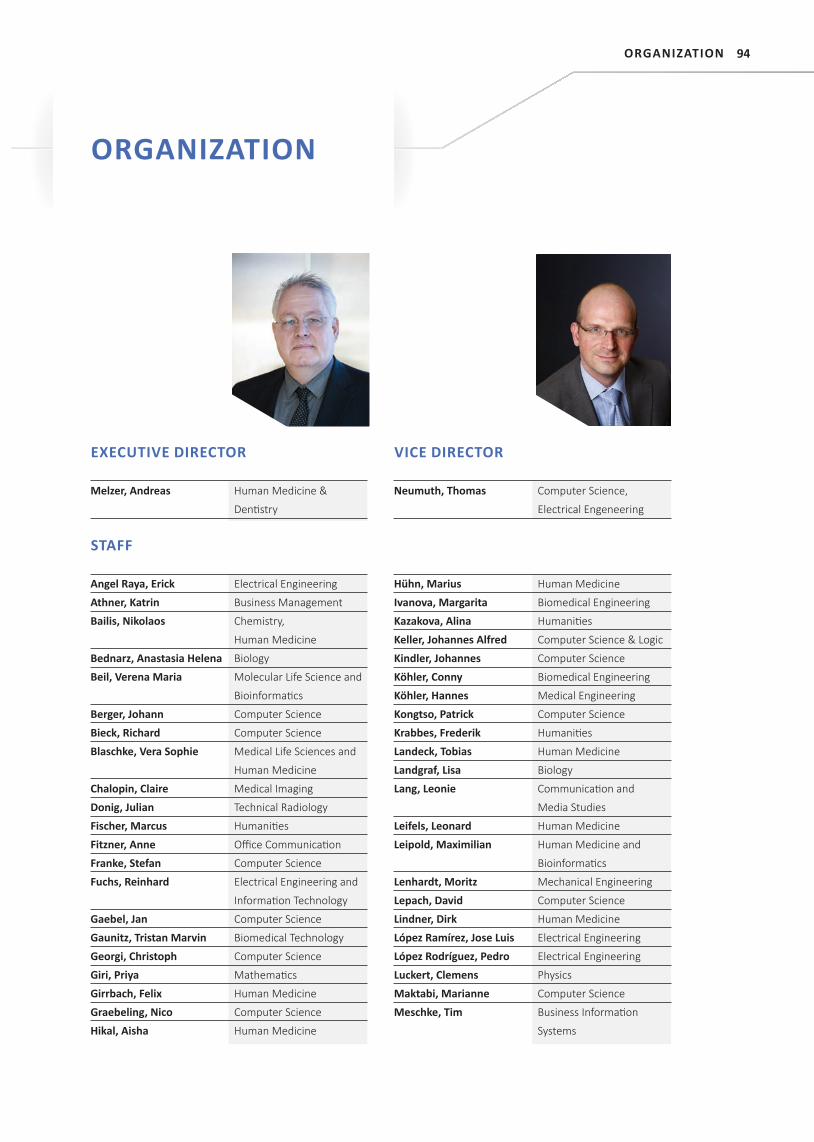
94
STAFF
Angel Raya, Erick Electrical Engineering
Athner, Katrin Business Management
Bailis, Nikolaos Chemistry,
Human Medicine
Bednarz, Anastasia Helena Biology
Beil, Verena Maria Molecular Life Science and
Bioinformati cs
Berger, Johann Computer Science
Bieck, Richard Computer Science
Blaschke, Vera Sophie Medical Life Sciences and
Human Medicine
Chalopin, Claire Medical Imaging
Donig, Julian Technical Radiology
Fischer, Marcus Humaniti es
Fitzner, Anne Offi ce Communicati on
Franke, Stefan Computer Science
Fuchs, Reinhard Electrical Engineering and
Informati on Technology
Gaebel, Jan Computer Science
Gaunitz, Tristan Marvin Biomedical Technology
Georgi, Christoph Computer Science
Giri, Priya Mathemati cs
Girrbach, Felix Human Medicine
Graebeling, Nico Computer Science
Hikal, Aisha Human Medicine
Hühn, Marius Human Medicine
Ivanova, Margarita Biomedical Engineering
Kazakova, Alina Humaniti es
Keller, Johannes Alfred Computer Science & Logic
Kindler, Johannes Computer Science
Köhler, Conny Biomedical Engineering
Köhler, Hannes Medical Engineering
Kongtso, Patrick Computer Science
Krabbes, Frederik Humaniti es
Landeck, Tobias Human Medicine
Landgraf, Lisa Biology
Lang, Leonie Communicati on and
Media Studies
Leifels, Leonard Human Medicine
Leipold, Maximilian Human Medicine and
Bioinformati cs
Lenhardt, Moritz Mechanical Engineering
Lepach, David Computer Science
Lindner, Dirk Human Medicine
López Ramírez, Jose Luis Electrical Engineering
López Rodríguez, Pedro Electrical Engineering
Luckert, Clemens Physics
Maktabi, Marianne Computer Science
Meschke, Tim Business Informati on
Systems
EXECUTIVE DIRECTOR
Melzer, Andreas Human Medicine &
Denti stry
VICE DIRECTOR
Neumuth, Thomas Computer Science,
Electrical Engeneering
ORGANIZATION
ORGANIZATION
95
Mrongrowius, Julia Medical Engineering
Science
Müller, Juliane Computer Science
Neumann, Juliane Computer Science
Oeltze-Jafra, Steffen Computer Science
Oeser, Alexander Media Management
Pabst, Tobias Computer Science
Patties, Ina Biology
Pfahl, Annekatrin Medical Engineering
Pogány, Gergely Biomedical Engineering
Prägler, Stefan Human Medicine
Reich, Martin Sensorics and Cognitive
Psychology
Rockstroh, Max Computer Science
Rosenow, Simon Media Management
Roy, Upasana Physics
Salz, Peter Computer Science
Schmierer, Lukas Computer Science
Schneider, Dominic Computer Science,
Human Medicine
Scholz, Kathrin Humanities
Schreiber, Erik Computer Science
Seifert, Andreas Physical Engineering
Selbmann, Lisa Human Medicine
Troitzsch, Hanna Jurisprudence
Tschachtli, Christine Office Communication
Unger, Chris Computer Science
Unger, Michael Computer Science
Weber, Sebastian Computer Science
Weiße, Karin Humanities
Wermke, Ludwig Computer Science
Wichmann, David Industrial Engineering and
Human Medicine
Wiegand, Ulrike Human Medicine
Zeumer, Christoph Humanities
Zhang, Xinrui Pharmacology
Ziemann, Martin Human Medicine
GUEST RESEARCHERS
Cabal Aragón, Electrical Engineering,
Jesús Guillermo CONACyT
Glaser, Bernhard Computer Science, ITQTIG
Hu, Guang Biomedical Science
Hu, Shaonan Pharmacology
Liebmann, Philipp Computer Science
Reske, Andreas Human Medicine,
HBK Zwickau
Thümmler, Moritz Electrical Engineering and
Information Technology,
HTWK
Tretbar, Steffen Biomedical Engineering,
Fraunhofer IBMT
ICCAS BOARD
Prof. Dr. Andreas MelzerDirector | Innovation Center Computer Assisted Surgery Director | Institute for Medical Science and Technology, University of Dundee and St. Andrews, Scotland
Prof. Dr. Jürgen MeixensbergerDirector | Department of Neurosurgery, Leipzig University Hospital
Prof. Dr. Andreas DietzDirector | Department of Otolaryngology, Leipzig University Hospital
Prof. em. Dr. Friedrich-Wilhelm MohrFormer Medical Director | Heart Center Leipzig
Prof. Dr. Heinz U. LemkeExecutive Director | International Foundation for Com-puter Assisted Radiology and Surgery (IFCARS)
ORGANIZATION
96
ADVISORY BOARD
Prof. Dr. Bernhard Preim (spokesman)Head | Visualization Group, Department of Simulation and Graphics, Faculty of Computer Science, Otto von
Guericke University Magdeburg
Prof. Dr. Volkmar FalkMedical Director | Department of Cardiothoracic and
Vascular Surgery, Deutsches Herzzentrum Berlin
Dr.-Ing. Klaus IrionDepartment Chief | Research and Technology, Karl Storz SE & Co. KG
Prof. Dr. Ron KikinisDirector | Fraunhofer Institute for Medical Image Com-puting MeVis | Surgical Planning Laboratory, Harvard Medical School, Boston, MA, USA
Dr. Heinrich KolemSiemens Healthcare GmbH | Director Advanced Ther-
apies
Prof. Dr. Nassir NavabHead | Chair for Computer Aided Medical Procedures & Augmented Reality, Department of Informatics, Techni-cal University München
Prof. Dr. Dipl.-Ing. Thomas Schmitz-RodeDirector | Institute of Applied Medical Engineering, Helmholtz-Institute of RWTH Aachen University & Hos-pital
CLINICAL COMMITTEE
Dr. Harald BusseMedical Physicist at the Department of Diagnostic and Interventional Radiology, Leipzig University Hospital
Prof. Dr. Christian EtzAttending Cardiac Surgeon and Associate Professor of Cardiac Surgery at the Department of Cardiac Surgery, Heart Center Leipzig; Director of the Saxonian Incubator for Clinical Translation (SIKT), Leipzig University
Prof. Dr. Ines GockelDirector of the Department of Visceral, Transplant, Tho-racic and Vascular Surgery, Leipzig University Hospital
Prof. Dr. Rainer HaakDirector of the Department of Dentistry and Parodon-tology, Leipzig University Hospital
Dr. Dirk HalamaSenior Physician at the Department for Head, Neck and Plastic Surgery, Leipzig University Hospital
Prof. Dr. Dr. Thomas HierlLeading Senior Physician at the Department for Oral & Maxillofacial Plastic Surgery, Leipzig University Hospital
Prof. Dr. Gerhard HindricksCo-Director of the Department of Electrophysiology, Heart Center Leipzig
Dr. Mathias HoferSenior Physician at the Department of Otorhinolaryn-gology, Head and Neck Surgery, Leipzig University Hos-pital
Prof. Dr. Albrecht HoffmeisterSenior Physician at Interdisciplinary Central Endoscopy, Leipzig University Hospital
Dr. Philipp KieferConsultant of Cardiac Surgery at the Department of Cardiac Surgery/ Senior Physician Transcatheter Aor-tic-Valve Implantation, Heart Center Leipzig
Prof. Dr. Martin LacherDirector of the Department for Paediatric surgery, Leipzig University Hospital
PD Dr. Dirk LindnerSenior Physician at the Department of Neurosurgery, Leipzig University Hospital
Dr. Tim-Ole PetersenConsultant at the Department for Diagnostic and Inter-ventional Radiology, Leipzig University Hospital
Prof. Dr. Andreas ReskeDirector of the Center of Anaesthesiology, Intensive Care Medicine, Emergency Medicine and Pain Therapy, Heinrich-Braun-Klinikum Zwickau
Prof. Dr. Bernhard SattlerHead of the Medical Physics Section of the Department of Nuclear Medicine, Leipzig University Hospital
Prof. Dr. Sebastian StehrDirector of the Department for Anesthesiology and In-tensive Care, Leipzig University Hospital
Dr. Patrick StumppPhysician at the Department for Diagnostic and Inter-ventional Radiology, Leipzig University Hospital
ORGANIZATION
97
NATIONAL COOPERATION PARTNERS
INDUSTRYACL GmbH, Leipzig
Diaspecti ve Vision GmbH
C.R.S. iiMoti on GmbH, Villingen-Schwenningen
Dornheim Medical Images GmbH, Magdeburg
EPfl ex GmbH, Detti ngen an der Erms
Fritz Stephan GmbH, Gackenbach
GADV - Gesellschaft für Automati sierung mit Datenverarbeitungsanlagen mbH,
Böblingen
Gesellschaft für Technische Visualisti k mbh (GTV), Dresden
Gesundheitsforen Leipzig GmbH, Leipzig
GMC Systems – Gesellschaft für medizinische Computersysteme mbH, Ilmenau
healthcare Consulti ng GmbH, Ebersberg
HEBUmedical GmbH, Tutt lingen
inomed Medizintechnik GmbH
Ilara GmbH, Herzogenrath
ITP GmbH, Weimar
Karl Storz SE & Co. KG
KLS Marti n Group, Tutt lingen
LOCALITE GmbH Biomedical Visualizati on Systems, Sankt Augusti n
MRComp GmbH, Gelsenkirchen
NUROMEDIA GmbH, Köln
OFFIS – Insti tut für Informati k e.V., Oldenburg
Optris GmbH, Berlin
PHACON GmbH, Leipzig
qcmed GmbH, Quality Consulti ng Medical, Aachen
Siemens AG, Healthcare Sector, Erlangen
SurgiTAIX AG
Steute Schaltgeräte GmbH & Co. KG, Löhne
Synagon GmbH, Aachen
TecVenture GmbH, Leipzig
UniTransferKlinik Lübeck GmbH (UTK), Lübeck
VISUS Health IT GmbH, Bochum
SCIENCEBochum University Hospital, Department for Anesthesiology and Intensive Care
Medicine
Fraunhofer Insti tute for Applied Opti cs and Precision Engineering IFO, Jena
Fraunhofer Insti tute for Biomedical Engineering, St. Ingbert
Fraunhofer Insti tute for Cell Therapy and Immunology IZI, Leipzig
Fraunhofer Insti tute for Computer Architecture and Soft ware Technology FIRST,
Berlin
Fraunhofer Insti tute for Integrated Electronic Circuits IIS, Erlangen
Fraunhofer Insti tute for Manufacturing Engineering and Automati on IPA,
Stutt gart
Fraunhofer Insti tute for Medical Image Computi ng MEVIS, Bremen
Fraunhofer Insti tute for Biomedical Engineering IBMT, St. Ingbert
Fraunhofer Insti tute for Open Communicati on Systems FOKUS, Berlin
Fraunhofer Insti tute for Telecommunicati ons, Heinrich Hertz Insti tute, HHI, Berlin
Freie Universität Berlin, Insti tute of Computer Science, Human-Centered
Computi ng
Heart Center Leipzig
Heidelberg University Hospital, Surgical Clinic, Departments of Radiology, Head-
Neck and Plasti c Surgery
Helmholtz Center Dresden Rossendorf, Neuroradiopharmaceuti cals
Jena University Hospital, Division Informati on Technology (IT), Department for
ENT
Johann Wolfgang von Goethe University Frankfurt, Department of Radiology
Johanniter-Unfall-Hilfe e.V.
Klinikum rechts der Isar, Munich, Department for Visceral Interventi ons
Leipzig University, Insti tutes for Anatomy, Computer Science, Medical
Informati cs, Stati sti cs and Epidemiology (IMISE), Center for Biotechnology and
Biomedicine (BBZ), Heart Center Leipzig, Saxon Incubator for Clinical Translati on
(SIKT), Clinical Trial Centre
Leipzig University of Applied Sciences (HTWK), Innovati ve Surgical Training
Technologies Faculty of Electrical Engineering and Informati on Technology
Leipzig University Hospital, Departments of Anesthesia, Angiology, Cardiology,
Denti stry, Diagnosti c and Interventi onal Radiology, Ear-Nose and Throat
Medicine, Head-Neck and Plasti c Surgery, Neurosurgery, Nuclear Medicine, Oral-
Maxillofacial and Plasti c Surgery, Department of Orthopaedics, Traumatology
and Reconstructi ve Surgery, Otorhinolaryngology, Surgery, Urology, Radiati on
Therapy, Visceral-Transplantati on-Thorax and Vascular Surgery
COOPERATION PARTNERS
COOPERATION PARTNERS
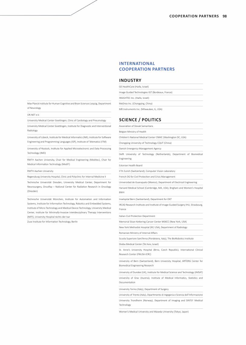
98
Max Planck Insti tute for Human Cogniti ve and Brain Sciences Leipzig, Department
of Neurology
OR.NET e.V.
University Medical Center Goetti ngen, Clinic of Cardiology and Pneumology
University Medical Center Goetti ngen, Insti tute for Diagnosti c and Interventi onal
Radiology
University of Lübeck, Insti tute for Medical Informati cs (IMI), Insti tute for Soft ware
Engineering and Programming Languages (ISP), Insti tute of Telemati cs (ITM)
University of Rostock, Insti tute for Applied Microelectronic and Data Processing
Technology (IMD)
RWTH Aachen University, Chair for Medical Engineering (Meditec), Chair for
Medical Informati on Technology (MedIT)
RWTH Aachen University
Regensburg University Hospital, Clinic and Polyclinic for Internal Medicine II
Technische Universität Dresden, University Medical Center, Department for
Neurosurgery, OncoRay – Nati onal Center for Radiati on Research in Oncology
(Dresden)
Technische Universität München, Insti tute for Automati on and Informati on
Systems, Insti tute for Informati on Technology, Roboti cs and Embedded Systems,
Insti tute of Micro Technology and Medical Device Technology, University Medical
Center, Insti tute for Minimally-Invasive Interdesciplinary Therapy Interventi ons
(MITI), University Hospital rechts der Isar
Zuse Insti tute for Informati on Technology, Berlin
INTERNATIONAL COOPERATION PARTNERS
INDUSTRYGE HealthCare (Haifa, Israel)
Image Guided Technologies IGT (Bordeaux, France)
INSIGHTEC Inc. (Haifa, Israel)
MeDrea Inc. (Chongqing, China)
MR Instruments Inc. (Milwaukee, IL, USA)
SCIENCE / POLITICSAssociati on of Slovak Samaritans
Belgian Ministry of Health
Children’s Nati onal Medical Center CNMC (Washington DC, USA)
Chongqing University of Technology CQUT (China)
Danish Emergency Management Agency
Delft University of Technology (Netherlands), Department of Biomedical
Engineering
Estonian Health Board
ETH Zurich (Switzerland), Computer Vision Laboratory
French DG for Civil Protecti on and Crisis Management
Universidad de Guanajuato (Mexico), Department of Electrical Engineering
Harvard Medical School (Cambridge, MA, USA), Brigham and Women’s Hospital
BWH
Inselspital Bern (Switzerland), Department for ENT
IRCAD Research Insti tute and Insti tute of Image-Guided Surgery IHU, Strasbourg,
France
Italian Civil Protecti on Department
Memorial Sloan Kett ering Cancer Center MSKCC (New York, USA)
New York Methodist Hospital (NY, USA), Department of Radiology
Romanian Ministry of Internal Aff airs
Scuola Superiore Sant’Anna (Pondetera, Italy), The BioRoboti cs Insti tute
Sheba Medical Center (Tel Aviv, Israel)
St. Anne’s University Hospital (Brno, Czech Republic), Internati onal Clinical
Research Center (FNUSA-ICRC)
University of Bern (Switzerland), Bern University Hospital, ARTORG Center for
Biomedical Engineering Research
University of Dundee (UK), Insti tute for Medical Science and Technology (IMSAT)
University of Graz (Austria), Insti tute of Medical Informati cs, Stati sti cs and
Documentati on
University Torino (Italy), Department of Surgery
University of Trento (Italy), Diparti mento di Ingegeria e Scienza dell’Informazione
University Trondheim (Norway), Department of Imaging and SINTEF Medical
Technology
Women’s Medical University and Waseda University (Tokyo, Japan)
COOPERATION PARTNERS

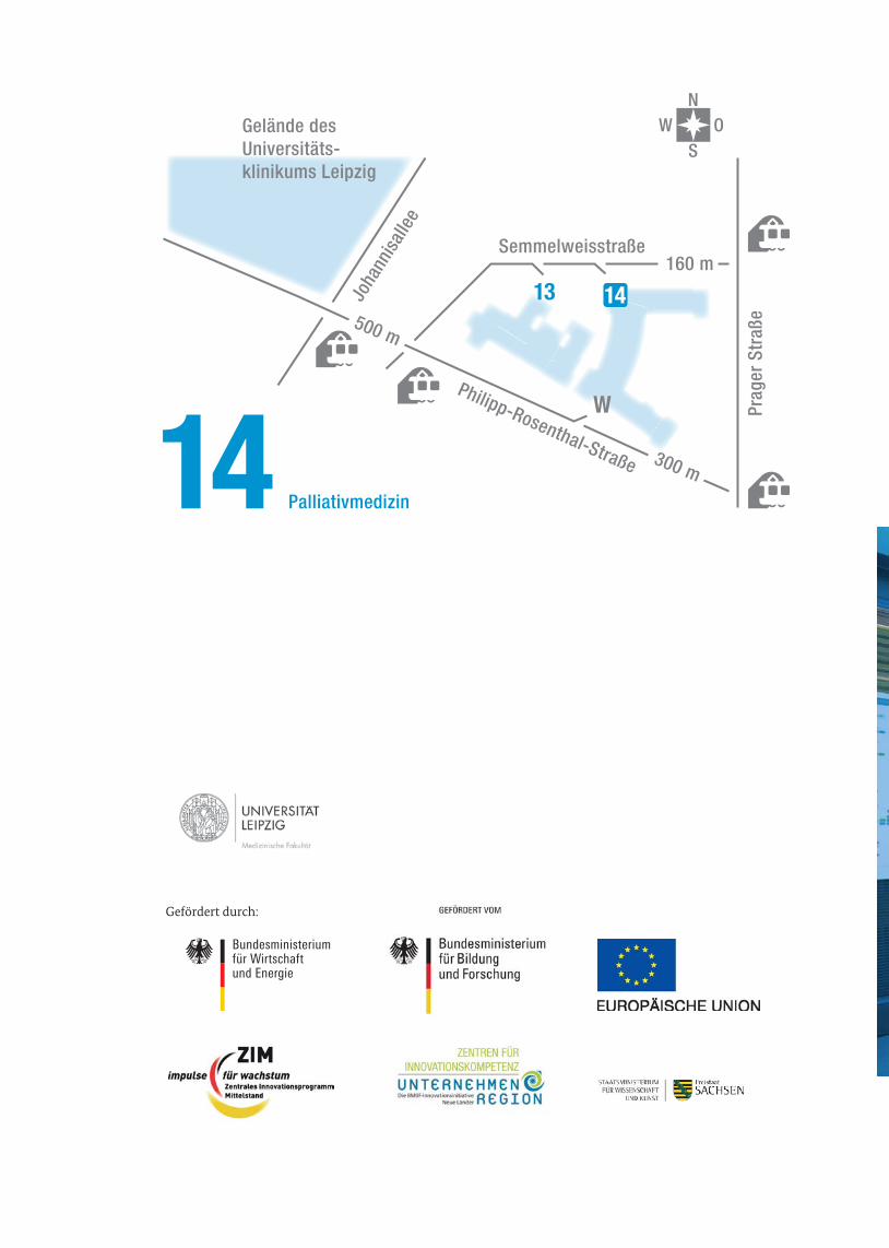


![Untitled-2 [] · Dr. Akshay Dr. Gade Dr. pratiksha Pat" Dr. Suresh Dr. AmitRajput Bensley Dr. Shefali Karkhanis Dr. Patil Dr. S Mulay Dr. Kamini Lakhiani Dr. Shah Dr. Jaydeep Rev](https://img.pdfslide.net/doc/110x75/5adee08b7f8b9a8f298c298a/untitled-2-akshay-dr-gade-dr-pratiksha-pat-dr-suresh-dr-amitrajput-bensley.jpg)