Embed Size (px)
Citation preview
BB46CH14-Discher ARI 13 April 2017 14:48
Matrix Mechanosensing: FromScaling Concepts in ’OmicsData to Mechanisms in theNucleus, Regeneration,and CancerDennis E. Discher,1 Lucas Smith,1 Sangkyun Cho,1
Mark Colasurdo,2 Andres J. Garcıa,2 and Sam Safran3
1Molecular and Cell Biophysics Lab, University of Pennsylvania, Philadelphia,Pennsylvania 19104; email: [email protected] H. Petit Institute for Bioengineering and Biosciences, Georgia Institute of Technology,Atlanta, Georgia 303323Department of Materials and Interfaces, Weizmann Institute of Science, Rehovet 76100, Israel
Annu. Rev. Biophys. 2017. 46:295–315
The Annual Review of Biophysics is online atbiophys.annualreviews.org
https://doi.org/10.1146/annurev-biophys-062215-011206
Copyright c© 2017 by Annual Reviews.All rights reserved
Keywords
polymer physics, collagen, lamins, myosin, contractility
Abstract
Many of the most important molecules of life are polymers. In animals, themost abundant of the proteinaceous polymers are the collagens, which con-stitute the fibrous matrix outside cells and which can also self-assemble intogels. The physically measurable stiffness of gels, as well as tissues, increaseswith the amount of collagen, and cells seem to sense this stiffness. An un-derstanding of this mechanosensing process in complex tissues, includingfibrotic disease states with high collagen, is now utilizing ’omics data sets andis revealing polymer physics–type, nonlinear scaling relationships betweenconcentrations of seemingly unrelated biopolymers. The nuclear structureprotein lamin A provides one example, with protein and transcript levelsincreasing with collagen 1 and tissue stiffness, and with mechanisms rootedin protein stabilization induced by cytoskeletal stress. Physics-based mod-els of fibrous matrix, cytoskeletal force dipoles, and the lamin A gene circuitillustrate the wide range of testable predictions emerging for tissues, cell cul-tures, and even stem cell–based tissue regeneration. Beyond the epigeneticsof mechanosensing, the scaling in cancer of chromosome copy number varia-tions and other mutations with tissue stiffness suggests that genomic changesare occurring by mechanogenomic processes that now require elucidation.
295
Click here to view this article'sonline features:
• Download figures as PPT slides• Navigate linked references• Download citations• Explore related articles• Search keywords
ANNUAL REVIEWS Further
Ann
u. R
ev. B
ioph
ys. 2
017.
46:2
95-3
15. D
ownl
oade
d fr
om w
ww
.ann
ualr
evie
ws.
org
Acc
ess
prov
ided
by
Uni
vers
ity o
f Pe
nnsy
lvan
ia o
n 05
/25/
17. F
or p
erso
nal u
se o
nly.
BB46CH14-Discher ARI 13 April 2017 14:48
Contents
INTRODUCTION . . . . . . . . . . . . . . . . . . . . . . . . . . . . . . . . . . . . . . . . . . . . . . . . . . . . . . . . . . . . . . . 296CORRELATED EXPRESSION OF STRUCTURAL BIOPOLYMERS:
MINING ’OMICS OF THREE-DIMENSIONAL TISSUES . . . . . . . . . . . . . . . . . . . 296SIMPLEST SIGNATURE OF MATRIX MECHANOSENSING:
2D CELL SPREADING . . . . . . . . . . . . . . . . . . . . . . . . . . . . . . . . . . . . . . . . . . . . . . . . . . . . . . . 301TISSUE STIFFNESS MEASUREMENTS . . . . . . . . . . . . . . . . . . . . . . . . . . . . . . . . . . . . . . . . 301COLLAGEN PERTURBATION: FIBROSIS AND MECHANOSENSING . . . . . . . 302CYTOSKELETAL ORDER IN TERMS OF FORCE DIPOLES . . . . . . . . . . . . . . . . . . 303NUCLEAR LINKAGES AND FORCES IN GENE REGULATION . . . . . . . . . . . . . . 305MATRIX MECHANOSENSING BY STEM CELLS IN
REGENERATIVE MEDICINE. . . . . . . . . . . . . . . . . . . . . . . . . . . . . . . . . . . . . . . . . . . . . . . . 307CONCLUSIONS: FROM THE EPIGENETICS OF MECHANOSENSING
TO THE MECHANOGENOMICS OF CANCER . . . . . . . . . . . . . . . . . . . . . . . . . . . . . 309
INTRODUCTION
Polymer physics has successfully focused for more than half a century on properties that areindependent of detailed chemistries (36). Cell and tissue functions have rarely been explained interms of polymer physics even though the solid-like nature of tissues is due to polymers, particularlyproteins, and most tissue cells are anchorage dependent in that viability requires adhesion to a solid(35). For mammals the most abundant protein is the structural protein outside of cells, collagen 1,which can self-assemble in water into macroscopic fibers and solidified networks or gels with astiffness that depends on concentration. Thus, the sensitivity of cells to matrix stiffness might seema logical consequence of anchorage dependence, but stiffness sensing has become a topic of greatinterest as well as some controversy.
From another perspective, the chemistry of a cell’s surroundings is often sensed by a cell,particularly the specific type and concentration of a ligand to which a cell receptor might bind.From the overwhelming accumulation of molecular biology data, it is perhaps tempting to believethat molecular detail is needed to explain all cell biological responses, with cells sensing only theprecise chemical details of their microenvironments. However, mammalian cells clearly exhibitan acute sensitivity to at least one physical variable of the environment, namely temperature.Every cell science lab sets their incubators at a physiological 37◦C rather than, for example, 47◦C,which would fry cells. The temperature of the surrounding medium propagates into a cell and,indeed, regulates conformational probabilities and pathway fluxes, even regulating gene expressionvia inducible transcription factors (79). The softness or stiffness of the surroundings to which acell adheres likewise seems to be sensed, propagated, and transduced into conformational andbiochemical changes within a cell, some of which are reviewed here.
CORRELATED EXPRESSION OF STRUCTURAL BIOPOLYMERS:MINING ’OMICS OF THREE-DIMENSIONAL TISSUES
For synthetic polymers, theory and basic experiments have advanced our understanding sufficientlyto guide the widespread use and generation of many polymer types, mixtures, and composites.
296 Discher et al.
Ann
u. R
ev. B
ioph
ys. 2
017.
46:2
95-3
15. D
ownl
oade
d fr
om w
ww
.ann
ualr
evie
ws.
org
Acc
ess
prov
ided
by
Uni
vers
ity o
f Pe
nnsy
lvan
ia o
n 05
/25/
17. F
or p
erso
nal u
se o
nly.
BB46CH14-Discher ARI 13 April 2017 14:48
Structural polymer systems generally sustain stresses and strains even at a scale much smaller thana cell, and synthetics in everyday use are sometimes soft or rubbery but others are hard and resistcracking. For biopolymers with structural roles in hydrated native tissues, such as collagens outsidecells or cytoskeletal components inside cells, disorder is often evident and mysteries abound con-cerning any relationship(s) between composition, order of architecture, and microscopic physicalproperties.
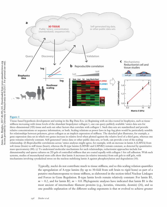
Proteomics, transcriptomics, genomics, and other emerging ’omics can perhaps offer insight asthese methods provide increasingly reproducible and accessible data sets about the relative concen-trations of specific proteins and nucleic acid species in various complex tissues with native matrixand multiple cell types. Mining such data sets, derived from three-dimensional (3D) functionaltissues, for correlations rooted in polymer physics can perhaps illustrate an emerging means forgenerating initial hypotheses for reductionist studies of mechanisms (Figure 1). Transcriptomedatabases, in particular, are standardized for many tissues, especially the US National Institutesof Health’s GEO database (Gene Expression Omnibus; https://www.ncbi.nlm.nih.gov/geo/).However, a top-down perspective should first seek to relate a collective physical property, such asthe stiffness of a tissue, to its protein composition.
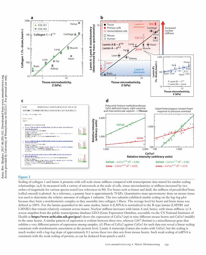
On the micron length scale relevant to a cell, the tissue stiffness is a microelasticity, E (orYoung’s modulus), that is measured in units of stress (kPa). For numerous tissues, E has beenmeasured by various labs using diverse methods, and the magnitude of E spans at least two to threelogs, from soft brain or marrow to the very stiff osteoid that bone-forming osteoblasts remodelinto bone (as summarized in 25). Bone is a good example of a tissue with very large differences inmicroscopic versus macroscopic mechanics: The microelasticity of osteoid is ∼20–50 kPa whereasthe macroscopic rigidity of bone is approximately a gigapascal (i.e., 106 kPa). Such log scalevariations are crucial to identifying any polymer physics–based trends (36). As an example, recentmass spectrometry–based proteomics studies of adult mouse tissue (80), indeed, indicate a typical,nonlinear polymer physics–type of scaling relationship for each of the two collagen 1 gene products,Col1a1 and Col1a2 (Figure 2a), which co-assemble stoichiometrically to make collagen 1 protein:
E ∼ collagen 1n with n ≈ 0.67.
In such scaling relations, prefactors with units of (stiffness/densityn) are ignored, as are small(but important) offsets that represent the critical concentrations needed to percolate a network.Similar magnitude exponents, n, are found for other fibrillar collagens, including collagens 3, 5,6, 11, and 12, making it clear that higher levels of fibrillar collagen are found in stiffer tissues(e.g., cardiac and skeletal muscle or bone-forming osteoid). Enzymatic degradation can be usedto decrease the concentration of fibrils in a tissue or tumor (25), or cross-linking can be usedto solidify fibril interactions, and both processes generally change tissue stiffness even for a softembryonic tissue, such as heart (55). Importantly, power-law scaling of tissue stiffness with theconcentration of these most abundant of structural biopolymers is overall consistent with thephysical properties of polymer networks. Indeed, gels of pure collagen 1 have exponents (n) thatdecrease from n = 2.8 at 22◦C to n = 2.1 at 37◦C (89), or that weaken to n ≈ 1 when the gelsare under stress (51).
Beyond the fibrillar collagenous matrix in tissues, mass spectrometry–based proteomic profil-ing of more than 100 of the most abundant proteins in adult mouse tissues revealed few otherproteins that also scaled with stiffness (80). A key exception is the nucleoskeletal protein lamin A(Figure 2b), which is an intermediate filament protein (like keratins in hair, hooves, and finger-nails) that assembles beneath the nuclear envelope and that scales with tissue stiffness:
lamin A ∼ Em with m ≈ 0.7.
www.annualreviews.org • Matrix Mechanosensing 297
Ann
u. R
ev. B
ioph
ys. 2
017.
46:2
95-3
15. D
ownl
oade
d fr
om w
ww
.ann
ualr
evie
ws.
org
Acc
ess
prov
ided
by
Uni
vers
ity o
f Pe
nnsy
lvan
ia o
n 05
/25/
17. F
or p
erso
nal u
se o
nly.
BB46CH14-Discher ARI 13 April 2017 14:48
3D TISSUEPublic ’omics: concentration
(standardized), sequence info
Biophysicsidea
Nonlinearscaling
Self-generated big data,or other public data sets
Query Discovery Validation
Mas
s sp
ectr
omet
ry p
rote
omes
LMN
A (r
elat
ive
inte
nsit
y)
Matrix E (kPa)
Soft gel Stiff gel
log
log
a
b Reproducible correlation
c
Soft
10x
1x
0.1x
Stiff
Mechanisms:Reductionist cell andtissue studies
Brain Liver Fat Lung Muscle HeartLMNALMNB1
LMNB2
1.4
1.2
1.0
0.3 10 40
Figure 1Tissue-based hypothesis development and testing in the Big Data Era. (a) Beginning with an idea rooted in biophysics, such as tissuestiffness increasing with tissue levels of the abundant biopolymer collagen 1, one can query publicly available ’omics data sets forthree-dimensional (3D) tissue and seek out other factors that correlate with collagen 1. Such data sets are standardized and providerelative concentrations or sequence information, or both. Scaling relations as power laws in log–log plots would be particularly sensiblefor relationships between polymers, given collagen as an implicit expression of stiffness. The sketched plot illustrates, for example, agene expression data set in which two genes increase in relative level when plotted against the relative level of a third gene, whereas onegene remains relatively constant. Self-generated ’omics data or other public data sets, or both, can provide a test of the scalingrelationship. (b) Reproducible correlations across ’omics analyses might agree, for example, with an increase in lamin A (LMNA) fromsoft tissue (brain) to stiff tissue (heart), whereas the B-type lamins (LMNB1 and LMNB2) remain constant, as detected by quantitativemass spectrometry (80). (c) To understand molecular mechanisms for such relationships, reductionist approaches include lowdimensionality and sparse cultures on 2D gels of controlled stiffness that are coated equally with collagen 1 for cell adhesion. With suchsystems, studies of mesenchymal stem cells show that lamin A increases (in relative intensity) from soft gels to stiff gels, withmechanisms involving cytoskeletal stress on the nucleus stabilizing lamin A against phosphorylation and degradation (10).
Typically, nuclei do not contribute much to tissue stiffness, and so this scaling relation quantifiesthe upregulation of A-type lamins (by up to 30-fold from soft brain to rigid bone) as part of aputative mechanoresponse to tissue stiffness, as elaborated in the section titled Nuclear Linkagesand Forces in Gene Regulation. B-type lamin levels remain relatively constant: For lamin B1,m ∼ 0.2, and for lamin B2, m ∼ 0.0. Phylogenetic analyses have indicated that lamin B1 is themost ancient of intermediate filament proteins (e.g., keratins, vimentin, desmin) (26), and soone possible explanation of the different scaling exponents is that m evolved to achieve greater
298 Discher et al.
Ann
u. R
ev. B
ioph
ys. 2
017.
46:2
95-3
15. D
ownl
oade
d fr
om w
ww
.ann
ualr
evie
ws.
org
Acc
ess
prov
ided
by
Uni
vers
ity o
f Pe
nnsy
lvan
ia o
n 05
/25/
17. F
or p
erso
nal u
se o
nly.
BB46CH14-Discher ARI 13 April 2017 14:48
a b
Col1a1Relative intensity (arbitrary units)
100
1,000
100 1,000
Col1a2
Lmna1,000
5,000
1,000 5,000
Polycomb histone methyltransferaseEzh2-deficient hearts: right ventricle
and interventricular septum → FibrosisGata4 heterozygous mutant heart
response to pressure overload
Col1a2 ~ Col1a10.96 (R2 = 0.96)
Lmna ~ Col1a10.23 (R2 = 0.93)
Col1a2 ~ Col1a11.16 (R2 = 0.96)
Lmna ~ Col1a10.35 (R2 = 0.75)
dc
Colla
gen
1 (%
<br
ain,
hear
t>)
Lam
in A
: La
min
B s
toic
hiom
etry
(det
erm
ined
by
mas
s sp
ectr
omet
ry)
Tissue microelasticity,E (kPa)
100kFemur Tissue
Primary cellsImmortalized cells
MouseHuman
Lamin A:B ~ E 0.6
Lamin A ~ E0.7
Collagen 1 ~ E 1.5
Lamin Bdominant
Lamin Adominant
Cartilage
Muscle MuscleHeart
Cartilage
Lung
Fat
LiverKidney
Brain
Heart
Lung
Fat
Kidney
Liver
Brain
10k
1k
100
1 10
Tissue microelasticity,E (kPa)
Rela
tive
inte
nsit
y(a
rbit
rary
uni
ts)
Increasednuclearstiffness
1 1010
1
10
1
1,000
100
1010
Tissue microelasticity,E (kPa)
Lam
in B
’s(%
<br
ain,
hear
t>)
FemurFemur
HSCPHSCP
U251*
A549
C2C12
MSC
SkullSkull
Fat
Brain
Femur
Liver Heart Cart.
Lamin B1 ~ E 0.2
Lamin B2 ~ const
COL1A1COL1A2
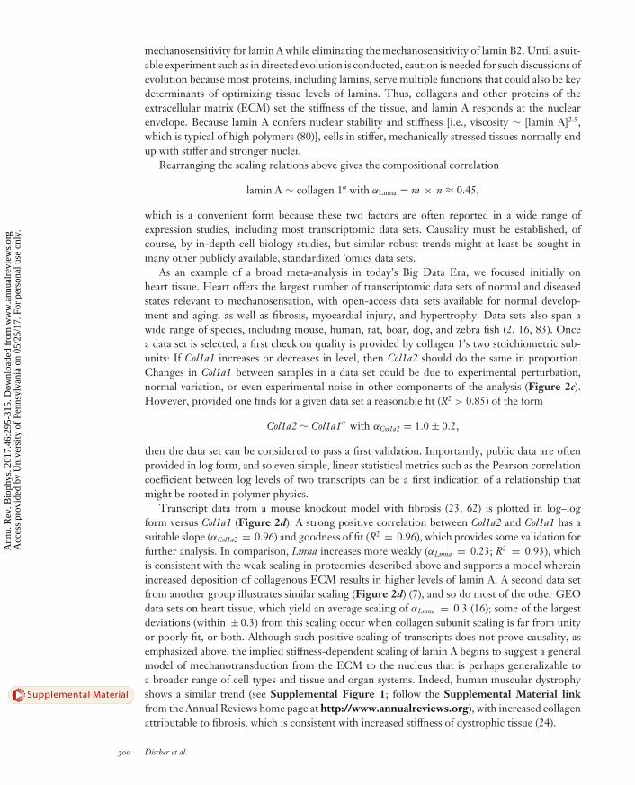
Figure 2Scaling of collagen 1 and lamin A proteins with cell-scale tissue stiffness compared with transcriptome data mined for similar scalingrelationships. (a,b) As measured with a variety of microtools at the scale of cells, tissue microelasticity or stiffness increased by twoorders of magnitude for various species tested (see references in 80). For bones such as femur and skull, the stiffness of precalcified bone(called osteoid) is plotted. As a reference, a gummy bear is approximately 70 kPa. Quantitative mass spectrometry done on mouse tissuewas used to determine the relative amounts of collagen 1 subunits. The two subunits exhibited similar scaling on the log–log plotbecause they form a stoichiometric complex as they assemble into collagen 1 fibers. The average level for heart and brain tissue wasdefined as 100%. For the lamins quantified in the same studies, lamin A (LMNA) is normalized to the B-type lamins (LMNB1 andLMNB2) that remain relatively constant across tissues. Nuclear stiffness increases with lamin A and, hence, with tissue stiffness. (c) Ascreen snapshot from the public transcriptome database GEO (Gene Expression Omnibus, accessible via the US National Institutes ofHealth at https://www.ncbi.nlm.nih.gov/geo/) shows the expression of Col1a2 (top) in nine different mouse hearts and Col1a1 (middle)in the same hearts. A similar pattern of expression is evident between these two, whereas Cd47 (bottom) is a miscellaneous gene thatexhibits a very different pattern of expression among samples. (d ) Plots of Col1a2 against Col1a1 for such data sets reveal a linear scalingconsistent with stoichiometric association at the protein level. Lamin A transcript (Lmna) also scales with Col1a1, but the scaling ismuch weaker with a log–log slope of approximately 0.3 across these two data sets from mouse hearts. Such weak scaling of mRNA isconsistent with the weak scaling of protein, as can be deduced from panels a and b.
www.annualreviews.org • Matrix Mechanosensing 299
Ann
u. R
ev. B
ioph
ys. 2
017.
46:2
95-3
15. D
ownl
oade
d fr
om w
ww
.ann
ualr
evie
ws.
org
Acc
ess
prov
ided
by
Uni
vers
ity o
f Pe
nnsy
lvan
ia o
n 05
/25/
17. F
or p
erso
nal u
se o
nly.
BB46CH14-Discher ARI 13 April 2017 14:48
mechanosensitivity for lamin A while eliminating the mechanosensitivity of lamin B2. Until a suit-able experiment such as in directed evolution is conducted, caution is needed for such discussions ofevolution because most proteins, including lamins, serve multiple functions that could also be keydeterminants of optimizing tissue levels of lamins. Thus, collagens and other proteins of theextracellular matrix (ECM) set the stiffness of the tissue, and lamin A responds at the nuclearenvelope. Because lamin A confers nuclear stability and stiffness [i.e., viscosity ∼ [lamin A]2.5,which is typical of high polymers (80)], cells in stiffer, mechanically stressed tissues normally endup with stiffer and stronger nuclei.
Rearranging the scaling relations above gives the compositional correlation
lamin A ∼ collagen 1α with αLmna = m × n ≈ 0.45,
which is a convenient form because these two factors are often reported in a wide range ofexpression studies, including most transcriptomic data sets. Causality must be established, ofcourse, by in-depth cell biology studies, but similar robust trends might at least be sought inmany other publicly available, standardized ’omics data sets.
As an example of a broad meta-analysis in today’s Big Data Era, we focused initially onheart tissue. Heart offers the largest number of transcriptomic data sets of normal and diseasedstates relevant to mechanosensation, with open-access data sets available for normal develop-ment and aging, as well as fibrosis, myocardial injury, and hypertrophy. Data sets also span awide range of species, including mouse, human, rat, boar, dog, and zebra fish (2, 16, 83). Oncea data set is selected, a first check on quality is provided by collagen 1’s two stoichiometric sub-units: If Col1a1 increases or decreases in level, then Col1a2 should do the same in proportion.Changes in Col1a1 between samples in a data set could be due to experimental perturbation,normal variation, or even experimental noise in other components of the analysis (Figure 2c).However, provided one finds for a given data set a reasonable fit (R2 > 0.85) of the form
Col1a2 ∼ Col1a1α with αCol1a2 = 1.0 ± 0.2,
then the data set can be considered to pass a first validation. Importantly, public data are oftenprovided in log form, and so even simple, linear statistical metrics such as the Pearson correlationcoefficient between log levels of two transcripts can be a first indication of a relationship thatmight be rooted in polymer physics.
Transcript data from a mouse knockout model with fibrosis (23, 62) is plotted in log–logform versus Col1a1 (Figure 2d). A strong positive correlation between Col1a2 and Col1a1 has asuitable slope (αCol1a2 = 0.96) and goodness of fit (R2 = 0.96), which provides some validation forfurther analysis. In comparison, Lmna increases more weakly (αLmna = 0.23; R2 = 0.93), whichis consistent with the weak scaling in proteomics described above and supports a model whereinincreased deposition of collagenous ECM results in higher levels of lamin A. A second data setfrom another group illustrates similar scaling (Figure 2d) (7), and so do most of the other GEOdata sets on heart tissue, which yield an average scaling of αLmna = 0.3 (16); some of the largestdeviations (within ± 0.3) from this scaling occur when collagen subunit scaling is far from unityor poorly fit, or both. Although such positive scaling of transcripts does not prove causality, asemphasized above, the implied stiffness-dependent scaling of lamin A begins to suggest a generalmodel of mechanotransduction from the ECM to the nucleus that is perhaps generalizable toa broader range of cell types and tissue and organ systems. Indeed, human muscular dystrophyshows a similar trend (see Supplemental Figure 1; follow the Supplemental Material linkfrom the Annual Reviews home page at http://www.annualreviews.org), with increased collagenattributable to fibrosis, which is consistent with increased stiffness of dystrophic tissue (24).
300 Discher et al.
Supplemental Material
Ann
u. R
ev. B
ioph
ys. 2
017.
46:2
95-3
15. D
ownl
oade
d fr
om w
ww
.ann
ualr
evie
ws.
org
Acc
ess
prov
ided
by
Uni
vers
ity o
f Pe
nnsy
lvan
ia o
n 05
/25/
17. F
or p
erso
nal u
se o
nly.
BB46CH14-Discher ARI 13 April 2017 14:48
SIMPLEST SIGNATURE OF MATRIX MECHANOSENSING:2D CELL SPREADING
Collagen 1 is both an adhesion ligand and a structural protein. A tissue with high collagen is thuslikely to present more adhesive ligand to a cell in such a tissue. The evident scaling relation betweenlamin A and collagen 1 could therefore reflect a purely biochemical signal from ligand densityrather than any implicit dependence on tissue stiffness, E. It is difficult to separate such variableswith intact tissue. However, many reductionist in vitro experiments with various materials allowseparate control over adhesion ligand and substrate elasticity E, showing that cells mechanosenseE as long as the ligand is not limiting.
The simplest experimental signature for mechanosensing substrate E is a saturable increase ofcell spread area as a function of E with the stiffest possible gels and rigid glass causing similar cellspreading as soon as a few hours and certainly after one day (e.g., 29, 30, 64). Importantly, forany given gel or substrate, the amount of adhesive ligand (e.g., gelatin or nonfibrillar collagen)should be widely varied, but the usual observation is that beyond a threshold in the density of suchligand, the cell spread area increases asymptotically to a maximum spread area as a function ofE. Inhibition of myosin II suppresses such spreading, and so actomyosin forces increase with E. Itis also useful and important to vary the thickness of soft gels bound to rigid glass, because for gelsthat are only a few microns thick or less, cells are seen to spread (9, 10, 30). This is because thecell’s actomyosin-driven distortions of the soft gels are constrained by the rigidity of the hidden,underlying glass. This latter approach maintains ligand biochemistry as well as gel physics andreveals the basic stiffness-sensing capabilities of adherent cells. It also highlights the need forcareful attention to physical heterogeneity in culture systems because cells generally respond torigid objects that are microns away (24).
A simple intuition into mechanosensing of matrix E is obtained by considering that actinpolymerization drives cell spreading at a near constant rate, vpolymer = A, whereas myosin II pullsback on the actin network. The latter occurs at a rate of vretract = B / (K + E) that follows the usualhyperbolic decrease (with constants B, K) as a function of resistance set by the extracellular load;in this case, the load is E that the cells engage via integrins and perhaps other adhesion receptors.As with muscle, contraction and retraction rate is low at high loads. The extent of cell spreadingrelates to a steady state vpolymer − vretract = A − B / (K + E) that yields minimal cell spreading forE � K and maximal spreading for E � K, which are limits widely observed for nearly all adherentcells (e.g., 29, 30, 64). A typical value for K ∼ 5 kPa multiplied by a typical cell-generated strainof ∼5% yields an estimate—through Netwon’s third law—of the typical actomyosin contractilestress of approximately hundreds of pascals generated within a cell protrusion. Assuming a typicalprotrusion curvature radius of ∼1 μm, such a stress can also be converted by the well-knownLaplace law to an effective cortical tension of ∼0.1 mN/m. A cortical tension of ∼0.1 mN/mhas indeed been measured for some cells, and this tension decreases dramatically with myosin IIinhibition (74).
TISSUE STIFFNESS MEASUREMENTS
Because alterations in the ECM can alter mechanosensing by cells (19), it is important to measuretissue mechanical properties on fresh samples. Bulk measurement methods include rheometry,with standard mechanical properties reported as elastic modulus, shear, or bulk modulus; theviscoelastic nature of soft tissue is characterized at times by viscosity, stress relaxation, and creep.However, the length scales of strains and stresses produced by bulk methods are much greater thanthe few micrometers that a cell can apply forces to and sense. Atomic force microscopy (AFM) hasenabled the mechanical characterization of fresh tissue on the cellular scale (44). The high spatial
www.annualreviews.org • Matrix Mechanosensing 301
Ann
u. R
ev. B
ioph
ys. 2
017.
46:2
95-3
15. D
ownl
oade
d fr
om w
ww
.ann
ualr
evie
ws.
org
Acc
ess
prov
ided
by
Uni
vers
ity o
f Pe
nnsy
lvan
ia o
n 05
/25/
17. F
or p
erso
nal u
se o
nly.
BB46CH14-Discher ARI 13 April 2017 14:48
resolution is able to decipher the high degree of mechanical heterogeneity in tissues that individualcells sense. The basic mode of action of AFM is indentation of a substrate with measurement of theforce applied from the bending of an AFM cantilever. The Hertz model or a related model is fitto the resulting force indentation curve to extract material stiffness. Although AFM has providedan abundance of elastic modulus data at a cellular scale, it is important to remember that AFM isprimarily used for indentation, and tensile properties can differ (57). Differences in indentationversus extension can be critical to understanding whether cells push or pull on their associatedECM and whether it is normal or diseased.
COLLAGEN PERTURBATION: FIBROSIS AND MECHANOSENSING
Fibrosis is the pathological accumulation of ECM, particularly fibrillar collagens, within any tissue,and it is associated with wound-healing processes that culminate in scars. Given the associationof fibrosis with a wide array of tissue injuries, fibroproliferative disease is a global health burden,accounting for nearly 50% of deaths in the developed world either directly or indirectly (87).Altered mechanosensing from the progressively stiffening, fibrotic ECM is responsible both for theimpaired ability of healthy cells to regenerate functioning tissue and for promoting the activationof fibroblasts to secrete even more ECM components (13).
Healthy lung has a stiffness of <5 kPa, but in fibrosis this increases to >15 kPa (8), with corre-sponding increases observed in fibrotic liver (22), striated muscle (78), and kidney (47). Althoughcollagen concentration is a major component of stiffness, explaining rheological measurementsof collagen gels requires additional properties (72). In terms of the rescaled bending modulusκ = κ/μl2, where κ is the collagen fiber bending modulus, μ is the collagen fiber stretchingmodulus, and l is the lattice spacing, the stiffness of the collagen network is represented by K:
κ
|�γ |φK
|�γ | f
(±1 + K 1/ f
|�γ |
)(φ− f )
, 1.
with an inflection point at a critical strain γc used in �γ = γ − γc. This critical strain is where thenetwork transitions from floppy to rigid, with more highly connected networks becoming rigidat smaller strains. The branch (±) also depends on γ ≶ γc . Network rigidity scales linearly withprotein concentration if the collagen fibers are elastic and it is relatively independent of fiber size(72). However, quantifying these parameters in tissues remains a challenge. Thus, although somemeasurements suggest that the extent of fibrosis does not correlate well with stiffness (76), collagencross-linking, which is related to network connectivity, can have strong effects on mechanicalproperties, as shown in the fibrotic heart (53). Indeed, collagen cross-linking and the enzymesresponsible for cross-linking are signatures of fibrotic disease (77) and provide therapeutic potentialacross many fibrotic conditions (3). Intriguingly, the ECM of cancerous tissue also becomes heavilycross-linked, leading to increased stiffness and oncogenic mechanosensing, with cross-linkinginhibitors showing therapeutic promise (50).
Fibrotic tissue also has a high degree of heterogeneity. For example, the reticular patternin idiopathic lung fibrosis is complex (18), with focal regions of increased stiffness measuredby AFM in mouse models (52). The heterogeneity of fibrotic scars in otherwise soft tissue hasbeen mimicked by copolymerizing polyacrylamide with collagen 1, which segregates into fibrouspatches (24). The culture system has been used to show that mechanoresponsive cells are greatlyaffected by even a small and stiff fibrotic patch.
Fibrous collagen networks can be locally aligned by cellular contraction, which results in ahigh degree of strain-stiffening behavior in these networks (84). This can be modeled by breakingdown the stress into components, σ = σi + σf , where σi represents the isotropic component of
302 Discher et al.
Ann
u. R
ev. B
ioph
ys. 2
017.
46:2
95-3
15. D
ownl
oade
d fr
om w
ww
.ann
ualr
evie
ws.
org
Acc
ess
prov
ided
by
Uni
vers
ity o
f Pe
nnsy
lvan
ia o
n 05
/25/
17. F
or p
erso
nal u
se o
nly.
BB46CH14-Discher ARI 13 April 2017 14:48
the network, which depends on the initial bulk and shear moduli. And σf represents the fibrouscomponent, which stiffens only in the direction of strain and depends on the initial modulus of thefibrous phase, the strain-hardening exponent, and the strain at which the fibers become aligned.In vitro experiments show that collagen is compacted and well aligned between contractile groupsof cells (73), creating an ECM architecture reminiscent of bridging fibrosis in the liver (32). Thealignment of collagen fibrils enables long-range force transmission to propagate contractile signalsmuch further than isotropic matrices (4, 84). Fibrotic matrices contain these highly anisotropicmechanical properties, likely produced from contractile cells.
Highly contractile myofibroblasts form the major fibrogenic cell, and the mechanosensitivity ofmyofibroblasts’ ECM production has been implicated in the fibrosis of skin, muscle, lung, liver, andkidney (46). This positions the mechanosensing of matrix stiffness as a critical cog in the positivefeedback loop that exists in fibroproliferative disorders (85). Further, myofibroblast contraction onstiff matrices also directly activates latent transforming growth factor-β, the primary profibroticsoluble factor, contributing additional positive feedback in fibrosis (86). The balance betweenECM production and degradation is further set off balance by myofibroblast contraction becausecollagen fibrils under load are protected from degradation by matrix metalloproteinases (31). This“use it or lose it” mechanism makes existing scar tissues even more difficult to remodel.
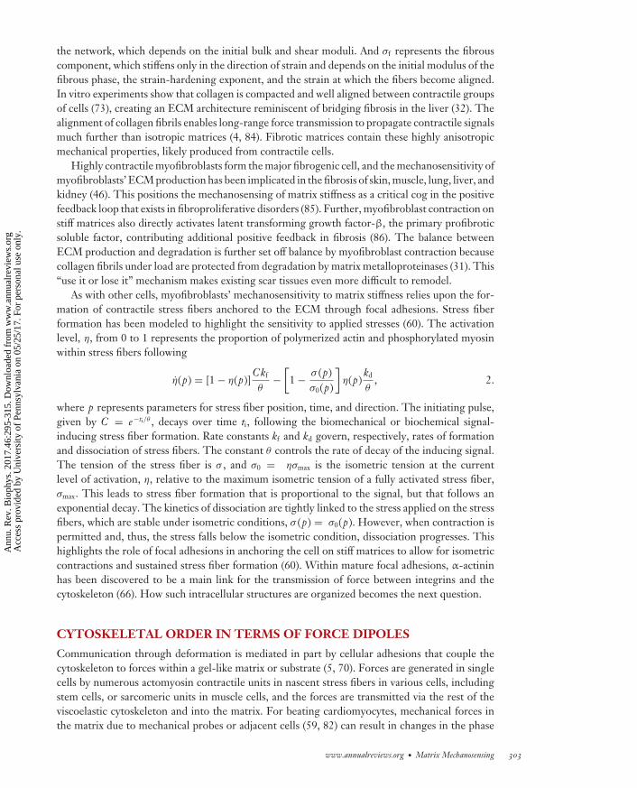
As with other cells, myofibroblasts’ mechanosensitivity to matrix stiffness relies upon the for-mation of contractile stress fibers anchored to the ECM through focal adhesions. Stress fiberformation has been modeled to highlight the sensitivity to applied stresses (60). The activationlevel, η, from 0 to 1 represents the proportion of polymerized actin and phosphorylated myosinwithin stress fibers following
η(p) = [1 − η(p)]Ckf
θ−[
1 − σ (p)σ0(p)
]η(p)
kd
θ, 2.
where p represents parameters for stress fiber position, time, and direction. The initiating pulse,given by C = e−ti/θ , decays over time ti, following the biomechanical or biochemical signal-inducing stress fiber formation. Rate constants kf and kd govern, respectively, rates of formationand dissociation of stress fibers. The constant θ controls the rate of decay of the inducing signal.The tension of the stress fiber is σ , and σ0 = ησmax is the isometric tension at the currentlevel of activation, η, relative to the maximum isometric tension of a fully activated stress fiber,σmax. This leads to stress fiber formation that is proportional to the signal, but that follows anexponential decay. The kinetics of dissociation are tightly linked to the stress applied on the stressfibers, which are stable under isometric conditions, σ (p) = σ0(p). However, when contraction ispermitted and, thus, the stress falls below the isometric condition, dissociation progresses. Thishighlights the role of focal adhesions in anchoring the cell on stiff matrices to allow for isometriccontractions and sustained stress fiber formation (60). Within mature focal adhesions, α-actininhas been discovered to be a main link for the transmission of force between integrins and thecytoskeleton (66). How such intracellular structures are organized becomes the next question.
CYTOSKELETAL ORDER IN TERMS OF FORCE DIPOLES
Communication through deformation is mediated in part by cellular adhesions that couple thecytoskeleton to forces within a gel-like matrix or substrate (5, 70). Forces are generated in singlecells by numerous actomyosin contractile units in nascent stress fibers in various cells, includingstem cells, or sarcomeric units in muscle cells, and the forces are transmitted via the rest of theviscoelastic cytoskeleton and into the matrix. For beating cardiomyocytes, mechanical forces inthe matrix due to mechanical probes or adjacent cells (59, 82) can result in changes in the phase
www.annualreviews.org • Matrix Mechanosensing 303
Ann
u. R
ev. B
ioph
ys. 2
017.
46:2
95-3
15. D
ownl
oade
d fr
om w
ww
.ann
ualr
evie
ws.
org
Acc
ess
prov
ided
by
Uni
vers
ity o
f Pe
nnsy
lvan
ia o
n 05
/25/
17. F
or p
erso
nal u
se o
nly.
BB46CH14-Discher ARI 13 April 2017 14:48
and frequency of beating (17). Cell motility has long been known to be regulated by substratestiffness (as in durotaxis) (61), and cells can also attract or repel each other depending on substraterigidity (68). Feedback occurring as resistance to force can lead to orientational order (34, 64, 90)or translational registry (21, 33, 55) that depends on matrix stiffness.
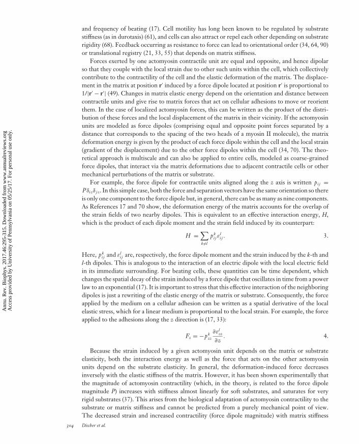
Forces exerted by one actomyosin contractile unit are equal and opposite, and hence dipolarso that they couple with the local strain due to other such units within the cell, which collectivelycontribute to the contractility of the cell and the elastic deformation of the matrix. The displace-ment in the matrix at position r′ induced by a force dipole located at position r′ is proportional to1/|r′ − r′| (49). Changes in matrix elastic energy depend on the orientation and distance betweencontractile units and give rise to matrix forces that act on cellular adhesions to move or reorientthem. In the case of localized actomyosin forces, this can be written as the product of the distri-bution of these forces and the local displacement of the matrix in their vicinity. If the actomyosinunits are modeled as force dipoles (comprising equal and opposite point forces separated by adistance that corresponds to the spacing of the two heads of a myosin II molecule), the matrixdeformation energy is given by the product of each force dipole within the cell and the local strain(gradient of the displacement) due to the other force dipoles within the cell (34, 70). The theo-retical approach is multiscale and can also be applied to entire cells, modeled as coarse-grainedforce dipoles, that interact via the matrix deformations due to adjacent contractile cells or othermechanical perturbations of the matrix or substrate.
For example, the force dipole for contractile units aligned along the z axis is written pi j =Pδi zδ j z. In this simple case, both the force and separation vectors have the same orientation so thereis only one component to the force dipole but, in general, there can be as many as nine components.As References 17 and 70 show, the deformation energy of the matrix accounts for the overlap ofthe strain fields of two nearby dipoles. This is equivalent to an effective interaction energy, H,which is the product of each dipole moment and the strain field induced by its counterpart:
H =∑k �=l
pki j ε
li j . 3.
Here, pki j and εl
i j are, respectively, the force dipole moment and the strain induced by the k-th andl-th dipoles. This is analogous to the interaction of an electric dipole with the local electric fieldin its immediate surrounding. For beating cells, these quantities can be time dependent, whichchanges the spatial decay of the strain induced by a force dipole that oscillates in time from a powerlaw to an exponential (17). It is important to stress that this effective interaction of the neighboringdipoles is just a rewriting of the elastic energy of the matrix or substrate. Consequently, the forceapplied by the medium on a cellular adhesion can be written as a spatial derivative of the localelastic stress, which for a linear medium is proportional to the local strain. For example, the forceapplied to the adhesions along the z direction is (17, 33):
Fz = −pkzz
∂εlzz
∂z. 4.
Because the strain induced by a given actomyosin unit depends on the matrix or substrateelasticity, both the interaction energy as well as the force that acts on the other actomyosinunits depend on the substrate elasticity. In general, the deformation-induced force decreasesinversely with the elastic stiffness of the matrix. However, it has been shown experimentally thatthe magnitude of actomyosin contractility (which, in the theory, is related to the force dipolemagnitude P) increases with stiffness almost linearly for soft substrates, and saturates for veryrigid substrates (37). This arises from the biological adaptation of actomyosin contractility to thesubstrate or matrix stiffness and cannot be predicted from a purely mechanical point of view.The decreased strain and increased contractility (force dipole magnitude) with matrix stiffness
304 Discher et al.
Ann
u. R
ev. B
ioph
ys. 2
017.
46:2
95-3
15. D
ownl
oade
d fr
om w
ww
.ann
ualr
evie
ws.
org
Acc
ess
prov
ided
by
Uni
vers
ity o
f Pe
nnsy
lvan
ia o
n 05
/25/
17. F
or p
erso
nal u
se o
nly.
BB46CH14-Discher ARI 13 April 2017 14:48
ultimately lead to the prediction of an optimal rigidity for which deformation-induced energies orforces are largest. This is a general feature of elastic interactions and is manifested experimentallyin the existence of an optimal rigidity for both the orientational order (34, 64, 90) and translationalregistry order (21, 55) of actomyosin units within single cells.
NUCLEAR LINKAGES AND FORCES IN GENE REGULATION
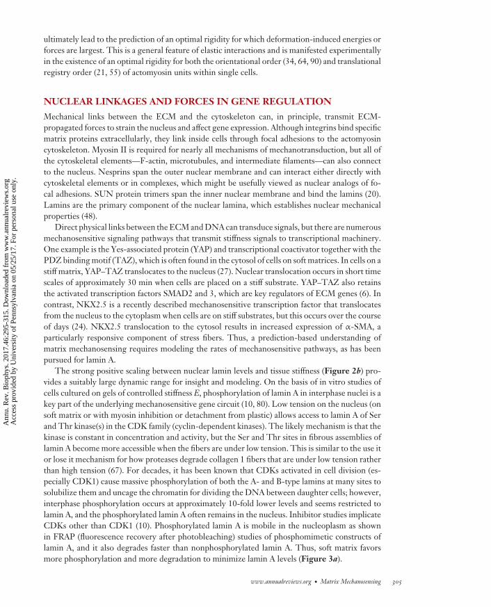
Mechanical links between the ECM and the cytoskeleton can, in principle, transmit ECM-propagated forces to strain the nucleus and affect gene expression. Although integrins bind specificmatrix proteins extracellularly, they link inside cells through focal adhesions to the actomyosincytoskeleton. Myosin II is required for nearly all mechanisms of mechanotransduction, but all ofthe cytoskeletal elements—F-actin, microtubules, and intermediate filaments—can also connectto the nucleus. Nesprins span the outer nuclear membrane and can interact either directly withcytoskeletal elements or in complexes, which might be usefully viewed as nuclear analogs of fo-cal adhesions. SUN protein trimers span the inner nuclear membrane and bind the lamins (20).Lamins are the primary component of the nuclear lamina, which establishes nuclear mechanicalproperties (48).
Direct physical links between the ECM and DNA can transduce signals, but there are numerousmechanosensitive signaling pathways that transmit stiffness signals to transcriptional machinery.One example is the Yes-associated protein (YAP) and transcriptional coactivator together with thePDZ binding motif (TAZ), which is often found in the cytosol of cells on soft matrices. In cells on astiff matrix, YAP–TAZ translocates to the nucleus (27). Nuclear translocation occurs in short timescales of approximately 30 min when cells are placed on a stiff substrate. YAP–TAZ also retainsthe activated transcription factors SMAD2 and 3, which are key regulators of ECM genes (6). Incontrast, NKX2.5 is a recently described mechanosensitive transcription factor that translocatesfrom the nucleus to the cytoplasm when cells are on stiff substrates, but this occurs over the courseof days (24). NKX2.5 translocation to the cytosol results in increased expression of α-SMA, aparticularly responsive component of stress fibers. Thus, a prediction-based understanding ofmatrix mechanosensing requires modeling the rates of mechanosensitive pathways, as has beenpursued for lamin A.
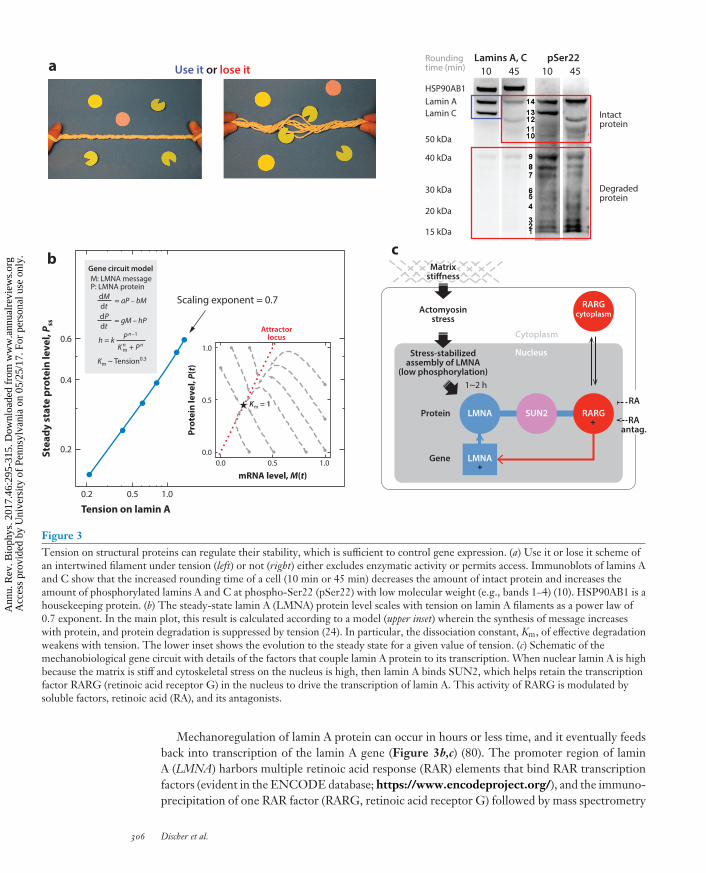
The strong positive scaling between nuclear lamin levels and tissue stiffness (Figure 2b) pro-vides a suitably large dynamic range for insight and modeling. On the basis of in vitro studies ofcells cultured on gels of controlled stiffness E, phosphorylation of lamin A in interphase nuclei is akey part of the underlying mechanosensitive gene circuit (10, 80). Low tension on the nucleus (onsoft matrix or with myosin inhibition or detachment from plastic) allows access to lamin A of Serand Thr kinase(s) in the CDK family (cyclin-dependent kinases). The likely mechanism is that thekinase is constant in concentration and activity, but the Ser and Thr sites in fibrous assemblies oflamin A become more accessible when the fibers are under low tension. This is similar to the use itor lose it mechanism for how proteases degrade collagen 1 fibers that are under low tension ratherthan high tension (67). For decades, it has been known that CDKs activated in cell division (es-pecially CDK1) cause massive phosphorylation of both the A- and B-type lamins at many sites tosolubilize them and uncage the chromatin for dividing the DNA between daughter cells; however,interphase phosphorylation occurs at approximately 10-fold lower levels and seems restricted tolamin A, and the phosphorylated lamin A often remains in the nucleus. Inhibitor studies implicateCDKs other than CDK1 (10). Phosphorylated lamin A is mobile in the nucleoplasm as shownin FRAP (fluorescence recovery after photobleaching) studies of phosphomimetic constructs oflamin A, and it also degrades faster than nonphosphorylated lamin A. Thus, soft matrix favorsmore phosphorylation and more degradation to minimize lamin A levels (Figure 3a).
www.annualreviews.org • Matrix Mechanosensing 305
Ann
u. R
ev. B
ioph
ys. 2
017.
46:2
95-3
15. D
ownl
oade
d fr
om w
ww
.ann
ualr
evie
ws.
org
Acc
ess
prov
ided
by
Uni
vers
ity o
f Pe
nnsy
lvan
ia o
n 05
/25/
17. F
or p
erso
nal u
se o
nly.
BB46CH14-Discher ARI 13 April 2017 14:48
a Use it or lose it
b
10 45 10 45Lamins A, C pSer22Rounding
time (min)
HSP90AB1Lamin A
Intactprotein
Degradedprotein
Lamin C
50 kDa
40 kDa
30 kDa
20 kDa
15 kDa
SUN2
Nucleus
Cytoplasm
Actomyosinstress
Stress-stabilizedassembly of LMNA
(low phosphorylation)1~2 h
LMNA
LMNA
RARG
RARGcytoplasm
c
Protein
Gene
MatrixstiffnessMatrix
stiffness
Stea
dy s
tate
pro
tein
leve
l, P s
s
Prot
ein
leve
l, P(t)
mRNA level, M(t)
Tension on lamin A
0.6
0.4
1.0
0.5
0.00.0 0.5
Attractorlocus
Km = 1
1.0
0.2
0.2 0.5
Scaling exponent = 0.7
1.0
Gene circuit modelM: LMNA messageP: LMNA protein
dM = aP – bMdt
Km ~ Tension0.3
dP = gM – hPdt
h = k P n–1
K nm + P n
RAantag.
RA
Figure 3Tension on structural proteins can regulate their stability, which is sufficient to control gene expression. (a) Use it or lose it scheme ofan intertwined filament under tension (left) or not (right) either excludes enzymatic activity or permits access. Immunoblots of lamins Aand C show that the increased rounding time of a cell (10 min or 45 min) decreases the amount of intact protein and increases theamount of phosphorylated lamins A and C at phospho-Ser22 (pSer22) with low molecular weight (e.g., bands 1–4) (10). HSP90AB1 is ahousekeeping protein. (b) The steady-state lamin A (LMNA) protein level scales with tension on lamin A filaments as a power law of0.7 exponent. In the main plot, this result is calculated according to a model (upper inset) wherein the synthesis of message increaseswith protein, and protein degradation is suppressed by tension (24). In particular, the dissociation constant, Km, of effective degradationweakens with tension. The lower inset shows the evolution to the steady state for a given value of tension. (c) Schematic of themechanobiological gene circuit with details of the factors that couple lamin A protein to its transcription. When nuclear lamin A is highbecause the matrix is stiff and cytoskeletal stress on the nucleus is high, then lamin A binds SUN2, which helps retain the transcriptionfactor RARG (retinoic acid receptor G) in the nucleus to drive the transcription of lamin A. This activity of RARG is modulated bysoluble factors, retinoic acid (RA), and its antagonists.
Mechanoregulation of lamin A protein can occur in hours or less time, and it eventually feedsback into transcription of the lamin A gene (Figure 3b,c) (80). The promoter region of laminA (LMNA) harbors multiple retinoic acid response (RAR) elements that bind RAR transcriptionfactors (evident in the ENCODE database; https://www.encodeproject.org/), and the immuno-precipitation of one RAR factor (RARG, retinoic acid receptor G) followed by mass spectrometry
306 Discher et al.
Ann
u. R
ev. B
ioph
ys. 2
017.
46:2
95-3
15. D
ownl
oade
d fr
om w
ww
.ann
ualr
evie
ws.
org
Acc
ess
prov
ided
by
Uni
vers
ity o
f Pe
nnsy
lvan
ia o
n 05
/25/
17. F
or p
erso
nal u
se o
nly.
BB46CH14-Discher ARI 13 April 2017 14:48
identified the nuclear envelope protein SUN2 as a possible binding partner. SUN2 is an integralmembrane protein known to bind lamin A protein, but SUN2 can also diffuse into the endoplas-mic reticulum, contiguous with the nuclear envelope but extended into much of the cytoplasm.Nuclear entry of RARG proved to be partially regulated by the levels of both SUN2 and laminA. This example of a mechanobiological gene circuit is perhaps a first and can be formalizedmathematically as (80)
d(lamin A)/dt = a LMNA – (tension-suppressed degradation),dLMNA/dt = b(lamin A) – c LMNA,
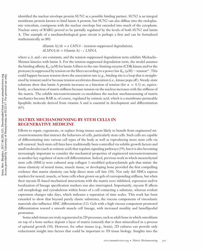
where a, b, and c are constants, and the tension-suppressed degradation term exhibits Michaelis–Menten kinetics with lamin A. For the tension-suppressed degradation term, the model assumesthe binding affinity Km (μM) for lamin A fibers to the rate-limiting enzyme (CDK kinase and/or theprotease) is suppressed by tension on the fibers according to a power law Km (μM) ∼ tensionα . Thiscould happen because tension slows the association rate (e.g., binding site is a loop that is straight-ened by tension) and/or because tension accelerates dissociation (i.e., kinase pops off ). Steady-statesolutions show that lamin A protein increases as a function of tension (for α = 0.3) or, equiva-lently, as a function of matrix stiffness because tension on the nucleus increases with the stiffness ofthe matrix. The soluble microenvironment co-modulates the nuclear mechanosensing of matrixmechanics because RAR is, of course, regulated by retinoic acid, which is a membrane-permeablelipophilic molecule derived from vitamin A and is essential in development and differentiation(65).
MATRIX MECHANOSENSING BY STEM CELLS INREGENERATIVE MEDICINE
Efforts to repair, regenerate, or replace living tissues seem likely to benefit from engineered mi-croenvironments that instruct the behaviors of cells, particularly stem cells. Such cells are capableof differentiating into various cell types of the body as well as reproducing more stem cells viaself-renewal. Such stem cell fates have traditionally been controlled via soluble growth factors andsmall molecules (such as retinoic acid) that regulate signaling pathways (39), but it is also becomingincreasingly important to consider the mechanical properties of engineered microenvironmentsas another key regulator of stem cell differentiation. Indeed, previous work in which mesenchymalstem cells (MSCs) were cultured atop collagen 1–modified polyacrylamide gels that mimic thetissue elasticity of neural tissue, muscle tissue, or developing bone provided the first compellingevidence that matrix elasticity can help direct stem cell fate (30). Not only did MSCs expressmarkers for neural, muscle, or bone cells when grown on gels of corresponding stiffness, but whentheir myosin II–based mechanical interactions with the matrix were inhibited, expression and/orlocalization of lineage specification markers was also interrupted. Importantly, myosin II affectscell morphology and cytoskeleton within hours of a cell contacting a substrate, whereas evidentexpression changes take days, which indicates a separation of time scales. This work has beenextended to show that beyond purely elastic substrates, the viscous components of viscoelasticmaterials also influence MSC differentiation (12). Gels with a high viscous component promoteddifferentiation toward a smooth muscle cell lineage, with increased motility and lamellipodialprotrusion.
Some adult tissues are truly regenerated in 2D processes, such as adult bone in which osteoblastson top of a bone surface deposit a layer of matrix (osteoid) that is then mineralized in a processof epitaxial growth (30). However, for other tissues (e.g., brain), 2D cultures can provide onlyreductionist insight into factors that could be important to 3D tissue biology. Insights into the
www.annualreviews.org • Matrix Mechanosensing 307
Ann
u. R
ev. B
ioph
ys. 2
017.
46:2
95-3
15. D
ownl
oade
d fr
om w
ww
.ann
ualr
evie
ws.
org
Acc
ess
prov
ided
by
Uni
vers
ity o
f Pe
nnsy
lvan
ia o
n 05
/25/
17. F
or p
erso
nal u
se o
nly.
BB46CH14-Discher ARI 13 April 2017 14:48
regeneration of 3D tissues could benefit from rationally engineered 3D culture systems that(a) eliminate apical–basal polarization while still paying attention to (b) access to soluble nutrientsand (c) physical caging constraints on cell morphology or proliferation, or both. The encapsulationof MSCs in 3D hydrogels of alginate (a carbohydrate commonly derived from brown seaweed)that was modified with Arg–Gly–Asp (or RGD) adhesion peptides (40) showed that soft gels withelasticity from 2.5 to 5 kPa favored adipogenesis (a soft tissue lineage), whereas stiff gels of 11 to30 kPa favored osteogenesis. Although 2D cultures on these gels were not studied and would allowone to measure any changes in gel mechanics caused by cells, the results are in close agreement withearlier 2D studies that used nondegradable polyacrylamide gels (80). Any degradation or extensivephysical remodeling of the matrix can be expected to change the matrix mechanics and, therefore,requires local measurements of the mechanics of the gel around the cell. The encapsulation ofMSCs in 3D hyaluronic acid–based gels indeed revealed that when the cell-mediated degradationof stiff gels was restricted so that cells remained spherically encaged, upregulation of myosin IItension was required to favor osteogenesis over adipogenesis (45). This is consistent with thetheory and experiments on MSCs that showed how cell shape influences cytoskeletal tension (90).
Matrix mechanotransduction pathways that affect stem cell fate must somehow enter the nu-cleus and coregulate gene expression. Such pathways could involve the nuclear accumulation andautocatalytic expression of basal levels of lineage-specific transcription factors (30) or perhaps moregeneric factors. YAP and TAZ are well-characterized examples of transcriptional regulators thatgenerally affect cell growth and differentiation. Human pluripotent stem cells (hPSCs) culturedon brain-like compliant polyacrylamide hydrogel (0.75 kPa elasticity) showed nuclear exclusionof YAP and differentiation into postmitotic neurons, whereas hPSCs on stiff gels (10 kPa) showedabundant YAP nuclear localization and maintenance of pluripotency (58). Compared with tradi-tional neurogenic induction methods that use soluble factors, hPSCs more rapidly and efficientlydifferentiated into neurons on the compliant gels in the absence of neurogenic induction factors.Furthermore, dynamic changes in substrate stiffness have highlighted an important window ofmechanosensitivity in stem cell neuronal differentiation (63). However, the results for hPSCs onthe stiff gels indicate differences from MSCs, which highlights the cell type–specific nature ofmechanoresponses. Indeed, hematopoietic stem and progenitor cells taken from marrow, whichis soft in this respect and similar to brain tissue, or else taken from a stiffer bone niche are alsomechanoresponsive to matrix elasticity, but these cells remain blood-lineage committed (75).
Tissues and gels can, of course, possess mechanical properties far more complex than simpleelasticity. If they experience too much strain or stress, they will yield, break, and/or flow. Sometissues, such as embryonic brain, are so soft that they creep and flow irreversibly (exhibiting plastic-ity) under microscale strain, whereas other tissues, such as embryonic heart, are resiliently elasticand recover completely from externally imposed strain (54). Bulk measurements of a few tissues ofintermediate stiffness, such as liver, have also indicated that these tissues exhibit stress relaxationwhen exposed to what might be a nonphysiologically high strain of 15% (14). This has motivatedthe development of alginate gels with tunable stress relaxation timescales (1 min to 40 min) butotherwise equivalent elastic moduli, ligand densities, and degradation characteristics (14). MSCsgrown in 9 kPa gels exhibited maximum adipogenesis in slow-relaxing gels, but MSCs grown in17 kPa gels exhibited maximum osteogenesis in fast-relaxing gels. Adhesive ligand clustering wasalso measured and could relate to the accumulation of gel around a cell—hence locally stiff matrix—but it is also clear that matrix relaxation permits cell protrusion and morphological changes so thatcell shape can again influence cytoskeletal tension (90). Matrix relaxation can also permit prolifer-ation that can modulate differentiation. Although the heterogeneous mechanics that arise after cellintegration requires careful measurement, these studies nevertheless underscore the fundamentalroles that matrix mechanics and physical properties have on stem cell fate.
308 Discher et al.
Ann
u. R
ev. B
ioph
ys. 2
017.
46:2
95-3
15. D
ownl
oade
d fr
om w
ww
.ann
ualr
evie
ws.
org
Acc
ess
prov
ided
by
Uni
vers
ity o
f Pe
nnsy
lvan
ia o
n 05
/25/
17. F
or p
erso
nal u
se o
nly.
BB46CH14-Discher ARI 13 April 2017 14:48
Matrix elasticity in vitro clearly influences transplantation in vivo. In particular, the self-renewalof proliferating stem cells is influenced by matrix elasticity, at least for mouse muscle stem cells(38). The growth of freshly isolated cells on muscle-like 12 kPa hydrogels (functionalized withthe ECM protein laminin) produced the greatest number of viable, transplant-competent cellscompared with softer or stiffer gels or even 2D culture plastic (approximately 106 kPa stiffness)coated with a very thin layer of gel to maintain the surface chemistry. A memory of in vitro matrixinteractions was demonstrated with MSCs derived from bone marrow (which is soft) and thencultured on rigid polystyrene dishes for a prolonged time, which thereby favored osteogenesis evenwhen the cells were transferred to a 2 kPa polyethylene glycol (PEG) hydrogel (88). Alternatively,if culture on plastic is kept sufficiently brief, then such differentiation can be suppressed.
Ensuring that a matrix is sufficiently malleable to cells can facilitate wound healing in vivo.Indeed, the myosin-dependent contraction and migration of fibroblasts around a wound gap areprimary mechanisms through which closure occurs, as opposed to the proliferation of cells (69).The forces transmitted through cell–cell contacts have proven to be critical factors in layers ofcells moving together, as in wound healing (81). Using void-forming alginate hydrogels, murineMSCs exhibited the greatest proliferation, collagen deposition, and mineralization, with constructelasticity ranging from 20 to 60 kPa (41). Transplantation into a bone defect model showedmaximal tissue regeneration with gels in the intermediate range of stiffness, perhaps similar instiffness to precalcified bone, or osteoid. More in-depth analysis is needed as other mechanisms,such as host cell infiltration and material degradation, could impact the response. The formationof epithelial cysts also exhibits maximum polarization and lumen formation in a narrow range ofECM elasticity when using PEG hydrogels, with abnormal morphogenesis observed for softerand stiffer gels (28). It seems that a matrix must be sufficiently rigid to provide appropriate cues tocells yet compliant enough to allow a given cell type—that might be strong or might be weak—to manipulate the matrix for migration, spreading, and proliferation. Adhesive ligand densityis generally important for cells to engage the matrix, and also for both adhesion and proteasedegradation of ECM-regulated apical–basal polarity and lumenogenesis. Thus, synthetic ECMtechnologies can provide insight into ECM regulation of complex morphogenetic behaviors andprovide potentially useful rules for regenerative medicine.
Reprogramming the epigenetic state of primary mouse fibroblasts to induced pluripotent stemcells (iPSCs) has also been examined in 3D synthetic hydrogels with modulation of matrix stiff-ness, degradability, and ligand (11). Using PEG hydrogels conjugated with adhesive peptidesand cross-linked with matrix metalloproteinase-cleavable peptides, the fibroblasts were encap-sulated and transduced with the four traditional Yamanaka factors. Compared with traditionalreprogramming in polystyrene dishes, the gels supported sustained proliferation and acceleratedthe reprogramming of the somatic cells. Compared with 128 microenvironmental conditionsspanning stiffness, biochemical presentation, and degradation, the highest efficiency of iPSC re-programming was achieved with hydrogels having a stiffness of 600 Pa, high degradability, andfunctionalization with epithelial cell adhesion molecule (EpCAM). Matrigel (Corning, New York,NY) produced similar efficiency but with less homogeneity in induction, which is interesting inthat Matrigel lacks EpCAM but is similarly soft, which mimics embryonic tissue.
CONCLUSIONS: FROM THE EPIGENETICS OF MECHANOSENSINGTO THE MECHANOGENOMICS OF CANCER
The above discussions often pertain to epigenetics in the sense that the genome is invariant asexpression varies with the mechanics of matrix and tissue. Epigenetics involves many layers ofgene expression regulation that have not yet been examined in relation to matrix mechanosensing,
www.annualreviews.org • Matrix Mechanosensing 309
Ann
u. R
ev. B
ioph
ys. 2
017.
46:2
95-3
15. D
ownl
oade
d fr
om w
ww
.ann
ualr
evie
ws.
org
Acc
ess
prov
ided
by
Uni
vers
ity o
f Pe
nnsy
lvan
ia o
n 05
/25/
17. F
or p
erso
nal u
se o
nly.
BB46CH14-Discher ARI 13 April 2017 14:48
such as methylation of DNA, histone modification, and spatial organization of chromosomes.The detailed biochemical kinetics of any changes in such increasingly established processes willrequire careful comparisons to the rapid kinetics of cytoskeletal changes (hours), but suitablychosen tissues should also be used to calibrate the epigenetic fine tuning of gene expression.For this reason among many others, we began this review by recognizing that tissues and theirconstituent matrices and cells are built from polymers with tissue-dependent concentrations (i.e.,epigenetically set levels). Standardized public transcriptome data together with mass spectrometryproteomics data and tissue stiffness measurements were then used to illustrate polymer physics–type scaling for the most abundant protein in tissue and collagen 1, as well as for one of themost abundant nuclear structural proteins, lamin A. Transcriptome data were discussed not onlyfor hearts from a wide range of species, which indicates common epigenetic pathways despitedifferent genomes, but also for all sorts of perturbations to try to highlight universality in scaling.Injury, including chronic disease, was briefly reviewed because it almost invariably leads to fibrosisdefined by increased collagen and/or cross-linking as well as altered tissue mechanics and function.
Som
atic
mut
atio
n ra
te(m
edia
n nu
mbe
r mut
atio
ns/M
b)
Brain Marrow Breast Kidney LungLiver Skin
10
0.2
1
10
100
Copy number variation (Mb)Number of structural variants
Brain Marrow Muscle Bone
SoftStiff
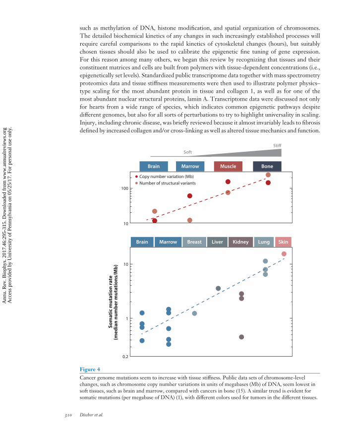
Figure 4Cancer genome mutations seem to increase with tissue stiffness. Public data sets of chromosome-levelchanges, such as chromosome copy number variations in units of megabases (Mb) of DNA, seem lowest insoft tissues, such as brain and marrow, compared with cancers in bone (15). A similar trend is evident forsomatic mutations (per megabase of DNA) (1), with different colors used for tumors in the different tissues.
310 Discher et al.
Ann
u. R
ev. B
ioph
ys. 2
017.
46:2
95-3
15. D
ownl
oade
d fr
om w
ww
.ann
ualr
evie
ws.
org
Acc
ess
prov
ided
by
Uni
vers
ity o
f Pe
nnsy
lvan
ia o
n 05
/25/
17. F
or p
erso
nal u
se o
nly.
BB46CH14-Discher ARI 13 April 2017 14:48
However, tissues are complex, which makes it difficult to distinguish coincidental correlations intissues from cell-level mechanisms.
Numerous culture systems using various materials of controlled elasticity and separately con-trolled biochemistry (from adhesive ligand to soluble factors) were reviewed as influencing isolatedcells in terms of morphology and cytoskeleton, on short time scales, and also in terms of expressionlevels and even differentiation of stem cells on longer time scales. The reductionist approaches inculture can certainly be relevant to tissue engineering as well as the basic science of development,but they also parse the independent variables sufficiently to enable theoretical physics approaches.Theory serves as always to formalize assumptions in fitting experimental results and in generatingpredictions for the next experiments. It has been particularly successful in modeling the organiza-tion of actomyosin cytoskeleton force dipoles in response to matrix elasticity, demonstrating thatsoft matrices suppress order as observed in experiments. Simpler scalar descriptions of stresseswere also integrated into otherwise conventional equations for regulation of gene and protein con-centrations, with calculations ultimately showing that actomyosin stress that increases with matrixstiffness tends to stabilize and thereby increase lamin A. Despite advances in our understanding,much more work is needed on the epigenetic mechanisms of matrix mechanosensing.
In cancer, the genomes of tumors have changed and likely continue to change, with large-scalesequencing (1, 15) revealing the extent of such changes, and, remarkably, genome changes incancer trend again with tissue stiffness (Figure 4). Stiff tissues, such as bone, muscle, and evenskin, lead to cancers that exhibit more genomic changes than tumors originating in soft tissues,such as brain and marrow. Even for skin cancer in which ultraviolet radiation (particularly withaging) clearly accounts for many or most mutations (56), changes in chromosome copy numberassociate with tumor stiffness, with point mutations increasing more weakly as all genomic changesreach a maximum in invasive melanoma (71). Epithelial tissues tend to be moderately stiff as partof a barrier function that also exposes them to carcinogens, but carcinogens cannot easily explainthe increases in chromosome copy number, from low rates in soft marrow and brain to elevatedrates in stiff muscle and higher rates in rigid bone.
Mutations can result from DNA damage and so the scaling of mutation rate with tissue stiffnessis likely explained by mechanoregulation of key DNA damage or repair processes, or both. Nuclearlocalization of DNA repair factors is indeed emerging as mechanosensitive (42, 43), although manyaspects of the mechanism(s) must still be addressed. The cancer genome data viewed from thismechanobiological perspective nonetheless suggest there are some underlying processes of matrixmechanosensing by factors that can alter the fundamental sequence of the genome in cells—thatis, a new field of mechanogenomics.
DISCLOSURE STATEMENT
The authors are not aware of any affiliations, memberships, funding, or financial holdings thatmight be perceived as affecting the objectivity of this review.
ACKNOWLEDGMENTS
Dr. P.C. Dave Dingal is very gratefully acknowledged for contributing Figure 3a. Seminal con-tributions from many groups to the topic of matrix mechanosensing could not be included dueto length restrictions, but some can be found within the references. This work was supported bythe National Institutes of Health, National Cancer Institute (U54-CA193417); National Heart,Lung, and Blood Institute (R01-HL124106 and R21-HL128187); the US-Israel Bilateral ScienceFoundation; the Charles E. Kaufman Foundation Grant (KA2015-79179); and the National Sci-ence Foundation (Materials Research Science and Engineering Center).
www.annualreviews.org • Matrix Mechanosensing 311
Ann
u. R
ev. B
ioph
ys. 2
017.
46:2
95-3
15. D
ownl
oade
d fr
om w
ww
.ann
ualr
evie
ws.
org
Acc
ess
prov
ided
by
Uni
vers
ity o
f Pe
nnsy
lvan
ia o
n 05
/25/
17. F
or p
erso
nal u
se o
nly.
BB46CH14-Discher ARI 13 April 2017 14:48
LITERATURE CITED
1. Alexandrov LB, Nik-Zainal S, Wedge DC, Aparicio SA, Behjati S, et al. 2013. Signatures of mutationalprocesses in human cancer. Nature 500:415–21
2. Barrett T, Wilhite SE, Ledoux P, Evangelista C, Kim IF, et al. 2013. NCBI GEO: Archive for functionalgenomics data sets—update. Nucleic Acids Res. 41:D991–95
3. Barry-Hamilton V, Spangler R, Marshall D, McCauley S, Rodriguez HM, et al. 2010. Allosteric inhibitionof lysyl oxidase-like-2 impedes the development of a pathologic microenvironment. Nat. Med. 16:1009–17
4. Bates JH, Davis GS, Majumdar A, Butnor KJ, Suki B. 2007. Linking parenchymal disease progression tochanges in lung mechanical function by percolation. Am. J. Respir. Crit. Care Med. 176:617–23
5. Ben-Yaakov D, Golkov R, Shokef Y, Safran SA. 2015. Response of adherent cells to mechanical pertur-bations of the surrounding matrix. Soft Matter 11:1412–24
6. Beyer TA, Weiss A, Khomchuk Y, Huang K, Ogunjimi AA, et al. 2013. Switch enhancers interpret TGF-βand Hippo signaling to control cell fate in human embryonic stem cells. Cell Rep. 5:1611–24
7. Bisping E, Ikeda S, Kong SW, Tarnavski O, Bodyak N, et al. 2006. Gata4 is required for maintenance ofpostnatal cardiac function and protection from pressure overload–induced heart failure. PNAS 103:14471–76
8. Booth AJ, Hadley R, Cornett AM, Dreffs AA, Matthes SA, et al. 2012. Acellular normal and fibrotic humanlung matrices as a culture system for in vitro investigation. Am. J. Respir. Crit. Care Med. 186:866–76
9. Buxboim A, Rajagopal K, Brown AE, Discher DE. 2010. How deeply cells feel: methods for thin gels.J. Phys. Condens. Matter 22:194116
10. Buxboim A, Swift J, Irianto J, Spinler KR, Dingal PC, et al. 2014. Matrix elasticity regulates lamin-A,Cphosphorylation and turnover with feedback to actomyosin. Curr. Biol. 24:1909–17
11. Caiazzo M, Okawa Y, Ranga A, Piersigilli A, Tabata Y, Lutolf MP. 2016. Defined three-dimensionalmicroenvironments boost induction of pluripotency. Nat. Mater. 15:344–52
12. Cameron AR, Frith JE, Gomez GA, Yap AS, Cooper-White JJ. 2014. The effect of time-dependentdeformation of viscoelastic hydrogels on myogenic induction and Rac1 activity in mesenchymal stemcells. Biomaterials 35:1857–68
13. Carver W, Goldsmith EC. 2013. Regulation of tissue fibrosis by the biomechanical environment. BioMedRes. Int. 2013:101979
14. Chaudhuri O, Gu L, Klumpers D, Darnell M, Bencherif SA, et al. 2016. Hydrogels with tunable stressrelaxation regulate stem cell fate and activity. Nat. Mater. 15:326–34
15. Chen X, Bahrami A, Pappo A, Easton J, Dalton J, et al. 2014. Recurrent somatic structural variationscontribute to tumorigenesis in pediatric osteosarcoma. Cell Rep. 7:104–12
16. Cho S, Irianto J, Discher DE. 2017. Mechanosensing by the nucleus: from pathways to scaling relation-ships. J. Cell Biol. 216:305–15
17. Cohen O, Safran SA. 2016. Elastic interactions synchronize beating in cardiomyocytes. Soft Matter12:6088–95
18. Cool CD, Groshong SD, Rai PR, Henson PM, Stewart JS, Brown KK. 2006. Fibroblast foci are notdiscrete sites of lung injury or repair: the fibroblast reticulum. Am. J. Respir. Crit. Care Med. 174:654–58
19. Cox TR, Erler JT. 2011. Remodeling and homeostasis of the extracellular matrix: implications for fibroticdiseases and cancer. Dis. Model. Mech. 4:165–78
20. Crisp M, Liu Q, Roux K, Rattner JB, Shanahan C, et al. 2006. Coupling of the nucleus and cytoplasm:role of the LINC complex. J. Cell Biol. 172:41–53
21. Dasbiswas K, Majkut S, Discher DE, Safran SA. 2015. Substrate stiffness–modulated registry phase cor-relations in cardiomyocytes map structural order to coherent beating. Nat. Commun. 6:6085
22. Degos F, Perez P, Roche B, Mahmoudi A, Asselineau J, et al. 2010. Diagnostic accuracy of FibroScanand comparison to liver fibrosis biomarkers in chronic viral hepatitis: a multicenter prospective study (theFIBROSTIC study). J. Hepatol. 53:1013–21
23. Delgado-Olguin P, Huang Y, Li X, Christodoulou D, Seidman CE, et al. 2012. Epigenetic repressionof cardiac progenitor gene expression by Ezh2 is required for postnatal cardiac homeostasis. Nat. Genet.44:343–47
312 Discher et al.
Ann
u. R
ev. B
ioph
ys. 2
017.
46:2
95-3
15. D
ownl
oade
d fr
om w
ww
.ann
ualr
evie
ws.
org
Acc
ess
prov
ided
by
Uni
vers
ity o
f Pe
nnsy
lvan
ia o
n 05
/25/
17. F
or p
erso
nal u
se o
nly.
BB46CH14-Discher ARI 13 April 2017 14:48
24. Dingal PC, Bradshaw AM, Cho S, Raab M, Buxboim A, et al. 2015. Fractal heterogeneity in minimalmatrix models of scars modulates stiff-niche stem-cell responses via nuclear exit of a mechanorepressor.Nat. Mater. 14:951–60
25. Discher DE, Mooney DJ, Zandstra PW. 2009. Growth factors, matrices, and forces combine and controlstem cells. Science 324:1673–77
26. Dittmer TA, Misteli T. 2011. The lamin protein family. Genome Biol. 12:22227. Dupont S, Morsut L, Aragona M, Enzo E, Giulitti S, et al. 2011. Role of YAP/TAZ in mechanotrans-
duction. Nature 474:179–8328. Enemchukwu NO, Cruz-Acuna R, Bongiorno T, Johnson CT, Garcia JR, et al. 2016. Synthetic matrices
reveal contributions of ECM biophysical and biochemical properties to epithelial morphogenesis. J. CellBiol. 212:113–24
29. Engler A, Bacakova L, Newman C, Hategan A, Griffin M, Discher D. 2004. Substrate compliance versusligand density in cell on gel responses. Biophys. J. 86:617–28
30. Engler AJ, Sen S, Sweeney HL, Discher DE. 2006. Matrix elasticity directs stem cell lineage specification.Cell 126:677–89
31. Flynn BP, Bhole AP, Saeidi N, Liles M, Dimarzio CA, Ruberti JW. 2010. Mechanical strain stabilizesreconstituted collagen fibrils against enzymatic degradation by mammalian collagenase matrix metallo-proteinase 8 (MMP-8). PLOS ONE 5:e12337
32. Fontana RJ, Goodman ZD, Dienstag JL, Bonkovsky HL, Naishadham D, et al. 2008. Relationship ofserum fibrosis markers with liver fibrosis stage and collagen content in patients with advanced chronichepatitis C. Hepatology 47:789–98
33. Friedrich BM, Buxboim A, Discher DE, Safran SA. 2011. Striated acto-myosin fibers can reorganize andregister in response to elastic interactions with the matrix. Biophys. J. 100:2706–15
34. Friedrich BM, Safran SA. 2012. How cells feel their substrate: spontaneous symmetry breaking of activesurface stresses. Soft Matter 8:3223–30
35. Frisch SM, Screaton RA. 2001. Anoikis mechanisms. Curr. Opin. Cell Biol. 13:555–6236. Gennes P-G. 1979. Scaling Concepts in Polymer Physics. Ithaca, NY: Cornell Univ. Press37. Ghibaudo M, Saez A, Trichet L, Xayaphoummine A, Browaeys J, et al. 2008. Traction forces and rigidity
sensing regulate cell functions. Soft Matter 4:1836–4338. Gilbert PM, Havenstrite KL, Magnusson KE, Sacco A, Leonardi NA, et al. 2010. Substrate elasticity
regulates skeletal muscle stem cell self-renewal in culture. Science 329:1078–8139. Guilak F, Cohen DM, Estes BT, Gimble JM, Liedtke W, Chen CS. 2009. Control of stem cell fate by
physical interactions with the extracellular matrix. Cell Stem Cell 5:17–2640. Huebsch N, Arany PR, Mao AS, Shvartsman D, Ali OA, et al. 2010. Harnessing traction-mediated
manipulation of the cell/matrix interface to control stem-cell fate. Nat. Mater. 9:518–2641. Huebsch N, Lippens E, Lee K, Mehta M, Koshy ST, et al. 2015. Matrix elasticity of void-forming
hydrogels controls transplanted-stem-cell-mediated bone formation. Nat. Mater. 14:1269–7742. Irianto J, Pfeifer CR, Bennett RR, Xia Y, Ivanovska IL, Liu AJ, et al. 2016. Nuclear constriction segregates
mobile nuclear proteins away from chromatin. Mol. Biol. Cell 27:401–2043. Irianto J, Xia Y, Pfeifer CR, Athirasala A, Ji J, et al. 2017. DNA damage follows repair factor depletion
and portends genome variation in cancer cells after pore migration. Curr. Biol. 27:210–2344. Jorba I, Uriarte JJ, Campillo N, Farre R, Navajas D. 2017. Probing micromechanical properties of the
extracellular matrix of soft tissues by atomic force microscopy. J. Cell. Physiol. 232:19–2645. Khetan S, Guvendiren M, Legant WR, Cohen DM, Chen CS, Burdick JA. 2013. Degradation-mediated
cellular traction directs stem cell fate in covalently crosslinked three-dimensional hydrogels. Nat. Mater.12:458–65
46. Klingberg F, Hinz B, White ES. 2013. The myofibroblast matrix: implications for tissue repair and fibrosis.J. Pathol. 229:298–309
47. Korsmo MJ, Ebrahimi B, Eirin A, Woollard JR, Krier JD, et al. 2013. Magnetic resonance elastographynoninvasively detects in vivo renal medullary fibrosis secondary to swine renal artery stenosis. Investig.Radiol. 48:61–68
48. Lammerding J, Fong LG, Ji JY, Reue K, Stewart CL, et al. 2006. Lamins A and C but not lamin B1regulate nuclear mechanics. J. Biol. Chem. 281:25768–80
www.annualreviews.org • Matrix Mechanosensing 313
Ann
u. R
ev. B
ioph
ys. 2
017.
46:2
95-3
15. D
ownl
oade
d fr
om w
ww
.ann
ualr
evie
ws.
org
Acc
ess
prov
ided
by
Uni
vers
ity o
f Pe
nnsy
lvan
ia o
n 05
/25/
17. F
or p
erso
nal u
se o
nly.
BB46CH14-Discher ARI 13 April 2017 14:48
49. Landau L, Lifshitz E. 1970. Theory of Elasticity. Oxford, UK: Pergamon Press50. Levental KR, Yu H, Kass L, Lakins JN, Egeblad M, et al. 2009. Matrix crosslinking forces tumor pro-
gression by enhancing integrin signaling. Cell 139:891–90651. Licup AJ, Munster S, Sharma A, Sheinman M, Jawerth LM, et al. 2015. Stress controls the mechanics of
collagen networks. PNAS 112:9573–7852. Liu F, Mih JD, Shea BS, Kho AT, Sharif AS, et al. 2010. Feedback amplification of fibrosis through matrix
stiffening and COX-2 suppression. J. Cell Biol. 190:693–70653. Lopez B, Querejeta R, Gonzalez A, Larman M, Diez J. 2012. Collagen cross-linking but not collagen
amount associates with elevated filling pressures in hypertensive patients with stage C heart failure: po-tential role of lysyl oxidase. Hypertension 60:677–83
54. Majkut S, Dingal PC, Discher DE. 2014. Stress sensitivity and mechanotransduction during heart devel-opment. Curr. Biol. 24:R495–501
55. Majkut S, Idema T, Swift J, Krieger C, Liu A, Discher DE. 2013. Heart-specific stiffening in early embryosparallels matrix and myosin expression to optimize beating. Curr. Biol. 23:2434–39
56. Martincorena I, Roshan A, Gerstung M, Ellis P, Van Loo P, et al. 2015. Tumor evolution: high burdenand pervasive positive selection of somatic mutations in normal human skin. Science 348:880–86
57. McKee CT, Last JA, Russell P, Murphy CJ. 2011. Indentation versus tensile measurements of Young’smodulus for soft biological tissues. Tissue Eng. Part B Rev. 17:155–64
58. Musah S, Wrighton PJ, Zaltsman Y, Zhong X, Zorn S, et al. 2014. Substratum-induced differentiation ofhuman pluripotent stem cells reveals the coactivator YAP is a potent regulator of neuronal specification.PNAS 111:13805–10
59. Nitsan I, Drori S, Lewis YE, Cohen S, Tzlil S. 2016. Mechanical communication in cardiac cell synchro-nized beating. Nat. Phys. 12:472–77
60. Pathak A, Deshpande VS, McMeeking RM, Evans AG. 2008. The simulation of stress fibre and focaladhesion development in cells on patterned substrates. J. R. Soc. Interface 5:507–24
61. Pelham RJ Jr., Wang Y. 1997. Cell locomotion and focal adhesions are regulated by substrate flexibility.PNAS 94:13661–65
62. Rajan S, Williams SS, Jagatheesan G, Ahmed RP, Fuller-Bicer G, et al. 2006. Microarray analysis of geneexpression during early stages of mild and severe cardiac hypertrophy. Physiol. Genom. 27:309–17
63. Rammensee S, Kang MS, Georgiou K, Kumar S, Schaffer DV. 2017. Dynamics of mechanosensitiveneural stem cell differentiation. Stem Cells 35:497–506
64. Rehfeldt F, Brown AE, Raab M, Cai S, Zajac AL, et al. 2012. Hyaluronic acid matrices show matrixstiffness in 2D and 3D dictates cytoskeletal order and myosin-II phosphorylation within stem cells. Integr.Biol. 4:422–30
65. Rhinn M, Dolle P. 2012. Retinoic acid signalling during development. Development 139:843–5866. Roca-Cusachs P, del Rio A, Puklin-Faucher E, Gauthier NC, Biais N, Sheetz MP. 2013. Integrin-
dependent force transmission to the extracellular matrix by α-actinin triggers adhesion maturation. PNAS110:E1361–70
67. Ruberti JW, Hallab NJ. 2005. Strain-controlled enzymatic cleavage of collagen in loaded matrix. Biochem.Biophys. Res. Commun. 336:483–89
68. Ryan J, Perez-Avila CA, Vaughan N. 1995. Insulin dependent diabetes mellitus and deliberate self-harm.J. Accid. Emerg. Med. 12:296–97
69. Sakar MS, Eyckmans J, Pieters R, Eberli D, Nelson BJ, Chen CS. 2016. Cellular forces and matrixassembly coordinate fibrous tissue repair. Nat. Commun. 7:11036
70. Schwarz US, Safran SA. 2013. Physics of adherent cells. Rev. Mod. Phys. 85:132771. Shain AH, Yeh I, Kovalyshyn I, Sriharan A, Talevich E, et al. 2015. The genetic evolution of melanoma
from precursor lesions. N. Engl. J. Med. 373:1926–3672. Sharma A, Licup AJ, Jansen KA, Rens R, Sheinman M, Koenderink GH. 2016. Strain-controlled criticality
governs the nonlinear mechanics of fibre networks. Nat. Phys. 12:584–8773. Shi Q, Ghosh RP, Engelke H, Rycroft CH, Cassereau L, et al. 2014. Rapid disorganization of mechanically
interacting systems of mammary acini. PNAS 111:658–63
314 Discher et al.
Ann
u. R
ev. B
ioph
ys. 2
017.
46:2
95-3
15. D
ownl
oade
d fr
om w
ww
.ann
ualr
evie
ws.
org
Acc
ess
prov
ided
by
Uni
vers
ity o
f Pe
nnsy
lvan
ia o
n 05
/25/
17. F
or p
erso
nal u
se o
nly.
BB46CH14-Discher ARI 13 April 2017 14:48
74. Shin JW, Swift J, Spinler KR, Discher DE. 2011. Myosin-II inhibition and soft 2D matrix maximizemultinucleation and cellular projections typical of platelet-producing megakaryocytes. PNAS 108:11458–63
75. Shin JW, Buxboim A, Spinler KR, Swift J, Christian DA, et al. 2014. Contractile forces sustain and polarizehematopoiesis from stem and progenitor cells. Cell Stem Cell 14:81–93
76. Smith LR, Barton ER. 2014. Collagen content does not alter the passive mechanical properties of fibroticskeletal muscle in mdx mice. Am. J. Physiol. Cell Physiol. 306:C889–98
77. Smith LR, Hammers DW, Sweeney HL, Barton ER. 2015. Increased collagen cross-linking is a signatureof dystrophin-deficient muscle. Muscle Nerve 54:71–78
78. Smith LR, Lee KS, Ward SR, Chambers HG, Lieber RL. 2011. Hamstring contractures in childrenwith spastic cerebral palsy result from a stiffer ECM and increased in vivo sarcomere length. J. Physiol.589:2625–39
79. Sonna LA, Fujita J, Gaffin SL, Lilly CM. 2002. Effects of heat and cold stress on mammalian geneexpression. J. Appl. Physiol. 92:1725–42
80. Swift J, Ivanovska IL, Buxboim A, Harada T, Dingal PC, et al. 2013. Nuclear lamin-A scales with tissuestiffness and enhances matrix-directed differentiation. Science 341:1240104
81. Tambe DT, Hardin CC, Angelini TE, Rajendran K, Park CY, et al. 2011. Collective cell guidance bycooperative intercellular forces. Nat. Mater. 10:469–75
82. Tang X, Bajaj P, Bashir R. 2016. How far cardiac cells can see each other mechanically. Soft Matter7:6151–58
83. Vizcaino JA, Csordas A, del-Toro N, Dianes JA, Griss J, et al. 2016. 2016 update of the PRIDE databaseand its related tools. Nucleic Acids Res. 44:D447–56
84. Wang H, Abhilash AS, Chen CS, Wells RG, Shenoy VB. 2014. Long-range force transmission in fibrousmatrices enabled by tension-driven alignment of fibers. Biophys. J. 107:2592–603
85. Wells RG. 2013. Tissue mechanics and fibrosis. Biochim. Biophys. Acta 1832:884–9086. Wipff PJ, Rifkin DB, Meister JJ, Hinz B. 2007. Myofibroblast contraction activates latent TGF-β1 from
the extracellular matrix. J. Cell Biol. 179:1311–2387. Wynn TA. 2007. Common and unique mechanisms regulate fibrosis in various fibroproliferative diseases.
J. Clin. Investig. 117:524–2988. Yang C, Tibbitt MW, Basta L, Anseth KS. 2014. Mechanical memory and dosing influence stem cell fate.
Nat. Mater. 13:645–5289. Yang YL, Leone LM, Kaufman LJ. 2009. Elastic moduli of collagen gels can be predicted from two-
dimensional confocal microscopy. Biophys. J. 97:2051–6090. Zemel A, Rehfeldt F, Brown AE, Discher DE, Safran SA. 2010. Optimal matrix rigidity for stress fiber
polarization in stem cells. Nat. Phys. 6:468–73
www.annualreviews.org • Matrix Mechanosensing 315
Ann
u. R
ev. B
ioph
ys. 2
017.
46:2
95-3
15. D
ownl
oade
d fr
om w
ww
.ann
ualr
evie
ws.
org
Acc
ess
prov
ided
by
Uni
vers
ity o
f Pe
nnsy
lvan
ia o
n 05
/25/
17. F
or p
erso
nal u
se o
nly.