Embed Size (px)
Citation preview
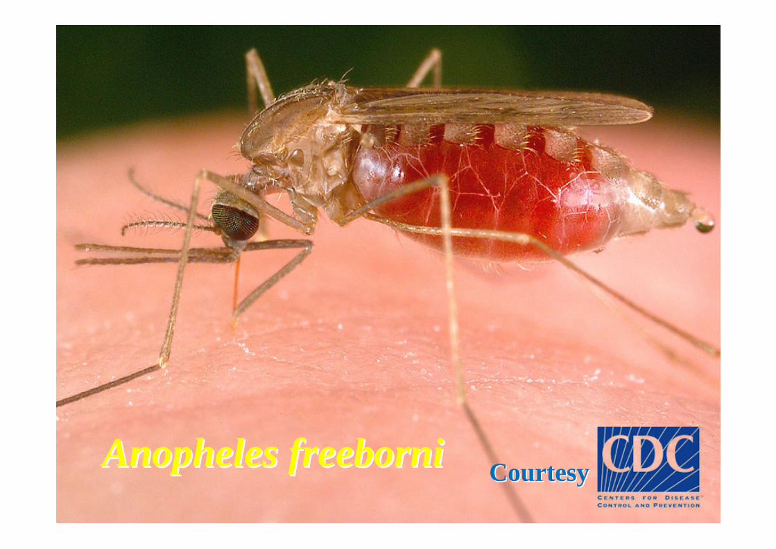
CourtesyCourtesyAnopheles freeborniAnopheles freeborni

Plasmodiaseen with the microscope
M. Lontie, MCH, Leuven, 2012
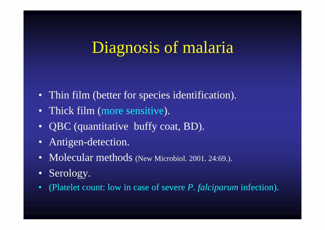
Diagnosis of malaria
• Thin film (better for species identification).• Thick film (more sensitive).• QBC (quantitative buffy coat, BD).• Antigen-detection.• Molecular methods (New Microbiol. 2001. 24:69.).
• Serology.• (Platelet count: low in case of severe P. falciparum infection).
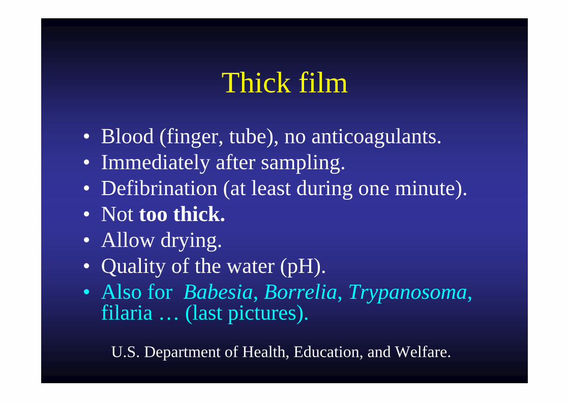
Thick film
• Blood (finger, tube), no anticoagulants.• Immediately after sampling.• Defibrination (at least during one minute).• Not too thick.• Allow drying.• Quality of the water (pH).• Also for Babesia, Borrelia, Trypanosoma,
filaria … (last pictures).
U.S. Department of Health, Education, and Welfare.
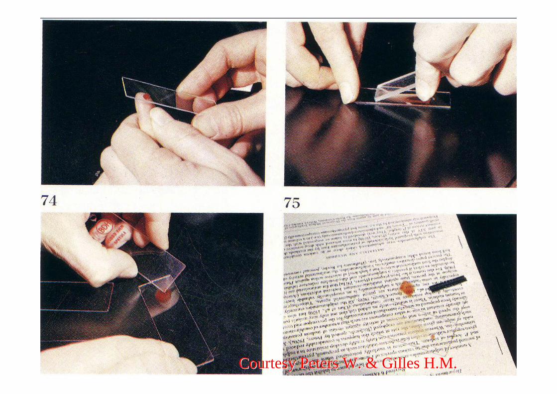
Courtesy Peters W. & Gilles H.M.Courtesy Peters W. & Gilles H.M.
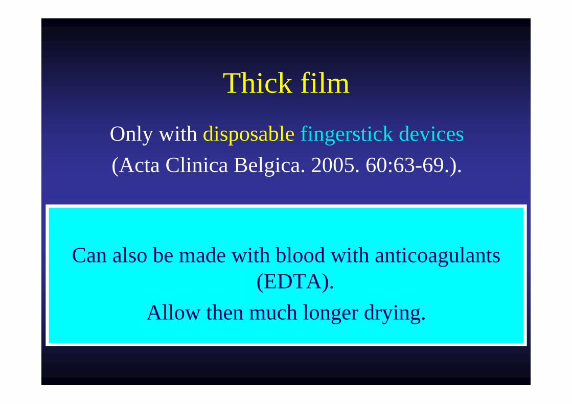
Thick film
Can also be made with blood with anticoagulants (EDTA).
Allow then much longer drying.
Only with disposable fingerstick devices(Acta Clinica Belgica. 2005. 60:63-69.).
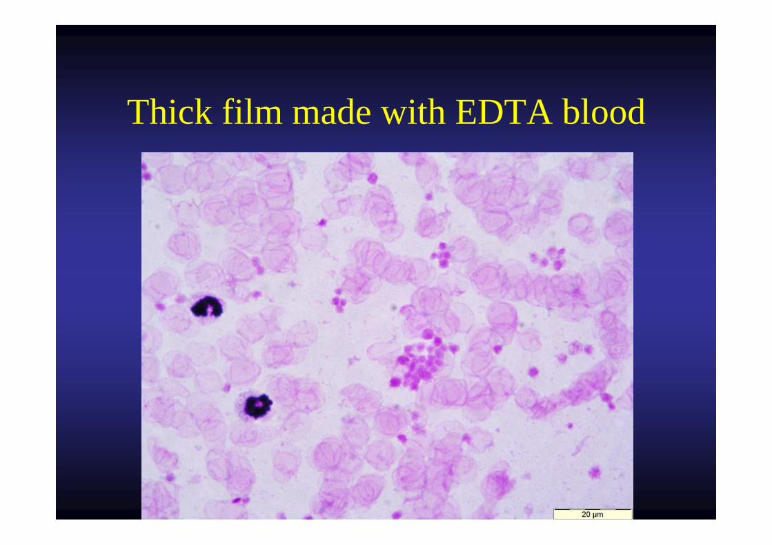
Thick film made with EDTA blood
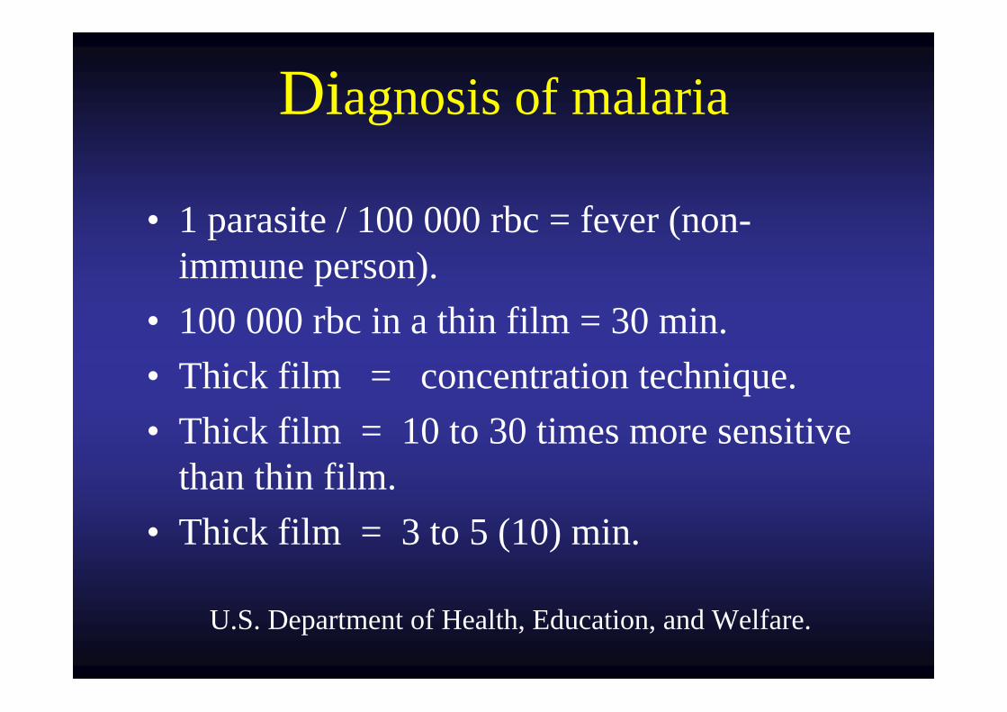
Diagnosis of malaria
• 1 parasite / 100 000 rbc = fever (non-immune person).
• 100 000 rbc in a thin film = 30 min. • Thick film = concentration technique.• Thick film = 10 to 30 times more sensitive
than thin film.• Thick film = 3 to 5 (10) min.
U.S. Department of Health, Education, and Welfare.
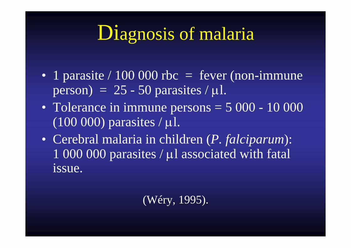
Diagnosis of malaria
• 1 parasite / 100 000 rbc = fever (non-immune person) = 25 - 50 parasites / μl.
• Tolerance in immune persons = 5 000 - 10 000 (100 000) parasites / μl.
• Cerebral malaria in children (P. falciparum): 1 000 000 parasites / μl associated with fatal issue.
(Wéry, 1995).
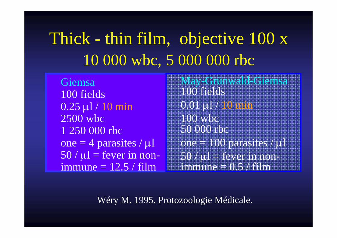
Thick - thin film, objective 100 x 10 000 wbc, 5 000 000 rbc
Giemsa100 fields 0.25 μl / 10 min 2500 wbc 1 250 000 rbc one = 4 parasites / μl 50 / μl = fever in non-immune = 12.5 / film
May-Grünwald-Giemsa100 fields
0.01 μl / 10 min 100 wbc
50 000 rbc one = 100 parasites / μl 50 / μl = fever in non-
immune = 0.5 / film
Wéry M. 1995. Protozoologie Médicale.
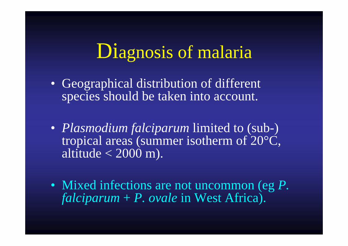
Diagnosis of malaria• Geographical distribution of different
species should be taken into account.
• Plasmodium falciparum limited to (sub-) tropical areas (summer isotherm of 20°C, altitude < 2000 m).
• Mixed infections are not uncommon (eg P. falciparum + P. ovale in West Africa).
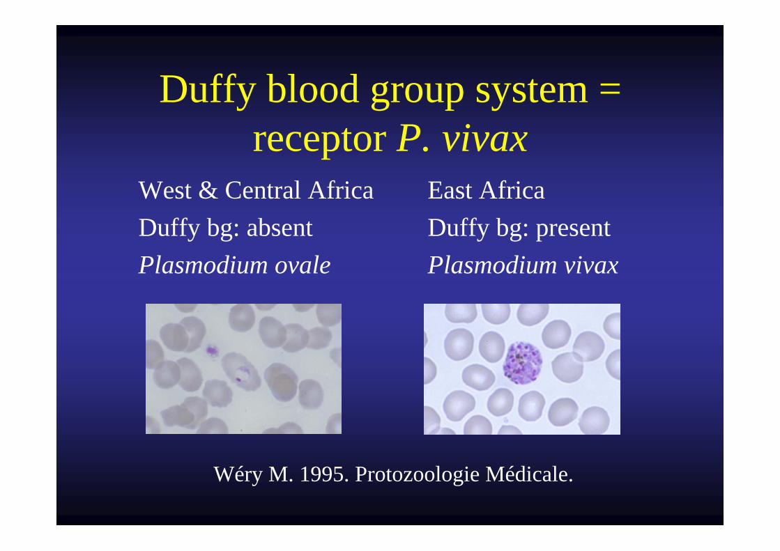
Duffy blood group system = receptor P. vivax
West & Central Africa Duffy bg: absent Plasmodium ovale
East Africa Duffy bg: present Plasmodium vivax
Wéry M. 1995. Protozoologie Médicale.
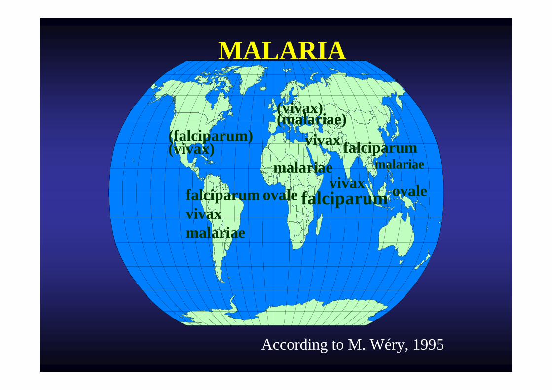
vivaxovale falciparum
falciparummalariae
vivax
falciparumvivaxmalariae
(vivax)(malariae)
(falciparum) (vivax)
MALARIA
ovale
malariae
According to M. Wéry, 1995
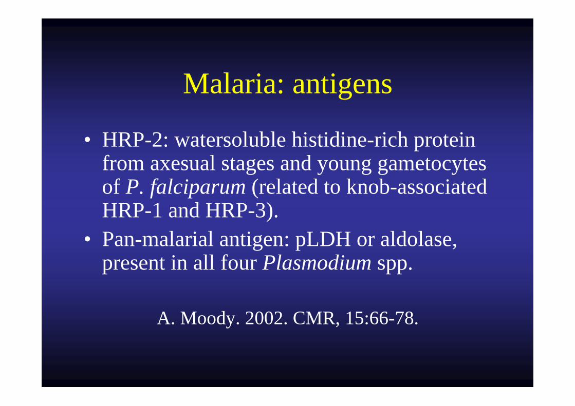
Malaria: antigens
• HRP-2: watersoluble histidine-rich protein from axesual stages and young gametocytes of P. falciparum (related to knob-associated HRP-1 and HRP-3).
• Pan-malarial antigen: pLDH or aldolase, present in all four Plasmodium spp.
A. Moody. 2002. CMR, 15:66-78.
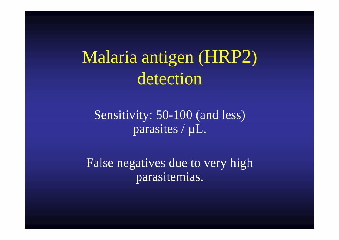
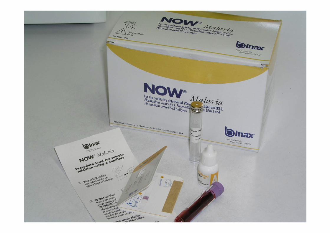
Malaria antigen (HRP2) detection
Sensitivity: 50-100 (and less) parasites / µL.
False negatives due to very high parasitemias.
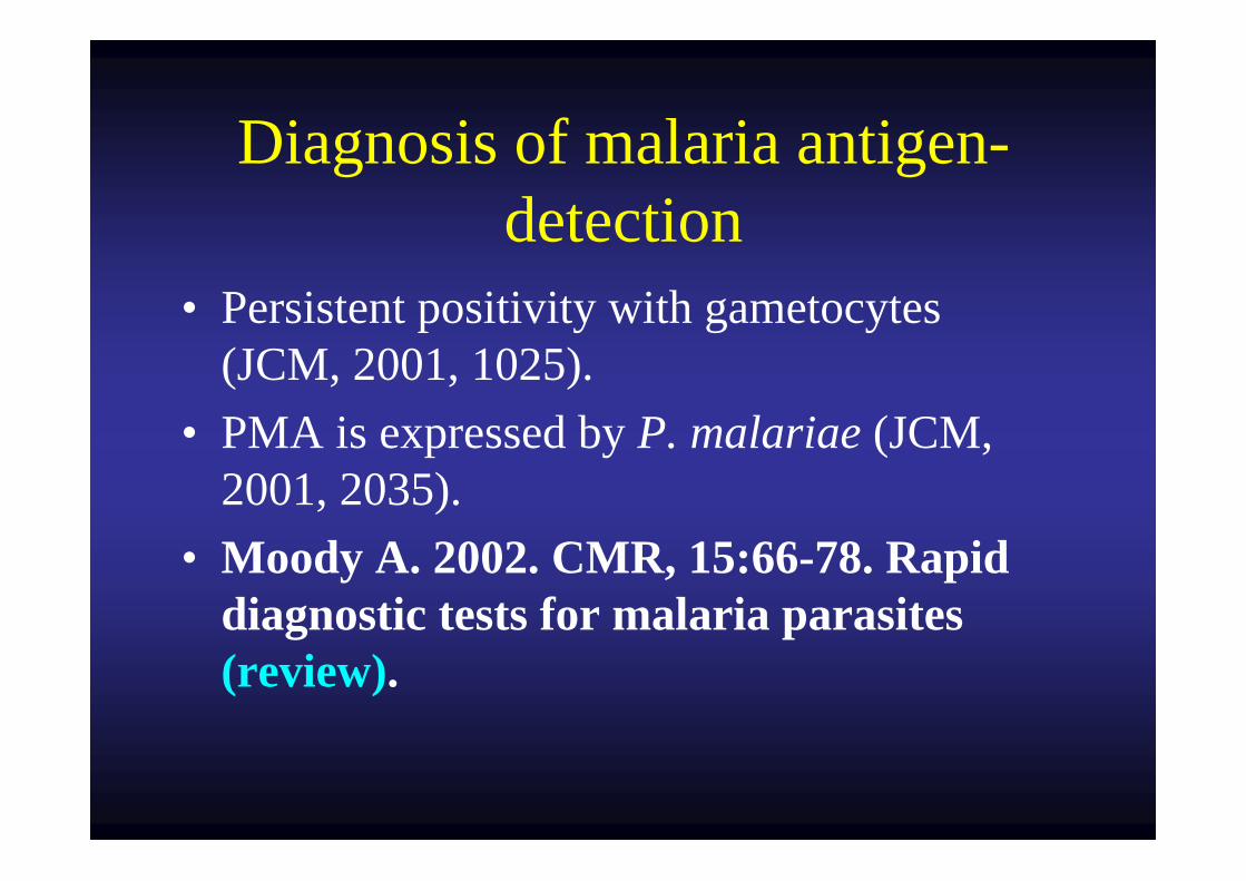
Diagnosis of malaria antigen-detection
• Persistent positivity with gametocytes (JCM, 2001, 1025).
• PMA is expressed by P. malariae (JCM, 2001, 2035).
• Moody A. 2002. CMR, 15:66-78. Rapid diagnostic tests for malaria parasites (review).
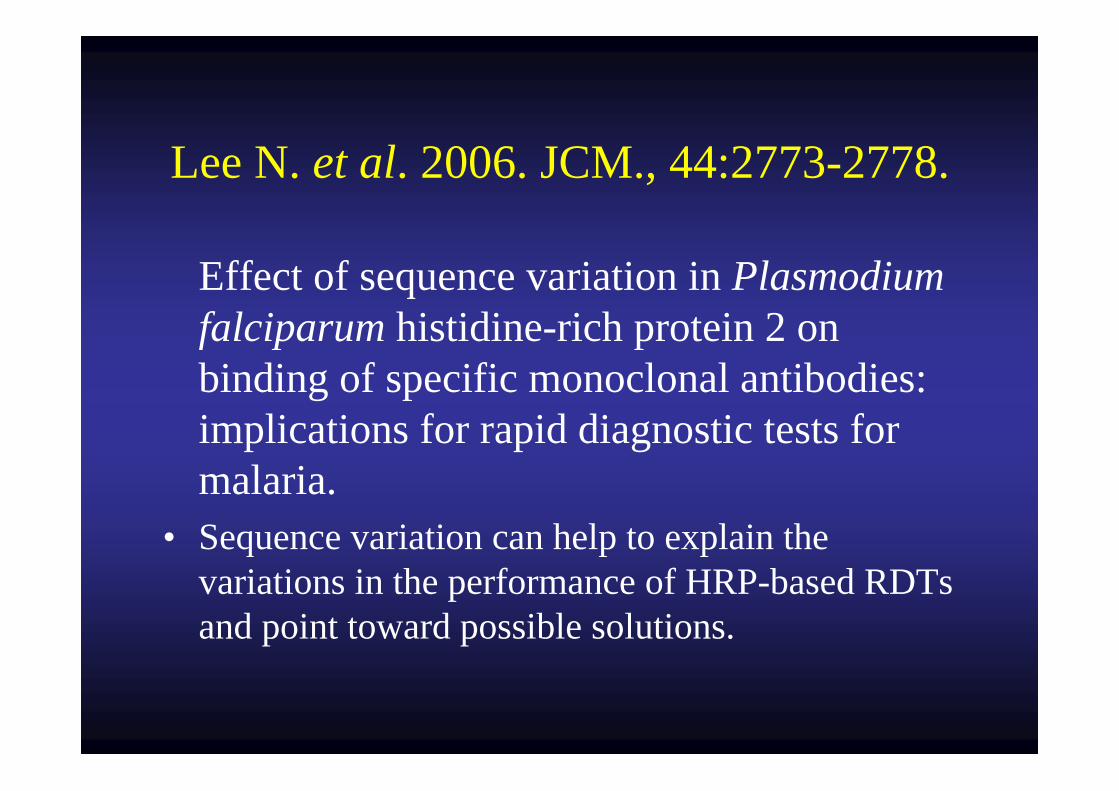
Lee N. et al. 2006. JCM., 44:2773-2778.
Effect of sequence variation in Plasmodiumfalciparum histidine-rich protein 2 on binding of specific monoclonal antibodies: implications for rapid diagnostic tests for malaria.
• Sequence variation can help to explain the variations in the performance of HRP-based RDTs and point toward possible solutions.
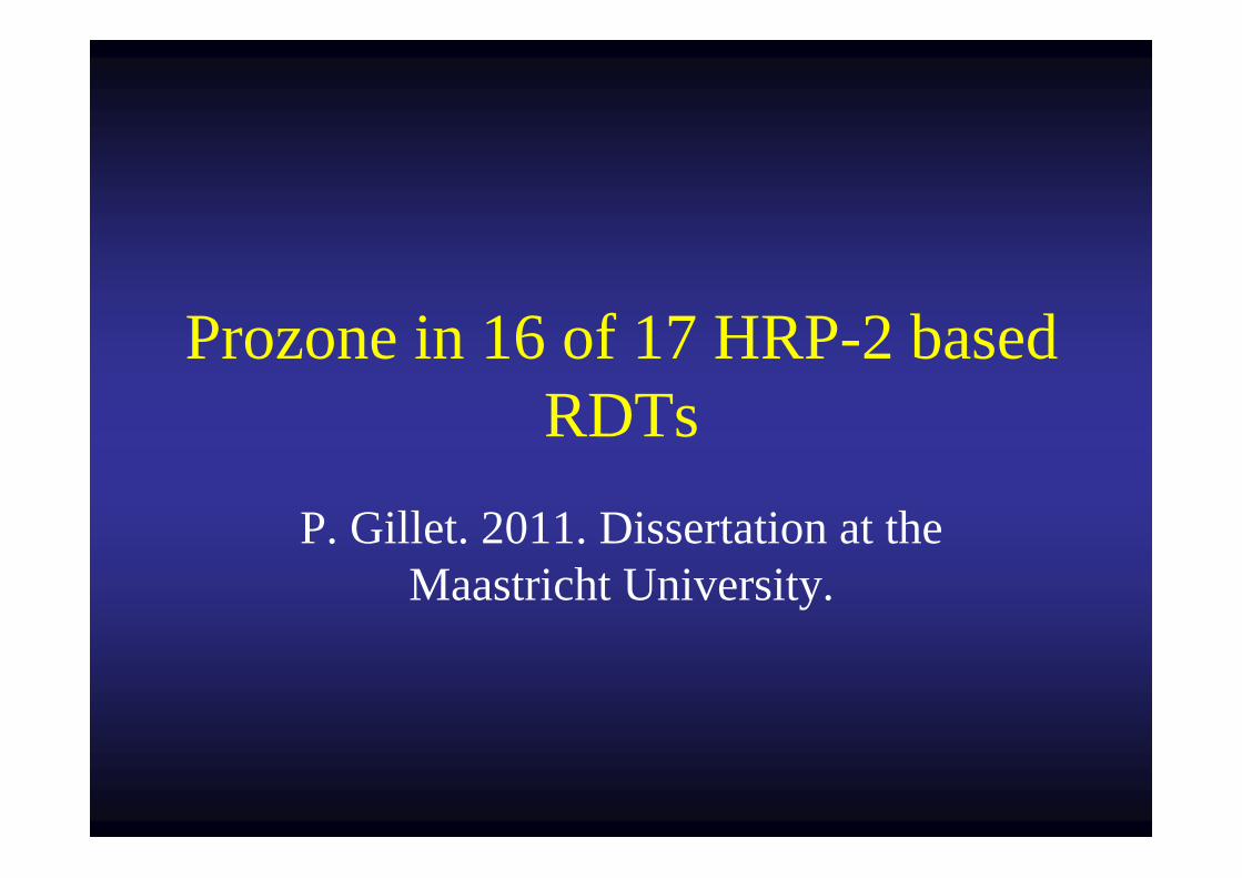
Prozone in 16 of 17 HRP-2 based RDTs
P. Gillet. 2011. Dissertation at the Maastricht University.
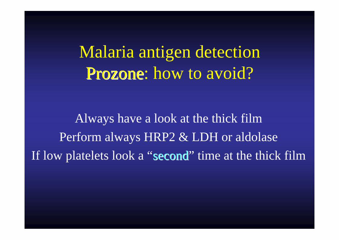
Malaria antigen detectionProzoneProzone: how to avoid?
Always have a look at the thick filmPerform always HRP2 & LDH or aldolase
If low platelets look a “secondsecond” time at the thick film
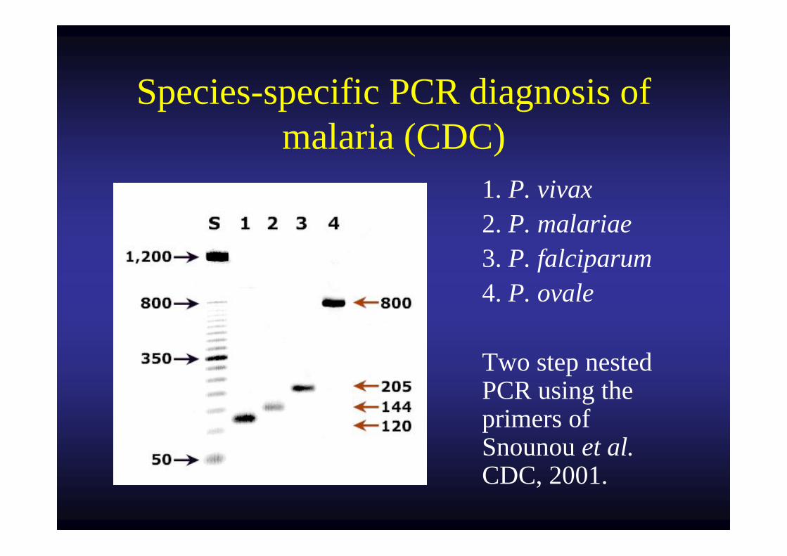
Species-specific PCR diagnosis of malaria (CDC)
1. P. vivax 2. P. malariae 3. P. falciparum 4. P. ovale
Two step nested PCR using the primers of Snounou et al. CDC, 2001.
Detection of four Plasmodiumspecies in blood from humans by
18S rRNA gene subunit-based and species-specific real time PCR
assays
Rougemont M. et al. 2004. JCM, 42:5636-5643.
Malaria serology
• Only P. falciparum cultivated in vitro.
• To exclude malaria e.g. among travellers. • Blood donation from travellers.• For epidemiological reasons.
Wéry M., 1995.
Malaria: staining procedures used
• Thick film: Giemsa staining.
• Thin film: May-Grünwald staining.
Inexpensive method of diluting Giemsa stain
• Petithory J.C. et al. 2005. JCM, 43:528.
29 bottles of Evian water.pH: 7.36Ready to use and stable for weeks.
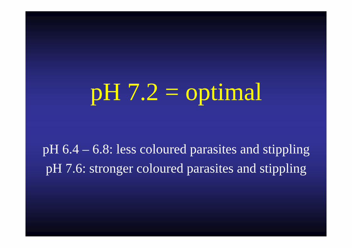
pH 7.2 = optimal
pH 6.4 – 6.8: less coloured parasites and stipplingpH 7.6: stronger coloured parasites and stippling
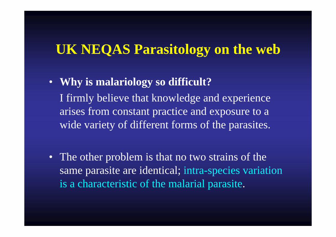
UK NEQAS Parasitology on the web
• Why is malariology so difficult?I firmly believe that knowledge and experience arises from constant practice and exposure to a wide variety of different forms of the parasites.
• The other problem is that no two strains of the same parasite are identical; intra-species variation is a characteristic of the malarial parasite.
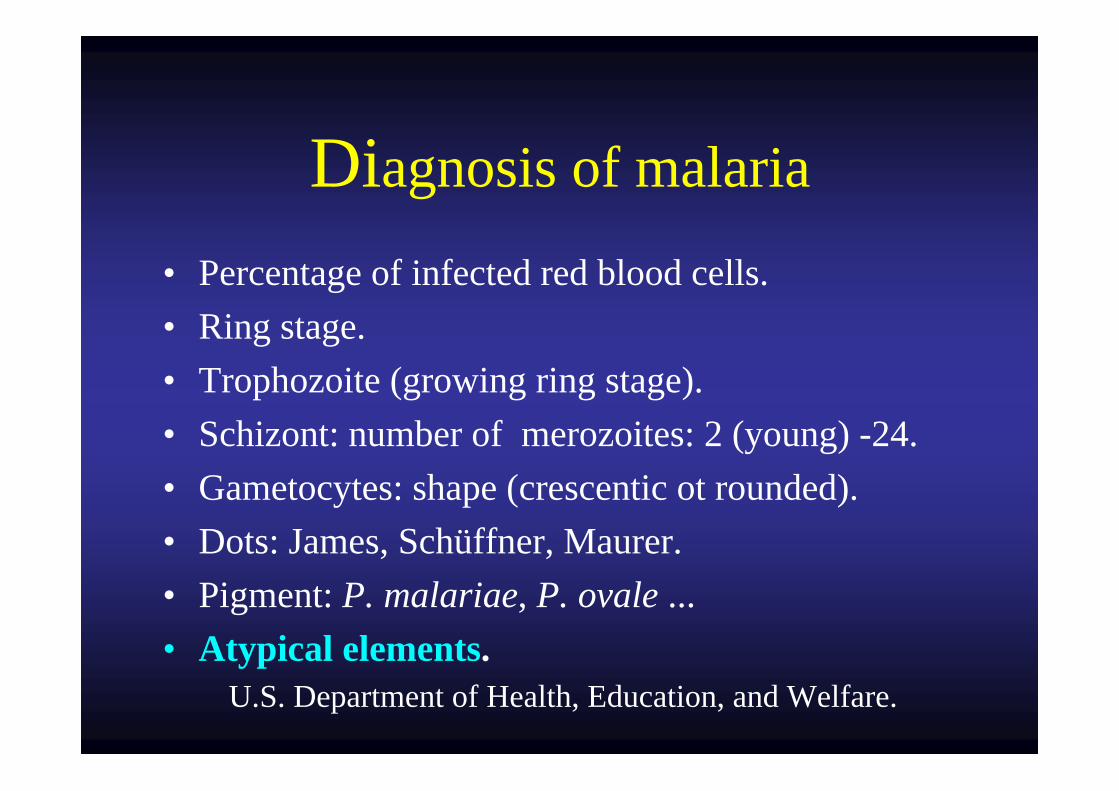
Diagnosis of malaria
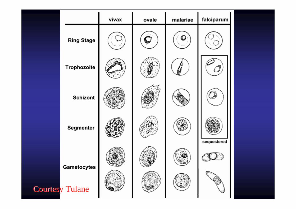
• Percentage of infected red blood cells.• Ring stage.• Trophozoite (growing ring stage).• Schizont: number of merozoites: 2 (young) -24.• Gametocytes: shape (crescentic ot rounded).• Dots: James, Schüffner, Maurer.• Pigment: P. malariae, P. ovale ...• Atypical elements.
U.S. Department of Health, Education, and Welfare.
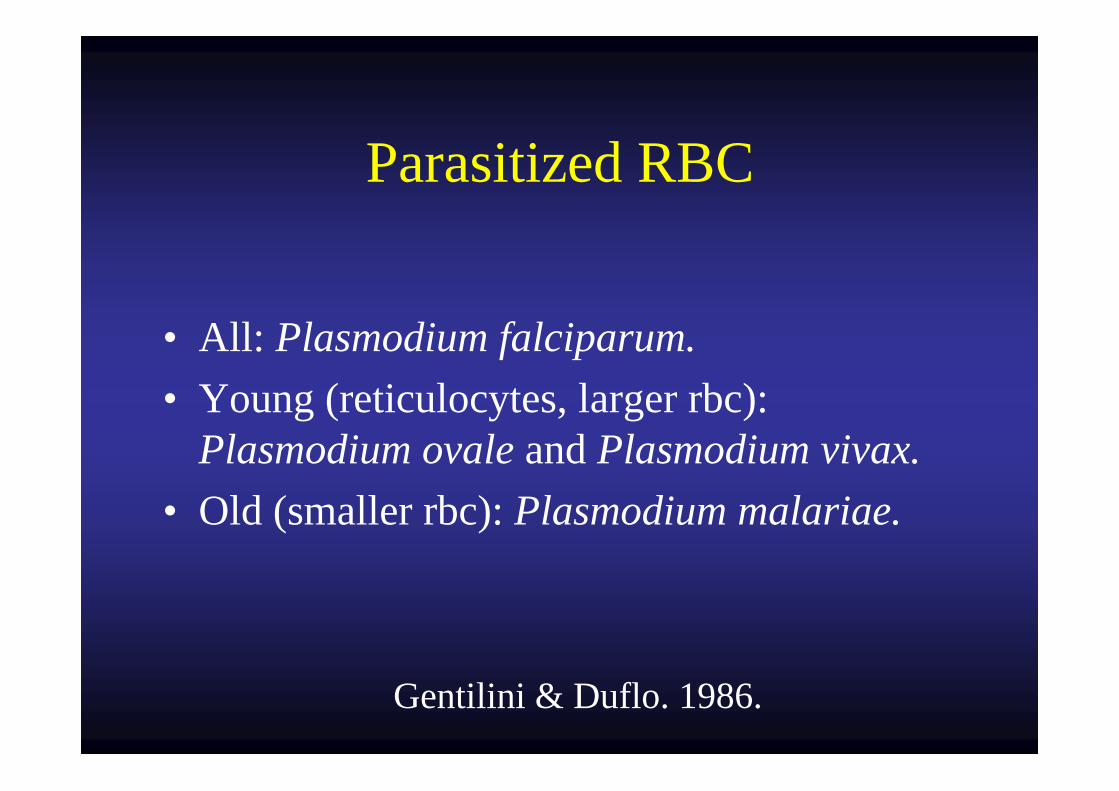
Parasitized RBC
• All: Plasmodium falciparum.• Young (reticulocytes, larger rbc):
Plasmodium ovale and Plasmodium vivax.• Old (smaller rbc): Plasmodium malariae.
Gentilini & Duflo. 1986.
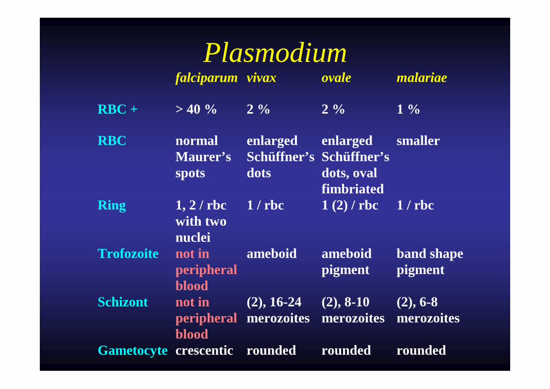
Plasmodium falciparum vivax ovale malariae
RBC + > 40 % 2 % 2 % 1 %
RBC normal Maurer’s spots
enlarged Schüffner’s dots
enlarged Schüffner’s dots, oval fimbriated
smaller
Ring 1, 2 / rbc with two nuclei
1 / rbc 1 (2) / rbc 1 / rbc
Trofozoite not in peripheral blood
ameboid ameboid pigment
band shape pigment
Schizont not in peripheral blood
(2), 16-24 merozoites
(2), 8-10 merozoites
(2), 6-8 merozoites
Gametocyte crescentic rounded rounded rounded
Courtesy TulaneCourtesy Tulane
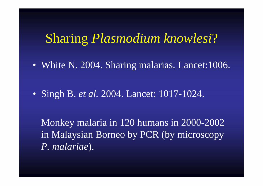
Sharing Plasmodium knowlesi?
• White N. 2004. Sharing malarias. Lancet:1006.
• Singh B. et al. 2004. Lancet: 1017-1024.
Monkey malaria in 120 humans in 2000-2002 in Malaysian Borneo by PCR (by microscopy P. malariae).
Plasmodium knowlesi: the fifth human malaria parasite.
N. J. White. 2008. CID 46:172-173.Island of Borneo, Malaysia.
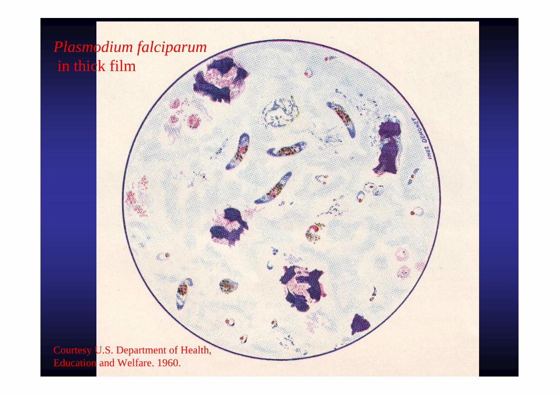
Plasmodium falciparumin thick film
Courtesy U.S. Department of Health, Education and Welfare. 1960.
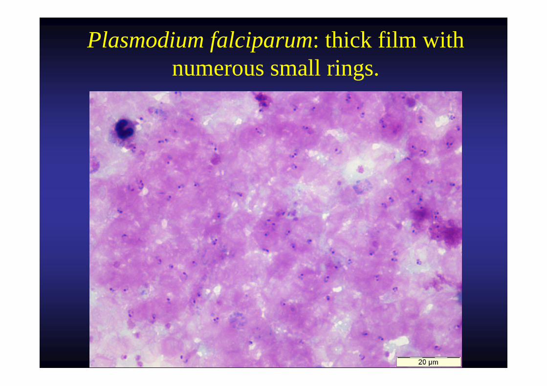
Plasmodium falciparum: thick film with numerous small rings.
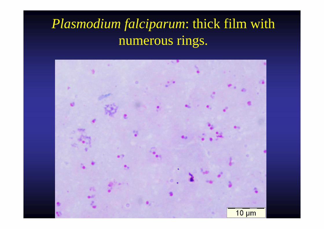
Plasmodium falciparum: thick film with numerous rings.
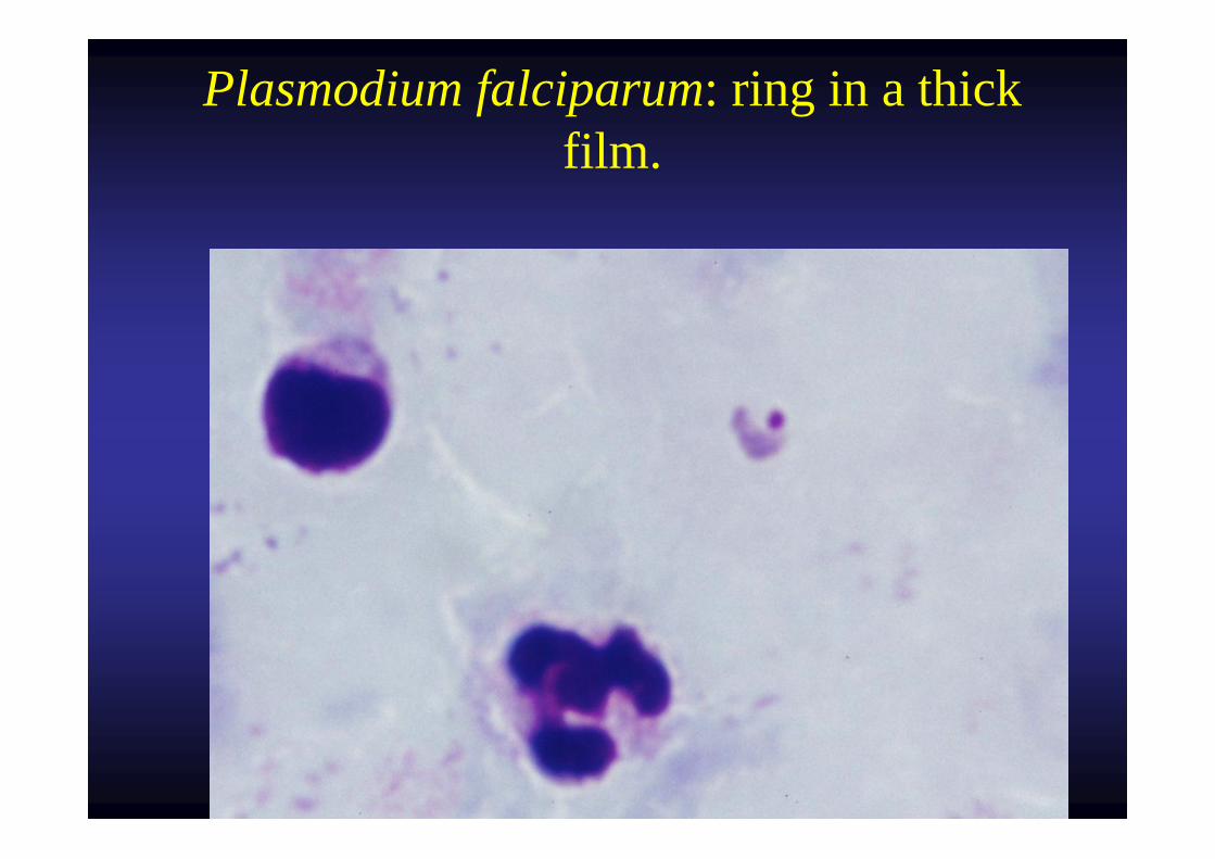
Plasmodium falciparum: ring in a thick film.
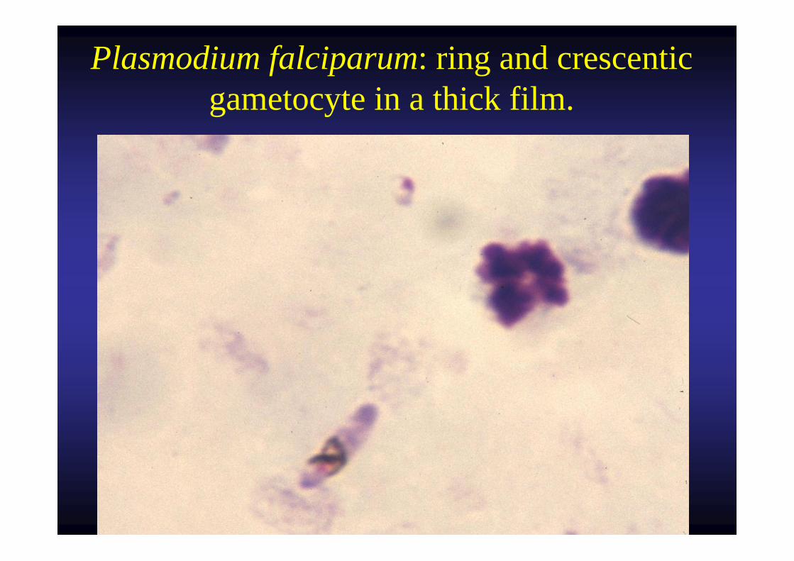
Plasmodium falciparum: ring and crescentic gametocyte in a thick film.
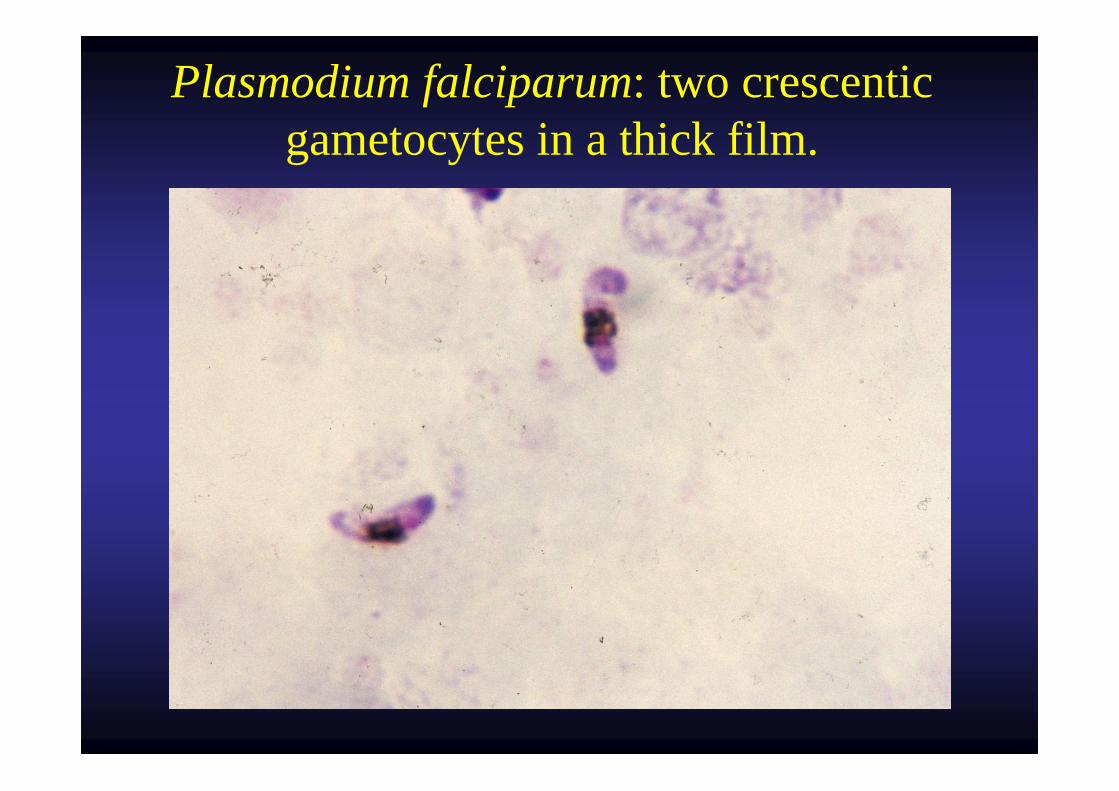
Plasmodium falciparum: two crescentic gametocytes in a thick film.
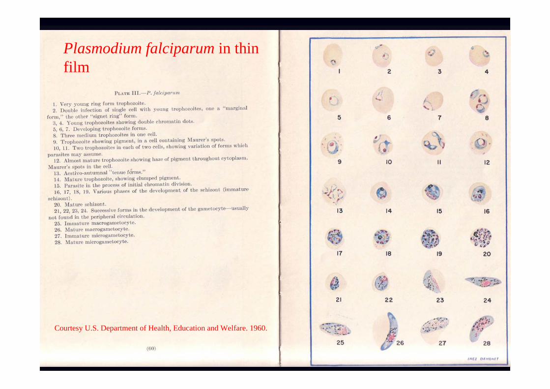
Plasmodium falciparum in thinfilm
Courtesy U.S. Department of Health, Education and Welfare. 1960.
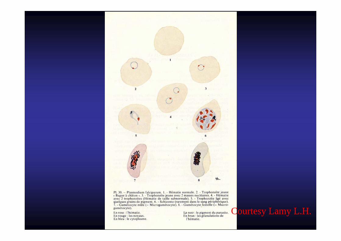
Courtesy Lamy L.H.
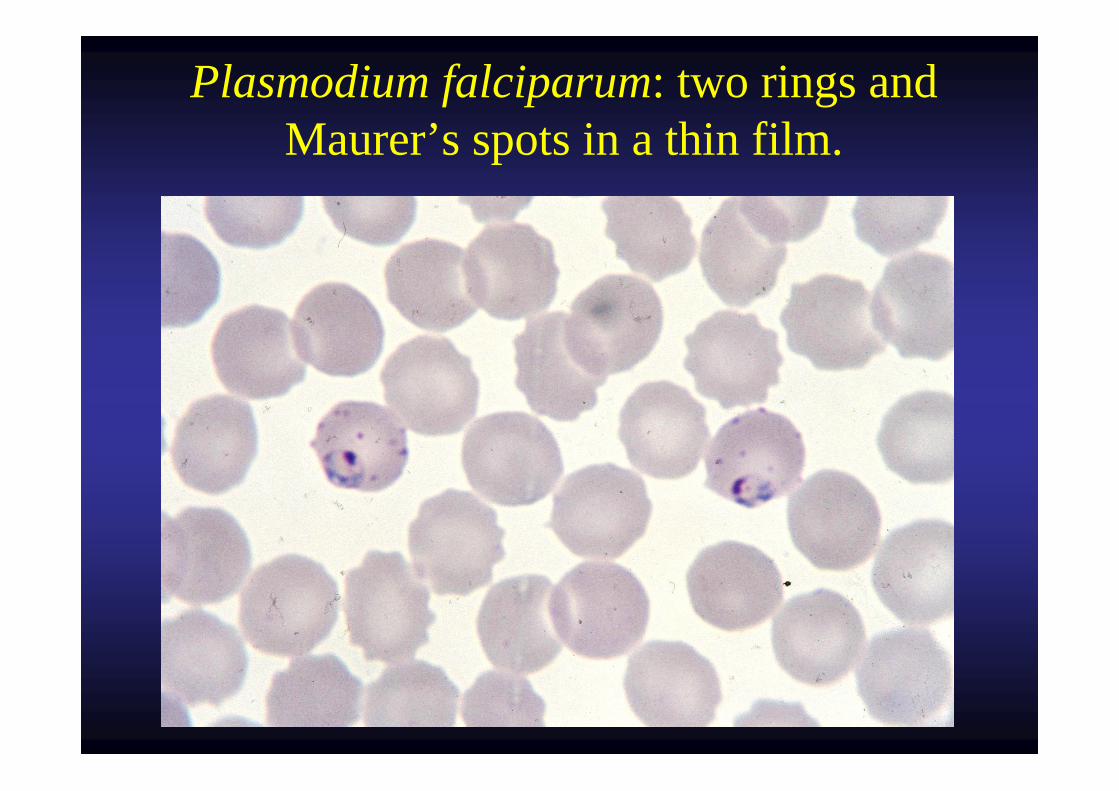
Plasmodium falciparum: two rings and Maurer’s spots in a thin film.
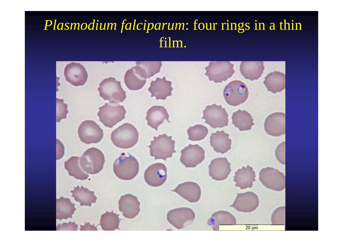
Plasmodium falciparum: four rings in a thin film.
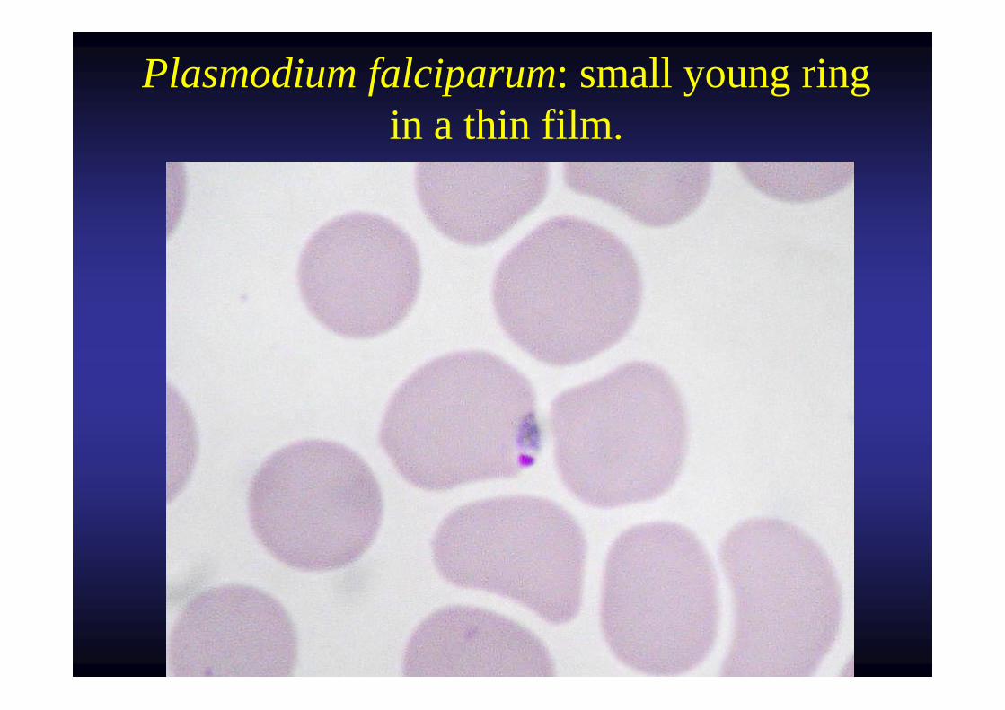
Plasmodium falciparum: small young ring in a thin film.
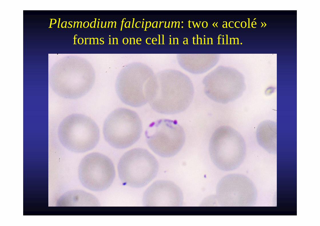
Plasmodium falciparum: two « accolé »forms in one cell in a thin film.
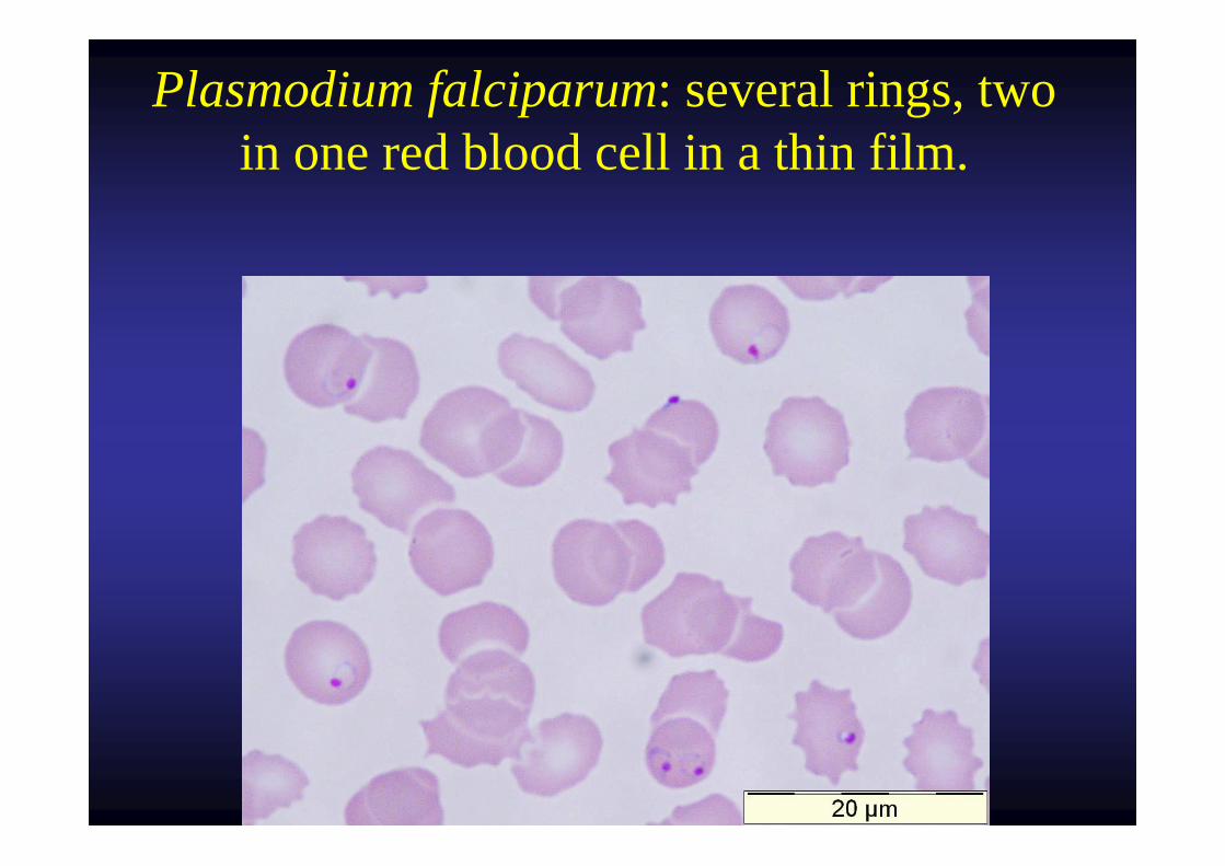
Plasmodium falciparum: several rings, two in one red blood cell in a thin film.
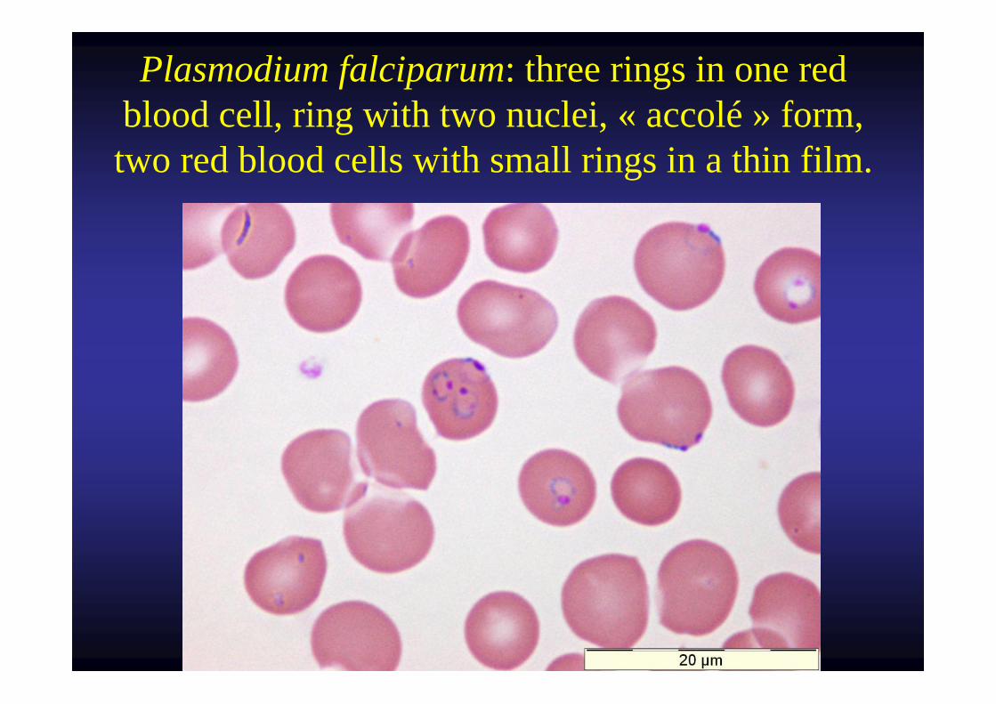
Plasmodium falciparum: three rings in one red blood cell, ring with two nuclei, « accolé » form, two red blood cells with small rings in a thin film.
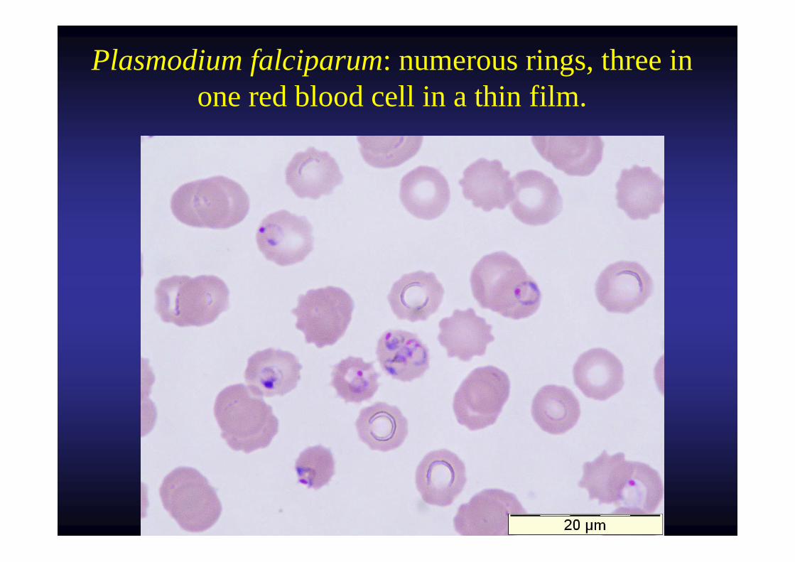
Plasmodium falciparum: numerous rings, three in one red blood cell in a thin film.
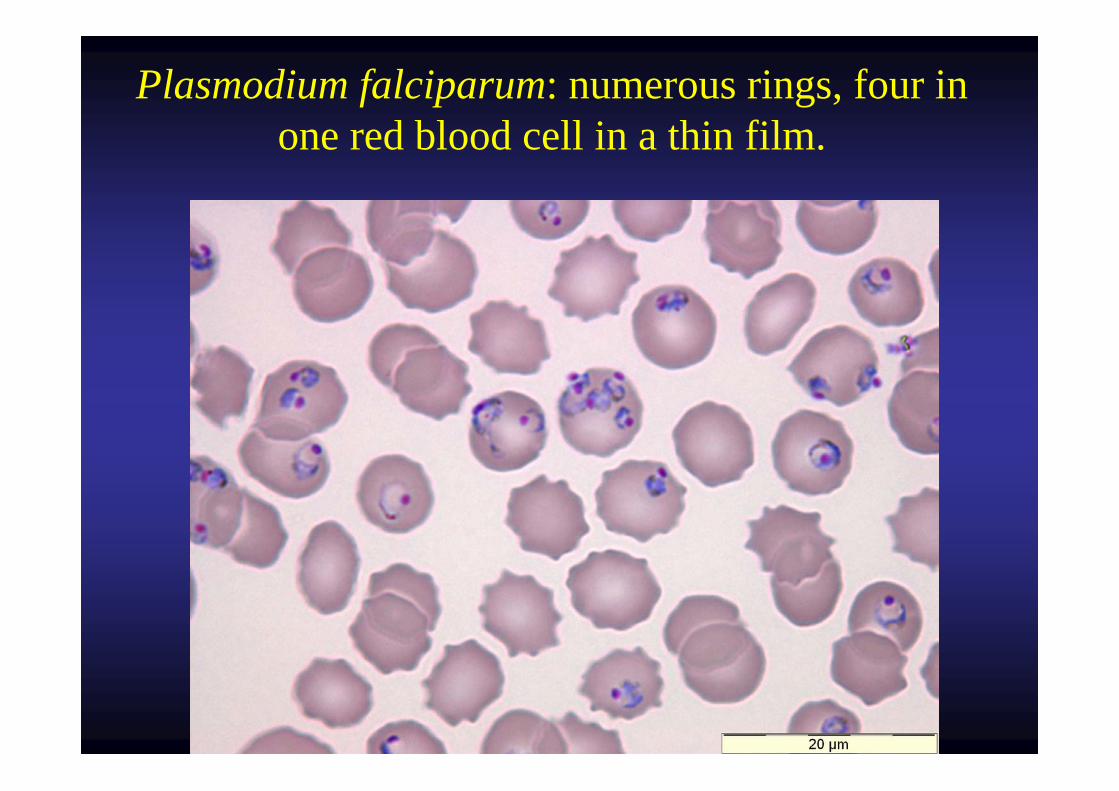
Plasmodium falciparum: numerous rings, four in one red blood cell in a thin film.
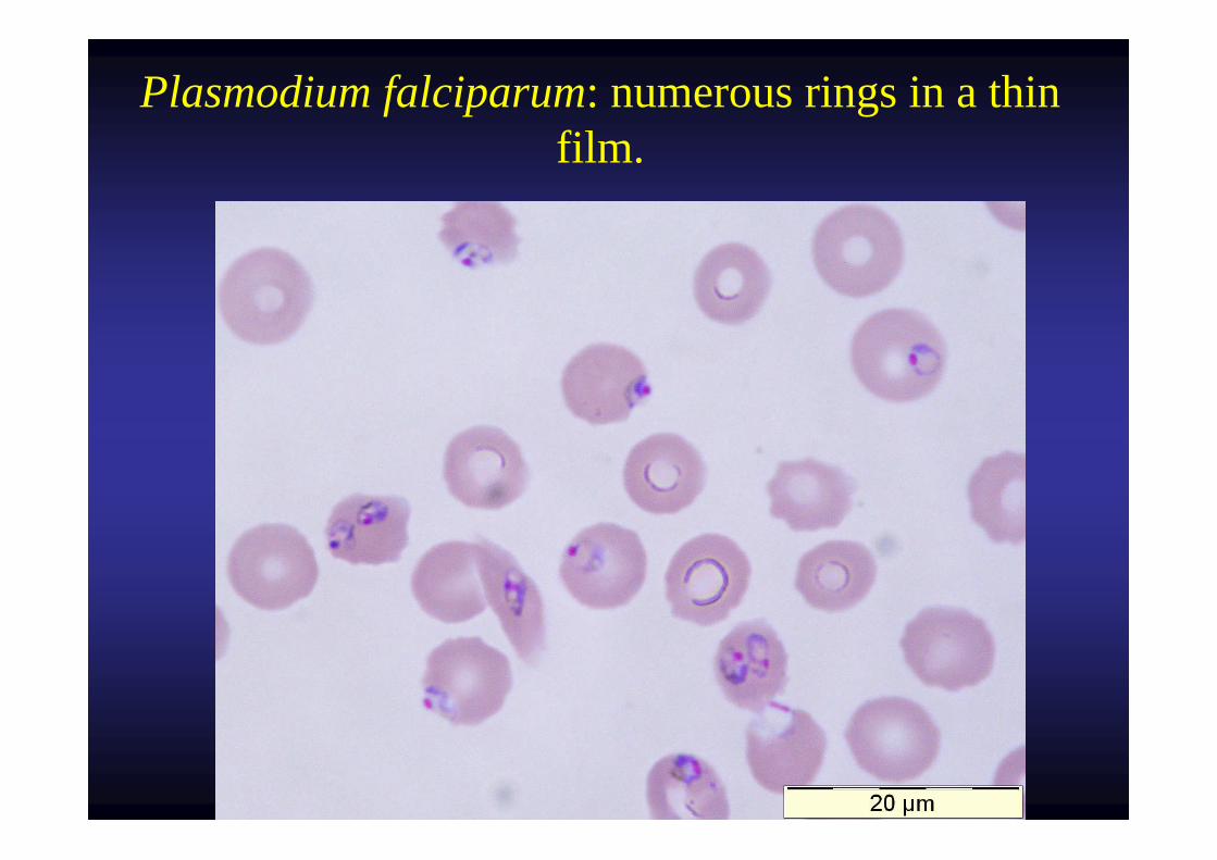
Plasmodium falciparum: numerous rings in a thin film.
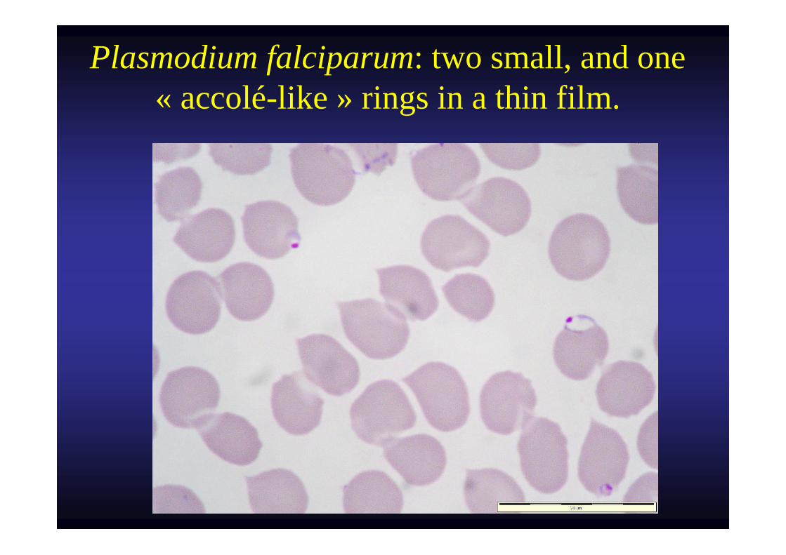
Plasmodium falciparum: two small, and one « accolé-like » rings in a thin film.
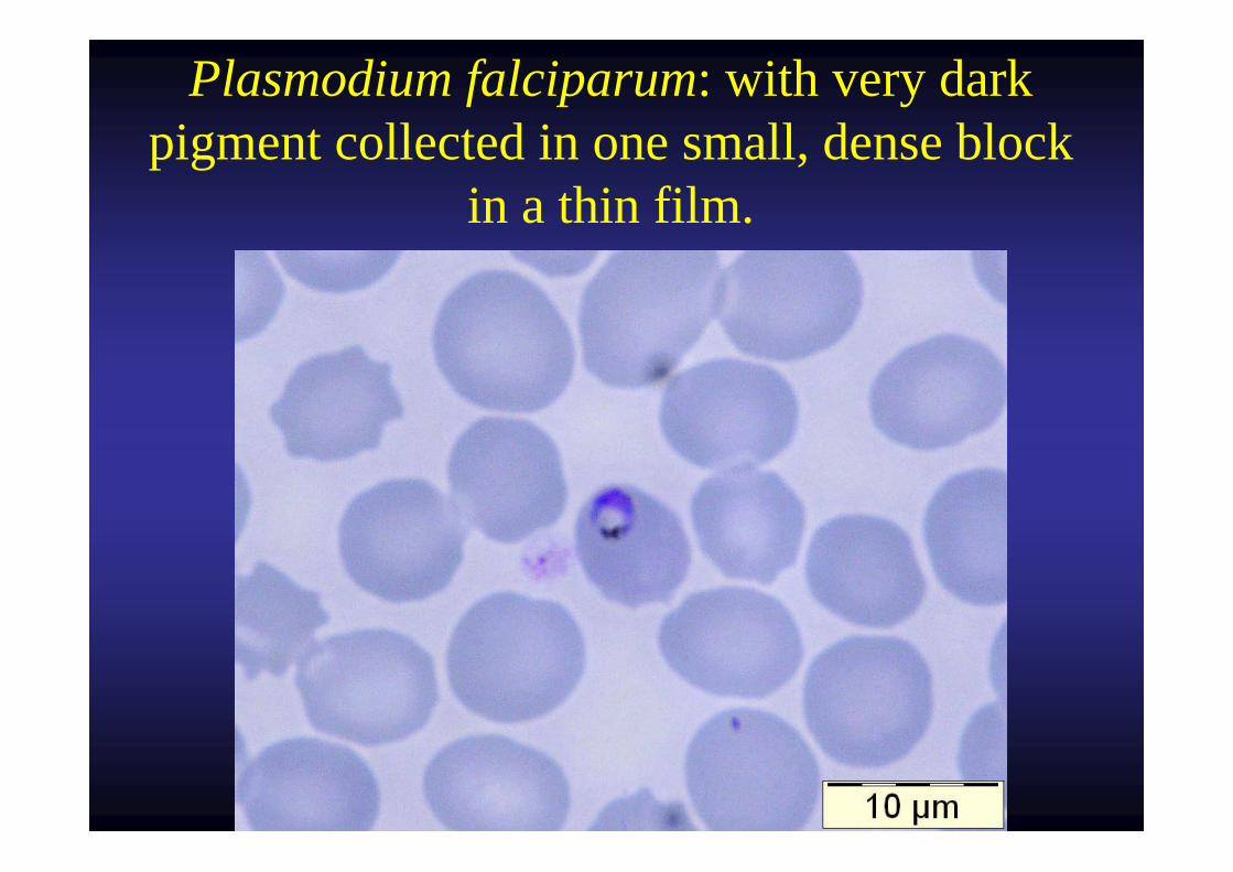
Plasmodium falciparum: with very dark pigment collected in one small, dense block
in a thin film.
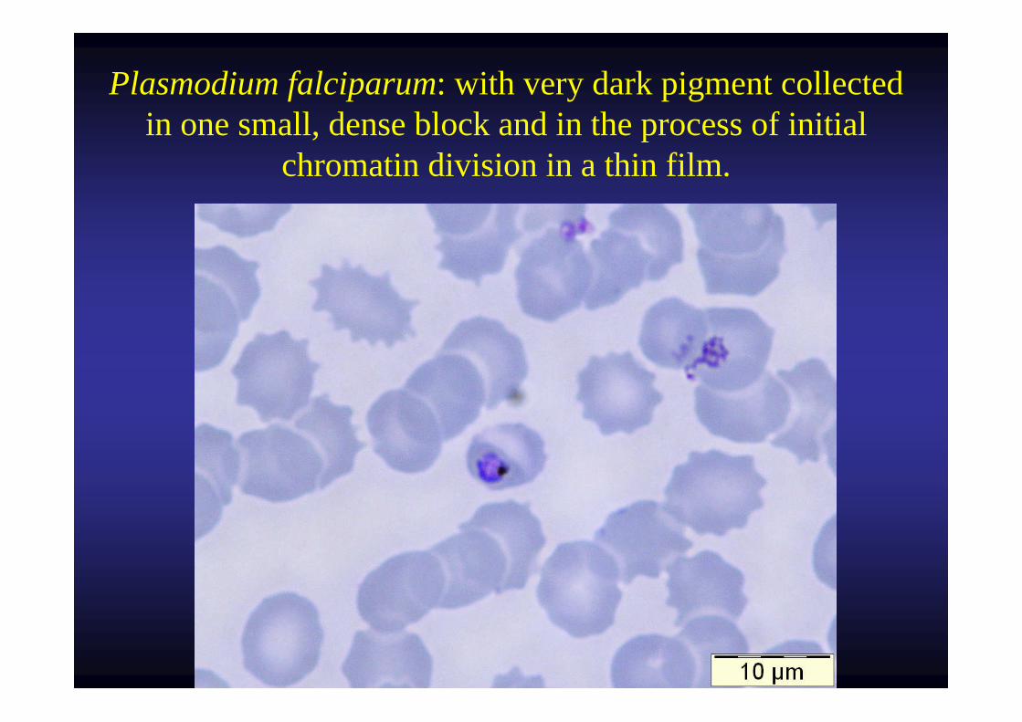
Plasmodium falciparum: with very dark pigment collected in one small, dense block and in the process of initial
chromatin division in a thin film.
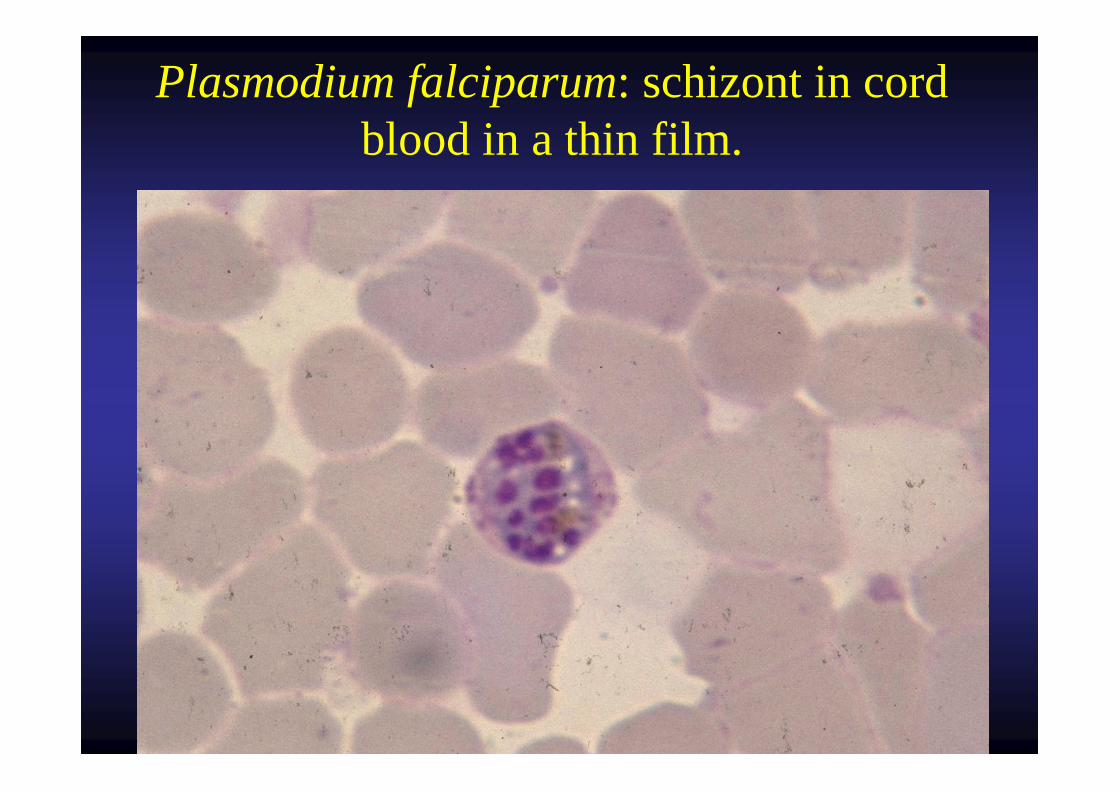
Plasmodium falciparum: schizont in cord blood in a thin film.
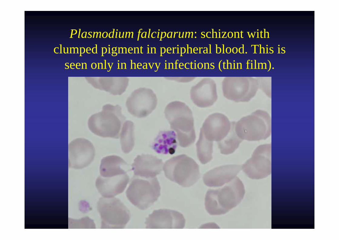
Plasmodium falciparum: schizont with clumped pigment in peripheral blood. This is
seen only in heavy infections (thin film).
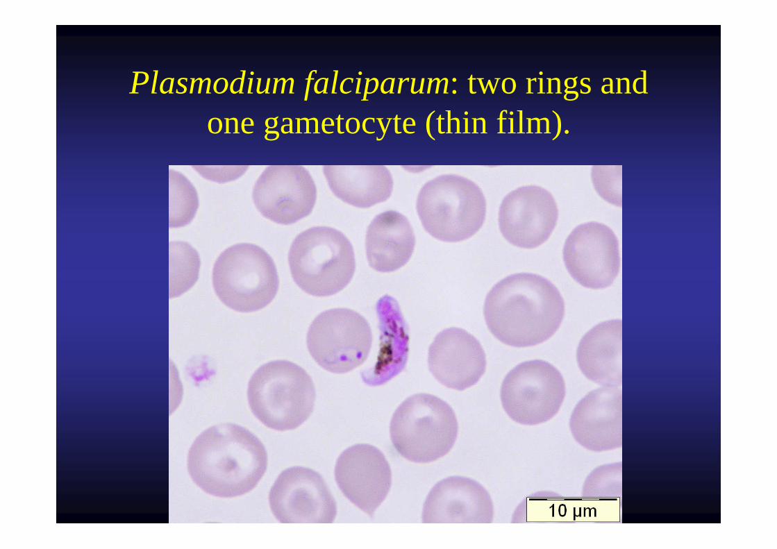
Plasmodium falciparum: two rings and one gametocyte (thin film).
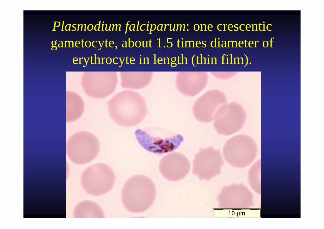
Plasmodium falciparum: one crescentic gametocyte, about 1.5 times diameter of
erythrocyte in length (thin film).
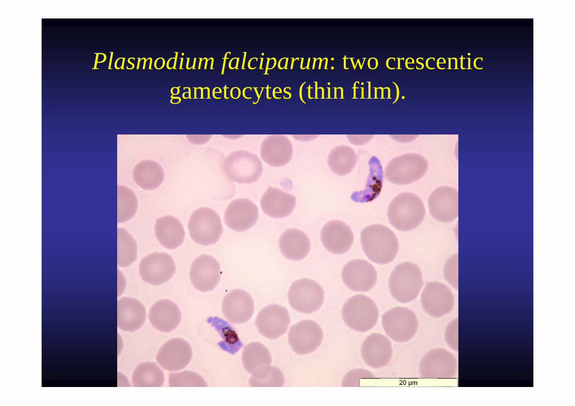
Plasmodium falciparum: two crescentic gametocytes (thin film).
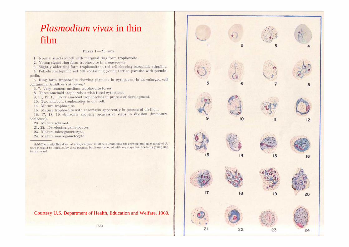
Plasmodium vivax in thinfilm
Courtesy U.S. Department of Health, Education and Welfare. 1960.
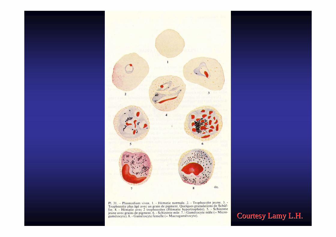
Courtesy Lamy L.H.Courtesy Lamy L.H.
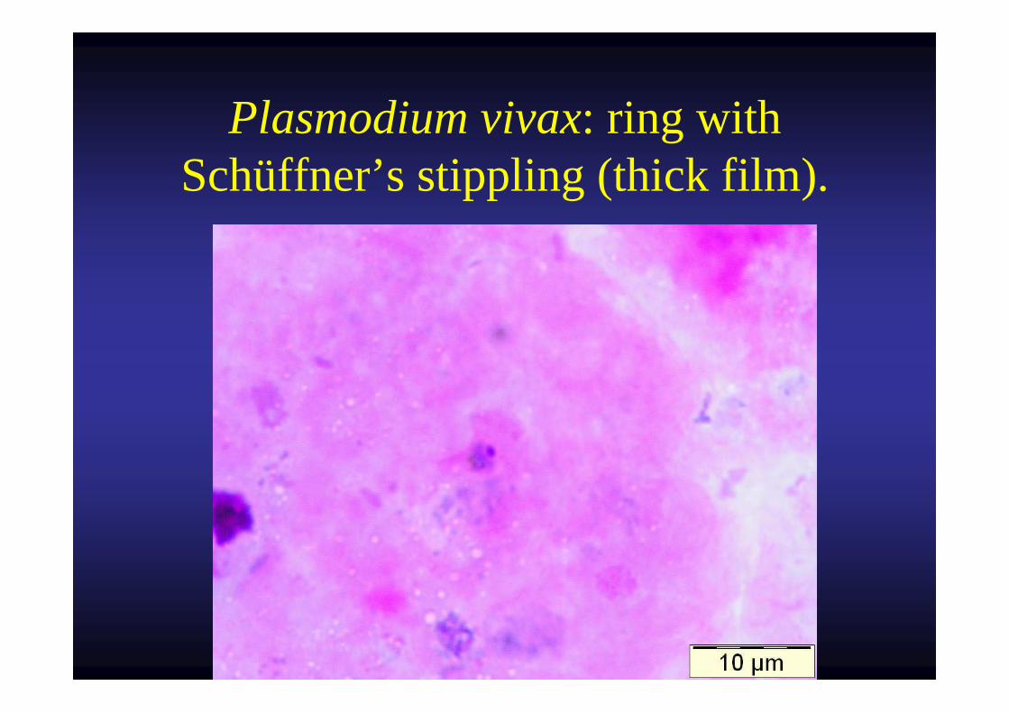
Plasmodium vivax: ring with Schüffner’s stippling (thick film).
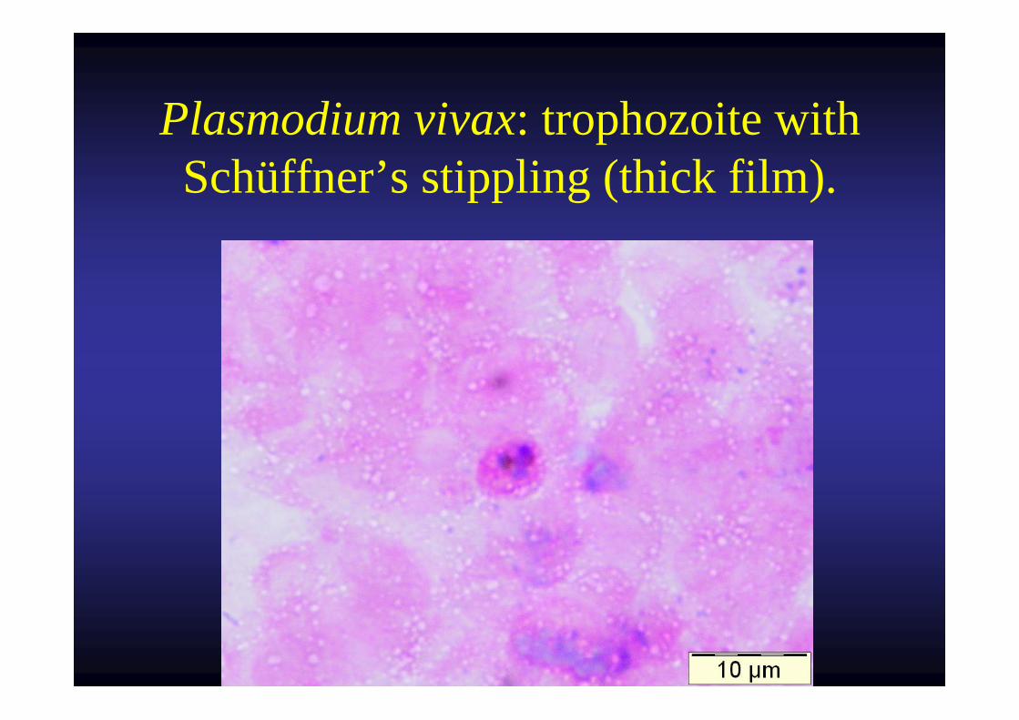
Plasmodium vivax: trophozoite with Schüffner’s stippling (thick film).
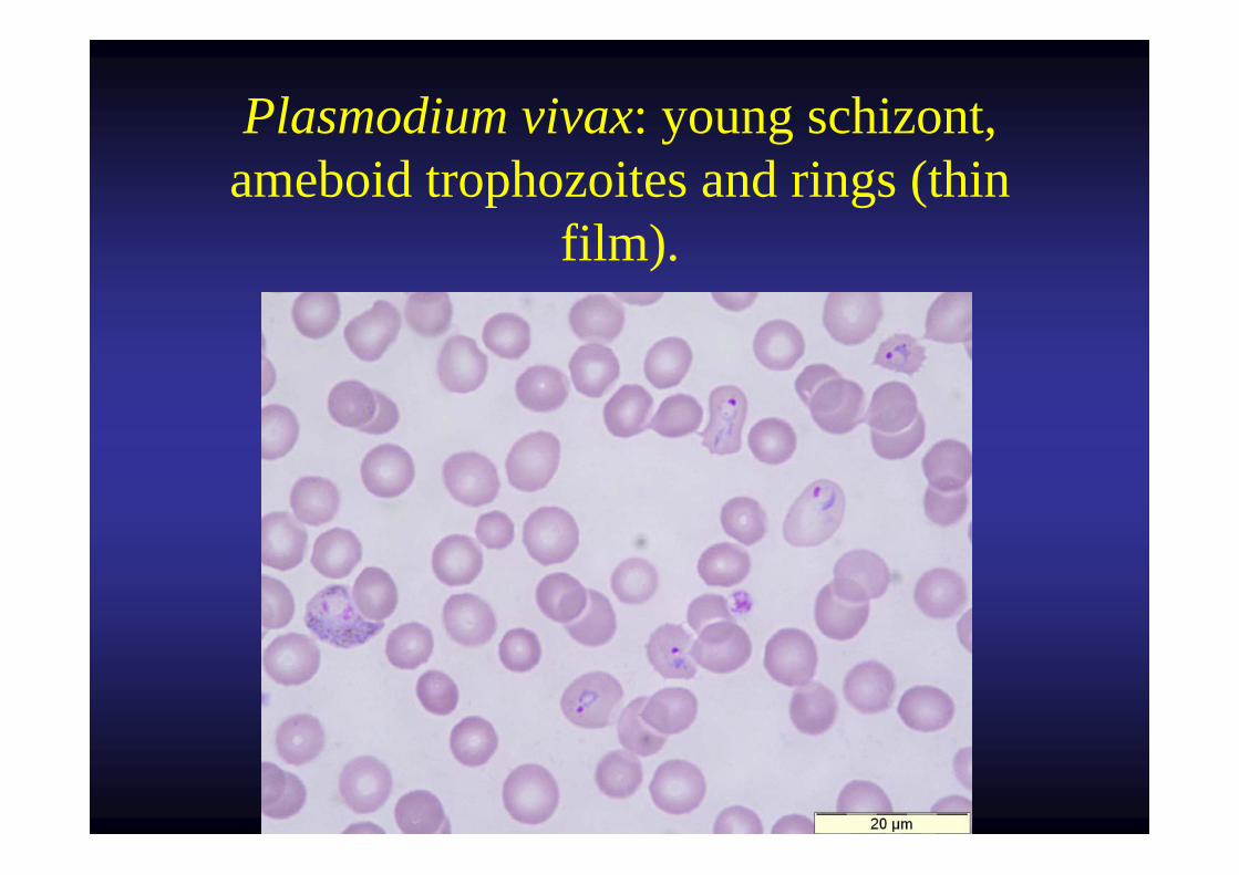
Plasmodium vivax: young schizont, ameboid trophozoites and rings (thin
film).

Plasmodium vivax: older ring and ameboid trophozoite (thin film).
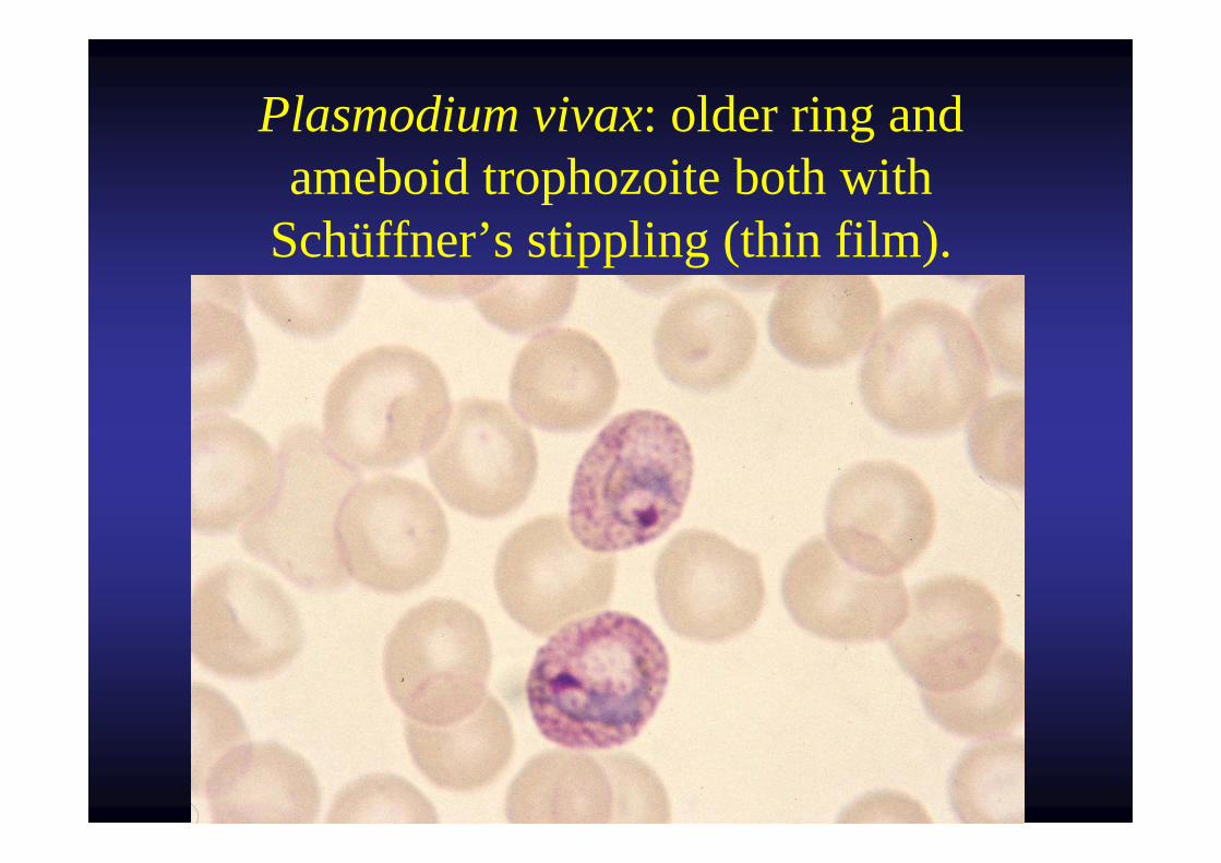
Plasmodium vivax: older ring and ameboid trophozoite both with
Schüffner’s stippling (thin film).
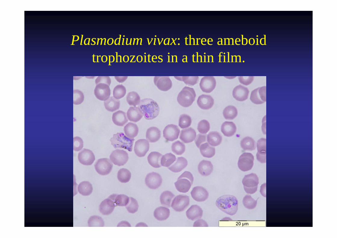
Plasmodium vivax: three ameboid trophozoites in a thin film.
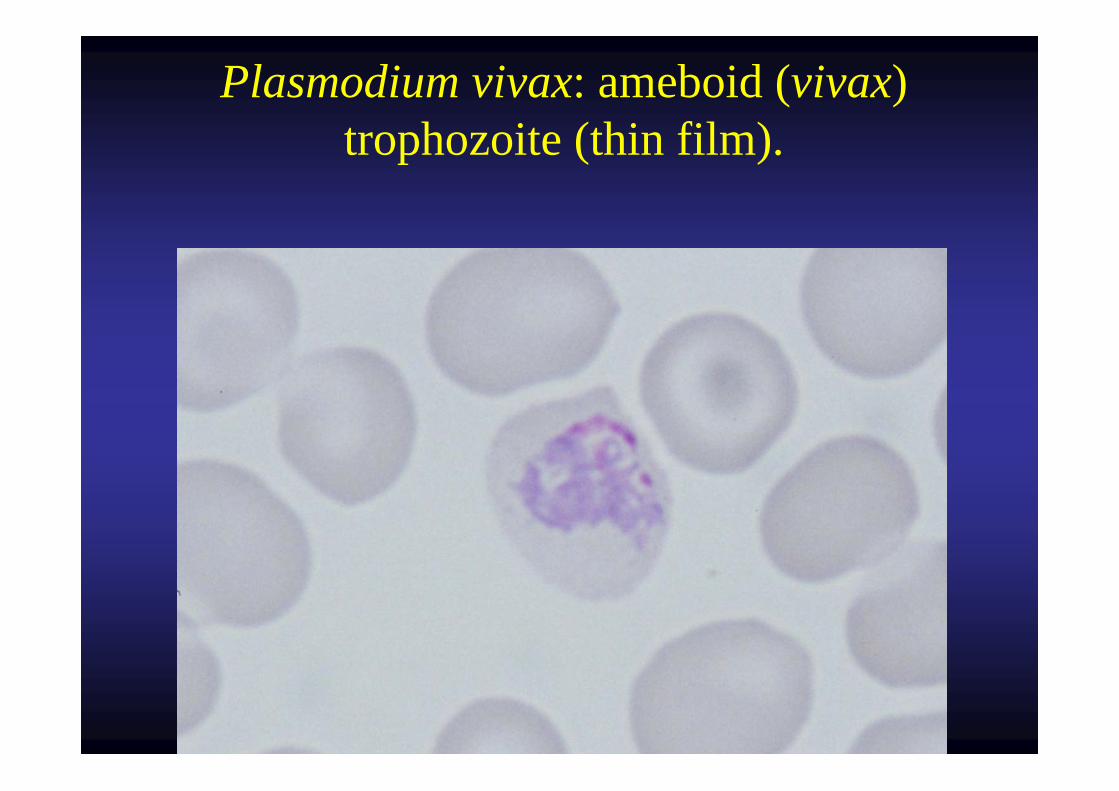
Plasmodium vivax: ameboid (vivax) trophozoite (thin film).
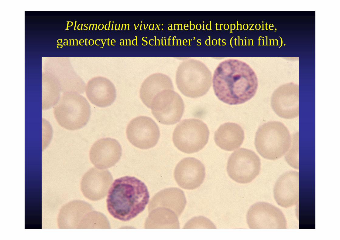
Plasmodium vivax: ameboid trophozoite, gametocyte and Schüffner’s dots (thin film).
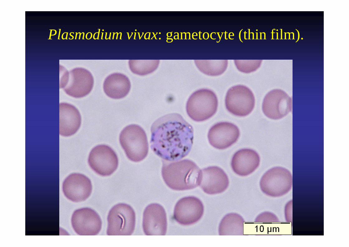
Plasmodium vivax: gametocyte (thin film).
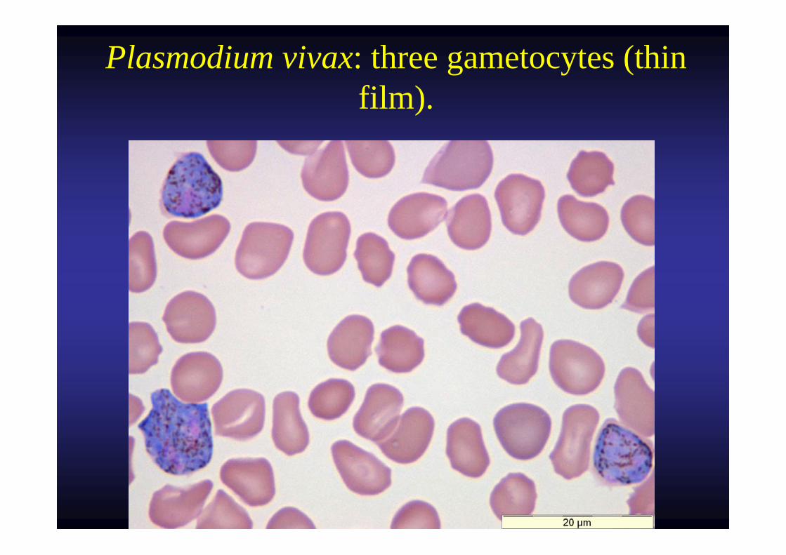
Plasmodium vivax: three gametocytes (thin film).
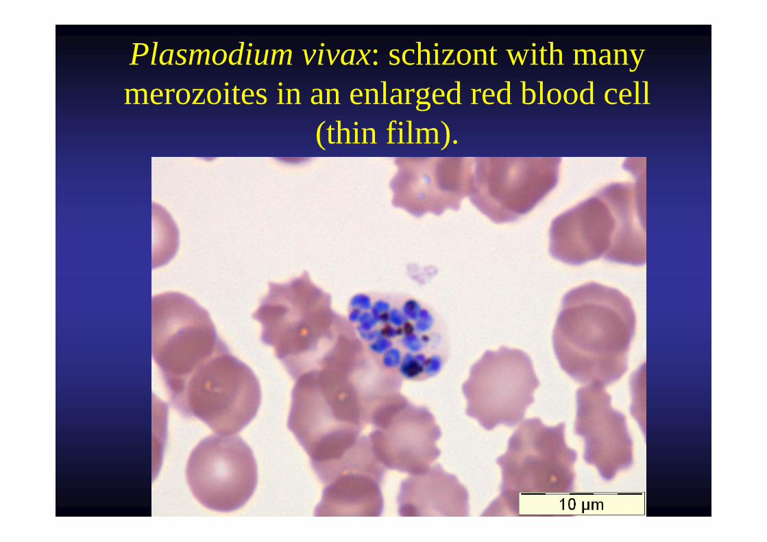
Plasmodium vivax: schizont with many merozoites in an enlarged red blood cell
(thin film).
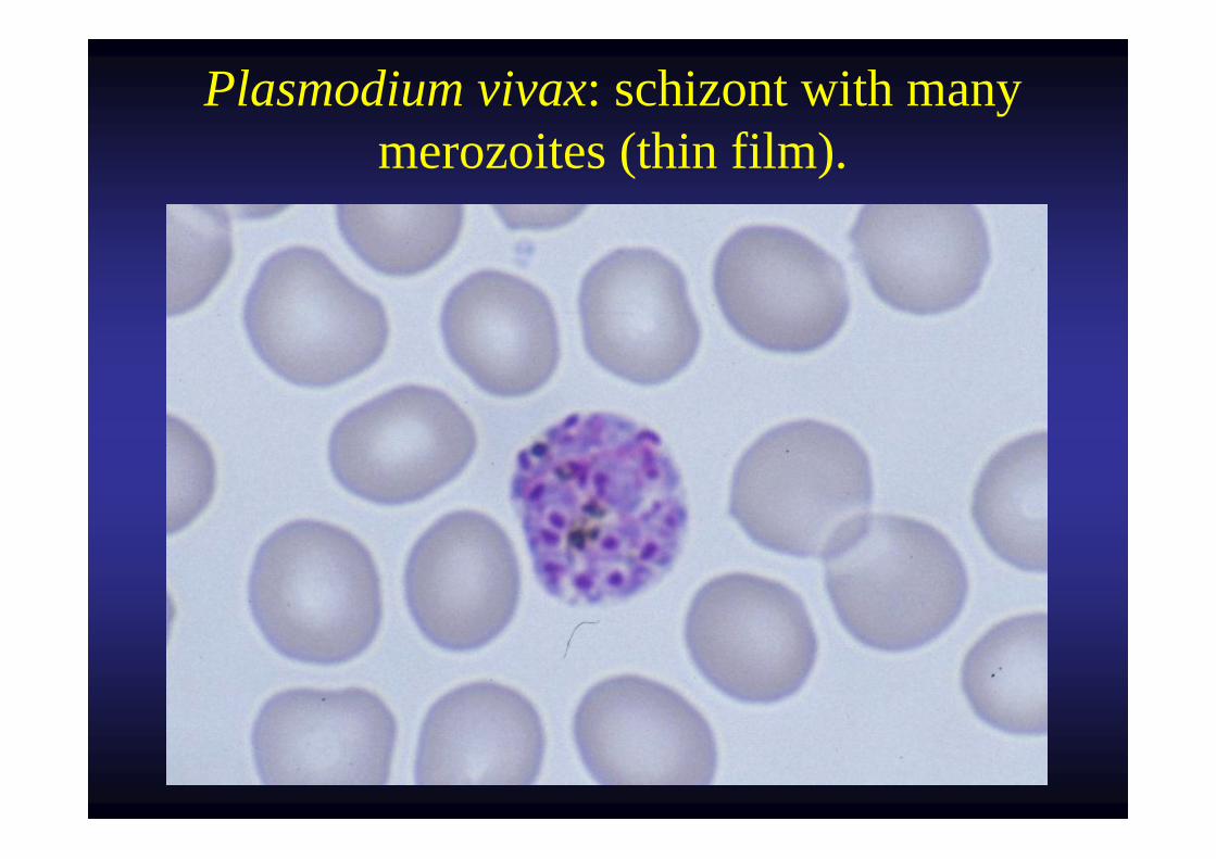
Plasmodium vivax: schizont with many merozoites (thin film).
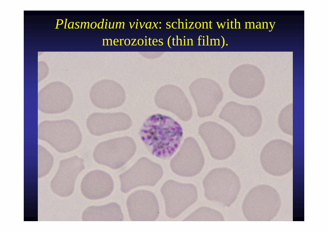
Plasmodium vivax: schizont with many merozoites (thin film).
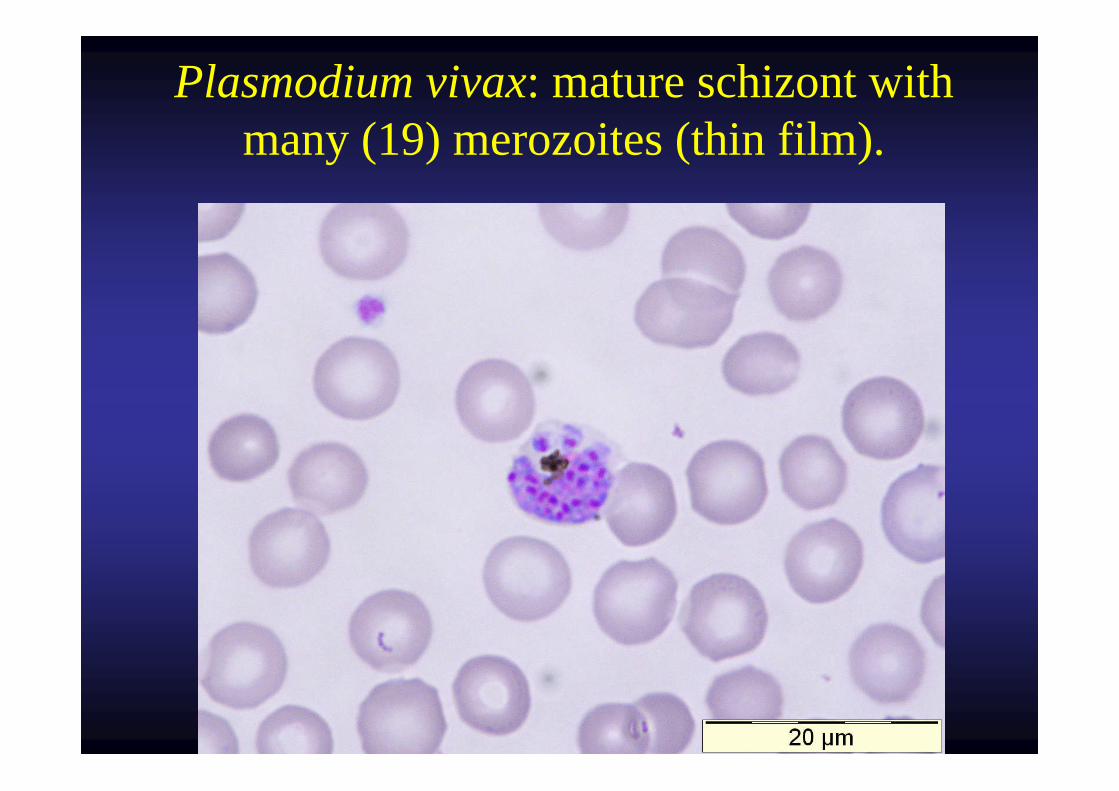
Plasmodium vivax: mature schizont with many (19) merozoites (thin film).
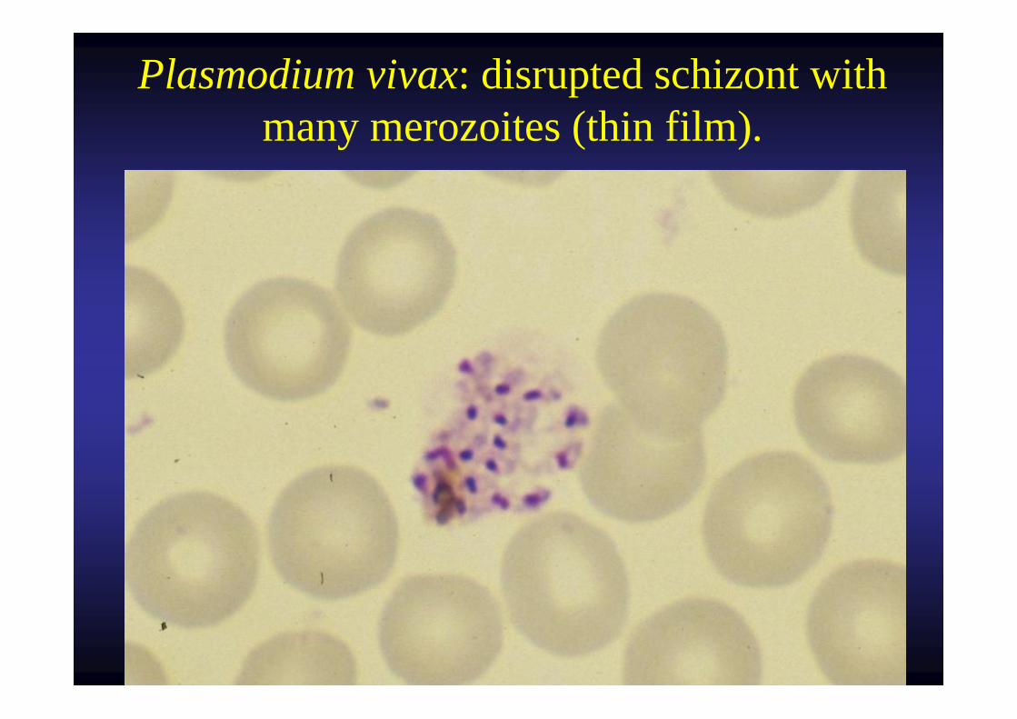
Plasmodium vivax: disrupted schizont with many merozoites (thin film).
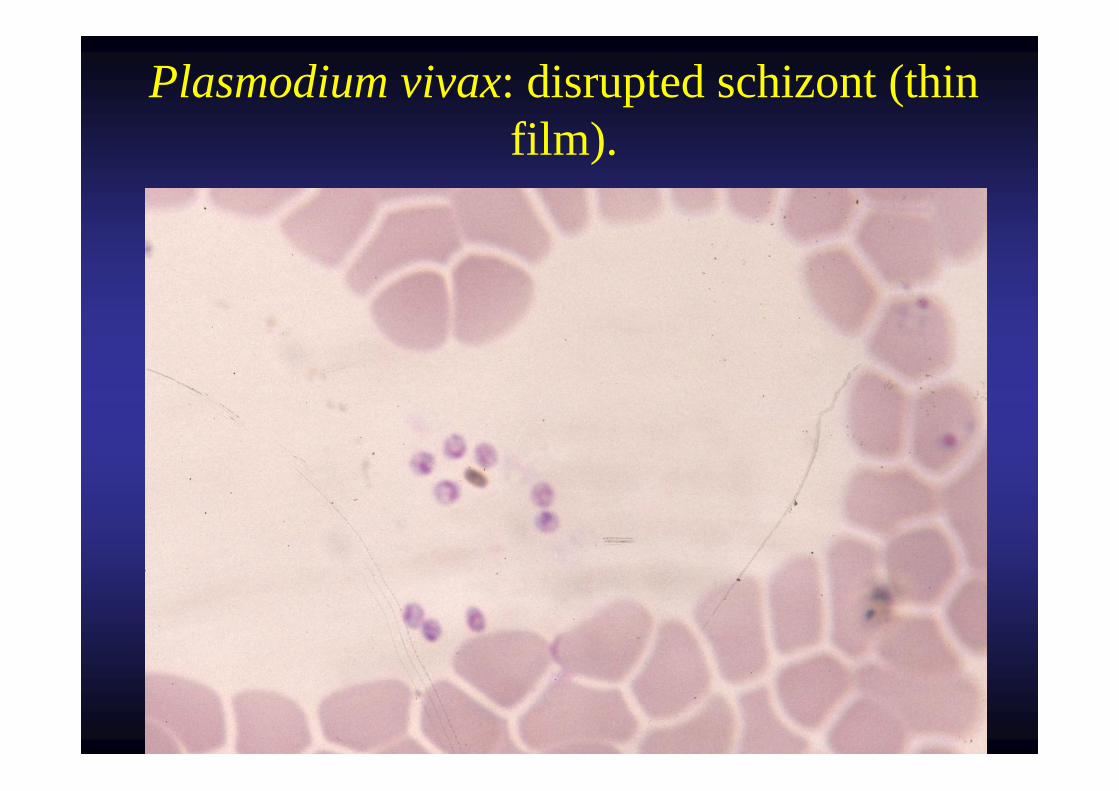
Plasmodium vivax: disrupted schizont (thin film).
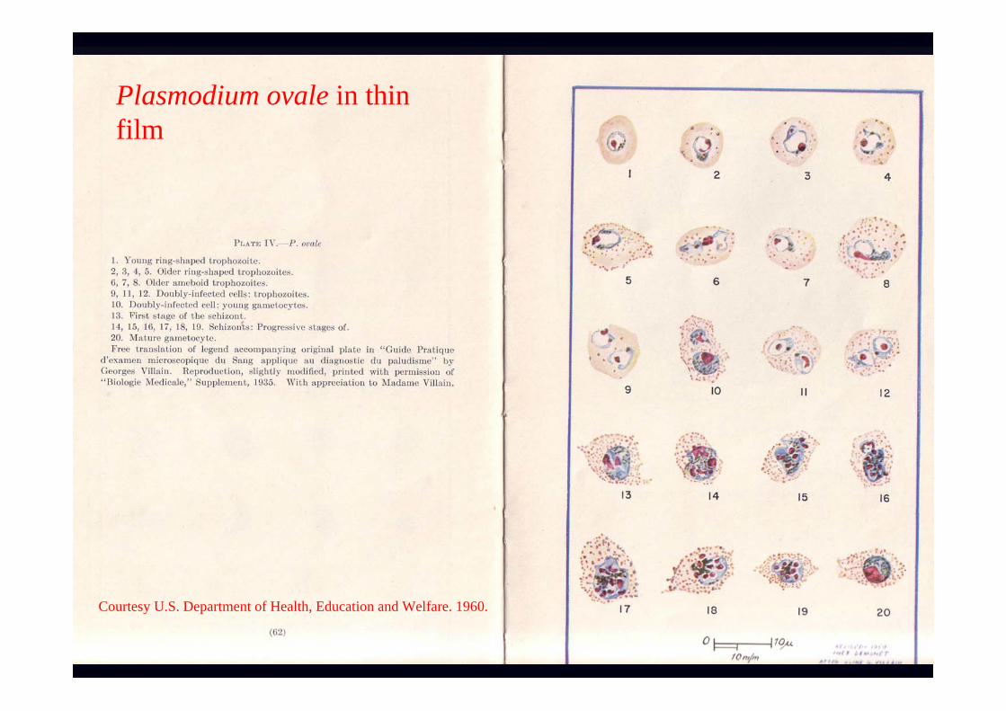
Plasmodium ovale in thinfilm
Courtesy U.S. Department of Health, Education and Welfare. 1960.
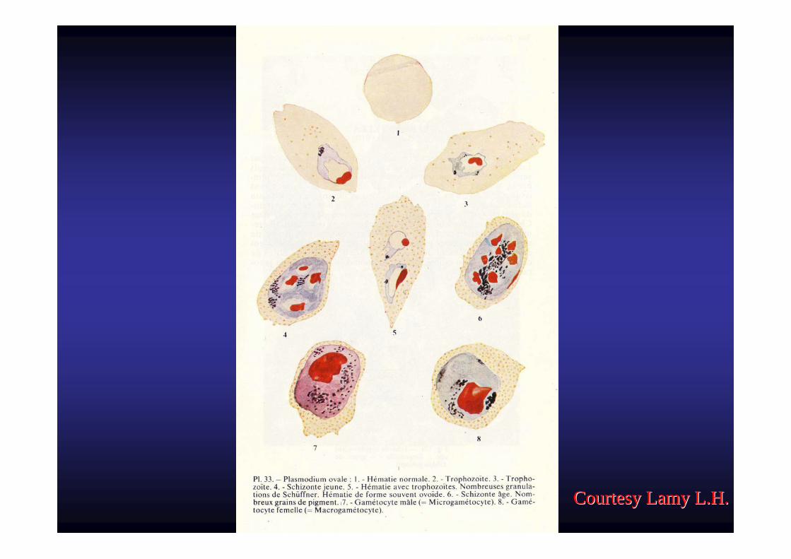
Courtesy Lamy L.H.Courtesy Lamy L.H.
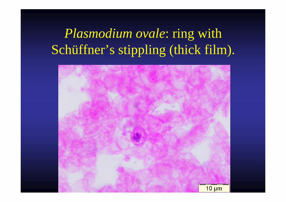
Plasmodium ovale: ring with Schüffner’s stippling (thick film).
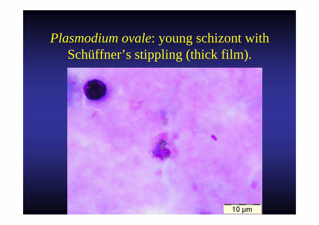
Plasmodium ovale: young schizont with Schüffner’s stippling (thick film).
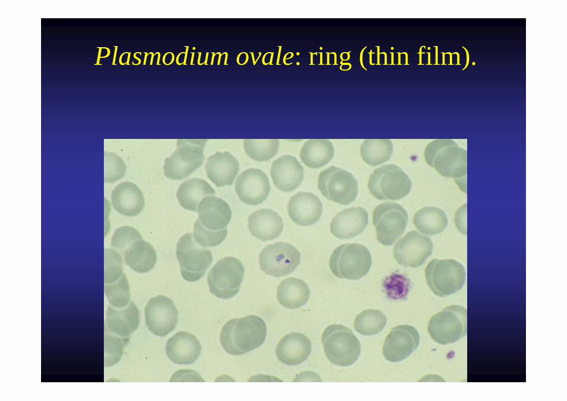
Plasmodium ovale: ring (thin film).
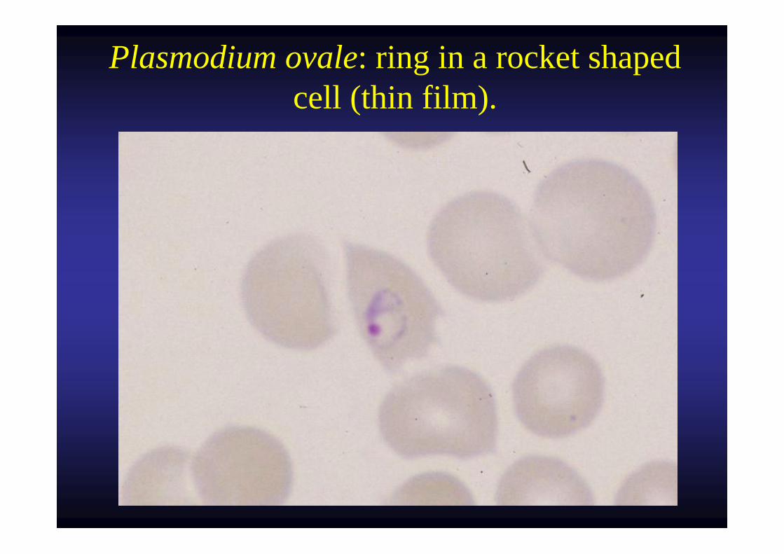
Plasmodium ovale: ring in a rocket shaped cell (thin film).
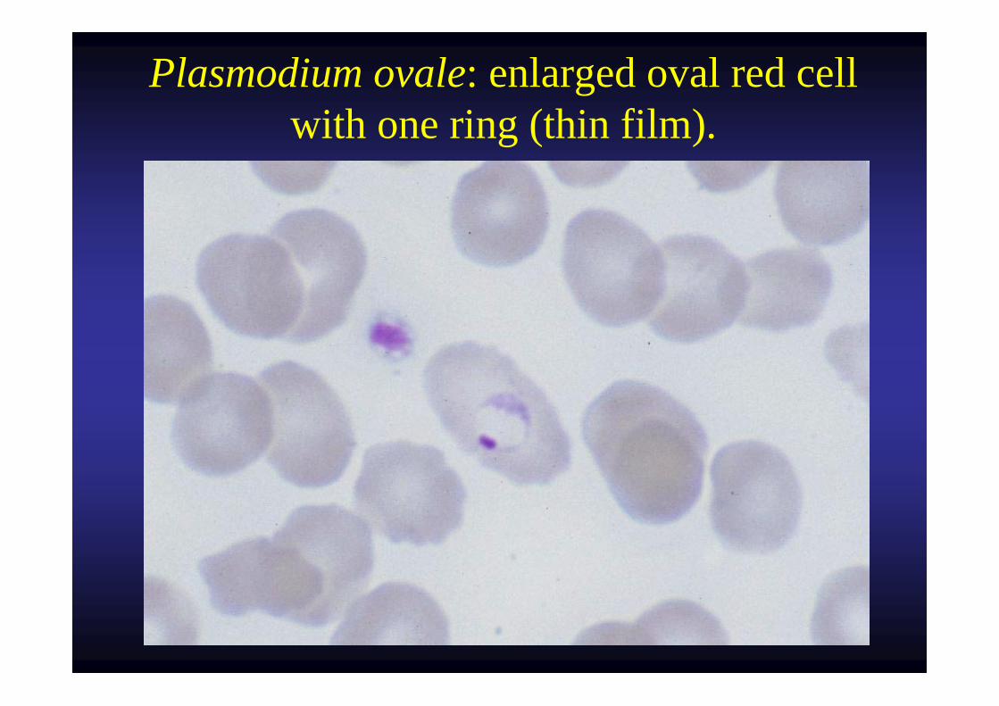
Plasmodium ovale: enlarged oval red cell with one ring (thin film).
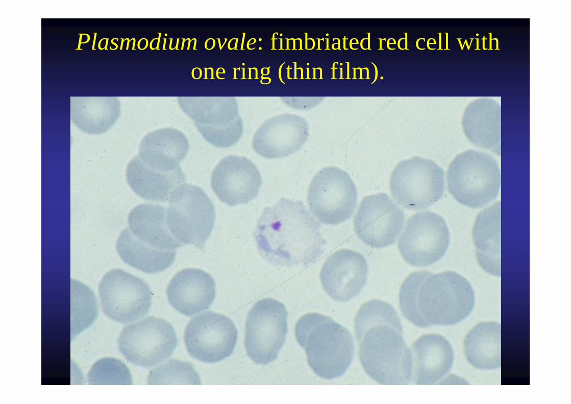
Plasmodium ovale: fimbriated red cell with one ring (thin film).
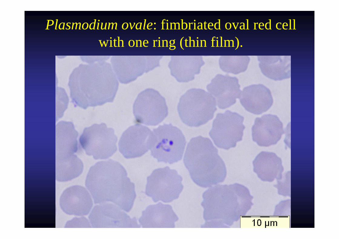
Plasmodium ovale: fimbriated oval red cell with one ring (thin film).
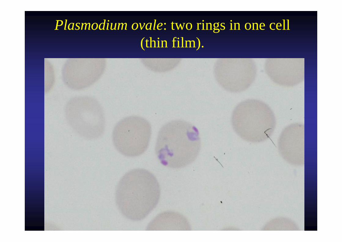
Plasmodium ovale: two rings in one cell (thin film).
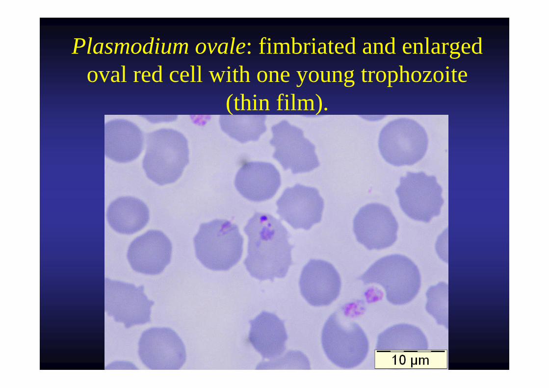
Plasmodium ovale: fimbriated and enlarged oval red cell with one young trophozoite
(thin film).
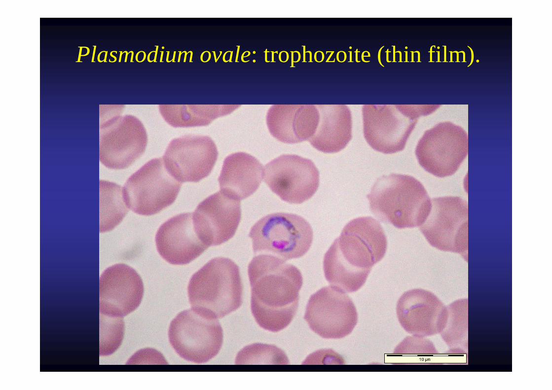
Plasmodium ovale: trophozoite (thin film).
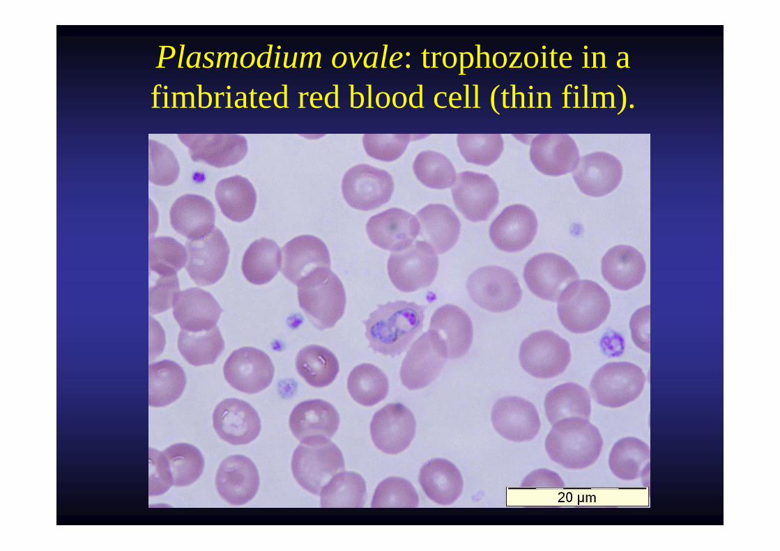
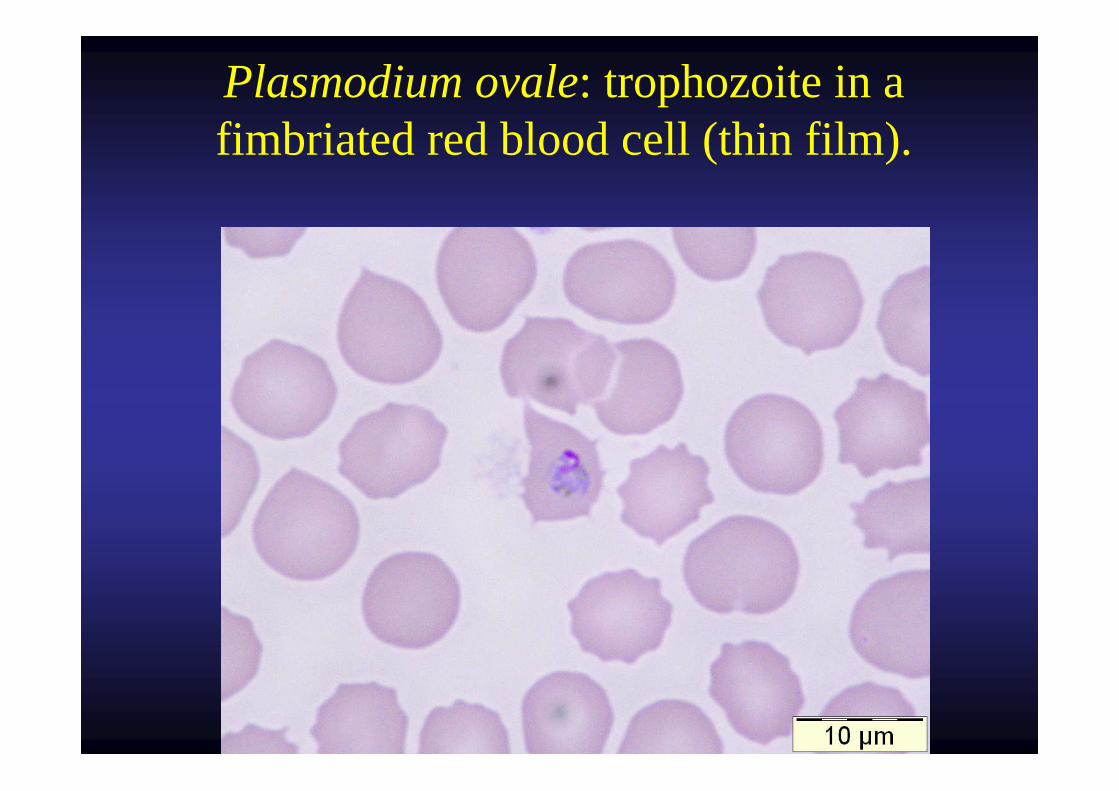
Plasmodium ovale: trophozoite in a fimbriated red blood cell (thin film).
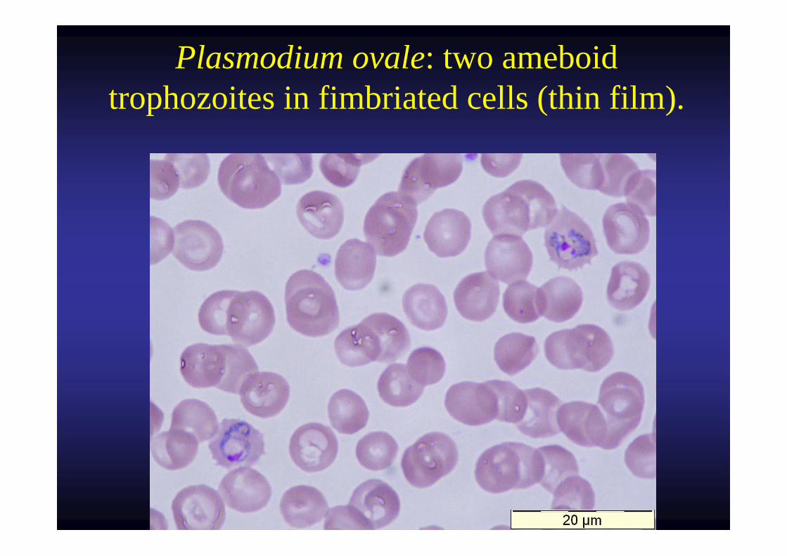
Plasmodium ovale: two ameboid trophozoites in fimbriated cells (thin film).
Plasmodium ovale: trophozoite in a fimbriated red blood cell (thin film).
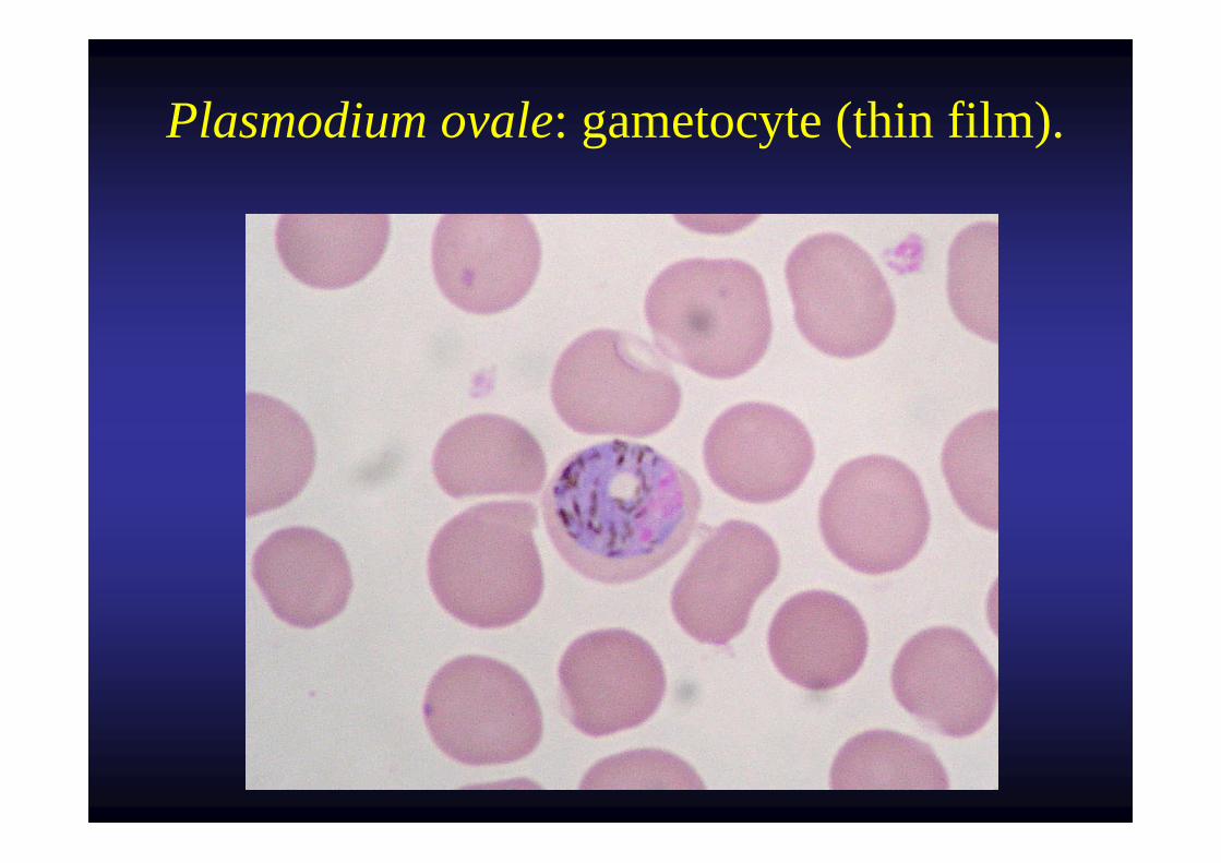
Plasmodium ovale: gametocyte (thin film).
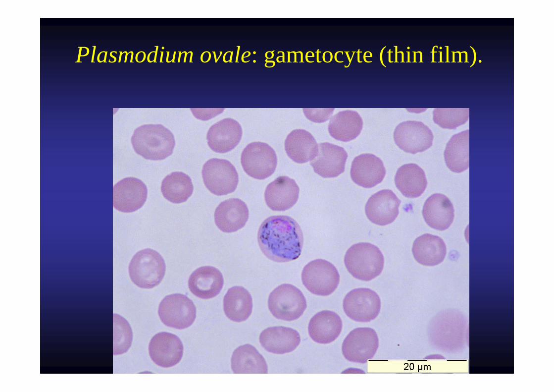
Plasmodium ovale: gametocyte (thin film).
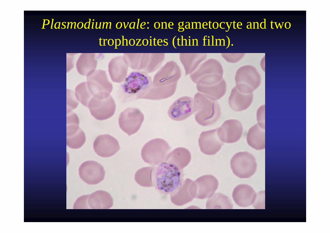
Plasmodium ovale: one gametocyte and two trophozoites (thin film).
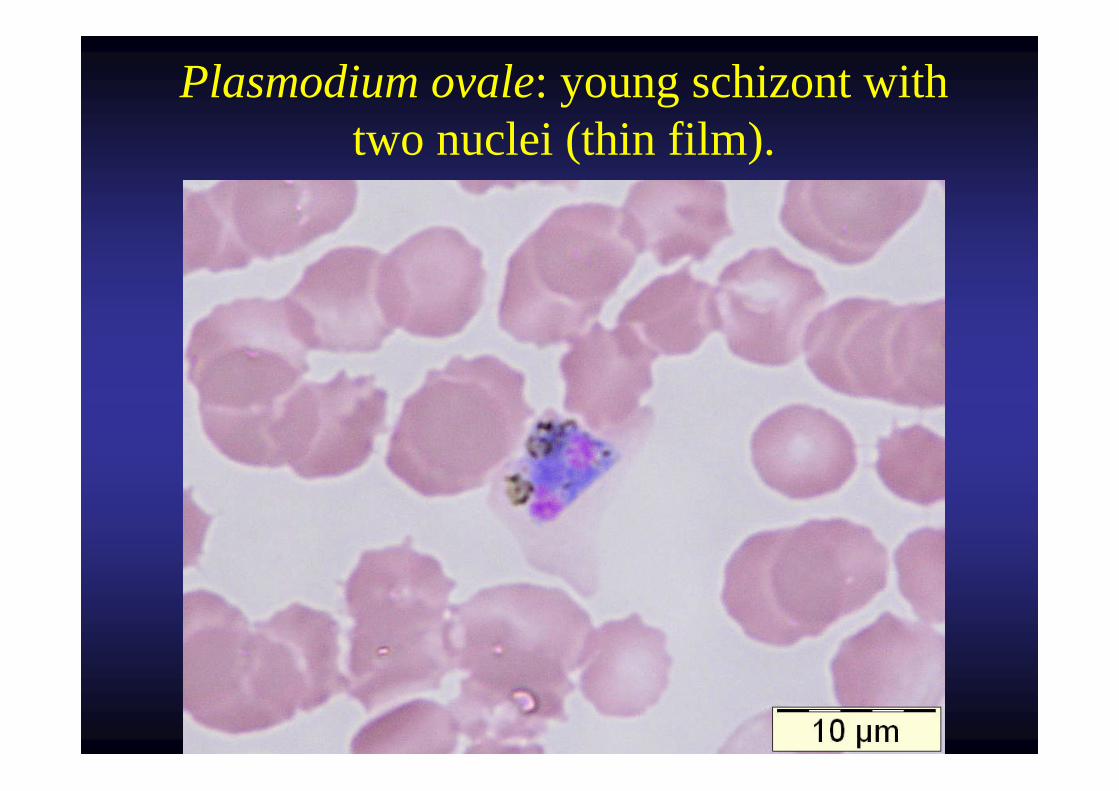
Plasmodium ovale: young schizont with two nuclei (thin film).
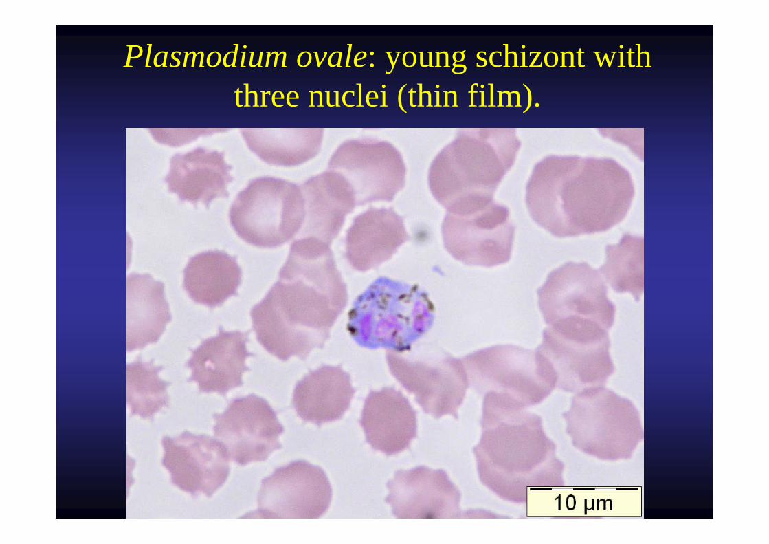
Plasmodium ovale: young schizont with three nuclei (thin film).
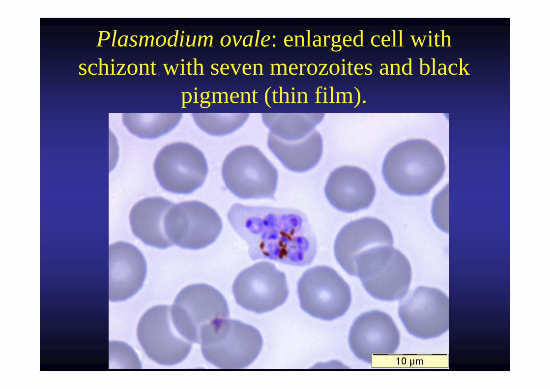
Plasmodium ovale: enlarged cell with schizont with seven merozoites and black
pigment (thin film).
Plasmodium malariae in thinfilm
Courtesy U.S. Department of Health, Education and Welfare. 1960.
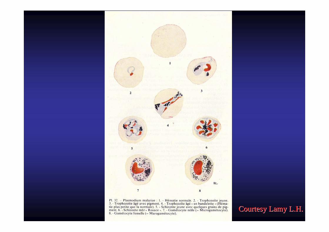
Courtesy Lamy L.H.Courtesy Lamy L.H.
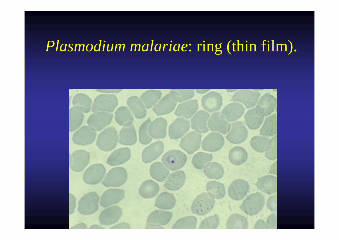
Plasmodium malariae: ring (thin film).
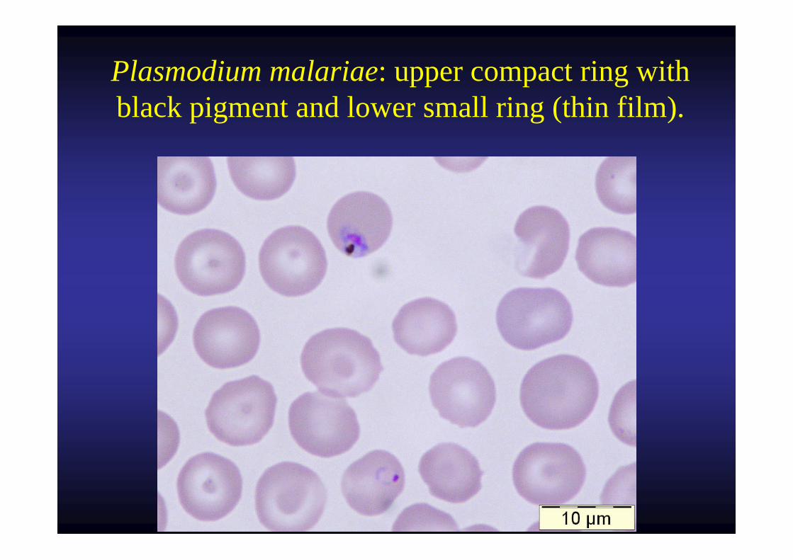
Plasmodium malariae: upper compact ring with black pigment and lower small ring (thin film).
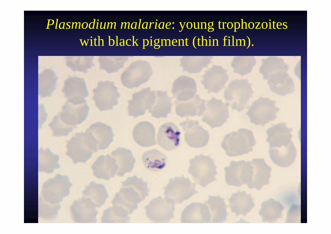
Plasmodium malariae: young trophozoites with black pigment (thin film).
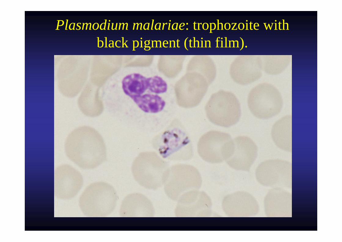
Plasmodium malariae: trophozoite with black pigment (thin film).
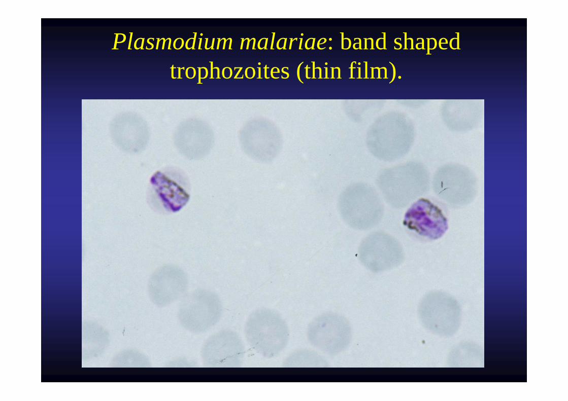
Plasmodium malariae: band shaped trophozoites (thin film).
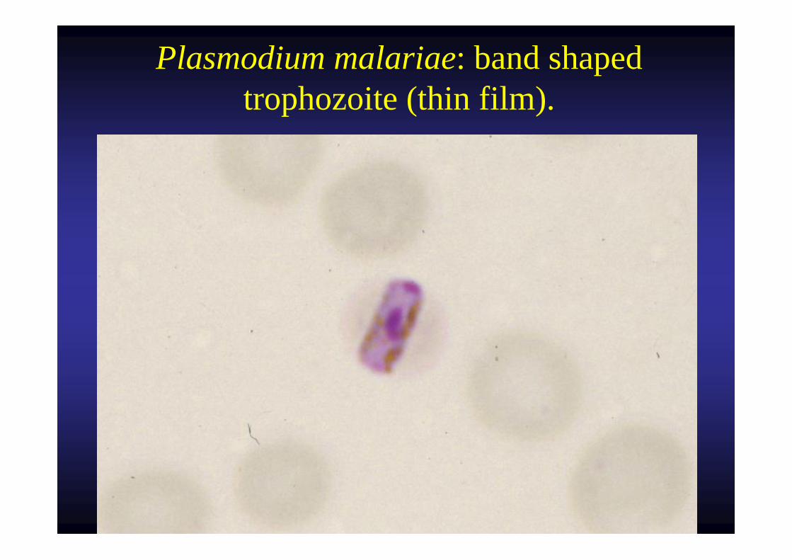
Plasmodium malariae: band shaped trophozoite (thin film).
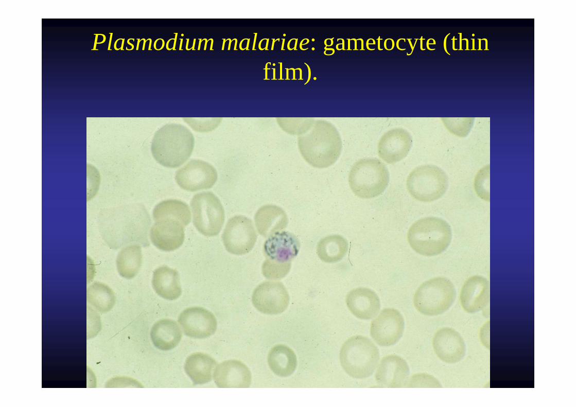
Plasmodium malariae: gametocyte (thin film).
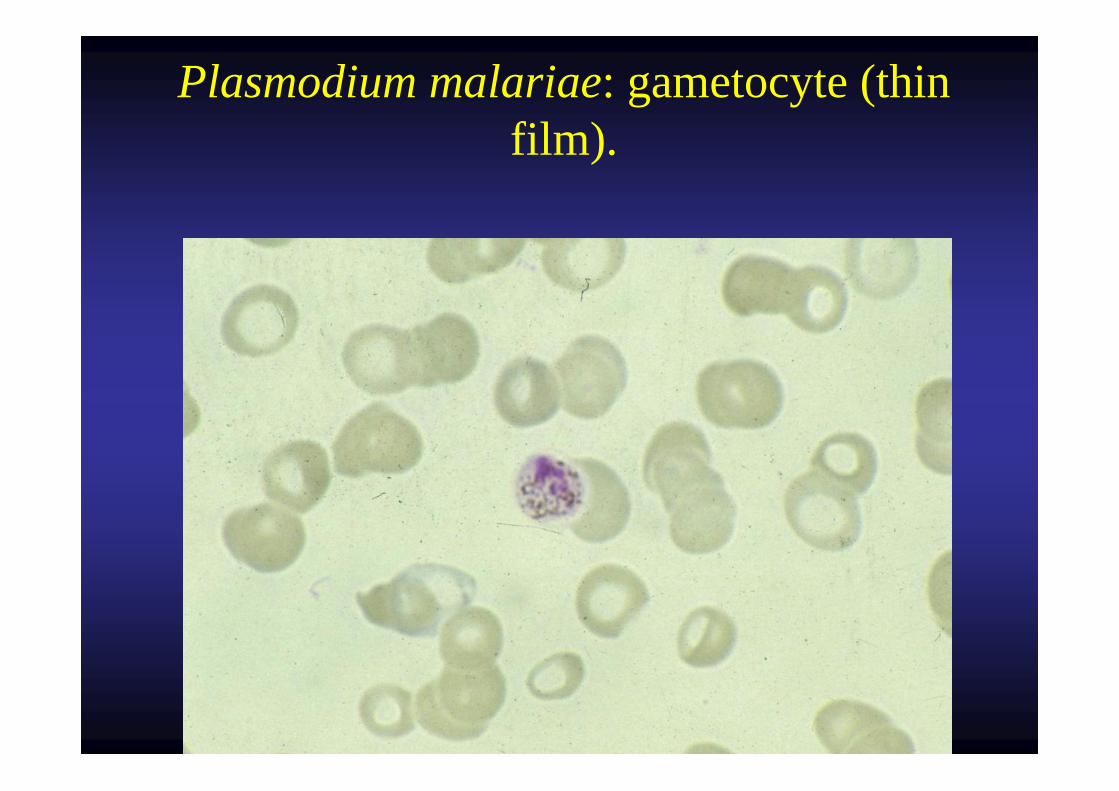
Plasmodium malariae: gametocyte (thin film).
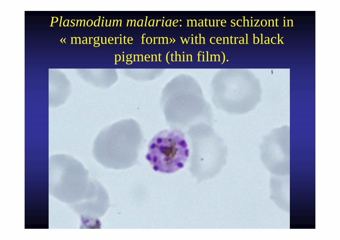
Plasmodium malariae: mature schizont in « marguerite form» with central black
pigment (thin film).
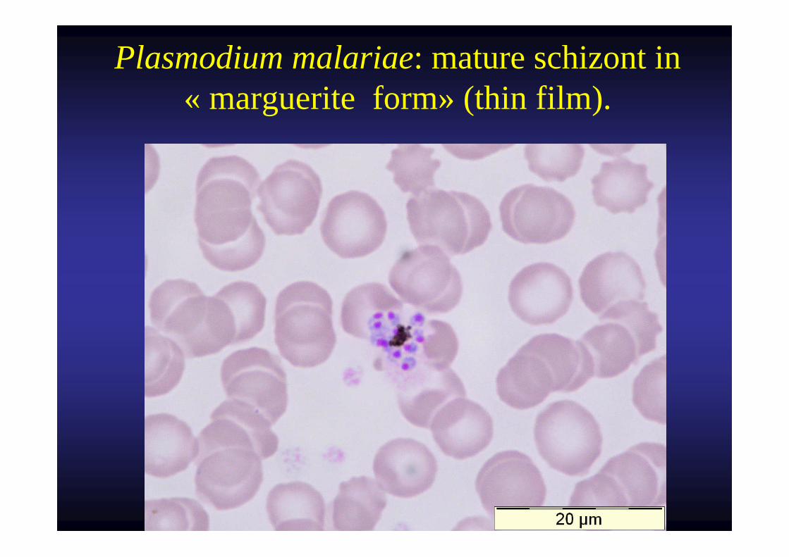
Plasmodium malariae: mature schizont in « marguerite form» (thin film).
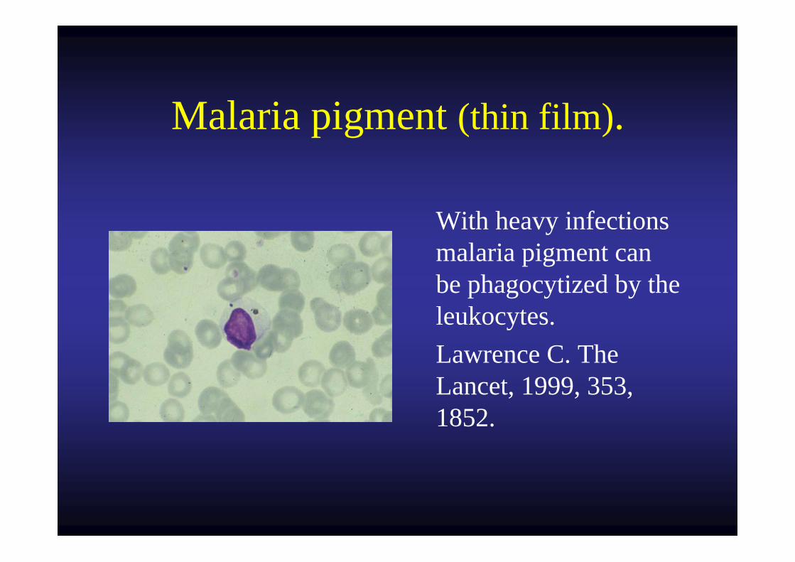
Malaria pigment (thin film).
With heavy infections malaria pigment can be phagocytized by the leukocytes.Lawrence C. The Lancet, 1999, 353, 1852.
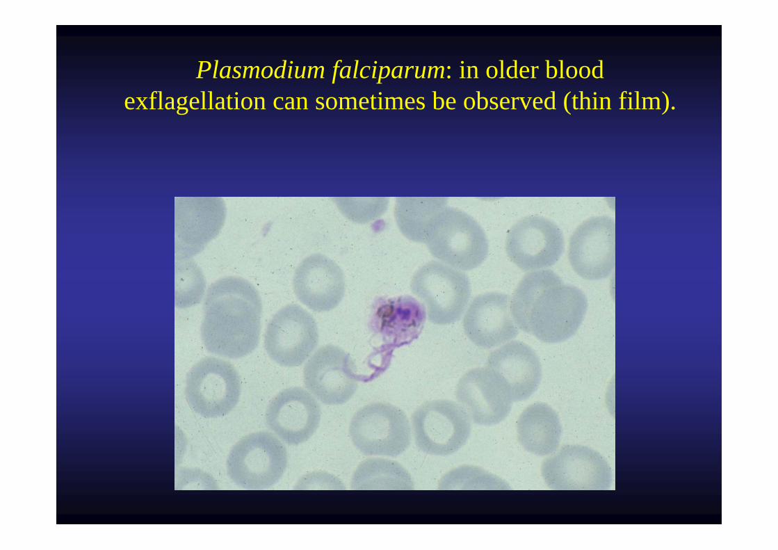
Plasmodium falciparum: in older bloodexflagellation can sometimes be observed (thin film).
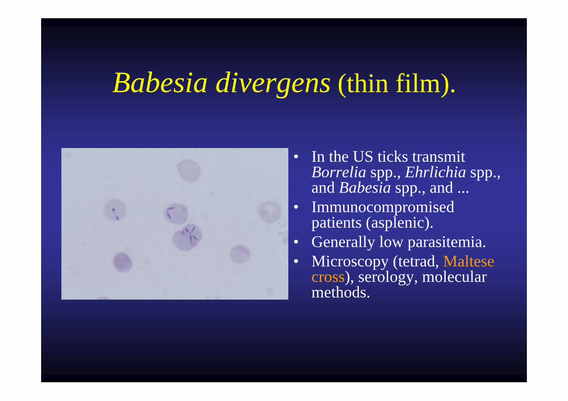
Babesia divergens (thin film).
• In the US ticks transmit Borrelia spp., Ehrlichia spp., and Babesia spp., and ...
• Immunocompromised patients (asplenic).
• Generally low parasitemia.• Microscopy (tetrad, Maltese
cross), serology, molecular methods.
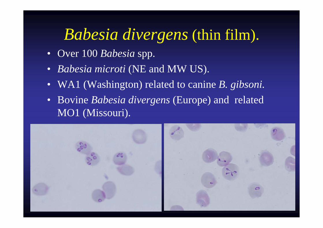
Babesia divergens (thin film).• Over 100 Babesia spp.• Babesia microti (NE and MW US).• WA1 (Washington) related to canine B. gibsoni.• Bovine Babesia divergens (Europe) and related
MO1 (Missouri).
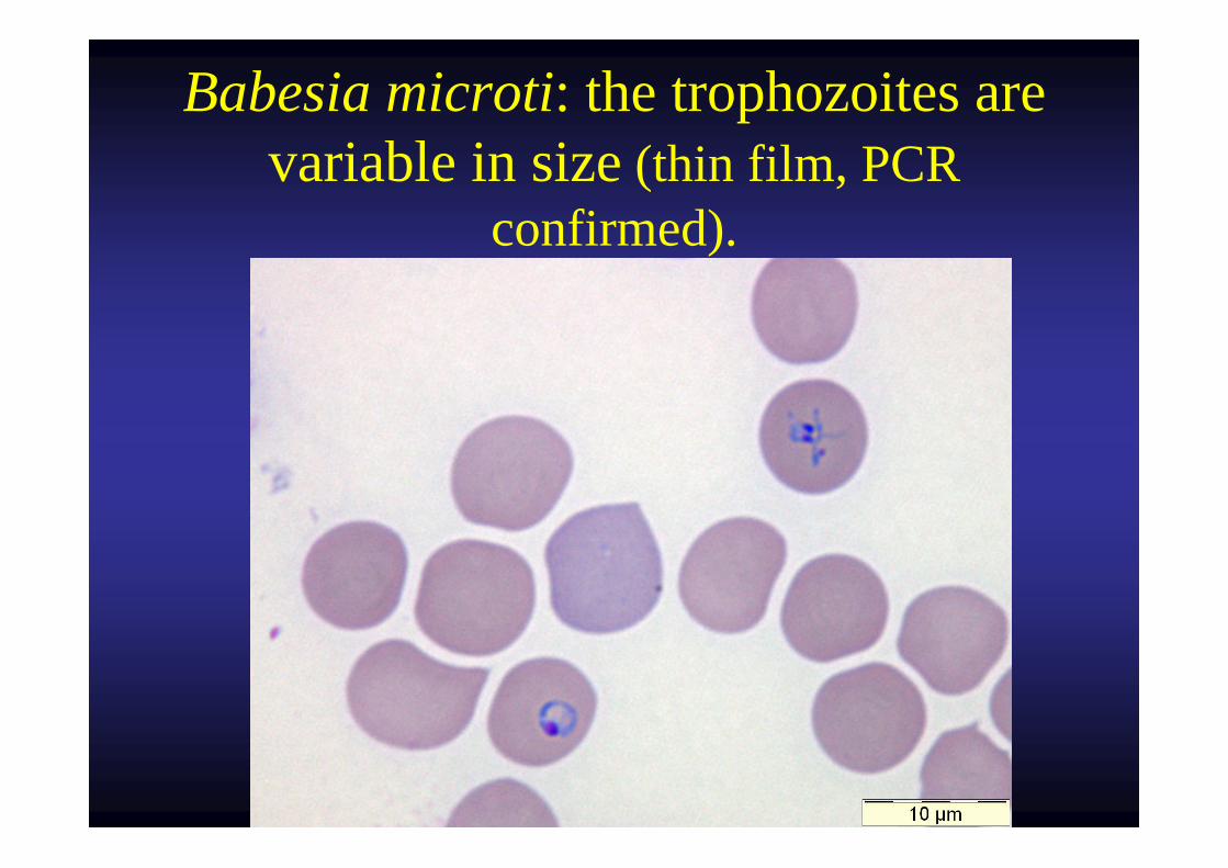
Babesia microti: the trophozoites are variable in size (thin film, PCR
confirmed).
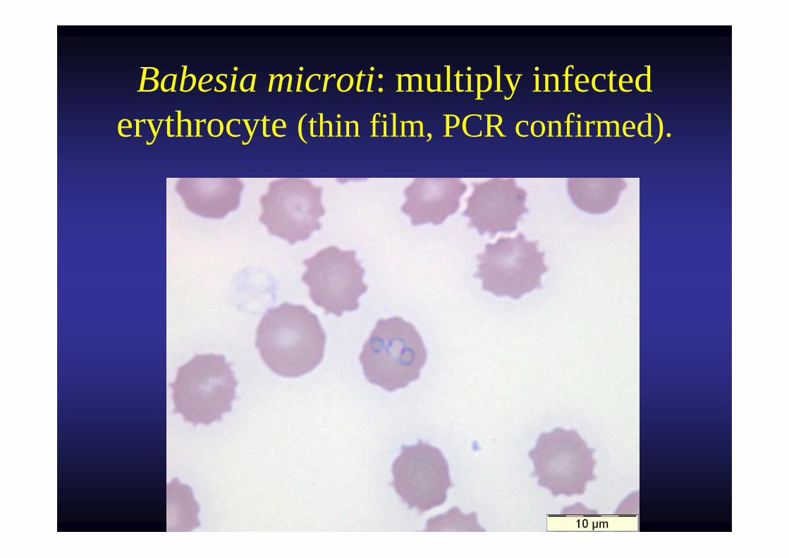
Babesia microti: multiply infected erythrocyte (thin film, PCR confirmed).
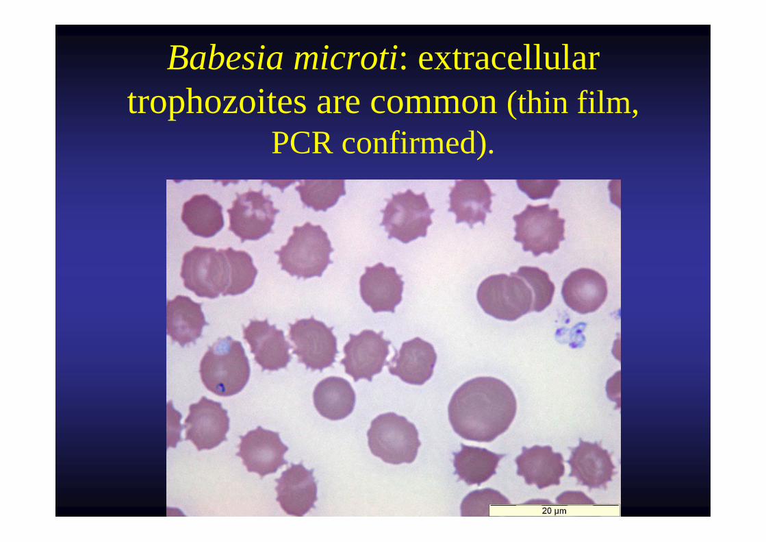
Babesia microti: extracellular trophozoites are common (thin film,
PCR confirmed).
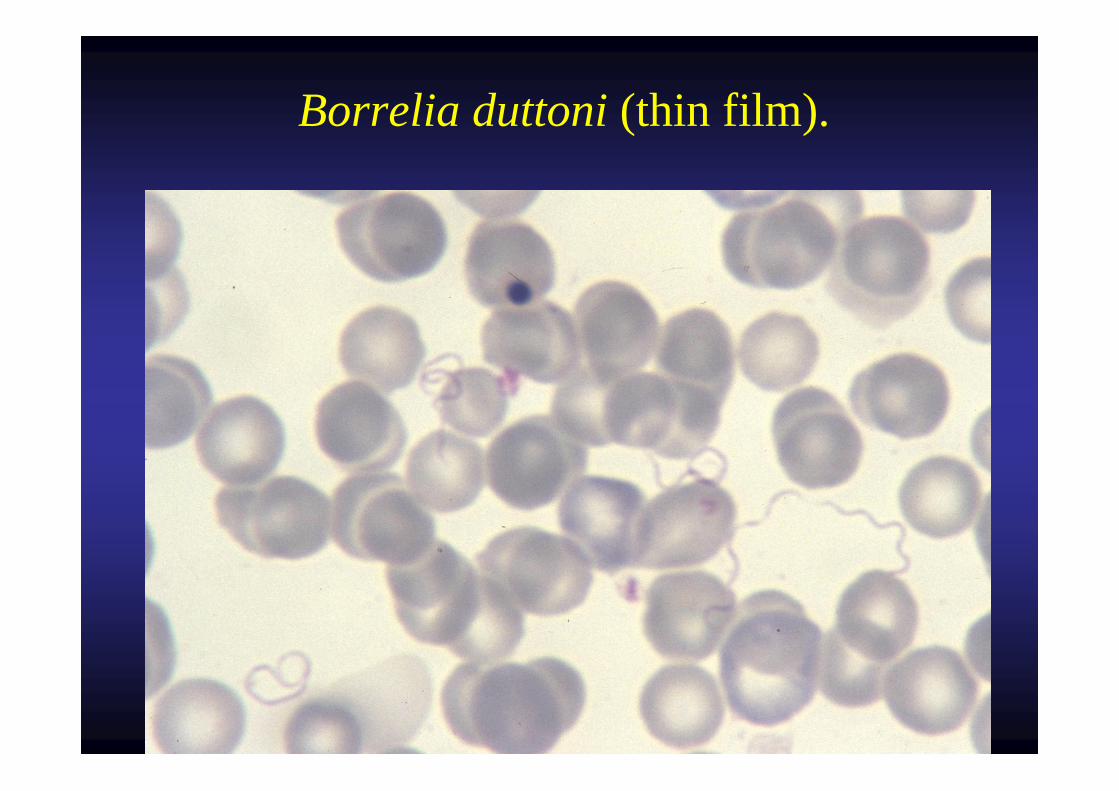
Borrelia duttoni (thin film).
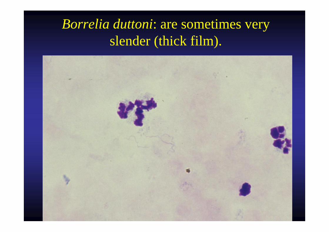
Borrelia duttoni: are sometimes very slender (thick film).
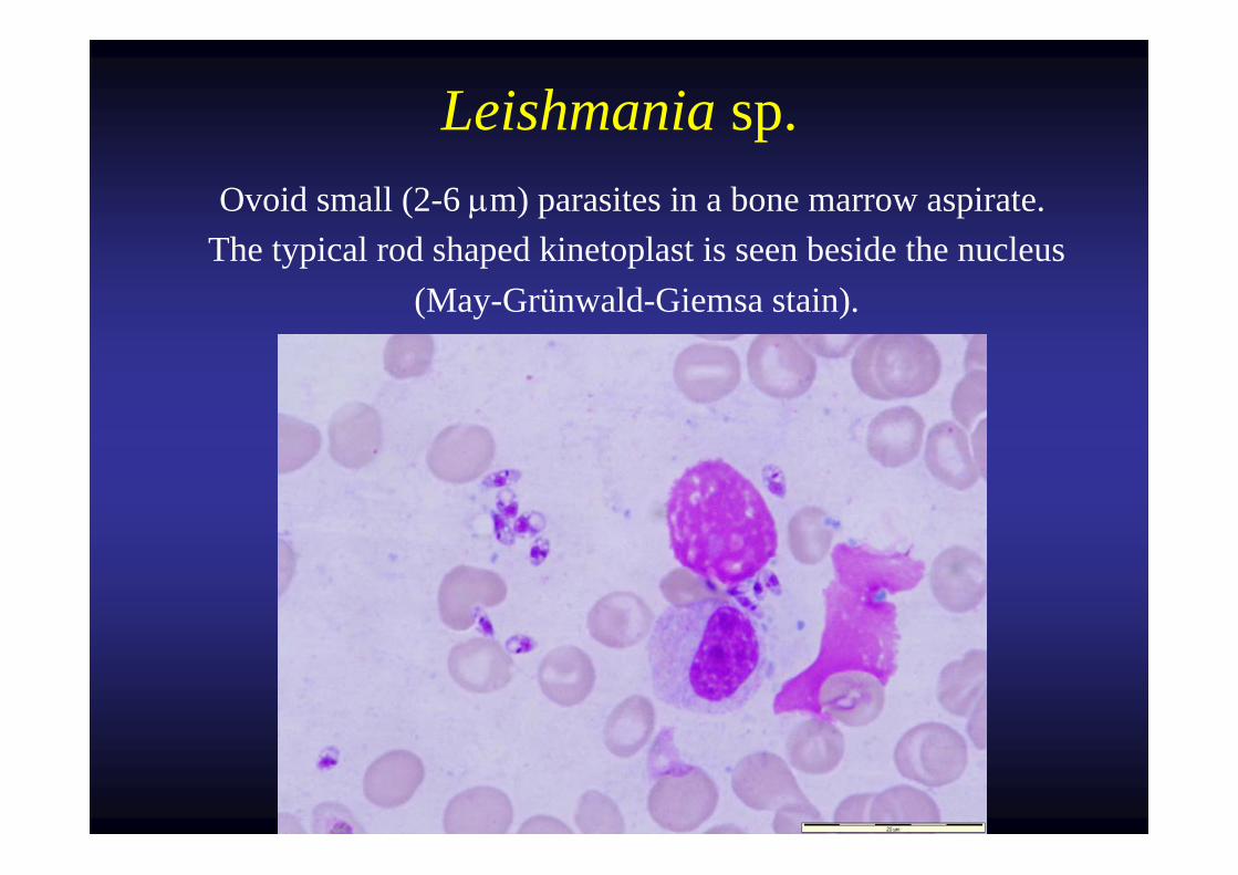
Leishmania sp. Ovoid small (2-6 μm) parasites in a bone marrow aspirate.
The typical rod shaped kinetoplast is seen beside the nucleus (May-Grünwald-Giemsa stain).
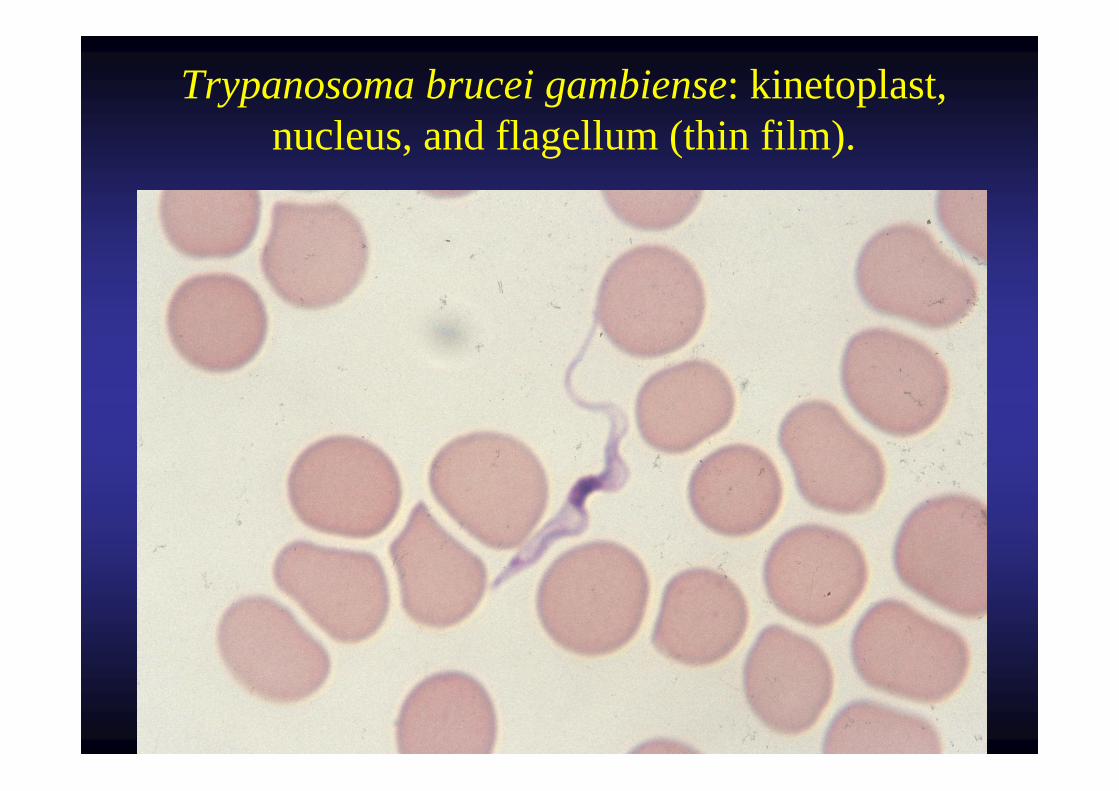
Trypanosoma brucei gambiense: kinetoplast, nucleus, and flagellum (thin film).
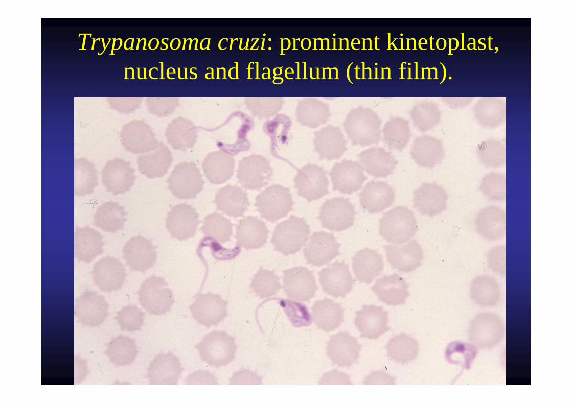
Trypanosoma cruzi: prominent kinetoplast, nucleus and flagellum (thin film).
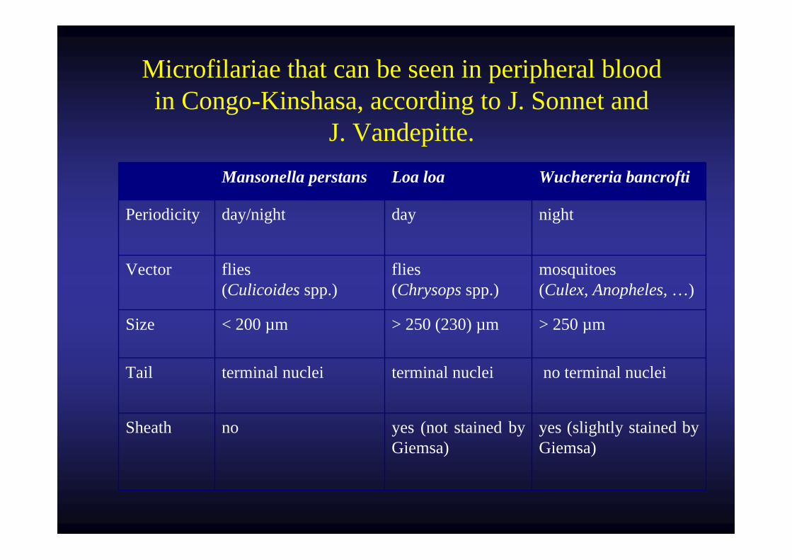
Microfilariae that can be seen in peripheral blood in Congo-Kinshasa, according to J. Sonnet and
J. Vandepitte.Mansonella perstans Loa loa Wuchereria bancrofti
Periodicity day/night day night
Vector flies(Culicoides spp.)
flies (Chrysops spp.)
mosquitoes (Culex, Anopheles, …)
Size < 200 µm > 250 (230) µm > 250 µm
Tail terminal nuclei terminal nuclei no terminal nuclei
Sheath no yes (not stained by Giemsa)
yes (slightly stained by Giemsa)
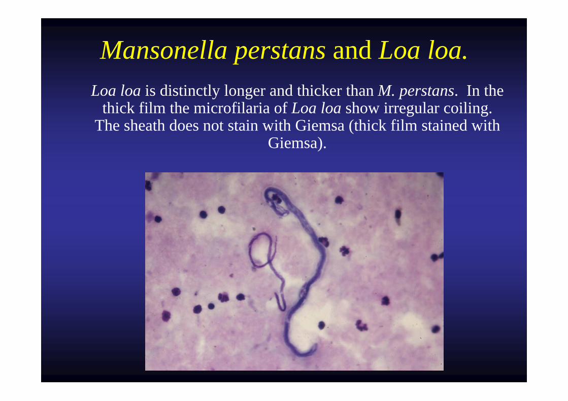
Mansonella perstans and Loa loa. Loa loa is distinctly longer and thicker than M. perstans. In the
thick film the microfilaria of Loa loa show irregular coiling. The sheath does not stain with Giemsa (thick film stained with
Giemsa).

2 Plasmodium ?
3 Plasmodium ?
4 Plasmodium ?
5 Plasmodium ?
Courtesy Gentilini M. & Duflo B.Courtesy Gentilini M. & Duflo B.
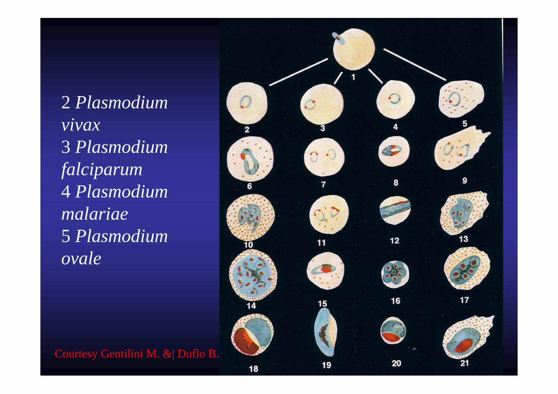
2 Plasmodium vivax3 Plasmodiumfalciparum4 Plasmodiummalariae5 Plasmodiumovale
Courtesy Gentilini M. &| Duflo B.
Comments mainly based on the
Manual for the Microscopical Diagnosis of Malaria in Man
1960 Edition
U.S. Department of Health, Education, and Welfare
Public Health Service


![Anopheles (Diptera: Culicidae), forest malaria vectors, in ... · complex [19] with Anopheles leucosphyrus Dönitz, 1901, Anopheles latens Sallum and Peyton, 2005, and Anopheles introlatus](https://img.pdfslide.net/doc/110x75/5d52cc0d88c993073e8b8565/anopheles-diptera-culicidae-forest-malaria-vectors-in-complex-19.jpg)