Embed Size (px)
Citation preview
Int.J.Curr.Microbiol.App.Sci (2017) 6(11): 2569-2586
2569
Original Research Article https://doi.org/10.20546/ijcmas.2017.611.302
Antibacterial Activity of Some Seaweed Extracts against Multidrug Resistant
Urinary Tract Bacteria and Analysis of their Virulence Genes
Wagih A. El-Shouny*, Reda M. Gaafar, Gehan A. Ismail and Marwa M. Elzanaty
Botany Department, Faculty of Science, Tanta University, Tanta, Egypt
*Corresponding author
A B S T R A C T
Introduction
Worldwide, urinary tract infections (UTI) are
among the most common infectious diseases.
About 150 million people are diagnosed with
UTI every year (Gonzalez and Schaeffer,
1999). A urinary tract infection is an infection
involving the kidneys, ureters, bladder, or
urethra. These are the structures that urine
passes through before being eliminated from
the body (Foxman and Brown, 2003).
Virtually, an infection occurs when Gram
negative bacteria (Escherichia coli, Klebsiella
pneumoniae and Proteus vulgaris) get into the
urine and begin to grow. The infection usually
starts at the opening of the urethra, where the
urine leaves the body and moves upward into
the urinary tract, these bacteria normally live
in the bowel (colon) and around the anus. The
symptoms develop rapidly including fairly
high fever, shaking chills, nausea and
vomiting (Kalaivani et al., 2016). UTI
infection may complicate into an acute or
chronic kidney infection (pyelonephritis) or
even permanent kidney damage due to
neglected or not correctly treated infection.
Many classes of antibiotics were classified as
typical treatments for such diseases. However,
the improper and uncontrolled use of many
antibiotics resulted in the occurrence of
International Journal of Current Microbiology and Applied Sciences ISSN: 2319-7706 Volume 6 Number 11 (2017) pp. 2569-2586 Journal homepage: http://www.ijcmas.com
Multidrug resistant bacteria raise a serious clinical problem for treating infectious diseases
worldwide. Different seaweed algae including Ulva lactuca (Chlorophyta), Petalonia
fascia (Phaeophyta) and Gelidium spinosum (Rhodophyta) were investigated as natural
sources for antibacterial compounds. Different solvents ethanol, methanol, acetone and
water were used to extract the antibacterial substances from these seaweeds and were
examined against urinary tract multidrug resistant bacterial isolates: Escherichia coli,
Klebsiella pneumoniae, and Proteus mirabilis. The ethanolic extract of Ulva lactuca
exhibited the most significant antibacterial activity against the tested urinary tract infection
(UTI) bacterial isolates. The extract was purified and chemically analyzed using UV, IR, 1H NMR and GC-MS. The results indicated that the extract was aromatic ester derivative
named Di-isooctylphthalate of molecular weight 390.56. PCR analysis of three virulence
factors (adhesion, urease and hemolysin) were employed in order to genetically compare
the virulence activities of the UTI isolates. The results showed that only faint PCR
amplicon of hemolysin virulence gene was observed in P. mirabilis sensitive isolate, while
two fragments were found in the multidrug and algal resistant corresponding isolates with
ampilicon sizes of 375 and 875 bp.
K e y w o r d s Antibacterial activity,
PCR analysis, Ulva
lactuca, UTI infection, Virulence genes.
Accepted:
20 September 2017
Available Online: 10 November 2017
Article Info
Int.J.Curr.Microbiol.App.Sci (2017) 6(11): 2569-2586
2570
antimicrobial drug resistance, which became a
major health problem worldwide (Dethlefsen
et al., 2008) and forced seeking for new
treatment sources.
Virulence factors are specific traits enabling
pathogenic bacteria to overcome host immune
system and cause various diseases (Ejrnaes,
2011). Virulence genes are located on
transmissible genetic elements and/or in
particular regions on the chromosome that are
called pathogenicity islands (Farshad et al.,
2012). Pathogenicity islands directed
coordinate horizontal transfer of virulence
genes between strains of one species or even
related species (Hacker et al., 1997). UTI
bacterial strains have various types of
virulence factors such as adhesions,
hemolysin, urease enzyme, toxins and iron
uptake systems that facilitate colonization and
persistence of the bacteria in the urinary tract
(Oliveira et al., 2011) and support their
resistance system and pathogenicity to the
regularly used synthetic drugs.
Nowadays, seaweeds are considered a novel
source of bioactive compounds and produce a
great variety of secondary metabolites
exhibiting broad spectrum of biological
activities (Khotimchenko et al., 2002;
Ramasamy and Kumar, 2009; El-Sheekh et
al., 2016). Many types of seaweed have been
screened extensively to isolate natural drugs
or biologically active substances from
different habitats around the world. Several
solvents with different polarities e.g. hexane,
chloroform, ethanol, methanol, acetone and
water were used to extract the antimicrobial
material from such seaweeds. Compounds
with cytostatic, antiviral, anthelminthic,
antifungal, bacteriostatic and bactericidal
activities have been detected in green, brown
and red seaweeds with a comparable
promising results with authentic antibiotics
(Newman et al., 2003; Manikandan et al.,
2011; Osman et al., 2013; Ismail et al., 2016).
Active antibacterial agents found in the
seaweeds include terpenoid, phlorotannins,
acrylic acid and phenolic compounds
(Cardoso et al., 2014). Also, steroids,
halogenated ketone, fatty acids and alkaline,
cyclic polysulphides were recorded (Cheung
et al., 2015; Khelil-Radji et al., 2017). The
present study aimed to explore the
antibacterial activity of some seaweed
extracts against urinary tract (UTI) infectious
bacteria. To compare the activity of these
extracts as natural drug sources with the
standard synthetic antibiotics against
multidrug resistant (MDR) isolates. Finally, to
compare the virulence gene profiles of the
UTI isolates and detect the effect of seaweeds
treatment on their virulence activities.
Materials and Methods
Isolation and identification of bacterial
isolates
The bacterial swaps were obtained from
microbiology laboratories, Faculty of
Pharmacy, Tanta University, Tanta, Egypt.
The bacterial isolates, generally Gram
negative, were cultivated on different
selective media. The morphological and
biochemical examinations were conducted
repeatedly according to the standard methods
until complete purification and identification
of these bacterial isolates (Oxoid, 1981;
Collee et al., 1996). Eight bacterial isolates
were selected of which Escherichia coli (3
isolates), Klebsiella pneumoniae (2 isolates)
and Proteus mirabilis (3 isolates).
Antibiotic susceptibility test of bacterial
isolates
The antibacterial susceptibility of the
pathogenic bacterial isolates were confirmed
against eight different classes of antibiotics
using the modified Kirby-Bauer disk diffusion
method according to the Clinical and
Int.J.Curr.Microbiol.App.Sci (2017) 6(11): 2569-2586
2571
Laboratory Standards Institute (CLSI, 2012)
on Mueller Hinton agar. The tested antibiotics
were commonly used in the treatments of UTI
cases and included: Carbapenems,
Cephalosporins, Monobactams,
Nitrofuratoins, Penicillins, Polyketide,
Quinolone/ Fluoroquinolone and
Sulfonamides. The plates were then incubated
overnight at 37ºC. The diameters of the
inhibition zones were measured (Vasquez and
Hand, 2004) and compared to differentiate the
bacterial isolates into sensitive, intermediate
and resistant and to calculate the multiple
antibiotic resistances (MAR) index for each
isolate (by dividing the number of antibiotics
to which the isolate is resistant by the total
number of tested antibiotics).
Preparation of algal extracts
Three species of seaweeds from different
divisions (Chlorophyta) Ulva lactuca,
(Phaeophyta) Petalonia fascia and
(Rhodophyta) Gelidium spinosum were
collected in 2016 from Abu Qir-Bay,
Alexandria, Egypt (N 31°19` E 30°03`). The
seaweeds were identified according to
(Aleem, 1993) and (Guiry and Guiry, 2016).
The different seaweeds were air dried in the
shade at room temperature (25ºC) and then
grounded to fine powder. The powders (1:10
w/v) were soaked with different polar organic
solvents (80% each of ethanol, methanol and
acetone in addition to water) for 24 hrs.on a
rotary shaker at 150 rpm. The extracts from
three consecutive soakings were pooled
together and filtered. The obtained filtrates
were evaporated using rotary evaporator at
45ºC until dryness, then weighed and stored at
-20ºC (Karthikaidevi et al., 2009). The
collected seaweeds were identified to be:
Ulva lactuca Linnaeus
Empire: Eukaryota, Kingdom: Plantae,
Phylum: Chlorophyta, Class: Ulvophyceae,
Order: Ulvales, Family: Ulvaceae Genus:
Ulva.
Petalonia fascia (O. F. Müller) Kuntze
Empire: Eukaryota, Kingdom: Chromista,
Phylum: Ochrophyta, Class: Phaeophyceae,
Order: Ectocarpales, Family:
Scytosiphonaceae, Genus: Petalonia.
Gelidium spinosum (S. G. Gmelin) P.C.
Silva.
Empire: Eukaryota, Kingdom: Plantae,
Phylum: Rhodophyta, Class:
Florideophyceae, Order: Gelidiales, Family:
Gelidiaceae, Genus: Gelidium.
Antibacterial activity test of seaweeds
extracts
The seaweeds antibacterial activity was
assayed using disk diffusion method as
described in the previous section. Suspensions
of the tested bacterial isolates were prepared
from overnight freshly incubated bacterial
cultures and mixed in sterilized saline
solution (0.9% w/v NaCl) to get cell counts of
106 to 10
8 CFU/ml for each bacterial isolate
(Gilbert, 1987).
Seaweed extract was prepared by re-
suspending the powder (50:1 w/v) in each
solvent. Previously sterilized filter paper disks
were immersed in different algal extracts (20
µl) and inoculated over Petri dishes surface.
The plates were incubated for 2 hours at 4ºC
to allow the diffusion of antibacterial
substance and then incubated overnight at
37ºC. Standard antibiotic discs and solvent
discs were used as positive and negative
controls, respectively.
The diameter of inhibition zones indicates the
antibacterial activity of different seaweeds in
different solvents.
Int.J.Curr.Microbiol.App.Sci (2017) 6(11): 2569-2586
2572
Identification of sensitive bacterial isolates
Bacterial isolates which were sensitive to
seaweed extracts were identified using
Biomerieux VITEK®2 system. The
Biomerieux VITEK®2 cards were filled with
the tested bacterial suspension and manually
loaded into the VITEK®2 system for
identification (Thomas et al., 2001 ).
Fractionation, purification and
characterization of antibacterial crude
extracts
Column chromatography and UV spectra
of algal antibacterial agent
Active crude seaweed extract (5 g) was
purified and fractioned by column
chromatography on silica gel G EDWC, 60-
200 mesh (Solomon and Santhi 2008). A
glass column (3 cm x 20 cm) was packed by
silica gel (30-40 g) in pure chloroform eluted
with gradients of mobile phase from 30:70 %
chloroform: ethanol to 70:30 % chloroform:
ethanol and the collected fractions were
evaporated at 40oC under vacuum. The dried
fractions were dissolved in 70 % ethanol
solvent and their absorption was measured
using UV spectrophotometer (UV 2101/pc) at
200- 900 nm wavelength (Blunt et al., 2007).
The antibacterial activity of the collected
fractions was examined against UTI bacterial
isolates using disk diffusion method.
Different active fractions with the same UV
absorption were collected together, dried and
subjected to subsequent analysis to determine
the chemical structure of the active
compounds.
Infra-red spectra (IR) of the algal
antibacterial agent
The infra-red spectrophotometer (FT-IR)
(Perkin Elmer 1430) was used to identify the
functional groups of the antibacterial algal
material (Boeriu et al., 2004). The pellet
which contains sample of active compound in
solid phase was mixed with KBr (FT-IR
grade) and FT-IR spectra were recorded in the
range of 4000- 400 cm-1
.
Proton nuclear magnetic resonance (1H
NMR) spectra of algal antibacterial agent
The sample was mixed with dehydrated
chloroform solvent (0.04 g/ml). Different
protons of the antibacterial material could be
identified by their corresponding nuclear
magnetic resonance (Atalah et al., 2007).
Gas chromatography mass spectrometry
(GC-MS)
Gas chromatography mass spectrometry (GC-
MS) analysis was used to determine the
chemical composition of the seaweeds
antibacterial material. The acquisition
parameters were done with column (30m x
250 µm x 0.25 µm film thickness) using
helium as carrier gas (0.8 ml/min) with Perkin
Elmer: Clarus 580/560 S model system. The
GC oven temperature was programed from
60oC to 250
oC at a rate of 2
oC/ min. Relative
area values (as a percentage of total volatile
composition) were directly obtained from
total ion current (TIC).
Molecular detection of the virulence factors
of the bacterial strains
DNA extraction and PCR amplification of
virulence factors
In order to extract genomic DNA of the
multidrug resistant (MDR) bacterial isolates,
which were sensitive to the seaweeds
antibacterial material, boiling method was
used. Before DNA extraction, the bacterial
isolates were activated by overnight culturing
on nutrient broth at 37oC, then it was
centrifuged at 5103 for 15 minutes. The
resulted pellets were mixed in 100 µl of
sterilized water and incubated in boiling water
Int.J.Curr.Microbiol.App.Sci (2017) 6(11): 2569-2586
2573
for 10 min at 100oC for complete DNA
extraction (Anderson et al., 2004).
Polymerase chain reaction (PCR) technique
was used to detect three virulence factors
genes including hemolysin, adhesion and
urease.
Amplification of bacterial virulence genes
was done in a total volume of 25 µl
containing 1 µl of bacterial suspension, 1.5 µl
of each of the primers, 8.5 µl sterilized
distilled water, and 12.5 µl of the red master
mix (Bioline); which contained 1000 x 50 µl
Reactions U Taq DNA polymerase (Sigma-
Aldrich) in 1 PCR buffer containing 1.5 mM
MgCl2 (Table 1).
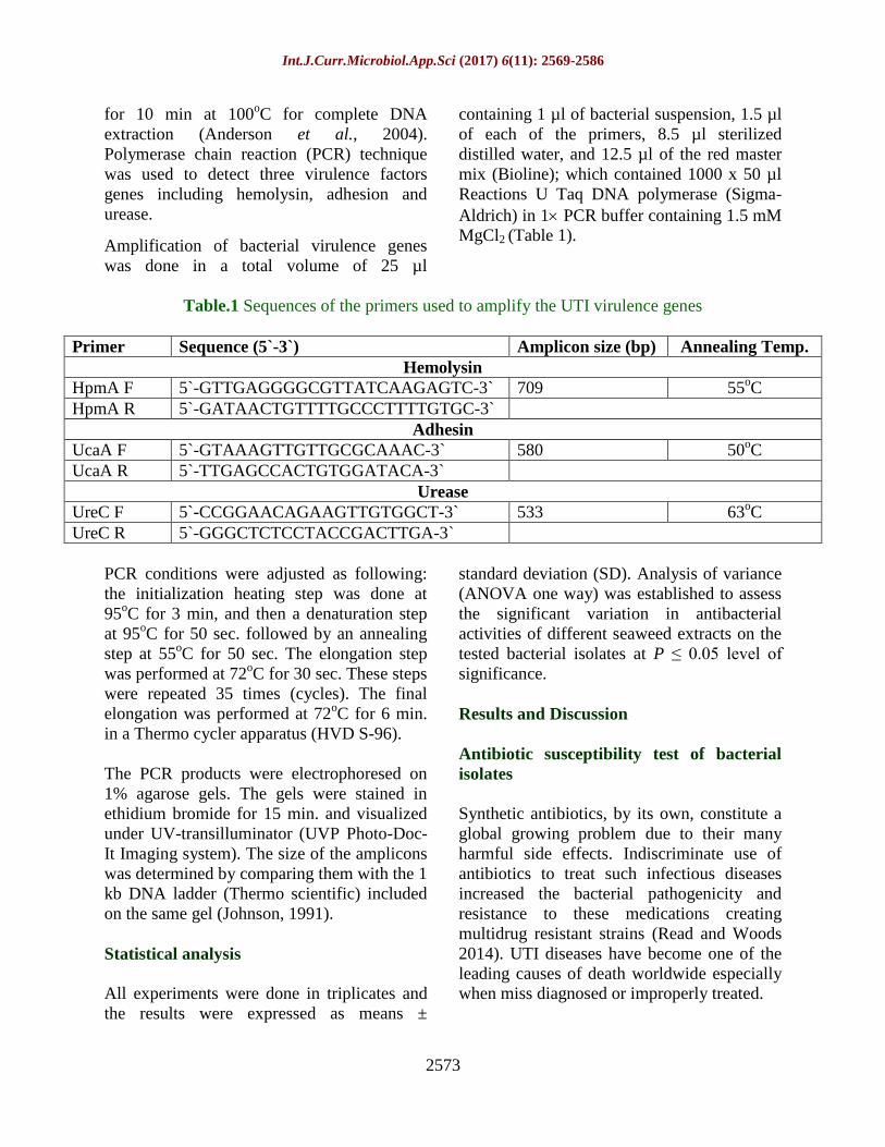
Table.1 Sequences of the primers used to amplify the UTI virulence genes
Primer Sequence (5`-3`) Amplicon size (bp) Annealing Temp.
Hemolysin
HpmA F 5`-GTTGAGGGGCGTTATCAAGAGTC-3` 709 55oC
HpmA R 5`-GATAACTGTTTTGCCCTTTTGTGC-3`
Adhesin
UcaA F 5`-GTAAAGTTGTTGCGCAAAC-3` 580 50oC
UcaA R 5`-TTGAGCCACTGTGGATACA-3`
Urease
UreC F 5`-CCGGAACAGAAGTTGTGGCT-3` 533 63oC
UreC R 5`-GGGCTCTCCTACCGACTTGA-3`
PCR conditions were adjusted as following:
the initialization heating step was done at
95oC for 3 min, and then a denaturation step
at 95oC for 50 sec. followed by an annealing
step at 55oC for 50 sec. The elongation step
was performed at 72oC for 30 sec. These steps
were repeated 35 times (cycles). The final
elongation was performed at 72oC for 6 min.
in a Thermo cycler apparatus (HVD S-96).
The PCR products were electrophoresed on
1% agarose gels. The gels were stained in
ethidium bromide for 15 min. and visualized
under UV-transilluminator (UVP Photo-Doc-
It Imaging system). The size of the amplicons
was determined by comparing them with the 1
kb DNA ladder (Thermo scientific) included
on the same gel (Johnson, 1991).
Statistical analysis
All experiments were done in triplicates and
the results were expressed as means ±
standard deviation (SD). Analysis of variance
(ANOVA one way) was established to assess
the significant variation in antibacterial
activities of different seaweed extracts on the
tested bacterial isolates at P ≤ 0.05 level of
significance.
Results and Discussion
Antibiotic susceptibility test of bacterial
isolates
Synthetic antibiotics, by its own, constitute a
global growing problem due to their many
harmful side effects. Indiscriminate use of
antibiotics to treat such infectious diseases
increased the bacterial pathogenicity and
resistance to these medications creating
multidrug resistant strains (Read and Woods
2014). UTI diseases have become one of the
leading causes of death worldwide especially
when miss diagnosed or improperly treated.
Int.J.Curr.Microbiol.App.Sci (2017) 6(11): 2569-2586
2574
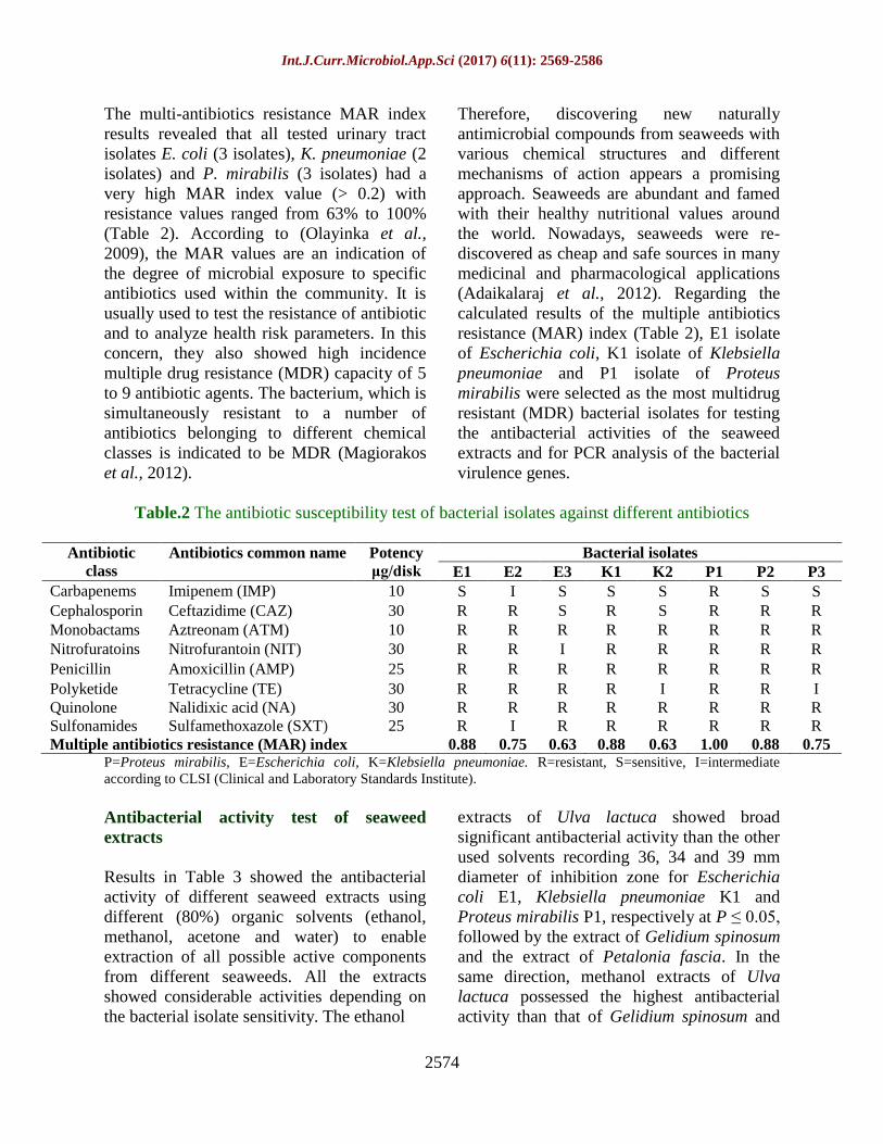
The multi-antibiotics resistance MAR index
results revealed that all tested urinary tract
isolates E. coli (3 isolates), K. pneumoniae (2
isolates) and P. mirabilis (3 isolates) had a
very high MAR index value (> 0.2) with
resistance values ranged from 63% to 100%
(Table 2). According to (Olayinka et al.,
2009), the MAR values are an indication of
the degree of microbial exposure to specific
antibiotics used within the community. It is
usually used to test the resistance of antibiotic
and to analyze health risk parameters. In this
concern, they also showed high incidence
multiple drug resistance (MDR) capacity of 5
to 9 antibiotic agents. The bacterium, which is
simultaneously resistant to a number of
antibiotics belonging to different chemical
classes is indicated to be MDR (Magiorakos
et al., 2012).
Therefore, discovering new naturally
antimicrobial compounds from seaweeds with
various chemical structures and different
mechanisms of action appears a promising
approach. Seaweeds are abundant and famed
with their healthy nutritional values around
the world. Nowadays, seaweeds were re-
discovered as cheap and safe sources in many
medicinal and pharmacological applications
(Adaikalaraj et al., 2012). Regarding the
calculated results of the multiple antibiotics
resistance (MAR) index (Table 2), E1 isolate
of Escherichia coli, K1 isolate of Klebsiella
pneumoniae and P1 isolate of Proteus
mirabilis were selected as the most multidrug
resistant (MDR) bacterial isolates for testing
the antibacterial activities of the seaweed
extracts and for PCR analysis of the bacterial
virulence genes.
Table.2 The antibiotic susceptibility test of bacterial isolates against different antibiotics
Antibiotic
class Antibiotics common name Potency
μg/disk
Bacterial isolates E1 E2 E3 K1 K2 P1 P2 P3
Carbapenems Imipenem (IMP) 10 S I S S S R S S
Cephalosporin Ceftazidime (CAZ) 30 R R S R S R R R
Monobactams Aztreonam (ATM) 10 R R R R R R R R
Nitrofuratoins Nitrofurantoin (NIT) 30 R R I R R R R R
Penicillin Amoxicillin (AMP) 25 R R R R R R R R
Polyketide Tetracycline (TE) 30 R R R R I R R I
Quinolone Nalidixic acid (NA) 30 R R R R R R R R
Sulfonamides Sulfamethoxazole (SXT) 25 R I R R R R R R
Multiple antibiotics resistance (MAR) index 0.88 0.75 0.63 0.88 0.63 1.00 0.88 0.75 P=Proteus mirabilis, E=Escherichia coli, K=Klebsiella pneumoniae. R=resistant, S=sensitive, I=intermediate
according to CLSI (Clinical and Laboratory Standards Institute).
Antibacterial activity test of seaweed
extracts
Results in Table 3 showed the antibacterial
activity of different seaweed extracts using
different (80%) organic solvents (ethanol,
methanol, acetone and water) to enable
extraction of all possible active components
from different seaweeds. All the extracts
showed considerable activities depending on
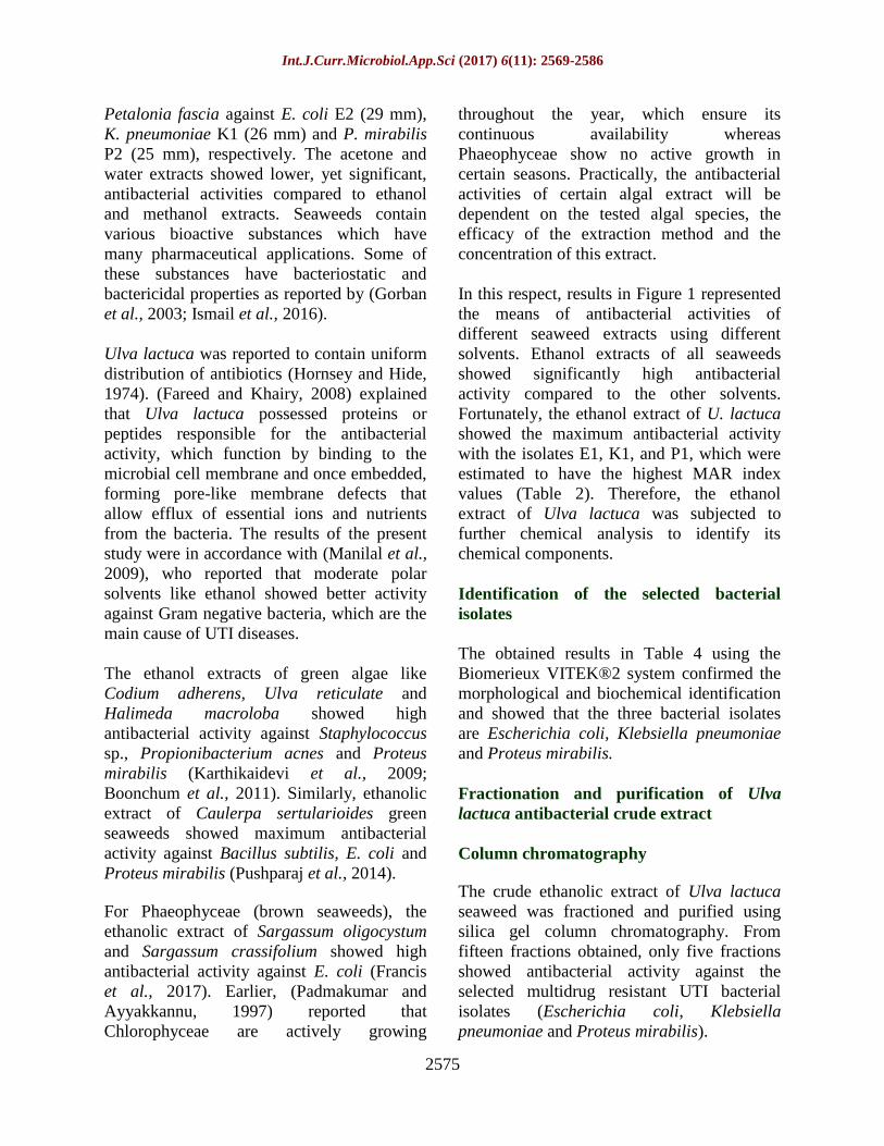
the bacterial isolate sensitivity. The ethanol
extracts of Ulva lactuca showed broad
significant antibacterial activity than the other
used solvents recording 36, 34 and 39 mm
diameter of inhibition zone for Escherichia
coli E1, Klebsiella pneumoniae K1 and
Proteus mirabilis P1, respectively at P ≤ 0.05,
followed by the extract of Gelidium spinosum
and the extract of Petalonia fascia. In the
same direction, methanol extracts of Ulva
lactuca possessed the highest antibacterial
activity than that of Gelidium spinosum and
Int.J.Curr.Microbiol.App.Sci (2017) 6(11): 2569-2586
2575
Petalonia fascia against E. coli E2 (29 mm),
K. pneumoniae K1 (26 mm) and P. mirabilis
P2 (25 mm), respectively. The acetone and
water extracts showed lower, yet significant,
antibacterial activities compared to ethanol
and methanol extracts. Seaweeds contain
various bioactive substances which have
many pharmaceutical applications. Some of
these substances have bacteriostatic and
bactericidal properties as reported by (Gorban
et al., 2003; Ismail et al., 2016).
Ulva lactuca was reported to contain uniform
distribution of antibiotics (Hornsey and Hide,
1974). (Fareed and Khairy, 2008) explained
that Ulva lactuca possessed proteins or
peptides responsible for the antibacterial
activity, which function by binding to the
microbial cell membrane and once embedded,
forming pore-like membrane defects that
allow efflux of essential ions and nutrients
from the bacteria. The results of the present
study were in accordance with (Manilal et al.,
2009), who reported that moderate polar
solvents like ethanol showed better activity
against Gram negative bacteria, which are the
main cause of UTI diseases.
The ethanol extracts of green algae like
Codium adherens, Ulva reticulate and
Halimeda macroloba showed high
antibacterial activity against Staphylococcus
sp., Propionibacterium acnes and Proteus
mirabilis (Karthikaidevi et al., 2009;
Boonchum et al., 2011). Similarly, ethanolic
extract of Caulerpa sertularioides green
seaweeds showed maximum antibacterial
activity against Bacillus subtilis, E. coli and
Proteus mirabilis (Pushparaj et al., 2014).
For Phaeophyceae (brown seaweeds), the
ethanolic extract of Sargassum oligocystum
and Sargassum crassifolium showed high
antibacterial activity against E. coli (Francis
et al., 2017). Earlier, (Padmakumar and
Ayyakkannu, 1997) reported that
Chlorophyceae are actively growing
throughout the year, which ensure its
continuous availability whereas
Phaeophyceae show no active growth in
certain seasons. Practically, the antibacterial
activities of certain algal extract will be
dependent on the tested algal species, the
efficacy of the extraction method and the
concentration of this extract.
In this respect, results in Figure 1 represented
the means of antibacterial activities of
different seaweed extracts using different
solvents. Ethanol extracts of all seaweeds
showed significantly high antibacterial
activity compared to the other solvents.
Fortunately, the ethanol extract of U. lactuca
showed the maximum antibacterial activity
with the isolates E1, K1, and P1, which were
estimated to have the highest MAR index
values (Table 2). Therefore, the ethanol
extract of Ulva lactuca was subjected to
further chemical analysis to identify its
chemical components.
Identification of the selected bacterial
isolates
The obtained results in Table 4 using the
Biomerieux VITEK®2 system confirmed the
morphological and biochemical identification
and showed that the three bacterial isolates
are Escherichia coli, Klebsiella pneumoniae
and Proteus mirabilis.
Fractionation and purification of Ulva
lactuca antibacterial crude extract
Column chromatography
The crude ethanolic extract of Ulva lactuca
seaweed was fractioned and purified using
silica gel column chromatography. From
fifteen fractions obtained, only five fractions
showed antibacterial activity against the
selected multidrug resistant UTI bacterial
isolates (Escherichia coli, Klebsiella
pneumoniae and Proteus mirabilis).
Int.J.Curr.Microbiol.App.Sci (2017) 6(11): 2569-2586
2576
The recorded inhibition zones for different
fractions were illustrated in Table 5. All five
fractions recorded remarkable inhibition
zones against the three-tested resistant
pathogenic isolates especially Fr2, Fr3 and
Fr8.
Determination of the chemical structure of
Ulva lactuca antibacterial compound
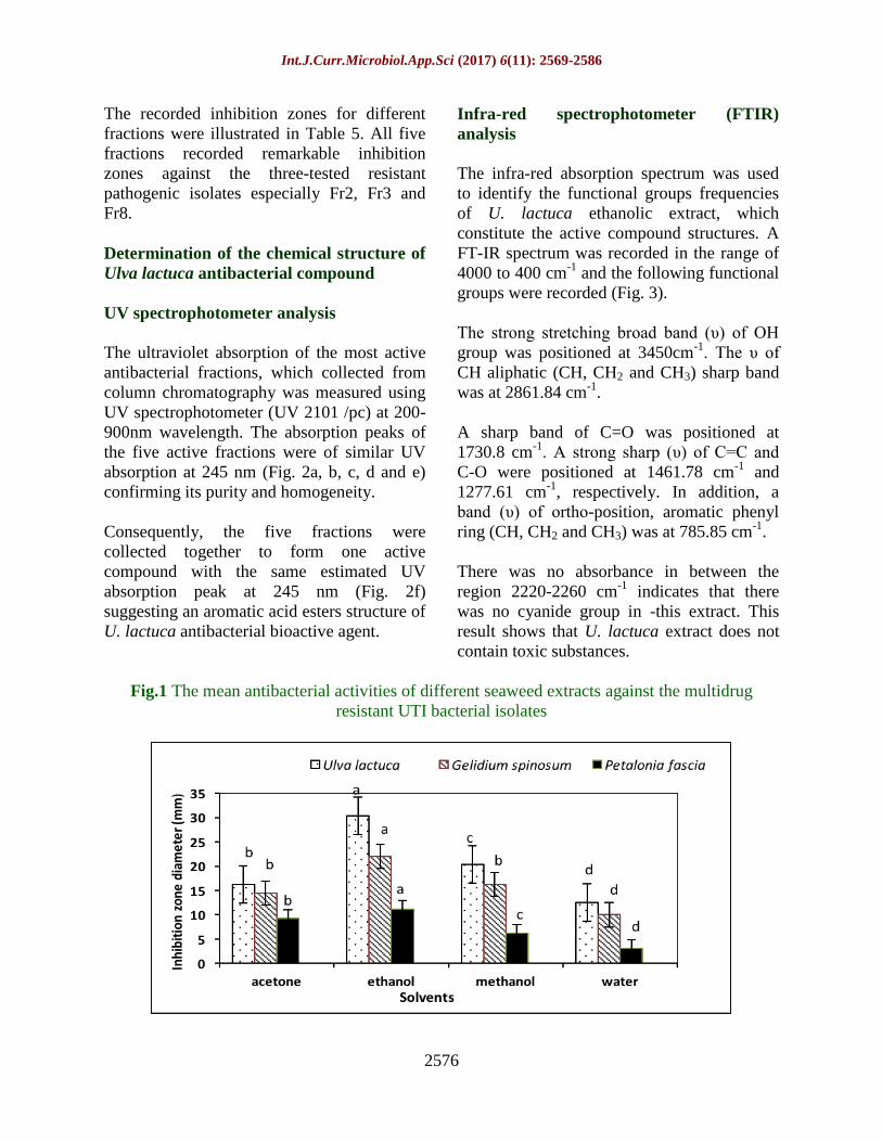
UV spectrophotometer analysis
The ultraviolet absorption of the most active
antibacterial fractions, which collected from
column chromatography was measured using
UV spectrophotometer (UV 2101 /pc) at 200-
900nm wavelength. The absorption peaks of
the five active fractions were of similar UV
absorption at 245 nm (Fig. 2a, b, c, d and e)
confirming its purity and homogeneity.
Consequently, the five fractions were
collected together to form one active
compound with the same estimated UV
absorption peak at 245 nm (Fig. 2f)
suggesting an aromatic acid esters structure of
U. lactuca antibacterial bioactive agent.
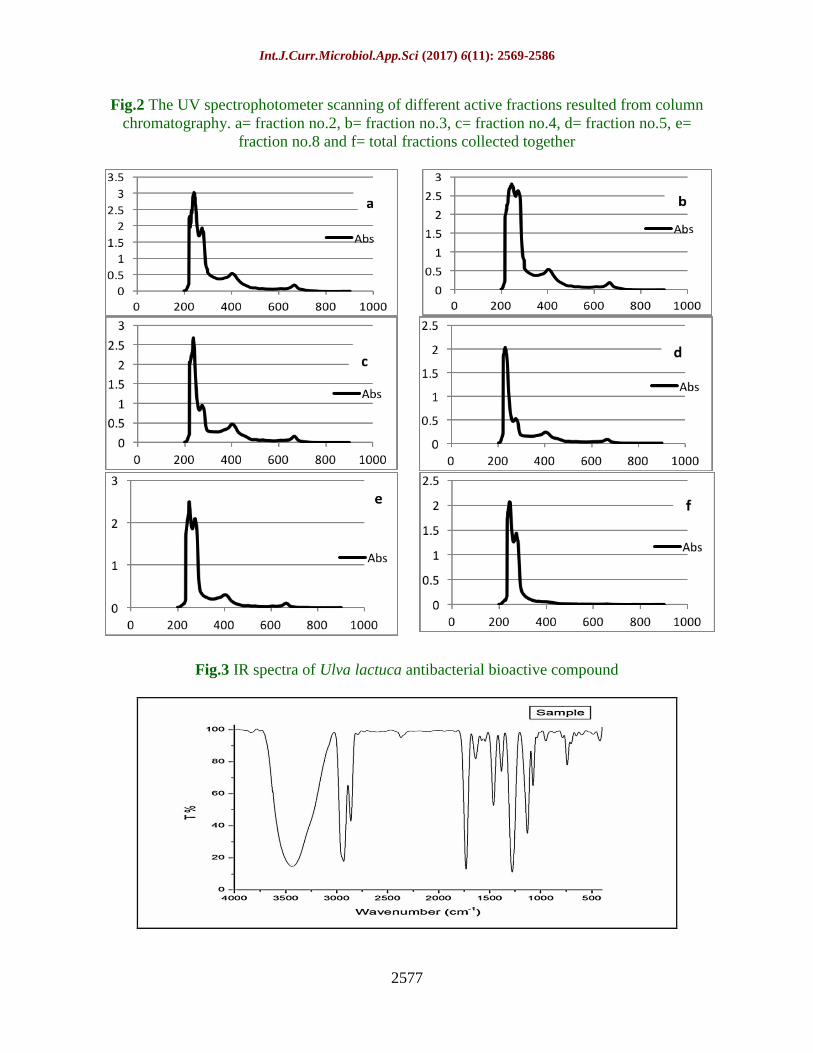
Infra-red spectrophotometer (FTIR)
analysis
The infra-red absorption spectrum was used
to identify the functional groups frequencies
of U. lactuca ethanolic extract, which
constitute the active compound structures. A
FT-IR spectrum was recorded in the range of
4000 to 400 cm-1
and the following functional
groups were recorded (Fig. 3).
The strong stretching broad band (υ) of OH
group was positioned at 3450cm-1
. The υ of
CH aliphatic (CH, CH2 and CH3) sharp band
was at 2861.84 cm-1
.
A sharp band of C=O was positioned at
1730.8 cm-1
. A strong sharp (υ) of C=C and
C-O were positioned at 1461.78 cm-1
and
1277.61 cm-1
, respectively. In addition, a
band (υ) of ortho-position, aromatic phenyl
ring (CH, CH2 and CH3) was at 785.85 cm-1
.
There was no absorbance in between the
region 2220-2260 cm-1
indicates that there
was no cyanide group in -this extract. This
result shows that U. lactuca extract does not
contain toxic substances.
Fig.1 The mean antibacterial activities of different seaweed extracts against the multidrug
resistant UTI bacterial isolates
Int.J.Curr.Microbiol.App.Sci (2017) 6(11): 2569-2586
2577
Fig.2 The UV spectrophotometer scanning of different active fractions resulted from column
chromatography. a= fraction no.2, b= fraction no.3, c= fraction no.4, d= fraction no.5, e=
fraction no.8 and f= total fractions collected together
Fig.3 IR spectra of Ulva lactuca antibacterial bioactive compound
Int.J.Curr.Microbiol.App.Sci (2017) 6(11): 2569-2586
2578
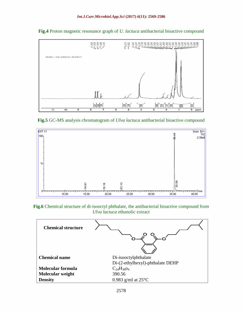
Fig.4 Proton magnetic resonance graph of U. lactuca antibacterial bioactive compound
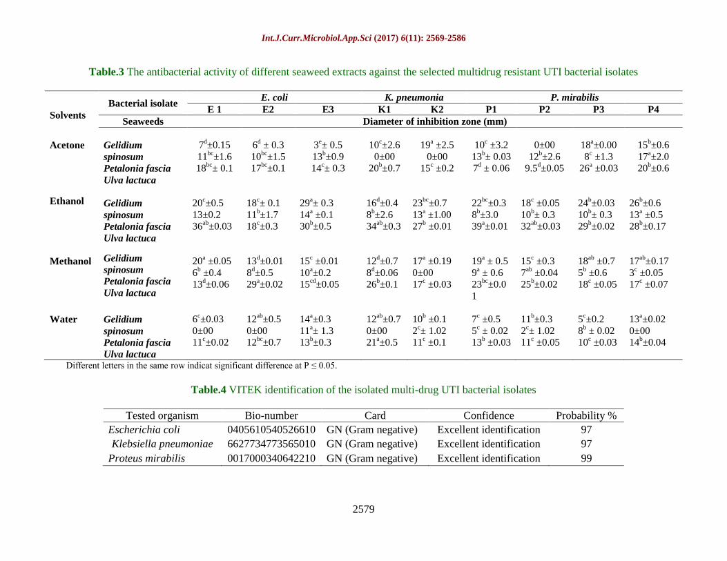
Fig.5 GC-MS analysis chromatogram of Ulva lactuca antibacterial bioactive compound
Fig.6 Chemical structure of di-isooctyl phthalate, the antibacterial bioactive compound from
Ulva lactuca ethanolic extract
Chemical structure
Di-isooctylphthalate
Di-(2-ethylhexyl)-phthalate DEHP Chemical name
C24H38o4 Molecular formula
390.56 Molecular weight
0.983 g/ml at 25°C Density
Int.J.Curr.Microbiol.App.Sci (2017) 6(11): 2569-2586
2579
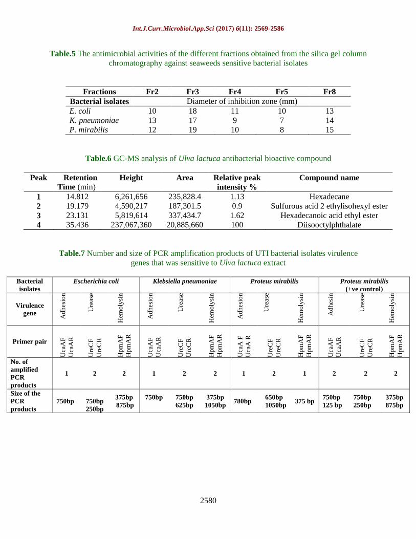
Table.3 The antibacterial activity of different seaweed extracts against the selected multidrug resistant UTI bacterial isolates
Solvents
Bacterial isolate E. coli K. pneumonia P. mirabilis
E 1 E2 E3 K1 K2 P1 P2 P3 P4
Seaweeds Diameter of inhibition zone (mm)
Acetone
Gelidium
spinosum
Petalonia fascia
Ulva lactuca
7d±0.15
11bc
±1.6
18bc
± 0.1
6d ± 0.3
10bc
±1.5
17bc
±0.1
3e± 0.5
13b±0.9
14c± 0.3
10c±2.6
0±00
20b±0.7
19a ±2.5
0±00
15c ±0.2
10c ±3.2
13b± 0.03
7d ± 0.06
0±00
12b±2.6
9.5d±0.05
18a±0.00
8c ±1.3
26a ±0.03
15b±0.6
17a±2.0
20b±0.6
Ethanol
Gelidium
spinosum
Petalonia fascia
Ulva lactuca
20c±0.5
13±0.2
36ab
±0.03
18c± 0.1
11b±1.7
18c±0.3
29a± 0.3
14a ±0.1
30b±0.5
16d±0.4
8b±2.6
34ab
±0.3
23bc
±0.7
13a ±1.00
27b ±0.01
22bc
±0.3
8b±3.0
39a±0.01
18c ±0.05
10b± 0.3
32ab
±0.03
24b±0.03
10b± 0.3
29b±0.02
26b±0.6
13a ±0.5
28b±0.17
Methanol
Gelidium
spinosum
Petalonia fascia
Ulva lactuca
20a ±0.05
6b ±0.4
13d±0.06
13d±0.01
8d±0.5
29a±0.02
15c ±0.01
10a±0.2
15cd
±0.05
12d±0.7
8d±0.06
26b±0.1
17a ±0.19
0±00
17c ±0.03
19a ± 0.5
9a ± 0.6
23bc
±0.0
1
15c ±0.3
7ab
±0.04
25b±0.02
18ab
±0.7
5b ±0.6
18c ±0.05
17ab
±0.17
3c ±0.05
17c ±0.07
Water
Gelidium
spinosum
Petalonia fascia
Ulva lactuca
6c±0.03
0±00
11c±0.02
12ab
±0.5
0±00
12bc
±0.7
14a±0.3
11a± 1.3
13b±0.3
12ab
±0.7
0±00
21a±0.5
10b ±0.1
2c± 1.02
11c ±0.1
7c ±0.5
5c ± 0.02
13b ±0.03
11b±0.3
2c± 1.02
11c ±0.05
5c±0.2
8b ± 0.02
10c ±0.03
13a±0.02
0±00
14b±0.04
Different letters in the same row indicat significant difference at P ≤ 0.05.
Table.4 VITEK identification of the isolated multi-drug UTI bacterial isolates
Tested organism Bio-number Card Confidence Probability %
Escherichia coli 0405610540526610 GN (Gram negative) Excellent identification 97
Klebsiella pneumoniae 6627734773565010 GN (Gram negative) Excellent identification 97
Proteus mirabilis 0017000340642210 GN (Gram negative) Excellent identification 99
Int.J.Curr.Microbiol.App.Sci (2017) 6(11): 2569-2586
2580
Table.5 The antimicrobial activities of the different fractions obtained from the silica gel column
chromatography against seaweeds sensitive bacterial isolates
Fractions Fr2 Fr3 Fr4 Fr5 Fr8
Bacterial isolates Diameter of inhibition zone (mm)
E. coli 10 18 11 10 13
K. pneumoniae 13 17 9 7 14
P. mirabilis 12 19 10 8 15
Table.6 GC-MS analysis of Ulva lactuca antibacterial bioactive compound
Peak Retention
Time (min)
Height Area Relative peak
intensity %
Compound name
1 14.812 6,261,656 235,828.4 1.13 Hexadecane
2 19.179 4,590,217 187,301.5 0.9 Sulfurous acid 2 ethylisohexyl ester
3 23.131 5,819,614 337,434.7 1.62 Hexadecanoic acid ethyl ester
4 35.436 237,067,360 20,885,660 100 Diisooctylphthalate
Table.7 Number and size of PCR amplification products of UTI bacterial isolates virulence
genes that was sensitive to Ulva lactuca extract
Bacterial
isolates
Escherichia coli Klebsiella pneumoniae Proteus mirabilis Proteus mirabilis
(+ve control)
Virulence
gene
Ad
hes
ion
Ure
ase
Hem
oly
sin
Ad
hes
ion
Ure
ase
Hem
oly
sin
Ad
hes
ion
Ure
ase
Hem
oly
sin
Ad
hes
in
Ure
ase
Hem
oly
sin
Primer pair
Uca
AF
Uca
AR
Ure
CF
Ure
CR
Hp
mA
F
Hp
mA
R
Uca
AF
Uca
AR
Ure
CF
Ure
CR
Hp
mA
F
Hp
mA
R
Uca
A F
Uca
A R
Ure
CF
Ure
CR
Hp
mA
F
Hp
mA
R
Uca
AF
Uca
AR
Ure
CF
Ure
CR
Hp
mA
F
Hp
mA
R
No. of
amplified
PCR
products
1 2 2 1 2 2 1 2 1 2 2 2
Size of the
PCR
products
750bp
750bp
250bp
375bp
875bp
750bp
750bp
625bp
375bp
1050bp 780bp
650bp
1050bp
375 bp 750bp
125 bp
750bp
250bp
375bp
875bp
Int.J.Curr.Microbiol.App.Sci (2017) 6(11): 2569-2586
2581
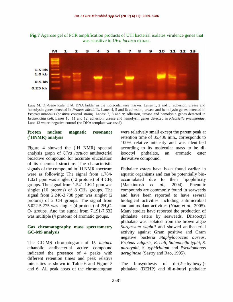
Fig.7 Agarose gel of PCR amplification products of UTI bacterial isolates virulence genes that
was sensitive to Ulva lactuca extract.
Lane M: O’-Gene Ruler 1 kb DNA ladder as the molecular size marker. Lanes 1, 2 and 3: adhesion, urease and
hemolysin genes detected in Proteus mirabilis. Lanes 4, 5 and 6: adhesion, urease and hemolysin genes detected in
Proteus mirabilis (positive control strain). Lanes: 7, 8 and 9: adhesion, urease and hemolysin genes detected in
Escherichia coli. Lanes 10, 11 and 12: adhesion, urease and hemolysin genes detected in Klebsiella pneumoniae.
Lane 13 water: negative control (no DNA template was used).
Proton nuclear magnetic resonance
(1HNMR) analysis
Figure 4 showed the (1H NMR) spectral
analysis graph of Ulva lactuca antibacterial
bioactive compound for accurate elucidation
of its chemical structure. The characteristic
signals of the compound in 1H NMR spectrum
were as following: The signal from 1.784-
1.321 ppm was singlet (12 protons) of 4 CH3
groups. The signal from 1.541-1.621 ppm was
singlet (16 protons) of 8 CH2 groups. The
signal from 2.246-2.738 ppm was singlet (2
protons) of 2 CH groups. The signal from
5.022-5.275 was singlet (4 protons) of 2H2C-
O- groups. And the signal from 7.191-7.632
was multiple (4 protons) of aromatic groups.
Gas chromatography mass spectrometry
GC-MS analysis
The GC-MS chromatogram of U. lactuca
ethanolic antibacterial active compound
indicated the presence of 4 peaks with
different retention times and peak relative
intensities as shown in Table 6 and Figure 5
and 6. All peak areas of the chromatogram
were relatively small except the parent peak at
retention time of 35.436 min., corresponds to
100% relative intensity and was identified
according to its molecular mass to be di-
isooctyl phthalate, an aromatic ester
derivative compound.
Phthalate esters have been found earlier in
aquatic organisms and can be potentially bio-
accumulated due to their lipophilicity
(Mackintosh et al., 2004). Phenolic
compounds are commonly found in seaweeds
and have been reported to have several
biological activities including antimicrobial
and antioxidant activities (Yuan et al., 2005).
Many studies have reported the production of
phthalate esters by seaweeds. Diisooctyl
phthalate was isolated from the brown algae
Sargassum wightii and showed antibacterial
activity against Gram positive and Gram
negative bacteria Staphylococcus aureus,
Proteus vulgaris, E. coli, Salmonella typhi, S.
paratyphi, S. typhiridium and Pseudomonas
aeruginosa (Sastry and Rao, 1995).
The biosynthesis of di-(2-ethylhexyl)-
phthalate (DEHP) and di-n-butyl phthalate
Int.J.Curr.Microbiol.App.Sci (2017) 6(11): 2569-2586
2582
(DBP) have been reported by (Chen, 2004) in
the red alga Bangia atropurpurea. According
to Osman et al., (2013), a high molecular
weight phthalate esters derivative of broad
spectrum antibacterial and antifungal
activities was synthesized by Ulva fasciata
green seaweed. The extract of Grateloupa
lithophila seaweed was recommended as
antibacterial substance for multidrug resistant
(MDR) microbes including the UTI bacteria
(Manikandan et al., 2011). Some marine
brown algae were screened by Kalaivani et
al., (2016) and were reported to produce
antagonistic bioactive compounds against
UTI pathogens.
Molecular detection of the virulence factors
of the bacterial strains
PCR amplification of virulence factors
Three different primer pairs (UcaAF UcaAR;
UreCF UreCR; HpmAF HpmAR) were used
to detect the virulence genes (adhesion,
hemolysin and urease) of the tested multidrug
resistant UTI bacterial isolates (E. coli, K.
pneumoniae and P. mirabilis). Various
virulence factors can be attributed to UTI
pathogenicity (Tarchouna et al., 2013). The
Polymerase Chain Reaction technique was
used to investigate the virulence genes
responsible for multidrug resistance in the
UTI bacterial isolates. Specific primers for
three virulence genes in the bacterial isolates
including ucaA (adhesion), ureC (urease), and
hpmA (hemolysin) were selected. These three
genes are responsible for the colonization of
bacteria in the urinary tract.
The seaweeds sensitive UTI bacterial isolates
were compared with the antibiotics and
seaweed resistant isolate (positive control)
carrying the virulence genes. The current
results (Table 7 and Fig. 7) showed that the
adhesion virulence gene was detected in all
the bacterial isolates (E. coli, K. pneumoniae
and P. mirabilis) with rate of 95% with one
amplicon (780 bp) for Proteus mirabilis and
750 bp for both of E. coli and Klebsiella
pneumoniae, while in the positive control
strain 2 PCR amplicons (750bp and faint
125bp) were observed, which indicated that
the resistant strain (positive control) utilize a
variety of adhesions to bind to the urinary
epithelial cells to start the infection causing
failure to the treatment with antibiotics and
seaweeds (Le Bouguenec et al., 1992;
Munkhdelger et al., 2017).
Regarding the ureC gene, which is considered
as a diagnostic feature of presence of UTI
bacteria due to the production of urease
enzyme; the results showed that all the
isolates in a rate 96.66% contained ureC gene.
Two PCR products (750bp and 625bp) were
observed in Klebsiella pneumoniae and in
Proteus mirabilis (650bp and 1050bp), while
two faint amplicons (750bp and 250bp) were
amplified in E. coli. Similarly, in the positive
(control) isolate the 2 faint PCR amplicons
(750bp and 250bp) were amplified (Table 7
and Fig. 7). Urease enzyme works on
changing pH of the urine to basic leading to
the deposition of calcium and magnesium
phosphate salts in the formed biofilm, which
in turn leads to the formation of more
complex crystalline biofilm that works to
close urinary catheter and protect the bacteria
from antibiotics (Stickler, 2008; Schaffera et
al., 2016).
HpmA gene is responsible for producing
hemolycin and considered as an important
virulence factor for UTI bacterial
pathogenicity. The hpmA gene was detected
in all bacterial isolates. In Proteus mirabilis,
only very faint amplicon (375bp) was
detected in this strain. Two PCR amplicons
(375bp and 875bp) of hpmA gene were
present in E. coli and in the positive control.
Similarly, two faint PCR products (375bp and
1050bp) were observed in case of K.
Int.J.Curr.Microbiol.App.Sci (2017) 6(11): 2569-2586
2583
pneumoniae. The hemolysin enzyme acts on
destroying the leukocyte cells by making
small holes in the membranes of leukocyte
and epithelial cells. Its presence is a very
important factor in providing the pathogenic
bacteria with iron; and because of its
cytotoxic, it leads to the destruction of the
kidney tissue of the host (Cestari et al., 2013;
Leclercq et al., 2016).
By linking the obtained results from
antibacterial activities of U. lactuca extract
with that of the PCR of the studied UTI
bacterial isolates, it revealed that presence of
the virulence genes (adhesion, urease and
hemolysin) is a crucial factor for successful
colonization of the uropathogenic bacteria and
its multidrug resistance property. Seaweed
derived compounds proved their ability as
new natural sources for antibacterial agents,
which reduce the virulence genes activity and
prevent (as for P. mirabilis) or mitigate (as for
E. coli and K. pneumoniae) the resistance and
pathogenicity of these kind of bacteria.
Marine algae are one of the biggest biomass
producers and proved a potential source of
new and unique compounds for many
applications. Many compounds derived from
seaweed have bacteriostatic or antibacterial,
antiviral, antitumor, anti-inflammatory and
antifouling activities. In this study, an
aromatic ester derivative was extracted from
Ulva lactuca green seaweed using ethanol.
The bioactive compound was chemically
identified as diisooctyl phthalate and showed
promising antibacterial activities against
multidrug resistant UTI infectious bacteria. In
addition, the virulence genes causing
pathogenicity of these bacteria were detected
using PCR. PCR results showed that
hemolysin, urease and adhesion virulence
factors were present in the treated P.
mirabilis, E. coli and K. pneumoniae UTI
isolates with high variations. Consequently,
the study recommends seaweeds as promising
treatment of human diseases or as new
antibacterial agents to replace synthetic
antibacterial drugs.
References
Adaikalaraj, G., Patric Raja, D., Johnson, M.,
Janakiraman, N., Babu, A., 2012.
Antibacterial potential of selected Red
seaweeds from Manapad coastal areas,
Thoothukudi, TamilNadu, India. Asian
Pacific Journal of Tropical Medicine
1077-1080.
Aleem, A.A., 1993. The Marine Algae of
Alexandria Egyptian Books House faculty
of science Alexandria. Egypt
Anderson, G., Martin, S., Hultgren, S., 2004.
Host subversion by formation of
intracellular bacterial communities in the
urinary tract. Microbes and Infection 6,
1094-1101.
Atalah, E., Hernández Cruz, C.M., Izquierdo,
M.S., Rosenlund, G., Caballero, M.J.,
Valencia, A., Robaina, L., 2007. Two
microalgae Crypthecodinium cohnii and
Phaeodactylum tricornutum as alternative
source of essential fatty acids in starter
feeds for Sea bream. Sparus aurata
Aquaculture 270, 178-185.
Blunt, B.R., Copp, W.P., Hu, M.H.G., Munro,
P.T., Northcote, M.R., 2007. UV–VIS
and HPLC studies on marine natural
product. Natural Product Reports 4, 31-
86.
Boeriu, C., Bravo, D., Gosselink, R.,
Gosselinkvan Dam, J., 2004.
Characterisation of structure dependent
functional properties of lignin with
infrared spectroscopy. Industrial Crops
and Products 20, 205-218.
Boonchum, W., Peerapornpisal, Y.,
Kanjanapothi, D., Pakkoh, J.,
Amornlerdpison, D., Pumas, C.,
Sangpaiboon, P., Vacharapiyasophon, P.,
2011. Antimicrobial and anti-
inflammatory properties of various
seaweeds from the Gulf of Thailand.
Int.J.Curr.Microbiol.App.Sci (2017) 6(11): 2569-2586
2584
International Journal of Agricultural and
Biology 13, 100-104.
Cardoso, M.S., Carvalho, G.L., Silva, J.P.,
Rodrigues, S.M., Pereira, R.O., Pereira,
L., 2014. Bioproducts from seaweeds: A
review with special focus on the Iberian
Peninsula. Current Organic Chemistry
18, 896-917.
Cestari, S.E., Ludovico, M.S., Martins, F.H., Da
Rocha, W.P., Elisa, W.P., Pelayo, J.S.,
2013. Molecular detection of HpmA and
hlyA hemolysin of uropathogenic Proteus
mirablis. Journal Microbial 67, 703-707.
Chen, C., 2004. Biosynthesis of di-(2-
ethylhexyl) phthalate (DEHP) and di-n-
butyl phthalate (DBP) from Red alga
Bangia atropurpurea. Water Research 38,
1014-1018.
Cheung, R.C.F., Wong, J.H., Pan, W., Chan,
Y.S., Yin, C., Dan, X., Ng, T.B., 2015.
Marine lectins and their medicinal
applications. Applied Microbiology and
Biotechnology 99, 3755-3773.
CLSI, 2012. Performance standards for
antimicrobial disk susceptibility tests;
approved standard 11th ed CLSI
document. Clinical and Laboratory
Standards Institute,Wayne, PA.
Collee, J., Fraser, G., Marmion, P., Simmons,
A., 1996. Practical Medical
Microbiology. 4th ed Churchill
Livingstone, New York,pp. 413–418.
Dethlefsen, L., Huse, S., Sogin, M., Relman, D.,
2008. The pervasive effects of an
antibiotic on the human gut microbiota, as
revealed by deep 16S rRNA sequencing.
PLOS Biology, 6:280.
Ejrnaes, K., 2011. Bacterial characteristics of
importance for recurrent urinary tract
infections caused by Escherichia coli.
Danish Medical Bulletin 58, B4187.
El-Sheekh, M.M., El Shafay, S.M., Ali, S.S.,
2016. Antimicrobial activity of some
seaweeds species from Red sea, against
multidrug resistant bacteria. Egyptian
Journal of Aquatic Research. 42, 65-74.
Fareed, M., Khairy, H., 2008. In vitro
antimicrobial activities of seaweeds
collected from Abu-Qir Bay Alexandria
Egypt. World Applied Sciences Journal 5,
389-396.
Farshad, S., Ranjbar, R., Japoni, A., Hosseini,
M., Anvarinejad, M.,
Mohammadzadegan, R., 2012. Microbial
susceptibility, virulence factors and
plasmid profiles of uropathogenic bacteria
strains isolated from children in Jahrom,
Iran. Archives of Iranian Medicine 15,
312-316.
Foxman, B., Brown, P., 2003. Epidemiology of
urinary tract infections: transmission and
risk factors, incidence, and costs.
Infectious Disease Clinics of North
America 41, 17-227.
Francis, N.B., Jonathan, M.B., Oliva, C.R.,
Amaro, N.a., Jayson, D.C., 2017.
Phytochemicals screening and
antimicrobial properties of Sargassum
oligocystum and Sargassum crassifolium
Extracts. Journal of medicinal plants
studies 5, 382-387.
Gilbert, P., 1987. Inocula for Antimicrobial
Sensitivity Testing: a Critical Review.
Journal of Antimicrobial Chemotherapy
20, 147-154.
Gonzalez, C.M., Schaeffer, A.J., 1999.
Treatment of urinary tract infection."
What's old, what's new and what works.
World Journal of Urology 6, 372-382.
Gorban, E., Kuprash, L., Gorban, N., 2003.
Spirulina: perspectives of the application
in medicine. Lik Sprava 7, 100-110.
Guiry, M., Guiry, G., 2016. AlgaeBase. World-
wide electronic publication, National
University of Ireland
http://www.algaebase.org.
Hacker, J.G., Oehler, B., Muhldorfer, I.,
Tschape, H., 1997. Pathogenicity islands
of virulent bacteria: structure, function
and impact on microbial evolution.
Molecular Microbiology 23, 1089-1097.
Hornsey, I.S., Hide, D., 1974. The Production
of antimicrobial compounds by British
marine algae. I. Antibiotic-Producing
marine algae. British Phycological
Society Journal of Environmental Biology
9, 353-361.
Int.J.Curr.Microbiol.App.Sci (2017) 6(11): 2569-2586
2585
Ismail, M.M., Gheda, S.F., Pereira, L., 2016.
Variation in bioactive compounds in
some seaweeds from Abo Qir bay,
Alexandria, Egypt. Rendiconti Fis. Acc.
Lincei 27, 269–279.
Johnson, J., 1991. Virulence factors in
Escherichia coli urinary tract infection.
Clinical Microbiology Reviews 4, 80-118.
Kalaivani, G., Hemalatha, N., Poongothai, E.,
2016. Screening of marine brown algae
associated potential bacteria producing
antagonistic bioactive compounds against
UTI pathogens. International Journal of
Pharma and Bio Sciences 7, 395-405.
Karthikaidevi, G., Manivannan, G.,
Thirumaran, G., Anantharaman, P.,
Balasubaramanian, T., 2009.
Antibacterial Properties of Selected Green
Seaweeds from Vedalai Coastal Waters;
Gulf of Mannar Marine Biosphere
Reserve. Global Journal of
Pharmacology 3, 107-112.
Khelil-Radji, F., Belhouari, M.Y., Chemlal-
Kherraz, D., Matallah-Boutiba, A.,
Boutiba, Z., 2017. Antimicrobial activity
of aqueous and ethanol extracts of
twomarine algae collected from Algerian
west coast. International Journal of
Biosciences 10, 100-104.
Khotimchenko, S.V., Vaskovsky, V.E.,
Titlyanavo, T.V., 2002. Fatty acids of
marine algae from the Pacific coast of
North California. Botanica Marina 45,
17-22.
Le Bouguenec, N.C., Archambaud, M.,
Labigne, A., 1992. Rapid and specific
detection of the pap, afa, and sfa adhesins
- encoding operons in uropathogenic
Escherichia coli strains by Polymerase
Chain Reaction. Journal of Clinical
Microbiology 30, 1189-1193.
Leclercq, S.Y., Sullivan, M.J., Ipe, D.S., Smith,
J.P., Cripps, A.W., Uletta, G.C., 2016.
Pathogenesis of Streptococcus urinary
tract infection depends on bacterial strain
and β-hemolysin/cytolysin that mediates
cytotoxicity cytokine synthesis
inflammation and virulence. Scientific
Reports 6:29000.
Mackintosh, C., Maldonado, J., Hongwu, J.,
Hoover, N., Chong, A., Ikonomou, M.,
Gobas, F., 2004. Distribution of phthalate
esters in a marine aquatic food web:
comparison to polychlorinated biphenyls.
Environmental Science and Technology
38, 2011-2020.
Magiorakos, A., Srinivasan, A., Carey, R.,
2012. Multidrug-resistant, extensively
drug-resistant and pandrug-resistant
bacteria: an international expert proposal
for interim standard definitions for
acquired resistance. Clinical
Microbiology and Infection 18, 268-281.
Manikandan, S., Ganesapandian, S., Singh, M.,
Kumaraguru, A.K., 2011. Antimicrobial
susceptibility pattern of urinary tract
infection causing human pathogenic
bacteria. Asian Journal of Medical
Sciences 3, 56-60.
Manilal, A., Sujith, S., Selvin, J., Kiran, G.S.,
Shakir, C., Gandhimathi, R., Panikkar,
M.V.N., 2009. Biopotentials Of Seaweeds
Collected From Southwest Coast Of
India. Journal of Marine Science and
Technology 17, 67-73.
Munkhdelger, Y., Gunregjav, N., Dorjpurev, A.,
Juniichiro, N., Sarantuya, J., 2017.
Detection of virulence genes,
phylogenetic group and antibiotic
resistance of uropathogenic Escherichia
coli in Mongolia. Journal of infection in
developing countries 11, 51-57.
Newman, D.J., Cragg, G.M., Snader, K.M.,
2003. Natural products as source of new
drugs over the period 1981-2002. Journal
of Natural Product 66, 1022-1037.
Olayinka, A., Olayinka, B., Onile, B., 2009.
Antibiotic susceptibility and plasmid
pattern of Pseudomonas aeruginosa from
the surgical unit of a University teaching
hospital in North Central Nigeria.
International Journal of Medical Science
1, 79-83.
Oliveira, F., Paludo, K., Arend, L., Farah, S.,
Pedrosa, F., Souza, E., 2011. Virulence
characteristics and antimicrobial
susceptibility of uropathogenic
Int.J.Curr.Microbiol.App.Sci (2017) 6(11): 2569-2586
2586
Escherichia coli strains. Genetics and
molecular research 10, 4114-4125.
Osman, M.E.H., Aboshady, A.M., Elshobary,
M.E., 2013. Production and
characterization of antimicrobial active
substance from some macroalgae
collected from Abu-Qir bay (Alexandria)
Egypt. African Journal of Biotechnology
12, 6847-6858.
Oxoid, 1981. The oxoid manual of culture
media ingredients and other laboratory
servies.. 5th Eds.Oxoid Manual,UK.
Padmakumar, k., Ayyakkannu, k., 1997.
Seasonal variation of antibacterial and
antifungal activities of the extracts of
marine algae from Southern coasts of
India. Botanica Marina 40, 507-515.
Pushparaj, A., Raubbin, R., Balasankar, T.,
2014. An antibacterial activity of the
green seaweed Caulerpha sertularioides
using five different solvents.
International Journal Pharm Tech,
Research 6, 01-05.
Ramasamy, M.S., Kumar, S.S., 2009. Anti-
inflammatory, antinociceptive and central
nervous system depressant activities of
marine bacterial extracts. Journal of
Pharmacology and Toxicology 4, 152-
159.
Read, A., Woods, R., 2014. Antibiotic
resistance management. Evolution,
Medicine, and Public Health 2014, 147.
Sastry, V., Rao, G., 1995. Dioctyl phthalate and
antibacterial compound from marine
brown alga Sargassum wightii. Journal of
Applied Phycology 7, 185-186.
Schaffera, J.N., Norsworthya, A.N., Sunb, T.-
T., Pearsona, M.M., 2016. Proteus
mirabilis fimbriae and urease dependent
clusters assemble in an extracellular niche
to initiate bladder stone formation. PNAS
113, 4494-4499.
Solomon, R., Santhi, V., 2008. Purification of
bioactive natural product against human
microbial pathogens from marine
seaweed, Dictyota acutiloba. World
Journal of Microbiology and
Biotechnology 24, 1747-1752.
Stickler, D.J., 2008. Bacterial biofilms in
patients with indwelling urinary catheters.
Nature Reviews Clinical Urology 11, 598-
608.
Tarchouna, M., Ferjani, A., Ben-Selma, W.,
Boukadida, 2013. Distribution of
uropathogenic virulence genes in
Escherichia coli isolated from patients
with urinary tract infection. International
Journal of Infectious Diseases 17, 450-
453.
Thomas, K.W.L., Tam, P.C., Liu, Z.K.,
Augustine, F.B.C., 2001 Evaluation of
VITEK 2 rapid identification and
susceptibility testing system against
Gram-Negative clinical isolates. Journal
of Clinical Microbiology 39, 2964-2966.
Vasquez, y., Hand, W., 2004. Antibiotic
susceptibility patterns of community-
acquired urinary tract infection isolates
from female patients on the US (Texas)-
Mexico border.. Journal of Applied
Research 4, 321-326.
Yuan, Y.V., Bone, D.E., Carrington, M.F.,
2005. Antioxidant activity of dulse
(Palmaria palmata) extract evaluated in
vitro. Food Chemistry 91, 485-494.
How to cite this article:
Wagih A. El-Shouny, Reda M. Gaafar, Gehan A. Ismail and Marwa M. Elzanaty. 2017.
Antibacterial Activity of Some Seaweed Extracts against Multidrug Resistant Urinary Tract
Bacteria and Analysis of their Virulence Genes. Int.J.Curr.Microbiol.App.Sci. 6(11): 2569-2586.
doi: https://doi.org/10.20546/ijcmas.2017.611.302