Embed Size (px)
Citation preview
International Journal of Bio-Science and Bio-Technology
Vol.8, No.2 (2016), pp.419-432
http://dx.doi.org/10.14257/ijbsbt.2016.8.2.39
ISSN: 2233-7849 IJBSBT
Copyright ⓒ 2016 SERSC
Antibacterial Screening of Soil Bacteria Isolates from Guimaras
Island, Philippines against Escherichia Coli and
Staphylococcus Aureus
Rey G. Tantiado1, Ma. Sophia Estella C. Tajolosa
1 and Kristine Joy R. Estrella
1
1College of Arts and Sciences, West Visayas State University,
La Paz, Iloilo City, Philippines
[email protected], [email protected]
Abstract
This study was conducted to screen and determine the antibacterial effects of soil
bacterial isolates from three sampling sites of Sinapsapan, Jordan, Guimaras against
Staphylococcus aureus and Escherichia coli. Test bacteria were isolated and
characterized based on colonial morphology and cell characteristics. Soil bacteria
isolates were screened for their antibacterial properties against S. aureus and E. coli
using agar disc diffusion method. Each treatment was done in three replicates and trials.
There were six bacterial isolates in each sampling site obtained and exhibited
antibacterial properties on S. aureus and E. coli after 72 hours of incubation. For S.
aureus, I4 (M=35.33 mm, active) from high sampling site; I2 (M=29.44 mm, partially
active) from intermediate sampling site, and I1 (M = 38.00 mm, active) from low
sampling site while for E. coli, I4 (M= 34.22, active) from high sampling site, I6 (32.78
mm, active) from intermediate sampling site, and I5 (M=34.78, active) from low sampling
site had the highest zones of inhibition.
Keywords: soil bacteria, agar disc, Guimaras, Escherichia coli, Staphylococcus aureus
1. Introduction
1.1 Background of the Study
Soil is a natural reservoir for microorganism and their antimicrobial products [1]. A
significant number of these organisms are undocumented and some of them may have an
effect on pathogenic bacteria [22]. In a single gram of soil, there can be billions of
bacteria [19]. It is estimated that there are about 60,000 different bacteria species, most
which have yet to be named, and each has its own particular roles and capabilities [19].
Most bacteria live in the top 10cm of soil where organic matter is present [19]. Soil
bacteria perform important services related to disease suppression [15].
Barangay Sinapsapan is located in Jordan, Guimaras Island, bordering Panay Gulf with
approximate coordinates of N 010 35’.00 E 122 30’.47. The distance from the sea is about
270 meters at an elevation of about 30 meters above sea level. The terrain is a typical
young coral island with the land rising rapidly to about 30-40 meters elevation when
proceeding inland from the coastland some 100 meters. The inland elevation varies up
and down forming valleys and ridges [8].
Barangay Sinapsapan has many unexplored territories which is most likely to yield
purposeful results towards isolation of new antibiotics. There are areas that are not
inhabited by people. Most of the soils in the area are rich in organic matter since leaves
and twigs cover most of the land. The texture of the soil is primarily coarse. The
International Journal of Bio-Science and Bio-Technology
Vol.8, No.2 (2016)
420 Copyright ⓒ 2016 SERSC
arrangement of the soil particles is in different aggregates wherein the soil granules clump
or bind together [9].
Bacteria are capable of causing disease. Humans are generally most interested in the
species of pathogenic bacteria in humans, although these bacteria can also infect other
animals and plants. Some notable pathogenic bacteria include Staphylococcus aureus and
Escherichia coli. Staphylococcus aureus, often referred to simply as “staph”, are gram-
positive spherical bacteria that occur in microscopic clusters resembling grapes.
Bacteriological culture of the nose and skin of normal humans invariably yields
staphylococci [25]. Escherichia coli is the head of the large bacterial family,
Enterobacteriaceae, the enteric bacteria which are facultatively anaerobic gram-negative
rods that live in the intestinal tracts of animals [25].
The science of antibiotics has remained and will remain for many years, one of the
most interesting natural sciences, in both theoretical and practical aspects. Microbial
natural products still appear as the most promising source of the future antibiotics that
society is expecting [16].
For the past years, pathogens underwent mutation which enabled them to resist
antimicrobials, thereby threatening millions of people worldwide [1,14]. The antibiotic
resistance problem demands to discover new antibacterial agents effective against
pathogenic bacteria resistant to current antibiotics. Majority of the bacteria in soil that
have the potential for drug sources remain uncultivable, and thereby inaccessible for
novel antibiotic discovery [1, 12, 16]. Thus, it is imperative to screen more and more
bacteria from different soil samples for antimicrobial activity with a hope of getting some
bacterial strains that produce antibiotics that have not yet been discovered and active
against drug resistant pathogens.
1.2. Objectives of the Study
This study was conducted to screen and determine the antibacterial effects of soil
bacterial isolates from three sampling sites of Sinapsapan, Jordan, Guimaras against S.
aureus and E. coli. Furthermore, soil bacterial isolates with antibacterial properties
against the test pathogenic bacteria were characterized. Specifically, it has the following
specific objectives:
1. To determine the number of bacterial isolates present in the three sampling sites of
Sinapsapan, Jordan, Guimaras.
2. To characterize (colonial morphology and gram staining reaction) bacterial isolates
that have antibacterial properties against S. aureus and E. coli.
3. To determine the effects of the bacterial isolates on S. aureus and E. coli after 72
hours of incubation.
4. To determine significant differences on the zone of inhibition of bacterial isolates
from the three sampling sites against S. aureus and E. coli.
2. Methodology
2.1. Sampling Site
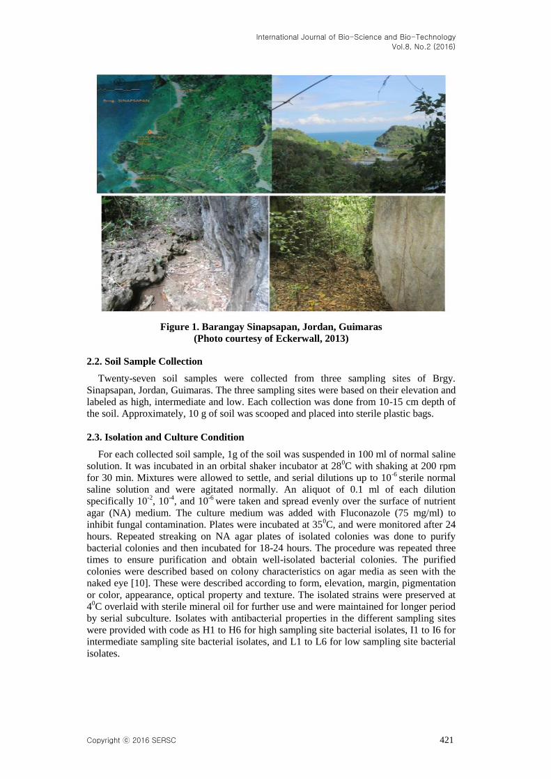
The sampling sites were located in Sinapsapan, Jordan, Guimaras Island, in the land
bordering Panay Gulf with approximate coordinates of N 010 35’.00 E 122 30’.47. The
distance from the sea is about 270 meters at an elevation of about 30 meters above sea
level [8].
International Journal of Bio-Science and Bio-Technology
Vol.8, No.2 (2016)
Copyright ⓒ 2016 SERSC 421
Figure 1. Barangay Sinapsapan, Jordan, Guimaras
(Photo courtesy of Eckerwall, 2013)
2.2. Soil Sample Collection
Twenty-seven soil samples were collected from three sampling sites of Brgy.
Sinapsapan, Jordan, Guimaras. The three sampling sites were based on their elevation and
labeled as high, intermediate and low. Each collection was done from 10-15 cm depth of
the soil. Approximately, 10 g of soil was scooped and placed into sterile plastic bags.
2.3. Isolation and Culture Condition
For each collected soil sample, 1g of the soil was suspended in 100 ml of normal saline
solution. It was incubated in an orbital shaker incubator at 280C with shaking at 200 rpm
for 30 min. Mixtures were allowed to settle, and serial dilutions up to 10-6
sterile normal
saline solution and were agitated normally. An aliquot of 0.1 ml of each dilution
specifically 10-2
, 10-4
, and 10-6
were taken and spread evenly over the surface of nutrient
agar (NA) medium. The culture medium was added with Fluconazole (75 mg/ml) to
inhibit fungal contamination. Plates were incubated at 350C, and were monitored after 24
hours. Repeated streaking on NA agar plates of isolated colonies was done to purify
bacterial colonies and then incubated for 18-24 hours. The procedure was repeated three
times to ensure purification and obtain well-isolated bacterial colonies. The purified
colonies were described based on colony characteristics on agar media as seen with the
naked eye [10]. These were described according to form, elevation, margin, pigmentation
or color, appearance, optical property and texture. The isolated strains were preserved at
40C overlaid with sterile mineral oil for further use and were maintained for longer period
by serial subculture. Isolates with antibacterial properties in the different sampling sites
were provided with code as H1 to H6 for high sampling site bacterial isolates, I1 to I6 for
intermediate sampling site bacterial isolates, and L1 to L6 for low sampling site bacterial
isolates.
International Journal of Bio-Science and Bio-Technology
Vol.8, No.2 (2016)
422 Copyright ⓒ 2016 SERSC
2.4. Test Organisms
Test organisms were the gram-positive bacterium, Staphylococcus aureus BIOTECH
1582 and the gram-negative bacterium, Escherichia coli BIOTECH 1634. The test
bacteria were purchased from the Philippine National Collection of Microorganisms
(PNCM), University of the Philippines Los Baños (UPLB) Biotech in Laguna. A letter for
requisition of purchase of bacteria was made prior to the antimicrobial assay. The bacteria
cultures were overlaid with sterile mineral oil and were sub-cultured for further use.
2.5. Broth Culture of Test Isolates and Test Pathogens
A loopful of test pathogens and characterized test bacteria isolates were inoculated in
each nutrient broth culture medium aseptically. Inoculation was done twice to ensure
growth of bacteria in broth solution. All bacterial broth cultures were incubated at 370C
for 24 hours. About 5 ml of the solution of the same proportions as those used in
preparing the culture suspension was transferred in a 25 ml screw-cap tube. The culture
tube was tightly sealed and stored in the dark at room temperature. Prior to use, the
turbidity standard was shaken thoroughly. The 0.5 Mc Farland Standard was used to
adjust the turbidity of the inoculum prior to microbial assay. The turbidity standard may
contain approximately 1.5 x 10⁸ CFU/ml of the cultured bacteria [18].
2.6. Preparation of Antibiotic Solution
An antibiotic ciprofloxacin was purchased from a local drug store. The preparation of
the solution was based on the indicated concentration i.e., 50 mg/ml [3], wherein 500 mg
of the antibiotic tablet was pulverized and dissolved in 10 ml sterile distilled water.
2.7. In vitro Screening of Soil Bacterial Isolates for Anti-bacterial Activity
Morphologically distinct colony of bacterial isolates was subjected for antibacterial
screening against the test pathogens using the modified agar disk diffusion method. The
test isolate was inoculated in an agar disk with a thickness of 2mm and a diameter of 10
mm on a plate of screening media spread-plated with the test pathogen. The plates were
incubated for 24 h at 37 0C. The procedure was also employed in making agar disc as the
positive control. Antagonism was measured by the size of the inhibition zone [14]. The
presence of zone of inhibition of the test isolate was derived using the formula as
indicated below and evaluated according to the observed and corresponding modified
inferences [18]:
𝑍𝑜𝑛𝑒 𝑜𝑓 𝐼𝑛ℎ𝑖𝑏𝑖𝑡𝑖𝑜𝑛 =(𝑋−𝑌)
𝑍
where X = highest mean diameter of the zone of inhibition (mm); Y = diameter of the
agar plate disc (mm); and Z = number of descriptive scale used.
Table 1. Descriptive Inferences of the Zone of Inhibition
Measurement Description
0-21.16 mm Inactive
21.17-32.33 mm Partially active
32.34-43.50 mm Active
43.51-54.67 mm Very active
Each bacteria isolate was tested on the test organism in three replicates and in three
trials. Zone of inhibition was recorded every after 24 hours of incubation for three
consecutive days.
International Journal of Bio-Science and Bio-Technology
Vol.8, No.2 (2016)
Copyright ⓒ 2016 SERSC 423
2.8. Gram Staining of Bacterial Isolates with Antibacterial Properties
A smear of bacteria isolate was taken in a clean glass slide and heated gently over a
flame. The smear was covered with a thin film of crystal violet for 1 min and was washed
gently in slow running tap water. Gram’s iodine solution was flooded over the smear for 1
min and was washed with tap water. A concentration of 80% ethanol was used to
decolorize the smear until the violet color ceased to flow away. The slide was flooded
with water and counter stained with safranin for 2 min. The slide was washed, drained, air
dried, and viewed under high power objective. The culture retaining the violet color
indicates a gram-positive organism while the culture with pink color indicates a gram-
negative organism [10].
2.9. Data Collection Procedure and Analyses
The data were collected based on the presence of zone of inhibition of bacterial isolates
on S. aureus and E. coli. The zone of inhibition was measured with the aid of a vernier
caliper. Each bacterial isolate was tested in three replicates and in three trials. The
measurement of the zone of inhibition on the test bacteria was recorded and subjected for
statistical analysis.
Descriptive Statistics. The mean zone of inhibition on each test bacterium was
tabulated. Furthermore, the zone of inhibition of each bacterial isolate was evaluated with
the aid of a modified descriptive scale on the zone of inhibition [18].
Inferential Statistics. One-Way Analysis of Variance with repeated measures
(rANOVA) was used to compare the average zone of inhibition of the different bacterial
isolates against E. coli BIOTECH 1634 and S. aureus BIOTECH 1582 over a seventy-
two hours incubation period. Post Hoc test using Least Significant Difference (LSD) for
pairwise comparison among the isolates was used. The level of significance was set at
0.05.
3. Results
3.1. Bacterial Isolates in the Three Sampling Sites of Sinapsapan, Jordan, Guimaras
Table 2 shows six bacterial isolates obtained from the three sampling sites of
Sinapsapan, Jordan, Guimaras.
Table 2. Bacterial Isolates Obtained in the Three Sampling Sites of
Brgy. Sinapsapan, Jordan, Guimaras
Sampling Site Number of Isolates
High Area 6
Intermediate Area 6
Low Area 6
3.2. Characteristics of Bacterial Isolates in the Different Sampling Sites in Barangay
Sinapsapan, Jordan, Guimaras
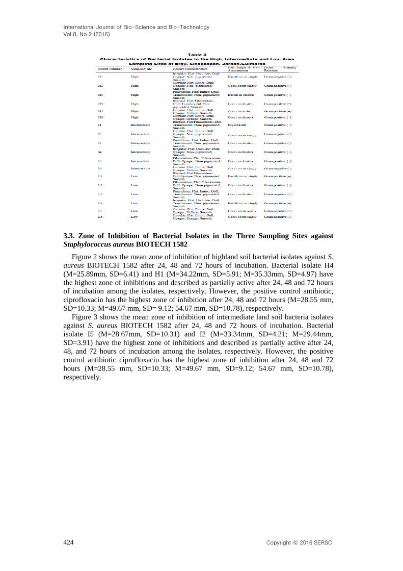
Table 3 shows the characteristics of the six bacterial isolates in each sampling site as
characterized according to shape, elevation, margin, color, pigmentation, texture, cell
arrangement, and Gram staining reaction.
International Journal of Bio-Science and Bio-Technology
Vol.8, No.2 (2016)
424 Copyright ⓒ 2016 SERSC
3.3. Zone of Inhibition of Bacterial Isolates in the Three Sampling Sites against
Staphylococcus aureus BIOTECH 1582
Figure 2 shows the mean zone of inhibition of highland soil bacterial isolates against S.
aureus BIOTECH 1582 after 24, 48 and 72 hours of incubation. Bacterial isolate H4
(M=25.89mm, SD=6.41) and H1 (M=34.22mm, SD=5.91; M=35.33mm, SD=4.97) have
the highest zone of inhibitions and described as partially active after 24, 48 and 72 hours
of incubation among the isolates, respectively. However, the positive control antibiotic,
ciprofloxacin has the highest zone of inhibition after 24, 48 and 72 hours (M=28.55 mm,
SD=10.33; M=49.67 mm, SD= 9.12; 54.67 mm, SD=10.78), respectively.
Figure 3 shows the mean zone of inhibition of intermediate land soil bacteria isolates
against S. aureus BIOTECH 1582 after 24, 48 and 72 hours of incubation. Bacterial
isolate I5 (M=28.67mm, SD=10.31) and I2 (M=33.34mm, SD=4.21; M=29.44mm,
SD=3.91) have the highest zone of inhibitions and described as partially active after 24,
48, and 72 hours of incubation among the isolates, respectively. However, the positive
control antibiotic ciprofloxacin has the highest zone of inhibition after 24, 48 and 72
hours (M=28.55 mm, SD=10.33; M=49.67 mm, SD=9.12; 54.67 mm, SD=10.78),
respectively.
International Journal of Bio-Science and Bio-Technology
Vol.8, No.2 (2016)
Copyright ⓒ 2016 SERSC 425
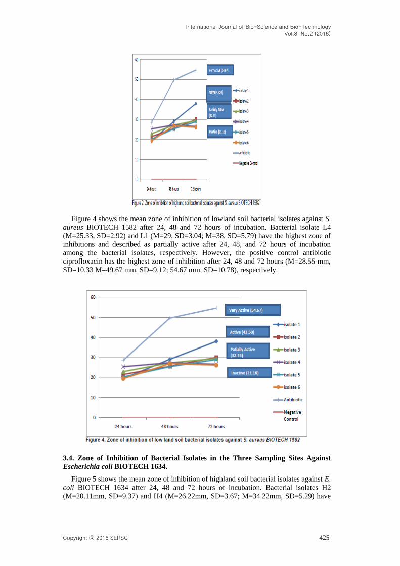
Figure 4 shows the mean zone of inhibition of lowland soil bacterial isolates against S.
aureus BIOTECH 1582 after 24, 48 and 72 hours of incubation. Bacterial isolate L4
(M=25.33, SD=2.92) and L1 (M=29, SD=3.04; M=38, SD=5.79) have the highest zone of
inhibitions and described as partially active after 24, 48, and 72 hours of incubation
among the bacterial isolates, respectively. However, the positive control antibiotic
ciprofloxacin has the highest zone of inhibition after 24, 48 and 72 hours (M=28.55 mm,
SD=10.33 M=49.67 mm, SD=9.12; 54.67 mm, SD=10.78), respectively.
3.4. Zone of Inhibition of Bacterial Isolates in the Three Sampling Sites Against
Escherichia coli BIOTECH 1634.
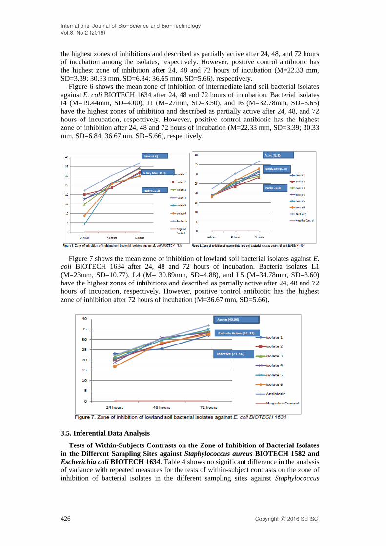
Figure 5 shows the mean zone of inhibition of highland soil bacterial isolates against E.
coli BIOTECH 1634 after 24, 48 and 72 hours of incubation. Bacterial isolates H2
(M=20.11mm, SD=9.37) and H4 (M=26.22mm, SD=3.67; M=34.22mm, SD=5.29) have
International Journal of Bio-Science and Bio-Technology
Vol.8, No.2 (2016)
426 Copyright ⓒ 2016 SERSC
the highest zones of inhibitions and described as partially active after 24, 48, and 72 hours
of incubation among the isolates, respectively. However, positive control antibiotic has
the highest zone of inhibition after 24, 48 and 72 hours of incubation (M=22.33 mm,
SD=3.39; 30.33 mm, SD=6.84; 36.65 mm, SD=5.66), respectively.
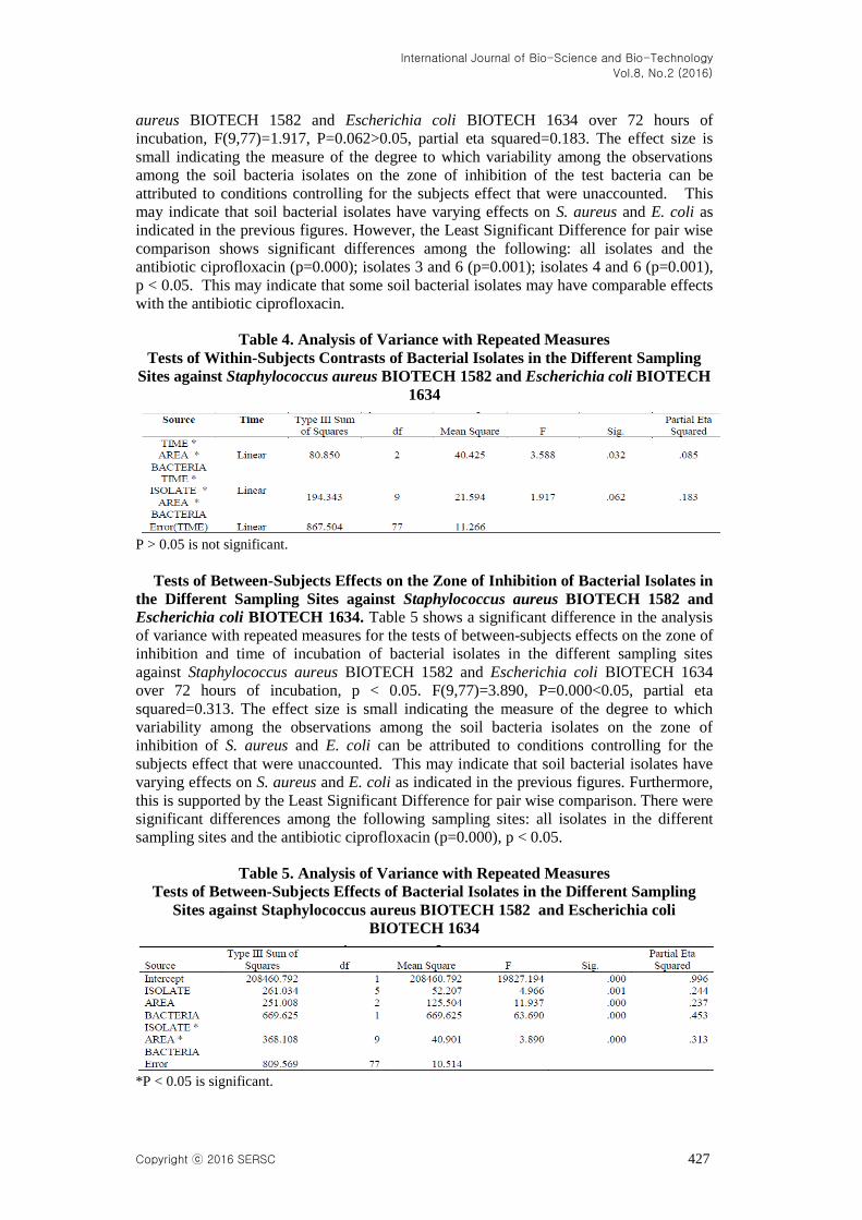
Figure 6 shows the mean zone of inhibition of intermediate land soil bacterial isolates
against E. coli BIOTECH 1634 after 24, 48 and 72 hours of incubation. Bacterial isolates
I4 (M=19.44mm, SD=4.00), I1 (M=27mm, SD=3.50), and I6 (M=32.78mm, SD=6.65)
have the highest zones of inhibition and described as partially active after 24, 48, and 72
hours of incubation, respectively. However, positive control antibiotic has the highest
zone of inhibition after 24, 48 and 72 hours of incubation (M=22.33 mm, SD=3.39; 30.33
mm, SD=6.84; 36.67mm, SD=5.66), respectively.
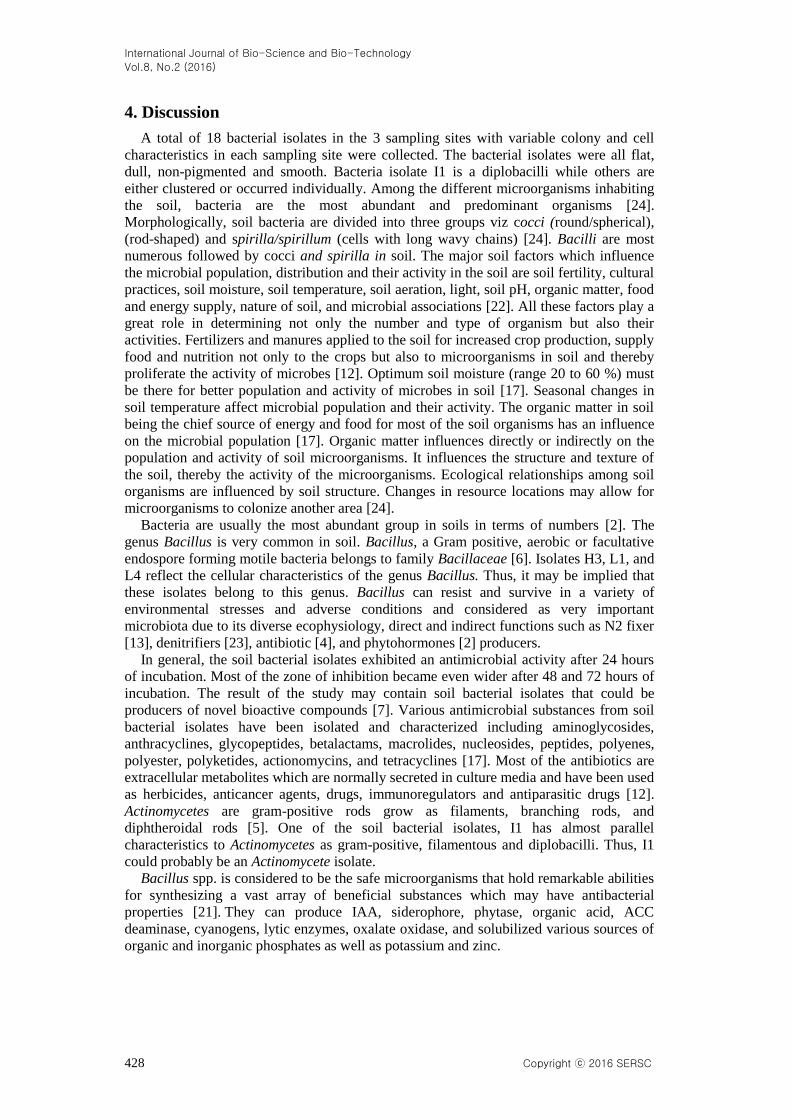
Figure 7 shows the mean zone of inhibition of lowland soil bacterial isolates against E.
coli BIOTECH 1634 after 24, 48 and 72 hours of incubation. Bacteria isolates L1
(M=23mm, SD=10.77), L4 (M= 30.89mm, SD=4.88), and L5 (M=34.78mm, SD=3.60)
have the highest zones of inhibitions and described as partially active after 24, 48 and 72
hours of incubation, respectively. However, positive control antibiotic has the highest
zone of inhibition after 72 hours of incubation (M=36.67 mm, SD=5.66).
3.5. Inferential Data Analysis
Tests of Within-Subjects Contrasts on the Zone of Inhibition of Bacterial Isolates
in the Different Sampling Sites against Staphylococcus aureus BIOTECH 1582 and
Escherichia coli BIOTECH 1634. Table 4 shows no significant difference in the analysis
of variance with repeated measures for the tests of within-subject contrasts on the zone of
inhibition of bacterial isolates in the different sampling sites against Staphylococcus
International Journal of Bio-Science and Bio-Technology
Vol.8, No.2 (2016)
Copyright ⓒ 2016 SERSC 427
aureus BIOTECH 1582 and Escherichia coli BIOTECH 1634 over 72 hours of
incubation, F(9,77)=1.917, P=0.062>0.05, partial eta squared=0.183. The effect size is
small indicating the measure of the degree to which variability among the observations
among the soil bacteria isolates on the zone of inhibition of the test bacteria can be
attributed to conditions controlling for the subjects effect that were unaccounted. This
may indicate that soil bacterial isolates have varying effects on S. aureus and E. coli as
indicated in the previous figures. However, the Least Significant Difference for pair wise
comparison shows significant differences among the following: all isolates and the
antibiotic ciprofloxacin (p=0.000); isolates 3 and 6 (p=0.001); isolates 4 and 6 (p=0.001),
p < 0.05. This may indicate that some soil bacterial isolates may have comparable effects
with the antibiotic ciprofloxacin.
Table 4. Analysis of Variance with Repeated Measures
Tests of Within-Subjects Contrasts of Bacterial Isolates in the Different Sampling
Sites against Staphylococcus aureus BIOTECH 1582 and Escherichia coli BIOTECH
1634
P > 0.05 is not significant.
Tests of Between-Subjects Effects on the Zone of Inhibition of Bacterial Isolates in
the Different Sampling Sites against Staphylococcus aureus BIOTECH 1582 and
Escherichia coli BIOTECH 1634. Table 5 shows a significant difference in the analysis
of variance with repeated measures for the tests of between-subjects effects on the zone of
inhibition and time of incubation of bacterial isolates in the different sampling sites
against Staphylococcus aureus BIOTECH 1582 and Escherichia coli BIOTECH 1634
over 72 hours of incubation, p < 0.05. F(9,77)=3.890, P=0.000<0.05, partial eta
squared=0.313. The effect size is small indicating the measure of the degree to which
variability among the observations among the soil bacteria isolates on the zone of
inhibition of S. aureus and E. coli can be attributed to conditions controlling for the
subjects effect that were unaccounted. This may indicate that soil bacterial isolates have
varying effects on S. aureus and E. coli as indicated in the previous figures. Furthermore,
this is supported by the Least Significant Difference for pair wise comparison. There were
significant differences among the following sampling sites: all isolates in the different
sampling sites and the antibiotic ciprofloxacin (p=0.000), p < 0.05.
Table 5. Analysis of Variance with Repeated Measures
Tests of Between-Subjects Effects of Bacterial Isolates in the Different Sampling
Sites against Staphylococcus aureus BIOTECH 1582 and Escherichia coli
BIOTECH 1634
*P < 0.05 is significant.
International Journal of Bio-Science and Bio-Technology
Vol.8, No.2 (2016)
428 Copyright ⓒ 2016 SERSC
4. Discussion
A total of 18 bacterial isolates in the 3 sampling sites with variable colony and cell
characteristics in each sampling site were collected. The bacterial isolates were all flat,
dull, non-pigmented and smooth. Bacteria isolate I1 is a diplobacilli while others are
either clustered or occurred individually. Among the different microorganisms inhabiting
the soil, bacteria are the most abundant and predominant organisms [24].
Morphologically, soil bacteria are divided into three groups viz cocci (round/spherical),
(rod-shaped) and spirilla/spirillum (cells with long wavy chains) [24]. Bacilli are most
numerous followed by cocci and spirilla in soil. The major soil factors which influence
the microbial population, distribution and their activity in the soil are soil fertility, cultural
practices, soil moisture, soil temperature, soil aeration, light, soil pH, organic matter, food
and energy supply, nature of soil, and microbial associations [22]. All these factors play a
great role in determining not only the number and type of organism but also their
activities. Fertilizers and manures applied to the soil for increased crop production, supply
food and nutrition not only to the crops but also to microorganisms in soil and thereby
proliferate the activity of microbes [12]. Optimum soil moisture (range 20 to 60 %) must
be there for better population and activity of microbes in soil [17]. Seasonal changes in
soil temperature affect microbial population and their activity. The organic matter in soil
being the chief source of energy and food for most of the soil organisms has an influence
on the microbial population [17]. Organic matter influences directly or indirectly on the
population and activity of soil microorganisms. It influences the structure and texture of
the soil, thereby the activity of the microorganisms. Ecological relationships among soil
organisms are influenced by soil structure. Changes in resource locations may allow for
microorganisms to colonize another area [24].
Bacteria are usually the most abundant group in soils in terms of numbers [2]. The
genus Bacillus is very common in soil. Bacillus, a Gram positive, aerobic or facultative
endospore forming motile bacteria belongs to family Bacillaceae [6]. Isolates H3, L1, and
L4 reflect the cellular characteristics of the genus Bacillus. Thus, it may be implied that
these isolates belong to this genus. Bacillus can resist and survive in a variety of
environmental stresses and adverse conditions and considered as very important
microbiota due to its diverse ecophysiology, direct and indirect functions such as N2 fixer
[13], denitrifiers [23], antibiotic [4], and phytohormones [2] producers.
In general, the soil bacterial isolates exhibited an antimicrobial activity after 24 hours
of incubation. Most of the zone of inhibition became even wider after 48 and 72 hours of
incubation. The result of the study may contain soil bacterial isolates that could be
producers of novel bioactive compounds [7]. Various antimicrobial substances from soil
bacterial isolates have been isolated and characterized including aminoglycosides,
anthracyclines, glycopeptides, betalactams, macrolides, nucleosides, peptides, polyenes,
polyester, polyketides, actionomycins, and tetracyclines [17]. Most of the antibiotics are
extracellular metabolites which are normally secreted in culture media and have been used
as herbicides, anticancer agents, drugs, immunoregulators and antiparasitic drugs [12].
Actinomycetes are gram-positive rods grow as filaments, branching rods, and
diphtheroidal rods [5]. One of the soil bacterial isolates, I1 has almost parallel
characteristics to Actinomycetes as gram-positive, filamentous and diplobacilli. Thus, I1
could probably be an Actinomycete isolate.
Bacillus spp. is considered to be the safe microorganisms that hold remarkable abilities
for synthesizing a vast array of beneficial substances which may have antibacterial
properties [21]. They can produce IAA, siderophore, phytase, organic acid, ACC
deaminase, cyanogens, lytic enzymes, oxalate oxidase, and solubilized various sources of
organic and inorganic phosphates as well as potassium and zinc.
International Journal of Bio-Science and Bio-Technology
Vol.8, No.2 (2016)
Copyright ⓒ 2016 SERSC 429
The ability of microorganisms to produce enzymes that may have antibacterial
properties is influenced by environmental conditions such as temperature, pH, and
presence of inductors or repressors [20].
A common statement of the competitive exclusion principle is that a species whose use
of resources is very similar cannot live in the same place for an extended period of time
[11]. Thus, if two or more species eat the same thing, use the same hiding places, occupy
the same habitats, etc. one species will be more efficient than the others and will fill the
niche with its offspring leaving no resources for other species. In this way, the more
efficient species will “competitively exclude” the less efficient species.
5. Conclusions and Recommendations
There were 6 soil bacterial isolates obtained in the high, intermediate, and low
sampling sites in Sinapsapan, Jordan, Guimaras. Isolate I1 is the only bacterium that is
diplobacilli while the rest are either clustered or occur singly. Isolates I1 and I5 were the
most effective soil bacterial isolate against S. aureus and E. coli, respectively. This is may
be due to the fact that they can degrade and inhibit bacterial growth due to their secretion
of digestive enzymes. After 72 hours of incubation, all bacterial isolates exhibited
variations on their antibacterial effect against S. aureus and E. coli. For S. aureus, the
following isolates had the widest zone of inhibitions after 72 hours of incubation: in the
highland sampling site, isolate H4; in the intermediate sampling site, isolate I2; and
lowland sampling site, isolate L1. For E. coli, the following isolates had the widest zone
of inhibitions after 72 hours of incubation: in the highland sampling site, isolate H4; in the
intermediate sampling site, isolate I6; and lowland sampling site, isolate L5.
There was a significant difference on the zone of inhibition of the different bacterial
isolates in the three sampling sites due to varying antibacterial effects of the isolates
against S. aureus and E. coli. This may be due to differences on the amount of substrates
produced by the different bacterial isolates which inhibit the growth of the test pathogens,
S. aureus and E. coli.
The findings have shown that different isolates were more effective on S. aureus than
on E. coli. It is therefore possible for the isolates to be considered as a prospective source
of antibiotic against on S. aureus than E. coli. Searching for new possible source of
antibiotics is necessary because it becomes a manner for bacteria to be resistant to certain
antibiotics. Antibiotic resistant bacteria are increasing nowadays in number that makes the
treatment for some diseases to be difficult.
Bacteria become resistant more quickly when antibiotics are used improperly. Since
there are kinds of bacteria that are resistant to antibiotics and are untreatable, there is a
greater must to search for more possible sources of antibiotics like from unexplored soil
sources in the nearby locality.
The study was limited only on the characterization of the colonies and gram staining
technique. It is recommended to do special staining methods such as flagella staining to
determine the arrangement of the flagella, endospore staining to determine whether the
isolates are spore-former and the position of their spores, capsule staining to determine
whether the isolates are virulent or not, and negative staining to reveal the morphology of
the cell.
The study did not determine the genera or the species of the isolates with antibacterial
properties. It is recommended to do biochemical tests to identify the genus of each isolate
and confirm using Bergey’s Manual of Systematic Bacteriology for the identification of
the isolate. Furthermore, molecular characterization of the isolates to reveal the identity of
each species of the isolate is also suggested. Phylogenetic analysis should be done to
determine the group of each isolate. This will also help to determine if the bacterial
isolates that were used in this study are newly discovered or not.
International Journal of Bio-Science and Bio-Technology
Vol.8, No.2 (2016)
430 Copyright ⓒ 2016 SERSC
Acknowledgment
The researchers would like to thank the staff of the Central Science Laboratory of West
Visayas State University, the panel members of the Biological Science Department,
Manong Ricky and Manang Sara, Johnny Boy Ybañez, Tito Ake and Tita Tinee, Eisha
and JC, and Mr. and Mrs. Estrella for the financial support.
References
[1] A. J. Alanis, “Resistance to Antibiotics: Are We in the Post-Antibiotic Era?, Archives Med Research,
vol. 36, (2005), pp. 697-705.
[2] F. F. Araujo, A. A. Henning and M. Hungria, “Phytohormones and antibiotics produced by Bacillus
subtilis and their effects on seed pathogenic fungi and on soybean root development”, World J. Microb.
Biot., vol. 21, (2005), pp. 1639-1645.
[3] L. V. Allen and H. C. Ansel, “Ansel’s pharmaceutical dosage forms and drug delivery system”, 10th ed.
USA: Wolters Kluwer, (2013).
[4] C. W. Bacon and D. M. Hinton, “Endophytic and biological control potential of Bacillus mojavensis and
related species”, Biol. Control, vol. 23, (2002), pp. 274-284.
[5] G. H. W. Bowden, “Actinomyces, Propionibacterium propionicus, and Streptomyces”, Baron S, editor.
Medical Microbiology. 4th edition. Galveston (TX): University of Texas Medical Branch at Galveston;
Chapter 34, Retrieved from: http://www.ncbi.nlm.nih.gov/books/NBK8385, (1996).
[6] D. Claus and R. C. W. Berkeley, “Genus Bacillus Cohn 1872, 174AL”, Bergey’s Manual of Systematic
Bacteriology, vol. 2, (1986), pp. 1105-1139.
[7] S. Das, P. S. Lyla and S. Ajmal Khan, “Marine microbial diversity and ecology: present status and
future perspectives. Current Science”, Department of Health (2009). National health data dictionary,
vol. 90, no. 10, (2006), pp. 1325-1335.
[8] Retrieved from:http://uhmis1.doh.gov.ph/standards/sdetails. php?data_element=BARANGAY
[9] Eckervall, Personal Communication, (2013) October 10.
[10] Española, Personal Communication, (2013) October 10.
[11] J. P. Harley, “Laboratory exercises in Microbiology”, (6thed.) Boston: McGraw Hill Higher Education,
(2005).
[12] C. Hoagstrom, “Competitive exclusion principle”, Retrieved from:
http://www.eoearth.org/view/article/151404, (2011).
[13] T. R. P. Kekuda, K. S. Shobha and R. Onkarappa, “Fascinating diversity and potent biological activities
of Actinomycete metabolites”, Journal of Pharmacy Research, vol. 3, no. 2, (2010), pp. 250-256.
[14] X. Liu, H. Zhao and S. Chen, “Colonization of maize and rice plants by strain Bacillus megaterium C4”,
Curr. Microbio., vol. 52, (2006), pp. 186-190.
[15] M. T. Madigan, J. M. Martinko, D. A. Stahl and D. P. Clark, “Brock’s biology of microorganism”,
(13thed.) Boston: Benjamin Cummings, (2012).
[16] J. P. Martin, “Soil Microbiology and Biochemistry”, SSSA; Madison, WI, (1976).
[17] F. Pela´ ez, “The historical delivery of antibiotics from microbial natural products—Can history repeat?,
Biochemical Pharmacology, vol. 71, (2006), pp. 981-990.
[18] T. R. Prashith Kekuda, K. S. Shobha, R. Onkarappa, S. A. Goutham and H. L. Raghavendra, “Screening
biological activities of a Streptomyces species isolated from soil of Agumbe”, Karnataka, India. Int. J.
Drug Dev. & Res., vol. 4, no. 3, (2012), pp. 104-114.
[19] E. A. Quinto and M. A. G. Santos, “A guidebook to plant screening: Phytochemical and biological”,
Microbiology section. España, Manila; University of Santo Tomas Publishing House, (2005).
[20] G. Reid and P. Wong, “Soil bacteria”, State of New South Wales Department of Primary Industries,
(2005).
[21] R. Sharma, Y. Chisti and U. C. Banerjee, “Production, purification, characterization, and applications of
lipases”, Biotechnology Advances, vol. 19, no. 8, (2001), pp. 627-662.
[22] T. Stein, “Bacillus subtilis antibiotics: structures, syntheses and specific functions”, MolMicrobiol, vol.
56, no. 4, (2005), pp. 1669-1679.
[23] A. Stephanie, E. J. A. Breznak and T. M. Schmidt, “Isolation and characterization of soilbacteria that
define ' Terriglobis ' gen. nov.”, in the Phyllum 'Acidobacteria' Appl. And Env. Microbiology, vol. 73,
no. 8, (2007), pp. 2708-2717.
[24] Suharti, H. A. Heering and S. de Vries, “NO Reductase from Bacillus azotoformans is a bifunctional
enzyme accepting electrons from menaquinol and a specific endogenous membrane-bound cytochrome
c551”, Biochem., vol. 43, (2004), pp. 13487-13495.
[25] D. M. Sylvia, J. F. Fuhrmann, P. G. Hartel and D. A. Zuberer, “Principles and applications of Soil
Microbiology”, New Jersey, Pearson Education Inc., (2005).
International Journal of Bio-Science and Bio-Technology
Vol.8, No.2 (2016)
Copyright ⓒ 2016 SERSC 431
[26] K. Todar, “An Introduction to Bacteriology: University of Wisconsin Inc.”, Retrieved at
http://textbookofbacteriology.net/index.html, (2008).
Authors
Ma. Sophia Estella C. Tajolosa. Graduate of BS Biology
(Microbiology Track) at West Visayas State University, Iloilo City
Philippines on March 2015. She is currently pursuing her Doctor of
Medicine in one of the medical schools in Iloilo City, Philippines.
Kristine Joy R. Estrella. Graduate of BS Biology
(Microbiology Track) at West Visayas State University, Iloilo
City Philippines on March 2015.
Rey G. Tantiado. Graduate of BS Biological Science and
Master of Arts in Biology at West Visayas State University,
Iloilo City Philippines on March 2003 and March 2006,
respectively. He graduated his Doctor of Philosophy in
Education (Biology) from the University of the Philippines Open
University, Los Banos, Laguna Philippines on May 2014. He is
currently an instructor at West Visayas State University.
International Journal of Bio-Science and Bio-Technology
Vol.8, No.2 (2016)
432 Copyright ⓒ 2016 SERSC