Embed Size (px)
Citation preview

@IJAPSA-2016, All rights Reserved Page 58
ANTIBIOTIC RESISTANCE AMONG ENTERIC BACTERIA AND THEIR
HEALTH IMPLICATION
Vishvas Hare*1, Pankaj Chowdhary
2and Vinay Singh Baghel
3
1,2,3Department of Environmental Microbiology, School for Environmental Sciences Babasaheb Bhimrao
Ambedkar University (A Central University), VidyaVihar, Raebareli Road, Lucknow, 226025, Uttar Pradesh,
India.
*Corresponding Author: Vishvas Hare
Abstract
Today antimicrobial agent resistance is an emerging global concern to both public and veterinary
health. The use of antibacterial drugs for prophylactic or therapeutic purposes in humans and for
veterinary and agricultural purposes has provided selective pressure favoring the survival and
spread of resistant organisms. However, resistant bacteria may transfer their resistance to
previously non-resistant pathogenic bacteria or directly infect humans with bacterialdiseases that
cannot be treated by conventional antimicrobial therapies. The potential for antibiotic exposure
and resistance development in human and animal gastrointestinal tracts, coupled with relatively
great abundance in waters contaminated with human and animal waste, makes the fecal coliform
bacteria a logical fecal groupfor studies of antibiotic resistance and transfer in aquatic
environments. The main bacteria present in human and animal feces discussed which indicators
of fecal pollution should be used in current drinking water microbiological analysis.This review
mainly focoused on antibiotic resistance of environmental isolates is imperative to explore the
antibiotic pressure in the environment. In addition methods to reduce bacteria resistant load in
wastewaters and the amount of antimicrobial agents is originated in most cases of hospitals and
farms that optimization the disinfection procedures and management of wastewater.
Key Words: Antibacterial durgs,Bacterial disease, contaminated water,feces, fecal coliform
bacteria,Resistance, wastes water
I. INTRODUCTION
As it is well known that antimicrobial resistance is a global problem in human and veterinary
medicine. Antibiotic resistance in bacteria provides one of the well-documented examples of an
evolutionary response to selection in natural populations. The selective agent can be easily
identified, changes in phenotype have occurred in a human generation, and the mode of transmission
of resistance determinants can be deduced [1-3].The antimicrobial agents used in animal care are
also significant, not only in increasing the resistance in animalpathogens, but also in bacteria
transmitted from animals to humans.Most studies show that not only the level of resistance of
pathogenic bacteria, but alsoof commensal bacteria increases.It is generally accepted that the main
risk factor for increase in the antibiotic resistance is extensive use of antibiotics. This has lead to the
emergence and dissemination of resistant bacteria and resistance genes in animals and
humans.Dramatically rapid increases in antibiotic resistance have been observed in a host of
phylogenetically diverse taxa and the process has occurred repeatedly within taxa as each is
challenged with new antibiotics [1, 4, 5].Antibiotic selection pressure can lead to a rapid reduction
in the initial fitness costs that were obtain as a pleiotropic effect of resistance. Additional
consequences of the rapid evolution of resistance may depend upon population structure. Bacterial
population structures range from distingguishablely clonal to non-clonal, depending on the relative
roles of horizontal and vertical transmission of geneticinformation within a species [4,6].Antibiotic

International Journal of Applied and Pure Science and Agriculture (IJAPSA) Volume 02, Issue 12, [December- 2016] e-ISSN: 2394-5532, p-ISSN: 2394-823X
@IJAPSA-2016, All rights Reserved Page 59
resistance determinants can spread via either clonalelaboration or horizontal transfer, the latter
mediated by conjugative plasmids, phage vectors, and natural transformation systems.In contrast, the
spread of resistance determinants via promiscuous horizontal exchange can be expected to both
maintain variation and show an absence of linkage disequilibrium between the selected and neutral
markers. Studies based on samples from a single time point often rely upon variation in resistance
genes and linkage disequilibrium to distinguish vertical from horizontal transmission. The
subsequent intraspecific spread of chromosomal resistance genes was most likely mediated by a
combination of clonal elaboration, as evidenced by linkage disequilibrium between resistance genes
and housekeeping genes, and horizontal gene transfer, deduced from the incongruence of
dendrogramsportayaling patterns of variation within the two types of loci [3, 7].Investigation of the
prevalence of resistance of certain indicator bacteria like E. coli and enterococci in the intestinal
tract of different populations of animals and humans makes it workable to compare the prevalence of
resistance in different populations. Because of the in avoidable high usage of antibiotics in hospitals,
selection and diffusion of resistant clones and resistance genes is high in hospitals. Therefore,
healthy individuals in the community outside hospitals hold not only a reservoir of resistant bacteria
and resistant genes, but are considered to be a suitable population to study the possibility of transfer
of resistant bacteria or resistance genes from animals to humans[1]
II. DETECTION OF MICROORGANISM
The detection and reckoning of microorganism trust on cultural methods. In these methods,
the microorganism isgrown on either a solid (agar) or liquid (broth) medium, which supplies the
nutritional requirements of the organisms or in the case of obligate parasites such as viruses, the
organism is grown in a culture of host cells. Once a microorganism has been grown and isolated as a
pure culture, identification of the organism is based on biochemical, immunological (serological),
and genetic characteristics of the isolate. In many illustrations, particularly for such well studied
organisms as the coliforms, specific compounds are obtain into the primary media, which allow for
selection and differentiation of the organisms of interest. For example, Endo agar is routinely used
for the reckoning of coliforms and otherenteric organisms. This agar has sodium sulfite and basic
fuchsinto inhibit the growth of Gram-positive bacteria. In addition, lactose is present as a primary
substrate for bacterial nutrition. Metabolism of lactose with the formation of acid and gas, a
hallmark characteristic of the coliform group, is detected by a color change due to the reaction
between acetaldehyde (an intermediate product of lactose fermentation) and the sodium
sulfite.Within the past several decades alternative, noncultural methods for the detection of
microorganisms have been developed and are now widely used. These methods are particularly
useful for the detection of microorganism for which no selective media has yet been developed.
These methods include the use of specific staining procedures, usually based on serological
properties of the organism (immunofluorescence), and molecular methods for the detection and
characterization of specific sequences within the DNA or RNA of the organism. Immunofluorescent
detection of microorganisms depends on the ability of antibody molecules to recognize and react
with specific three-dimensional regions (epitopes) on the surface of microbial cells. Antibodies can
be labelled with fluorescent dyes. Specific-microorganisms stained with immunofluorescent dyes
can be enumerated by direct counts under an epifluorescentmicroscope.Detection of nucleic acid
sequences unique to a particular organism involve several distingguishable procedures. Nucleic acid
probes containing nucleotide sequences complementary to a unique sequence of a specific
microorganism can be coupled with a variety of reporter molecules (fluorescent dyes and
radioisotopes). When mixed with a solution of DNA elicit from an environmental sample, the probe
will only bind to those specific target sequences that are complementary to it. Thus, the presence of
specific microbial nucleic acid, and presumably then specific microorganisms, can be determined.
Frequently, the amounts of specific nucleic acids present in environmental samples are too low to be
detected directly by gene probes. In this case, there is a need to increase, or amplify, the specific

International Journal of Applied and Pure Science and Agriculture (IJAPSA) Volume 02, Issue 12, [December- 2016] e-ISSN: 2394-5532, p-ISSN: 2394-823X
@IJAPSA-2016, All rights Reserved Page 60
sequences to detectable levels.This is accomplished through the use of the polymerase chain reaction
(PCR).
III. INDICATOR BACTERIA
Indicator bacteria belonging to the normal intestinal flora of humans and animals. These
bacteria not only constitute an tremendous reservoir of resistance genes for (potentially) pathogenic
bacteria, but also the level of resistance in the endogenous flora is considered to be a good indicator
for the selection pressure exerted by antibiotic use in that population [8] and for the resistance
problems to be expected in pathogens [9].Indicator organisms that can be transmitted by a
waterborne route, (e.g., thermotolerant coliforms) are used widely as deputy for the detection of
pathogens.[10].Compared two media, mFC and mTEC, for the reckoning of fecal coliforms in tidal
creeks.Counts by mTEC were systematically higher than mFC counts at all salinity ranges. In
addition, a significant number of false positives were associated with using mFC in middle and high
salinity areas.Lifshitz and Joshi found the ColiPlate (CP) kit gave estimates of E. coli that were 20%
higher than standard membrane filtration when the two methods were compared for testing water
samples [11]. The difference increased when samples were impaled with injured cells. They
concluded that the CP kit is a more reliablemethod for use with samples having high levels of
injured or weakened cells. m-ColiBlue24 (m-CB) was compared to m-Endo medium and an
International Organization for Standardization (ISO) standard conform medium, lactose agar with
Tergitol 7, for the analysis of indicator organisms in bottled water.Pruss reviewed studies on
uncontrolled waters, such asseas, lakes, and rivers, to evaluate the health risks caused by poor
microbiological quality of recreational water. Most studies reported a dose-related increase of health
risk in swimmers with an increase in the indicator bacteria count in recreational waters. The
indicator microorganisms that correlated best with health out come were Enterococci/Fecal
streptococci for both marine and freshwater and E. coli for freshwater [10].
(1) Fecal Indicator Bacteria
In 1892, Schardinger proposed that since-Bacterium coli was a characteristic component of
the fecal flora, its presence in water could be taken as an indication of the presence of fecal pollution
and therefore of the potential presence of enteric pathogens [12].Various classification schemes for
coliforms have emerged. The earliest were those of MacConkey in 1909, which recognized 128
different coliform types, while Bergey and Deehan in 1908 identified 256. By the early 1920s,
differentiation of coliforms had come to a series of correlations that suggested that indole
production, gelatin liquefaction, sucrose fermentation and the Voges-Proskauer reaction were among
the more important tests for determining fecal contamination. These developments culminated in the
IMViC (Indole, Methyl Red, Voges–Proskauer and Citrate) tests for the differentiation of so-called
fecal coliforms and intermediates [13].Traditional tests for total and fecal coliforms are carried out
either by the multiple-tube fermentation technique or by filtration through membrane. The multiple-
tube fermentation technique is used for medium or highly contaminated waters, and the filtration
through membrane for low or very low contaminated waters. Filtration through membrane is a very
sensitive technique since can detect one (culturable) cell in 500 or even 1,000 mL of water.
However, both methods take several days to complete and do not detect viable but non-culturable
bacteria [14].These limitations stimulate the discovery of alternative methods, faster and, if possible,
less prone to false negative results such as those caused by the viable but non-culturable bacteria.
Fecal Indicator bacteria are generally three types that found on the basis of different test and
describe below following.
3.1.1 Total Coliforms Total coliforms are Gram-negative, oxidase-negative, non-sporeformingrods, which ferment
lactose with gas production at 35-37 °C, after 48h, in a medium with bile salts and detergents [15].
When the test of coliforms is carried out with environmental waters, several species of the four
Enterobacteriaceae genera Escherichia, Klebsiella, Enterobacter and Citrobacter give positive

International Journal of Applied and Pure Science and Agriculture (IJAPSA) Volume 02, Issue 12, [December- 2016] e-ISSN: 2394-5532, p-ISSN: 2394-823X
@IJAPSA-2016, All rights Reserved Page 61
results and therefore are coliforms according to this definition. However, the environmental
significance of these four genera is very disparate as discussed in the present text. Therefore, total
coliform counts are not necessarily a measure of fecal pollution and indeed can have no relation with
this cause [16].The detection of β-D-galactosidase activity (at 37 °C) is usually a good marker for
total coliforms in environmental waters, since most of these bacteria display this enzymatic activity
[2, 9]. Most Escherichia, Citrobacter, Enterobacter, Klebsiella andRaoultellastrains have
galactosidase. Hafnia, Serratiaand Yersinia also possess this enzymatic activity. Most Proteus,
Salmonella andEdwardsiella strains do not display β-galactosidase [17, 18]. β-galactosidase cleaves
lactose in glucose and galactose, and can be detected by using colored or fluorescent markers that
change color after enzyme action.In environmental waters, the presence of Aeromonas or Vibrio
cholerae can be a source of false positives in the β-D-galactosidase assay, since these bacteria have
galactosidase, but are not coliforms. Additionally, in particular environments, such as estuaries, β-
galactosidase activity can overestimate total coliform count due to UV-stimulated enzymatic activity
in certain bacteria such as E. coli.
3.1.2 Fecal coliforms
Fecal coliforms (or thermotolerant coliforms) are traditionally defined as coliforms that
ferment lactose at 44.5 °C in a medium with bile salt[15].The range of species detected by the
experimental procedure is much lower than that of total coliforms. With environmental polluted
waters, only E. coli, andKlebsellaoxytocaandKlebsellapneumoniae gave positive results in the test
[19].The detection of β-D-glucuronidase activity (at 44.5 °C) is, generally, a good marker for fecal
coliforms in environmental polluted waters and very specific forE.coli[20].In Gram-negative
bacteria, this enzymatic activity if found in most E. coli strains and in some Salmonella and Shigella
strains [21]. Aeromonas, Citrobacter, Enterobacter, non-coli Escherichia, Hafnia, Klebsiella,
Proteus, Serratia, Vibrio, Yersinia, and most Salmonella strains do not display β-glucuronidase
activity [20].β-D-glucuronidase activity can be detected by using colored or fluorescent markers that change color after enzyme action. The presence of this enzyme in some strains of Bacteroides,
Flavobacterium, Staphylococcus, Streptococcus, in anaerobiccorynebacteria and Clostridium, has
also been reported [22]. β-D-glucuronidase activity in fecal bacteria other thenE. coli (Bacteroides,
Bifidobacteria, Clostridia, Enterococci and Lactobacillus) is very limited [23].
3.1.3 Streptococci and Enterococci
Fecal streptococci also belong to the traditional indicators of fecal pollution. Fecal
streptococci are Gram-positive, catalase-negative, non-sporeformingcocci that grows at 35 °C in a
medium containing bile salts and sodium azide[15] Azide is a strong inhibitor of the respiratory
chain. Since streptococci are one of the very few bacteria that have no respiratory chain, the test is
very specific for this group, and false positives are rarely found [24].Fecal enterococci (E. faecalis,
E. faecium, E. avium and E. gallinarum) are fecal streptococci that grow in the presence of 6.5%
NaCl at 45 °C. Selective media use these particular characteristics in order to separate enterococci
from the other streptococci. Several studies have reported on the microbiological composition of
human and animal (cattle, chicken, deer, dog, fowl, goose, and swine) feces. E. faecalisandE.
faecium were present in human and animal feces. However, whereas human feces almost have only
these two enterococci, in the animals others species co-occur, like E. avium, E. cecorum, E. durans,
E. gallinarumandE. Hirae[25].
IV. ANTIMICROBIAL RESISTANCE
Many retrospective and prospective studies have been performed to study the Egression and
selection of resistance in bacteria by antibiotic usage.Despite large differences in methodology, most
results show that after the introduction of an antibiotic in veterinary practise, the resistance in
pathogenic bacteria and the faecal flora increases, as in human medicine.Some bacteria, most
enterobacteriaceae, staphylococci and Pasteurella spp. become more readily resistant to certain
antibiotics than others like Clostridium sps.andstreptococci which are still fully susceptible to

International Journal of Applied and Pure Science and Agriculture (IJAPSA) Volume 02, Issue 12, [December- 2016] e-ISSN: 2394-5532, p-ISSN: 2394-823X
@IJAPSA-2016, All rights Reserved Page 62
penicillinG.The literature on resistance against APE is very limited as most of these molecules are
not used for therapy and therefore, susceptibility testing is not performed regularly.Linton et al
found a significant increase in the prevalence of resistance against tylosin and bacitracin in faecal
enterococci of fed these molecules [26]. In this study, virginiamycin usage did not result in an
increase in resistance. Ohmae et al. noticed an increase of resistance against carbadox in faecal E.
coli isolates of pigs after its introduction as APE [27]. All resistant isolates from six farms that fed
carbadox continuously to pigs either as APE or for prevention of swine dysentery carried the same
transferable plasmid consultringcarbadoxresistance.Carbadox is not used in poultry and no
carbadoxresistance was found in E. coli isolates from poultry in the same region. Mills and Kelly
[28] also reported an increase in resistance in E. coli isolates from 37 to 61% after the introduction
of carbadox. Carbadox, however, was not only used as an APE, but also for prevention of swine
dysentery and therapy for salmonellosis. Interest in the selection of resistance by APE increased
after the Egression of vancomycin resistant enterococci (VRE) in human infections. It was soon
recognised that avoparcin was until recently commonly used as APE in most EU-member states,
selects for VRE in the intestinal flora of animals. In countries where avoparcin was used as APE,
VRE was not only found in food animals fed with avoparcin, but also in the faecal flora of healthy
humans and pet animals [29, 30].Also resistance against MLS-antibiotics like erythromycin and
quinupristin-dalfopristinis quite common in enterococci from animals fed with related antibiotics as
APE like tylosin (a macrolide) or virginiamycin[30].According to WHO the resistance to antibiotics
is an ability of bacterial population to survive the effect of inhibitory concentration of antimicrobial
agents.Antimicrobial resistance in bacteria may emerge by several pathways. Some bacterial species
are normally and inherently resistant to certain antibiotics, whereas other are sensitive. Sensitivity
has 3 requirements: a target for reaction, a mechanism for transport into the cell before the antibiotic
action takes place and absence of enzymes that could in-activate or modify the antibiotic. A change
in any of these prerequisites could render an antibiotic-sensitive bacterium resistant to the drug
[31].Water bacteria might be endemic to aquatic environments or exogenous, transiently and
occasionally present in the water as a result of shedding from animal, vegetal,or soil surfacess. The
study of antibiotic resistance in endemic water organisms is important, as it might indicate the extent
of modificationof water ecosystems by human action. Aeromonas strains from Portuguese estuarine
water carry less frequently beta-lactamase genes than Enterobacteriaceae[32]. In water reservoirs a-
half of Aeromonas strains might present multiple antibiotic resistance [33].Resistance profiles of
aquatic pseudomonads depend on the species composition, but also from the site in which they were
isolated, being more antibiotic-resistant along shorelines and in sheltered bays than in the open
water, indicating the influence of nonaquatic organisms or pollutants. Nevertheless, such influences
can be found in the more remote water environments; psychrotrophic bacteria from Antarctic show
various degrees of resistance to industrial antibiotics and metals [34].The connexion of antibiotic-
resistance and resistance to heavy metals is very frequent in the same organism (also in the same
plasmid, transposon, or integron) so that industrial pollution probably selects for antibiotic-
resistance and vice versa [35].Indeed metal contamination represents a long-standing, widespread,
and fractious selection pressure for multiresistant organisms. For the nonaquatic organisms,
obviously the density of antibiotic-resistance organisms and antibiotic-resistance genes in fresh
water varies with the proximity to areas with increased antibiotic expenditure, metal pollution, and
between seasons, being more frequently found in rainy seasons [1]. Very little work has been done
to illuminate the role of bacterial biofilms in water environments and its role in antibiotic resistance.
Phenotypic antibiotic resistance in bacterial biofilms might indeed protect the water environment
from selective events caused by the antibiotic release, which probably are acting more effectively on
planktonic bacteria.
4.1 Mechanism of antibiotic resistance in bacteria
The development of antibiotic resistance is the ability of infectious organisms to adapt
quickly to new environmental conditions. Bacteria are single-celled organisms that, compared with
higher life forms, have small numbers of genes. Therefore, even a single random genetic mutation

International Journal of Applied and Pure Science and Agriculture (IJAPSA) Volume 02, Issue 12, [December- 2016] e-ISSN: 2394-5532, p-ISSN: 2394-823X
@IJAPSA-2016, All rights Reserved Page 63
can greatly affect their ability to cause disease and because most microbes reproduce by dividing
every few hours, bacteria can evolve rapidly. A mutation that helps a microbe surviving to an
antibiotic exposure will quickly become dominant throughout the microbial population. Microbes
also often evolve resistance genes from each other through horizontal gene transfer mechanism
which might enable them to be a multiple antibiotic resistant strain. It is also noted that the
specificity of the interactions between antibiotics and various protein sequences within a bacterium
resultse in significantly high ratio of mutations in its genome which leads to antibiotic resistance.
There is also a relatively high possibility that a particular mutation in a certain target sequence will
result in antibiotic resistance.Antibiotics generally target a variety of essential bacterial functions.
For instance, the β-lactam antibiotics and vancomycininterrupte cell wall synthesis of pathogens,
whereas macrolides and tetracyclines disrupt the protein synthesis at ribosomal level. Bacteria may
develop their antibiotic properties by a variety of mechanisms. One mechanism ofresistance is by
degrading the antibiotic in a step by step process. This degradation starts when bacterial β-
lactamases hydrolyzes the β-lactam ring thus rendering these antibiotics ineffective. A secondary
resistance mechanism is then triggered when the antibiotic target is altered. As the next step, bacteria
may block the entry of antibiotic to the site of action, resulting in decreasedabsorption, which in turn
results in bacteria with decreased sensitivity to vancomycin due to thicker cell walls. Finally,
bacteria may develop efflux pumps that actively pump antibiotics out of the cell so that they do
notreach their target[1].
4.1.1Horizontal gene transfer of antibiotic resistance in water environments
Unlike eukaryotes, prokaryotes do not reproduce sexually, nor do they undergo meiosis.
Horizontal gene transfer occur in prokaryotes have evolved three different mechanism conjugation,
transformation and transduction for creating recombination. Horizontal gene transfer occurs
primarily between members of same species. Horizontal gene transfer will become an important in
evolution of many species. Antimicrobial resistance in bacteria associated with different ecological niches will be a global concern. Theegression of antimicrobial resistant strains of pathogenic
bacteria will become a great threat to the public health. The detection of rising trends in
antimicrobial resistance of bacterial strains facilitates implementation of effective control measures.
The antibiotic susceptibility testing contributes directly to patient care, and of antimicrobial drug
resistance. However, in our region, the study of antibiotic resistance of bacteria from environment
like soil, water or from fish is bare. Therefore, study pertaining to antibiotic resistance of
environmental isolates is imperative to explore the antibiotic pressure in the environment. Methods
to reduce bacteria resistant load in waste waters and the amount of antimicrobial agents is originated
in most cases of hospitals and farms that optimization the disinfection procedures and management
of waste water.
Estuarine water-borne Aeromonas strains carry almost as frequently as Enterobacteriaceae
class 1 integron platforms carrying antibiotic-resistance genes [32].Exclusively environmentally
based organisms, as Delftia, also harbor class 3 integrons.The continuity of such genetic structures
cannot probably be explained entirely by antibiotic selection, suggesting that activities resulting in
antibiotic resistance might have other physiological roles or that they are placed in multifunctional
plasmids. The most frequent gene cassette found involves aminoglycoside- resistance genes, rarely
under positive selection in our days, and there is a distrust that some other resistance genes, as
integronsul genes, might provide benefits for the bacteria, unrelated with resistance. However,some
of these mobile gene cassettes in Aeromonas might involve important mechanisms of resistance, as
Qnr, involved in fluoroquinolone resistance, which might behorizontally propagated by IncU-type
plasmids [36].Certainly the dense bacterial populations in sewage treatment plants favor genetic
exchange among bacterial populations and communities, integrons predating transposons and
plasmid diffusion. Multiresistance plasmids of broad host-range are systematically recovered in
sewage [37]. Interestingly, antibiotic-resistance genes from muck influence the lagoons and
groundwater gene pool, but this pool also contains antibiotic-resistance genes from endemic
bacteria. Aeromonas from aquaculture water systems (fish, eel farming) are particularly resistant to
International Journal of Applied and Pure Science and Agriculture (IJAPSA) Volume 02, Issue 12, [December- 2016] e-ISSN: 2394-5532, p-ISSN: 2394-823X
@IJAPSA-2016, All rights Reserved Page 64
antibiotics [38] (Penders and Stobbering, found that frequently contain plasmids and integrons with
multiple genes for antibiotic resistance [39],Jacobs and Chenia and the connexion with heavy-metal
resistance is not uncommon [35]. Water originated in transgenic plant fields may constitute a matter
of concern, but no significant differences have been found in bacterial antibiotic-resistance levels
between transgenic and nontransgenic corn fields [40].
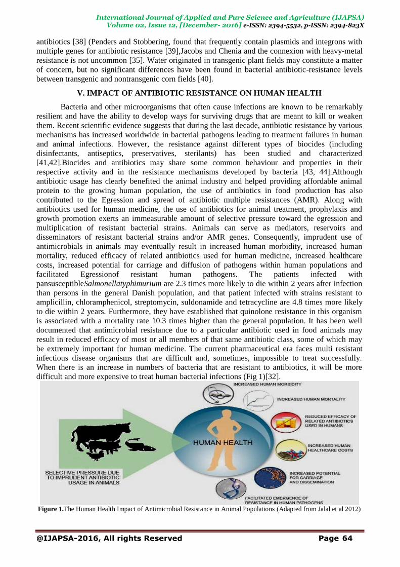
V. IMPACT OF ANTIBIOTIC RESISTANCE ON HUMAN HEALTH
Bacteria and other microorganisms that often cause infections are known to be remarkably
resilient and have the ability to develop ways for surviving drugs that are meant to kill or weaken
them. Recent scientific evidence suggests that during the last decade, antibiotic resistance by various
mechanisms has increased worldwide in bacterial pathogens leading to treatment failures in human
and animal infections. However, the resistance against different types of biocides (including
disinfectants, antiseptics, preservatives, sterilants) has been studied and characterized
[41,42].Biocides and antibiotics may share some common behaviour and properties in their
respective activity and in the resistance mechanisms developed by bacteria [43, 44].Although
antibiotic usage has clearly benefited the animal industry and helped providing affordable animal
protein to the growing human population, the use of antibiotics in food production has also
contributed to the Egression and spread of antibiotic multiple resistances (AMR). Along with
antibiotics used for human medicine, the use of antibiotics for animal treatment, prophylaxis and
growth promotion exerts an immeasurable amount of selective pressure toward the egression and
multiplication of resistant bacterial strains. Animals can serve as mediators, reservoirs and
disseminators of resistant bacterial strains and/or AMR genes. Consequently, imprudent use of
antimicrobials in animals may eventually result in increased human morbidity, increased human
mortality, reduced efficacy of related antibiotics used for human medicine, increased healthcare
costs, increased potential for carriage and diffusion of pathogens within human populations and
facilitated Egressionof resistant human pathogens. The patients infected with
pansusceptibleSalmonellatyphimurium are 2.3 times more likely to die within 2 years after infection
than persons in the general Danish population, and that patient infected with strains resistant to
amplicillin, chloramphenicol, streptomycin, suldonamide and tetracycline are 4.8 times more likely
to die within 2 years. Furthermore, they have established that quinolone resistance in this organism
is associated with a mortality rate 10.3 times higher than the general population. It has been well
documented that antimicrobial resistance due to a particular antibiotic used in food animals may
result in reduced efficacy of most or all members of that same antibiotic class, some of which may
be extremely important for human medicine. The current pharmaceutical era faces multi resistant
infectious disease organisms that are difficult and, sometimes, impossible to treat successfully.
When there is an increase in numbers of bacteria that are resistant to antibiotics, it will be more
difficult and more expensive to treat human bacterial infections (Fig 1)[32].
Figure 1.The Human Health Impact of Antimicrobial Resistance in Animal Populations (Adapted from Jalal et al 2012)

International Journal of Applied and Pure Science and Agriculture (IJAPSA) Volume 02, Issue 12, [December- 2016] e-ISSN: 2394-5532, p-ISSN: 2394-823X
@IJAPSA-2016, All rights Reserved Page 65
VI. EMERGING WATERBORNE BACTERIAL PATHOGENS
The rising pathogenic bacteria of concern outlined here have the potential to be spread
through drinking water, but they do not correlate with the presence of E. coli or with other
commonly used drinking water quality indicators, such as coliform bacteria. In most cases, there are
no satisfactory microbiological indicators of their presence. More studies are needed in order to
understand the real significance and dimension of the diseases caused by water contaminated with
these bacteria, and the ecology of these pathogens [45].
6.1 Escherichia
Escherichia, a member of Enterobacteriaceae, are oxidase-negative catalase-positive straight
rods that ferment lactose. Cells are positive in the Methyl-Red test, but negative in the Voges-
Proskauer assay. Cells do not use citrate, do not produce H2S or lipase, and do not hydrolyze urea
[46]. E. coli is a natural and essential part of the bacterial flora in the gut of humans and animals.
Most E. coli strains are nonpathogenic and reside harmlessly in the colon. However, certain
serotypes do play a role in intestinal and extra-intestinal diseases, such as urinary tract infections
[47].
6.1.1 Pathogenic Escherichia coli Strains
E. coli strains isolated from intestinal diseases have been grouped into at least six different
main groups, based on epidemiological evidence, phenotypic traits, clinical features of the disease
and specific virulence factors. From these, enterotoxigenic (ETEC, namely O148),
enterohemorrhagic (EHEC, namely O157) and enteroinvasive serotypes (EIEC, namely O124) are of
outstanding importance and can be transmitted through contaminated water [47, 48].
6.1.1a EnterotoxigenicE. coli (ETEC) Strains
EnterotoxigenicE. coli (ETEC) serotypes can cause infantile gastroenteritis. The number of
reports of their occurrence in developed countries is comparatively small, but it is an extremely
important cause of diarrhea in the developing world, where there is no tolerable clean water and
poor sanitation. Disease caused by ETEC follows ingestion of contaminated food or water and is
characterized by riotous watery diarrhea lasting for several days that often leads to dehydration and
malnutrition in young children. ETEC also are the most common cause of -travelersdiarrhea that
affects individuals from industrialized countries travelling to developing regions of the World [48,
49].
6.1.1b EnterohemorrhagicE. coli (EHEC) Strains
These organisms produce a toxin known as verocytotoxin which is similar to the toxin
produced by Shigella. Infection with this organism is associated with haemorrhagic colitis. In a
small proportion of the cases, particularly in children, the infection can progress to haemolytic
uraemic syndrome, a life threatening disease. E. coli serotype O157:H7 causes abdominal pain,
bloody diarrhea, and hemolytic uremic syndrome. Although E. coli O157:H7 is not usually a
concern in treated drinking water, outbreaks involving expenditure of drinking water contaminated
with human sewage or cattle feces have been documented. An increasing number of outbreaks are
associated with the expenditure of fruits and vegetables (sprouts, lettuce, coleslaw, salad)
contaminated with feces from domestic or wild animals at some stage during cultivation or handling.
EHEC has also been isolated from bodies of water (ponds, streams), wells and water troughs, and
has been found to survive for months in muck and water-trough sediments [45,49].Person-to-person
contact is an important mode of transmission through the oral-fecal route.
6.1.1c EnteroinvasiveE. coli (EIEC) Strains
EnteroinvasiveE. coli (EIEC) are capable of invading and multiplying in the intestinal
epithelial cells of the distal large bowel in humans. The illness is characterized by abdominal
cramps, diarrhea, vomiting, fever, chills, a generalized malaise, and the appearance of blood and
mucus in the stools of infected individuals. [47, 48]. An investigation in Croatia showed that E. coli

International Journal of Applied and Pure Science and Agriculture (IJAPSA) Volume 02, Issue 12, [December- 2016] e-ISSN: 2394-5532, p-ISSN: 2394-823X
@IJAPSA-2016, All rights Reserved Page 66
O124 could frequently be isolated from cases of gastroenteritis, enterocolitis, and dysentery. The
dysentery was more common among the older age groups, while the two other types of disease
occurred equally in all age groups.Any food contaminated with human feces from an ill individual,
either directly or via contaminated water, could cause disease in others. Outbreaks have been
associated with hamburger meat and unpasteurized milk.
6.2Salmonella.
Salmonella was discovered between the illness and expenditure of water from an aqueduct
that flowed near thecamp. The risk of suffering from the illness rose with the amount of water
consumed. Chemical and bacteriological analyses of the aqueduct water indicated the presence of
fecal contamination. An outbreak of gasteroenteritis due to S. ohio whose origin was the expenditure
of water from a drinking fountain was described for the first time by [50]. This fountain had no
chlorination system. S. ohio was isolated from the water and from 2 of the 13 stool specimens
analysed. A molecular epidemiology study of Salmonella serotype Enteritidis was carried out by
ribotyping and randomly amplified polymorphic DNA (RAPD) typing of 38 food and 25 water
strains, which were epidemiologically unrelated and collected in Spain from 1985 to 1996 [51] Their
results supported the fact that organisms representing at least 40 genomic groups are currently
circulating in Spain but that only the organisms of five groups preponderant and these fall into a
single subcluster or lineage. Organisms of four infrequentgroups were only collected from sewage or
environmental waters.Typhoid fever, a severe systemic illness transmitted through food or water, is
caused by the bacterium Salmonella serotype typhi. Luby et al. [52] evaluated risk factors for
developing typhoid fever in a setting where the disease is endemic (Karachi, Pakistan). Eating ice
cream, eating food from a roadside cabin during the summer months, taking antimicrobials in the 2
weeks preceding the onset of symptoms, and drinking water at the worksite were all independently
associated with typhoid fever.
6.3 Shigella. Children under 5 years of age infected with either Shigella dysenteriae type I or
Shigellaflexneri attending a diarrhea treatment center from 1993 to 1995 in Dhaka, Bangladesh. Use
of antibiotics at home, use of water from tube wells or pipe-water for drinking, and lack of sanitary
facilities were the behavioral and environmental factors strongly associated with S. dysenteriae type
I infection. Tshimanga et al[53] investigated a July 1994 outbreak of Shigella dysenteriae type I at a
textile factory in Bulawayo, Zimbabwe. Thirty seven of 58 workers who drank borehole water were
ill compared to 1 of the 17 who did not. Water samples from the two boreholes yielded numerous
fecal coliforms.
6.4 Vibrio. Cholera, caused by certain strains of Vibrio cholerae, is the first disease for which a
waterborne route of transmission was shown. Paneth et al[54] reviewed the history of John Snow's
1854 investigation proving a waterborne route of transmission for cholera. Epidemic cholera is
caused by toxigenic strains of V. cholerae, of which strains 01 and 0139 are associated most often,
but not exclusively, with epidemic outbreaks. Faruque et al[55] reviewed the epidemiology,
genetics, and ecology of toxigenic V.cholerae. They emphasized the close connexion among V.
cholera, surface water, and the population interacting with the water.They also noted that molecular
epidemiological studies have disclosed significant clonal diversity among toxigenic strains and
continual Egression of new epidemic clones.
6.5 Campylobacter.
Campylobacter spp.are now recognized as a major cause of gastroenteritis associated with
the ingestion of contaminated food and water. Furtado et al[56] reported that Campylobacter was
associated with the majority of waterborne disease outbreaks from private water supplies. Hazeleger
et al.[57] examined the physiological activities of Campylobacter at several environmental
temperatures. Cellular activity, and hencecontinuity, could be measured at temperatures as low as

International Journal of Applied and Pure Science and Agriculture (IJAPSA) Volume 02, Issue 12, [December- 2016] e-ISSN: 2394-5532, p-ISSN: 2394-823X
@IJAPSA-2016, All rights Reserved Page 67
40C. In addition, the organism was capable of chemotaxis and aerotaxis at all temperatures and thus
may be able to move to more favourable environments. Both osmolality and temperature were found
to be significant factors in the continuity of Campylobacter spp.[58].None of the Campylobacter
examined (C.jejuni, C. lari, and E. coli) grew in media with an osmolality of 130 mosmol and a
temperature below 42°C. In media with low osmolalities, the number of viable cells declined rapidly
at any of the temperatures examined.
6.6 Acrobacter Arcobacter spp. isolated from the treatment plants showed the same serotypes as described
for human isolates.Therefore, the spread of Arcobacter via the drinking water pathmust be
suspected.Arcobacter enrichment medium (AM), newly developed by Oxoid, was compared with
twoCampylobacter enrichment media (Preston broth [Oxoid] and LabM broth) and with
Arcobacterbasal medium (ABM) as a control by [59] Atabay and Corry.Twenty strains of
ArcobacterandCampylobacter spp. were tested for growth, with target inocula of less than 4
CFU/mL of medium. None of the Campylobacter spp. grew in the complete AM, and only one grew
(very poorly) in the ABM. However, AM supported good growth of all three species of Arcobacter
(A. butzleri, A. skirrowii, and A. cryaerophilus), which have been associated with human and animal
disease.
6.7 Helicobacter pylori
Helicobacter pylori has been cited as a major etiologic agent for gastritis and has been
implicated in the pathogenesis of peptic and duodenal ulcer disease and gastric carcinoma. H. pylori
has not been isolated from environmental sources, including water [60].On the contrary, molecular
methods have been successful in detecting this pathogen. Fluorescence in situ hybridization has been
successfully used to detect this pathogen in drinking water distribution systems and other water
bodies. Polymerase chain reaction has also been used to detect the presence of H. pylori DNA in
drinking water, especially associated with biofilms [61].In drinking-water biofilms, H.pyloricells
rapidly lose culturability, entering a viable but non-culturablestate.How the organism is transmitted
is still not fully understood. However, the fact that it has been recovered from saliva, dental plaques,
the stomach, and fecal samples strongly indicates oral-oral or fecal-oral transmission. Water and
food appear to be of lesser direct importance, but they can still play a significant role in situations
with improper sanitation and hygiene [45]. The survival of Helicobacter pylori in artificially
contaminated milk and tap water was investigated by Fan et al. [62]. Helicobacter pyloricould
survive for up to 10 days in milk at 40C storage but only 4 days in tap water with a steady decrease
of colony-forming units. However, electron microscopy clearly showed that the
nonculturablecoccoid form was present in tap water that had been kept at 4°C for 7 days.
6.8 Mycobacterium avium complex.
Mycobacterium avium complex (MAC) organisms have been isolated from water and soil. It
is now generallyaccepted that environmental sources, especially natural waters, arethe reservoirs for
most human infections caused by MAC.A typical mycobacteria are responsible for a variety of
diseases, particularly in immune compromised individuals. [63] Lin et al. found that Mycobacterium
avium was signify cantly more resistant to disinfection with copper-silver ions
thanwasLegionellapneumophila. Water, both in the city water supplyand hospital environment, was
found to be the major source oftransmission of Mycobacterium xenopi, an opportunistic
pathogenthat causes pulmonary infections [64].Steinert et al. (1998b) [65] compared that the growth
of Mycobacteriumavium in coculture with the free-living amoeba Acanthamoebapolyphaga with the
growth of M. avium when it was separated fromamoebae. Although viable mycobacteria were
observed within amoebal vacuoles, there was no significant difference between bacterialgrowth in
coculture and bacterial growth separately.Mac organisms has ability to survive and grow under
varied conditions. Mac organisms can proliferate in water at temperatures up to 51 °C and can grow
in natural waters over a wide pH range [45].These mycobacteria are highly resistant to chlorine and
the other chemical disinfectants used for the treatment of drinking-water. Standard drinking-water

International Journal of Applied and Pure Science and Agriculture (IJAPSA) Volume 02, Issue 12, [December- 2016] e-ISSN: 2394-5532, p-ISSN: 2394-823X
@IJAPSA-2016, All rights Reserved Page 68
treatments will not eliminate Mac organisms but, if operating satisfactorily, will significantly reduce
the numbers that may be present in the source water to a level that represents a negligible risk to the
general population. The entryway of these mycobacteria in distribution systems is through leaks.
Growth of Mac organisms in biofilms is probably important for their continuous presence in
distribution systems. The symptoms encountered with Mac infections result from colonization of
either the respiratory or the gastrointestinal tract, with possible diffusion to other locations in the
body. Exposure to Mac organisms may occur through the expenditure of contaminated food, the
inhalation of air with contaminated soil particles, or contact with or ingestion, aspiration, or
aerosolization of potable water containing the organisms [45].With respect to water supplies,
infection with M. avium and M.intracellulare has been well documented. Unlike gastrointestinal
pathogens, where E. coli can be used to indicate their potential presence, no suitable indicators have
been identified to signal increasing concentrations of Mac organisms in water systems [45].
VII. FECAL BACTERIA IN THEIR HOSTS AND IN THE ENVIRONMENT
7.1 Bacteroides
Bacteroides are Gram-negative, non-sporeforming, anaerobic pleiomorphic rods.
Bacteroides are the most abundant bacteria in human feces. In animal feces, on the contrary,
Bacteroides are present at low numbers. Although anaerobic, Bacteroides are among the most
tolerant to oxygen of all anaerobic human gastrointestinal species. B. thetaiotaomicron is one of the
most abundant species in the lower regions of the human gastrointestinal tract. Bacteroides have a
high pathogenic potential and account for approximately two-thirds of all anaerobes isolated from
clinical specimens. The most frequently isolated species has been B. fragilis. The survival of
Bacteroides in environmental waters is usually much lower than the survival of coliforms [66].
7.2 Eubacterium
Eubacterium are anaerobic non-sporeforming Gram-positive rods. Some species have been transferred to other genera-Actinobaculum, Atopobium, Collinsella, Dorea, Eggerthella,
Mogibacterium, Pseudoramibacter and Slackia. Cells are not very aerotolerant. Species isolated
from the human gastrointestinal tract include: E. barkeri, E. biforme, E. contortum, E. cylindrioides,
E. hadrum, E. limosum, E. moniliforme, E. rectal and E. ventricosum.
7.3 Bifidobacterium
Bifidobacteria are Gram-positive, non-sporeforming, pleiomorphicrods.The optimum growth
temperature is 35-39 °C. The genus Bifidobacterium contains ca. 25 species, most of which have
been detected in the human gastrointestinal tract.Bifidobacteria are present in high numbers in the
feces of humans and some animals. Several Bifidobacterium species are specific either for humans
or for animals.Bifidobacteria have been found in sewage and polluted environmental waters, but
appears to be absent from unpolluted or pristine environments such as springs and unpolluted soil.
This results from the facts that upon introduction into the environment bifidobacteriadecrease
appreciably in numbers probably due to their stringent growth requirements.Bifidobacteria grow
poorly below 30 °C and have stringent nutrient requirements. Reports on the survival of
bifidobacteria in environmental waters indicate that their survival is lower than that of coliforms
(Biavati, Mattarelli, 2003 and Wilson UK, 2005).The presence of bifidobacteria in the environment
is therefore considered an indicator of fecal contamination. Since some species are specific for
humans and animals, the identification of Bifidobacterium species present in the polluted water
could, in principle, provide information on the origin of fecal pollution.Bifidobacteria are the less
studied of all fecal bacteria, due to the technical difficulties in their isolation and cultivation. Other
Gram-positive bacteria, such as Streptococcus and Lactobacillus, which may occur in higher
numbers than bifidobacteria, can inhibit their growth. Although selective media has been designed
for the isolation of bifidobacteria from environmental waters, the outcome is still unsatisfactory,
with appreciable numbers of false positives and low recovery percentages (Biavati, Mattarelli, 2003
and Wilson UK, 2005).

International Journal of Applied and Pure Science and Agriculture (IJAPSA) Volume 02, Issue 12, [December- 2016] e-ISSN: 2394-5532, p-ISSN: 2394-823X
@IJAPSA-2016, All rights Reserved Page 69
7.4 Clostridia
The genus Clostridium is one of the largest genera of the prokaryotes containing 168 validly
published species.Clostridiaare Gram-positive rods, forming endospores. The genus Clostridium
includes psychrophilic, mesophilic, and thermophilic species. Most of the clostridial species are
motile with peritrichous flagellation. Cells are catalase-negative and do not carry out a dissimilatory
sulphate reduction. Clostridia usually produce mixtures of organic acids and alcohols from
carbohydrates and proteins. Many species are saccharolytic and proteolytic. Some species fix
atmospheric dinitrogen. The major role of these organisms in nature is in the degradation of organic
material to acids, alcohols, CO2, H2, and minerals. Frequently, a butyric acid smell is associated
with the proliferation of clostridia. The ability to form spores that resist dryness, heat, and aerobic
conditions makes the clostridia ubiquitous (Wilson UK, 2005, and Hippeet al, 2003).Most species
are obligate anaerobic, although tolerance to oxygen occurs. Oxygen sensitivity restricts the habitat
of the clostridia to anaerobic areas or areas with low oxygen tensions. Growing and dividing
clostridia will, therefore, not be found in air saturated surface layers of lakes and rivers or on the
surface of organic material and soil. Clostridialspores, however, are present with high probability in
these environments, and will germinate when oxygen is exhausted and when appropriate nutrients
are present (Wilson UK, 2005 andHippeet al,2003).C. perfringens ferment lactose, sucrose and
inositol with the production of gas, produce a stormy clot fermentation with milk, reduce nitrate,
hydrolyzegelatin and produce lecithinase and acid phosphatase. The species is divided into five
types, A to E, on the basis of production of major lethal toxins (Rainey et al,2009 and
Smith,2003).C. perfringens appears to be a universal component of the human and animal intestine,
since has been isolated from the intestinal contents of every animal that has been studied. Humans
carry C. perfringens as part of the normal endogenous flora.
7.5 Lactobacillus
Lactobacilli are non-sporeforming Gram-positive long rods. There are more than thirty species in the genus. Most are microaerophillic, although some are obligate anaerobes. Cells are
catalase-negative and obtain their energy by the fermentation of sugars, producing a variety of acids,
alcohol and carbon dioxide. Lactobacilli have complex nutritional requirements and in agarized
media may need the supplementation with aminoacids, peptides, fatty-acid esters, salts, nucleic acid
derivatives and vitamins. Lactobacilli very rarely cause infections in humans (Wilson UK,2005).
7.6 Enterococci
Enterococci are Gram-positive, non-sporeforming, catalase-negative ovoid cells. Cells occur
singly, in pairs or short chains. Optimal growth for most species is 35–37 °C. Some will grow at 42–
45 °C and at 10 °C. Growth requires complex nutrients but is usually abundant on commonly used
bacteriological media. The enterococci are facultative anaerobic but prefer anaerobic conditions
(Wilson UK,2005 and Švec, P.; Devriese, L.A,2009).The genus was separated from Streptococcus in
the 1980s. Enterococci form relatively distingguishable groups. Members of such groups exhibit
similar phenotypic characteristics and species delimitation can be difficult.Enterococci are naturally
present in many kinds of foods, especially those of animal origin such as milk and milk products,
meat and fermented sausages. Enterococci are usually considered secondary contaminants of food,
although they often play a positive role in ripening and aroma development of some types of cheeses
[66, 67] (Wilson UK,2005 and Švec, P.; Devriese, L.A,2009). Although soil is not a natural habitat
for enterococci, cells can be found in this habitat due to the transport by rain.
7.7 Citrobacter
Citrobacter, a member of Enterobacteriaceae, are motile straight rods. Cells are oxidase-
negative, catalase-positive and positive in the Methyl-Red test. Cells use citrate, are negative in the
Voges-Proskauer test and do not decarboxylate lysine [34] (Bergey’s Manual,1994). Citrobacter
species can be isolated from different clinical sites. In particular, C. freundii is intestinal inhabitants
of humans that may sometimes haveevolvedthe ability to produce an enterotoxin and thus become

International Journal of Applied and Pure Science and Agriculture (IJAPSA) Volume 02, Issue 12, [December- 2016] e-ISSN: 2394-5532, p-ISSN: 2394-823X
@IJAPSA-2016, All rights Reserved Page 70
an intestinal pathogen. Citrobacter is reported to occur in environments such as water, sewage, soil
and food [68] (Frederiksen et al, 2003).
7.8 Klebsiella and Raoultella
Klebsiella and Raoultellaare Enterobacteriaceae, oxidase-negative catalase-positive non-
motile straight rods, surrounded by a capsule. Cells decarboxylate lysine, but are ornithine and
arginine dihydrolase negative. Cells grow on KCN, do not produce H2S and ferment most
carbohydrates [34] (Bergey’s Manual,1994). Klebsiellae are ubitiquous in the environment. They
have been found in a variety of environmental situations, such as soil, vegetation, or water, and they
influence many biochemical and geochemical processes. They have been recovered from aquatic
environments receiving industrial wastewaters,plant products, fresh vegetables, food with a high
content of sugars and acids, frozen orange juice concentrate, sugarcane wastes, living trees, and
plants and plant byproducts. They are commonly associated with wood, sawdust, and waters
receiving industrial effluents from pulp and paper mills and textile finishing plants. Klebsiella have
been isolated from the root surfaces of various plants. K. pneumoniae, K. oxytoca, and R. planticola
are all capable of fixing dinitrogen[69] (Grimont et al, 2005).
7.9 Enterobacter
Enterobacter a member of Enterobacteriaceae, are motile straight rods. Cells are positive in
the Voges-Proskauer test VP and in Simmons citrate agar. Cells do not decarboxylate lysine, but are
ornithine positive. Malonate is usually utilized and gelatin is slowly liquefied. Cells do not produce
H2S, deoxyribonuclease and lipase [34] (Bergey’s Manual,1994).Before the widespread use of
antibiotics, Enterobacter species were rarely found as pathogens, but these organisms are now
increasingly encountered, causing nosocomial infections such as urinary tract infections and
bacteremia. Enterobacter species were the second most common gram-negative organism, behind
Pseudomonas aeruginosa. Both bacteria were reported to each represent 4.7% of bloodstream
infections in intensive care units. Enterobacter species represented 3.1% of bloodstream infections in
non-intensive care units.They found Enterobacter species to be the eighth most common cause of
healthcare-associated infections (5% of all infections) and the fourth most common gram-negative
cause of these infections [70] (Hidron et al, 2008).An Enterobacter cloacae subsp. cloaca (E.
cloacae) occurs in the intestinal tracts of humans and animals, in hospital environments, the skin, in
water, sewage, soil, meat. Nitrogen-fixing strains have been isolated from the roots of rice pdlants.
E. amnigenus has been mostly isolated from water, but some strains were isolated from clinical
specimens from the respiratory tract, wounds and feces. E. asburiae strains were isolated from
clinical specimens, mostly urine, respiratory tract, feces, wounds, and blood [69] (Grimont et
al,2003).
VIII. CONCLUSION
1. It was concluded that safe drinking water for all is one of the major challenges of the 21st
century and that microbiological control of drinking water should be the norm everywhere.
2. In this review a general characterization of the most important enteric bacteria transmitted
through water is presented, focusing on the biology and ecology of the causal agents and on the
diseases characteristics.
3. Currently, it is thought that the input of antibiotics in general as well as from hospitals seems to
be of minor importance, at least in terms of resistance. Up to now, antibiotics have not been
detected in drinking water.
4. There is insufficient information available to reach a final conclusion on the significance and
impact of the presence of resistant bacteria in the environment, which would allow the
assessment of the potential risks related for instance, to human health and ecosystem functions.
5. The impact of antibiotics present in the aquatic environment on the frequency of resistance
transfer is questionable.

International Journal of Applied and Pure Science and Agriculture (IJAPSA) Volume 02, Issue 12, [December- 2016] e-ISSN: 2394-5532, p-ISSN: 2394-823X
@IJAPSA-2016, All rights Reserved Page 71
6. The present date suggests that the input of resistant bacteria into the environment from different
sources seems to be the most important source of resistance in the environment. Therefore, the
prudent use of antibiotics and disinfectants will significantly reduce the risk for the general
public and for the environment.
7. This not only means limiting the duration of selective pressure by reducing the treatment period
and the continuous use of sub-therapeutically concentrations, but also includes controlling the
dissemination of antibiotics being used, as well as prudent monitoring of resistance.
8. However, a full environmental risk assessment cannot be performed on the basis of the data
available; the availability of such data is a prerequisite if proper risk assessment and risk
management programs for both humans and the environment are to be undertaken. Therefore,
the careful use of antibiotics and the restriction of their input into the aquatic environment are
the matters of necessity.
IX. ACKNOWLEDGEMENT
Financial support from UGC fellowship to Mr. Vishvas Hare for Ph.D. work is dully acknowledged.
BIBLIOGRAPHY
[1] Casey, C. L.; Hernandez, S. M.; Yabsley, M. J.; Smith, K.F. and Sanchez, S. 2015. The carriage of antibiotic
re1sistance by enteric bacteria from imported tokay geckos (Gekko gecko) destined for the pet trade. Sci Total
Environ. 505: 299-305.
[2] Van, Poucke, S.O. and Nelis, H. J. 2000. Rapid Detection of Fluorescent and Chemiluminescent Total Coliforms
and Escherichia coli on Membrane Filters. J. Microbiol. Method, 42: 233-244.
[3] Spratt, B. G.; Smith, N. H.; Zhou, J.; Rourke, M. O. and Feil. E. 1995. The population genetics of the pathogenic
Neisseria. pp. 143-160 in S. Baumberg, J. P. W. Young, E. M. H. Wellington, and J. R. Saunders, eds. Population
genetics of bacteria. Cambridge Univ. Press, Cambridge, U.K.
[4] Iruka, N.; Okeke, O. A.; Aboderin, D. K.; Byarugaba, K. K. O. and Japheth, A. O. 2007. Growing Problem of
Multidrug Resistant Enteric Pathogens in Africa. Emerging Infectious Diseases www.cdc.gov/eid 13:11.
[5] Bennett, P. M. 1995. The spread of drug resistance. S. Baumberg, J. P. W. Young, E. M. H. Wellington, and J. R.
Saunders, eds. Population genetics of bacteria. Cambridge Univ. Press, Cambridge, U.K., pp. 317-344.
[6] Milkman, R. and Bridges, M. M. 1999 Molecular evolution of the Escherichia coli chromosome. IV. Sequence
comparisons. Genetics. 133(3):455-68.
[7] Coffey, T. J.; Daniels, M.; Enright, M. C. and Spratt, B. G. 1999. Serotype 14 variants of the Spanish penicillin-
resistant serotype 9V clone of Streptococcus pneumoniae arose by large recom- binational replacements of the
cpsA-pbp 1 a region. Microbiology. 145: 2023-2031.
[8] Fernández, M. C.; Beatriz, N.; Giampaolo, S. B.; Ibañez, M.; Guagliardo, V.; Esnaola, M. M.; Conca, L.; Valdivia,
P.; Stagnaro, S. M.; Chiale, C. and Frade, H. 2000. AeromonasHydrophila and its Relation with Drinking Water
Indicators of Microbiological Quality in Argentine. Genetica. 108: 35-40.
[9] George, I.; Petit, M. and Servais, P. 2000. Use of Enzymatic Methods for Rapid Enumeration of Coliforms in
Freshwaters. Lett.Appl. Microbiol. 88: 404-413.
[10] Esham, E. C. and Sizemore, R. K. 1998. Evaluation of Two Techniques: mFC and mTEC for Determining
Distributions of Fecal Pollution in Small, N. Carolina Tidal Creeks. WASP.106, 179.
[11] Lifshitz, R. and Joshi, R. 1998. Comparison of a Novel ColiPlateKitand the Standard Membrane Filter Technique
for Enumerating Total Coliforms and Escherichia coli Bacteria in Water. Environ. Toxic.Water Qual, 13: 157.
[12] Medema, G. J.; Payment, P.; Dufour, A.; Robertson, W.; Waite, M.; Hunter, P.; Kirby, R. and Anderson, Y. 2003.
Safe drinking water: an ongoing challenge. In Assessing Microbial Safety of Drinking Water. Improving
Approaches and Method; WHO & OECD, IWA Publishing: London, UK pp. 11-45.
[13] Ashbolt, N. J.; Grabow, O. K.; and Snozzi, M. 2001. Indicators of microbial water quality. In Water Quality:
Guidelines, Standards and Health; Fewtrell, L., Bartram, J., Eds.; World Health Organization (WHO), IWA
Publishing: London, UK, pp. 289-316.
[14] Villarino, A.; Toribio, A. L.; Brena, B. M.; Grimont, P. A. D. and Bouvet, O. M. M. 2003. On the Relationship
Between the Physiological State of Bacteria and Rapid Enzymatic Assays of Faecal Coliforms in the Environment.
Biotechnol.Lett, 25: 1329-1334.
[15] George, I. and Servais, P. 2002. Sources et Dynamique des Coliformesdans le Basin de la Sein; Rapport de
Synthèse; Programme PIREN-Seine 1998-2001, Sources et dynamique des coliformesdand le bassin de la Seine; C.
N. R. S.: Paris, France,.
[16] Payment, P.; Waite, M. and Dufour, A. 2003. Introducing parameters for the assessment of drinking water quality.
In Assessing Microbial Safety of Drinking Water Improving Approaches and Method; WHO & OECD, IWA
Publishing: London, UK,; pp. 47-77.

International Journal of Applied and Pure Science and Agriculture (IJAPSA) Volume 02, Issue 12, [December- 2016] e-ISSN: 2394-5532, p-ISSN: 2394-823X
@IJAPSA-2016, All rights Reserved Page 72
[17] Kampfer, P., Rauhoff, O., Dott, W. 1991 Glycosidase Profiles of Members of the Family Enterobacteriaceae J. Clin.
Microbiol, 29, 2877-2879.
[18] Tryland, I. and Fiksdal, L. 1998. Enzyme Characteristics of Beta-D Galactosidase- and Beta-D-Glucuronidase-
Positive Bacteria and Their Interference in Rapid Methods for Detection of Waterborne Coliforms and Escherichia
coli. Appl. Environ.MicrobioL, 64: 1018.
[19] Cabral, J. P. and Marques, C. 2006. Faecal Coliform Bacteria in Febros river (Northwest Portugal) Temporal
Variation, Correlation with Water Parameters, and Species Identification. Environ. Monit. Assess. 118: 21-36.
[20] Farnleitner, A. H.; Hocke, L.; Beiwl, C.; Kavka, G. G.; Zechmeister, T.; Kirschner, A. K.T. and Mach, R. L. 2001.
Rapid Enzymatic Detection of Escherichia coli Contamination in Polluted River Water. Lett.Appl. Microbiol. 33:
246-250.
[21] Manafi, M.; Kneifel, W. And Bascomb, S. 1991. Fluorogenic and Chromogenic Substrates Used in Bacterial
Diagnostics. Microbiol. Rev. 55: 335-348.
[22] Edberg, S.C. and Kontnick, C. M. 1986. Comparison of β-Glucuronidase-Based Substrate Systems for Identification
of Escherichia coli. J. Clin. Microbiol. 24: 368–371.
[23] Hawksworth, G.; Drasar, B. S. and Hill, M. J. 1971. Intestinal Bacteria and the Hydrolysis of Glycoside Bonds. J.
Med. Microbiol, 4: 451-459.
[24] Klein, G. 2003. Ecology and Antibiotic Resistance of Enterococci from Food and the Gastro-Intestinal Tract. Int. J.
Food Microbiol. 88: 123-131.
[25] Wheeler, A. L.; Hartel, P.G.; Godfrey, D. G.; Hill, J. L. and Segars, W. I. 2002 Potentital of Enterococcus faecalis
as a Human Fecal Indicator for Microbial Source Tracking. J. Environ. Qual, 31: 1286-1293.
[26] Linton, A. H.; Hedges, A. J. and Bennet, P. M. 1988. Monitoring for the development of resistance during the use of
olaquindox as a feed additive on commercial pig farms J.Appl Bacteriol. 64:311-27.
[27] Ohmae, K.; Yonezawa, S. and Terakado, N. 1983. Epizootiological studies on R plasmid with carbadox resistance.
Jpn J Vet Sci, 45: 165-1701.
[28] Mills, K. W. and Kelly, B. L. 2003. Antibiotic susceptibilities of swine Salmonella isolates from 1979 to 1983. Am
J Vet Res 47, 1986, 2349-50.Nagabhushanam.13-32.
[29] Van Belkom A.; Van den Braak N.; Thomassen N, et al. 1996. Vancomycin resistant enterococci in dogs and cats.
Lancet. 348: 1038-9.
[30] Van den Bogaard, A. E.; Mertens, P.; London, N., et al. 1997. High prevalence of colonization with vancomycin-
and pristinamycin resistant enterococci in healthy humans and pigs in the Netherlands. J.Antimicrobe Chemother,
40:453-4.
[31] Levy, D. A.; Bens, M. S.; Craun, G. F.; Calderon, R. L. and Herwaldt, B. L. 1998. Surveillance for Waterborne-
Disease Outbreaks United States, 1995-1996. Mor. Mortal. Wkly. Rep. CDC Surveill. Summ, 47:5,1.
[32] Wheeler, E.; Pei-Ying Hong.; Lenin Cruz Bedon. and Roderick I. Mackie. 2012. Carriage of antibiotic-resistant
enteric bacteria varies among sites in galapagos reptiles. Journal of Wildlife Diseases, 48(1): 56-67.
[33] Blasco, M. D.; Esteve, C. and Alcaide, E. 2008. Multiresistant waterborne pathogens isolated from water reservoirs
and cooling systems. J.Appl Microbiol. 105(2): 469-75.
[34] De Souza, M. J.; Nair, S.; Loka Bharati, P. A. and Chandramohan, D. 2006. Metal and antibiotic resistance in
psychotrophic bacteria from Antarctic marine waters Ecotoxicology. 15: 379-384.
[35] Baker-Austin, C.; Wright, M.S.; Stepanauskas, R.; and Mcarthur, J. V. 2006. Coselection of antibiotic and metal
resistance Trends Microbiol, 14: 176-182.
[36] Cattoir, V.; Poirel, L.; Aubert, C.; Soussy, C. J; Nordmann, P. 2008. Unexpected occurrence of plasmid-mediated
quinolone resistance determinants in environmental Aeromonas spp. Emerg Infect Dis. 14: 231-237.
[37] Schluter, A.; Szczepanowski, R.; Kurz, N.; Schneiker, S.; Krahn, I.; Puhler. 2007. Erythromycin resistance-
conferring plasmid pRSB105, isolated from a sewage treatment plant, harbors a Southern Sweden. Appl. Environ.
Microbiol, 64: 3079.
[38] Penders, J. and Stobbering, E. E. 2007. Antibiotic resistance of motile aeromonads in indoor catfish and eel farms
in the southern part of The Netherlands. Int J Antimicrob Agents. 31: 261-265.
[39] Jacobs, L. and Chenia, H. Y. 2007. Characterization of integrons and tetracycline resistance determinants in
Aeromonas spp. isolated from South African aquaculture systems. Int J Food Microbiol. 114: 295-306.
[40] Demaneche, S.; Sanguin, H.; Potej, Navarro, E.; Bernillon, D.; Mavingui, P.; Wildi, W.; Vogel, T. M. and Simonet,
P. 1979. Antibiotic-resistant soil bacteria in transgenic plant fields. ProcNatlAcadSci U S A Doran, J.W.; Linn,
D.M. Bacteriological Quality of Runoff Water from Pasteure land Appl. Environ. Microbiol. 37: 985-991.
[41] Russell, A. D. 1990. Mechanisms of bacterial resistance to biocides. International Biodeterioration 26: 101-110.
[42] Russell, A. D. 1995. Mechanisms of bacterial resistance to biocides. International Biodeterioration &
Biodegradation 36: 247-265.
[43] Russell, A. D. 2003. Biocide use and antibiotic resistance: the relevance of laboratory findings to clinical
environmental situations. Lancet Infectious Disease. 3: 794-803.
[44] Sheldon, A.T. Jr. 2005. Antiseptic "resistance": real or perceived threat? Clinical Infectious Diseases. 40: 1650-6.
[45] Health Canada. 2006. Guidelines for Canadian Drinking Water Quality: Guideline Technical Document. Bacterial
Waterborne Pathogens. Current and Emerging Organisms of Concern. Health Canada: Ottawa, ON, Canada.

International Journal of Applied and Pure Science and Agriculture (IJAPSA) Volume 02, Issue 12, [December- 2016] e-ISSN: 2394-5532, p-ISSN: 2394-823X
@IJAPSA-2016, All rights Reserved Page 73
[46] Bergey’s Manual of Determinative Bacteriology 1994, 9th ed.; Holt, J.G., et al., Eds.; Williams & Wilkins:
Baltimore, MD, USA, pp. 175-190.
[47] Scheutz, F., Strockbine, N. A. 2005. Genus Escherichia. In Bergey’s Manual of Systematic Bacteriology, 2nd ed.;
Brenner, D.J., Krieg, N.R., Staley, J.T., Eds.; Springer: New York, NY, USA; 2, pp. 607-623.
[48] Bettelheim, K. A. 2003. The genus Escherichia. In The Prokaryotes: An Evolving Electronic Resource for the
Microbiological Community, electronic release 3.14, 3th ed.; Dworkin, M., Falkow, S., Rosenberg, E., Eds.;
Springer-Verlag: New York, NY, USA.
[49] WHO (World Health Organization). 2008. Guidelines for Drinking-water Quality, Incorporating 1st and 2nd
Addenda, Volume 1, Recommendations, 3rd ed.; WHO: Geneva, Switzerland.
[50] Molinero, M. E., Fernandez, I., Garcia-Calabuig, M. A. and Peiro. E. 1998. Investigation of a Water-Borne
Salmonella ohio Outbreak. Enferm. Infec. Microbiol Clin. 16, 230.
[51] Landers, E.; Gonzalez-Hevia, M. A. and Mendoza, M. C. 1998. Molecular Epidemiology of Salmonella Serotype
Enteritidis. Relationships Between Food, Water and Pathogenic Strains. Int. J. Food Microbiol, 43(1-2): 81-90
[52] Luby, S. P.; Faizan, M. K.; Fisher-Hoch, S. P.; Syed, A.; Mintz, E. D.; Bhutta, Z. A. and McCormick, J. B. 1998.
Risk Factors for Typhoid Fever in an Endemic Setting, Karachi, Pakistan. Epidemiol. Infect, 120, 129.
[53] Tshimanga, M.; Peterson, D. E. and Dlodlo, R. A. 1997. Using Epidemiologie Tools to Control an Outbreak of
Diarrhoea in a Textile Factory,Bulawayo, Zimbabwe. East Afr. Med. J. 74, 719.
[54] Paneth, N. Vinten-Johansen, P.; Brody, H. and Rip, M. 1998. A Rivalry of Foulness: Official and Unofficial
Investigations of the London Cholera Epidemic of 1854. Am. J. Public Health, 88(10):1545-53.
[55] Faruque, A. S.; Teka, T.; and Fuchs, G. J. 1998. Shigellosis in Children: A Clinico-Epidemiological Comparison
Between Shigella dysenteriae type I and Shigella flexneri. Ann. Trop. Paediatr. 18(3):197-201.
[56] Furtado, C.; Adak, G. K.; Stuart, J. M.; Wall, P. G.; Evans, H. S. and Case More, D. P. 1998. Outbreaks of
Waterborne Infectious Intestinal Disease in England and Wales, 1992-5. Epidemiol.Infect. 134(6): 1141-9.
[57] Hazeleger, W. C.; Wouters, J. A.; Rombouts, F. M. And Abee, T. 1998. Physiological Activity of Campylobacter
jejuni Far Below the Mini mal Growth Temperature. Appl. Environ. Microbiol. 64(10): 3917-3922.
[58] Reezal, A.; McNeil, B. and Anderson, J. G. 1998. Effect of Low-Osmolality Nutrient Media on Growth and
Culturability of Campylobacter Species. Appl. Environ. Microbiol, 64(12): 4643-4649.
[59] Atabay, H. I. and Corry, J.E. 1998. Evaluation of a New Arcobacter Enrichment Medium and Comparison with Two
Media Developed for Enrichment of Campylobacter spp. Int. J. Food Microbiol, 41(1): 53-58.
[60] Gião, M.S.; Azevedo, N.F.; Wilks, S. A.; Vieira, M.J. and Keevil, C.W. 2008. Continuity of Helicobacter pylori in
Heterotrophic Drinking Water Biofilms. Appl. Environ. Microbiol, 74. 5898-5904. York, NY, USA.
[61] Hulten, K.; Han, S.W.; Enroth, H.; Klein, P. D.; Opekun, A. R.; Gilman, R.H.; Evans, D. G.; Engstrand, L.;
Graham, D.Y. and El-Zaatari, F.A. 1996. Helicobacter pylori in the Drinking Water in Peru. Gastroenterology,
110: 1031-1035.
[62] Fan, X. G.; Chua, A.; Li, T. G. and Zeng, Q. S. 1998. Survival of Helicobacter pylori in Milk and Tap Water. J.
Gastroenterol. Hepatol.13(11):1096-8.
[63] Lin, Y. S. E.; Vidic, R. D.; Stout, J. E.; McCartney, C. A. and Lu, V. L. 1998. Inactivation of Mycobacterium avium
by Copper and Silver Ions. Water Res. 32, (7), 1997-2000.
[64] Badalik, L.; Svejnochova, M.; Honzatkova, Z. and Kristufek, P. 1998. Epidemiologie and Microbiologie Aspects of
Mycobacteriosis in Slovakia, M. xenopi.Bratisl.Lek.Listy. 99, 563.
[65] Steinert, M., Birkness, K., White, E., Fields, B., & Quinn, F. 1998. Mycobacterium avium Bacilli Grow
Saprozoically in Coculture with Acanthamoeba polyphaga and Survive Within Cyst Walls. Appl Environ.
Microbiol, 64(6): 2256-61.
[66] Sinton, L.W.; Finlay, R. K. and Hannah, D. J. 1998. Distinguishing Human from Faecal Contamination in Water: A
Review. New Zealand J. Marine Freshwater Res. 32: 323-348.
[67] Švec, P. and Devriese, L. A. 2009. Genus Enterococcus. In Bergey’s Manual of Systematic Bacteriology, 2nd ed.;
DE Vos, P.; Garrity, G.M.; Jones, D.; Krieg, N.R.; Ludwig, W.; Rainey, F.A.; Schleifer, K. H., Whitman, W.B.,
Eds.; Springer: New York, NY, USA, 3: 594-607.
[68] Frederickson, W. and Sogaard, P. 2003. The genus Citrobacter. In The Prokaryotes: An Evolving Electronic
Resource for the Microbiological Community, electronic release 3.14, 3th ed.; Dworkin, M., Falkow, S., Rosenberg,
E., Eds.; Springer-Verlag: New York, NY, USA.
[69] Grimont, F.; Grimont, P. A. D. and Richard, C. 2003. The Genus Klebsiella. In The Prokaryotes: An Evolving
Electronic Resource for the Microbiological Community, electronic release 3.14, 3th ed.; Dworkin, M., Falkow, S.,
Rosenberg, E., Eds.; Springer-Verlag: New York, NY, USA.
[70] Hidron, A. I.; Edwards, J. R.; Patel, J.; Horan, T. C.; Sievert, D. M. and Pollock, D. A. 2008. NHSN Annual Update:
Antimicrobial Resistant Pathogens Associated with Healthcare-Associated Infections: Annual Summary of Data
Reported to the National Healthcare Safety Network at the Centers for Disease Control and Prevention, 2006–2007.
Infect. Control Hosp. Epidemiol, 29: 996-1011.


![Virulence Profile, Fluoroquinolone and Quinolone ......biochemical tests such as IMViC tests, H2S production were used [19]. 2.2 Phylogenetic Groups and Antibiotic Resistance Antimicrobial](https://img.pdfslide.net/doc/110x75/5eba7b1541a63f6b3e277261/virulence-profile-fluoroquinolone-and-quinolone-biochemical-tests-such.jpg)