Embed Size (px)
Citation preview

The Journal of Neuroscience, November 1991, 7 I(1 1): 35073519
Antibody Markers Identify a Common Progenitor to Sympathetic Neurons and Chromaffin Cells in viva and Reveal the Timing of Commitment to Neuronal Differentiation in the Sympathoadrenal Lineage
David J. Anderson,’ Josette F. Carnahan, z-a Arie Michelsohn,* and Paul H. Patterson*
‘Howard Hughes Medical Institute and 2Division of Biology, California Institute of Technology, Pasadena, California 91125
Using specific antibody markers and double-label immu- nofluorescence microscopy, we have followed the fate of progenitor cells in the sympathoadrenal (SA) sublineage of the neural crest in developing rat embryos. Such progenitors are first recognizable in the primordial sympathetic ganglia at embryonic day 11.5 (El 1.5), when they express tyrosine hydroxylase. At this stage, the progenitors also coexpress neuronal markers such as SCGlO and neurofilament, to- gether with a series of chromaffin ceil markers called SAl- 5 (Carnhan and Patterson, 1991 a). The observation of such doubly labeled cells is consistent with the hypothesis that these cells represent a common progenitor to sympathetic neurons and adrenal chromaffin cells. Subsequent to El 1.5, expression of the chromaffin markers is extinguished in the sympathetic ganglia but retained by cells within the adrenal gland. Concomitant with the loss of the SAl-5 immuno- reactivity in sympathetic ganglia, a later sympathetic neu- ron-specific marker, B2, appears. In dissociated cell sus- pensions, some 82’ cells that coexpress SAl are seen. This implies a switch in the antigenic phenotype of developing sympathetic neurons, rather than a replacement of one cell population by another. The SAl + B2 transition does not oc- cur for the majority of cells within the adrenal primordium. In vitro, most B2+ cells fail to differentiate into chromaffin cells in response to glucocorticoid. Instead, they continue to extend neurites and then die. Taken together, these data imply that the SAl -82 transition correlates with a loss of competence to respond to an inducer of chromaffin differ- entiation. Thus, the development of SA derivatives is con- trolled both by environmental signals and by changes in the ability of differentiating cells to respond to such signals.
Received Jan. 25, 1991; revised May 1, 1991; accepted June 11, 1991. We are grateful to Joan Roach for assistance with histology and immunofluo-
rescence, to Shelly Diamond for help with cell sorting, and to Li-Ching Lo for the preparation of anti-SCGlO antibodies. We also thank Dr. Jane Dodd (Columbia Universitv. New York) for the aenerous sift of monoclonal antibodv B2 and for helpful &&estions. Wk thank members ,f the Anderson laboratoj for helpful discussions. This work was supported by NIH Grant NS23476 to D.J.A., and an NINDS grant (Javits Neuroscience Investigator Award) and a M&night Foun- dation Neuroscience Research Project Award to P.H.P. D.J.A. is an Assistant Investigator of the Howard Hughe; Medical Institute.
Correspondence should be addressed to David J. Anderson, Division of Biology, 2 16-76, California Institute of Technology, Pasadena, CA 9 1125.
a Present address: AMGEN, Inc., Amgen Center, Thousand Oaks, CA 9 1320- 1789. Copyright 0 1991 Society for Neuroscience 0270-6474/91/l 13507-13%05.00/O
An important problem in developmental neurobiology concerns the mechanism of diversification of the cell types that derive from the neural crest (Le Douarin, 1982). The dissection of this process has been aided by the use of monoclonal antibodies, which have proven useful tools for the study of neural devel- opment (Mirsky, 1982; Reichardt, 1984). Such antibodies can provide molecular markers of differentiation and can reveal important differences between cells that are otherwise indistin- guishable. The preceding article (Camahan and Patterson, 199 1 a) describes the generation and characterization of a novel set of monoclonal antibodies (SAl-5) that, in the postnatal rat, spe- cifically label adrenal chromaffin cells, the major endocrine de- rivative of the neural crest. These antibodies provide a set of reagents that can be used to identify chromaffin cells and their precursors in developing embryos.
Studies of postnatal (Unsicker et al., 1978; Doupe et al., 1985a,b) and ofembryonic chromaffin cells (Anderson and Axel, 1986; Seidl and Unsicker, 1989a,b) have suggested that these adrenal medullary cells derive from a progenitor whose alternate fate is to become a sympathetic neuron. However, it has been difficult to establish whether this lineage relationship holds in vivo. To address this issue, we have examined in sections of developing rat embryos the pattern of expression of the chro- maffin-specific sympathoadrenal (SA) antigens in relationship to that of neuron-specific markers. Neuronal markers in this lineage include intracellular proteins such as neurofilament 68 kDa (NF68) (Cochard and Paulin, 1984) and SCG 10 (Anderson and Axel, 1985; Stein et al., 1988b), as well as cell surface antigens such as B2 (Anderson and Axel, 1986; Anderson, 1988). Because several of these markers are sequentially expressed in an overlapping manner, the developmental segregation of sym- pathetic neurons and chromaffin cells from their precursor(s) can be traced in situ.
We found that SAl+ cells within the sympathetic ganglion primordium initially coexpress SCG 10 and tyrosine hydroxy- lase (TH), suggesting that they have the potential to become either sympathetic neurons or chromaffin cells. Subsequently, SA 1 immunoreactivity is lost and B2 immunoreactivity appears in these sympathetic neuroblasts. By contrast, this antigenic switch does not occur for most of the developing adrenal med- ullary population. Experiments in vitro suggest that the SA 1 -B2 switch correlates with a loss of competence to respond to glu- cocorticoids, and commitment to the neuronal pathway of dif- ferentiation.

Anderson et al. l Identification of a Sympathoadrenal Progenitor in viva
SA-1 TH
SA-1 SCGlO
SA-1 SCGI 0
Figure 1. Initial expression of SAl occurs in developing sympathetic ganglia. Sections are through sympathetic ganglion primordia, at El 1.5. The same section was doubly labeled with SAl (A) and TH (B). The same section was doubly labeled with SAl (C) and polyclonal anti-SCGlO (0). Arrows indicate individual doubly labeled cells. E and F, Higher-magnification view illustrating two process-bearing cell bodies (arrows) doubly labeled by SAl Q and SCGlO (F). Arrowheads indicate the process from one cell, which is also doubly labeled. Asterisk in C and D indicates autofluorescent blood cells in the dorsal aorta. The specific staining patterns illustrated were not observed in controls lacking primary antibody. Scale bars, 33 pm.
Materials and Methods Immunocytochemistry. Staged embryos were fixed in 4% paraformal- dehyde, embedded, and sectioned in a cryostat as described previously (Carnahan and Patterson, 199 la). Double labeling was performed using an SAl ascites fluid diluted 1:2000, together with undiluted B2 hybrid- oma supematant (a gift from J. Dodd, Columbia University), polyclonal rabbit anti-TH diluted 1:250 (Eugene Tech Inc., Allendale, NJ), or polyclonal rabbit anti-SCGlO diluted 1:250 (Stein et al., 1988a). Sec- tions were incubated overnight at 4°C with primary antibody, in the
presence of 1% normal goat serum (NGS) and 0.1% Nonidet P-40. After thorough washing to remove unbound primary antibody, sections in- cubated with SAl plus anti-TH or with SAl plus anti-SCGlO were developed using rhodamine-conjugated goat anti-rabbit IgG at 1:500 plus fluorescein isothiocyanate (PITC)-conjugated goat anti-mouse IgG at 1:200 (TAGG, Inc., Burlingame, CA). Sections incubated with SAl plus B2 were developed using rhodamine goat anti-mouse Igh4 (TAGO) and PITC-conjugated goat anti-mouse IgG (Southern Biotech, Inc., At- lanta, GA), both at 1: 100. To reduce nonspecific binding, dilutions of secondary antibody were precleared by incubating for 1 hr at 4°C in

The Journal of Neuroscience, November 1991, 1 I(1 1) 3509
10% normal rat serum plus 5% NGS, and then centrifuged for 10 min at 14,000 x g. Controls lacking primary antibody exhibited no specific staining. Sections were mounted in paru-phenylenediamme/glycerol and examined with an Olympus OMT-2 inverted microscope. All micro- graphs in this article illustrating SAl versus B2 staining were photo- graphed and printed using identical exposure times, so that staining intensity is directly comparable among the figures. For the detection of phenylethanolamine N-methyl transferase (PNMT) immunoreactivity, a rabbit anti-PNMT antiserum (the generous gift of Dr. Martha Bohn, University of Rochester) was used at a dilution of 1: 1000 and visualized using an FITC-conjugated goat anti-rabbit IgG secondary antibody (TAGO) at 1:200.
Isolation and culture of 82’ and BZ- cells by FACS. B2+ cells were isolated from dissociated suspensions of embryonic day 14.5 (E14.5) adrenal glands by surface labeling and fluorescence-activated cell sorting (FACS) as previously described (Anderson and Axel, 1986). B2- cells were isolated using monoclonal antibody HNK- 1 (Abo.aud B&h, 198 l), which labels all TH+ cells within the E14.5 adrenal eland (Anderson and Axel, 1986; Birren and Anderson, 1990). We used HNK-1 rather than the SA( 1,2,4) cocktail (Camahan and Patterson, 199 1 b) to isolate B2- progenitors, because the SA antigens are poorly expressed on the cell surface in E14.5 adrenal suspensions (J. F. Camahan, unpublished observations). As all B2+ cells are also HNK-l+, however, in order to isolate the B2- fraction the B2+ cells first had to be eliminated by complement lysis using monoclonal antibody B2. The remaining B2- cells were then labeled with HNK-1 ascites diluted 1:500 and FITC- conjugated goat anti-mouse IgM (TAGO). Virtually all HNK-I+, B2- cells are TH+. and over 80% are SAl+. indicating that HNK-1 fin com- bination with B2 elimination) is a sympatho&enal lineage‘ surface marker comparable to the SA antibodies, for the E14.5 adrenal gland. B2+ and HNK-l+ (B2-) cells were separated from unlabeled cells by FACS usina an Ortho instrument (Becton-Dickinson. Inc.. Mountain- view, CA).Approximately l-2% of the input cells were B2+, while 6- 7% were B2-, HNK-I+. Cells were cultured on a collagen/poly-n-lysine/ laminin substratum in steroid-stripped L- 15-C0, complete medium as previously described (Anderson and Axel, 1986), extent that fetal calf serum was substituted for rat serum. When inchtded,-dexamethasone (DEX) was diluted from a stock solution of lo-’ M in 95% ethanol. to a final concentration of 1 WM. For quantifying the inhibition of process outgrowth, a cell cluster was defined as “process bearing” if any cyto- plasmic extension could be detected originating from the cluster. It was not possible to determine whether every cell in a cluster was process bearing, because the tight clumping of the cells precluded assignment of processes to individual cells. Nonetheless, for the purposes of this study this measurement represents a highly stringent criterion, in that only clusters containing exclusively rounded cells were counted as non- process bearing. Any systematic error introduced by this method would, if anything, underestimate the difference in process outgrowth between B2- and B2+ cells. For single-cell tracking experiments, cells were iden- tified 24 hr after plating and their positions marked by means of a gridded coordinate system embossed in the bottom of the dish using a BB press. Cells were photographed at that time and every 24 hr thereafter for the next 2 d.
Results Transient coexpression of neuronal- and chromajin-specific markers by cells in embryonic sympathetic ganglia In the postnatal rat, the SAl-5 series of monoclonal antibodies specifically labels adrenal chromaffin cells but not sympathetic neurons (Carnahan and Patterson, 1991a). In developing em- bryos, however, expression of the SAl antigen is detectable in cells of the primordial sympathetic ganglia, beginning at El 1.5 (Fig. 1A; see also Camahan and Patterson, 199 la). At this time, neural crest cells have aggregated in clusters adjacent to the dorsal aorta and can be recognized by their expression of TH, a lineage marker for adrenergic neural crest derivatives (Cochard et al., 1979). Double labeling indicates that all SAl+ cells appear to coexpress TH, and vice versa (Fig. 1B). No expression of SA 1 can be detected in premigratory or in migrating neural crest cells 1 d earlier in development (data not shown).
The initial expression of SAl coincides not only with that of TH, but also with that of neural-specific markers such as SCG 10
(Anderson and Axel, 1985; Stein et al., 1988b) and NF68 (Co- chard and Paulin, 1984). To determine whether such neuronal markers are coexpressed in the same cells that express SAl, double-label immunohistochemistry was performed using a rab- bit anti-SCGlO antibody (Stein et al., 1988b), mouse monoclo- nal SA 1 antibody, and species-specific secondary antibodies. Within the ganglionic cluster, SCGlO (Fig. 1D) is expressed in an overall pattern that is coextensive with that of SAl (Fig. 1 C). At higher magnification (Fig. lE,F), individual cells labeled by both anti-SCGlO and SAl are visible; many of these cells are process bearing (Fig. lE,F, arrows). It is difficult to determine whether all SAl+ cells are also SCGlO+, however, because the SCGlO antigen is membrane bound (Stein et al., 1988b) and concentrated in the perinuclear region (Fig. 1 D, arrow) whereas the SAl antigen exhibits a punctate, cytoplasmic distribution (Fig. lC, arrowhead). Nevertheless, a substantial number of double-positive cells can be observed. For technical reasons, double labeling with monoclonal SA 1 and NF68 antibodies was not possible. However, the timing of TH and NF68 appearance is coincident (Cochard et al., 1979; Cochard and Paulin, 1984), and TH and NF68 are coexpressed in SA precursors (Anderson and Axel, 1986). It is therefore likely that SA 1+ ganglionic cells coexpress NF68 as well as SCGlO.
The coexpression of SA 1, TH, and SCGl 0 in individual gan- glionic cells persists through E 12.5 (Fig. 2A-D). After this time, the expression of SAl in the sympathetic ganglia begins to de- cline (see also Camahan and Patterson, 1991a). However, ex- pression of SCGlO, TH, and NF is maintained, as previously described (Cochard et al., 1979; Cochard and Paulin, 1984; Anderson and Axel, 1986).
Cells in sympathetic ganglia begin to express B2 coincident with the loss of SAl Previously, monoclonal antibody B2 (J. Dodd, unpublished ob- servations) was shown to label specifically a population of neu- ronal precursors in E 14.5 sympathetic ganglia, as well as a subset of cells within the adrenal gland (Anderson and Axel, 1986). As B2, like the SA antibodies, appears to be highly specific for cells of the SA lineage, it was of interest to determine the relative timing of expression of these two markers. In contrast to SA 1, no staining with B2 can be detected in sympathetic ganglia, or elsewhere in the embryo, at El 1.5 (not shown). At E12.5, faint patches of punctate B2 staining can occasionally be seen (Fig. 2F). These patches overlap some of the SAl+ cells (Fig. 2E). Over the next few days of development (E13.5-15.5), the in- tensity of B2 expression in the sympathetic ganglia increases, while that of SAl decreases (compare exposure-matched Figs. 2F, 3B, 4B), until SAl expression appears completely replaced by that of B2.
The increase in B2 expression appears to occur in a rostro- caudal gradient, with more anteriorly located ganglia (Fig. 3B) expressing higher levels than those located more posteriorly (Fig. 30). SAl staining does not appear to fade in a complementary rostrocaudal manner, however (Fig. 3A, C’). Within “transition- al” sympathetic ganglia at E14.5, a complementary pattern of SA 1 and B2 expression is seen: regions of a given ganglion that are high in SAl (Fig. 4A, arrow) are low in B2 (Fig. 4B, arrow); conversely, regions that are high in B2 are low in SAl (Fig. 4A,B). However, the overall domains of SA 1 and B2 expression in such ganglia appear roughly coextensive. By El 5.5-16.5, however, virtually all detectable SAl staining has disappeared, and the ganglia are uniformly strongly B2+. Taken together, these

3510 Anderson et al. l Identification of a Sympathoadrenal Progenitor in viva
SA-1
TH
SCG 10
82
Figure 2. Coexpression of SAl with TH and SCGlO in E12.5 sympathetic ganglia. A and B, C and D, and E and F represent doubly labeled sections stained with the antibodies indicated below the panels. In all cases, the fluorochrome used for SAl was FITC. In F, a faint patch of punctate B2 staining can be detected in a region containing SA 1 + cells (arrow, E and F). However, many ganglia showed no detectable staining with B2 at this stage of development. Asterisks indicate autofluorescent blood cells. Scale bar, 33 pm.
data indicate that expression of B2 occurs subsequent to that of SAI during SA development but that there is a period of tran- sient overlap between these two markers.
The SAI -B2 transition does not occur in the adrenal medulla Chromaffin cells derive from SA progenitors that continue mi- grating from the vicinity of the sympathetic ganglia to invade the adrenal gland primordium. This invasion begins between E13.5 and E14.5 (Bohn et al., 1981; Teitelman et al., 1982). These invading progenitors express SA 1 (not shown). In contrast to their counterparts in the sympathetic ganglia, however, they appear for the most part to maintain expression of this antigen after settling within the adrenal (Fig. 4C). For example, at E14.5, many cells expressing high levels of SAl can be observed deep
within the adrenal gland (Fig. 4C,E; arrowhead), whereas in the adjacent sympathetic ganglion virtually no SAl staining is de- tectable (Fig. 4C, arrow). Conversely, the sympathetic ganglia exhibit a high level of B2 labeling (Fig. 40, arrow), whereas only faint B2 staining is detected within the adrenal gland. In some sections, areas of intense B2 staining could be observed within the adrenal primordium (Fig. 4F, arrowhead). However, these regions correspond to the future extraadrenal ganglionic complex (EAGC) (Lempinen, 1964), a structure that segregates from the adrenal gland proper and eventually degenerates to- ward the end of gestation (Aloe and Levi-Montalcini, 1979).
Cells classified as “chromaffin cells” by histochemical stains have been identified in the embryo in locations outside of the adrenal gland (Lempinen, 1964). Such “extraadrenal chromaffin
The Journal of Neuroscience, November 1991, 1 I(1 1) 3511
SA-1
SA-1 B2 Figure 3. Initial appearance of B2 in sympathetic ganglia. A and B, and C and D represent doubly labeled sections through two different axial levels in the trunk region of an El 3.5 embryo. The A, B pair lies more anterior (rostral) than the C,D pair. A and C were photographed and printed using identical exposure times, as were B and D. Note that the B2 staining in B overlaps the region of the sympathetic ganglion containing SAl+ cells in A. The B2 staining shows a characteristic punctate appearance. In the more caudally located ganglion (D), the intensity of B2 staining is much weaker (compare with B). Compare the intensity of B2 staining in B to that in Figure 2F, one day earlier in development. Arrowheads (C and D) indicate SAl+ cells that exhibit faint B2 labeling. da, dorsal aorta. Scale bar, 33 pm.
cells” also appear to undergo the SAl -B2 transition, like the cells in the sympathetic ganglia but unlike those within the adrenal gland (Fig. 5). For example, clusters of cells expressing SAl can be observed in the paraaortic region (Fig. 5A, arrows) at E14.5. Patches of B2+ cells are present in some, but not all, of these SAl+ paraaortic clusters (Fig. 5B, arrows). At higher magnification, the domains of B2 and SAl staining appear to be partially overlapping (Fig. 5C,D; arrow), similar to the pat- terns in the transitional sympathetic ganglia (see above).
The appearance of B2 correlates with a loss of competence for chromafin dlrerentiation The foregoing data indicated that neuroblasts in the sympathetic ganglia undergo a developmental change beginning on E13.5, in which they gradually extinguish expression of SAl and ac- quire expression of B2. By contrast, this change does not appear to occur for the majority of cells that migrate to the adrenal gland. In order to determine whether this change in antigenic phenotype reflects changes in the developmental properties of sympathoadrenal progenitors, we studied in culture the behavior
of B2+ and B2- cell populations isolated from E14.5 adrenal glands. The adrenal gland was chosen for this experiment be- cause previous studies had indicated that, in addition to the majority B2- chromaffin precursor population, a subset of B2+ cells is present (Anderson and Axel, 1986; see also Fig. 4F); thus, both populations can be obtained from the same tissue. After isolating the B2+ cells, the B2-, TH+ cells were separated from non-SA lineage B2- cells using monoclonal antibody HNK-1 (see Materials and Methods). This antibody identifies SA lineage cells in the adrenal gland, since all HNK- 1 + cells are also TH+ (Birren and Anderson, 1990). The majority (over 80%) of the B2-, HNK- 1+ cells are also strongly SA 1+, as determined by staining of fixed, permeabilized cells (n = 300). By contrast, 81% of freshly isolated B2+ cells are SAl-. This immunochem- ical fractionation thus separated the SA progenitor population from the El 4.5 adrenal gland into predominantly B2+, SAl- and B2-, SA 1 + cells.
B2+ cells appeared more advanced in their state of neuronal differentiation, and less able to develop into chromaffin cells, than the B2-, SAl+ cells. For example, after 24 hr in culture,

3512 Anderson et al. l Identification of a Sympathoadrenal Progenitor in viva
SA-1 SA-1 SA-1
B2 B2
F&m 4. The SAl -B2 switch occurs in sympathetic ganglia, but not in the adrenal gland. A and B, Doubly labeled pair from a section through an E14.5 sympathetic ganglion. Note that the levels of B2 staining by this stage are quite high (compare with exposure-matched Figs. 2F and 4&D). Note also that faint SAl staining in A can still be detected in the region of the ganglion that is strongly B2+. The arrows indicate a region that is still strongly SAl+ (A), but weak for B2 (B). C and D, and E and F represent doubly labeled pairs from two nearby sections through the adrenal gland at E14.5. The arrowheads (C and E) indicate strongly SAl+ chromaffin cells deep within the adrenal medullary region. Note that these same regions are negative or only faintly positive for B2 (D and F). The arrows (C and D) indicate the nearby sympathetic ganglion; note the intense B2 staining (0) and low SAl staining (C). In F, the arrowhead indicates a patch of B2+ in the region that will become the EAGC, note that this region also contains some brightly SAl+ cells. Photographic exposure times are identical with those in Figures 2 and 4 (E and F) for SAl and B2, respectively. Scale bar, 63 pm.
8 1% of B2+ cell clusters were process bearing, whereas only 47% ofthe B2- clusters bore neurites (Fig. 6A, NO ADD). In addition, the processes on B2+ cells tended to be longer, on average, than those on B2- cells (Anderson, 1988). By day 3, however, 76% of cell clusters in the B2- population had extended processes (data not shown), indicating that most if not all of these cells are neuronal precursors but that their differentiation lags behind that of the B2+ cells.
The two cell populations also differed significantly in their ability to differentiate into chromaffin cells, as assessed by mor- phology and antigenic phenotype, in response to the synthetic glucocorticoid DEX. For example, after 24 hr of culture in 1 PM DEX, the majority of B2- cell clusters exhibited a round,
chromaffin morphology; only 13% were process bearing (Fig. 6A, DEX, B2-). By contrast, under these same conditions, over 60% of the B2+ cells bore neurites (Fig. 6A, DEX, B2+). Thus, DEX inhibited process outgrowth 72% in the B2- population, but only 24% in the B2+ population. Chromaffin differentiation was also assessed by staining cells with antibodies to the chro- maffin-specific marker enzyme PNMT. After 3 d of culture in DEX, over 80% of the B2- cells exhibited PNMT immuno- reactivity, whereas less than 50% of the B2+ cells expressed PNMT (Fig. 6B). Taken together, these data indicate that the B2- population is less advanced in neuronal differentiation, and has a greater capacity for chromaffin differentiation, than the B2+ population.

SA-1 82
The Journal of Neuroscience, November 1991, 1 f(11) 3513
SA-1 82
Although the B2+ population on average is more advanced in neuronal differentiation than the B2- population, at the single cell level it is heterogeneous. Previous studies indicated that many B2+ cells continue to extend neurites and then die in glucocorticoid but that a subset of cells can undergo chromaffin differentiation (Anderson and Axel, 1986). Serial observations of identified cells now indicate that these two subpopulations can be identified prospectively by their different morphologies.
Figure 5. Expression of SAl and B2 by extraadrenal chromaffin cells in the paraaortic region. A and B, and C and D represent doubly labeled pairs from two nearby sections through the lumbar region of an El 4.5 embryo, posterior to the region containing the adrenal gland. Arrowheads in A indicate the sympa- thetic ganglia. da, dorsal aorta. Arrows in B indicate patches of B2+ cells in the paraaortic region; some of these patch- es are also SAl+ in A (arrows). Note, however, a large SAl+ patch in A (left arrow) that is not labeled by B2 in B. Similar patches in other sections are also labeled by anti-TH and anti-SCG 10 (not shown). C and D are a higber-magni- fication view of paraaortic cells. Note the large patch of SAl+ cells that is not labeled by B2 (0, arrow), and the in- tense B2 staining that overlaps the SAl + region. Scale bar: 134 pm for A and B, 33.5 pm for C and D.
We followed the fates of 72 individual B2+ cells in low-density cultures with DEX, over a 3 d period. Forty-three of these cells were initially process bearing; of those, 86% died over the en- suing culture period (Fig. 7, arrow). The process-bearing cells that survived did not express detectable PNMT (not shown). On the other hand, of the 19 cells that survived and displayed a chromaffin phenotype, 84% derived from precursors initially lacking processes (Fig. 7, arrowhead). (The remaining 16% de-

3514 Anderson et al. l Identification of a Sympathoadrenal Progenitor in viva
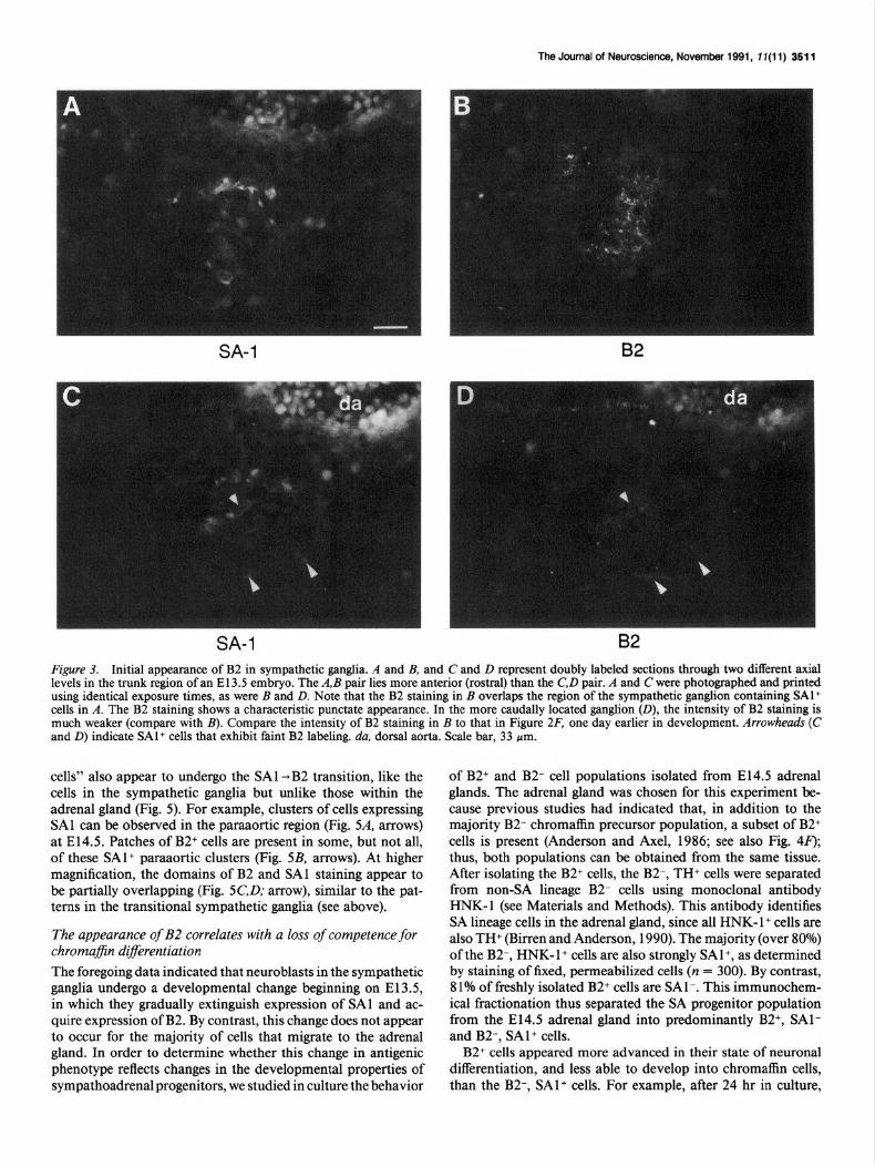
Expression of SAI and B2 in late gestational emb yos The preceding observations revealed a good correlation between the position of SA lineage cells in the embryo and their antigenic phenotype, around E 14.5-l 5.5. Cells that migrate to the adrenal gland maintain expression of SA 1 and do not induce B2, whereas cells that migrate to the sympathetic ganglia extinguish expres- sion of SAl and induce B2. At later stages of gestation, however (ca. E18.5), some violations of this “rule” were observed. For example, scattered patches of B2+ cells can be observed deep within the adrenal medulla (Fig. 8B). Conversely, patches of SAl+ cells can be observed outside of the adrenal microenvi- ronment, in the EAGC (Fig. 8C). The EAGC also contains patches of brightly stained B2+ cells (Fig. 80, arrow). However, these patches exhibit a complementary, nonoverlapping distri- bution relative to the patches of SA 1 + cells (Fig. 8D, arrowheads; compare to 8c). Thus, the mutual exclusivity of high B2 and high SAl labeling persists, despite the fact that some cells of each phenotype appear in the “inappropriate” location.
The SAl+ and B2+ patches can also be distinguished by their relative levels of TH expression: SAl+ patches are intensely stained with anti-TH antibody (Fig. 8E,F’), whereas B2+ patches are more weakly stained (not shown). B2+ patches, conversely, stain more intensely than SA 1 + patches with neuronal markers such as SCGlO and NF68 (not shown). This pattern of marker expression supports the idea that the B2+ patches contain cells with a more neuronal phenotype than the SAl+ patches.
A
80
80
NO ADD DEX
100
81
80
80
0 B2+ BZ- B2+ B2-
NO ADD DEX
Figure 6. B2+ cells show a reduced capacity for chromaffin differen- tiation compared to B2- cells. B2* and B2- (HNK- 1’) cells were isolated from E14.5 adrenal glands (see Materials and Methods) and cultured with (DEm or without (NO ADD) 1 KM DEX. A, After 24 hr in culture, the population was scored for the percentage of cell clusters bearing processes (see Materials and Methods). Only the TH+ cell population was counted, contaminating flat cells were not included. Note that in DEX. onlv 13% of B2- clusters had nrocesses. whereas 61% of B2+ clusters had processes. The method of analysis probably underestimates the difference between B2+ and B2- cells (see Materials and Methods). B, After 3 d in culture, the population was fixed and stained with an anti-PNMT antibody, and the percentage of surviving cells expressing PNMT was determined. Time course experiments indicate that most cells that can express PNMT have done so by 3 d of culture in DEX (Michelsohn and Anderson, unpublished observations). Data are the mean + SEM of four to six determinations from two independent ex- periments.
rived from cells initially bearing short processes.) These data indicate a strong correlation between the initial morphology of B2+ cells and their response to glucocorticoid: cells with short processes or a round morphology are likely to survive and dif- ferentiate into chromaffin cells, whereas process-bearing cells are likely to continue to extend neurites and then die. The re- sponse of SA precursors to glucocorticoid can therefore be pre- dicted based upon a combination of cell morphology and an- tigenic phenotype.
Discussion Evidence for a bipotential SA progenitor in vivo Neural crest cells in the ventrolateral migration pathway make a late developmental decision between the expression of an endocrine or a neuronal phenotype. In vitro studies (Doupe et al., 1985b, Anderson and Axel, 1986; Seidl and Unsicker, 1989b, Carnahan and Patterson, 1991b) have provided evidence that this decision is made by a progenitor cell whose choice of cell fate is determined, at least in part, by environmental factors. However, it has been difficult to verify this lineage relationship in vivo, because of the difficulties involved in tracing the fates of migratory cells in the mammalian embryo. In this study, we report the observation that at least some cells in embryonic sympathetic ganglia transiently coexpress neural-specific and chromaffin-specific markers. Cells that remain in the ganglion primordia lose expression of the chromaffin markers (SAl-5) and retain expression of the neuronal markers. Conversely, cells that migrate to the adrenal primordium lose expression of the neuronal markers (Anderson and Axel, 1986; Vogel and Weston, 1990) and retain expression of SAl-5. These data are consistent with the idea that the transiently dual-phenotype cells are bi- potential progenitors of chromaffin cells and sympathetic neu- rons, which selectively repress expression of the neuronal or chromaffin markers according to their final choice of cell fate.
An alternative explanation is that the disappearance of SAl immunoreactivity from the ganglion primordium at El 4.5 is due to the death or emigration of SA 1 + cells. This seems unlikely, because initially the SA 1 + cells are also TH+ and SCG 1 O+, and cells with a TH+, SCG 1 O+ phenotype remain in the ganglia even as SAl disappears. Moreover, we observe the concomitant ap- pearance of a second marker, B2, in cells that are still weakly SAl+ (see below). Thus, the cells that are initially SAl+ appear to remain in the ganglion primordium and to change their an- tigenic phenotype, rather than to disappear. Furthermore, the

The Journal of Neuroscience, November 1991, 17(11) 3515
isolation of SAl+ cells from El 3.5 sympathetic ganglia has re- vealed that 90% of these cells initially express neurofilament 140 kDa and that these cells have the capacity to develop either into chromaffin cells or sympathetic neurons, depending upon culture conditions (Carnahan and Patterson, 199 lb). Taken to- gether, these data indicate that the SA 1 +, SCG 10’ cells observed in embryonic sympathetic ganglia are likely to be precursors of sympathetic neurons in vivo.
The phenomenon of transient marker coexpression followed by mutually exclusive segregation is reminiscent of several other cell lineages, wherein bi- or multipotential progenitors have been shown to give rise to closely related but distinct cell types. In the immune system, for example, CD4+ and CD8+ peripheral T-lymphocytes have been shown to derive from a progenitor that is initially double positive for both of these cell-surface markers (Carbone et al., 1988; Fowlkes et al., 1988). In the pituitary gland, somatotrophs expressing growth hormone and lactotrophs expressing prolactin are thought to develop from precursors that transiently coexpress both hormones (Hoeffler et al., 1985). Finally, in the endocrine pancreas, embryonic islet progenitor cells have been shown to initially coexpress peptides, such as insulin and glucagon, which later segregate to different subpopulations of mature endocrine cells (Alpert et al., 1988).
This phenomenon could reflect the fact that the coexpressed genes are regulated, in part, by transcription factors that are common to all cells of a given lineage (Bodner et al., 1988; Ingraham et al., 1988). The later restriction of these markers to different subtypes of cells within the lineage could reflect the superposition of additional regulatory mechanisms, which fine- tune the pattern of gene expression. In the SA lineage, for ex- ample, the expression of neuronal markers such as SCG 10 can be upregulated by fibroblast growth factor (FGF) and NGF (fac- tors which promote neuronal differentiation) and downregulated by glucocorticoid (which promotes chromaffin differentiation) (Stein et al., 1988a). Conversely, chromaffin-abundant markers such as TH (Fig. 8EJJ are upregulated by glucocorticoid and downregulated by NGF (Leonard et al., 1987; Stein et al., 1988a). In this way, a repertoire of lineage-specific gene expression es- tablished in progenitor cells by a common transcriptional pro- gram may be fine-tuned by differences in local environment, generating further phenotypic diversification.
Developing sympathetic neuroblasts undergo a switch in antigenic phenotype Our antibody markers revealed a relatively late developmental event in the differentiation of sympathetic ganglionic neuro- blasts: as expression of the SAl antigen declines, the cells induce the expression of a surface antigen recognized by monoclonal antibody B2. At intermediate times, both SAl and B2 are ex- pressed in the same ganglia, and the pattern of expression of these two markers appears coextensive. Eventually, SAl ex- pression becomes undetectable and the cells in the ganglia uni- formly express high levels of B2. This pattern strongly suggests that the SA 1 -+ B2 transition reflects a change in marker expres-
Figure 7. B2+ cells bearing processes fail to respond to glucocorticoid. Individual B2+ cells were identified 24 hr after plating in 1 PM DEX and photographed every 24 hr for the next 2 d. Numbers indicate hours of culture. Arrows indicate an example of a pair of process-bearing cells that failed to respond to DEX. Note at 48 hr the phase-bright pycnotic figures near the remaining two phase-gray living cells, suggestive of cell
division followed by cell death. At 72 hr, only a single pycnotic figure is visible (arrow). The arrowheadindicates an example of anon-process- hearing cell (or cell cluster) that survived and displayed a rounded chromaffin morphology. Antibody staining indicated that cells of this phenotype were PNMT+ (not shown). Note that process-bearing cells were also observed to die in cultures lacking DEX (not shown), indi- cating that the death of these cells is not caused by glucocorticoid. The extent of cell death is greater in the low-density cultures used in these single-cell tracking experiments than in the mass cultures used in pop- ulation experiments; therefore, the results of the two types of experi- ments are not quantitatively comparable.
3518 Anderson et al. l Identification of a Sympathoadrenal Progenitor in viva
SA-1 B2
SA-1 B2
SA-1 TH
Figure 8. Patches of B2+ and SAl+ cells that violate the positional “rule.” Shown are sections from an E18.5 embryo. A and B, Doubly labeled pair through the adrenal gland. Note the large patch of B2+ cells (B), which is SAl- (A). In A, the SAl staining is obscured by a high level of autofluorescence but is clearly visible as punctate labeling at the margin of the B2+ zone. C and D, Doubly labeled pair through the extraadrenal ganglionic region. Note the intensely stained patch of B2+ cells (arrow, D), which is SAl- (m-row, C). Arrowheads in D indicate two less-intensely stained B2+ patches, which flank a SAl+ patch (C). E and F, Doubly labeled pair through an EAGC, illustrating adjacent patches of SAl+, THh’ cells, and SAl-, Thl” cells. The SAl- patches would be B2+ (as, e.g., in C and D, arrowheads). Scale bar, 62 pm.
sion within a single population of cells, rather than the replace- ment of one population by another. This interpretation is re- inforced by the complementary patterns of SA 1 and B2 staining seen within individual transitional ganglia: regions that are in- tensely SAl+ are weakly B2+, and vice versa. Such nonhomo- geneous staining further suggests that the SA l-+ B2 switch occurs asynchronously for cells within a given ganglion.
The SAl -B2 transition provides an indication that the cells of the developing sympathetic ganglia are changing, in a way that is not otherwise detectable in vivo. In what way are the cells
changing? The fact that expression of SAl precedes that of B2 suggests that cells expressing the former antigen are at an earlier stage of neuronal differentiation than cells expressing the latter. Consistent with this idea, cells isolated by sorting with a cocktail of SA antibodies have a rounded morphology (Carnahan and Patterson, 199 1 b), whereas cells isolated by sorting with B2 tend to extend processes shortly after plating (Anderson, 1988). Moreover, most B2-, SAl+ cells can be induced to differentiate into chromaffin cells at high frequency by glucocorticoid (see also Camahan and Patterson, 199 1 b). By contrast, process-bear-
ing B2+ cells are refractory to such an induction. As demon- strated previously (Anderson and Axel, 1986), a minority of B2+ cells remain responsive to glucocorticoid, but most of these cells initially exhibit a rounded morphology. The developmental het- erogeneity within the adrenal B2+ population is consistent with the fact that 19% of freshly isolated B2+ cells express SAl. This could reflect the gradual nature of the SAl -B2 transition, as suggested by the overlapping expression of SAl and B2 in sec- tions of El45 sympathetic ganglia. Nevertheless, cells with a B2+, SAl- antigenic phenotype and a process-bearing mor- phology have lost competence to respond to glucocorticoid and appear committed to neuronal differentiation (Fig. 9). The loss of responsiveness to glucocorticoid may serve to “insulate” im- mature sympathetic neurons from the steep rise in circulating fetal corticosterone levels, which begins on El65 (Teitelman et al., 1982). Loss of competence is thought to be a feature of inductive processes in other embryonic systems (Gurdon, 1987; Grainger and Gurdon, 1989).
Control of the SAl -B2 transition
Cells that arrest migration in the sympathetic ganglia extinguish SAl and induce B2, whereas those that migrate into the adrenal primordium mostly fail to make this switch. Instead, chromaffin cells maintain expression of SAl into adulthood. This obser- vation suggests that the SAl -B2 switch may be controlled by factors in the microenvironments encountered by SA progeni- tors. SA antigen-positive progenitors from sympathetic ganglia reduce levels of SAl expression in the absence of DEX but maintain expression in the presence of DEX (Carnahan and Patterson, 199 1 b). Moreover, chromaffin cells from neonatal rats lose SA staining in the presence of NGF and absence of glucocorticoid (Carnahan and Patterson, 199 la). These in vitro results suggest that in vivo, progenitors within the adrenal pri- mordium may be prevented from undergoing the SA 1 - B2 tran- sition by the high local concentration of adrenal corticosteroids. The SAl -B2 transition may also be promoted by factors that stimulate neuronal differentiation, such as FGF and NGF (Stemple et al., 1988; Birren and Anderson, 1990; Carnahan and Patterson, 199 1 b).
The maintenance of SAl and failure to induce B2 are first apparent in the adrenal gland as early as E14.5 (Fig. 4), sug- gesting that glucocorticoids and their receptor are functional at this early stage. mRNA encoding the glucocorticoid receptor has been detected in the adrenal gland as early as E 15.5 (An- derson and Michelsohn, 1989). However, using a radioligand binding assay, Seidl and Unsicker (1989b) have reported that glucocorticoid receptor is undetectable in adrenal medullary precursors prior to E16.5. The reason for this apparent discrep- ancy is not clear. Chromaffin precursors may contain a low level of receptor that is sufficient to mediate the inhibition of neuronal differentiation; alternatively, the SAl -B2 transition may be suppressed in the early adrenal gland by factors other than ste- roid hormones. However, recent radioimmunoassay data in- dicate that the E 14.5 adrenal gland contains micromolar quan- tities of glucocorticoid (A. Michelsohn and D. J. Anderson, unpublished observations). Whatever the case, the SAl and B2 markers provide evidence that progenitors in the adrenal gland are phenotypically distinct from their ganglionic counterparts 2 d before they express PNMT, the only other marker previously able to distinguish chromaffin cells from sympathetic neurons (Bohn et al., 198 1; Teitelman et al., 1982).
The correlation between the antigenic phenotype of SA pro-
The Journal of Neuroscience, November 1991, 1 I(11) 3517
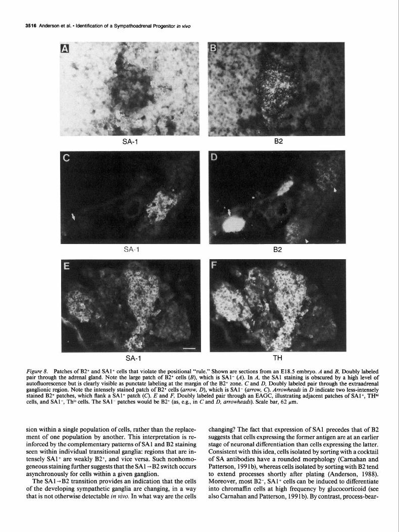
Bipotential sympathoaclrenal progenitor
\
glucocorticoid
committed neuroblast
I
SA-I+
NGF+? Adrenal chromaffin cell
Figure 9. Schematic diagram illustrating sequence of antigenic changes in the SA lineage. The earliest bipotential progenitors are SAl+ and B2-. They coexpress neuron-specific markers such as SCGl 0 and chro- maffin-specific markers such as SAl-5, as illustrated by the crosshatch- ing. In this study, progenitor cells were isolated from E14.5 adrenal glands using monoclonal antibody HNK-1 in combination with B2- mediated complement lysis. Most but not all of these cells are also SAl +; those cells lacking SA 1 may have initiated neuronal differentiation. In the absence of glucocorticoids, these progenitors differentiate along the neuronal pathway, a process promoted by FGF and perhaps other, as yet unidentified factors. Cells that are B2+ and SAl- and that bear processes have lost competence to respond to glucocorticoid (committed neuroblust). For clarity, intermediate stages of development between the bipotential progenitor and committed neuroblast are omitted.
genitors and their location in the embryo is not perfect: occa- sional clusters of B2+ cells can be found within the adrenal gland, and clusters of SAl+ cells are found outside ofit, late in gestation. The B2+ clusters probably correspond to clusters of Neuropeptide Y+ cells observed by Henion and Landis (1990). Similar clusters of cells with a neuronal phenotype have been found within the human fetal adrenal gland as well (Cooper et al., 1990). Our in vitro results provide a possible explanation for this phenomenon: these B2+ neuronal cells derive from pre- cursors that have lost competence to respond to glucocorticoid, either prior to or shortly after migration to the adrenal pri- mordium. The majority of these cells disappear by the first postnatal week, probably being eliminated by cell death (Henion and Landis, 1989).
By the same token, at E18.5 some SAl+ cells can be found outside of the adrenal environment. Since the SAl -B2 switch appears to occur slowly and asynchronously, these cells may

3518 Anderson et al. * Identification of a Sympathoadrenal Progenitor in viva
have failed to induce B2 by E 16.5 and then have been prevented from doing so subsequently by the surge in fetal GC levels that occurs on E16.5-17.5 (Teitelman et al., 1982). The decision to retain the SAl antigen may, moreover, become stabilized with further chromaffin development. Interestingly, the SAl+ cells in the paraaortic and extraadrenal region are found in clusters, as are the B2+ cells. This suggests that once cells decide whether or not to make the SAl -B2 switch, they may reinforce their immediate neighbors to do likewise. Such a “community effect” has been demonstrated for the process of mesodermal induction in Amphibia (Gurdon, 1988). Alternatively, such clusters could reflect an aggregation (Henion and Landis, 1990) or clonal ex- pansion of phenotypically similar cells.
Using a series of antibody markers, we have defined a se- quence of events during the development of the SA lineage in the rat embryo (Fig. 9). Bipotential SA progenitors initially coex- press both chromaffin-specific and neuron-specific markers and then differentiate into neurons or endocrine cells depending upon the environment to which they migrate. Subsequently, addi- tional changes in marker expression are detected: cells in the sympathetic ganglia switch from an SAl+ to a B2+ phenotype, whereas this switch fails to occur for the majority of cells within the adrenal gland. Studies of isolated SA progenitors in vitro indicate that this switch correlates with a loss of competence to respond to glucocorticoids, an inducing signal for chromaffin differentiation. Taken together, these data indicate that the de- velopment of sympathetic neurons and adrenal chromaffin cells is controlled not only by environmental signals, but also by changes in the ability of cells to respond to such signals. Such changes illustrate one way in which the developmental capacities of initially multipotent cells can become restricted during dif- ferentiation.
References Abo T, Balch CM (198 1) A differentiation antigen of human NK and
K cells identified by a monoclonal antibody (HNK-1). J Immunol 127:1024-1029.
Aloe L, Levi-Montalcini R (1979) Nerve growth factor-induced trans- formation of immature chromaffin cells in vivo into sympathetic neu- rons: effects of antiserum to nerve growth factor. Proc Nat1 Acad Sci USA 76:1246-1250.
Aluert S. Hanahan D. Teitelman G ( 1988) Hvbrid insulin genes reveal a developmental lineage for pancreatic’endocrine cells and imply a relationship with neurons. Cell 53:295-308.
Anderson DJ (1988) Cell fate and gene expression in the developing neural crest. In: Neural development and regeneration, NATO AS1 series, Series H, Vol 22 (Gorio A, ed), pp 188-198. Berlin: Springer.
Anderson DJ, Axe1 R (1985) Molecular probes for the development and plasticity of neural crest derivatives. Cell 42:649-662.
Anderson DJ, Axe1 R (1986) A bipotential neuroendocrine precursor whose choice of cell fate is determined by NGF and glucocorticoids. Cell 47:1079-1090.
Anderson DJ, Michelsohn A (1989) Role of glucocorticoids in the chromaffin-neuron developmental decision. Int J Dev Neurosci 12: 83-94.
Birren SJ, Anderson DJ (1990) A v-myc-immortalized sympathoad- renal progenitor cell line in which neuronal differentiation is initiated by FGF but not NGF. Neuron 4: 189-20 1.
Bodner M, Castrillo J-L, Theill LE, Deerinck T, Ellisman M, Karin M (1988) The pituitary-specific transcription factor GHF- 1 is a homeo- box-containing protein. Cell 55:505-5 18.
Bohn MC, Goldstein M, Black IB (198 1) Role of glucocorticoids in expression of the adrenergic phenotype in rat embryonic adrenal gland. Dev Biol 82:1-10.
Carbone AM, Marrack P, Kappler JW (1988) Demethylated CD8 gene in CD4+ T cells suggests that CD4+ cells develop from CDS+ pre- cursors. Science 242: 1174-l 176.
Camahan JF, Patterson PH (1991a) The generation of monoclonal antibodies that bind preferentially to adrenal chromaffin cells and the cells of embryonic sympathetic ganglia. J Neurosci 11:3493-3506.
Camahan JF, Patterson PH (199 1 b) Isolation of the progenitor cells of the sympathoadrenal lineage from embryonic sympathetic ganglia with the SA monoclonal antibodies. J Neurosci 11:3520-3530.
Cochard P, Paulin D (1984) Initial expression of neurofilaments and vimentin in the central and peripheral nervous system of the mouse embrvo in viva J Neurosci 4:2080-2094.
Cochari P, Goldstein M, Black IB (1979) Initial development of the noradrenergic phenotype in autonomic neuroblasts of the rat embryo in viva Dev Biol 7 1: 100-l 14.
Cooper MJ, Hutchins GM, Cohen PS, Helman LJ, Mennie RJ, Israel MA (1990) Human neuroblastoma tumor cell lines correspond to the arrested differentiation of chromaffin adrenal medullary neuro- blasts. Cell Growth Differ 1: 149-159.
Doupe AJ, Landis SC, Patterson PH (1985a) Environmental influences in the development ofneural crest derivatives: glucocorticoids, growth factors, and chromaffin cell plasticity. J Neurosci 5:2 119-2 142.
Doupe AJ, Patterson PH, Landis SC (1985b) Small intensely fluores- cent (SIF) cells in culture: role of glucocorticoids and growth factors in their development and phenotypic interconversions with other neural crest derivatives. J Neurosci 5:2 143-2 160.
Fowlkes BJ, Schwartz PH, Pardoll DM (1988) Deletion of self-reactive thymocytes occurs at a CD4+ CD8+ precursor stage. Nature 334: 620-623.
Grainger RM, Gurdon JB (1989) Loss of competence in amphibian induction can take place in single nondividing cells. Proc Nat1 Acad Sci USA 86: 1900-l 904.
Gurdon JB (1987) Embryonic induction-molecular prospects. De- velopment 99:285-306.
Gurdon JB (1988) A community effect in animal development. Nature 336~772-774.
Henion PD, Landis SC (1989) Evidence for death of committed neu- ronal precursors in the developing adrenal gland. Sot Neurosci Abstr 15:884.
Henion PD, Landis SC (1990) Asynchronous appearance and topo- graphic segregation of neuropeptide-containing cells in the developing rat adrenal medulla. J Neurosci 10:2886-2896.
Hoeffler JP, Boockfor RR, Frawley S (1985) Ontogeny of prolactin cells in neonatal rats: initial prolactin secretors also release growth hormone. Endocrinology 117: 187-l 95.
Ingraham HA, Chen R, Mangalam HJ, Elsholtz HP, Flynn SE, Lin CR, Simmons DM, Swanson L, Rosenfeld MG (1988) A tissue-specific transcription factor containing a homeodomain specifies a pituitary nhenotvne. Cell 55:5 19-529.
GmpinenM (1964) Extra-adrenal chromaffin tissue of the rat and the effect of cortical hormones on it. Acta Phvsiol Stand ~SUDD~~ 23 1: 1-9.
Le Douarin NM (1982) The neural crest. Cambridge:-Cambridge UP. Leonard DGB, Ziff EB, Greene LA (1987) Identification and char-
acterization of mRNAs regulated by nerve growth factor in PC12 cells. Mol Cell Biol 7:3 156-3 157.
Mirsky R (1982) The use of antibodies to define and study major cell types in the central and peripheral nervous system. In: Neuroim- munology (Brockes JP, ed), pp 14 l-l 8 1. New York: Plenum.
Reichardt LF (1984) Immunological approaches to the nervous sys- tem. Science 225:1294-1299.
Seidl K, Unsicker K (1989a) Survival and neuritic growth of sym- pathoadrenal (chromaffin) precursor cells in vitro. Int J Dev Neurosci 7~465-473.
Seidl K, Unsicker K (1989b) The determination of the adrenal med- ullary cell fate during embryogenesis. Dev Biol 136:48 l-490.
Stein R, Orit S, Anderson DJ (1988a) The induction of a neural- specific gene, SCGlO, by nerve growth factor in PC12 cells is tran- scriptional, protein synthesis dependent, and glucocorticoid inhibit- able. Dev Biol 127:3 16-325.
Stein R, Mori N, Matthews K, Lo L-C, Anderson DJ (1988b) The NGF-inducible SCGlO mRNA encodes a novel membrane-bound protein present in growth cones and abundant in developing neurons. Neuron 1~463-476.
Stemple DL, Mahanthappa NK, Anderson DJ (I 988) Basic FGF in- duces neuronal differentiation, cell division, and NGF dependence in chromaffin cells: a sequence of events in sympathetic development. Neuron 1:517-525.
Teitelman G, Joh TH, Park D, Brodsky M, New M, Reis DJ (1982)

The Journal of Neuroscience, November 1991, 7 I(1 1) 3519
Expression of the adrenergic phenotype in cultured fetal adrenal med- cells: impairment by glucocorticoids. Proc Nat1 Acad Sci USA 75: ullary cells: role of intrinsic and extrinsic factors. Dev Biol 80:450- 3498-3502. 459. Vogel KS, Weston JA (1990) The sympathoadrenal lineage in avian
Unsicker K, Drisch B, Otten J, Thoenen H (1978) Nerve growth embryos. I. Adrenal chromaffin cells lose neuronal traits during em- factor-induced fiber outgrowth from isolated rat adrenal chromaffin bryogenesis. Dev Biol 139: l-1 2.