Embed Size (px)
Citation preview
Antibody-Mediated Protection against PlasmodiumSporozoites Begins at the Dermal Inoculation Site
Yevel Flores-Garcia,a Gibran Nasir,a Christine S. Hopp,a* Christian Munoz,a* Amanda E. Balaban,a Fidel Zavala,a Photini Sinnisa
aDepartment of Molecular Microbiology & Immunology, Johns Hopkins Bloomberg School of Public Health,Baltimore, Maryland, USA
ABSTRACT Plasmodium sporozoites are injected into the skin as mosquitoes probefor blood. From here, they migrate through the dermis to find blood vessels whichthey enter in order to be rapidly carried to the liver, where they invade hepatocytesand develop into the next life cycle stage, the exoerythrocytic stage. Once sporozo-ites enter the blood circulation, they are found in hepatocytes within minutes. Incontrast, sporozoite exit from the inoculation site resembles a slow trickle and oc-curs over several hours. Thus, sporozoites spend the majority of their extracellulartime at the inoculation site, raising the hypothesis that this is when the malarial par-asite is most vulnerable to antibody-mediated destruction. Here, we investigate thishypothesis and demonstrate that the neutralizing capacity of circulating antibodiesis greater at the inoculation site than in the blood circulation. Furthermore, theseantibodies are working, at least in part, by impacting sporozoite motility at the inoc-ulation site. Using actively and passively immunized mice, we found that most para-sites are either immobilized at the site of injection or display reduced motility, par-ticularly in their net displacement. We also found that antibodies severely impair theentry of sporozoites into the bloodstream. Overall, our data suggest that antibodiestargeting the migratory sporozoite exert a large proportion of their protective effectat the inoculation site.
IMPORTANCE Studies in experimental animal models and humans have shown thatantibodies against Plasmodium sporozoites abolish parasite infectivity and providesterile immunity. While it is well documented that these antibodies can be inducedafter immunization with attenuated parasites or subunit vaccines, the mechanismsby and location in which they neutralize parasites have not been fully elucidated.Here, we report studies indicating that these antibodies display a significant portionof their protective effect in the skin after injection of sporozoites and that onemechanism by which they work is by impairing sporozoite motility, thus diminishingtheir ability to reach blood vessels. These results suggest that immune protectionagainst malaria begins at the earliest stages of parasite infection and emphasize theneed of performing parasite challenge in the skin for the evaluation of protectiveimmunity.
KEYWORDS antibodies, malaria, preerythrocytic, skin, sporozoites, vaccine
Malaria remains one of the most important infectious diseases in the world, causingsignificant morbidity and mortality, particularly in resource-poor settings. Plas-
modium parasites, the causative agents of malaria, cycle between mosquito andmammalian hosts. In the mammalian host, infection has two distinct phases, anasymptomatic preerythrocytic stage when parasite numbers are low, and a symptom-atic erythrocytic stage responsible for all clinical symptoms of the disease. Efforts togenerate a malaria vaccine have focused on both of these stages, with vaccinecandidates targeting sporozoites demonstrating some promise (1). Though short-lived
Received 5 October 2018 Accepted 8October 2018 Published 20 November 2018
Citation Flores-Garcia Y, Nasir G, Hopp CS,Munoz C, Balaban AE, Zavala F, Sinnis P. 2018.Antibody-mediated protection againstPlasmodium sporozoites begins at the dermalinoculation site. mBio 9:e02194-18. https://doi.org/10.1128/mBio.02194-18.
Editor Patricia J. Johnson, University ofCalifornia Los Angeles
Copyright © 2018 Flores-Garcia et al. This is anopen-access article distributed under the termsof the Creative Commons Attribution 4.0International license.
Address correspondence to Fidel Zavala,[email protected], or Photini Sinnis,[email protected].
* Present address: Christine S. Hopp, Laboratoryof Immunogenetics, National Institute ofAllergy and Infectious Diseases, NationalInstitutes of Health, Rockville, Maryland, USA;Christian Munoz, Medical TechnologyDepartment, Faculty of Health Sciences,University of Antofagasta, Antofagasta, Chile.
Y.F.-G. and G.N. contributed equally to thisarticle.
This article is a direct contribution from aFellow of the American Academy ofMicrobiology. Solicited external reviewers:Carole Long, NIAID/NIH; Johanna Daily, AlbertEinstein College of Medicine.
RESEARCH ARTICLEHost-Microbe Biology
crossm
November/December 2018 Volume 9 Issue 6 e02194-18 ® mbio.asm.org 1
on June 22, 2020 by guesthttp://m
bio.asm.org/
Dow
nloaded from
in the mammalian host, their low numbers and extracellular residence time likely makethem more susceptible than other life cycle stages to the effect of antibodies. Indeed,the protection observed in human vaccine recipients closely correlates with antibodytiters against sporozoites (2–4).
Sporozoites are the infective stage of the malarial parasite and must make aremarkable journey from the site at which they are deposited by infected mosquitoesto the liver, where they invade hepatocytes and transform into the next life cycle stage.This is a bottleneck for the parasite, with 10 to 100 sporozoites being inoculated (5) andonly a fraction ultimately making it to the liver and developing to mature liver-stageparasites (6, 7). The barriers faced by sporozoites are only beginning to be appreciated,with the first hurdle being exit from the inoculation site. Several lines of evidencedemonstrate that sporozoites are deposited into the skin and not directly into theblood circulation, including direct visualization of the process by intravital imaging(5–9). After their inoculation, sporozoites actively move in the skin to find and penetrateblood vessels to enter the blood circulation and be transported to the liver (6, 7). Likeall apicomplexan parasites, sporozoites move by a substrate-based motility calledgliding motility, powered by an actin-myosin motor beneath the plasma membrane(10). Plasmodium sporozoites are faster and move for longer periods of time than otherPlasmodium life cycle stages, suggesting that their fast robust motility may haveevolved for exit from the inoculation site. Indeed, this notion is supported by thephenotype of two motility mutants, a thrombospondin-related anonymous protein(TRAP) mutant that moves more slowly (11) and a deletion mutant of TRAP-like protein(TLP [12]). Both mutants are significantly more attenuated in their ability to causeinfection, after inoculation into the skin, thus highlighting the role of sporozoitemotility in exit from the inoculation site.
Investigation into the kinetics with which sporozoites exit the inoculation siterevealed that although some sporozoites leave within minutes, many take 30 to 120min to exit (13). These data from experiments with rodent malaria parasites aresupported by studies in humans and monkeys. In monkeys, transplantation of thedermal bite site 2 h after the bites of Plasmodium cynomolgi-infected mosquitoesresulted in infection in naive recipients (14). Additional experiments in humans thatwere fed upon by Plasmodium vivax- and Plasmodium falciparum-infected mosquitoesshowed that blood removed from these subjects 1 h post-mosquito bite could initiatemalaria infection in naive recipients (15). In contrast to the time it takes for sporozoitesto transit from the dermis to the bloodstream, once in the blood circulation, sporozo-ites are arrested in the liver and enter hepatocytes within minutes (13, 16, 17). Thus, forsporozoites, the inoculation site is where the parasite is extracellular for the longestperiod of time and therefore likely to be most vulnerable to antibody-mediatedneutralization.
An efficacious malaria vaccine would significantly contribute to the control andpossibly elimination of malaria. Early studies showed that immunization of birds andmice with radiation-attenuated sporozoites conferred protection, and this model hasserved as the gold standard for preerythrocytic-stage vaccine candidates (18, 19).Follow-up studies demonstrated that antibodies targeting the major surface protein ofsporozoites, the circumsporozoite protein (CSP), and T cells specific for infected hepa-tocytes were the basis of this protection (20, 21). These studies led to the developmentof a CSP-based subunit vaccine candidate called RTS,S. Phase III clinical trials of RTS,Sdemonstrated 40% to 50% efficacy in preventing clinical disease for 1 year, withprotection waning significantly at later time points (2). Though there is need forimprovement, this is a milestone for the malaria vaccine field and validates thesporozoite and its major surface proteins as targets. Follow-up studies of protected andunprotected children participating in this trial demonstrated that antibody titers cor-relate with protection (3, 4). We believe that the low sporozoite inoculum together withthe length of time the parasite is extracellular at the inoculation site make the migratorysporozoite the most vulnerable Plasmodium life cycle stage in the mammalian host. Inthis study, we use the rodent malaria model to investigate the effect of circulating
Flores-Garcia et al. ®
November/December 2018 Volume 9 Issue 6 e02194-18 mbio.asm.org 2
on June 22, 2020 by guesthttp://m
bio.asm.org/
Dow
nloaded from
anti-sporozoite antibodies on sporozoite motility and infectivity at the dermal inocu-lation site.
RESULTSSporozoites have impaired movement in the skin of immunized mice. With the
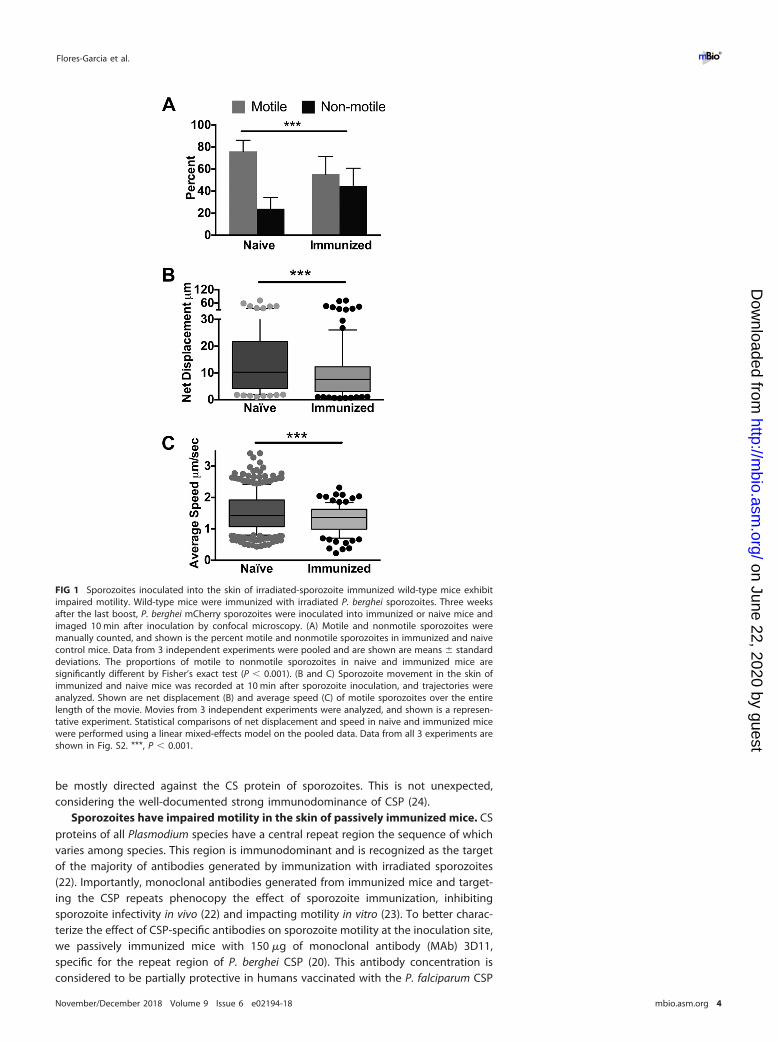
knowledge that sporozoites spend some time at the inoculation site and must bemotile to find and enter blood vessels, we used the rodent model to investigate theimpact of immunization on sporozoite motility at the inoculation site. We immunizedmice with radiation-attenuated Plasmodium berghei sporozoites, and 3 weeks after thelast immunization, when mice are protected from challenge with live sporozoites (seeFig. S1 in the supplemental material), we performed intravital imaging of P. bergheisporozoites expressing mCherry in immunized and age-matched naive mice. Sporozo-ites were inoculated into the ear pinna of mice, and their motility was visualized in5-min movies, beginning 10 min post-inoculation. We found that sporozoites inocu-lated into the skin of immunized mice display significantly altered motility compared tosporozoites inoculated into the skin of naive mice (Fig. 1A to C). Significantly highernumbers of sporozoites were not moving in immunized mice at 10 min after inocula-tion (Fig. 1A). With those sporozoites that were motile, we quantified their displace-ment and speed in immunized and naive mice. Net displacement, defined as thedistance along a straight line between the initial and final positions of motile sporo-zoites over the duration of a 5-min movie, was significantly reduced in sporozoites inimmunized mice (Fig. 1B). Quantification of the average speed of each sporozoite overthe course of 5-min movies showed that motile sporozoites in immunized mice alsohad a reduced average speed (Fig. 1C). Interestingly, the reductions in sporozoitedisplacement and speed in immunized mice were largely in the upper quartiles; thedistances and speeds reached by the upper quartiles of moving sporozoites weresignificantly lower in immunized mice. (A representative experiment is shown in Fig. 1Band C, and results from all 3 biological replicates are shown in Fig. S2). Overall, thesedata suggested that immune responses generated by immunization with radiation-attenuated sporozoites impact parasite motility at the inoculation site.
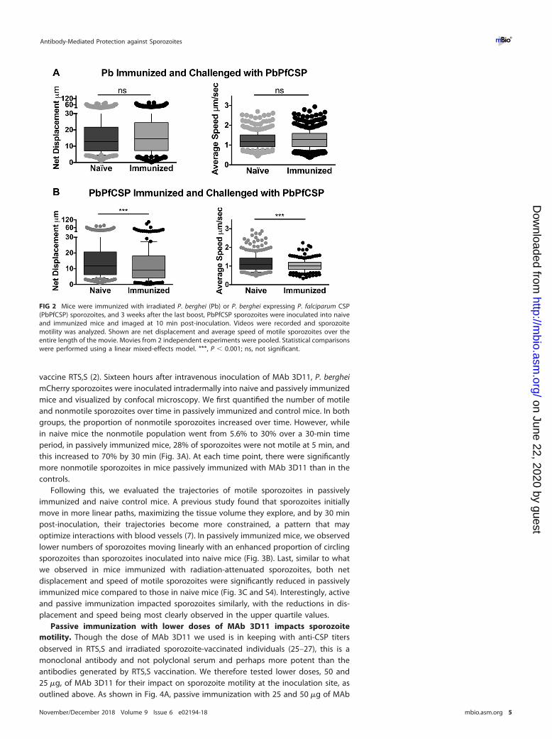
We hypothesized that the inhibitory effect on sporozoite motility in immunized micewas due to antibodies targeting the major surface protein of sporozoites, the circum-sporozoite protein (CSP). Indeed, previous studies have shown that antibodies to CSPare a prominent component of the immune response observed after immunizationwith irradiated sporozoites (22). Furthermore, antibodies specific for CSP can conferprotection and have been shown to immobilize sporozoites in vitro (20, 23). Todetermine if the antibody response that impacted sporozoite motility after immuniza-tion with irradiated sporozoites was specific to CSP, we generated a fluorescent P.berghei parasite in which the endogenous csp gene was replaced by the csp codingsequence from the human malaria parasite Plasmodium falciparum (PbPfCSP; Fig. S3).Importantly, antibodies to P. falciparum CSP do not cross-react with P. berghei CSP, andvice versa (Fig. S3). Transfections were performed in the P. berghei mCherry line usedfor the intravital imaging experiments, as this line does not contain a selection cassetteand can therefore be used to generate fluorescent mutant or transgenic lines (7).Transgenic PbPfCSP parasites develop normally in the mosquito and display normalinfectivity in mice (Fig. S3). To look at the role of CSP-specific antibodies on sporozoitemotility in the skin, mice were immunized with P. berghei sporozoites, and followingimmunization, the motility of PbPfCSP sporozoites in immunized and naive mice wasassessed by confocal microscopy. In contrast to the significant differences in netdisplacement and average speed of wild-type P. berghei sporozoites (Fig. 1B and C), wefound no significant difference in either parameter when PbPfCSP mCherry parasiteswere inoculated into P. berghei-immunized mice (Fig. 2, top). In contrast, when micewere immunized with PbPfCSP parasites, net displacement and speed of PbPfCSPsporozoites were significantly decreased (Fig. 2, bottom). Taken together, sporozoiteimmunization impacts the motility of sporozoites at the inoculation site and appears to
Antibody-Mediated Protection against Sporozoites ®
November/December 2018 Volume 9 Issue 6 e02194-18 mbio.asm.org 3
on June 22, 2020 by guesthttp://m
bio.asm.org/
Dow
nloaded from
be mostly directed against the CS protein of sporozoites. This is not unexpected,considering the well-documented strong immunodominance of CSP (24).
Sporozoites have impaired motility in the skin of passively immunized mice. CSproteins of all Plasmodium species have a central repeat region the sequence of whichvaries among species. This region is immunodominant and is recognized as the targetof the majority of antibodies generated by immunization with irradiated sporozoites(22). Importantly, monoclonal antibodies generated from immunized mice and target-ing the CSP repeats phenocopy the effect of sporozoite immunization, inhibitingsporozoite infectivity in vivo (22) and impacting motility in vitro (23). To better charac-terize the effect of CSP-specific antibodies on sporozoite motility at the inoculation site,we passively immunized mice with 150 �g of monoclonal antibody (MAb) 3D11,specific for the repeat region of P. berghei CSP (20). This antibody concentration isconsidered to be partially protective in humans vaccinated with the P. falciparum CSP
FIG 1 Sporozoites inoculated into the skin of irradiated-sporozoite immunized wild-type mice exhibitimpaired motility. Wild-type mice were immunized with irradiated P. berghei sporozoites. Three weeksafter the last boost, P. berghei mCherry sporozoites were inoculated into immunized or naive mice andimaged 10 min after inoculation by confocal microscopy. (A) Motile and nonmotile sporozoites weremanually counted, and shown is the percent motile and nonmotile sporozoites in immunized and naivecontrol mice. Data from 3 independent experiments were pooled and are shown are means � standarddeviations. The proportions of motile to nonmotile sporozoites in naive and immunized mice aresignificantly different by Fisher’s exact test (P � 0.001). (B and C) Sporozoite movement in the skin ofimmunized and naive mice was recorded at 10 min after sporozoite inoculation, and trajectories wereanalyzed. Shown are net displacement (B) and average speed (C) of motile sporozoites over the entirelength of the movie. Movies from 3 independent experiments were analyzed, and shown is a represen-tative experiment. Statistical comparisons of net displacement and speed in naive and immunized micewere performed using a linear mixed-effects model on the pooled data. Data from all 3 experiments areshown in Fig. S2. ***, P � 0.001.
Flores-Garcia et al. ®
November/December 2018 Volume 9 Issue 6 e02194-18 mbio.asm.org 4
on June 22, 2020 by guesthttp://m
bio.asm.org/
Dow
nloaded from
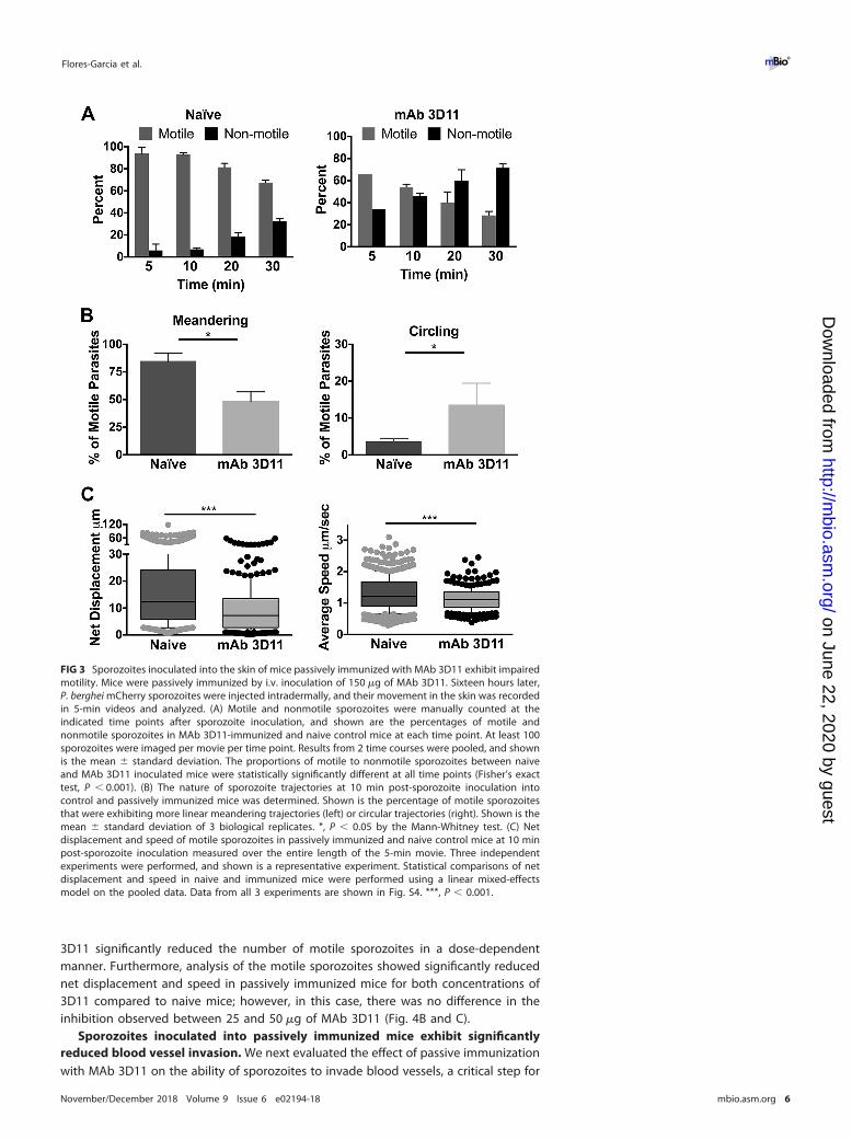
vaccine RTS,S (2). Sixteen hours after intravenous inoculation of MAb 3D11, P. bergheimCherry sporozoites were inoculated intradermally into naive and passively immunizedmice and visualized by confocal microscopy. We first quantified the number of motileand nonmotile sporozoites over time in passively immunized and control mice. In bothgroups, the proportion of nonmotile sporozoites increased over time. However, whilein naive mice the nonmotile population went from 5.6% to 30% over a 30-min timeperiod, in passively immunized mice, 28% of sporozoites were not motile at 5 min, andthis increased to 70% by 30 min (Fig. 3A). At each time point, there were significantlymore nonmotile sporozoites in mice passively immunized with MAb 3D11 than in thecontrols.
Following this, we evaluated the trajectories of motile sporozoites in passivelyimmunized and naive control mice. A previous study found that sporozoites initiallymove in more linear paths, maximizing the tissue volume they explore, and by 30 minpost-inoculation, their trajectories become more constrained, a pattern that mayoptimize interactions with blood vessels (7). In passively immunized mice, we observedlower numbers of sporozoites moving linearly with an enhanced proportion of circlingsporozoites than sporozoites inoculated into naive mice (Fig. 3B). Last, similar to whatwe observed in mice immunized with radiation-attenuated sporozoites, both netdisplacement and speed of motile sporozoites were significantly reduced in passivelyimmunized mice compared to those in naive mice (Fig. 3C and S4). Interestingly, activeand passive immunization impacted sporozoites similarly, with the reductions in dis-placement and speed being most clearly observed in the upper quartile values.
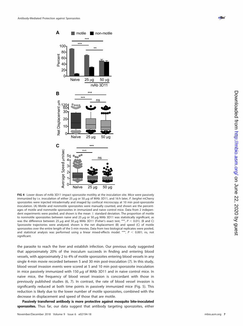
Passive immunization with lower doses of MAb 3D11 impacts sporozoitemotility. Though the dose of MAb 3D11 we used is in keeping with anti-CSP titersobserved in RTS,S and irradiated sporozoite-vaccinated individuals (25–27), this is amonoclonal antibody and not polyclonal serum and perhaps more potent than theantibodies generated by RTS,S vaccination. We therefore tested lower doses, 50 and25 �g, of MAb 3D11 for their impact on sporozoite motility at the inoculation site, asoutlined above. As shown in Fig. 4A, passive immunization with 25 and 50 �g of MAb
FIG 2 Mice were immunized with irradiated P. berghei (Pb) or P. berghei expressing P. falciparum CSP(PbPfCSP) sporozoites, and 3 weeks after the last boost, PbPfCSP sporozoites were inoculated into naiveand immunized mice and imaged at 10 min post-inoculation. Videos were recorded and sporozoitemotility was analyzed. Shown are net displacement and average speed of motile sporozoites over theentire length of the movie. Movies from 2 independent experiments were pooled. Statistical comparisonswere performed using a linear mixed-effects model. ***, P � 0.001; ns, not significant.
Antibody-Mediated Protection against Sporozoites ®
November/December 2018 Volume 9 Issue 6 e02194-18 mbio.asm.org 5
on June 22, 2020 by guesthttp://m
bio.asm.org/
Dow
nloaded from
3D11 significantly reduced the number of motile sporozoites in a dose-dependentmanner. Furthermore, analysis of the motile sporozoites showed significantly reducednet displacement and speed in passively immunized mice for both concentrations of3D11 compared to naive mice; however, in this case, there was no difference in theinhibition observed between 25 and 50 �g of MAb 3D11 (Fig. 4B and C).
Sporozoites inoculated into passively immunized mice exhibit significantlyreduced blood vessel invasion. We next evaluated the effect of passive immunizationwith MAb 3D11 on the ability of sporozoites to invade blood vessels, a critical step for
FIG 3 Sporozoites inoculated into the skin of mice passively immunized with MAb 3D11 exhibit impairedmotility. Mice were passively immunized by i.v. inoculation of 150 �g of MAb 3D11. Sixteen hours later,P. berghei mCherry sporozoites were injected intradermally, and their movement in the skin was recordedin 5-min videos and analyzed. (A) Motile and nonmotile sporozoites were manually counted at theindicated time points after sporozoite inoculation, and shown are the percentages of motile andnonmotile sporozoites in MAb 3D11-immunized and naive control mice at each time point. At least 100sporozoites were imaged per movie per time point. Results from 2 time courses were pooled, and shownis the mean � standard deviation. The proportions of motile to nonmotile sporozoites between naiveand MAb 3D11 inoculated mice were statistically significantly different at all time points (Fisher’s exacttest, P � 0.001). (B) The nature of sporozoite trajectories at 10 min post-sporozoite inoculation intocontrol and passively immunized mice was determined. Shown is the percentage of motile sporozoitesthat were exhibiting more linear meandering trajectories (left) or circular trajectories (right). Shown is themean � standard deviation of 3 biological replicates. *, P � 0.05 by the Mann-Whitney test. (C) Netdisplacement and speed of motile sporozoites in passively immunized and naive control mice at 10 minpost-sporozoite inoculation measured over the entire length of the 5-min movie. Three independentexperiments were performed, and shown is a representative experiment. Statistical comparisons of netdisplacement and speed in naive and immunized mice were performed using a linear mixed-effectsmodel on the pooled data. Data from all 3 experiments are shown in Fig. S4. ***, P � 0.001.
Flores-Garcia et al. ®
November/December 2018 Volume 9 Issue 6 e02194-18 mbio.asm.org 6
on June 22, 2020 by guesthttp://m
bio.asm.org/
Dow
nloaded from
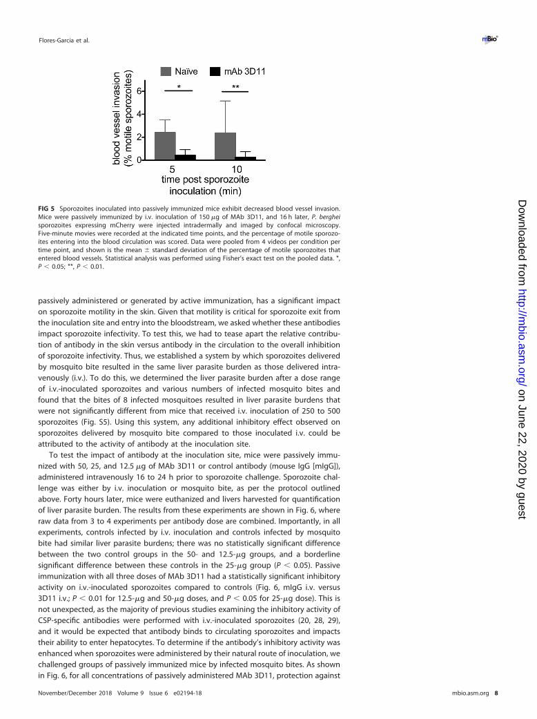
the parasite to reach the liver and establish infection. Our previous study suggestedthat approximately 20% of the inoculum succeeds in finding and entering bloodvessels, with approximately 2 to 4% of motile sporozoites entering blood vessels in anysingle 4-min movie recorded between 5 and 30 min post-inoculation (7). In this study,blood vessel invasion events were scored at 5 and 10 min post-sporozoite inoculationin mice passively immunized with 150 �g of MAb 3D11 and in naive control mice. Innaive mice, the frequency of blood vessel invasion is concordant with those inpreviously published studies (6, 7). In contrast, the rate of blood vessel invasion issignificantly reduced at both time points in passively immunized mice (Fig. 5). Thisreduction is likely due to the lower number of motile sporozoites, combined with thedecrease in displacement and speed of those that are motile.
Passively transferred antibody is more protective against mosquito bite-inoculatedsporozoites. Thus far, our data suggest that antibody targeting sporozoites, either
FIG 4 Lower doses of mAb 3D11 impact sporozoite motility at the inoculation site. Mice were passivelyimmunized by i.v. inoculation of either 25 �g or 50 �g of MAb 3D11, and 16 h later, P. berghei mCherrysporozoites were injected intradermally and imaged by confocal microscopy at 10 min post-sporozoiteinoculation. (A) Motile and nonmotile sporozoites were manually counted, and shown are the percent-ages of motile and nonmotile sporozoites in immunized and naive control mice. Data from 2 indepen-dent experiments were pooled, and shown is the mean � standard deviation. The proportion of motileto nonmotile sporozoites between naive and 25 �g or 50 �g MAb 3D11 was statistically significant, aswas the difference between 25 �g and 50 �g MAb 3D11 (Fisher’s exact test, ***, P � 0.01). (B and C)Sporozoite trajectories were analyzed; shown is the net displacement (B) and speed (C) of motilesporozoites over the entire length of the 5-min movies. Data from two biological replicates were pooled,and statistical analysis was performed using a linear mixed-effects model. ***, P � 0.001; ns, notsignificant.
Antibody-Mediated Protection against Sporozoites ®
November/December 2018 Volume 9 Issue 6 e02194-18 mbio.asm.org 7
on June 22, 2020 by guesthttp://m
bio.asm.org/
Dow
nloaded from
passively administered or generated by active immunization, has a significant impacton sporozoite motility in the skin. Given that motility is critical for sporozoite exit fromthe inoculation site and entry into the bloodstream, we asked whether these antibodiesimpact sporozoite infectivity. To test this, we had to tease apart the relative contribu-tion of antibody in the skin versus antibody in the circulation to the overall inhibitionof sporozoite infectivity. Thus, we established a system by which sporozoites deliveredby mosquito bite resulted in the same liver parasite burden as those delivered intra-venously (i.v.). To do this, we determined the liver parasite burden after a dose rangeof i.v.-inoculated sporozoites and various numbers of infected mosquito bites andfound that the bites of 8 infected mosquitoes resulted in liver parasite burdens thatwere not significantly different from mice that received i.v. inoculation of 250 to 500sporozoites (Fig. S5). Using this system, any additional inhibitory effect observed onsporozoites delivered by mosquito bite compared to those inoculated i.v. could beattributed to the activity of antibody at the inoculation site.
To test the impact of antibody at the inoculation site, mice were passively immu-nized with 50, 25, and 12.5 �g of MAb 3D11 or control antibody (mouse IgG [mIgG]),administered intravenously 16 to 24 h prior to sporozoite challenge. Sporozoite chal-lenge was either by i.v. inoculation or mosquito bite, as per the protocol outlinedabove. Forty hours later, mice were euthanized and livers harvested for quantificationof liver parasite burden. The results from these experiments are shown in Fig. 6, whereraw data from 3 to 4 experiments per antibody dose are combined. Importantly, in allexperiments, controls infected by i.v. inoculation and controls infected by mosquitobite had similar liver parasite burdens; there was no statistically significant differencebetween the two control groups in the 50- and 12.5-�g groups, and a borderlinesignificant difference between these controls in the 25-�g group (P � 0.05). Passiveimmunization with all three doses of MAb 3D11 had a statistically significant inhibitoryactivity on i.v.-inoculated sporozoites compared to controls (Fig. 6, mIgG i.v. versus3D11 i.v.; P � 0.01 for 12.5-�g and 50-�g doses, and P � 0.05 for 25-�g dose). This isnot unexpected, as the majority of previous studies examining the inhibitory activity ofCSP-specific antibodies were performed with i.v.-inoculated sporozoites (20, 28, 29),and it would be expected that antibody binds to circulating sporozoites and impactstheir ability to enter hepatocytes. To determine if the antibody’s inhibitory activity wasenhanced when sporozoites were administered by their natural route of inoculation, wechallenged groups of passively immunized mice by infected mosquito bites. As shownin Fig. 6, for all concentrations of passively administered MAb 3D11, protection against
FIG 5 Sporozoites inoculated into passively immunized mice exhibit decreased blood vessel invasion.Mice were passively immunized by i.v. inoculation of 150 �g of MAb 3D11, and 16 h later, P. bergheisporozoites expressing mCherry were injected intradermally and imaged by confocal microscopy.Five-minute movies were recorded at the indicated time points, and the percentage of motile sporozo-ites entering into the blood circulation was scored. Data were pooled from 4 videos per condition pertime point, and shown is the mean � standard deviation of the percentage of motile sporozoites thatentered blood vessels. Statistical analysis was performed using Fisher’s exact test on the pooled data. *,P � 0.05; **, P � 0.01.
Flores-Garcia et al. ®
November/December 2018 Volume 9 Issue 6 e02194-18 mbio.asm.org 8
on June 22, 2020 by guesthttp://m
bio.asm.org/
Dow
nloaded from
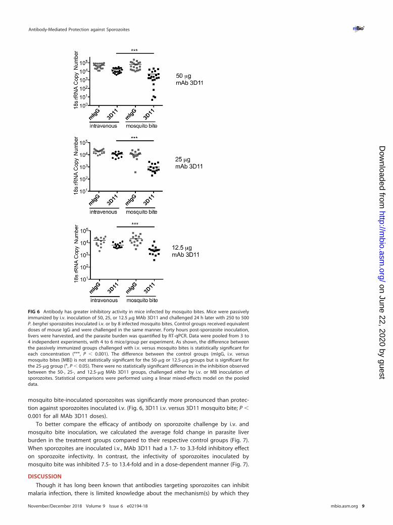
mosquito bite-inoculated sporozoites was significantly more pronounced than protec-tion against sporozoites inoculated i.v. (Fig. 6, 3D11 i.v. versus 3D11 mosquito bite; P �
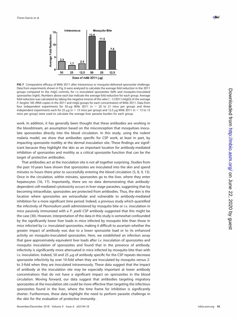
0.001 for all MAb 3D11 doses).To better compare the efficacy of antibody on sporozoite challenge by i.v. and
mosquito bite inoculation, we calculated the average fold change in parasite liverburden in the treatment groups compared to their respective control groups (Fig. 7).When sporozoites are inoculated i.v., MAb 3D11 had a 1.7- to 3.3-fold inhibitory effecton sporozoite infectivity. In contrast, the infectivity of sporozoites inoculated bymosquito bite was inhibited 7.5- to 13.4-fold and in a dose-dependent manner (Fig. 7).
DISCUSSION
Though it has long been known that antibodies targeting sporozoites can inhibitmalaria infection, there is limited knowledge about the mechanism(s) by which they
FIG 6 Antibody has greater inhibitory activity in mice infected by mosquito bites. Mice were passivelyimmunized by i.v. inoculation of 50, 25, or 12.5 �g MAb 3D11 and challenged 24 h later with 250 to 500P. berghei sporozoites inoculated i.v. or by 8 infected mosquito bites. Control groups received equivalentdoses of mouse IgG and were challenged in the same manner. Forty hours post-sporozoite inoculation,livers were harvested, and the parasite burden was quantified by RT-qPCR. Data were pooled from 3 to4 independent experiments, with 4 to 6 mice/group per experiment. As shown, the difference betweenthe passively immunized groups challenged with i.v. versus mosquito bites is statistically significant foreach concentration (***, P � 0.001). The difference between the control groups (mIgG, i.v. versusmosquito bites [MB]) is not statistically significant for the 50-�g or 12.5-�g groups but is significant forthe 25-�g group (*, P � 0.05). There were no statistically significant differences in the inhibition observedbetween the 50-, 25-, and 12.5-�g MAb 3D11 groups, challenged either by i.v. or MB inoculation ofsporozoites. Statistical comparisons were performed using a linear mixed-effects model on the pooleddata.
Antibody-Mediated Protection against Sporozoites ®
November/December 2018 Volume 9 Issue 6 e02194-18 mbio.asm.org 9
on June 22, 2020 by guesthttp://m
bio.asm.org/
Dow
nloaded from
work. In addition, it has generally been thought that these antibodies are working inthe bloodstream, an assumption based on the misconception that mosquitoes inocu-late sporozoites directly into the blood circulation. In this study, using the rodentmalaria model, we show that antibodies specific for CSP work, at least in part, byimpacting sporozoite motility at the dermal inoculation site. These findings are signif-icant because they highlight the skin as an important location for antibody-mediatedinhibition of sporozoites and motility as a critical sporozoite function that can be thetarget of protective antibodies.
That antibodies act at the inoculation site is not all together surprising. Studies fromthe past 10 years have shown that sporozoites are inoculated into the skin and spendminutes to hours there prior to successfully entering the blood circulation (5, 8, 9, 13).Once in the circulation, within minutes, sporozoites go to the liver, where they enterhepatocytes (16, 17). Importantly, there are no data demonstrating that antibody-dependent cell-mediated cytotoxicity occurs in liver-stage parasites, suggesting that bybecoming intracellular, sporozoites are protected from antibodies. Thus, the skin is thelocation where sporozoites are extracellular and vulnerable to antibody-mediatedinhibition for a more significant time period. Indeed, a previous study which quantifiedthe infectivity of Plasmodium yoelii administered by mosquito bite or i.v. inoculation inmice passively immunized with a P. yoelii CSP antibody suggested that this might bethe case (30). However, interpretation of the data in this study is somewhat confoundedby the significantly lower liver loads in mice infected by mosquito bite than those inmice infected by i.v. inoculated sporozoites, making it difficult to ascertain whether thegreater impact of antibody was due to a lower sporozoite load or to its enhancedactivity on mosquito-inoculated sporozoites. Here, we established an infection assaythat gave approximately equivalent liver loads after i.v. inoculation of sporozoites andmosquito inoculation of sporozoites and found that in the presence of antibody,infectivity is significantly more attenuated in mice infected by mosquito bite than withi.v. inoculation. Indeed, 50 and 25 �g of antibody specific for the CSP repeats decreasesporozoite infectivity by over 10-fold when they are inoculated by mosquito versus 2-to 3-fold when they are inoculated intravenously. These data suggest that the impactof antibody at the inoculation site may be especially important at lower antibodyconcentrations that do not have a significant impact on sporozoites in the bloodcirculation. Moving forward, our data suggest that antibodies targeting migratorysporozoites at the inoculation site could be more effective than targeting the infectioussporozoites found in the liver, where the time frame for inhibition is significantlyshorter. Furthermore, these data highlight the need to perform parasite challenge inthe skin for the evaluation of protective immunity.
FIG 7 Comparative efficacy of MAb 3D11 after intravenous or mosquito-delivered sporozoite challenge.Data from experiments shown in Fig. 6 were analyzed to calculate the average fold reduction in the 3D11groups compared to the mIgG controls, for i.v.-inoculated sporozoites (left) and mosquito-inoculatedsporozoites (right). Numbers above each bar indicate the average fold reduction for each group. Averagefold reduction was calculated by taking the negative inverse of the ratio [�1/(3D11:mIgG)] of the averageP. berghei 18S rRNA copies in the 3D11 and mIgG groups for each concentration of MAb 3D11. Data fromfour independent experiments for 50 �g MAb 3D11 (n � 20 to 21 mice per group) and threeindependent experiments each for 25 �g (n � 15 mice per group) and 12.5 �g MAb 3D11 (n � 13 to 15mice per group) were used to calculate the average liver parasite burden for each group.
Flores-Garcia et al. ®
November/December 2018 Volume 9 Issue 6 e02194-18 mbio.asm.org 10
on June 22, 2020 by guesthttp://m
bio.asm.org/
Dow
nloaded from
We also demonstrate that an important mechanism by which CSP repeat antibodiesact at the inoculation site is via their impact on sporozoite motility. We observed thatantibody specific for the CSP repeats decreases the percentage of parasites that aremotile, with the remaining motile parasites exhibiting reduced displacement andspeed. Interestingly, the greatest impact of antibody was on those sporozoites movingfarthest and fastest, suggesting that it may be these sporozoites that disproportionatelysucceed in entering blood vessels. Supporting this hypothesis, we did find significantinhibition of blood vessel invasion in passively immunized mice. These results suggestthat sporozoites with high displacements may be more likely to find and enter bloodvessels, consistent with previous data demonstrating that sporozoite dispersal earlyafter their inoculation maximizes the tissue volume they explore, which may beimportant in locating blood vessels (7). Though the precise relationship betweendecreases in displacement and inhibition of blood vessel entry to degree of inhibitionof sporozoite infectivity remains unclear, it would be an important area of investigationfor future study. Previous in vitro studies and one in vivo study using supraphysiologicconcentrations of antibody found that antibodies specific for the CSP repeats canimmobilize sporozoites (23, 31). Here, we show that at titers more in line with thosefound in immunized individuals, antibodies specific for the CSP repeats have moresubtle effects on motility, and these effects can be associated with a significant impacton infection. Importantly, these in vivo data could be used to establish parameters inthe rodent model for screening vaccine candidates targeting the migratory sporozoite.
Importantly, we cannot conclude that the impact of antibody on sporozoite motilityin the skin is the only mechanism by which antibody acts to inhibit sporozoite infection.As shown both in this study and many previous studies, when sporozoites are injectedintravenously in mice with circulating anti-sporozoite antibodies, there is also a signif-icant inhibition of parasite infection in the liver, and this suggests an effect of anti-bodies on the migration of sporozoites to the liver parenchyma, which also requiresparasite motility. Importantly, early studies with MAb 3D11 indicated that Fab mono-mers in vivo had a protective effect comparable to that observed with intact MAb 3D11IgG1 (28). This indicates that Fc functions are not needed for the protective activity ofthis antibody. Nevertheless, we cannot rule out that protection conferred by otherantibodies requires complement fixation, leading to sporozoite destruction via directlysis or phagocytosis. Additionally, it remains to be determined whether Fc-mediatedmechanisms found in vitro are relevant to protection observed in vivo. Our currentexperiments suggest that in vivo, a direct effect of antibody on sporozoite motility atthe inoculation site is one of the mechanisms by which antibody protects.
Our findings may be relevant to the malaria vaccine effort targeting preerythrocyticstages of Plasmodium species. To date, RTS,S, a subunit vaccine based on CSP, is theonly malaria vaccine candidate to show efficacy in phase III clinical trials, conferring50% protection in preventing malaria and 45% efficacy in preventing severe disease(32). Nonetheless, the efficacy of RTS,S wanes significantly after 1 year (33) and fallsshort of community-established benchmarks, indicating that more work is needed toproduce a fully efficacious vaccine. Follow-up studies on RTS,S vaccinees suggest thatantibody titers specific for the CSP repeat region correlate with protection (2–4). Ourdata raise the possibility that the partial efficacy of RTS,S is due to the impact ofantibody on sporozoite motility at the inoculation site. Further studies with sera fromprotected and unprotected RTS.S-immunized volunteers are needed to determine howprotective antibodies are functioning. In vivo studies in mice with P. berghei sporozoitesexpressing P. falciparum CSP could be employed to elucidate whether, as we observedin our studies, antibodies from RTS,S-immunized volunteers are functioning by impact-ing sporozoite motility at the inoculation site.
MATERIALS AND METHODSAnimals. Five- to 8-week-old female C57BL/6nTac and C57BL/6j mice were purchased from Taconic
Farms (Derwood, MD) and Charles River Laboratories (Frederick, MD), respectively, and housed in theanimal facility at the Johns Hopkins Bloomberg School of Public Health. C57BL/6j mice were used for allimaging studies, and C57BL/6nTac mice were used for all infectivity studies. For all experiments, mice in
Antibody-Mediated Protection against Sporozoites ®
November/December 2018 Volume 9 Issue 6 e02194-18 mbio.asm.org 11
on June 22, 2020 by guesthttp://m
bio.asm.org/
Dow
nloaded from
the control and experimental groups were age and sex matched. All animal work was performed inaccordance with the Institutional Animal Care and Use Committee guidelines (protocols M017H325 andM016H35).
Parasites and mosquitoes. Sporozoite infectivity studies were performed with wild-type P. bergheiANKA strain parasites. Imaging studies were performed with P. berghei ANKA strain parasites expressingmCherry under the uis4 promoter (PBANKA_0501200) previously described by Hopp et al. (7). For someof the imaging studies, P. berghei parasites expressing P. falciparum CSP were used. These weregenerated in the mCherry line outlined above, and the methodology for their generation and verificationcan be found below and in Fig. S1. Anopheles stephensi mosquitoes were reared in the insectary at theJohns Hopkins Bloomberg School of Public Health, infected with the indicated parasites, and used forexperiments between days 18 and 22 postinfectious blood meal.
Generation of a P. berghei mCherry parasite expressing P. falciparum CSP. P. berghei parasitesexpressing mCherry (7) were used to generate transgenic parasites expressing P. falciparum CSP.Transfections were performed as described by Janse et al. (34), using pR-CSPfFL containing the P.falciparum csp coding region with a P. berghei signal sequence, the hDHFR selection cassette, and csp 5=and 3= untranslated regions (UTRs) (35). pR-CSPfFL was cut with XhoI and KasI and transfected intoschizont cultures of mCherry P. berghei parasites by electroporation using an Amaxa Nucleofector.Following transfection, parasites were inoculated into Swiss Webster mice and selected with pyrimeth-amine. Parental populations of parasites were cloned by limiting dilution in mice, and clones wereverified by PCR and sequencing.
Confocal microscopy and cell tracking. Sporozoites were imaged in the ear pinna of mice, aspreviously described (6, 7). Mice were anesthetized by intraperitoneal injection of ketamine (35 to50 mg/kg body weight) and xylazine-hydrochloride (6 to 10 mg/kg body weight), and P. berghei mCherrysporozoites were injected into the ear at multiple sites in volumes of 0.5 to 2 �l, using a NanoFil 10-�lsyringe with an NF33BV-2 needle (World Precision Instruments). Mice were kept in a microscope chamberwarmed to 28°C, and their ears were fixed onto a cover glass and imaged with a 10� objective on aninverted Zeiss Axio Observer Z1 microscope with a Yokogawa CSU22 spinning disk. Ten minutesfollowing intradermal inoculation of sporozoites, 5-min videos were recorded, with each Z-stack con-sisting of 3 slices spanning a total depth of 30 to 50 �m, with a frame rate of 1,000 ms/frame, using anelectron-multiplying charge-coupled-device (EMCCD) camera (Photometrics, Tucson, AZ, USA) and the 3iSlideBook 5.0 software. Videos were analyzed using Fiji and the ICY 1.8.6.0 softwares, which are freelyavailable from https://fiji.sc and http://icy.bioimageanalysis.org (36), respectively. Speed and net dis-placement were determined by automated tracking of sporozoites from recorded videos, while thepercentage of total sporozoites that were motile and nonmotile and the number that exhibited circularor meandering motility were manually counted. Blood vessel invasion was also quantified manually andscored when a rapid change in sporozoite speed was observed as previously outlined (7). For all intravitalimaging experiments, each biological replicate was performed with two mice, one immunized and onenaive.
Immunization of mice. For immunization with irradiated sporozoites, 6- to 7-week-old femaleC57BL/6j mice were immunized intravenously with 10,000 �-attenuated sporozoites 3 times at 2-weekintervals with either wild-type P. berghei parasites or transgenic P. berghei parasites expressing full-lengthP. falciparum CSP. Mice immunized with this regimen were protected from sporozoite challenge. Threeweeks following the final immunization, mice were used for confocal microscopy to investigate sporo-zoite motility. For passive immunization experiments, the indicated concentration of MAb 3D11 (20), anIgG1 subclass, was inoculated intravenously 16 to 24 h prior to the inoculation of sporozoites.
Infectivity experiments. For each experiment, the same cage of P. berghei ANKA-infected Anophelesstephensi mosquitoes was used to infect mice by i.v. or mosquito bite inoculation. The prevalence andintensity of infection in the cage were determined on day 18 to 22 postinfectious blood meal bymicroscopically examining the salivary gland sporozoite load from each of 20 mosquitoes. Mosquitoeswere only used if the average sporozoite load per mosquito was greater than 10,000 sporozoites and thecage had a prevalence of infection of 90% or greater. For intravenous challenge, infected mosquitoeswere dissected in cold Leibovitz’s L-15 medium (catalog no. 11415064; Thermo Fisher Scientific), andsporozoites were counted using a hemocytometer. A working solution with 250 to 500 sporozoites per200 �l was prepared in RPMI 1640 and inoculated into mice through the tail vein. For mosquito bitechallenge, infected mosquitoes were anesthetized on ice and transferred to feeding tubes 1 day prior tochallenge. No more than four mosquitoes were placed in a single tube. Mosquitoes were sugar-starvedovernight but provided with water until �6 h prior to mosquito bite challenge. Female C57BL/6nTacmice were anesthetized with intraperitoneal injection of ketamine (35 to 50 mg/kg body weight) andxylazine-hydrochloride (6 to 10 mg/kg body weight) and placed on a warming plate set to 37°C. Eachmouse was exposed to 8 mosquito bites, 4 bites per ear. Visualization of the biting process ensured thateach mouse was probed upon by 8 mosquitoes, and if a mosquito did not probe, it was replaced byanother mosquito.
Quantification of parasite liver burden by RT-qPCR. Parasite liver burden was measured byquantitative reverse transcription-PCR (RT-qPCR), as outlined by Bruña-Romero et al. (37). Briefly, 40 hafter parasite challenge, mice were sacrificed, and their livers were harvested, washed twice in coldphosphate-buffered saline (pH 7.4), weighed, and homogenized in 10 ml of prechilled Tri-Reagent(Molecular Research Center, Inc., Cincinnati, OH) for 1 min at full speed using a homogenizer (Kinematica,Bohemia, NY). RNA was extracted from 1 ml of the liver homogenate by phenol-chloroform extraction,and the concentration was measured using a NanoDrop spectrophotometer (Thermo Fisher Scientific).RNA (1.5 �g) was reverse transcribed using random hexamers (Thermo Fisher Scientific) and the
Flores-Garcia et al. ®
November/December 2018 Volume 9 Issue 6 e02194-18 mbio.asm.org 12
on June 22, 2020 by guesthttp://m
bio.asm.org/
Dow
nloaded from
following cycling profile: 25°C (10 min), 42°C (20 min), 95°C (5 min), and 5°C (5 min). Following this,quantitative PCR (qPCR) was performed using primers specific for the P. berghei 18S rRNA gene, aspreviously described by Kumar et al. (38). Quantification of P. berghei 18S rRNA copy number was basedon a standard curve prepared using 10-fold serial dilutions (107 to 102) of the P. berghei 18S rRNA gene.
Statistical analysis. Statistical comparisons of motility-based parameters for Fig. 1, 4, S2, and S4 wereperformed using a linear mixed-effects model on the pooled data, accounting for experimental variationamong biological replicates as a random effect using STATA version 13.0 software. Since the distributionof sporozoite net displacement in both naive and immunized mice was right-skewed, the data weresquare root-transformed prior to analysis. Comparisons of motility type between immunized and naivegroups in Fig. 3B were performed using nonparametric Mann-Whitney tests (two-tailed, � � 0.05) withthe GraphPad Prism 6 software. Statistical analysis of the proportion of motile and nonmotile sporozoitesin naive and immunized mice (Fig. 1A, 3A, and 4A) was performed using Fisher’s exact test. Statisticalcomparisons of P. berghei 18S rRNA copy number between control and passively immunized mice inFig. 6 were performed using a linear multilevel mixed-effects model, accounting for experimentalvariation and interaction of treatment and route, using the STATA version 13.0 software.
SUPPLEMENTAL MATERIALSupplemental material for this article may be found at https://doi.org/10.1128/mBio
.02194-18.FIG S1, DOCX file, 0.03 MB.FIG S2, DOCX file, 0.1 MB.FIG S3, DOCX file, 7.2 MB.FIG S4, DOCX file, 0.2 MB.FIG S5, DOCX file, 0.04 MB.
ACKNOWLEDGMENTSWe thank Godfree Mlambo, Abhai Tripathi, and Chris Kizito, the team of the
parasitology and insectary core facilities at the Johns Hopkins Malaria Research Institutefor their outstanding work and Bloomberg Family Philanthropies for their support ofthese facilities. We acknowledge the Johns Hopkins School of Medicine MicroscopyFacility (MicFac) and thank Scott Kuo and Barbara Smith for their invaluable assistance.
This work was supported by the National Institutes of Health (grant R01 AI132359 toP.S.) and by a Johns Hopkins Malaria Research Institute fellowship (to C.S.H.).
REFERENCES1. Long CA, Zavala F. 2016. Malaria vaccines and human immune re-
sponses. Curr Opin Microbiol 32:96 –102. https://doi.org/10.1016/j.mib.2016.04.006.
2. White MT, Verity R, Griffin JT, Asante KP, Owusu-Agyei S, Greenwood B,Drakeley C, Gesase S, Lusingu J, Ansong D, Adjei S, Agbenyega T, OgutuB, Otieno L, Otieno W, Agnandji ST, Lell B, Kremsner P, Hoffman I,Martinson F, Kamthunzu P, Tinto H, Valea I, Sorgho H, Oneko M, OtienoK, Hamel MJ, Salim N, Mtoro A, Abdulla S, Aide P, Sacarlal J, Aponte JJ,Njuguna P, Marsh K, Bejon P, Riley EM, Ghani AC. 2015. Immunogenicityof the RTS,S/AS01 malaria vaccine and implications for duration ofvaccine efficacy: secondary analysis of data from a phase 3 randomisedcontrolled trial. Lancet Infect Dis 15:1450 –1458. https://doi.org/10.1016/S1473-3099(15)00239-X.
3. White MT, Bejon P, Olotu A, Griffin JT, Riley EM, Kester KE, OckenhouseCF, Ghani AC. 2013. The relationship between RTS,S vaccine-inducedantibodies, CD4� T cell responses and protection against Plasmodiumfalciparum infection. PLoS One 8:e61395. https://doi.org/10.1371/journal.pone.0061395.
4. Foquet L, Hermsen CC, van Gemert GJ, Van Braeckel E, Weening KE,Sauerwein R, Meuleman P, Leroux-Roels G. 2014. Vaccine-inducedmonoclonal antibodies targeting circumsporozoite protein prevent Plas-modium falciparum infection. J Clin Invest 124:140 –144. https://doi.org/10.1172/JCI70349.
5. Medica DL, Sinnis P. 2005. Quantitative dynamics of Plasmodium yoeliisporozoite transmission by infected anopheline mosquitoes. Infect Im-mun 73:4363– 4369. https://doi.org/10.1128/IAI.73.7.4363-4369.2005.
6. Amino R, Thiberge S, Martin B, Celli S, Shorte S, Frischknecht F,Menard R. 2006. Quantitative imaging of Plasmodium transmissionfrom mosquito to mammal. Nat Med 12:220 –224. https://doi.org/10.1038/nm1350.
7. Hopp CS, Chiou K, Ragheb DR, Salman A, Khan SM, Liu AJ, Sinnis P. 2015.
Longitudinal analysis of Plasmodium sporozoite motility in the dermisreveals component of blood vessel recognition. elife 4:e07789. https://doi.org/10.7554/eLife.07789.
8. Matsuoka H, Yoshida S, Hirai M, Ishii A. 2002. A rodent malaria, Plasmodiumberghei, is experimentally transmitted to mice by merely probing of infec-tive mosquito, Anopheles stephensi. Parasitol Int 51:17–23. https://doi.org/10.1016/S1383-5769(01)00095-2.
9. Vanderberg J, Frevert U. 2004. Intravital microscopy demonstratingantibody-mediated immobilisation of Plasmodium berghei sporozoitesinjected into skin by mosquitoes. Int J Parasitol 34:991–996. https://doi.org/10.1016/j.ijpara.2004.05.005.
10. Douglas RG, Amino R, Sinnis P, Frischknecht F. 2015. Active migrationand passive transport of malaria parasites. Trends Parasitol 31:357–362.https://doi.org/10.1016/j.pt.2015.04.010.
11. Ejigiri I, Ragheb DR, Pino P, Coppi A, Bennett BL, Soldati-Favre D,Sinnis P. 2012. Shedding of TRAP by a rhomboid protease from themalaria sporozoite surface is essential for gliding motility and sporo-zoite infectivity. PLoS Pathog 8:e1002725. https://doi.org/10.1371/journal.ppat.1002725.
12. Moreira CK, Templeton TJ, Lavazec C, Hayward RE, Hobbs CV, Kroeze H,Janse CJ, Waters AP, Sinnis P, Coppi A. 2008. The Plasmodium TRAP/MIC2family member, TRAP-like protein (TLP), is involved in tissue traversal bysporozoites. Cell Microbiol 10:1505–1516. https://doi.org/10.1111/j.1462-5822.2008.01143.x.
13. Yamauchi LM, Coppi A, Snounou G, Sinnis P. 2007. Plasmodium sporo-zoites trickle out of the injection site. Cell Microbiol 9:1215–1222. https://doi.org/10.1111/j.1462-5822.2006.00861.x.
14. Lloyd OC, Sommerville T. 1949. The fate of sporozoites of Plasmodiumcynomolgi injected into the skin of Rhesus monkeys. Proc Pathol Soc61:144 –146.
15. Fairley NH. 1947. Sidelights on malaria in man obtained by subinocula-
Antibody-Mediated Protection against Sporozoites ®
November/December 2018 Volume 9 Issue 6 e02194-18 mbio.asm.org 13
on June 22, 2020 by guesthttp://m
bio.asm.org/
Dow
nloaded from
tion experiments. Trans R Soc Trop Med Hyg 40:621– 676. https://doi.org/10.1016/0035-9203(47)90025-4.
16. Shin SCJ, Vanderberg JP, Terzakis JA. 1982. Direct infection of hepato-cytes by sporozoites of Plasmodium berghei. J Protozool 29:448 – 454.https://doi.org/10.1111/j.1550-7408.1982.tb05431.x.
17. Sinnis P, Willnow TE, Briones MRS, Herz J, Nussenzweig V. 1996. Remnantlipoproteins inhibit malaria sporozoite invasion of hepatocytes. J ExpMed 184:945–954. https://doi.org/10.1084/jem.184.3.945.
18. Nussenzweig RS, Vanderberg J, Most H, Orton C. 1967. Protective im-munity produced by the injection of x-irradiated sporozoites of Plasmo-dium berghei. Nature 216:160 –162. https://doi.org/10.1038/216160a0.
19. Mulligan HW, Russell PF, Mohan BN. 1941. Active immunization of fowlsagainst Plasmodium gallinaceum by injections of killed homologoussporozoites. J Mal Inst India 4:25–34.
20. Yoshida N, Nussenzweig RS, Potocnjak P, Nussenzweig V, Aikawa M.1980. Hybridoma produces protective antibodies directed against thesporozoite stage of malaria parasite. Science 207:71–73. https://doi.org/10.1126/science.6985745.
21. Romero P, Maryanski JL, Corradin G, Nussenzweig RS, Nussenzweig V,Zavala F. 1989. Cloned cytotoxic T cells recognize an epitope in thecircumsporozoite protein and protect against malaria. Nature 341:323–326. https://doi.org/10.1038/341323a0.
22. Zavala F, Cochrane AH, Nardin EH, Nussenzweig RS, Nussenzweig V.1983. Circumsporozoite proteins of malaria parasites contain a singleimmunodominant region with two or more identical epitopes. J ExpMed 157:1947–1957. https://doi.org/10.1084/jem.157.6.1947.
23. Stewart MJ, Nawrot RJ, Schulman S, Vanderberg JP. 1986. Plasmodiumberghei sporozoite invasion is blocked in vitro by sporozoite-immobilizing antibodies. Infect Immun 51:859 – 864.
24. Zavala F, Tam JP, Hollingdale MR, Cochrane AH, Quakyi I, NussenzweigRS, Nussenzweig V. 1985. Rationale for the development of a syntheticvaccine against Plasmodium falciparum malaria. Science 228:1436 –1440.https://doi.org/10.1126/science.2409595.
25. Kester KE, Cummings JF, Ofori-Anyinam O, Ockenhouse CF, Krzych U,Moris P, Schwenk R, Nielsen RA, Debebe Z, Pinelis E, Juompan L, WilliamsJ, Dowler M, Stewart VA, Wirtz RA, Dubois MC, Lievens M, Cohen J, BallouWR, Heppner DG, Jr, RTS,S Vaccine Evaluation Group. 2009. Randomized,double-blind, phase 2a trial of falciparum malaria vaccines RTS,S/AS01Band RTS,S/AS02A in malaria-naive adults: safety, efficacy, and immuno-logic associates of protection. J Infect Dis 200:337–346. https://doi.org/10.1086/600120.
26. Olotu A, Lusingu J, Leach A, Lievens M, Vekemans J, Msham S, Lang T,Gould J, Dubois MC, Jongert E, Vansadia P, Carter T, Njuguna P,Awuondo KO, Malabeja A, Abdul O, Gesase S, Mturi N, Drakeley CJ,Savarese B, Villafana T, Lapierre D, Ballou WR, Cohen J, Lemnge MM,Peshu N, Marsh K, Riley EM, von Seidlein L, Bejon P. 2011. Efficacy ofRTS,S/AS01E malaria vaccine and exploratory analysis on anti-circumsporozoite antibody titres and protection in children aged 5–17months in Kenya and Tanzania: a randomised controlled trial. LancetInfect Dis 11:102–109. https://doi.org/10.1016/S1473-3099(10)70262-0.
27. Moorthy VS, Ballou WR. 2009. Immunological mechanisms underlyingprotection mediated by RTS,S: a review of the available data. Malar J8:312. https://doi.org/10.1186/1475-2875-8-312.
28. Potocnjak P, Yoshida N, Nussenzweig RS, Nussenzweig V. 1980. Mon-ovalent fragments (Fab) of monoclonal antibodies to a sporozoite sur-face antigen (Pb44) protect mice against malarial infection. J Exp Med151:1504 –1513. https://doi.org/10.1084/jem.151.6.1504.
29. Zavala F, Tam JP, Barr PJ, Romero PJ, Ley V, Nussenzweig RS, Nussenz-weig V. 1987. Synthetic peptide vaccine confers protection againstmurine malaria. J Exp Med 166:1591–1596. https://doi.org/10.1084/jem.166.5.1591.
30. Sack BK, Miller JL, Vaughan AM, Douglass A, Kaushansky A, MikolajczakS, Coppi A, Gonzalez-Aseguinolaza G, Tsuji M, Zavala F, Sinnis P, KappeSH. 2014. Model for in vivo assessment of humoral protection againstmalaria sporozoite challenge by passive transfer of monoclonal antibod-ies and immune serum. Infect Immun 82:808 – 817. https://doi.org/10.1128/IAI.01249-13.
31. Vanderberg J, Nussenzweig R, Most H. 1969. Protective immunity pro-duced by the injection of x-irradiated sporozoites of Plasmodium ber-ghei. V. In vitro effects of immune serum on sporozoites. Mil Med134:1183–1190. https://doi.org/10.1093/milmed/134.9.1183.
32. RTS,S Clinical Trials Partnership. 2011. First results of phase 3 trial ofRTS,S/AS01 malaria vaccine in African children. N Engl J Med 365:1863–1875. https://doi.org/10.1056/NEJMoa1102287.
33. RTS,S Clinical Trials Partnership. 2015. Efficacy and safety of RTS,S/AS01malaria vaccine with or without a booster dose in infants and children inAfrica: final results of a phase 3, individually randomised, controlled trial.Lancet 386:31– 45. https://doi.org/10.1016/S0140-6736(15)60721-8.
34. Janse CJ, Franke-Fayard B, Waters AP. 2006. Selection by flow-sorting ofgenetically transformed, GFP-expressing blood stages of the rodentmalaria parasite, Plasmodium berghei. Nat Protoc 1:614 – 623. https://doi.org/10.1038/nprot.2006.88.
35. Espinosa DA, Christensen D, Muñoz C, Singh S, Locke E, Andersen P,Zavala F. 2017. Robust antibody and CD8� T-cell responses induced byP. falciparum CSP adsorbed to cationic liposomal adjuvant CAF09 confersterilizing immunity against experimental rodent malaria infection. NPJVaccines 2:10. https://doi.org/10.1038/s41541-017-0011-y.
36. de Chaumont F, Dallongeville S, Chenouard N, Hervé N, Pop S, ProvoostT, Meas-Yedid V, Pankajakshan P, Lecomte T, Le Montagner Y, LagacheT, Dufour A, Olivo-Marin JC. 2012. Icy: an open bioimage informaticsplatform for extended reproducible research. Nat Methods 9:690 – 696.https://doi.org/10.1038/nmeth.2075.
37. Bruña-Romero O, Hafalla JC, Gonzalez-Aseguinolaza G, Sano G, Tsuji M,Zavala F. 2001. Detection of malaria liver-stages in mice infectedthrough the bite of a single Anopheles mosquito using a highly sensitivereal-time PCR. Int J Parasitol 31:1499 –1502. https://doi.org/10.1016/S0020-7519(01)00265-X.
38. Kumar KA, Oliveira GA, Edelman R, Nardin E, Nussenzweig V. 2004.Quantitative Plasmodium sporozoite neutralization assay (TSNA). J Im-munol Methods 292:157–164.
Flores-Garcia et al. ®
November/December 2018 Volume 9 Issue 6 e02194-18 mbio.asm.org 14
on June 22, 2020 by guesthttp://m
bio.asm.org/
Dow
nloaded from