Embed Size (px)
Citation preview

Hindawi Publishing CorporationClinical and Developmental ImmunologyVolume 2008, Article ID 723539, 15 pagesdoi:10.1155/2008/723539
Review ArticleAntigen-Induced Immunomodulation inthe Pathogenesis of Atherosclerosis
Natalia Milioti,1 Alexandra Bermudez-Fajardo,1 Manuel L. Penichet,2, 3 and Ernesto Oviedo-Orta1
1 Faculty of Heath and Medical Sciences, University of Surrey, Guildford, Surrey GU2 7XH, UK2 Department of Surgery, David Geffen School of Medicine, University of California, Los Angeles, CA 90095-6904, USA3 Department of Microbiology, Immunology, and Molecular Genetics, David Geffen School of Medicine,University of California, Los Angeles, CA 90095 -1678, USA
Correspondence should be addressed to Ernesto Oviedo-Orta, [email protected]
Received 17 December 2007; Revised 2 April 2008; Accepted 30 April 2008
Recommended by Shyam Mohapatra
Atherosclerosis is a chronic inflammatory disorder characterised by the accumulation of monocytes/macrophages, smooth musclecells, and lymphocytes within the arterial wall in response to the release of proinflammatory molecules. Such accumulation resultsin the formation of the atherosclerotic plaque, which would eventually evolve to complications such as total artery occlusion,rupture, calcification, or aneurysm. Although the molecular mechanism responsible for the development of atherosclerosis isnot completely understood, it is clear that the immune system plays a key role in the development of the atherosclerotic plaqueand in its complications. There are multiple antigenic stimuli that have been associated with the pathogenesis of atherosclerosis.Most of these stimuli come from modified self-molecules such as oxidised low-density lipoproteins (oxLDLs), beta2glycoprotein1(β2GP1), lipoprotein a (LP(a)), heat shock proteins (HSPs), and protein components of the extracellular matrix such as collagenand fibrinogen in the form of advanced glycation-end (AGE) products. In addition, several foreign antigens including bacteriasuch as Porphyromonas gingivalis and Chlamydia pneumoniae and viruses such as enterovirus and cytomegalovirus have beenassociated with atherosclerosis as potentially causative or bystander participants, adding another level of complexity to the analysisof the pathophysiology of atherosclerosis. The present review summarises the most important scientific findings published withinthe last two decades on the importance of antigens, antigen stimulation, and adaptive immune responses in the development ofatherosclerotic plaques.
Copyright © 2008 Natalia Milioti et al. This is an open access article distributed under the Creative Commons Attribution License,which permits unrestricted use, distribution, and reproduction in any medium, provided the original work is properly cited.
1. ATHEROSCLEROTIC PLAQUE FORMATION
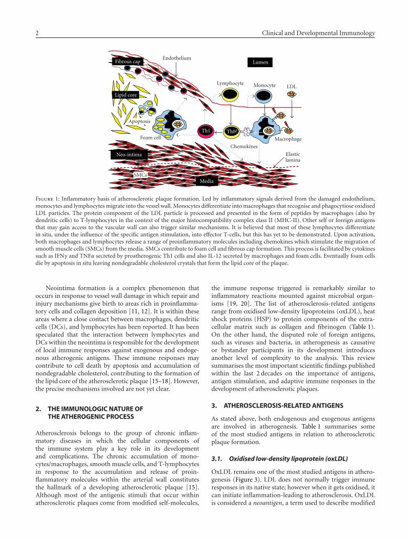
It is difficult to identify the factors responsible for theinitiation of the atheroma lesion and/or the order in whichthese factors contribute to plaque formation. Nevertheless,it is known that endothelial dysfunction and high levelsof circulating cholesterol, as oxLDL, play a key role in theproinflammatory process that triggers the first steps in thedevelopment of atherosclerotic plaques [1, 2]. Whateverthe cause, these steps are characterised by an initiallyreversible accumulation of lipid-laden macrophages in thesubendothelial space as a consequence of the increasingmigration of blood-derived monocytes. These cells accu-mulate at focal points within the vascular wall of mediumand small size arteries driven by chemokines and adhesionmolecules produced by the damaged endothelium [3–5].Monocytes differentiate in situ into macrophages which
express membrane receptors such as Toll-like receptorsand scavenger receptors that participate in the clearanceof oxLDL [6, 7]. Lymphocytes can also transmigrate andaccumulate within the arterial wall from the very earlieststages (Figure 1) [8].
As the inflammatory process becomes chronic, smoothmuscle cells also start to migrate from the media intothe intima layer of the vessel, in response to chemokinesand aided by the release of membrane metalloproteinases(MMPs) that enable them to break through the elastic laminainto the subendothelial space (Figure 1) [9, 10]. Persistenceof inflammation creates a vicious circle of cell migration,dedifferentiation of smooth muscle cells, production ofchemotactic and proinflammatory mediators, and cell deathleading to vascular wall remodelling and formation of a newlayer called neointima (Figures 1 and 2) [11, 12].
2 Clinical and Developmental Immunology
Fibrous cap
Lipid core
Apoptosis
Neo-intima
SMCs
Foam cell
Endothelium
Lymphocyte Monocyte LDL
Th1 Th0?
Chemokines
Media
Macrophage
Lumen
Elasticlamina
Figure 1: Inflammatory basis of atherosclerotic plaque formation. Led by inflammatory signals derived from the damaged endothelium,monocytes and lymphocytes migrate into the vessel wall. Monocytes differentiate into macrophages that recognise and phagocytiose oxidisedLDL particles. The protein component of the LDL particle is processed and presented in the form of peptides by macrophages (also bydendritic cells) to T-lymphocytes in the context of the major histocompatibility complex class II (MHC-II). Other self or foreign antigensthat may gain access to the vascular wall can also trigger similar mechanisms. It is believed that most of these lymphocytes differentiatein situ, under the influence of the specific antigen stimulation, into effector T-cells, but this has yet to be demonstrated. Upon activation,both macrophages and lymphocytes release a range of proinflammatory molecules including chemokines which stimulate the migration ofsmooth muscle cells (SMCs) from the media. SMCs contribute to foam cell and fibrous cap formation. This process is facilitated by cytokinessuch as IFNγ and TNFα secreted by proatherogenic Th1 cells and also IL-12 secreted by macrophages and foam cells. Eventually foam cellsdie by apoptosis in situ leaving nondegradable cholesterol crystals that form the lipid core of the plaque.
Neointima formation is a complex phenomenon thatoccurs in response to vessel wall damage in which repair andinjury mechanisms give birth to areas rich in proinflamma-tory cells and collagen deposition [11, 12]. It is within theseareas where a close contact between macrophages, dendriticcells (DCs), and lymphocytes has been reported. It has beenspeculated that the interaction between lymphocytes andDCs within the neointima is responsible for the developmentof local immune responses against exogenous and endoge-nous atherogenic antigens. These immune responses maycontribute to cell death by apoptosis and accumulation ofnondegradable cholesterol, contributing to the formation ofthe lipid core of the atherosclerotic plaque [15–18]. However,the precise mechanisms involved are not yet clear.
2. THE IMMUNOLOGIC NATURE OFTHE ATHEROGENIC PROCESS
Atherosclerosis belongs to the group of chronic inflam-matory diseases in which the cellular components ofthe immune system play a key role in its developmentand complications. The chronic accumulation of mono-cytes/macrophages, smooth muscle cells, and T-lymphocytesin response to the accumulation and release of proin-flammatory molecules within the arterial wall constitutesthe hallmark of a developing atherosclerotic plaque [15].Although most of the antigenic stimuli that occur withinatherosclerotic plaques come from modified self-molecules,
the immune response triggered is remarkably similar toinflammatory reactions mounted against microbial organ-isms [19, 20]. The list of atherosclerosis-related antigensrange from oxidised low-density lipoproteins (oxLDL), heatshock proteins (HSP) to protein components of the extra-cellular matrix such as collagen and fibrinogen (Table 1).On the other hand, the disputed role of foreign antigens,such as viruses and bacteria, in atherogenesis as causativeor bystander participants in its development introducesanother level of complexity to the analysis. This reviewsummarises the most important scientific findings publishedwithin the last 2 decades on the importance of antigens,antigen stimulation, and adaptive immune responses in thedevelopment of atherosclerotic plaques.
3. ATHEROSCLEROSIS-RELATED ANTIGENS
As stated above, both endogenous and exogenous antigensare involved in atherogenesis. Table 1 summarises someof the most studied antigens in relation to atheroscleroticplaque formation.
3.1. Oxidised low-density lipoprotein (oxLDL)
OxLDL remains one of the most studied antigens in athero-genesis (Figure 3). LDL does not normally trigger immuneresponses in its native state; however when it gets oxidised, itcan initiate inflammation-leading to atherosclerosis. OxLDLis considered a neoantigen, a term used to describe modified
Natalia Milioti et al. 3
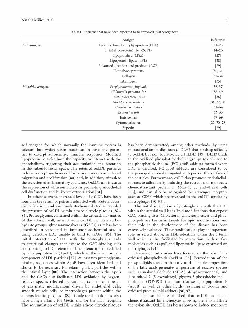
Table 1: Antigens that have been reported to be involved in atherogenesis.
Antigen Reference
Autoantigens Oxidised low-density lipoprotein (LDL) [21–23]
Beta2glycoprotein1 (beta2GP1) [24–26]
Lipoprotein a (LP(a)) [27]
Lipoprotein-lipase (LPL) [28]
Advanced glycation-end products (AGE) [29]
Heat-shock proteins [30, 31]
Collagen [32–34]
Fibrinogen [35]
Microbial antigens Porphyromonas gingivalis [36, 37]
Chlamydia pneumoniae [38–49]
Bacteroides forsynthus [36]
Streptococcus mutans [36, 37, 50]
Helicobacter pylori [51–64]
Echerichia coli [65, 66]
Enterovirus [67–69]
Cytomegalovirus [22, 70–78]
Viperin [79]
self-antigens for which normally the immune system istolerant but which upon modification have the poten-tial to excerpt autoreactive immune responses. Modifiedlipoprotein particles have the capacity to interact with theendothelium, triggering their accumulation and retentionin the subendothelial space. The retained oxLDL particlesinduce macrophage foam cell formation, smooth muscle cellmigration and proliferation [80] and, in addition, stimulatethe secretion of inflammatory cytokines. OxLDL also inducesthe expression of adhesion molecules promoting endothelialcell dysfunction and leukocyte extravasation [81].
In atherosclerosis, increased levels of oxLDL have beenfound in the serum of patients admitted with acute myocar-dial infarction, and immunohistochemical studies revealedthe presence of oxLDL within atherosclerotic plaques [82–85]. Proteoglycans, contained within the extracellular matrixof the arterial wall, interact with oxLDL via their carbo-hydrate groups, glycosamynoglycans (GAGs) as it has beendescribed in vitro and in immunohistochemical studiesusing defective LDL unable to bind to GAGs [86]. Theinitial interaction of LDL with the proteoglycans leadsto structural changes that expose the GAG-binding sitescontributing to LDL retention. This interaction is mediatedby apolipoprotein B (ApoB), which is the main proteincomponent of LDL particles [87]. At least two proteoglycan-binding sequences within ApoB have been identified andshown to be necessary for retaining LDL particles withinthe intimal layer [80]. The interaction between the ApoBand the GAGs also facilitates LDL oxidation by oxygen-reactive species released by vascular cells or as a resultof enzymatic modifications driven by endothelial cells,smooth muscle cells, or macrophages present within theatherosclerotic plaques [88]. Cholesterol molecules alsohave a high affinity for GAGs and for the LDL receptor.The accumulation of oxLDL within atherosclerotic plaques
has been demonstrated, among other methods, by usingmonoclonal antibodies such as DLH3 that binds specificallyto oxLDL but non to native LDL (nLDL) [89]. DLH3 bindsto the oxidised phosphatidylcholine groups (oxPC) and tothe phosphatidylcholine (PC)-apoB adducts formed whenLDL is oxidised. PC-apoB adducts are considered to bethe principal antibody targeted epitopes on the surface ofthe particles. Furthermore, oxPC also promote endothelial-monocyte adhesion by inducing the secretion of monocytechemoattractant protein 1 (MCP-1) by endothelial cells[23], and can also be recognised by scavenger receptorssuch as CD36 which are involved in the oxLDL uptake bymacrophages [90–93].
The initial interaction of proteoglycans with the LDLwithin the arterial wall leads lipid modifications that exposeGAG binding sites. Cholesterol, cholesteryl esters and phos-pholipids are the main targets for lipid modifications andtheir role in the development of the disease has beenextensively evaluated. These modifications play an importantrole, as stated above, in LDL retention within the arterialwall which is also facilitated by interactions with surfacemolecules such as apoE and lipoprotein lipase expressed onmacrophages [94].
However, most studies have focused on the role of theoxidised phospholipids (oxPLs) [95]. Peroxidation of thephospholipids starts in the fatty acids. The decompositionof the fatty acids generates a spectrum of reactive speciessuch as malondialdehyde (MDA), 4-hydroxynonenal, and1-palmitoyl-2-(5-oxovaleroyl)-glycero-3-phosphocholinemolecule (POVPC) that can oxidise apolipoprotein B(ApoB) as well as other lipids, resulting in ox-PLs andoxidised protein-lipid adducts [96, 97].
It has also been established that oxLDL acts as achemoattractant for monocytes allowing them to infiltratethe lesion site. OxLDL has been shown to induce monocyte

4 Clinical and Developmental Immunology
LC
FC
SR
M
L
(a)
LC
FC
SR
M
L
(b)
LC
FC
SR
ML
(c)
LC
FCSR
ML
(d)
Figure 2: Morphological features of advanced atheroscleroticplaques. (a) and (b) show sections from a human carotid artery; (c)and (d) are sections from an apoE deficient mouse brachiocephalicartery. Sections (a) and (c) have been stained with haematoxylinand eosin and sections (b) and (d) with van Gieson staining (usedto demonstrate the increase of collagen deposition and developmentof elastic fibres, a characteristic feature of the atherogenic process.A positive staining is depicted by a brown colour). L: lumen of thevessel; SR: shoulder region (it is believed to contain large numbersof proinflammatory cells including macrophages and lymphocytes,and it is the site related with the onset of the developmentof the atherosclerotic plaque); FC: fibrous cap (It also containslarge numbers of mononuclear infiltrate and smooth muscle cellsthat have migrated from the media layer (M) and proliferated inresponse to the local inflammatory stimuli. It is also characterisedby high collagen deposition and little or no endothelial cells); LC:lipid core (it contains mainly macrophage and smooth musclecell-derived foam cells, apoptotic cells, and cholesterol crystals.Older lesions may also display signs of calcification). Contrastingdifferences can be recognised in the anatomic development ofatherosclerotic plaques between human and mouse including thehypertrophy associated with the proliferation of the smooth musclecells in the media layer and the fibrous cap. In humans, some lesionsmay also contain signs of intraplaque haemorrhage. Signs of plaquerupture are usually best recognised in mouse (reviewed in [13, 14]).
adhesion to the endothelium by upregulating the expres-sion of adhesion molecules on their surface, by inducingmacrophage major histocompatibility class II (MHC-II) &LeuM3 cell surface expression and by accelerating monocytedifferentiation in to macrophages [22]. Endothelial cells arealso capable of oxidising LDL, contributing to a continuousgeneration of oxLDL within the lesion site, and to attract
apoB-100
apoB-100 apoB-100
apoB-100
Unesterifiedcholesterol
Triglycerides
Phospholipids
Cholesterolesters
Figure 3: Schematic representation of the low-density lipoproteinparticle (LDL). The LDL particle has a size of approximately 21–24 nm and is the main transporter of unesterified cholesterol,cholesterol esters, and triglycerides in the blood. It contains anouter layer composed of phospholipids and unesterified cholesterolin which a single protein is embedded, the apolipoprotein B-100(apoB-100). These components are more susceptible to oxidationby free radicals in the subendothelial space during inflammation.They are also targets for the recognition of the LDL by scavengerreceptors, proteoglycans, and low-density lipoprotein receptor(LDLr). The core of the particle contains primarily cholesterolesters and triglycerides. In atherogenesis, a large number of IgMantibodies are created in response to oxidative stress-modifiedphospholipids, whereas IgG antibodies and T-cell clones aregenerated against apoB-100.
more monocytes. Other inflammatory mediators such asinterleukin 1β (IL-1β), tumor necrosis factor alpha (TNF-α) and monocyte colony stimulating factor (M-CSF) caninduce the expression of the oxLDL receptor (oxLDL-R) onthe surface of endothelial cells, thus further contributing tooxLDL accumulation [22, 98].
Scavenger receptors expressed by macrophages play a piv-otal role in LDL accumulation; such receptors include CD36(a membrane glycoprotein), CD68, CXCL16, the scavengerreceptors A & B1 (SR-A & SR-B1), and the lectin-type oxi-dised low-density lipoprotein receptor 1 (LOX1) [98, 99]. Itis believed that the high expression of the scavenger receptorson macrophages mediates lipid accumulation and foam cellformation [100]. Following proteolytic processing inside thecell, fragments of the oxidised-modified ApoB protein aredisplayed on the surface of macrophages bound on MHC-II molecules. This process also leads to the upregulation ofimportant molecules such as toll-like receptors TLR2 andTLR4 that induce proatherogenic immune responses [36].There is also evidence that products of the inflammatoryprocess such as endogenous HSP60 and LDL oxidationderivatives bind TLR4-CD14 complexes on monocytes andmacrophages eliciting proinflammatory responses [30, 101,102]. This has been associated with an enhanced productionof cytokines, an enhancement of oxLDL uptake, and anincrease adhesion of these cells to the endothelium mediatedby IL-8 and NFκB synthesis [101, 103].

Natalia Milioti et al. 5
The large size of the LDL molecule (2 × 10⁶ kDa)favours the exposure of many epitopes recognised by theantibodies generated, mainly IgM. OxLDL is known to bea very potent immunogen and the antibodies generated inresponse to its modifications are able to bind to many othersimilarly modified endogenous proteins [104]. It has beendemonstrated that there is a molecular mimicry between thehead of the PC groups of oxLDL and the PC groups expressedon the surface of many pathogens such as Streptococcuspneumoniae [105], which indicates that during an infection,more autoantibodies against oxLDL might be generated.Studies using experimental animal models have shown thatepitopes generated during LDL oxidation, such as oxPC, arealso generated on the surface of bacteria and on the surfaceof endothelial cells [21]. These epitopes bind to antibodiesthat will mediate removal of oxLDL and apoptotic cells [106].Some of the oxidation-specific epitopes present on oxLDLare also presented on the surface of apoptotic cells in thelesion site, and play a role in the clearance of the damagedoxidised lipid molecules and of apoptotic cells generatedduring the inflammatory response within plaques [21].
3.2. Immunisation using oxLDL confersatheroprotection
A series of studies have shown the beneficial side of oxLDL.These studies have demonstrated that immune responsesagainst this lipoprotein may protect against the developmentof the disease [107–111]. The first report came from Palinskiet al. which immunised LDL receptor-deficient rabbits usinghomologous MDA-LDL. This treatment induced high titresof antibodies displaying equal specificity as those risen bythe native particle and significantly reduced atheroscleroticplaque development [107]. Studies from other laboratoriesconfirmed these results and showed that immunisation ofhypercholesterolemic rabbits reduced T cell and oxLDLimmunoreactivity within the neointima of immunized ani-mals [108].
The effect of LDL immunisation on atheroprotection hasalso been assessed using mouse models of the disease. Georgeet al. was the first to report the effect of MDA modified LDLimmunisation in apoE-deficient mice (apoE−/−). Immunisedmice developed high titres of anti-MDA-LDL antibodies andthe treatment significantly reduced lesion size at the aorticsinus by more than half when compared with their controllittermates immunised with PBS. However, they did notfind differences between the groups with respect to cellularcomposition of the atherosclerotic plaques [109]. Later on,Freigang et al. showed that LDL receptor-deficient mice(LDLR−/−) immunised with homologous malondialdehyde-modified LDL (MDA-LDL) induced the synthesis of anti-bodies of different classes against distinctive epitopes onoxLDL and that this antibody response is significantlycorrelated with a reduction by approximately 40% of lesionsize. However, they also showed that immunisation withMDA-LDL raised equivalent amounts of both T helper 1(Th1)-related IgG2a and Th2-dependent IgG1 antibodies[110]. On the other hand, an elegant study carried outby Zhou et al. provided evidence of the involvement and
control of the production of oxLDL-induced antibodies byT cells. They immunised apoE−/− mice with homologousplaque homogenates or homologous MDA-LDL. They foundthat both antigen preparations reduced lesion development.The protective effect was associated with a specific raiseof T-cell-dependent IgG antibodies against MDA-LDL andoxidised phospholipids which are correlated with the reduc-tion in plaque size and circulating cholesterol levels [111].Despite these demonstrations, the protective role of oxLDLduring physiological conditions remains unknown and theimmunological mechanisms related with it have not yet beenfully studied.
3.3. β2-glycoprotein I (β2GpI)
Rheumatic patients suffering from the antiphospholipidsyndrome produce large amounts of antiphospholipid anti-bodies. The standard phospholipid used to detect antiphos-pholipid antibodies is cardiolipin, which is prone to per-oxidation and is also an important component of theoxLDL molecule [21]. A cofactor involved in anticardiolipinbinding is the β2-glycoprotein I (β2GpI), a positively chargedplasma protein circulating in the blood and also present inplatelets and endothelial cells in atherosclerotic plaques [25].Binding to the aPL antibodies requires a structural changein β2GpI which occurs when the protein binds to negativelycharged phospholipids present in the atherosclerotic plaques.When transgenic animals are immunised with β2GpI, theatherosclerosis process is accelerated [26]. It has beenreported that β2GpI can function as a scavenger receptorto mediate lipid engulfment by macrophages. Furthermore,histological studies showed that β2GpI is located in thesubendothelial space in areas rich in CD4+ T cells [24]. Arecent study showed that the adoptive transfer of β2GpIreactive T cells can promote the generation of fatty streaksin LDL−/− mice, indicating that cellular autoimmunity isinvolved in the pathogenesis of atherosclerosis [25].
3.4. Lipoprotein(a) [Lp(a)]
Lp(a) is an antigen of relevance to atherosclerosis devel-opment [27]. Lp(a) is associated with apolipoprotein(a)(Apo-A), another glycoprotein. Lp(a) is present in theatherosclerotic plaques bound to fibrin. Furthermore, itmay be internalised by macrophages within the plaques andinduce the expression and secretion of chemoattractantsfrom endothelial cells, thus triggering the attraction ofmonocytes in to atherosclerotic plaques. This effect isspecifically attributed to the Apo-A component and suggeststhat in the presence of high levels of Apo-A, monocyterecruitment in to the vascular wall is favoured. The precisenature of the chemoattractant involved is not known yet, butGM-CSF and MCP-1 have already been discarded [27].
3.5. Lipoprotein-lipase (LPL)
A further self-antigen involved in lupus-related atheroscle-rosis is LPL [28]. It is a member of the lipase family thathydrolyses triglyceride molecules on lipoprotein molecules.

6 Clinical and Developmental Immunology
LPL activity is significantly decreased with the progressionof the disease due to the generation of anti-LPL antibodies[112, 113]. The hypothesis that has been formulated is thatthese antibodies might bind to LPL molecules on the surfaceof endothelial cells and obstruct lipid degradation by LPL,thus promoting lipid accumulation in the atheroscleroticplaques [113].
3.6. Advanced glycation end (AGE) products
A recent study suggests a possible role of AGE as facilitatorsof antigenic stimulation in atherosclerosis by promoting thematuration of dendritic cells (DCs) [29]. AGE productsstimulate the upregulation of costimulatory and antigenpresenting molecules on DCs which in turn causes Tcell proliferation through the secretion of proinflammatorycytokines. This activation is mediated, at least in part bythe upregulation of the receptors for AGE (RAGE) andthe scavenger receptor A (SR-A), which is responsible forregulating cholesterol accumulation on DCs through theJnk signaling pathway [29]. Immunohistological studies haveconfirmed the expression of AGE, as well as AGE receptorswithin atherosclerotic lesions. Cells expressing high levels ofRAGE have been found located close to AGE, where normalRAGE is expressed in low levels in the endothelium [114,115]. SR-A knock-out mice have decreased atheroscleroticlesions, therefore suggesting an indirect link between AGEstimulation and the development of atherosclerosis in theseanimals [116].
3.7. Heat shock proteins (HSPs)
HSPs are released from stressed endothelial cells and can actas chaperones in the process of denaturation of other pro-teins. They can induce the production of specific antibodieswhich usually accelerate atherosclerotic plaque developmentwhen used to immunise experimental animals [26]. Humanand microbial HSP60 activate vascular endothelial cellsand macrophages directly through CD14 and p38 mitogen-activated protein kinase signalling pathway in a similar man-ner as bacterial lipopolysaccharide (LPS) [30], leading to IL-6and TNF-α secretion and promotion of atherosclerosis. HSPsare highly conserved among different species. Antibodiesinvolved in the atherosclerotic development recognise bothhuman and microbial HSPs [31].
3.8. Bacteria-derived antigens
The potential relationship between bacterial infections andthe induction of atherosclerosis has been studied in differentgroups of cardiovascular patients including those whodevelop the disease but that lack the conventional risk factorsassociated with it such as hypercholesterolemia, high bloodpressure, smoking and diabetes [51]. It has been speculatedthat bacterial infection may have a direct cytopathic effecton the vascular wall or that could act indirectly through theinduction of an autoimmune inflammatory response involv-ing mechanisms such as molecular mimicry and epitopespreading to generate atherosclerosis [19]. Several microbial
components known to ligate pattern recognition receptors orheat shock proteins and unmethylated CpG DNA have beenreported as ligands for toll-like receptors (TLRs), and there-fore, have the potential to induce atherosclerosis [117]. TLRsare part of the sensing mechanisms in response to infectionsbut it has been suggested that they may also play a contradic-tory role in inflammation leading to atherosclerosis. Thereis evidence showing that endothelial cells and macrophagesin atherosclerotic lesions can upregulate TLR expressionin response to microbial antigens [36]. It is known thatautoantibodies such as those binding to endogenous humanHSP60 and oxidised LDL can also activate TLR4 and induceproatherogenic immune responses. The response involvesthe secretion of proinflammatory cytokines, MMPs, andother inflammatory mediators (nitric oxide, endothelin-1)[118, 119].
An example is Porphyromonas gingivalis which has beendetected within atherosclerotic plaques [37]. The inflamma-tory action of P. gingivalis fimbriae was shown to be mediatedby ligation of TLR2, TLR4, CD14, and beta2-integrins andalso by the upregulation of nuclear factor kappa-B (NF-κB). It has also been observed that P. gingivalis fimbriaemay promote atherosclerotic plaque rupture by inducing thesecretion of MMPs [120–122]. Other pathogens studied inrelation to atherosclerosis are Bacteroides forsynthus, wherethe protein A secreted by the bacterium acts through CD14and TLR2 ligations to induce atherosclerosis, whereas in thecase of Streptococcus mutans it is the protein AgI/II that actsthrough CD14 and TLR4 [37].
Similar mechanisms have been described for Chlamy-dophila pneumoniae (C. pneumoniae) infection where LPSand bacterial HSP act as ligands to TLRs. C. pneumoniae isan intracellular prokaryotic pathogen that infects humansprovoking distinct forms of pneumonia, and it has beenalso proposed that it may cause chronic inflammatorydiseases such as atherosclerosis [38]. Chlamydial LPS hasbeen shown to induce macrophage foam cell formationand chlamydial HSP60 is known to contribute to LDLoxidation in the presence of macrophages on the lesion site.The presence of C. pneumoniae within atheroma lesionshas been detected by PCR and immunohistochemistry.However, detection is sometimes difficult due to phasesof activity and latency of the pathogen [123, 124]. Thepathogen has been located within DCs in close proximityto T cells [125] but the precise mechanisms responsiblefor the induction of immune activation and atherosclerosisdevelopment remain to be clarified. There is evidence thatpatients with acute myocardial infarction have higher titresof antibodies against C. pneumoniae than control patients[126]. Moreover, C. pneumoniae has been also extractedand cultured from atherosclerotic plaques [125, 127, 128].Experiments carried out in animal models demonstrated theinduction of atherosclerosis by inoculation of C. pneumoniae[129]. C. pneumoniae can persistently infect epithelial cellsand macrophages within human atherosclerotic plaquescausing a chronic and nonlytic infection [128]. Immuneresponses against Chamydia spp. infection mainly involveCD4+ T-helper (Th)1 cells and antibodies, although othercomponents such as CD8+ T cells also play a key role
Natalia Milioti et al. 7
[130–132]. The relative contribution of these componentsto protection depends on several factors, such as the site ofinfection, whether it is a primary or secondary infection,and whether the infection is acute or persistent [133].However, despite all the evidence supporting the role ofChlamydia infection in the development of atherosclero-sis, its correlation with the development of complicationsremains controversial. A key element to the debate is thefailure of recent human clinical trials and animal studiesaiming to investigate the secondary preventive effect ofantibiotics on atherosclerosis [128, 129, 134]. In thesestudies, antibiotic therapy was effective in clearing the acuteinfection, but failed to influence the atherogenic propertiesof C. pneumoniae unless the therapy was started early duringthe acute infection [134]. It has been hypothesised thatthis may be due to a sequestration of the organism withinatherosclerotic plaques, that makes it inaccessible to bothantibiotics and the cellular components of the immuneresponse.
The microorganism Helicobacter pylori—a cause of gas-trointestinal infections—has been also found to be presentin atherosclerotic lesions but completely absent from healthyarteries [52, 53]. However, immunohistochemical studiescould not detect its presence in the lesions but insteadthere was a strong cross-reactivity of the antibodies to thedifferent elements of the plaque related to the acceleration ofinflammatory events and plaque destabilisation.
Cross-reactivity has also been observed with antibodiesagainst human HSP60 and E. coli-derived GroEL, an HSP[65]. The generation of anti-HSP antibodies can induceautoimmune reactions binding to HSP on endothelial cellsat the lesion site where it is expressed at high levels dueto shear stress triggered by blood pressure, stimulation byoxLDL in situ or by inducing the secretion of proinflam-matory cytokines. The down stream effects are endothelialand macrophage damage and subsequent inflammatoryevents that lead to the pathogenesis of atherosclerosis[135–137].
3.9. Virus-derived antigens
Viruses have also been postulated as promoters of atheroscle-rosis. One of the most closely linked to this disease iscytomegalovirus. This virus infects the majority of thehuman population by targeting SMCs and endothelial cellsproducing a latent type of infection [138–142]. US28, oneof the viral proteins expressed on the cell surface of thecytomegalovirus after infection, is a chemoattractant forSMCs. US28 and UL122 proteins were found to have an11aa sequence homologous to human HSP60, and it isthought that antibodies against these viral proteins canbind to human HSP60 expressed on stressed endothe-lial cells [143]. The proteins also share some homologywith nonstressed endothelial cell markers such as CD151,CD49f, and connexin 45 (Cx45). It is believed that dur-ing cytomegalovirus infection, antibodies generated againstthese proteins can bind, by molecular mimicry, to thesurface markers on both nonstressed and already-stressedendothelial cells causing apoptosis of endothelial cells, which
is considered to be one of the key early events in atheroscle-rotic plaque formation. It has been also suggested thatendothelial cell stress induces HSP60 expression enhancingthe binding of circulating autoantibodies and amplifyingthe endothelial cell damage [143]. Interesting results relat-ing cytomegalovirus with atherosclerosis are derived fromstudies investigating the expression of the viperin gene. Thehuman viperin gene has been suggested as a potential markerfor cytomegalovirus infection [144]. Viperin, which is highlyconserved among species, has a well-known antiviral effectand its use for the local treatment of cytomegalovirus hasbeen recently proposed. Viperin is expressed by endothelialcells and SMCs in the vascular wall of disease vessels butno expression has been detected in the normal arteries[144].
Another pathogen associated with atherosclerosis isenterovirus, especially the enterovirus group coxsackie Bvirus [67]. High levels of enterovirus antibodies have beendetected in patients with myocardial infraction but it has notyet been fully established whether the virus contributes to thepathogenesis of the disease [67, 68, 145]. Other infectiousorganisms that have been implicated in the pathogenesis ofatherosclerosis involve a member of the herpes virus familythat is known to induce atherosclerosis in chickens [146].The virus alters cellular metabolism resulting in cholesterolaccumulation which is a common mechanism proposed forall virally-induced atherosclerosis. There is also evidence thatthe virus promotes smooth muscle cells (SMCs) proliferation[147–150].
4. T-CELL ANTIGEN IMMUNE RESPONSES ANDATHEROSCLEROTIC PLAQUE DEVELOPMENT
T lymphocytes are present in atherosclerotic plaques atall stages of its development [15]. Most T cells withinatherosclerotic plaques are CD4+ and a small fraction ofthe population consists of CD8+ T cells. CD4+ cells isolatedfrom human plaques have been found to express the αβT cell receptor (αβ TCR) [8, 151] that recognises antigenspresented in the context of HLA-DR in the surface ofAPCs. The close proximity between T lymphocytes andAPCs within atherosclerotic plaques supports the viewthat these lymphocytes are involved in antigen recognitionand antigen-specific proliferation in the shoulder regionsof atherosclerotic lesions [81]. They are attracted to thetissues by chemokines and adhesion molecules expressed onthe surface of endothelial cells. Although the productionof IgM, also called natural auto-antibodies, seems to bepredominant in atherosclerosis, the presence in the serumof IgG antibodies specific to oxLDL epitopes is indicative ofthe involvement of CD4+ T cells in the process of affinitymaturation and isotype class switching of B-cell clonesspecific to atherogenic antigens [21].
The unbalance between pro- and anti-inflammatoryimmune responses appears to be responsible for the develop-ment of atherosclerosis. The activation of naıve CD4+ T cellsgenerates one of the two major types of functionally differenteffector T cells, the T-herper1 (Th1) or the Th2. The responseof the former cells is considered to be proinflammatory
8 Clinical and Developmental Immunology
in the context of atherosclerosis and is characterised bythe secretion of IFN-γ, IL-12 and TNF-α which are allinvolved in macrophage activation. IFN-γ also drives Th1cell differentiation that can be inhibited by IL-10 [21]. Th2response is considered to be anti-inflammatory due to thesecretion and action of IL-10 and other cytokines such asIL-4, IL-5, and IL-13, all linked to B cell activation anddifferentiation. Th2 differentiation is favoured by IL-4 andit can be inhibited by IFN-γ. Th1 is the predominant T cellsubset found in atherosclerotic lesions [70, 152, 153].
The type of antibody produced, driven by Th1 or Th2immune responses, also plays a key role in atherogenesis [21].The synthesis of IgG1 antibodies indicates a predominantTh2 response while IgG2a is indicative of Th1 responses [21].The regulation of the balance between Th1 and Th2 immuneresponses appears to be controlled by another T cell subsetreferred to as regulatory T cells (Tregs) (reviewed in [154]).It has been suggested that plaque size correlates with thenumber of Th1 cells present within the lesions [155].
On the other hand, Th2 activation and proliferationappears to be triggered by epitope-specific stimulation[7, 156–160] or by the induction of natural antibodiesinvolved in the clearance of lipoprotein particles [95]. Auto-antibodies against oxLDL have been found circulating in theplasma. There is a correlation between the concentrationsof these antibodies in plasma and lesion size [161]. Recentexperimental evidence shows that pneumococcal vaccina-tion using an animal model of atherosclerosis induces theproduction of anti-oxLDL IgM antibodies, which inverselycorrelates with the development of atherosclerotic plaques[162]. It has been also proposed that IgM antibodies maybind to oxLDL preventing its binding and degradation bymacrophages, or even prevent the uptake of apoptotic cells bymacrophages [163, 164]. However, the mechanisms involvedin the production of these antibodies or their precise role inatherogenesis have not yet been addressed. Th2 responses arealso recognised in advanced stages of atherosclerosis, whenhypercholesterolemia is prominent and there seems to be ashift of the immune response towards a Th2 type, indicatingthat in late stages the immune system is trying to overcomethe pro-inflammatory damage [152].
Noticeably, the therapeutic correction of the balancebetween these two types of responses has been pivotal forthe development of novel interventions, such as vaccinesagainst the development of the disease. Experiments carriedout using inbred stains of mice show that C57BL/6 mice aremore prone to develop Th1 responses and more atheroscle-rosis than BALB/c mice which are prone to develop Th2responses and consequently atheroresistant [165]. It has alsobeen noted that deletion of STAT6, a transcription factorrequired for the activation of Th2 responses, prone thesemice to develop atherosclerosis [166]. Treatment of hyperc-holesteremic mice with recombinant IFN-γ also acceleratesatherosclerotic plaque development [167], an effect that isreversed when mice receive the drug pentoxyfyllin, a potentTh1 blocker [155].
Switching the immune response to a Th2 type canbe achieved by the expression of the anti-inflammatorycytokine IL-10 which suppresses the effect of proinflam-
matory cytokines such as IL-12 and IFN-γ [168, 169]. Theathero-protective effect of IL-10 was noted even in mice feda high-fat diet. However, the treatment failed to influenceplasma cholesterol levels indicating that the IL-10 effectsare due to modulation of the immune response involved inintraplaque inflammation mechanisms [169]. Deficiency ofT-bet, a transcription factor required for Th1 differentiation,in experimental animals significantly reduced atheroscleroticlesions. This effect was linked to a reduction in numberof proliferating smooth muscle cells in the intima layer[170]. T-bet deficient mice have also shown a skewedimmune response towards the Th2-type when HSPs wereadministered to these mice [170]. These findings suggestthat transcriptional regulation in T cell differentiation canrepresent a good target to immunomodulate atherosclerosis.
Just recently the first report appeared on the possible roleof Th17 cells in cardiovascular disease [171]. Th17 cells arecharacterised by IL-17 (or IL-17A), IL-17F, IL-6, TNF-α, andIL-22 expressions. Their discovery has contributed to explaincrucial regulatory mechanisms which until now the classiccontrol by Th1 and Th2 or Treg cell-mediated mechanismscould not explain. Th17 cells have been suggested to playa key role in inflammation and autoimmunity. They havealso been involved in the pathogenesis of hypersensitivityreactions. The study of their role in host defence mechanismshas just recently started and promises to be another areaof high interest in cardiovascular biology research (see thefollowing articles for a comprehensive review of the recentlypublished literature on Th17 [172–177]). Cheng et al. havesuggested that Th17/Treg balance may play a key role incontrolling inflammation, plaque destabilization, and theonset of acute coronary syndrome. They investigated thishypothesis by assessing Th17/Treg functions through theanalysis of T cell frequencies, secretion of specific cytokines,and production of key transcription factors in patientswith acute myocardial infarction, unstable angina and stableangina. They found that Th17 cell numbers as well as itscytokines (IL-17, IL-6, and IL-23) and transcription factor(RORgammat) levels were significantly higher in patientswith acute coronary syndrome as compared to controls. Thestudy also showed a significant decrease in Treg number,Treg-related cytokines (IL-10 and TGF-β1), and Foxp3 levelsin these patients as compared to stable angina and controlssuggesting a potential role for Th17/Treg imbalance in plaquedestabilization and the onset of ACS [171].
5. CONCLUSIONS
The multifactorial nature of atherosclerosis also applies tothe number and quality of antigens capable of inducingproinflammatory/proatherogenic immune responses. Theevidence accumulated so far supports the view that oxLDLis one of the most important atherogenic antigens, by virtueof being the main trigger of monocytes/macrophage andSMC infiltration, proliferation, and conversion in to foamcells in the neointima layer. The key role of other self-derived antigens such as HSPs and β2-GpI and the presenceof circulating antibodies against them, that in most casescorrelates with the clinical outcome, have been used to

Natalia Milioti et al. 9
justify the classification of atherosclerosis as an inflammatorydisease with an important autoimmune component. No lessimportant is the role of foreign antigens derived, amongothers, from bacteria and viruses which might play a causaland/or at least a bystander effect contributing to the chronicinflammatory process and its complications. Finally, therole of T lymphocytes and the pro- and anti-inflammatorybalances controlled by their different subsets has been shownto be crucial in the development of the disease. Theseresponses are ultimately driven by the nature of the initialstimuli (the antigen) and supported by a complex cascadeof events involving cytokines, components of the extra-cellular matrix, and even gene expression regulators suchas transcription factors. Our current understanding of theimmunopathogenic mechanisms involved in atheroscleroticplaque development has witnessed an enormous advance inthe last decade, and some of this knowledge constitutes thefoundation for the design of the next generation of drugsto combat cardiovascular disease and reduce its devastatingconsequences for the benefit of mankind.
REFERENCES
[1] P. Shashkin, B. Dragulev, and K. Ley, “Macrophage differen-tiation to foam cells,” Current Pharmaceutical Design, vol. 11,no. 23, pp. 3061–3072, 2005.
[2] W. Shi, X. Wang, D. M. Shih, V. E. Laubach, M. Navab, and A.J. Lusis, “Paradoxical reduction of fatty streak formation inmice lacking endothelial nitric oxide synthase,” Circulation,vol. 105, no. 17, pp. 2078–2082, 2002.
[3] J. H. Pan, G. K. Sukhova, J. T. Yang, et al., “Macrophagemigration inhibitory factor deficiency impairs atherosclerosisin low-density lipoprotein receptor-deficient mice,” Circula-tion, vol. 109, no. 25, pp. 3149–3153, 2004.
[4] A. D. Lucas and D. R. Greaves, “Atherosclerosis: role ofchemokines and macrophages,” Expert Reviews in MolecularMedicine, vol. 3, no. 25, pp. 1–18, 2001.
[5] J. Fan and T. Watanabe, “Inflammatory reactions in thepathogenesis of atherosclerosis,” Journal of Atherosclerosis andThrombosis, vol. 10, no. 2, pp. 63–71, 2003.
[6] K. S. Michelsen and M. Arditi, “Toll-like receptor signalingand atherosclerosis,” Current Opinion in Hematology, vol. 13,no. 3, pp. 163–168, 2006.
[7] G. Stoll and M. Bendszus, “Inflammation and atherosclero-sis: novel insights into plaque formation and destabilization,”Stroke, vol. 37, no. 7, pp. 1923–1932, 2006.
[8] L. Jonasson, J. Holm, O. Skalli, G. Bondjers, and G. K.Hansson, “Regional accumulations of T cells, macrophages,and smooth muscle cells in the human atheroscleroticplaque,” Arteriosclerosis, vol. 6, no. 2, pp. 131–138, 1986.
[9] Z.-Q. Yan and G. K. Hansson, “Innate immunity,macrophage activation, and atherosclerosis,” ImmunologicalReviews, vol. 219, no. 1, pp. 187–203, 2007.
[10] A. C. Newby, “Matrix metalloproteinases regulate migration,proliferation, and death of vascular smooth muscle cells bydegrading matrix and non-matrix substrates,” CardiovascularResearch, vol. 69, no. 3, pp. 614–624, 2006.
[11] M. H. Hoofnagle, J. A. Thomas, B. R. Wamhoff, and G. K.Owens, “Origin of neointimal smooth muscle: we’ve comefull circle,” Arteriosclerosis, Thrombosis, and Vascular Biology,vol. 26, no. 12, pp. 2579–2581, 2006.
[12] G. R. De Meyer and H. Bult, “Mechanisms of neointimaformation—lessons from experimental models,” VascularMedicine, vol. 2, no. 3, pp. 179–189, 1997.
[13] C. L. Jackson, M. R. Bennett, E. A. L. Biessen, J. L. Johnson,and R. Krams, “Assessment of unstable atherosclerosis inmice,” Arteriosclerosis, Thrombosis, and Vascular Biology, vol.27, no. 4, pp. 714–720, 2007.
[14] C. L. Jackson, “Is there life after plaque rupture?” BiochemicalSociety Transactions, vol. 35, no. 5, pp. 887–889, 2007.
[15] G. K. Hansson and P. Libby, “The immune responsein atherosclerosis: a double-edged sword,” Nature ReviewsImmunology, vol. 6, no. 7, pp. 508–519, 2006.
[16] L. J. Pinderski, M. P. Fischbein, G. Subbanagounder, et al.,“Overexpression of interleukin-10 by activated T lympho-cytes inhibits atherosclerosis in LDL receptor-deficient miceby altering lymphocyte and macrophage phenotypes,” Circu-lation Research, vol. 90, no. 10, pp. 1064–1071, 2002.
[17] H. M. Dansky, S. A. Charlton, M. M. Harper, and J. D. Smith,“T and B lymphocytes play a minor role in atheroscleroticplaque formation in the apolipoprotein E-deficient mouse,”Proceedings of the National Academy of Sciences of the UnitedStates of America, vol. 94, no. 9, pp. 4642–4646, 1997.
[18] T. Esaki, T. Hayashi, E. Muto, K. Yamada, M. Kuzuya, andA. Iguchi, “Expression of inducible nitric oxide synthase inT lymphocytes and macrophages of cholesterol-fed rabbits,”Atherosclerosis, vol. 128, no. 1, pp. 39–46, 1997.
[19] B. Ludewig, P. Krebs, and E. Scandella, “Immunopathogen-esis of atherosclerosis,” Journal of Leukocyte Biology, vol. 76,no. 2, pp. 300–306, 2004.
[20] Q. Xu, “Infections, heat shock proteins, and atherosclerosis,”Current Opinion in Cardiology, vol. 18, no. 4, pp. 245–252,2003.
[21] S. Horkko, C. J. Binder, P. X. Shaw, et al., “Immunolog-ical responses to oxidized LDL,” Free Radical Biology andMedicine, vol. 28, no. 12, pp. 1771–1779, 2000.
[22] M. Aikawa and P. Libby, “The vulnerable atheroscleroticplaque: pathogenesis and therapeutic approach,” Cardiovas-cular Pathology, vol. 13, no. 3, pp. 125–138, 2004.
[23] N. Leitinger, “Oxidized phospholipids as triggers of inflam-mation in atherosclerosis,” Molecular Nutrition and FoodResearch, vol. 49, no. 11, pp. 1063–1071, 2005.
[24] J. George, Y. Shoenfeld, and D. Harats, “The involvementof β2-glycoprotein I (β2-GPI) in human and murineatherosclerosis,” Journal of Autoimmunity, vol. 13, no. 1, pp.57–60, 1999.
[25] J. George, D. Harats, B. Gilburd, et al., “Adoptive transferof β2-glycoprotein i-reactive lymphocytes enhances earlyatherosclerosis in LDL receptor-deficient mice,” Circulation,vol. 102, no. 15, pp. 1822–1827, 2000.
[26] L. J. Jara, G. Medina, O. Vera-Lastra, and M.-C. Amigo,“Accelerated atherosclerosis, immune response and autoim-mune rheumatic diseases,” Autoimmunity Reviews, vol. 5, no.3, pp. 195–201, 2006.
[27] M. Poon, X. Zhang, K. G. Dunsky, M. B. Taubman, and P. C.Harpel, “Apolipoprotein(a) induces monocyte chemotacticactivity in human vascular endothelial cells,” Circulation, vol.96, no. 8, pp. 2514–2519, 1997.
[28] J. F. de Carvalho, E. F. Borba, V. S. T. Viana, C. Bueno, E.P. Leon, and E. Bonfa, “Anti-lipoprotein lipase antibodies: anew player in the complex atherosclerotic process in systemiclupus erythematosus?” Arthritis & Rheumatism, vol. 50, no.11, pp. 3610–3615, 2004.

10 Clinical and Developmental Immunology
[29] J. Ge, Q. Jia, C. Liang, et al., “Advanced glycosylation endproducts might promote atherosclerosis through inducingthe immune maturation of dendritic cells,” Arteriosclerosis,Thrombosis, and Vascular Biology, vol. 25, no. 10, pp. 2157–2163, 2005.
[30] A. Kol, A. H. Lichtman, R. W. Finberg, P. Libby, and E.A. Kurt-Jones, “Cutting edge: heat shock protein (HSP) 60activates the innate immune response: CD14 is an essentialreceptor for HSP60 activation of mononuclear cells,” Journalof Immunology, vol. 164, no. 1, pp. 13–17, 2000.
[31] D. J. Lamb, W. El-Sankary, and G. A. A. Ferns, “Molecularmimicry in atherosclerosis: a role for heat shock proteins inimmunisation,” Atherosclerosis, vol. 167, no. 2, pp. 177–185,2003.
[32] G. E. Sander and T. D. Giles, “Cardiovascular complicationsof collagen vascular disease,” Current Treatment Options inCardiovascular Medicine, vol. 4, no. 2, pp. 151–159, 2002.
[33] E. P. Amento, N. Ehsani, H. Palmer, and P. Libby, “Cytokinesand growth factors positively and negatively regulate inter-stitial collagen gene expression in human vascular smoothmuscle cells,” Arteriosclerosis and Thrombosis, vol. 11, no. 5,pp. 1223–1230, 1991.
[34] A. H. Thomas, E. R. Edelman, and C. M. Stultz, “Collagenfragments modulate innate immunity,” Experimental Biologyand Medicine, vol. 232, no. 3, pp. 406–411, 2007.
[35] T. Temelkova-Kurktschiev, C. Koehler, E. Henkel, and M.Hanefeld, “Leukocyte count and fibrinogen are associatedwith carotid and femoral intima-media thickness in a riskpopulation for diabetes,” Cardiovascular Research, vol. 56, no.2, pp. 277–283, 2002.
[36] G. Hajishengallis, A. Sharma, M. W. Russell, and R. J. Genco,“Interactions of oral pathogens with Toll-like receptors:possible role in atherosclerosis,” Annals of Periodontology, vol.7, no. 1, pp. 72–78, 2002.
[37] H. Yumoto, H.-H. Chou, Y. Takahashi, M. Davey, F. C.Gibson III, and C. A. Genco, “Sensitization of human aorticendothelial cells to lipopolysaccharide via regulation of Toll-like receptor 4 by bacterial fimbria-dependent invasion,”Infection and Immunity, vol. 73, no. 12, pp. 8050–8059, 2005.
[38] G. I. Byrne and M. V. Kalayoglu, “Chlamydia pneumoniae andatherosclerosis: links to the disease process,” American HeartJournal, vol. 138, no. 5, supplement 1, pp. S488–S490, 1999.
[39] H. C. Jha, H. Vardhan, R. Gupta, R. Varma, J. Prasad, andA. Mittal, “Higher incidence of persistent chronic infectionof Chlamydia pneumoniae among coronary artery diseasepatients in India is a cause of concern,” BMC InfectiousDiseases, vol. 7, article 48, pp. 1–8, 2007.
[40] A. Ciervo, F. Mancini, and A. Cassone, “Transcription,expression, localization and immunoreactivity of Chlamy-dophila pneumoniae Phospholipase D protein,” MicrobialPathogenesis, vol. 43, no. 2-3, pp. 96–105, 2007.
[41] R. J. Gagliardi, D. R. Silveira, R. A. Caffaro, V. P. dosSantos, and H. H. Caiaffa-Filho, “Chlamydia pneumoniae andsymptomatic carotid atherosclerotic plaque: a prospectivestudy,” Arquivos de Neuro-Psiquiatria, vol. 65, no. 2, pp. 385–389, 2007.
[42] R. Ezzahiri, F. R. M. Stassen, H. R. M. Kurvers, V. Dolmans, P.J. E. H. M. Kitslaar, and C. A. Bruggeman, “Chlamydia pneu-moniae infections augment atherosclerotic lesion formation:a role for serum amyloid P,” APMIS, vol. 114, no. 2, pp. 117–126, 2006.
[43] T. Yoshida, N. Koide, I. Mori, H. Ito, and T. Yokochi,“Chlamydia pneumoniae infection enhances lectin-like oxi-dized low-density lipoprotein receptor (LOX-1) expression
on human endothelial cells,” FEMS Microbiology Letters, vol.260, no. 1, pp. 17–22, 2006.
[44] M. T. Yavuz, O. Yavuz, M. Yazici, et al., “Interaction betweenChlamydia pneumoniae seropositivity, inflammation and riskfactors for atherosclerosis in patients with severe coronarystenosis,” Scandinavian Journal of Clinical and LaboratoryInvestigation, vol. 66, no. 6, pp. 523–534, 2006.
[45] S. S. Wang, M. L. C. Tondella, A. Bajpai, et al., “CirculatingChlamydia pneumoniae DNA and advanced coronary arterydisease,” International Journal of Cardiology, vol. 118, no. 2,pp. 215–219, 2007.
[46] A. D. Hauer, P. de Vos, N. Peterse, et al., “Delivery of Chlamy-dia pneumoniae to the vessel wall aggravates atherosclerosisin LDLr−/− mice,” Cardiovascular Research, vol. 69, no. 1, pp.280–288, 2006.
[47] S. Poppert, K. Schlaupitz, R. Marre, et al., “Chlamydiapneumoniae in an ex vivo human artery culture model,”Atherosclerosis, vol. 187, no. 1, pp. 50–56, 2006.
[48] D. Nazzal, A.-V. Cantero, N. Therville, et al., “Chlamydiapneumoniae alters mildly oxidized low-density lipoprotein-induced cell death in human endothelial cells, leading tonecrosis rather than apoptosis,” The Journal of InfectiousDiseases, vol. 193, no. 1, pp. 136–145, 2006.
[49] R. Sessa, M. Di Pietro, G. Schiavoni, et al., “Chlamydiapneumoniae in asymptomatic carotid atherosclerosis,” Inter-national Journal of Immunopathology and Pharmacology, vol.19, no. 1, pp. 111–118, 2006.
[50] K. Nakano, H. Inaba, R. Nomura, et al., “Detection ofcariogenic Streptococcus mutans in extirpated heart valveand atheromatous plaque specimens,” Journal of ClinicalMicrobiology, vol. 44, no. 9, pp. 3313–3317, 2006.
[51] F. Franceschi, A. R. Sepulveda, A. Gasbarrini, et al., “Cross-reactivity of anti-CagA antibodies with vascular wall anti-gens: possible pathogenic link between Helicobacter pyloriinfection and atherosclerosis,” Circulation, vol. 106, no. 4, pp.430–434, 2002.
[52] M. Kowalski, “Helicobacter pylori (H. pylori) infection incoronary artery disease: influence of H. pylori eradicationon coronary artery lumen after percutaneous transluminalcoronary angioplasty. The detection of H. pylori specificdna in human coronary atherosclerotic plaque,” Journal ofPhysiology and Pharmacology, vol. 52, no. 1, supplement 1,pp. 3–31, 2001.
[53] B. Farsak, A. Yildirir, Y. Akyon, et al., “Detection ofChlamydia pneumoniae and Helicobacter pylori DNA inhuman atherosclerotic plaques by PCR,” Journal of ClinicalMicrobiology, vol. 38, no. 12, pp. 4408–4411, 2000.
[54] T. Okada, K. Ayada, S. Usui, et al., “Antibodies againstheat shock protein 60 derived from Helicobacter pylori:diagnostic implications in cardiovascular disease,” Journal ofAutoimmunity, vol. 29, no. 2-3, pp. 106–115, 2007.
[55] A. Kilic, O. Onguru, H. Tugcu, S. Kilic, C. Guney, and Y. Bilge,“Detection of cytomegalovirus and Helicobacter pylori DNAin arterial walls with grade III atherosclerosis by PCR,” PolishJournal of Microbiology, vol. 55, no. 4, pp. 333–337, 2006.
[56] A. K. Adiloglu, A. Ocal, R. Can, H. Duver, T. Yavuz, and B. C.Aridogan, “Detection of Helicobacter pylori and Chlamydiapneumoniae DNA in human coronary arteries and evaluationof the results with serologic evidence of inflammation,” SaudiMedical Journal, vol. 26, no. 7, pp. 1068–1074, 2005.
[57] M. H. Park, J. Y. Min, S. B. Koh, et al., “Helicobacter pyloriinfection and the CD14 C(-260)T gene polymorphism inischemic stroke,” Thrombosis Research, vol. 118, no. 6, pp.671–677, 2006.
Natalia Milioti et al. 11
[58] K. Cassar, P. Bachoo, I. Ford, M. McGee, M. Greaves, andJ. Brittenden, “Helicobacter pylori seropositivity is associatedwith enhanced platelet activation in patients with intermit-tent claudication,” Journal of Vascular Surgery, vol. 39, no. 3,pp. 560–564, 2004.
[59] F. Mach, G. K. Sukhova, M. Michetti, P. Libby, and P.Michetti, “Influence of Helicobacter pylori infection duringatherogenesis in vivo in mice,” Circulation Research, vol. 90,no. 1, pp. E1–E4, 2002.
[60] H. S. Markus, P. Risley, M. A. Mendall, H. Steinmetz, andM. Sitzer, “Helicobacter pylori infection, the cytotoxin gene Astrain, and carotid artery intima-media thickness,” Journal ofCardiovascular Risk, vol. 9, no. 1, pp. 1–6, 2002.
[61] T. Rechcinski, J. D. Kasprzak, M. Chmiela, M. Krzeminska-pakuła, and W. Rudnicka, “Patients with unstable anginapectoris present increased humoral response against Heli-cobacter pylori in comparison with patients with aggravateddyspepsia,” Acta Microbiologica Polonica, vol. 51, no. 4, pp.339–344, 2002.
[62] A. R. Folsom, F. J. Nieto, P. Sorlie, L. E. Chambless, and D.Y. Graham, “Helicobacter pylori seropositivity and coronaryheart disease incidence,” Circulation, vol. 98, no. 9, pp. 845–850, 1998.
[63] G. Cammarota, V. Pasceri, A. Papa, et al., “Helicobacter pyloriinfection and ischaemic heart disease,” Italian Journal ofGastroenterology and Hepatology, vol. 30, supplement 3, pp.S304–S306, 1998.
[64] N. Ossei-Gerning, P. Moayyedi, S. Smith, et al., “Helicobacterpylori infection is related to atheroma in patients undergoingcoronary angiography,” Cardiovascular Research, vol. 35, no.1, pp. 120–124, 1997.
[65] M. Mayr, B. Metzler, S. Kiechl, et al., “Endothelial cytotox-icity mediated by serum antibodies to heat shock proteinsof Escherichia coli and Chlamydia pneumoniae: immunereactions to heat shock proteins as a possible link betweeninfection and atherosclerosis,” Circulation, vol. 99, no. 12, pp.1560–1566, 1999.
[66] A. Unlu, N. Turkozkan, B. Cimen, U. Karabicak, and H.Yaman, “The effect of Escherichia coli-derived lipopolysac-charides on plasma levels of malondialdehyde and 3-nitrotyrosine,” Clinical Chemistry and Laboratory Medicine,vol. 39, no. 6, pp. 491–493, 2001.
[67] M. Roivainen, G. Alfthan, P. Jousilahti, M. Kimpimaki, T.Hovi, and J. Tuomilehto, “Enterovirus infections as a possiblerisk factor for myocardial infarction,” Circulation, vol. 98, no.23, pp. 2534–2537, 1998.
[68] T. W. Kwon, D. K. Kim, J. S. Ye, et al., “Detection ofenterovirus, cytomegalovirus, and Chlamydia pneumoniae inatheromas,” Journal of Microbiology, vol. 42, no. 4, pp. 299–304, 2004.
[69] F. Cainelli and S. Vento, “Infections and solid organ trans-plant rejection: a cause-and-effect relationship?” The LancetInfectious Diseases, vol. 2, no. 9, pp. 539–549, 2002.
[70] J. Frostegard, J. Nilsson, A. Haegerstrand, A. Hamsten, H.Wigzell, and M. Gidlund, “Oxidized low density lipoproteininduces differentiation and adhesion of human monocytesand the monocytic cell line U937,” Proceedings of the NationalAcademy of Sciences of the United States of America, vol. 87,no. 3, pp. 904–908, 1990.
[71] S. Gredmark, L. Jonasson, D. Van Gosliga, J. Ernerudh, andC. Soderberg-Naucler, “Active cytomegalovirus replication inpatients with coronary disease,” Scandinavian CardiovascularJournal, vol. 41, no. 4, pp. 230–234, 2007.
[72] C. Grahame-Clarke, “Human cytomegalovirus, endothelialfunction and atherosclerosis,” Herpes, vol. 12, no. 2, pp. 42–45, 2005.
[73] I. Vliegen, A. Duijvestijn, G. Grauls, S. Herngreen, C. Brugge-man, and F. Stassen, “Cytomegalovirus infection aggravatesatherogenesis in apoE knockout mice by both local andsystemic immune activation,” Microbes and Infection, vol. 6,no. 1, pp. 17–24, 2004.
[74] Ts. Altannavch, K. Roubalova, J. Broz, D. Hruba, andM. Andel, “Serological markers of Chlamydia pneumoniae,cytomegalovirus and Helicobacter pylori infection in diabeticand non-diabetic patients with unstable angina pectoris,”Central European Journal of Public Health, vol. 11, no. 2, pp.102–106, 2003.
[75] D. Taylor-Robinson and B. J. Thomas, “Antibodies tocytomegalovirus or Chlamydia pneumoniae and coronaryheart disease,” The Lancet, vol. 351, no. 9096, p. 143, 1998.
[76] E. Adam, J. L. Melnick, and M. E. DeBakey, “Cytomega-lovirus infection and atherosclerosis,” Central EuropeanJournal of Public Health, vol. 5, no. 3, pp. 99–106, 1997.
[77] Y. F. Zhou, E. Guetta, Z. X. Yu, T. Finkel, and S. E.Epstein, “Human cytomegalovirus increases modified lowdensity lipoprotein uptake and scavenger receptor mRNAexpression in vascular smooth muscle cells,” Journal ofClinical Investigation, vol. 98, no. 9, pp. 2129–2138, 1996.
[78] P. Koskinen, K. Lemstrøm, S. Mattila, P. Hayry, and M.S. Nieminen, “Cytomegalovirus infection associated accel-erated heart allograft arteriosclerosis may impair the latefunction of the graft,” Clinical Transplantation, vol. 10, no.6, pp. 487–493, 1996.
[79] L. G. Puskas, L. Tiszlavicz, Zs. Razga, L. L. Torday, T. Krenacs,and Gy. Papp, “Detection of nanobacteria-like particles inhuman atherosclerotic plaques,” Acta Biologica Hungarica,vol. 56, no. 3-4, pp. 233–245, 2005.
[80] S.-O. Olofsson and J. Boren, “Apolipoprotein B: a clini-cally important apolipoprotein which assembles atherogeniclipoproteins and promotes the development of atherosclero-sis,” Journal of Internal Medicine, vol. 258, no. 5, pp. 395–410,2005.
[81] E. Groyer, G. Caligiuri, J. Laschet-Khallou, and A. Nico-letti, “Immunological aspects of atherosclerosis,” La PresseMedicale, vol. 35, no. 3, part 2, pp. 475–486, 2006.
[82] A. Hammer, G. Kager, G. Dohr, H. Rabl, I. Ghassempur, andG. Jurgens, “Generation, characterization, and histochemicalapplication of monoclonal antibodies selectively recognizingoxidatively modified apoB-containing serum lipoproteins,”Arteriosclerosis, Thrombosis, and Vascular Biology, vol. 15, no.5, pp. 704–713, 1995.
[83] A. Festa, H. P. Kopp, G. Schernthaner, and E. J. Men-zel, “Autoantibodies to oxidised low density lipoproteinsin IDDM are inversely related to metabolic control andmicrovascular complications,” Diabetologia, vol. 41, no. 3,pp. 350–356, 1998.
[84] L. Iuliano, A. Signore, and F. Violi, “Uptake of oxidized LDLby human atherosclerotic plaque,” Circulation, vol. 96, no. 6,pp. 2093–2094, 1997.
[85] S. Yla-Herttuala, “Is oxidized low-density lipoprotein presentin vivo?” Current Opinion in Lipidology, vol. 9, no. 4, pp. 337–344, 1998.
[86] U. Olsson, G. Ostergren-Lunden, and J. Moses, “Glycosam-inoglycan-lipoprotein interaction,” Glycoconjugate Journal,vol. 18, no. 10, pp. 789–797, 2001.

12 Clinical and Developmental Immunology
[87] S.-O. Olofsson, L. Asp, and J. Boren, “The assembly andsecretion of apolipoprotein B-containing lipoproteins,” Cur-rent Opinion in Lipidology, vol. 10, no. 4, pp. 341–346, 1999.
[88] D. Steinberg, T. E. Carew, C. Fielding, et al., “Lipoproteinsand the pathogenesis of atherosclerosis,” Circulation, vol. 80,no. 3, pp. 719–723, 1989.
[89] H. Itabe, “Oxidized low-density lipoproteins: what is under-stood and what remains to be clarified,” Biological andPharmaceutical Bulletin, vol. 26, no. 1, pp. 1–9, 2003.
[90] S. O. Rahaman, D. J. Lennon, M. Febbraio, E. A. Podrez, S. L.Hazen, and R. L. Silverstein, “A CD36-dependent signalingcascade is necessary for macrophage foam cell formation,”Cell Metabolism, vol. 4, no. 3, pp. 211–221, 2006.
[91] H.-J. Lim, S. Lee, K.-S. Lee, et al., “PPARγ activation inducesCD36 expression and stimulates foam cell like changes inrVSMCs,” Prostaglandins & Other Lipid Mediators, vol. 80,no. 3-4, pp. 165–174, 2006.
[92] E. S. Wintergerst, J. Jelk, and R. Asmis, “Differential expres-sion of CD14, CD36 and the LDL receptor on humanmonocyte-derived macrophages: a novel cell culture systemto study macrophage differentiation and heterogeneity,”Histochemistry and Cell Biology, vol. 110, no. 3, pp. 231–241,1998.
[93] N. Ruiz-Velasco, A. Domınguez, and M. A. Vega, “Statinsupregulate CD36 expression in human monocytes, an effectstrengthened when combined with PPAR-γ ligands Putativecontribution of Rho GTPases in statin-induced CD36 expres-sion,” Biochemical Pharmacology, vol. 67, no. 2, pp. 303–313,2004.
[94] M. Kaplan and M. Aviram, “Retention of oxidized LDLby extracellular matrix proteoglycans leads to its uptake bymacrophages: an alternative approach to study lipoproteinscellular uptake,” Arteriosclerosis, Thrombosis, and VascularBiology, vol. 21, no. 3, pp. 386–393, 2001.
[95] C. J. Binder, P. X. Shaw, M.-K. Chang, et al., “The roleof natural antibodies in atherogenesis,” Journal of LipidResearch, vol. 46, no. 7, pp. 1353–1363, 2005.
[96] P. Holvoet, G. Perez, Z. Zhao, E. Brouwers, H. Bernar, and D.Collen, “Malondialdehyde-modified low density lipoproteinsin patients with atherosclerotic disease,” Journal of ClinicalInvestigation, vol. 95, no. 6, pp. 2611–2619, 1995.
[97] E. Lecomte, Y. Artur, Y. Chancerelle, et al., “Malondialdehydeadducts to, and fragmentation of, apolipoprotein B fromhuman plasma,” Clinica Chimica Acta, vol. 218, no. 1, pp. 39–46, 1993.
[98] R. P. Choudhury, J. M. Lee, and D. R. Greaves, “Mechanismsof disease: macrophage-derived foam cells emerging as ther-apeutic targets in atherosclerosis,” Nature Clinical PracticeCardiovascular Medicine, vol. 2, no. 6, pp. 309–315, 2005.
[99] K. J. Moore and M. W. Freeman, “Scavenger receptorsin atherosclerosis: beyond lipid uptake,” Arteriosclerosis,Thrombosis, and Vascular Biology, vol. 26, no. 8, pp. 1702–1711, 2006.
[100] Y. V. Bobryshev, “Monocyte recruitment and foam cellformation in atherosclerosis,” Micron, vol. 37, no. 3, pp. 208–222, 2006.
[101] Y. I. Miller, S. Viriyakosol, C. J. Binder, J. R. Feramisco, T. N.Kirkland, and J. L. Witztum, “Minimally modified LDL bindsto CD14, induces macrophage spreading via TLR4/MD-2, and inhibits phagocytosis of apoptotic cells,” Journal ofBiological Chemistry, vol. 278, no. 3, pp. 1561–1568, 2003.
[102] A. F. Pasini, M. Anselmi, U. Garbin, et al., “Enhancedlevels of oxidized low-density lipoprotein prime monocytes
to cytokine overproduction via upregulation of CD14 andToll-like receptor 4 in unstable angina,” Arteriosclerosis,Thrombosis, and Vascular Biology, vol. 27, no. 9, pp. 1991–1997, 2007.
[103] Q. W. Yang, L. Mou, F.-L. Lv, et al., “Role of Toll-likereceptor 4/NF-κB pathway in monocyte-endothelial adhe-sion induced by low shear stress and ox-LDL,” Biorheology,vol. 42, no. 3, pp. 225–236, 2005.
[104] P. Libby, “Inflammation and cardiovascular disease mecha-nisms,” American Journal of Clinical Nutrition, vol. 83, no. 2,pp. 456S–460S, 2006.
[105] G. Caligiuri, J. Khallou-Laschet, M. Vandaele, et al.,“Phosphorylcholine-targeting immunization reduces athe-rosclerosis,” Journal of the American College of Cardiology, vol.50, no. 6, pp. 540–546, 2007.
[106] P. X. Shaw, C. S. Goodyear, M.-K. Chang, J. L. Witz-tum, and G. J. Silverman, “The autoreactivity of anti-phosphorylcholine antibodies for atherosclerosis-associatedneo-antigens and apoptotic cells,” Journal of Immunology,vol. 170, no. 12, pp. 6151–6157, 2003.
[107] W. Palinski, E. Miller, and J. L. Witztum, “Immunizationof low density lipoprotein (LDL) receptor-deficient rabbitswith homologous malondialdehyde-modified LDL reducesatherogenesis,” Proceedings of the National Academy of Sci-ences of the United States of America, vol. 92, no. 3, pp. 821–825, 1995.
[108] J. Nilsson, F. Calara, J. Regnstrom, et al., “Immunization withhomologous oxidized low density lipoprotein reduces neoin-timal formation after balloon injury in hypercholesterolemicrabbits,” Journal of the American College of Cardiology, vol. 30,no. 7, pp. 1886–1891, 1997.
[109] J. George, A. Afek, B. Gilburd, et al., “Hyperimmunizationof apo-E-deficient mice with homologous malondialdehydelow-density lipoprotein suppresses early atherogenesis,”Atherosclerosis, vol. 138, no. 1, pp. 147–152, 1998.
[110] S. Freigang, S. Horkko, E. Miller, J. L. Witztum, and W.Palinski, “Immunization of LDL receptor-deficient mice withhomologous malondialdehyde-modified and native LDLreduces progression of atherosclerosis by mechanisms otherthan induction of high titers of antibodies to oxidativeneoepitopes,” Arteriosclerosis, Thrombosis, and Vascular Biol-ogy, vol. 18, no. 12, pp. 1972–1982, 1998.
[111] X. Zhou, G. Caligiuri, A. Hamsten, A. K. Lefvert, and G.K. Hansson, “LDL immunization induces T-cell-dependentantibody formation and protection against atherosclerosis,”Arteriosclerosis, Thrombosis, and Vascular Biology, vol. 21, no.1, pp. 108–114, 2001.
[112] M. Reichlin, J. Fesmire, A. I. Quintero-Del-Rio, and M.Wolfson-Reichlin, “Autoantibodies to lipoprotein lipase anddyslipidemia in systemic lupus erythematosus,” Arthritis &Rheumatism, vol. 46, no. 11, pp. 2957–2963, 2002.
[113] J. C. Mamputu, A. C. Desfaits, and G. Renier, “Lipopro-tein lipase enhances human monocyte adhesion to aorticendothelial cells,” Journal of Lipid Research, vol. 38, no. 9, pp.1722–1729, 1997.
[114] G. Basta, A. M. Schmidt, and R. De Caterina, “Advanced gly-cation end products and vascular inflammation: implicationsfor accelerated atherosclerosis in diabetes,” CardiovascularResearch, vol. 63, no. 4, pp. 582–592, 2004.
[115] M. Sun, M. Yokoyama, T. Ishiwata, and G. Asano, “Depo-sition of advanced glycation end products (AGE) andexpression of the receptor for AGE in cardiovascular tissueof the diabetic rat,” International Journal of ExperimentalPathology, vol. 79, no. 4, pp. 207–222, 1998.
Natalia Milioti et al. 13
[116] A. Miyazaki, H. Nakayama, and S. Horiuchi, “Scavengerreceptors that recognize advanced glycation end products,”Trends in Cardiovascular Medicine, vol. 12, no. 6, pp. 258–262, 2002.
[117] H. Tlaskalova-Hogenova, R. Stepankova, T. Hudcovic, et al.,“Commensal bacteria (normal microflora), mucosal immu-nity and chronic inflammatory and autoimmune diseases,”Immunology Letters, vol. 93, no. 2-3, pp. 97–108, 2004.
[118] S. C. G. Hollestelle, M. R. de Vries, J. K. van Keulen,et al., “Toll-like receptor 4 is involved in outward arterialremodeling,” Circulation, vol. 109, no. 3, pp. 393–398, 2004.
[119] R. de Graaf, G. Kloppenburg, P. J. H. M. Kitslaar, C. A.Bruggeman, and F. Stassen, “Human heat shock protein 60stimulates vascular smooth muscle cell proliferation throughToll-like receptors 2 and 4,” Microbes and Infection, vol. 8, no.7, pp. 1859–1865, 2006.
[120] M. Triantafilou, F. G. J. Gamper, P. M. Lepper, et al.,“Lipopolysaccharides from atherosclerosis-associated bacte-ria antagonize TLR4, induce formation of TLR2/1/CD36complexes in lipid rafts and trigger TLR2-induced inflamma-tory responses in human vascular endothelial cells,” CellularMicrobiology, vol. 9, no. 8, pp. 2030–2039, 2007.
[121] P. J. Ford, E. Gemmell, P. Timms, A. Chan, F. M. Preston, andG. J. Seymour, “Anti-P. gingivalis response correlates withatherosclerosis,” Journal of Dental Research, vol. 86, no. 1, pp.35–40, 2007.
[122] B. Soder, S. Airila Mansson, P.-O. Soder, K. Kari, and J.Meurman, “Levels of matrix metalloproteinases-8 and -9with simultaneous presence of periodontal pathogens ingingival crevicular fluid as well as matrix metalloproteinase-9 and cholesterol in blood,” Journal of Periodontal Research,vol. 41, no. 5, pp. 411–417, 2006.
[123] M. Ieven, “Chlamydia pneumoniae and atherosclerosis,”Verhandelingen—Koninklijke Academie voor Geneeskunde vanBelgie, vol. 63, no. 5, pp. 433–445, 2001, discussion 446.
[124] I. Kaklikkaya, N. Kaklikkaya, K. Buruk, et al., “Investigationof Chlamydia pneumoniae DNA, chlamydial lipopolisaccha-ride antigens, and Helicobacter pylori DNA in atheroscleroticplaques of patients with aortoiliac occlusive disease,” Cardio-vascular Pathology, vol. 15, no. 2, pp. 105–109, 2006.
[125] Y. V. Bobryshev, W. Cao, M. C. Phoon, et al., “Detection ofChlamydophila pneumoniae in dendritic cells in atheroscle-rotic lesions,” Atherosclerosis, vol. 173, no. 2, pp. 185–195,2004.
[126] R. J. Belland, S. P. Ouellette, J. Gieffers, and G. I. Byrne,“Chlamydia pneumoniae and atherosclerosis,” Cellular Micro-biology, vol. 6, no. 2, pp. 117–127, 2004.
[127] R. Sessa, M. Di Pietro, G. Schiavoni, et al., “Detection ofChlamydia pneumoniae in atherosclerotic coronary arteries,”International Journal of Immunopathology and Pharmacology,vol. 17, no. 3, pp. 301–306, 2004.
[128] F. F. Mussa, H. Chai, X. Wang, Q. Yao, A. B. Lumsden, andC. Chen, “Chlamydia pneumoniae and vascular disease: anupdate,” Journal of Vascular Surgery, vol. 43, no. 6, pp. 1301–1307, 2006.
[129] M. D. de Kruif, E. C. M. van Gorp, T. T. Keller, J. M.Ossewaarde, and H. Ten Cate, “Chlamydia pneumoniaeinfections in mouse models: relevance for atherosclerosisresearch,” Cardiovascular Research, vol. 65, no. 2, pp. 317–327, 2005.
[130] H. Su and H. D. Caldwell, “CD4+ T cells play a significant rolein adoptive immunity to Chlamydia trachomatis infection ofthe mouse genital tract,” Infection and Immunity, vol. 63, no.9, pp. 3302–3308, 1995.
[131] S. G. Morrison and R. P. Morrison, “Resolution of secondaryChlamydia trachomatis genital tract infection in immunemice with depletion of both CD4+ and CD8+ T cells,”Infection and Immunity, vol. 69, no. 4, pp. 2643–2649, 2001.
[132] R. C. Brunham and J. Rey-Ladino, “Immunology of Chlamy-dia infection: implications for a Chlamydia trachomatisvaccine,” Nature Reviews Immunology, vol. 5, no. 2, pp. 149–161, 2005.
[133] G. Entrican, D. Buxton, and D. Longbottom, “Chlamydialinfection in sheep: immune control versus fetal pathology,”Journal of the Royal Society of Medicine, vol. 94, no. 6, pp.273–277, 2001.
[134] V. C. Kuppuswamy and S. Gupta, “Antibiotic therapy forcoronary heart disease: the myth and the reality,” TimelyTopics in Medicine. Cardiovascular Diseases, vol. 10, p. E2,2006.
[135] R. Rigano, E. Profumo, B. Buttari, et al., “Heat shock proteinsand autoimmunity in patients with carotid atherosclerosis,”Annals of the New York Academy of Sciences, vol. 1107, pp. 1–10, 2007.
[136] F. Hauet-Broere, L. Wieten, T. Guichelaar, S. Berlo, R. vander Zee, and W. Van Eden, “Heat shock proteins induce T cellregulation of chronic inflammation,” Annals of the RheumaticDiseases, vol. 65, supplement 3, pp. iii65–iii68, 2006.
[137] P. J. Ford, E. Gemmell, A. Chan, et al., “Inflammation, heatshock proteins and periodontal pathogens in atheroscle-rosis: an immunohistologic study,” Oral Microbiology andImmunology, vol. 21, no. 4, pp. 206–211, 2006.
[138] R. Liu, M. Moroi, M. Yamamoto, et al., “Presence and severityof Chlamydia pneumoniae and cytomegalovirus infectionin coronary plaques are associated with acute coronarysyndromes,” International Heart Journal, vol. 47, no. 4, pp.511–519, 2006.
[139] D. Virok, Z. Kis, L. Kari, et al., “Chlamydophila pneumo-niae and human cytomegalovirus in atherosclerotic carotidplaques—combined presence and possible interactions,”Acta Microbiologica et Immunologica Hungarica, vol. 53, no.1, pp. 35–50, 2006.
[140] M. Westphal, I. Lautenschlager, C. Backhaus, et al.,“Cytomegalovirus and proliferative signals in the vascularwall of CABG patients,” Thoracic and Cardiovascular Surgeon,vol. 54, no. 4, pp. 219–226, 2006.
[141] B. T. Muller, R. Huber, B. Henrich, et al., “Chlamydiapneumoniae, herpes simplex virus and cytomegalovirus insymptomatic and asymptomatic high-grade internal carotidartery stenosis. Does infection influence plaque stability?”Vasa, vol. 34, no. 3, pp. 163–169, 2005.
[142] Y. Sun, W. Pei, T. Welte, Y. Wu, S. Ye, and Y. Yang,“Cytomegalovirus infection is associated with elevatedinterleukin-10 in coronary artery disease,” Atherosclerosis,vol. 179, no. 1, pp. 133–137, 2005.
[143] C. Bason, R. Corrocher, C. Lunardi, et al., “Interaction ofantibodies against cytomegalovirus with heat-shock protein60 in pathogenesis of atherosclerosis,” The Lancet, vol. 362,no. 9400, pp. 1971–1977, 2003.
[144] P. S. Olofsson, K. Jatta, D. Wagsater, et al., “The antiviralcytomegalovirus inducible gene 5/viperin is expressed inatherosclerosis and regulated by proinflammatory agents,”Arteriosclerosis, Thrombosis, and Vascular Biology, vol. 25, no.7, pp. e113–e116, 2005.
[145] D. Fairweather, S. Frisancho-Kiss, and N. R. Rose, “Viruses asadjuvants for autoimmunity: evidence from Coxsackievirus-induced myocarditis,” Reviews in Medical Virology, vol. 15,no. 1, pp. 17–27, 2005.

14 Clinical and Developmental Immunology
[146] J. L. Melnick, E. Adam, and M. E. DeBakey, “Cytome-galovirus and atherosclerosis,” Archivum Immunologiae etTherapiae Experimentalis, vol. 44, no. 5-6, pp. 297–302, 1996.
[147] A. I. Ibrahim, M. T. Obeid, M. J. Jouma, et al., “Detectionof herpes simplex virus, cytomegalovirus and Epstein-Barrvirus DNA in atherosclerotic plaques and in unaffectedbypass grafts,” Journal of Clinical Virology, vol. 32, no. 1, pp.29–32, 2005.
[148] O. N. Scheglovitova, Y. A. Romanov, E. V. Maksianina, V.A. Svintsitskaya, and A. G. Pronin, “Herpes simplex typeI virus infected human vascular endothelial cells inducethe production of anti-viral and proinflammatory factorsby peripheral blood leukocytes in vitro,” Russian Journal ofImmunology, vol. 7, no. 2, pp. 115–122, 2002.
[149] Y. Shi and O. Tokunaga, “Herpesvirus (HSV-1, EBV andCMV) infections in atherosclerotic compared with non-atherosclerotic aortic tissue,” Pathology International, vol. 52,no. 1, pp. 31–39, 2002.
[150] D. Kotronias and N. Kapranos, “Herpes simplex virus as adeterminant risk factor for coronary artery atherosclerosisand myocardial infarction,” In Vivo, vol. 19, no. 2, pp. 351–357, 2005.
[151] S. Stemme, B. Faber, J. Holm, O. Wiklund, J. L. Witztum, andG. K. Hansson, “T lymphocytes from human atheroscleroticplaques recognize oxidized low density lipoprotein,” Proceed-ings of the National Academy of Sciences of the United States ofAmerica, vol. 92, no. 9, pp. 3893–3897, 1995.
[152] J. Nilsson, G. K. Hansson, and P. K. Shah, “Immunomod-ulation of atherosclerosis: implications for vaccine develop-ment,” Arteriosclerosis, Thrombosis, and Vascular Biology, vol.25, no. 1, pp. 18–28, 2005.
[153] P. Szodoray, O. Timar, K. Veres, et al., “Th1/Th2 imbalance,measured by circulating and intracytoplasmic inflammatorycytokines—immunological alterations in acute coronarysyndrome and stable coronary artery disease,” ScandinavianJournal of Immunology, vol. 64, no. 3, pp. 336–344, 2006.
[154] Z. Mallat, H. Ait-Oufella, and A. Tedgui, “Regulatory T-cell immunity in atherosclerosis,” Trends in CardiovascularMedicine, vol. 17, no. 4, pp. 113–118, 2007.
[155] E. Laurat, B. Poirier, E. Tupin, et al., “In vivo downregulationof T helper cell 1 immune responses reduces atherogenesis inapolipoprotein E-knockout mice,” Circulation, vol. 104, no.2, pp. 197–202, 2001.
[156] G. N. Fredrikson, G. Berglund, R. Alm, J.-A. Nilsson, P.K. Shah, and J. Nilsson, “Identification of autoantibodiesin human plasma recognizing an apoB-100 LDL receptorbinding site peptide,” Journal of Lipid Research, vol. 47, no.9, pp. 2049–2054, 2006.
[157] G. N. Frederikson, L. Andersson, I. Soderberg, et al.,“Atheroprotective immunization with MDA-modified apo B-100 peptide sequences is associated with activation of Th2specific antibody expression,” Autoimmunity, vol. 38, no. 2,pp. 171–179, 2005.
[158] J. Nilsson, “Regelating protective immunity in atherosclero-sis,” Circulation Research, vol. 96, no. 4, pp. 395–397, 2005.
[159] G. N. Fredrikson, B. Hedblad, G. Berglund, et al., “Identifica-tion of immune responses against aldehyde-modified peptidesequences in apoB associated with cardiovascular disease,”Arteriosclerosis, Thrombosis, and Vascular Biology, vol. 23, no.5, pp. 872–878, 2003.
[160] G. Caligiuri, A. Nicoletti, B. Poirier, and G. K. Hansson, “Pro-tective immunity against atherosclerosis carried by B cells of
hypercholesterolemic mice,” Journal of Clinical Investigation,vol. 109, no. 6, pp. 745–753, 2002.
[161] W. Palinski and J. L. Witztum, “Immune responses tooxidative neoepitopes on LDL and phospholipids modulatethe development of atherosclerosis,” Journal of InternalMedicine, vol. 247, no. 3, pp. 371–380, 2000.
[162] C. J. Binder, S. Horkko, A. Dewan, et al., “Pneumococ-cal vaccination decreases atherosclerotic lesion formation:molecular mimicry between Streptococcus pneumoniae andoxidized LDL,” Nature Medicine, vol. 9, no. 6, pp. 736–743,2003.
[163] K. Kobayashi, K. Tada, H. Itabe, et al., “Distinguishedeffects of antiphospholipid antibodies and anti-oxidized LDLantibodies on oxidized LDL uptake by macrophages,” Lupus,vol. 16, no. 12, pp. 929–938, 2007.
[164] P. X. Shaw, S. Horkko, M.-K. Chang, et al., “Naturalantibodies with the T15 idiotype may act in atherosclerosis,apoptotic clearance, and protective immunity,” Journal ofClinical Investigation, vol. 105, no. 12, pp. 1731–1740, 2000.
[165] B. Paigen, A. Morrow, C. Brandon, D. Mitchell, and P.Holmes, “Variation in susceptibility to atherosclerosis amonginbred strains of mice,” Atherosclerosis, vol. 57, no. 1, pp. 65–73, 1985.
[166] S. A. Huber, P. Sakkinen, C. David, M. K. Newell, and R.P. Tracy, “T helper-cell phenotype regulates atherosclerosisin mice under conditions of mild hypercholesterolemia,”Circulation, vol. 103, no. 21, pp. 2610–2616, 2001.
[167] S. C. Whitman, P. Ravisankar, H. Elam, and A. Daugh-erty, “Exogenous interferon-γ enhances atherosclerosis inapolipoprotein E-/- mice,” American Journal of Pathology, vol.157, no. 6, pp. 1819–1824, 2000.
[168] Y. Liu, D. Li, J. Chen, et al., “Inhibition of atherogene-sis in LDLR knockout mice by the systemic delivery ofadeno-associated virus type 2-hIL-10,” Atherosclerosis, vol.188, no. 1, pp. 19–27, 2006.
[169] L. J. Pinderski Oslund, C. C. Hedrick, T. Olvera, et al.,“Interleukin-10 blocks atherosclerotic events in vitro and invivo,” Arteriosclerosis, Thrombosis, and Vascular Biology, vol.19, no. 12, pp. 2847–2853, 1999.
[170] C. Buono, C. J. Binder, G. Stavrakis, J. L. Witztum, L. H.Glimcher, and A. H. Lichtman, “T-bet deficiency reducesatherosclerosis and alters plaque antigen-specific immuneresponses,” Proceedings of the National Academy of Sciences ofthe United States of America, vol. 102, no. 5, pp. 1596–1601,2005.
[171] X. Cheng, X. Yu, Y. Ding, et al., “The Th17/Treg imbalance inpatients with acute coronary syndrome,” Clinical Immunol-ogy, vol. 127, no. 1, pp. 89–97, 2008.
[172] Y. Bi, G. Liu, and R. Yang, “Th17 cell induction and immuneregulatory effects,” Journal of Cellular Physiology, vol. 211, no.2, pp. 273–278, 2007.
[173] B. Afzali, G. Lombardi, R. I. Lechler, and G. M. Lord, “Therole of T helper 17 (Th17) and regulatory T cells (Treg)in human organ transplantation and autoimmune disease,”Clinical & Experimental Immunology, vol. 148, no. 1, pp. 32–46, 2007.
[174] B. Stockinger and M. Veldhoen, “Differentiation and func-tion of Th17 T cells,” Current Opinion in Immunology, vol.19, no. 3, pp. 281–286, 2007.
[175] C. B. Schmidt-Weber, M. Akdis, and C. A. Akdis, “Th17 cellsin the big picture of immunology,” Journal of Allergy andClinical Immunology, vol. 120, no. 2, pp. 247–254, 2007.

Natalia Milioti et al. 15
[176] E. Bettelli, T. Korn, and V. K. Kuchroo, “Th17: the thirdmember of the effector T cell trilogy,” Current Opinion inImmunology, vol. 19, no. 6, pp. 652–657, 2007.
[177] C. T. Weaver, L. E. Harrington, P. R. Mangan, M. Gavrieli,and K. M. Murphy, “Th17: an effector CD4 T cell lineage withregulatory T cell ties,” Immunity, vol. 24, no. 6, pp. 677–688,2006.