Embed Size (px)
Citation preview
Therapeutics, Targets, and Chemical Biology
Antitumor Effects in Hepatocarcinoma of Isoform-SelectiveInhibition of HDAC2
Yun-Han Lee1,2, Daekwan Seo1, Kyung-Ju Choi2, Jesper B. Andersen1, Min-Ah Won2, Mitsuteru Kitade1,Luis E. G�omez-Quiroz1, Adam D. Judge3, Jens U. Marquardt1, Chiara Raggi1, Elizabeth A. Conner1,Ian MacLachlan3, Valentina M. Factor1, and Snorri S. Thorgeirsson1
AbstractHistone deacetylase 2 (HDAC2) is a chromatin modifier involved in epigenetic regulation of cell cycle, apop-
tosis, and differentiation that is upregulated commonly in human hepatocellular carcinoma (HCC). In this study,we show that specific targeting of this HDAC isoform is sufficient to inhibit HCC progression. siRNA-mediatedsilencing of HDAC inhibited HCC cell growth by blocking cell-cycle progression and inducing apoptosis. Theseeffects were associated with deregulation of HDAC-regulated genes that control cell cycle, apoptosis, and lipidmetabolism, specifically, by upregulation of p27 and acetylated p53 and by downregulation of CDK6 and BCL2.We found that HDAC2 silencing in HCC cells also strongly inhibited PPARg signaling and other regulators ofglycolysis (ChREBPa and GLUT4) and lipogenesis (SREBP1C and FAS), eliciting a marked decrease in fataccumulation. Notably, systemic delivery of HDAC2 siRNA encapsulated in lipid nanoparticles was sufficientto blunt the growth of human HCC in a murine xenograft model. Our findings offer preclinical proof-of-conceptfor HDAC2 blockade as a systemic therapy for liver cancer. Cancer Res; 74(17); 1–10. �2014 AACR.
IntroductionHepatocellular carcinoma (HCC) is the third most lethal
neoplasm causing an estimated 700,000 deaths annually (1). Inthe United States, the incidence of HCC has doubled over thepast two decades, with only 30% to 40% of patients beingeligible for curative treatments due to the late diagnosis,underlying liver disease, and lack of effective treatment options(2–4). HCCs are phenotypically and genetically heterogeneoustumors driven by diverse molecular mechanisms (5). However,HCCs exhibit certain common traits selected through genomicand epigenetic alterations (6, 7). Identification of both com-mon and HCC subtype-specific genomic alterations may pro-vide an opportunity for anticancer treatment through targetedtherapy (8).Histone deacetylases (HDAC) belong to a family of chroma-
tin-modifying enzymes that repress gene expression by remov-
ing acetyl groups from histone substrates (9). There are 11known human HDAC isoforms, which are subdivided into fourclasses based on function and DNA sequence similarity. Theseinclude class I (HDAC1, 2, 3, 8), class IIa (HDAC4, 5, 7, 9), classIIb (HDAC6, 10), and class IV (HDAC11). Within the family,HDAC class I enzymes show widespread activity throughoutthe body and are frequently overexpressed in many diseases,including cancer (10). Given the functional significance ofHDACs for epigenetic regulation of a large number of genesand signaling cascades in human cancer (9, 11), the pharma-cologic targeting of HDACs is emerging as a promising anti-cancer strategy (12).
Currently, two small-molecule inhibitors of HDACs (HDACi),including SAHA (vorinostat) and FK-228 (romidepsin), havebeen approvedby theU.S. Food andDrugAdministration (FDA)for treating subcutaneousT lymphomaand/or peripheralT-celllymphoma, and many more HDACi are undergoing evaluationfor clinical application (12, 13). However, despite someprogressin preclinical models (14), most of the known first-generationHDACi showed limitations in clinical trials, mainly due toineffectiveness at low concentrations in solid tumors andunselective binding to all or several HDAC isoforms, therebyincreasing the off-target effects (15). Panobinostat (LBH589), anovel cinnamic hydroxamic acidHDACi, has shown tobe activeon HDAC class I and II isoforms at nanomolar concentrations(16) and to overcome imatinib-resistant KIT mutation in acombinatorial trial (17). In addition to efforts for reduction inpharmacologic concentration, a number of HDAC isoform–selective or -specific inhibitors are currently in development toabolish the adverse effects in clinic (18).
In the course of our studies of gene expression signatures aspredictors of survival classes of patientswithHCC,we identified
1Laboratory of Experimental Carcinogenesis, Center for Cancer Research,National Cancer Institute, NIH, Bethesda, Maryland. 2Department of Radi-ation Oncology, Yonsei University College of Medicine, Seoul, Korea.3Tekmira Pharmaceuticals, Corp., Burnaby, British Columbia, Canada.
Note: Supplementary data for this article are available at Cancer ResearchOnline (http://cancerres.aacrjournals.org/).
Corresponding Authors: Snorri S. Thorgeirsson, Laboratory of Experi-mental Carcinogenesis, Center for Cancer Research, National CancerInstitute, NIH, 37 Convent Drive Dr MSC 4262, Room 4146A, Bethesda,MD 20892. Phone: 301-496-1935; Fax: 301-496-0734; E-mail:[email protected], and Yun-Han Lee, Department of RadiationOncology, Yonsei University College of Medicine, 50-1 Yonsei-ro, Seo-daemun-gu, Seoul 120-749, Korea. Phone: 82-2-2228-0834; Fax: 82-2-2227-7823; E-mail: [email protected]
doi: 10.1158/0008-5472.CAN-13-3531
�2014 American Association for Cancer Research.
CancerResearch
www.aacrjournals.org OF1
Research. on June 3, 2017. © 2014 American Association for Cancercancerres.aacrjournals.org Downloaded from
Published OnlineFirst June 23, 2014; DOI: 10.1158/0008-5472.CAN-13-3531
a limited number of genes with expression patterns that weresignificantly associated with disease prognosis (19). We thenvalidated thesefindings in apreclinicalmodel of liver cancer (20,21) by therapeutically targeting two of the identified "survival"genes (COP1orCSN5) using RNA interference (RNAi). RNAi is anintrinsic cellular mechanism that triggers a sequence-specificdegradation of target mRNA (22, 23). Lipid nanoparticles (LNP)were used as an effective delivery vehicle for the small inter-fering RNA molecules (siCOP1 and the siCSN5) minimizingsystemic toxicity (24–27).
Here, we extended our studies to address the therapeuticutility of the selective targeting of another survival gene,HDAC2, the overexpression of which significantly correlatedwith length of HCC patient survival (19). HDAC2 has beenshown to prevent apoptosis and promote cell-cycle progres-sion by inhibiting p53 tumor suppressor and increasing MYCexpression (28–30). Given our past experience in several pre-clinical studies, we used RNAi-based LNP as a treatmentstrategy to allow liver tissue selective drug accumulation andeffective silencing of a target RNA in HCC cells while reducingunwanted immune response (20, 31, 32). We also performedmicroarray profiling and a subsequent protein analysis tobetter understand the mechanism and therapeutic utility ofHDAC inhibition in HCC. HDAC2 knockdown by siRNA wasisoform selective and caused a strong suppression of cancercell growth. The growth-inhibitory effects were driven by asubset of molecular alterations, including upregulation of p27,acetylated p53, and downregulation of CDK6, BCL-2, andperoxisome proliferator-activated receptor g (PPARg). In vivodelivery of HDAC2 siRNA effectively reduced liver tumorgrowth in mice.
Materials and MethodssiRNA
All native HDAC2 siRNA duplexes used for in vitro studieswere chemically synthesized by Ambion (HDAC2-1, siRNA ID#120208; HDAC2-2, siRNA ID# 120209; HDAC2-3, siRNA ID#120210). For in vivo applications, HDAC2-1 siRNA was synthe-sized in a large scale by Integrated DNA Technologies andcontained 20-O-methyl (20-OMe) modifications (31). Modifiedtarget siRNAs were encapsulated into LNP as described by Jeffsand colleagues (24). Negative control siRNA molecules that donot target any endogenous transcript were used for controlexperiments. SilencerNegative Control #1 siRNA (Ambion) andLNP-formulated bgal 478siRNA (32) were used for in vitroand in vivo studies, respectively.
Cell culture and transfection of siRNA in vitroLiver cancer cell lines, PLC and HepG2, were obtained from
the American Type Culture Collection (ATCC), Huh7 fromRiken Cell Bank Japan (deposited by Dr. Nam-Ho Huh), andHuh1 fromHealth ScienceResearchResource Bank Japanwerepassaged for <6 months, and maintained as recommended bythe provider. ATCC performed cell line authentication usingDNA fingerprinting by short tandem repeat analysis. Riken CellBank and Health Science Research Resource Bank did notprovide information on the method of authentication. All celllines were karyotyped upon receipt for future reference. Cells
were plated at 30% density 24 hours before transfection.Lipofectamine 2000 was mixed with siRNA molecules in avolume of 50 mL Opti-MEM I (both from Invitrogen). Themediumwas replaced 24 hours after transfection. The negativecontrol siRNAs (NCsiRNA) were used in the same quantity andtransfected to the cells simultaneously.
Microarray analysisTotal RNA was extracted, quality controlled, in vitro tran-
scribed, and hybridized on Sentrix whole-genome BeadChips,human Ref-8v3 (Illumina), as previously described (33). Imageanalysis and data extraction were performed automaticallyusing Illumina GenomeScan Software. All microarray datawere submitted to the Gene Expression Omnibus databasewith the accession number GSE52232. The genomic data wereanalyzed using the Bootstrap t test and ANOVA (10,000 repeti-tions) between treatment and control (n¼ 4, each). To explorethe functional relationships among the significant genes withaltered expression in HCC cells treated with HDAC2 siRNAcompared with controls, their functional associations werevalidated by independent pathway analysis tools, PathwayStudio (Ariadne Genomics) and Ingenuity Pathway Analysis(Ingenuity Systems).
Systemic administration of LNP-formulated siRNA andbioluminescence imaging in vivo
All procedures were performed in accordance with theguidelines of the NIH animal care committee. Huh7-lucþ
(5 � 105) cells were injected into the splenic pulp of 6-week-old male SCID/Beige mice (Charles River Laboratories) aspreviously described (20, 21, 33). LNP-formulated siRNAs(2 mg/kg) were injected into the lateral tail vein, every 3 days,for a total of three doses. Tumor growth was monitoredby bioluminescence imaging (BLI) for 4 weeks with 3 or 4 dayintervals using an IVIS Imaging System and analyzed byLiving Image Software (Xenogen).
Statistical analysisStatistical comparisons were conducted using a Bootstrap t
test with 10,000 repetitions. The Mann–Whitney U test wasused when normality assumptions were not satisfied. All dataare shown as means� SEM. Analyses were conducted using Rstatistical software (v. 3.0.1). P values� 0.05 (�) and� 0.01 (��)were considered as statistically significant.
Measurement of cell proliferation and apoptotic celldeath, real-time RT-PCR, Western blot analysis, andgeneration of HCC reporter cell line expressingluciferase
See Supplementary Materials and Methods.
ResultssiRNA silencing of HDAC2 inhibits proliferation andinduces apoptosis in HCC cells
To address the biologic functions of HDAC2, two HCC celllines, Huh7 and HepG2, were transfected with three variantsof HDAC2-specific siRNA (HDAC2-1, -2, or -3). To select thesiRNA eliciting the highest treatment efficacy and target
Lee et al.
Cancer Res; 74(17) September 1, 2014 Cancer ResearchOF2
Research. on June 3, 2017. © 2014 American Association for Cancercancerres.aacrjournals.org Downloaded from
Published OnlineFirst June 23, 2014; DOI: 10.1158/0008-5472.CAN-13-3531

gene knockdown, we tested a 5 to 20 nmol/L dose range foreach HDAC2 siRNA at optimal experimental conditions.Cells were plated at 30% confluence 24 hours before trans-fection and treated with siRNA mixed with the same amountof cationic lipids. HDAC2-1 siRNA at a concentration of15 nmol/L caused a maximum growth suppression ofapproximately 80% in both the Huh7 and HepG2 cells after4 days of treatment (Fig. 1A). This dose was, therefore,selected for all subsequent studies. In a concordance withthe phenotypic assay result, HDAC2-1 siRNA was the mosteffective at target gene silencing in both of the examinedtumor cell lines (Fig. 1B). HDAC2 knockdown was specific as
transfection with NCsiRNA did not affect target gene expres-sion. The decrease in cell viability was affirmed by micro-scopic observation in 4 days of target gene silencing (Fig.1C). The growth-suppressing effect was reproduced in twoadditional HCC cell lines, Huh1 and PLC/PRF/5 (Fig. 1D).The growth inhibition in HDAC2-depleted HCC cells wasattributed, in part, to blockade of cell-cycle progression.Flow cytometry analysis revealed that Huh7 and HepG2cells transfected with HDAC2-1siRNA for 48 hours hadincreased G0–G1 and reduced G2–M fractions, suggestingcell-cycle arrest in the G1 phase (Fig. 2A). More significantly,HCC cells with silenced HDAC2 showed a strong induction
B
C
A
Fol
d ch
ange
0
0.2
0.4
0.6
0.8
1
1.2
Day 1 Day 2 Day 3 Day 4
Huh7Huh1HepG2PLC
********
D
Nor
mal
ized
HD
AC
2 ex
pres
sion
Huh7
0
0.2
0.4
0.6
0.8
1
1.2
**
HepG2
0
0.2
0.4
0.6
0.8
1
1.2
* *
NC 21 3
siRNA
Nor
mal
ized
HD
AC
2 ex
pres
sion
HepG2
Huh7
** *
0
0.2
0.4
0.6
0.8
1
1.2
** * *
0
0.2
0.4
0.6
0.8
1
1.2
5 nmol/L siRNA 10 nmol/L siRNA
NT 32NC NT1 2 3NC NT1 2 3NC NT1 2 3NC 1
15 nmol/L siRNA 20 nmol/L siRNA
Fol
d ch
ange
F
old
chan
ge
NCsiRNA HDAC2-1siRNA
Huh
7H
epG
2
Figure 1. siRNA knockdown of HDAC2 inhibits growth of HCC cells. A, growth inhibition of Huh7 and HepG2 cells measured by an MTT assay 4 days aftertransfection of indicated doses of HDAC2 siRNAs. NT, no treatment; NC, negative control siRNA; 1, HDAC2-1; 2, HDAC2-2; and 3, HDAC2-3siRNA.Thedata are shownasmeans�SEMof triplicate experiments.�,P <0.05; Bootstrap t testwith 10,000 repetitions. B, downregulationofHDAC2mRNA inHuh7and HepG2 cells 2 days after transfection with 15 nmol/L HDAC2-1siRNA. Data expressed relative to GAPDH and normalized to NCsiRNA treatment(�, P < 0.05; Bootstrap t test). C, representative light microscopy images of Huh7 and HepG2 cells 4 days after transfection. Scale bar, 100 mm. D, kinetics ofgrowth inhibition in various HCC cell lines treated with 15 nmol/L of HDAC2-1siRNA expressed as fold changes relative to NCsiRNA. Data representthree independent experiments. ��, P < 0.01; Bootstrap t test.
Isoform-Selective HDAC2 RNAi Targeting in HCC cells
www.aacrjournals.org Cancer Res; 74(17) September 1, 2014 OF3
Research. on June 3, 2017. © 2014 American Association for Cancercancerres.aacrjournals.org Downloaded from
Published OnlineFirst June 23, 2014; DOI: 10.1158/0008-5472.CAN-13-3531
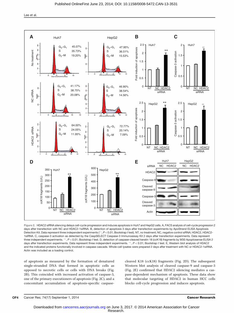
of apoptosis as measured by the formation of denaturedsingle-stranded DNA that formed in apoptotic cells asopposed to necrotic cells or cells with DNA breaks (Fig.2B). This coincided with increased activation of caspase-3,one of the primary executioners of apoptosis (Fig. 2C), and aconcomitant accumulation of apoptosis-specific caspase-
cleaved K18 (ccK18) fragments (Fig. 2D). The subsequentWestern blot analysis of cleaved caspase-9 and caspase-3(Fig. 2E) confirmed that HDAC2 silencing mediates a cas-pase-dependent mechanism of apoptosis. These data showthat molecular targeting of HDAC2 in human HCC cellsblocks cell-cycle progression and induces apoptosis.
Huh7
72.77%
20.14%
7.09%
SG0–G1 45.07%
35.73%
G2–M 19.20%
41.17%
38.75%
20.08%
64.00%
24.05%
11.95%
47.95%
36.51%
15.53%
46.90%
38.54%
14.56%
No
trea
tmen
t H
DA
C2
siR
NA
NC
siR
NA
HepG2
0
0.5
1
1.5
2
2.5
**
0
0.5
1
1.5
2
**
NT NC HDAC2
Fol
d in
duct
ion
of a
popt
osis
Huh7
CBA
siRNA
0
0.5
1
1.5
2
2.5**
Cas
pase
-3 a
ctiv
atio
n
HepG2
NT NC HDAC2siRNA
D
HepG2
E
0
0.5
1
1.5
2**
Huh7
Cas
pase
-3 a
ctiv
atio
n
Fol
d in
duct
ion
of a
popt
osis
2.5
NT NC HDAC2siRNA
NT NC HDAC2siRNA
Huh7
HDAC2
Caspase-9
Cleaved caspase-9
Caspase-3
Cleaved caspase-3
Actin
siRNA NC HDAC2 NC HDAC2
HepG2
****
0
50
100
150
200
250
300
350
0
100
200
300
400
500
ccK
18 fr
agm
ents
(U
/L)
Huh7 HepG2
NT NC HDAC2siRNA
NT NC HDAC2
siRNA
600
ccK
18 fr
agm
ents
(U
/L)
SG0–G1
G2–M
SG0–G1
G2–MSG0–G1
G2–M
SG0–G1
G2–MSG0–G1
G2–M
Figure 2. HDAC2 siRNA silencing delays cell-cycle progression and induces apoptosis in Huh7 and HepG2 cells. A, FACS analysis of cell-cycle progression 2days after transfection with NC and HDAC2-1siRNA. B, detection of apoptosis 3 days after transfection experiments by ApoStrand ELISA ApoptosisDetection Kit. Data represent three independent experiments (��, P < 0.01; Bootstrap t test). NT, no treatment; NC, negative control siRNA; HDAC2, HDAC2-1siRNA. C, caspase-3 activation as detected by the CaspSELECT Caspase-3 Immunoassay Kit 2 days after transfection experiments. Data representthree independent experiments. ��, P < 0.01; Bootstrap t test. D, detection of caspase-cleaved keratin 18 (ccK18) fragments by M30 Apoptosense ELISA 2days after transfection experiments. Data represent three independent experiments. ��, P < 0.01; Bootstrap t test. E, Western blot analysis of HDAC2and the indicated proteins functionally involved in caspase cascade. Whole-cell lysates were prepared 2 days after treatment with NC or HDAC2-1siRNA.Actin was included as a loading control.
Lee et al.
Cancer Res; 74(17) September 1, 2014 Cancer ResearchOF4
Research. on June 3, 2017. © 2014 American Association for Cancercancerres.aacrjournals.org Downloaded from
Published OnlineFirst June 23, 2014; DOI: 10.1158/0008-5472.CAN-13-3531
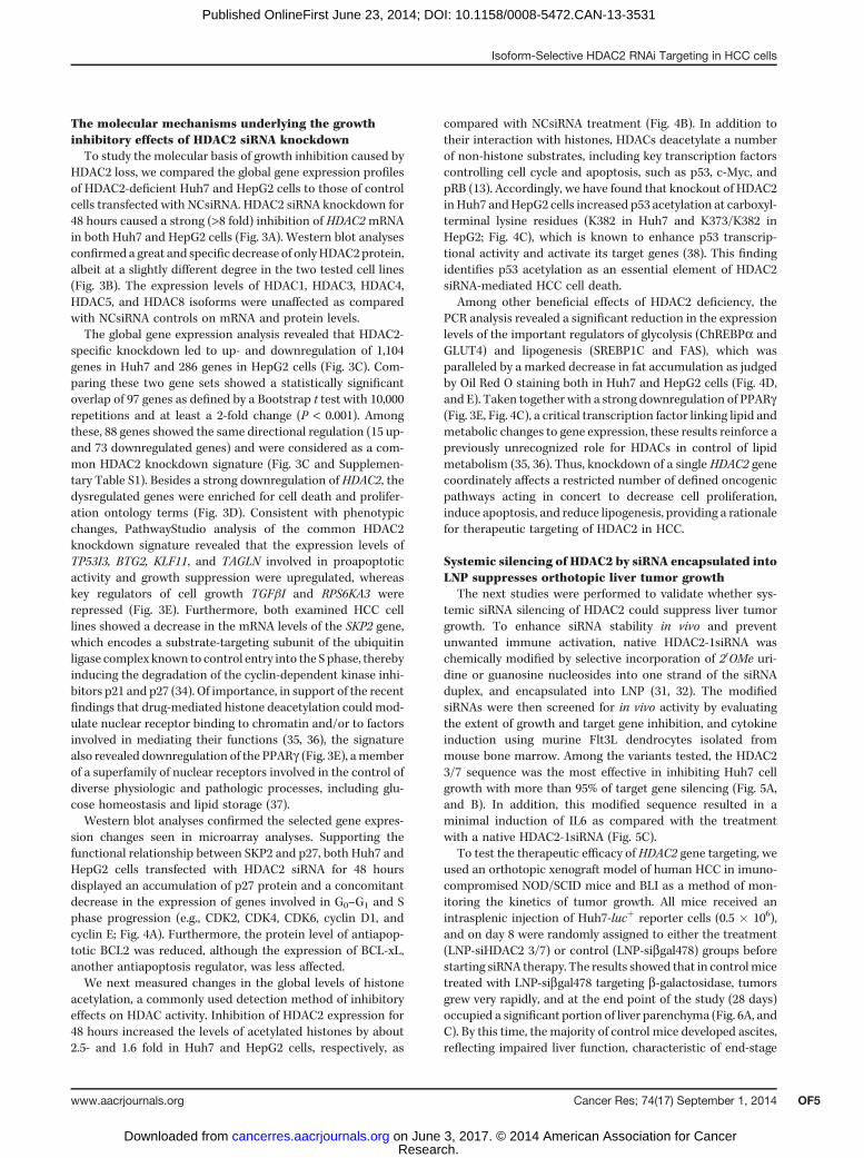
The molecular mechanisms underlying the growthinhibitory effects of HDAC2 siRNA knockdownTo study the molecular basis of growth inhibition caused by
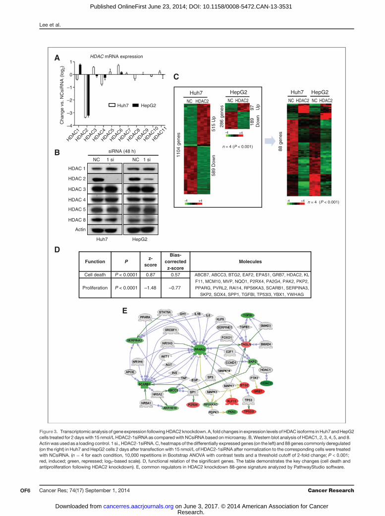
HDAC2 loss, we compared the global gene expression profilesof HDAC2-deficient Huh7 and HepG2 cells to those of controlcells transfected with NCsiRNA. HDAC2 siRNA knockdown for48 hours caused a strong (>8 fold) inhibition of HDAC2mRNAin both Huh7 and HepG2 cells (Fig. 3A). Western blot analysesconfirmed a great and specific decrease of onlyHDAC2protein,albeit at a slightly different degree in the two tested cell lines(Fig. 3B). The expression levels of HDAC1, HDAC3, HDAC4,HDAC5, and HDAC8 isoforms were unaffected as comparedwith NCsiRNA controls on mRNA and protein levels.The global gene expression analysis revealed that HDAC2-
specific knockdown led to up- and downregulation of 1,104genes in Huh7 and 286 genes in HepG2 cells (Fig. 3C). Com-paring these two gene sets showed a statistically significantoverlap of 97 genes as defined by a Bootstrap t test with 10,000repetitions and at least a 2-fold change (P < 0.001). Amongthese, 88 genes showed the same directional regulation (15 up-and 73 downregulated genes) and were considered as a com-mon HDAC2 knockdown signature (Fig. 3C and Supplemen-tary Table S1). Besides a strong downregulation of HDAC2, thedysregulated genes were enriched for cell death and prolifer-ation ontology terms (Fig. 3D). Consistent with phenotypicchanges, PathwayStudio analysis of the common HDAC2knockdown signature revealed that the expression levels ofTP53I3, BTG2, KLF11, and TAGLN involved in proapoptoticactivity and growth suppression were upregulated, whereaskey regulators of cell growth TGFbI and RPS6KA3 wererepressed (Fig. 3E). Furthermore, both examined HCC celllines showed a decrease in the mRNA levels of the SKP2 gene,which encodes a substrate-targeting subunit of the ubiquitinligase complex known to control entry into the S phase, therebyinducing the degradation of the cyclin-dependent kinase inhi-bitors p21 and p27 (34). Of importance, in support of the recentfindings that drug-mediated histone deacetylation could mod-ulate nuclear receptor binding to chromatin and/or to factorsinvolved in mediating their functions (35, 36), the signaturealso revealed downregulation of the PPARg (Fig. 3E), amemberof a superfamily of nuclear receptors involved in the control ofdiverse physiologic and pathologic processes, including glu-cose homeostasis and lipid storage (37).Western blot analyses confirmed the selected gene expres-
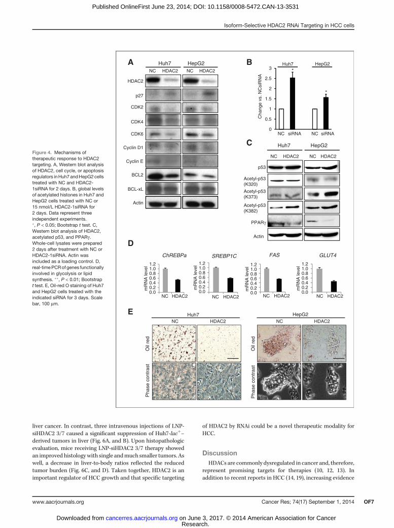
sion changes seen in microarray analyses. Supporting thefunctional relationship between SKP2 and p27, both Huh7 andHepG2 cells transfected with HDAC2 siRNA for 48 hoursdisplayed an accumulation of p27 protein and a concomitantdecrease in the expression of genes involved in G0–G1 and Sphase progression (e.g., CDK2, CDK4, CDK6, cyclin D1, andcyclin E; Fig. 4A). Furthermore, the protein level of antiapop-totic BCL2 was reduced, although the expression of BCL-xL,another antiapoptosis regulator, was less affected.We next measured changes in the global levels of histone
acetylation, a commonly used detection method of inhibitoryeffects on HDAC activity. Inhibition of HDAC2 expression for48 hours increased the levels of acetylated histones by about2.5- and 1.6 fold in Huh7 and HepG2 cells, respectively, as
compared with NCsiRNA treatment (Fig. 4B). In addition totheir interaction with histones, HDACs deacetylate a numberof non-histone substrates, including key transcription factorscontrolling cell cycle and apoptosis, such as p53, c-Myc, andpRB (13). Accordingly, we have found that knockout of HDAC2inHuh7 andHepG2 cells increased p53 acetylation at carboxyl-terminal lysine residues (K382 in Huh7 and K373/K382 inHepG2; Fig. 4C), which is known to enhance p53 transcrip-tional activity and activate its target genes (38). This findingidentifies p53 acetylation as an essential element of HDAC2siRNA-mediated HCC cell death.
Among other beneficial effects of HDAC2 deficiency, thePCR analysis revealed a significant reduction in the expressionlevels of the important regulators of glycolysis (ChREBPa andGLUT4) and lipogenesis (SREBP1C and FAS), which wasparalleled by a marked decrease in fat accumulation as judgedby Oil Red O staining both in Huh7 and HepG2 cells (Fig. 4D,and E). Taken together with a strong downregulation of PPARg(Fig. 3E, Fig. 4C), a critical transcription factor linking lipid andmetabolic changes to gene expression, these results reinforce apreviously unrecognized role for HDACs in control of lipidmetabolism (35, 36). Thus, knockdown of a single HDAC2 genecoordinately affects a restricted number of defined oncogenicpathways acting in concert to decrease cell proliferation,induce apoptosis, and reduce lipogenesis, providing a rationalefor therapeutic targeting of HDAC2 in HCC.
Systemic silencing of HDAC2 by siRNA encapsulated intoLNP suppresses orthotopic liver tumor growth
The next studies were performed to validate whether sys-temic siRNA silencing of HDAC2 could suppress liver tumorgrowth. To enhance siRNA stability in vivo and preventunwanted immune activation, native HDAC2-1siRNA waschemically modified by selective incorporation of 20OMe uri-dine or guanosine nucleosides into one strand of the siRNAduplex, and encapsulated into LNP (31, 32). The modifiedsiRNAs were then screened for in vivo activity by evaluatingthe extent of growth and target gene inhibition, and cytokineinduction using murine Flt3L dendrocytes isolated frommouse bone marrow. Among the variants tested, the HDAC23/7 sequence was the most effective in inhibiting Huh7 cellgrowth with more than 95% of target gene silencing (Fig. 5A,and B). In addition, this modified sequence resulted in aminimal induction of IL6 as compared with the treatmentwith a native HDAC2-1siRNA (Fig. 5C).
To test the therapeutic efficacy of HDAC2 gene targeting, weused an orthotopic xenograft model of human HCC in imuno-compromised NOD/SCID mice and BLI as a method of mon-itoring the kinetics of tumor growth. All mice received anintrasplenic injection of Huh7-lucþ reporter cells (0.5 � 106),and on day 8 were randomly assigned to either the treatment(LNP-siHDAC2 3/7) or control (LNP-sibgal478) groups beforestarting siRNA therapy. The results showed that in controlmicetreated with LNP-sibgal478 targeting b-galactosidase, tumorsgrew very rapidly, and at the end point of the study (28 days)occupied a significant portion of liver parenchyma (Fig. 6A, andC). By this time, the majority of control mice developed ascites,reflecting impaired liver function, characteristic of end-stage
Isoform-Selective HDAC2 RNAi Targeting in HCC cells
www.aacrjournals.org Cancer Res; 74(17) September 1, 2014 OF5
Research. on June 3, 2017. © 2014 American Association for Cancercancerres.aacrjournals.org Downloaded from
Published OnlineFirst June 23, 2014; DOI: 10.1158/0008-5472.CAN-13-3531
A
C
B
Function Pz-
score
Bias-correctedz-score
Molecules
Cell death P < 0.0001 0.87 0.57 ABCB7, ABCC3, BTG2, EAF2, EPAS1, GRB7, HDAC2, KL
F11, MCM10, MVP, NQO1, P2RX4, PA2G4, PAK2, PKP2,
PPARG, PVRL2, RAI14, RPS6KA3, SCARB1, SERPINA3,
SKP2, SOX4, SPP1, TGFBI, TP53I3, YBX1, YWHAG
Proliferation P < 0.0001 –1.48 –0.77
D
HDAC 1
HDAC 2
HDAC 3
HDAC 4
HDAC 5
HDAC 8
Actin
NC 1 si
Huh7 HepG2
NC 1 si
siRNA (48 h)
Cha
nge
vs. N
Csi
RN
A (
log 2
)HDAC mRNA expression
1
0
–1
–2
–3
–4
HepG2Huh7
HepG2
NC HDAC2
Huh7 HepG2
NC HDAC2 NC HDAC2
Huh7
NC HDAC2
286
gene
s 97 Up
189
Dow
n
88 g
enes
n = 4 (P < 0.001)
1104
gen
es
589
Dow
n51
5 U
p
n = 4 (P < 0.001)
E
HDAC1
HDAC2
HDAC3
HDAC4
HDAC5
HDAC6
HDAC7
HDAC8
HDAC9
HDAC10
HDAC11
-4 +4 -4 +4
-4 +4
Figure3. Transcriptomic analysis of gene expression followingHDAC2knockdown.A, fold changes in expression levels of HDAC isoforms inHuh7andHepG2cells treated for 2 days with 15 nmol/L HDAC2-1siRNA as comparedwith NCsiRNA based onmicroarray. B,Western blot analysis of HDAC1, 2, 3, 4, 5, and 8.Actin was used as a loading control. 1 si., HDAC2-1siRNA.C, heatmaps of the differentially expressed genes (on the left) and 88 genes commonly deregulated(on the right) in Huh7 and HepG2 cells 2 days after transfection with 15 nmol/L of HDAC2-1siRNA after normalization to the corresponding cells were treatedwith NCsiRNA. (n ¼ 4 for each condition, 10,000 repetitions in Bootstrap ANOVA with contrast tests and a threshold cutoff of 2-fold change; P < 0.001;red, induced; green, repressed; log2-based scale). D, functional relation of the significant genes. The table demonstrates the key changes (cell death andantiproliferation following HDAC2 knockdown). E, common regulators in HDAC2 knockdown 88-gene signature analyzed by PathwayStudio software.
Lee et al.
Cancer Res; 74(17) September 1, 2014 Cancer ResearchOF6
Research. on June 3, 2017. © 2014 American Association for Cancercancerres.aacrjournals.org Downloaded from
Published OnlineFirst June 23, 2014; DOI: 10.1158/0008-5472.CAN-13-3531
liver cancer. In contrast, three intravenous injections of LNP-siHDAC2 3/7 caused a significant suppression of Huh7-lucþ–derived tumors in liver (Fig. 6A, and B). Upon histopathologicevaluation, mice receiving LNP-siHDAC2 3/7 therapy showedan improved histologywith single andmuch smaller tumors. Aswell, a decrease in liver-to-body ratios reflected the reducedtumor burden (Fig. 6C, and D). Taken together, HDAC2 is animportant regulator of HCC growth and that specific targeting
of HDAC2 by RNAi could be a novel therapeutic modality forHCC.
DiscussionHDACs are commonly dysregulated in cancer and, therefore,
represent promising targets for therapies (10, 12, 13). Inaddition to recent reports in HCC (14, 19), increasing evidence
D
A B
ENC HDAC2NC HDAC2
HepG2
Huh7 HepG2NC HDAC2
HDAC2
CDK2
CDK6
Cyclin E
Actin
BCL2
BCL-xL
p27
CDK4
Cyclin D1
NC HDAC2
HDAC2NC
Huh7 HepG2
NC HDAC2
p53
Actin
PPARγ
Acetyl-p53 (K320)
Acetyl-p53 (K373)
Acetyl-p53 (K382)
0.00.20.40.60.81.01.2
GLUT4
mR
NA
leve
lNC HDAC2
ChREBPa
NC HDAC20.00.20.40.60.81.01.2
mR
NA
leve
l
FAS
NC HDAC20.00.20.40.60.81.01.2
mR
NA
leve
l
SREBP1C
NC HDAC20.00.20.40.60.81.01.2
mR
NA
leve
l
Huh7
Pha
se c
ontr
ast
Cha
nge
vs. N
Csi
RN
A
Huh7 HepG2
*
*
0
0.5
1
1.5
2
2.5
3
CNC siRNA NC siRNA
*
Oil
red
Pha
se c
ontr
ast
Oil
red
Figure 4. Mechanisms oftherapeutic response to HDAC2targeting. A, Western blot analysisof HDAC2, cell cycle, or apoptosisregulators inHuh7andHepG2cellstreated with NC and HDAC2-1siRNA for 2 days. B, global levelsof acetylated histones in Huh7 andHepG2 cells treated with NC or15 nmol/L HDAC2-1siRNA for2 days. Data represent threeindependent experiments.�, P < 0.05; Bootstrap t test. C,Western blot analysis of HDAC2,acetylated p53, and PPARg .Whole-cell lysates were prepared2 days after treatment with NC orHDAC2-1siRNA. Actin wasincluded as a loading control. D,real-timePCRof genes functionallyinvolved in glycolysis or lipidsynthesis. ��, P < 0.01; Bootstrapt test. E, Oil-red O staining of Huh7and HepG2 cells treated with theindicated siRNA for 3 days. Scalebar, 100 mm.
Isoform-Selective HDAC2 RNAi Targeting in HCC cells
www.aacrjournals.org Cancer Res; 74(17) September 1, 2014 OF7
Research. on June 3, 2017. © 2014 American Association for Cancercancerres.aacrjournals.org Downloaded from
Published OnlineFirst June 23, 2014; DOI: 10.1158/0008-5472.CAN-13-3531

demonstrates aberrant expression of HDAC2 in diverse cancertypes such as cutaneous T-cell lymphoma, gastric cancer,colorectal cancer, prostate cancer, ovarian cancer, and endo-metrial cancer (39), implying that HDAC2 targeting may be aneffective therapeutic strategy against various cancer types.However, the anticancer therapy by a selective inhibition ofa disease-specific HDAC is still lacking.
The present study provides evidence that siRNA targetingof HDAC2 in HCC cell lines elicits a strong target specificityand inhibits only one HDAC2 target protein among thetested class I and class IIa HDAC isoenzymes. Over the pastdecade, at least 21 siRNAs have been evaluated for more thana dozen diseases, including recent preclinical studies target-ing polo-like kinase 1 (PLK1) and kinesin spindle protein(KSP; ref. 27). The primary obstacle for therapeutic appli-cation of RNAi is the successful in vivo delivery to the
targeted cell (40, 41). In this study, we used LNP technologyto protect the systemically administered siRNA from glo-merular filtration and serum nucleases, thereby extendingsiRNA half-life and increasing its potency while suppressingunwanted immune responses (24–26, 31, 32). Indeed, treat-ment with a moderate (2 mg/kg) dose of 20OMe-modifiedHDAC2 siRNA encapsulated in LNP was sufficient to medi-ate a potent silencing of the target mRNA and the effectivesuppression of HCC growth in vivo without cytokine induc-tion or toxicity associated with unmodified siRNA. Althoughmore work is needed to optimize the therapeutic dose andaddress potential side-effects, these findings suggest theclinical utility of HDAC2 siRNA targeting for treatment ofHCC disease with high target selectivity.
In agreement with published data (28, 42, 43), inhibition ofHDAC2 protein caused a block in cell-cycle progression andextensive apoptosis in all the examined HCC cell lines. Onthe molecular level, it was paralleled by a coordinateddysregulation of genes acting downstream of HDAC2-regu-lated transcription factors. Microarray profiling showed thatphenotypic changes triggered by HDAC2 siRNA silencingwere associated with an increased expression of proapop-totic effectors KLF11, BTG2, and TP53I3, and downregulationof key regulators of cell growth such as SKP2 and TGFbI. Thiswas paralleled by a strong reduction in the protein levels ofCDK2, CDK4, CDK6, cyclin D1, and cyclin E, whereas theexpression of cdk inhibitor p27 was increased, providing amechanism for HDAC2 silencing-mediated inhibition of cell-cycle progression. We also show that HDAC2 siRNA knock-down increased the levels of p53 acetylation, which is knownto boost the p53 recruitment to promoter-associated com-plexes and upregulate the expression of p53 target genesinvolved in cell-cycle control and apoptosis. Of note, HDAC2knockdown enhanced p53 acetylation both in HepG2 (withwild-type p53) and Huh7 cells (with mutant p53). Further-more, siRNA-mediated HDAC2 silencing caused a down-regulation of PPARg , which was associated with reductionin expressions of lipogenic enzymes and decreased lipidaccumulation. This is in agreement with recent findingsthat genetic deletion of HDAC1 and HDAC2 in culturedmesenchymal precursor cells (35) or in vivo treatment witha novel class II-selective inhibitor MC1568 (36) interferedwith PPARg signaling pathways. There is increasing appre-ciation that PPARg signaling in addition to control of lipidhomeostasis also influences inflammation, hepatic fibrosis,and cancer. Given the involvement of PPARg signaling net-works in diverse physiologic and pathologic processes(37, 44, 45) and the conflicting data about the role of PPARgas either an anti- or protumorigenic factor in HCC (46, 47),the mechanisms whereby HDACi interferes with the PPARgsignaling pathways may be of significance for the clinical useof HDACi and require thorough investigation.
In conclusion, this is thefirst study to report that targeting ofHDAC2 by RNAi-based nanoparticles allowed a selective inac-tivation of only one disease-specific HDAC isoform andachieved consistent therapeutic benefits in a preclinal modelof HCC, providing an attractive therapeutic modality for livercancer.
C
0
0.2
0.4
0.6
0.8
1
1.2
Nor
mal
ized
HA
DC
2 ex
pres
sion
**
B
A
020406080
100120140160
IL6
(pg/
mL)
**
0
0.2
0.4
0.6
0.81.0
1.2
Cel
l via
bilit
y
Untre
ated
LNP-L
uc
LNP-H
DAC2-1
LNP-H
DAC2 3/
6
Untre
ated
LNP-L
uc
LNP-H
DAC2-3/
7PBS
LNP-H
DAC2-1
LNP-H
DAC2-3/
7
LNP-H
DAC2 3/
7
LNP-H
DAC2 3/
8
LNP-H
DAC2 4/
6
LNP-H
DAC2 4/
7
LNP-H
DAC2 4/
8
LNP-H
DAC2 5/
6
LNP-H
DAC2 5/
7
LNP-H
DAC2 5/
8
* * *
Figure 5. Selection of HDAC2 3/7 for in vivo application based on theinhibition of tumor cell growth and minimal cytokine induction. A,inhibition of Huh7-lucþ cell growth after transfection with 30 nmol/L ofSNALP-formulated HDAC2-1 (native) or its modified variants (HDAC2-3/6�8, HDAC2-4/6�8, HDAC2-5/6�8). The siRNA transfectants wereexamined by MTT assay at 4 days after treatment [�, P < 0.05 (n ¼ 3) byBootstrap t test]. LNP-Luc, LNP-formulated siRNA targeting luciferase.B, downregulation of HDAC2 mRNA in Huh7 cells treated withHDAC2 3/7. Total RNAs were extracted at 48 hours after treatment with15 nmol/L of siRNA (��, P < 0.01, Bootstrap t test). C, quantification of IL6levels after HDAC2 siRNA treatment. Culture supernatants of Flt3L-derived dendrocytes were assayed for IL6 using ELISA 24 hours aftersiRNA treatment. The data are shown as means � SEM of triplicateexperiments (��, P < 0.01, Bootstrap t test).
Lee et al.
Cancer Res; 74(17) September 1, 2014 Cancer ResearchOF8
Research. on June 3, 2017. © 2014 American Association for Cancercancerres.aacrjournals.org Downloaded from
Published OnlineFirst June 23, 2014; DOI: 10.1158/0008-5472.CAN-13-3531

Disclosure of Potential Conflicts of InterestI. MacLachlan received commercial research support from Tekmira
Pharmaceuticals Corp. No potential conflicts of interests were disclosed bythe other authors.
Authors' ContributionsConception and design: Y.-H. Lee, A.D. Judge, I. MacLachlan, S.S. ThorgeirssonDevelopment of methodology: J.B. Andersen, J.U. Marquardt, I. MacLachlanAcquisition of data (provided animals, acquired and managed patients,provided facilities, etc.): Y.-H. Lee, K.-J. Choi, J.B. Andersen, M.-A. Won,M. Kitade, J.U. Marquardt, C. Raggi, E.A. Conner, I. MacLachlanAnalysis and interpretation of data (e.g., statistical analysis, biostatistics,computational analysis): Y.-H. Lee, D. Seo, J.B. Andersen, J.U. Marquardt,V.M. FactorWriting, review, and/or revision of the manuscript: Y.-H. Lee, D. Seo, J.B.Andersen, V.M. Factor, S.S. Thorgeirsson
Administrative, technical, or material support (i.e., reporting or orga-nizing data, constructing databases): A.D. Judge, E.A. ConnerStudy supervision: I. MacLachlan, S.S. ThorgeirssonOther (Western blot analysis): L.E. G�omez-Quiroz
Grant SupportThis project was supported by the Intramural Research Programof the Center
for Cancer Research, NCI, and by a grant of the Korean Health Technology R&DProject, Ministry of Health & Welfare, Republic of Korea (A121982).
The costs of publication of this article were defrayed in part by the payment ofpage charges. This article must therefore be hereby marked advertisement inaccordance with 18 U.S.C. Section 1734 solely to indicate this fact.
Received December 9, 2013; revised May 8, 2014; accepted May 20, 2014;published OnlineFirst June 23, 2014.
References1. Parkin DM, Bray F, Ferlay J, Pisani P. Global cancer statistics. CA
Cancer J Clin 2005;55:74–108.2. Llovet JM, Bruix J. Molecular targeted therapies in hepatocellular
carcinoma. Hepatology 2008;48:1312–27.
LNP
-βga
l478
LN
P-H
DA
C2
3/7
Day 14 Day 21 Day 28Day 11Day 8βg
al47
8 H
DA
C2
3/7
Gross morphology H&E
Days after transplantation11 15 17 21 25 28
Mea
n p
hoto
n pe
r se
cond
(x1
08)
βgal478
(n = 8)
HDAC2 3/7
(n = 6) 0
0.5
1.0
1.5
2.0
2.5
*
B
DC
A
0
0.02
0.04
0.06
0.08
0.1
0.12
Live
r w
eigh
t /bo
dy w
eigh
t
*
Figure 6. Systemic silencing of HDAC2 by siRNA-based LNP suppresses orthotopic liver tumor growth. A, representative bioluminescence images of miceafter intrasplenic injection of 0.5 � 106 Huh7-lucþ reporter cells, resulting in tumorous growth in the liver. Eight days after transplantation, mice wererandomly assigned either to control (bgal478, n¼8) or target siRNA treatment (HDAC23/7, n¼ 6) group based on the bioluminescence intensity. LNPs loadedwith bgal478 or HDAC2 3/7 siRNA were injected at a dose of 2 mg/kg at 8, 11, and 14 days. B, quantification of bioluminescence intensity. The totalflux is plotted as photon/second. �, P < 0.05, (n ¼ 8 vs. n ¼ 6) by Mann–Whitney U test. C, gross liver morphology and histopathology at 28 days aftertransplantation. H&E, hematoxylin & eosin staining; scale bar, 50 mm. D, liver-to-body weight ratios. The data are shown as the means � SEM in eachtreatment group. �, P < 0.05, (n ¼ 8 vs. n ¼ 6) by Mann–Whitney U test.
www.aacrjournals.org Cancer Res; 74(17) September 1, 2014 OF9
Isoform-Selective HDAC2 RNAi Targeting in HCC cells
Research. on June 3, 2017. © 2014 American Association for Cancercancerres.aacrjournals.org Downloaded from
Published OnlineFirst June 23, 2014; DOI: 10.1158/0008-5472.CAN-13-3531
3. Bruix J, ShermanM.Management of hepatocellular carcinoma. Hepa-tology 2005;42:1208–36.
4. Llovet JM, Ricci S, Mazzaferro V, Hilgard P, Gane E, Blanc JF, et al.Sorafenib in advanced hepatocellular carcinoma. N Engl J Med 2008;359:378–90.
5. Farazi PA, DePinho RA. Hepatocellular carcinoma pathogenesis: fromgenes to environment. Nat Rev Cancer 2006;6:674–87.
6. Thorgeirsson SS, Grisham JW. Molecular pathogenesis of humanhepatocellular carcinoma. Nat Genet 2002;31:339–46.
7. Feitelson MA, Sun B, Satiroglu Tufan NL, Liu J, Pan J, Lian Z. Geneticmechanisms of hepatocarcinogenesis. Oncogene 2002;21:2593–604.
8. Roberts LR,GoresGJ. Hepatocellular carcinoma:molecular pathwaysand new therapeutic targets. Semin Liver Dis 2005;25:212–25.
9. Kramer OH. HDAC2: a critical factor in health and disease. TrendsPharmacol Sci 2009;30:647–55.
10. Delcve GP, Khan DH, Davie JR. Targeting class I histone deacetylasesin cancer therapy. Expert Opin Ther Targets 2013;17:29–41.
11. Trivedi CM, Luo Y, Yin Z, Zhang M, Zhu W, Wang T, et al. Hdac2regulates the cardiac hypertrophic response by modulating Gsk3bactivity. Nat Med 2007;13:324–31.
12. Ververis K, Hiong A, Karagiannis TC, Licciardi PV. Histone deacetylaseinhibitors (HDACIs): multitargeted anticancer agents. Biologics 2013;7:47–60.
13. Glass E, Viale PH. Histone deacetylase inhibitors: novel agents incancer treatment. Clin J Oncol Nurs 2013;17:34–40.
14. Lachenmayer A, Toffanin S, Cabellos L, Alsinet C, Hoshida Y, Villa-nueva A, et al. Combination therapy for hepatocellular carcinoma:additive preclinical efficacy of the HDAC inhibitor panobinostat withsorafenib. J Hepatol 2012;56:1343–50.
15. Gryder BE, Sodji QH, Oyelere AK. Targeted cancer therapy: givinghistone deacetylase inhibitors all they need to succeed. Future MedChem 2012;4:505–24. Review. Erratum in: Future Med Chem 2012;4:1369–70.
16. Geng L, Cuneo KC, Fu A, Tu T, Atadja PW, Hallahan DE. Histonedeacetylase (HDAC) inhibitor LBH589 increases duration of g-H2AXFoci and confines HDAC4 to the cytoplasm in irradiated non–small celllung cancer. Cancer Res 2006;66:11298–304.
17. Bauer S, Hilger RA, M€uhlenberg T, Grabellus F, Nagarajah J, HoiczykM, et al. Phase I study of panobinostat and imatinib in patients withtreatment-refractory metastatic gastrointestinal stromal tumors. Br JCancer 2014;110:1155–62.
18. Marks PA, Xu WS. Histone deacetylase inhibitors: Potential in cancertherapy. J Cell Biochem 2009;107:600–8.
19. Lee JS, Chu IS, Heo J, Calvisi DF, Sun Z, Roskams T, et al. Classi-fication and prediction of survival in hepatocellular carcinoma by geneexpression profiling. Hepatology 2004;40:667–76.
20. Lee YH, Andersen JB, Song HT, Judge AD, Seo D, Ishikawa T, et al.Definition of ubiquitination modulator COP1 as a novel therapeutictarget in human hepatocellular carcinoma. Cancer Res 2010;70:8264–9.
21. Lee YH, Judge AD, Seo D, Kitade M, Gomez-Quiroz LE, Ishikawa T,et al. Molecular targeting of CSN5: A mechanism of therapeuticresponse. Oncogene 2011;30:4175–84.
22. Fire A, Xu S, MontgomeryMK, Kostas SA, Driver SE, Mello CC. Potentand specific genetic interference by double stranded RNA in Caenor-habditis elegans. Nature 1998;391:806–11.
23. Elbashir SM, Harborth J, Lendeckel W, Yalcin A, Weber K, Tuschl T.Duplexes of 21-nucleotide RNAsmediate RNA interference in culturedmammalian cells. Nature 2001;411:494–8.
24. Jeffs LB, Palmer LR, Ambegia EG, Giesbrecht C, Ewanick S, MacLa-chlan I. A scalable, extrusion free method for efficient liposomalencapsulation of plasmid DNA. Pharm Res 2005;22:362–72.
25. Morrissey DV, Lockridge JA, Shaw L, Blanchard K, Jensen K, BreenW,et al. Potent and persistent in vivo anti-HBV activity of chemicallymodified siRNAs. Nat Biotechnol 2005;23:1002–7.
26. Zimmermann TS, Lee AC, Akinc A, Bramlage B, Bumcrot D, FedorukMN, et al. RNAi-mediated gene silencing in non-human primates.Nature 2006;441:111–4.
27. Judge AD, Robbins M, Tavakoli I, Levi J, Hu L, Fronda A, et al.Confirming the RNAi-mediated mechanism of action of siRNA-basedcancer therapeutics in mice. J Clin Invest 2009;119:661–73.
28. Huang BH, Laban M, Leung CH, Lee L, Lee CK, Salto-Tellez M, et al.Inhibition of histone deacetylase 2 increases apoptosis and p21Cip1/WAF1 expression, independent of histone deacetylase 1. Cell DeathDiffer 2005;12:395–404.
29. Harms KL, Chen X. Histone deacetylase 2 modulates p53 transcrip-tional activities through regulationof p53–DNAbinding activity. CancerRes 2007;67:3145–52.
30. Ropero S, Ballestar E, Alaminos M, Arango D, Schwartz S Jr, EstellerM. Transforming pathways unleashed by a HDAC2mutation in humancancer. Oncogene 2008;27:4008–12.
31. Judge AD, Bola G, Lee AC, MacLachlan I. Design of noninflammatorysynthetic siRNA mediating potent gene silencing in vivo. Mol Ther2005;13:494–504.
32. Judge AD, Sood V, Shaw JR, Fang D, McClintock K, MacLachlan I.Sequence-dependent stimulation of the mammalian innate immuneresponse. Nat Biotechnol 2005;23:457–62.
33. Andersen JB, Factor VM, Marquardt JU, Raggi C, Lee YH, Seo D, et al.An integrated genomic and epigenomic approach predicts therapeuticresponse to zebularine in human liver cancer. Sci Transl Med 2010;2:54ra77.
34. Bashir T, DorrelloNV, Amador V,GuardavaccaroD, PaganoM.Controlof the SCF(Skp2-Cks1) ubiquitin ligase by the APC/C(Cdh1) ubiquitinligase. Nature 2004;428:190–3.
35. Haberland M, Carrer M, Mokalled MH, Montgomery RL, Olson EN.Redundant control of adipogenesis by histone deacetylases 1 and 2. JBiol Chem 2010;285: 14663–70.
36. Nebbioso A, Dell'Aversana C, Bugge A, Sarno R, Valente S, Rotili D,et al.HDACsclass II-selective inhibition alters nuclear receptor-depen-dent differentiation. J Mol Endocrinol 2010;45:219–28.
37. Grygiel-G�orniak B. Peroxisome proliferator-activated receptors andtheir ligands: nutritional and clinical implications-a review. Nutr J2014;13:17.
38. Li P, Wang D, Yao H, Doret P, Hao G, Shen Q, et al. Coordination ofPAD4 and HDAC2 in the regulation of p53-target gene expression.Oncogene 2010;29:3153–62.
39. Weichert W. HDAC expression and clinical prognosis in human malig-nancies. Cancer Lett 2009;280:168–76.
40. Pai SI, Lin YY,MacaesB,Meneshian A, HungCF,Wu TC. Prospects ofRNA interference therapy for cancer. Gene Ther 2006;13:464–77.
41. Hokaiwado N, Takeshita F, Banas A, Ochiya T. RNAi-based drugdiscovery and its application to therapeutics. IDrugs 2008;11:274–8.
42. Zhu P, Martin E, Mengwasser J, Schlag P, Janssen KP, G€ottlicher M.Induction of HDAC2 expression upon loss of APC in colorectal tumor-igenesis. Cancer Cell 2004;5: 455–63.
43. Fritsche P, Seidler B, Sch€uler S, Schnieke A, G€ottlicherM, Schmid RM,et al. HDAC2 mediates therapeutic resistance of pancreatic cancercells via the BH3-only protein NOXA. Gut 2009;58:1399–1409.
44. Kidani Y, Bensinger SJ. Liver X receptor and peroxisome proliferator-activated receptor as integrators of lipid homeostasis and immunity.Immunol Rev 2012;249:72–83.
45. Zhang F, Kong D, Lu Y, Zheng S. Peroxisome proliferator-activatedreceptor-g as a therapeutic target for hepatic fibrosis: from bench tobedside. Cell Mol Life Sci 2013;70:259–76.
46. Baffy G, Brunt EM, Caldwell SH. Hepatocellular carcinoma in non-alcoholic fatty liver disease: an emerging menace. J Hepatol 2012;56:1384–91.
47. Wu CW, Farrell GC, Yu J. Functional role of peroxisome-proliferator-activated receptor g in hepatocellular carcinoma. J GastroenterolHepatol 2012;27:1665–69.
Cancer Res; 74(17) September 1, 2014 Cancer ResearchOF10
Lee et al.
Research. on June 3, 2017. © 2014 American Association for Cancercancerres.aacrjournals.org Downloaded from
Published OnlineFirst June 23, 2014; DOI: 10.1158/0008-5472.CAN-13-3531
Published OnlineFirst June 23, 2014.Cancer Res Yun-Han Lee, Daekwan Seo, Kyung-Ju Choi, et al. Inhibition of HDAC2Antitumor Effects in Hepatocarcinoma of Isoform-Selective
Updated version
10.1158/0008-5472.CAN-13-3531doi:
Access the most recent version of this article at:
Material
Supplementary
http://cancerres.aacrjournals.org/content/suppl/2014/06/27/0008-5472.CAN-13-3531.DC1
Access the most recent supplemental material at:
E-mail alerts related to this article or journal.Sign up to receive free email-alerts
Subscriptions
Reprints and
To order reprints of this article or to subscribe to the journal, contact the AACR Publications
Permissions
To request permission to re-use all or part of this article, contact the AACR Publications
Research. on June 3, 2017. © 2014 American Association for Cancercancerres.aacrjournals.org Downloaded from
Published OnlineFirst June 23, 2014; DOI: 10.1158/0008-5472.CAN-13-3531