Embed Size (px)
Citation preview
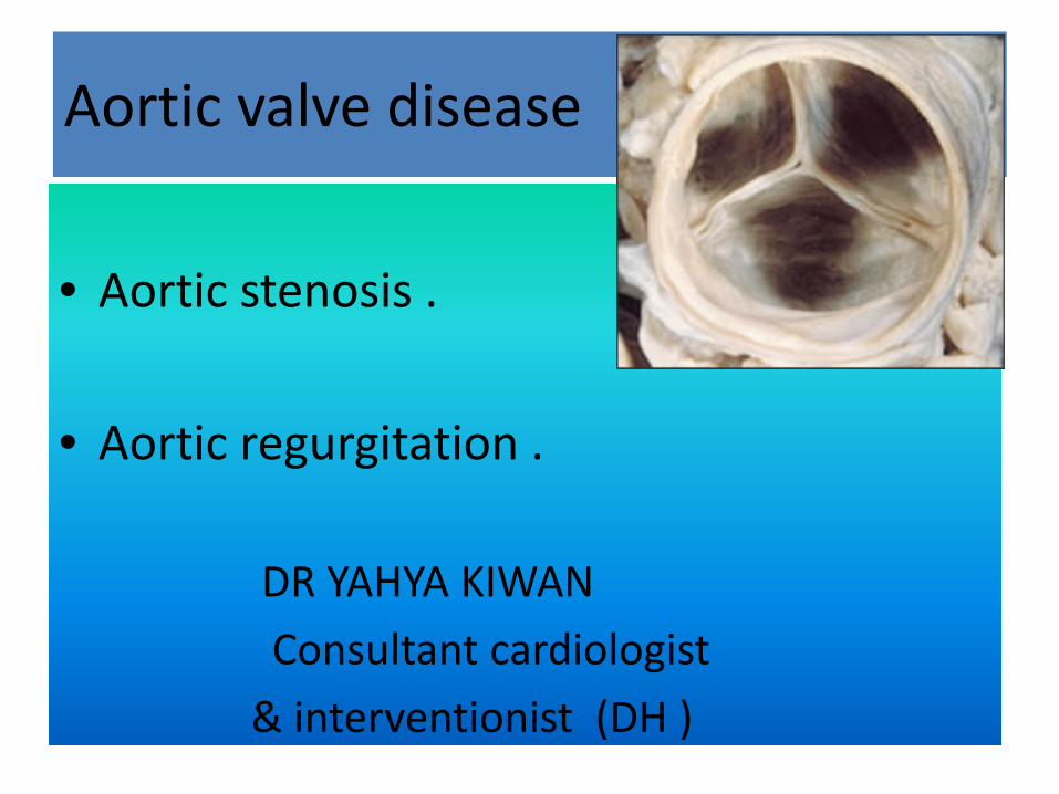
Aortic valve disease
• Aortic stenosis .
• Aortic regurgitation .
DR YAHYA KIWANConsultant cardiologist
& interventionist (DH )
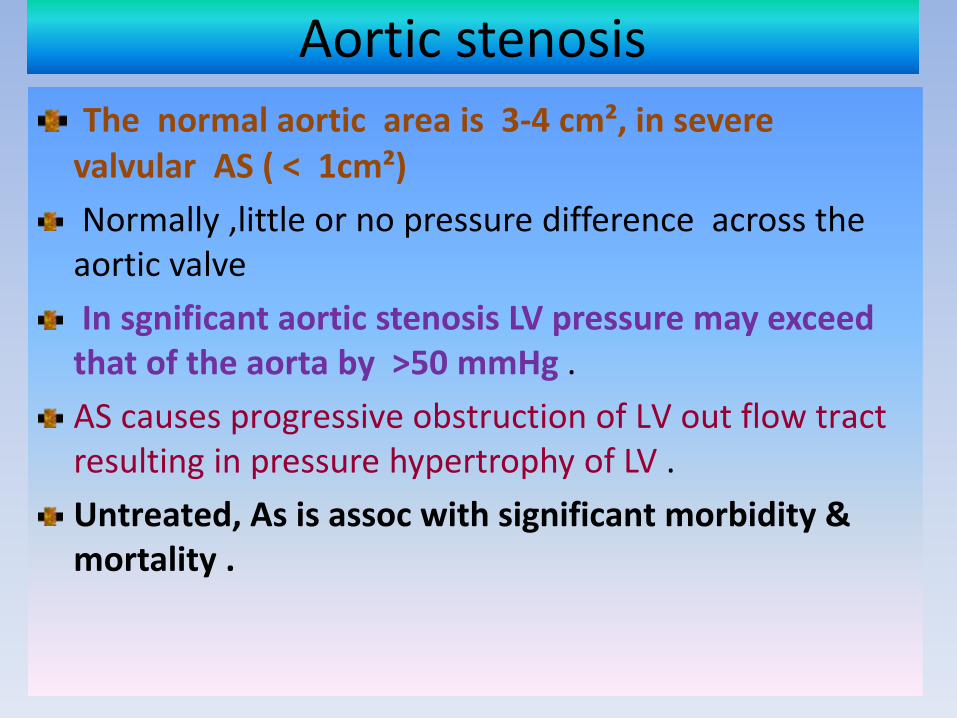
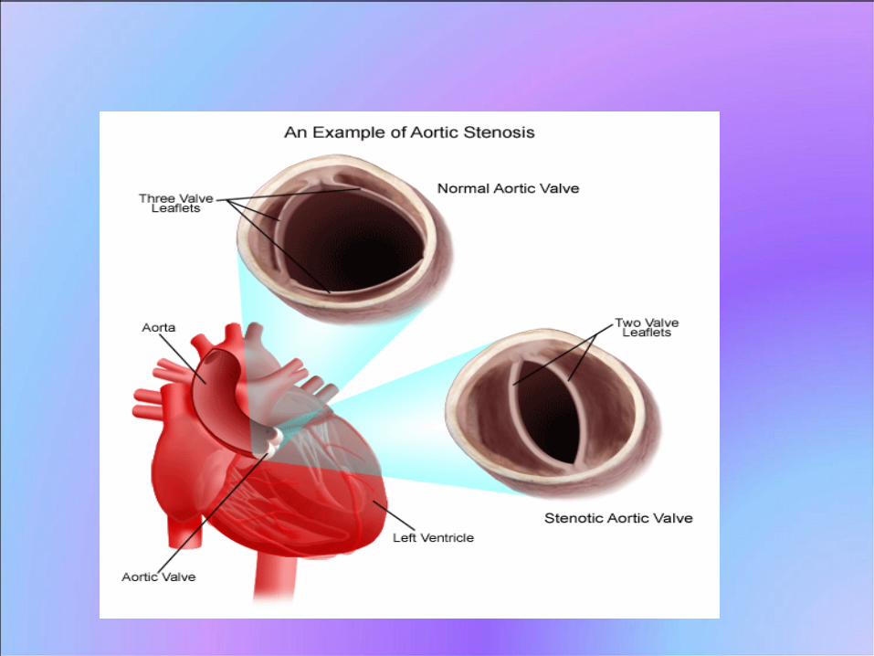
Aortic stenosis The normal aortic area is 3-4 cm², in severe
valvular AS ( < 1cm²)Normally ,little or no pressure difference across the
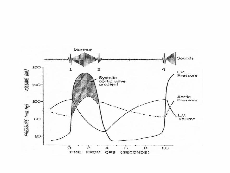
aortic valveIn sgnificant aortic stenosis LV pressure may exceed
that of the aorta by >50 mmHg .AS causes progressive obstruction of LV out flow tract resulting in pressure hypertrophy of LV . Untreated, As is assoc with significant morbidity & mortality .
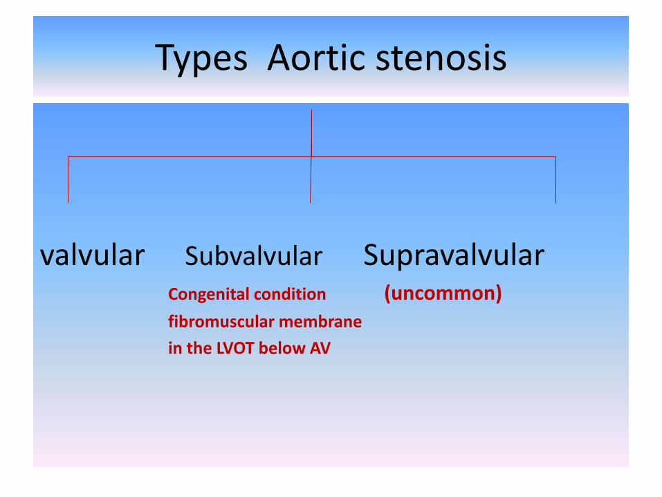
Types Aortic stenosis
valvular Subvalvular Supravalvular Congenital condition (uncommon)fibromuscular membranein the LVOT below AV
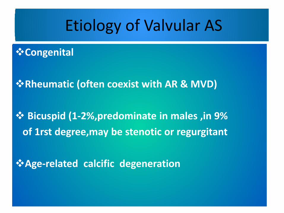
Etiology of Valvular ASCongenital
Rheumatic (often coexist with AR & MVD)
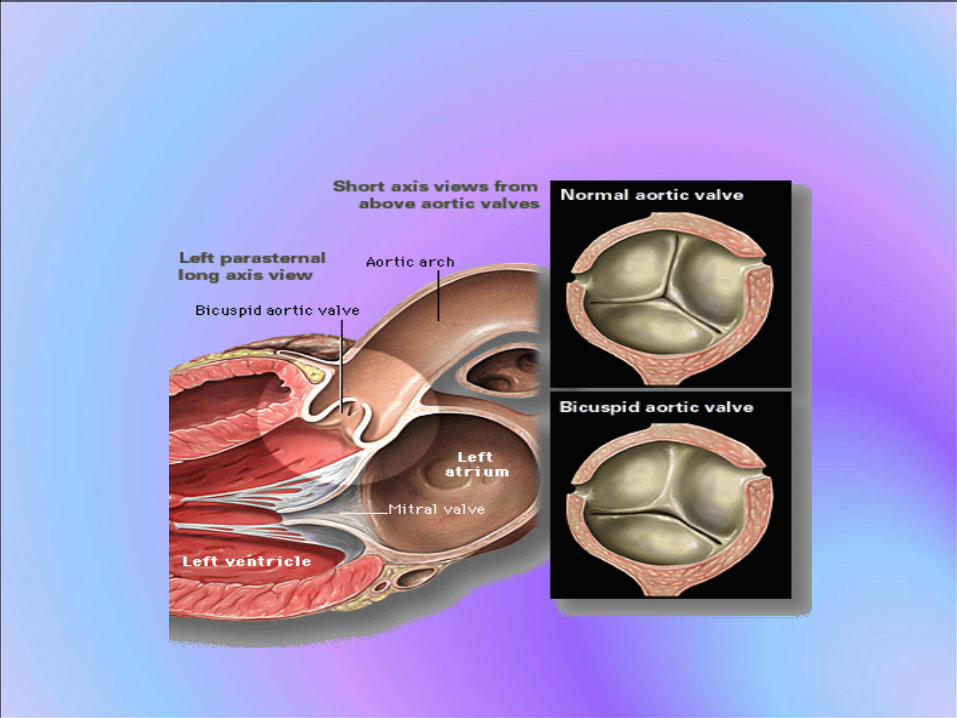
Bicuspid (1-2%,predominate in males ,in 9% of 1rst degree,may be stenotic or regurgitant
Age-related calcific degeneration
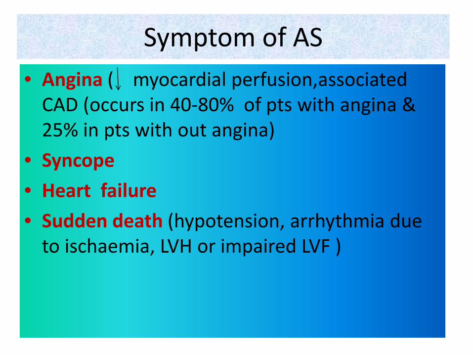
Symptom of AS• Angina ( myocardial perfusion,associated
CAD (occurs in 40-80% of pts with angina & 25% in pts with out angina)
• Syncope• Heart failure• Sudden death (hypotension, arrhythmia due
to ischaemia, LVH or impaired LVF )
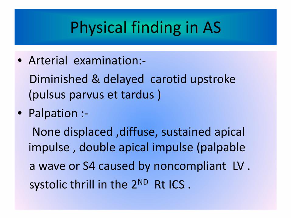
Physical finding in AS
• Arterial examination:-Diminished & delayed carotid upstroke (pulsus parvus et tardus )
• Palpation :-None displaced ,diffuse, sustained apical
impulse , double apical impulse (palpable a wave or S4 caused by noncompliant LV .systolic thrill in the 2ND Rt ICS .
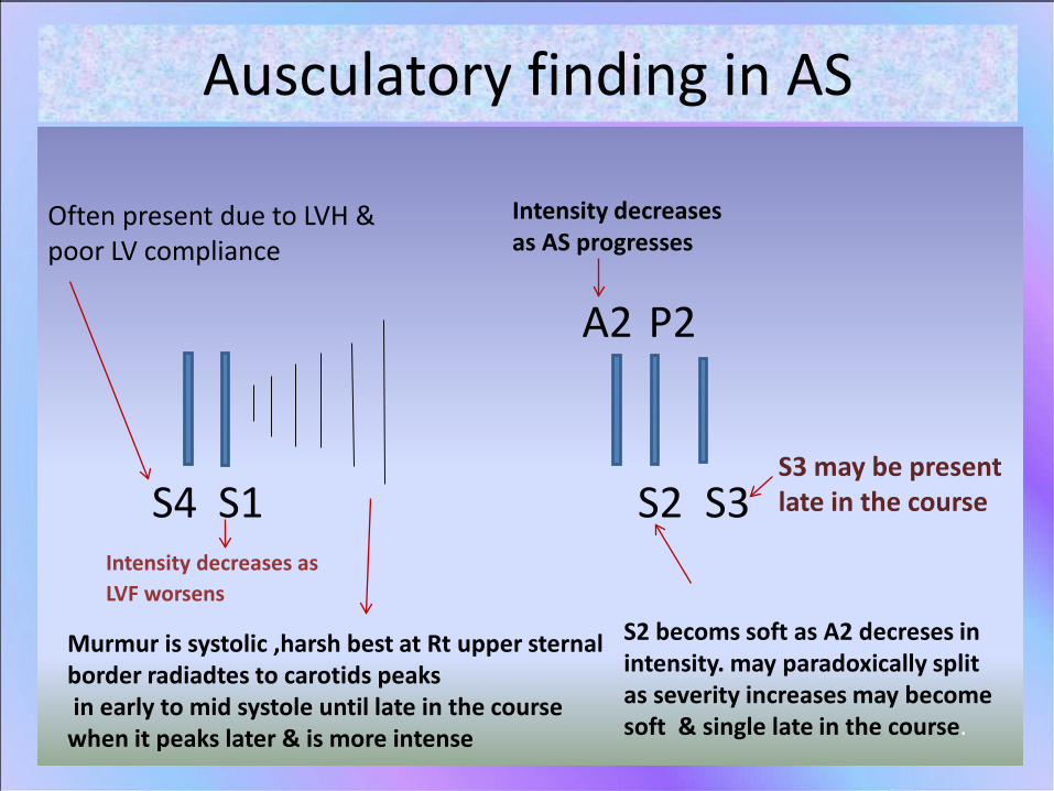
Ausculatory finding in AS
S2 becoms soft as A2 decreses in intensity. may paradoxically split as severity increases may become soft & single late in the course.
Intensity decreases as AS progresses
A2
S2
P2
S3
Murmur is systolic ,harsh best at Rt upper sternal border radiadtes to carotids peaksin early to mid systole until late in the course
when it peaks later & is more intense
S1S4Intensity decreases as LVF worsens
Often present due to LVH & poor LV compliance
S3 may be present late in the course
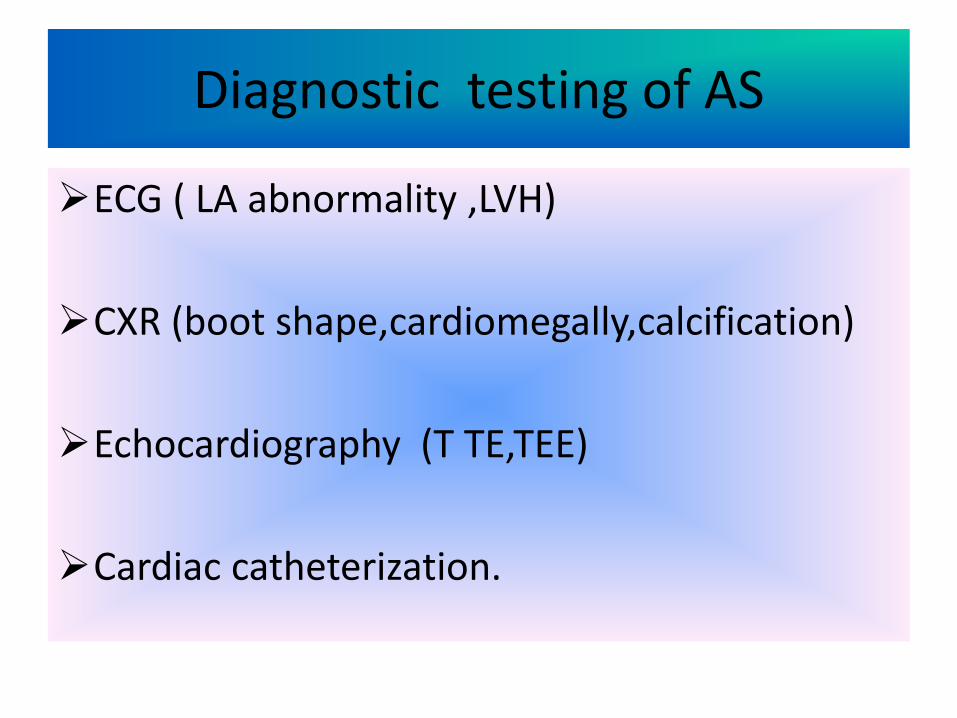
Diagnostic testing of AS
ECG ( LA abnormality ,LVH)
CXR (boot shape,cardiomegally,calcification)
Echocardiography (T TE,TEE)
Cardiac catheterization.
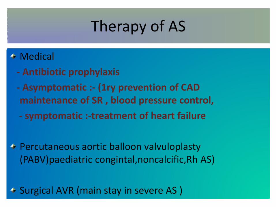
Therapy of AS
Medical - Antibiotic prophylaxis- Asymptomatic :- (1ry prevention of CAD maintenance of SR , blood pressure control,- symptomatic :-treatment of heart failure
Percutaneous aortic balloon valvuloplasty (PABV)paediatric congintal,noncalcific,Rh AS)
Surgical AVR (main stay in severe AS )
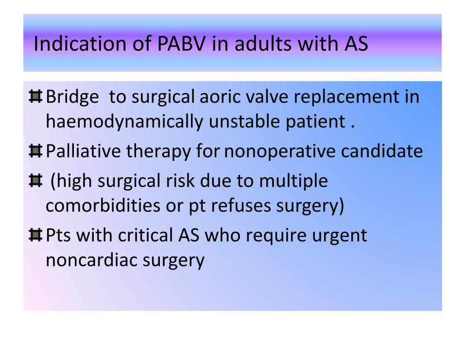
Indication of PABV in adults with AS
Bridge to surgical aoric valve replacement in haemodynamically unstable patient .Palliative therapy for nonoperative candidate(high surgical risk due to multiple comorbidities or pt refuses surgery)Pts with critical AS who require urgent noncardiac surgery
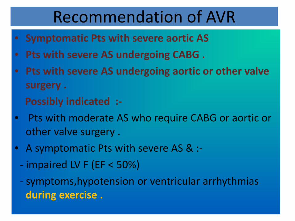
Recommendation of AVR• Symptomatic Pts with severe aortic AS • Pts with severe AS undergoing CABG .• Pts with severe AS undergoing aortic or other valve
surgery .Possibly indicated :-
• Pts with moderate AS who require CABG or aortic or other valve surgery .
• A symptomatic Pts with severe AS & :-- impaired LV F (EF < 50%)- symptoms,hypotension or ventricular arrhythmias
during exercise .
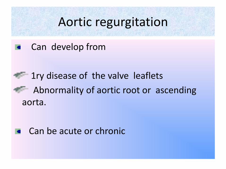
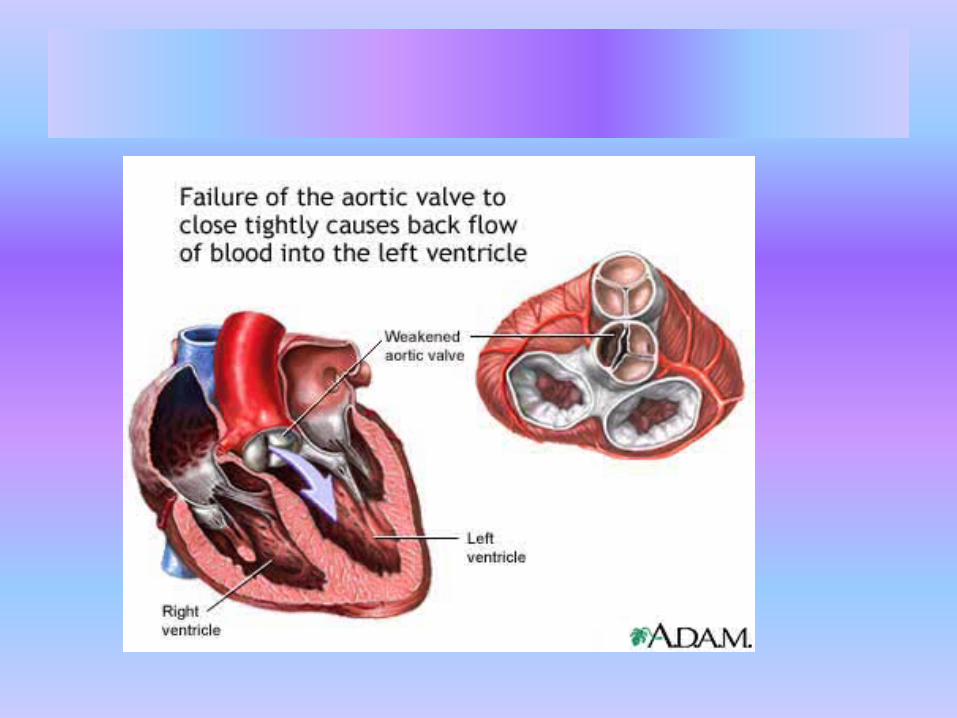
Aortic regurgitation
Can develop from
1ry disease of the valve leaflets Abnormality of aortic root or ascending
aorta.
Can be acute or chronic
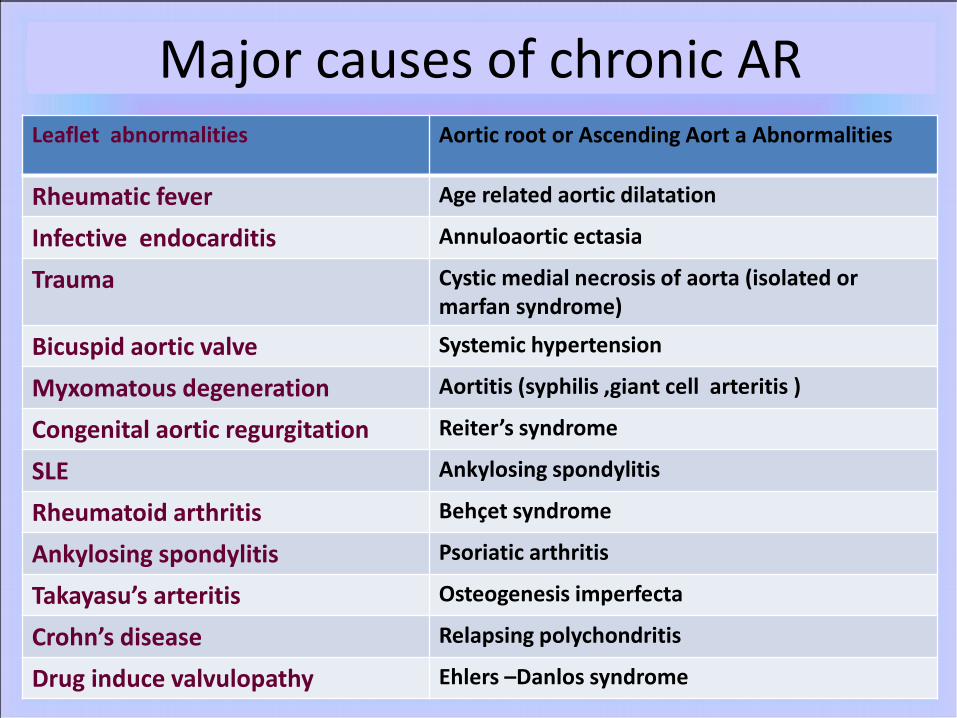
Major causes of chronic ARLeaflet abnormalities Aortic root or Ascending Aort a Abnormalities
Rheumatic fever Age related aortic dilatation
Infective endocarditis Annuloaortic ectasia
Trauma Cystic medial necrosis of aorta (isolated or marfan syndrome)
Bicuspid aortic valve Systemic hypertension
Myxomatous degeneration Aortitis (syphilis ,giant cell arteritis )
Congenital aortic regurgitation Reiter’s syndrome
SLE Ankylosing spondylitis
Rheumatoid arthritis Behcet syndrome
Ankylosing spondylitis Psoriatic arthritis
Takayasu’s arteritis Osteogenesis imperfecta
Crohn’s disease Relapsing polychondritis
Drug induce valvulopathy Ehlers –Danlos syndrome
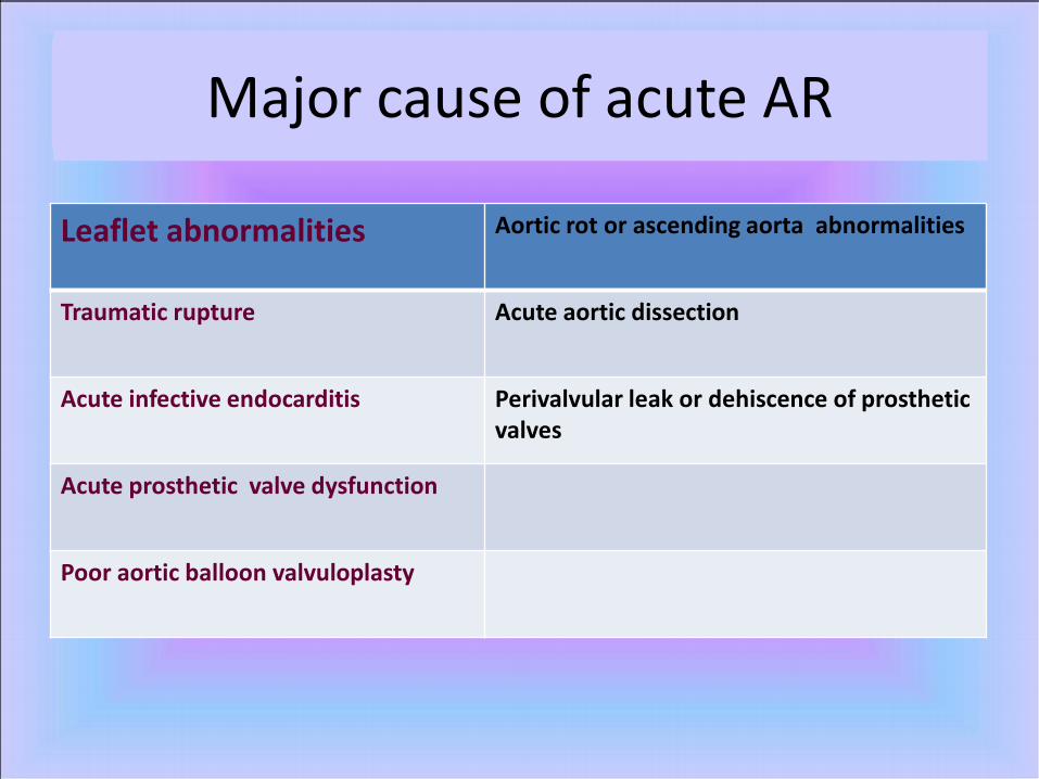
Major cause of acute AR
Leaflet abnormalities Aortic rot or ascending aorta abnormalities
Traumatic rupture Acute aortic dissection
Acute infective endocarditis Perivalvular leak or dehiscence of prosthetic valves
Acute prosthetic valve dysfunction
Poor aortic balloon valvuloplasty
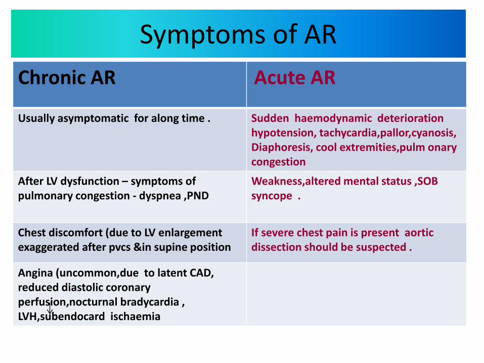
Symptoms of AR Chronic AR Acute AR
Usually asymptomatic for along time . Sudden haemodynamic deterioration hypotension, tachycardia,pallor,cyanosis,Diaphoresis, cool extremities,pulm onary congestion
After LV dysfunction – symptoms of pulmonary congestion - dyspnea ,PND
Weakness,altered mental status ,SOB syncope .
Chest discomfort (due to LV enlargement exaggerated after pvcs &in supine position
If severe chest pain is present aortic dissection should be suspected .
Angina (uncommon,due to latent CAD, reduced diastolic coronary perfusion,nocturnal bradycardia , LVH,subendocard ischaemia
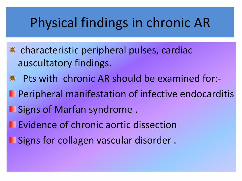
Physical findings in chronic AR
characteristic peripheral pulses, cardiac auscultatory findings.
Pts with chronic AR should be examined for:-Peripheral manifestation of infective endocarditis Signs of Marfan syndrome .Evidence of chronic aortic dissection Signs for collagen vascular disorder .
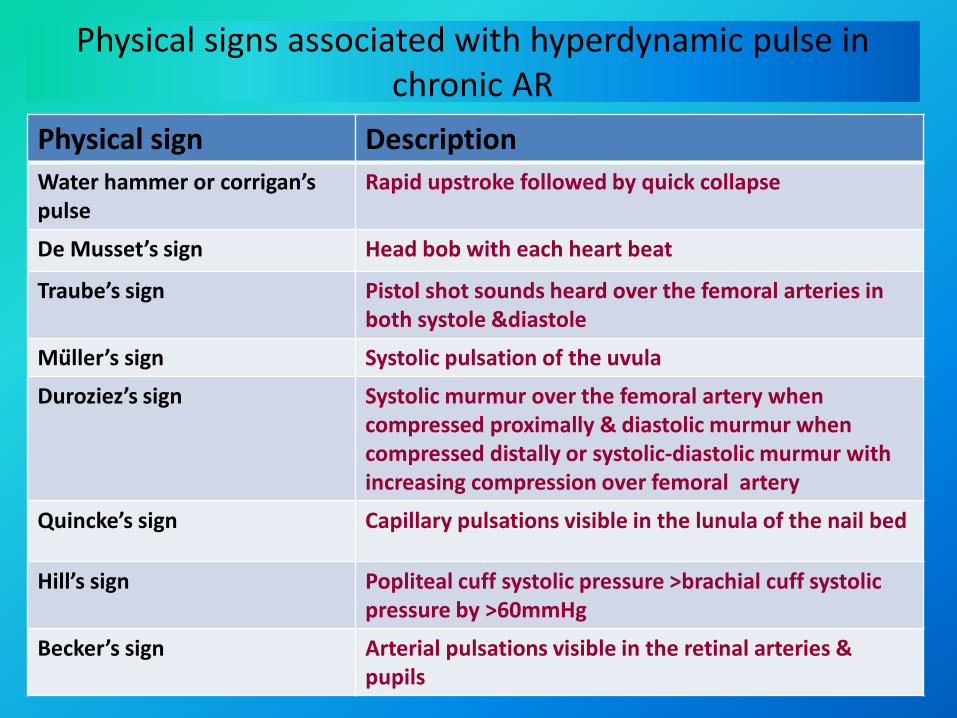
Physical signs associated with hyperdynamic pulse in chronic AR
Physical sign Description Water hammer or corrigan’spulse
Rapid upstroke followed by quick collapse
De Musset’s sign Head bob with each heart beat
Traube’s sign Pistol shot sounds heard over the femoral arteries in both systole &diastole
Muller’s sign Systolic pulsation of the uvula
Duroziez’s sign Systolic murmur over the femoral artery when compressed proximally & diastolic murmur when compressed distally or systolic-diastolic murmur with increasing compression over femoral artery
Quincke’s sign Capillary pulsations visible in the lunula of the nail bed
Hill’s sign Popliteal cuff systolic pressure >brachial cuff systolic pressure by >60mmHg
Becker’s sign Arterial pulsations visible in the retinal arteries & pupils
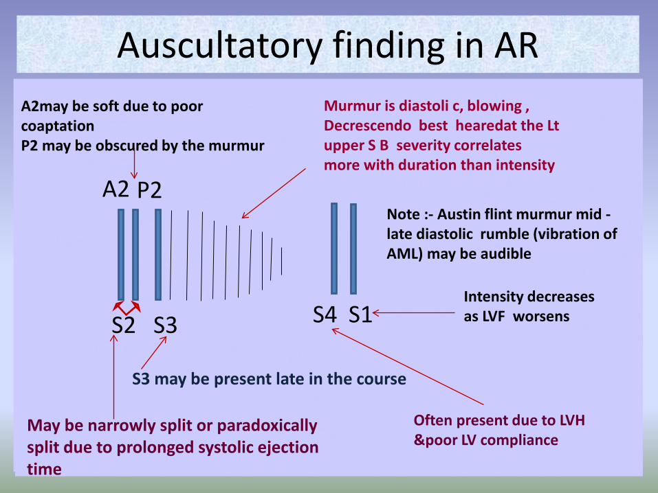
Auscultatory finding in AR
S1
P2A2
S4S2 S3
May be narrowly split or paradoxically split due to prolonged systolic ejection time
S3 may be present late in the course
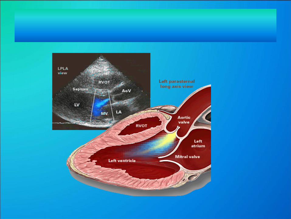
Murmur is diastoli c, blowing ,Decrescendo best hearedat the Lt upper S B severity correlates more with duration than intensity
Intensity decreases as LVF worsens
Often present due to LVH &poor LV compliance
A2may be soft due to poor coaptationP2 may be obscured by the murmur
Note :- Austin flint murmur mid -late diastolic rumble (vibration of AML) may be audible
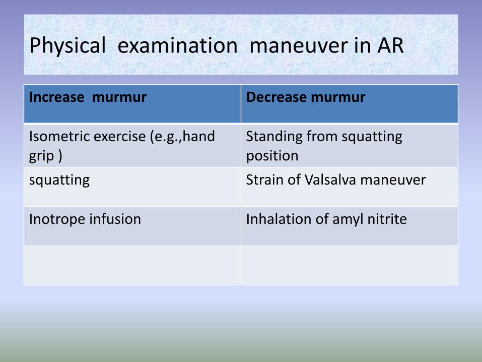
Physical examination maneuver in AR
Increase murmur Decrease murmur
Isometric exercise (e.g.,handgrip )
Standing from squatting position
squatting Strain of Valsalva maneuver
Inotrope infusion Inhalation of amyl nitrite
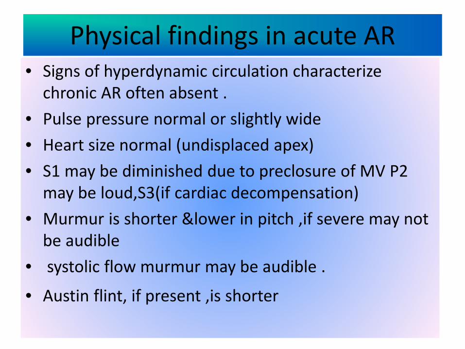
Physical findings in acute AR • Signs of hyperdynamic circulation characterize
chronic AR often absent .• Pulse pressure normal or slightly wide• Heart size normal (undisplaced apex)• S1 may be diminished due to preclosure of MV P2
may be loud,S3(if cardiac decompensation)• Murmur is shorter &lower in pitch ,if severe may not
be audible• systolic flow murmur may be audible .
• Austin flint, if present ,is shorter
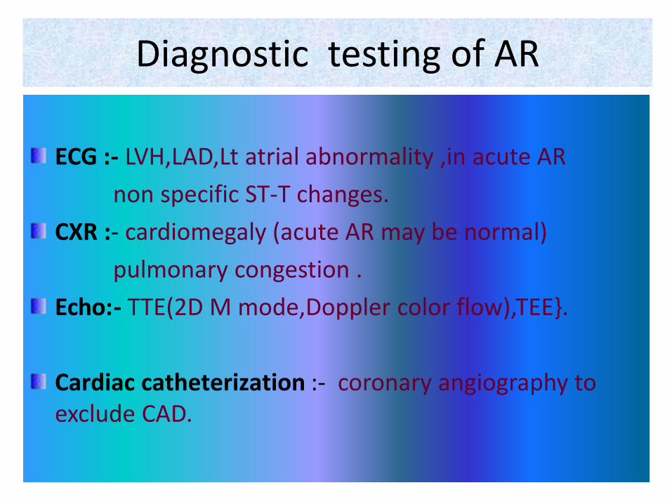
Diagnostic testing of AR
ECG :- LVH,LAD,Lt atrial abnormality ,in acute AR non specific ST-T changes.
CXR :- cardiomegaly (acute AR may be normal)pulmonary congestion .
Echo:- TTE(2D M mode,Doppler color flow),TEE}.
Cardiac catheterization :- coronary angiography to exclude CAD.
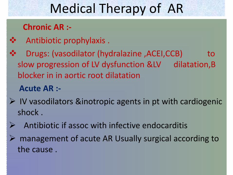
Medical Therapy of AR Chronic AR :-
Antibiotic prophylaxis . Drugs: (vasodilator (hydralazine ,ACEI,CCB) to
slow progression of LV dysfunction &LV dilatation,B blocker in in aortic root dilatation Acute AR :-
IV vasodilators &inotropic agents in pt with cardiogenic shock .
Antibiotic if assoc with infective endocarditis management of acute AR Usually surgical according to
the cause .
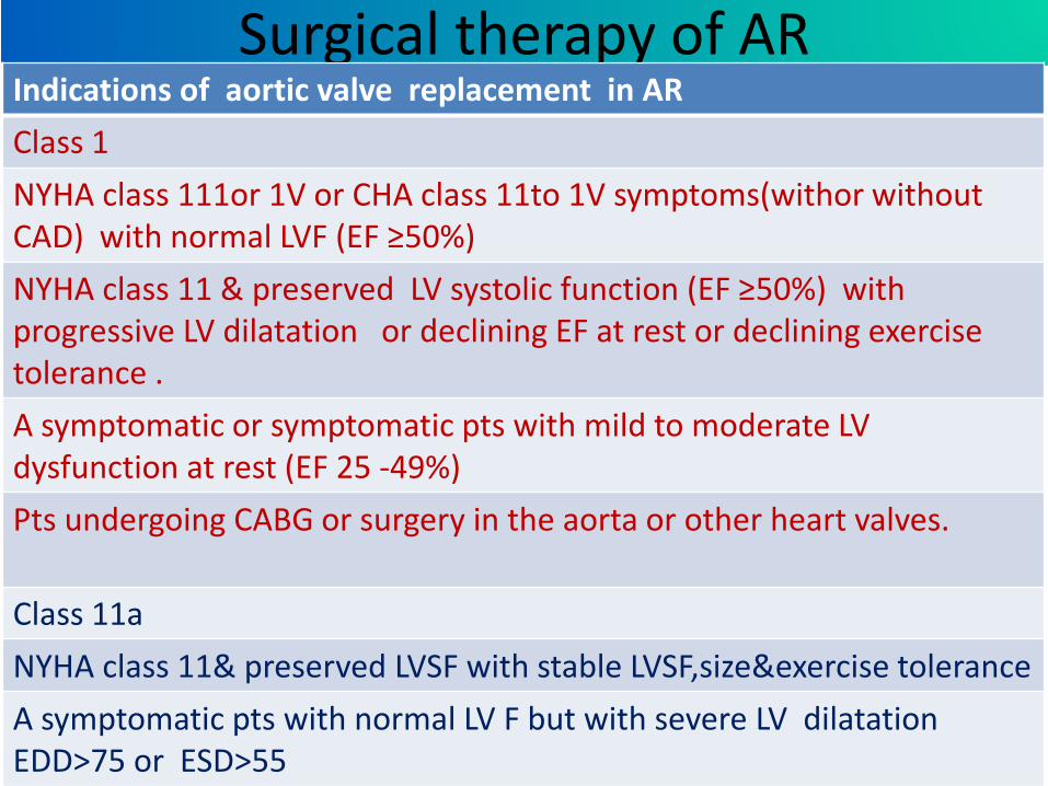
Surgical therapy of AR Indications of aortic valve replacement in AR Class 1NYHA class 111or 1V or CHA class 11to 1V symptoms(withor without CAD) with normal LVF (EF ≥50%)NYHA class 11 & preserved LV systolic function (EF ≥50%) with progressive LV dilatation or declining EF at rest or declining exercise tolerance .A symptomatic or symptomatic pts with mild to moderate LV dysfunction at rest (EF 25 -49%)Pts undergoing CABG or surgery in the aorta or other heart valves.
Class 11aNYHA class 11& preserved LVSF with stable LVSF,size&exercise toleranceA symptomatic pts with normal LV F but with severe LV dilatation EDD>75 or ESD>55
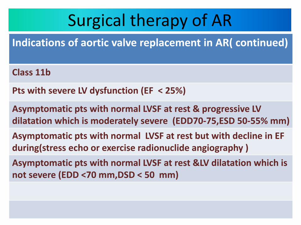
Surgical therapy of AR Indications of aortic valve replacement in AR( continued)
Class 11b
Pts with severe LV dysfunction (EF < 25%)
Asymptomatic pts with normal LVSF at rest & progressive LV dilatation which is moderately severe (EDD70-75,ESD 50-55% mm)Asymptomatic pts with normal LVSF at rest but with decline in EF during(stress echo or exercise radionuclide angiography )Asymptomatic pts with normal LVSF at rest &LV dilatation which is not severe (EDD <70 mm,DSD < 50 mm)
Pt with chronic AR should be obserevedclosely for the development of LV systolic dysfunction.Follow up evaluation typically is conducted with serial echocardiography .If signs o of LV systolic dysfunction develop surgical therapy should be considered .